Embed Size (px)
Citation preview
[1]
nurseCe4Less.com nurseCe4Less.com nurseCe4Less.com nurseCe4Less.com
Blood Transfusions:
An Overview
Jassin M. Jouria, MD
Dr. Jassin M. Jouria is a medical doctor, professor of
academic medicine, and medical author. He
graduated from Ross University School of Medicine
and has completed his clinical clerkship training in
various teaching hospitals throughout New York, including King’s County Hospital Center
and Brookdale Medical Center, among others. Dr. Jouria has passed all USMLE medical
board exams, and has served as a test prep tutor and instructor for Kaplan. He has
developed several medical courses and curricula for a variety of educational institutions. Dr.
Jouria has also served on multiple levels in the academic field including faculty member and
Department Chair. Dr. Jouria continues to serves as a Subject Matter Expert for several
continuing education organizations covering multiple basic medical sciences. He has also
developed several continuing medical education courses covering various topics in clinical
medicine. Recently, Dr. Jouria has been contracted by the University of Miami/Jackson
Memorial Hospital’s Department of Surgery to develop an e-module training series for
trauma patient management. Dr. Jouria is currently authoring an academic textbook on
Human Anatomy & Physiology.
Abstract
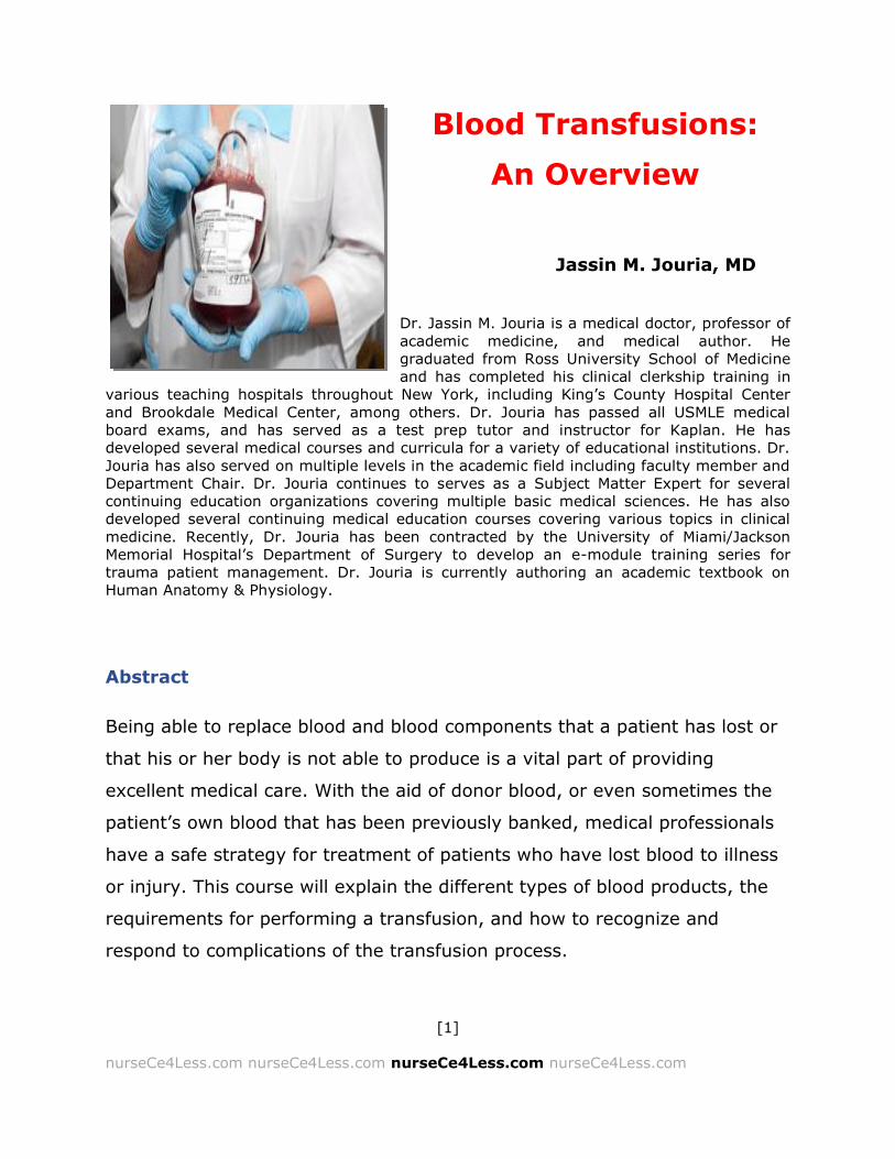
Being able to replace blood and blood components that a patient has lost or
that his or her body is not able to produce is a vital part of providing
excellent medical care. With the aid of donor blood, or even sometimes the
patient’s own blood that has been previously banked, medical professionals
have a safe strategy for treatment of patients who have lost blood to illness
or injury. This course will explain the different types of blood products, the
requirements for performing a transfusion, and how to recognize and
respond to complications of the transfusion process.

[2]
nurseCe4Less.com nurseCe4Less.com nurseCe4Less.com nurseCe4Less.com
Continuing Nursing Education Course Planners
William A. Cook, PhD, Director, Douglas Lawrence, MA, Webmaster,
Susan DePasquale, MSN, FPMHNP-BC, Lead Nurse Planner
Policy Statement
This activity has been planned and implemented in accordance with the
policies of NurseCe4Less.com and the continuing nursing education
requirements of the American Nurses Credentialing Center's Commission on
Accreditation for registered nurses. It is the policy of NurseCe4Less.com to
ensure objectivity, transparency, and best practice in clinical education for
all continuing nursing education (CNE) activities.
Continuing Education Credit Designation
This educational activity is credited for 3 hours. Nurses may only claim credit
commensurate with the credit awarded for completion of this course activity.
Statement of Learning Need
The transfusion of blood and blood products provide life-saving measures
and treat various medical conditions. Nurses are required to follow blood
transfusion guidelines and protocol to ensure patient safety and wellbeing.
Course Purpose
To provide nursing professionals with knowledge of blood and blood product
indications and administration protocol for safe administration.

[3]
nurseCe4Less.com nurseCe4Less.com nurseCe4Less.com nurseCe4Less.com
Target Audience
Advanced Practice Registered Nurses and Registered Nurses
(Interdisciplinary Health Team Members, including Vocational Nurses and
Medical Assistants may obtain a Certificate of Completion)
Course Author & Planning Team Conflict of Interest Disclosures
Jassin M. Jouria, MD, William S. Cook, PhD, Douglas Lawrence, MA,
Susan DePasquale, MSN, FPMHNP-BC – all have no disclosures
Acknowledgement of Commercial Support
There is no commercial support for this course.
Activity Review Information
Reviewed by Susan DePasquale, MSN, FPMHNP-BC
Release Date: 1/1/2016 Termination Date: 5/14/2018
Please take time to complete a self-assessment of knowledge, on page 4, sample questions before reading the article.
Opportunity to complete a self-assessment of knowledge learned will be provided at the end of the course.
[4]
nurseCe4Less.com nurseCe4Less.com nurseCe4Less.com nurseCe4Less.com
1. GM is a 32-year-old AA female who is typed with B+ blood. As a
donor, GM can MOST SAFELY provide packed RBC for which of
the following individuals?
a. TS, a 21 year old Asian female with AB+ blood
b. KT, a 56 year old European male with AB - blood
c. PL, a 76 year old white female with A - blood
d. MN, a 5-year-old Hispanic male with B+ blood
2. Your patient is an otherwise healthy 46-year-old male with β-
thalassemia and is scheduled for a transfusion of packed RBC.
You are with him and are explaining some of the risks and
benefits of his upcoming transfusion. Which of the following
would be true?
a. He runs a high risk of graft-versus-host disease along with
allergic reactions, a fever, a hemolytic reaction and some very
rare reactions but that he will be observed and monitored
throughout the transfusion.
b. He runs a high risk of Transfusion-Related Acute Lung Injury
(TRALI) or Transfusion Associated Circulatory Overload (TACO)
but that he will be observed and monitored throughout the
transfusion.
c. You should counsel him not to get a transfusion, but to get a
shot of iron for his iron deficiency anemia.
d. The transfusion should be completed within 4 hours and there
are some risks associated with an RBC transfusion, including
allergic reactions, a fever, a hemolytic reaction and some very
rare reactions but that he will be observed and monitored
throughout the transfusion.
[5]
nurseCe4Less.com nurseCe4Less.com nurseCe4Less.com nurseCe4Less.com
3. Your patient is typed AB— and is scheduled for an infusion of
plasma. Which of the following plasma types is most suitable
for your patient?
a. AB+
b. O -
c. A+
d. B –
4. A patient who is thrombocytopenic is scheduled to receive two
units of irradiated platelets from which the leukocytes (WBC)
have been removed. You spike the unit with a standard infusion
set and get ready to connect directly to her right arm.
Something makes you stop—what question do you need to ask
yourself?
a. Don’t I need to use a standard blood filter?
b. Shouldn’t I use another leukocyte reduction filter just to be sure
her chances of GVHD are reduced?
c. I should probably ask the blood bank if these platelets have been
tested for ABO compatibility, shouldn’t I?
d. I wonder if her platelet level is low enough to get two units of
platelets?
5. An acute hemolytic reaction during a transfusion for acute blood
loss is most likely due to:
a. ABO incompatibility
b. Too rapid infusion
c. Rh0D incompatibility
d. Massive sepsis

[6]
nurseCe4Less.com nurseCe4Less.com nurseCe4Less.com nurseCe4Less.com
Introduction
According to a report from the Agency for Healthcare Research and Quality
(AHRQ), blood transfusions were the most common hospital procedure in
2010, indicating that 11% of all hospital stays underwent at least one
transfusion procedure.1 This represented more than a 126% increase since
1997 and reached across all age groups, except for individuals less than one
year old.
The American Association of Blood Banks (AABB) estimates that over 9.2
million volunteers donate blood and blood components every year.
Approximately 30% of those are first time donors; together, all these donors
represent about 15.7 million units of blood and blood components.2 There is,
however, significant variability in transfusion outcomes in a number of
clinical settings. This variability may reflect deviations from practice
guidelines, training differences, differences in recommendations of various
medical societies, differences in availability of inventory and disagreements
about the validity of the practice guidelines.3
While blood transfusions are generally accepted as being life-saving, this has
actually not be tested in any prospective controlled trial.3 While the number
of deaths associated with blood transfusions is small and seems to be
decreasing,4 blood transfusions still carry significant risk, are expensive, and
face potential shortages in emergencies. The American Association of Blood
Banks (AABB) has initiated a Patient Blood Management (PBM) program with
the goal of optimizing the care of transfusion patients and has produced
guidelines for PBM and blood utilization. Blood management can be defined
as a “patient-centered standard of care in which strategies and techniques
[7]
nurseCe4Less.com nurseCe4Less.com nurseCe4Less.com nurseCe4Less.com
are used to reduce, eliminate, or optimize blood transfusions to improve
patient outcomes.”5 The guidelines include:6
Development of guidelines specific to individual facilities and for quality
improvement
Providing thresholds and assessment strategies for the transfusion
review process
Both surgical and non-surgical strategies for PBM
Procedures for auditing blood utilization
Successful PBM programs most often have a committed leader, and this is
most often a registered nurse (RN) or an advanced practice registered nurse
(APRN). The nursing staff often has the responsibility to implement the PBM
programs, assess and manage transfusions and the potential reactions, as
well as communicate to the patient and their family the benefits and
potential adverse reactions to a blood transfusion. This activity is designed
to review basic information about transfusions, provide information on
potential complications and the current best practice guidelines, procedures
and policies to manage complications and to inform health teams and
patients regarding new developments in alternatives to allogeneic blood
transfusions, including autologous transfusions, erythropoiesis-stimulating
agents and hemostatic agents.7
Overview Of Transfusions Of Blood And Blood Components
Whole blood is rarely transfused anymore because of safety issues and
practical concerns about the effective use of a unit of donated whole blood.
It is much more efficient to use components rather than whole blood

[8]
nurseCe4Less.com nurseCe4Less.com nurseCe4Less.com nurseCe4Less.com
because more patients can benefit from this process. The different types of
transfusions include:
Packed red blood cells or packed RBCs (most often washed free of
plasma)
Plasma
Clotting factors
Platelets
Intravenous Immunoglobulins or IVIG (i.e., RhoGam™)
Cellular therapies, often considered forms of transfusions and defined as
using components derived from whole blood donations include hematopoietic
stem cell transplants and bone marrow transplants.
Current Guidelines For Packed Red Blood Cell Transfusion
Recommendations for blood transfusion undergo constant debate and
revision. The oldest (and often still repeated) rule quoted is the “10/30” rule,
which states that the hemoglobin levels of surgical patients should be at or
above 10g/dL and the hematocrit should be at or about 30%. However, this
rule is not based on any direct evidence. Various professional organizations
have developed their own criteria or guidelines for transfusions. There are
many overlaps in these criteria. One of the broadest and all-encompassing
sets of guidelines is from the American Society of Hematology (ASH) and the
American Association of Blood Banks (AABB) and are cited in this review. It
should be remembered that the nursing staff must also bear in mind the
policies and guidelines held to by their individual hospitals and institutions,
and that it is the professional and ethical responsibility of the nursing staff to

[9]
nurseCe4Less.com nurseCe4Less.com nurseCe4Less.com nurseCe4Less.com
adhere to their institutional policies and guidelines. The ultimate clinically
relevant verdict on safety and efficacy of transfusion should be based on
effect on a patient's outcome, and whether transfusion improves it.8 For
reference, a comparison of some of the clinical guidelines from other
professional organizations is given in Table 1.
Table 1: Guideline Comparisons from individual professional organizations. Adapted from
From the American Society of Hematology:8-12
AABB College of
American
Pathologists
(CAP)
Society of Critical
Care Medicine
(SCCM)
Society of
Thoracic
Surgeons
Target
Population
for
guidelines
Hospitalized, hemodynamically stable
General Critically Ill Cardiac surgery
Hemoglobin
levels
requiring
RBC
transfusions
Hb ≤7 g/dL in critically ill patients; Hb ≤8 g/dL in surgical
patients or patients with pre-existing
cardiovascular disease; When symptoms are present. Based on clinic decision: Patients with
acute coronary syndrome
Hb <6 g/dL
Rarely for Hb>10g/dL Based on clinical decisions: Hb 6-
10g/dL
Hb <7 g/dL if ventilated, trauma, or stable
cardiac disease (Hb <8 g/dL in
acute coronary syndrome) Rarely for Hb>10g/dL
Hb <6 g/dL (Hb <7 g/dL in postoperative
patients and higher if risk of
end-organ ischemia) Rarely for Hb>10g/dL
Other
Factors to
Consider
Hb levels as well as symptoms (chest pain, orthostatic hypotension, unresponsive
tachycardia, heart
failure)
Peripheral tissue oxygenation, clinical signs and symptoms, Hb, extent/rate of
bleeding
Volume status, shock, duration/ extent of anemia, cardiopulmonary parameters
Age, severity of illness, cardiac function, ischemia, extent/ rate of blood loss,
Hb, SVO2

[10]
nurseCe4Less.com nurseCe4Less.com nurseCe4Less.com nurseCe4Less.com
Proper Uses Of Red Blood Cell (RBC) Transfusion9
Treatment of symptomatic anemia
Prophylaxis in life-threatening anemia
Restoration of oxygen-carrying capacity in case of hemorrhage
RBC are also indicated for exchange transfusion
o Sickle cell disease
o Severe parasitic infection (malaria, babesiosis)
o Severe methemoglobinemia
o Severe hyperbilirubinemia of newborn
RBC transfusion is not routinely indicated for pharmacologically treatable
anemia, such as:
Iron deficiency anemia
Vitamin B12 or folate deficiency anemia
Dosage and Administration9
One unit of RBC will raise the hemoglobin of an average-size adult by
~1g/dL (or raise HCT ~3%)
ABO group of RBC products must be compatible with ABO group of
recipient
RBC product must be serologically compatible with the recipient (see
Pretransfusion Testing). Exceptions can be made in emergencies (see
Emergency Release of Blood Products).
Rate of transfusion
o Transfuse slowly for first 15 minutes
Complete transfusion within 4 hours (per FDA)
[11]
nurseCe4Less.com nurseCe4Less.com nurseCe4Less.com nurseCe4Less.com
Special Processing Requirements of RBC for Transfusions9
There are times when special processing requirements exist for RBC
transfusions. These are designed to minimize the risk of transfusion
reactions in various ways. Leukocyte reduction, or the removal of white
blood cells (WBC) from the donated blood minimizes the potential that a
transfusion reaction based on the immune responses of those WBC can
occur. These include reactions such as the febrile, non-hemolytic transfusion
reactions. Leukocyte reduction can also minimize the risk that viruses, such
as Cytomegalovirus (CMV) or herpes virus, are inadvertently transferred.13,14
However, leukocyte reduction does not prevent the possibility of Graft
Versus Host Disease (GVHD); blood products and components must be
irradiated in order to prevent GVHD.
Washing the blood cells removes plasma and removes pre-formed antibodies
that may be present in the donor serum. This is particularly important to
prevent the risk of anaphylaxis in IgA-deficient patient with anti-IgA
antibodies.15,16 Washing the blood cells can also decrease the potential for
reactions in patients sensitized by a history of previous transfusions.17
Washing the cells must be performed under sterile conditions and using
isotonic solutions to prevent contamination and hemolysis respectively.
Finally, blood and blood components may be irradiated to prevent graft-
versus-host disease. This is most commonly done if the donor is a family
member, from an HLA-selected donor or if the donor relationship to the
recipient has not been established. It may also be done in preparation for an
intrauterine transfusion and is commonly done for a number of pediatric
conditions. Potassium (K+) levels may be elevated in these samples, though
relatively rarely to clinically significant levels.18
[12]
nurseCe4Less.com nurseCe4Less.com nurseCe4Less.com nurseCe4Less.com
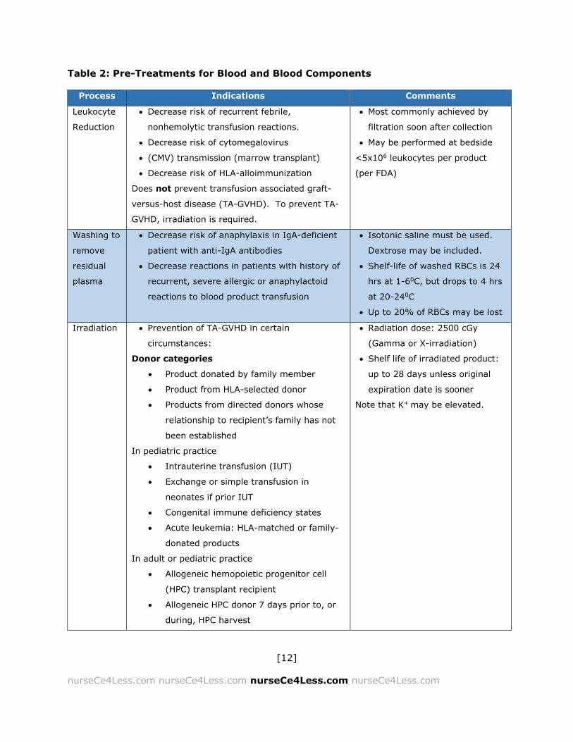
Table 2: Pre-Treatments for Blood and Blood Components
Process Indications Comments
Leukocyte
Reduction
Decrease risk of recurrent febrile,
nonhemolytic transfusion reactions.
Decrease risk of cytomegalovirus
(CMV) transmission (marrow transplant)
Decrease risk of HLA-alloimmunization
Does not prevent transfusion associated graft-
versus-host disease (TA-GVHD). To prevent TA-
GVHD, irradiation is required.
Most commonly achieved by
filtration soon after collection
May be performed at bedside
<5x10⁶ leukocytes per product
(per FDA)
Washing to
remove
residual
plasma
Decrease risk of anaphylaxis in IgA-deficient
patient with anti-IgA antibodies
Decrease reactions in patients with history of
recurrent, severe allergic or anaphylactoid
reactions to blood product transfusion
Isotonic saline must be used.
Dextrose may be included.
Shelf-life of washed RBCs is 24
hrs at 1-60C, but drops to 4 hrs
at 20-240C
Up to 20% of RBCs may be lost
Irradiation Prevention of TA-GVHD in certain
circumstances:
Donor categories
Product donated by family member
Product from HLA-selected donor
Products from directed donors whose
relationship to recipient’s family has not
been established
In pediatric practice
Intrauterine transfusion (IUT)
Exchange or simple transfusion in
neonates if prior IUT
Congenital immune deficiency states
Acute leukemia: HLA-matched or family-
donated products
In adult or pediatric practice
Allogeneic hemopoietic progenitor cell
(HPC) transplant recipient
Allogeneic HPC donor 7 days prior to, or
during, HPC harvest
Radiation dose: 2500 cGy
(Gamma or X-irradiation)
Shelf life of irradiated product:
up to 28 days unless original
expiration date is sooner
Note that K+ may be elevated.

[13]
nurseCe4Less.com nurseCe4Less.com nurseCe4Less.com nurseCe4Less.com
Autologous HPC recipient
Hodgkin disease
History of treatment with purine
analogues and related drugs:
o Fludarabine
o 2CDA (Cladribine®)
o Deoxycoformycin (Pentostatin®)
o Clofarabine (Clolar®)
o Bendamustine (Treanda®)
o Nelarabine (Arranon®)
History of treatment with alemtuzumab
(anti-CD52)
Aplastic anemia or rabbit antithymocyte
globulin
When is Packed RBC Transfusion Required?
In general, packed RBC transfusions are required for anemias of any
etiology. It should be remembered that anemia is not a disease, but a
complex of signs and symptoms of cardiovascular and pulmonary
compensatory mechanisms in response to some illness, condition or injury.
Anemia, or the lack of sufficient red blood cells (RBC’s) to provide adequate
oxygen to the tissues, can be caused by:
Deficiencies in erythropoiesis, or RBC production:
These can include iron deficiency anemia and anemias due to folate,
vitamin B12 and copper deficiencies. This type of anemia can also include
aplastic anemia and the thalassemias.

[14]
nurseCe4Less.com nurseCe4Less.com nurseCe4Less.com nurseCe4Less.com
Acute blood loss from injury or chronic blood loss from, for example,
gastric or duodenal ulcers, coagulation disorders, gastritis,
hypermenorrhea or hypothyroid disease.
Excessive hemolysis due to:
Extrinsic defects or those not due to defects within the RBCs
(these would include some autoimmune hemolysis, paroxysmal
nocturnal hemoglobinuria, trauma and infections)
Intrinsic defects due to:
o Congenital RBC membrane defects such as hereditary
elliptocytosis or acquired defects such as stomatocytosis or
hypophosphatemia
o Metabolic disorders such as hexose-monophosphate shunt
defects (Glucose-6-phosphate deficiency [G6PD])
o Hemoglobinopathies including sickle-cell anemia (SCA) and
thalassemias (α, β, and β-δ)
When is an Other Blood Component Required?
Platelets
Platelets may be pooled from a number of donors or they may be derived
from a single donor by platelet apheresis (see below). The platelets derived
through single donor apheresis have the advantage of being technically
easier to pre-test (as opposed to the pooled platelets which must be tested
at each donation). An apheresis platelet unit equals six or more platelet
units from whole blood. Another related advantage is that platelets from

[15]
nurseCe4Less.com nurseCe4Less.com nurseCe4Less.com nurseCe4Less.com
apheresis effectively reduce the potential for transfusion-transmitted
infections and the incidence of allo-immunization by a larger number of
platelet antigens from a pooled source.19 The platelet products should be
leukoreduced (i.e., have the WBCs removed) to reduce the likelihood of allo-
immunization. The platelet products should also be irradiated to prevent
graft-versus-host disease, which is nearly 100% fatal.19
Platelet transfusion should not be based solely on the platelet count but
should also be based on the presence, absence or likelihood of clinically
relevant bleeding.20 Indications for platelet transfusion include the following
conditions.19,20
Prophylaxis for individuals with counts less than 20,000/ μL
(microliter):
o Acute leukemia (except for acute promyelocytic leukemia in
unstable patients when the risk of alloimmunization is high)
o Bone marrow aplasia
o Myelodysplasias
o Autologous bone marrow transplants
o Autologous peripheral blood stem cell transplantation
o Bladder cancers or necrotic tumors
o Major surgery
o Bone marrow biopsy
o Other:
Lumbar puncture, epidural anesthesia, endoscopy with
biopsy, placement of a central venous catheter, liver biopsy

[16]
nurseCe4Less.com nurseCe4Less.com nurseCe4Less.com nurseCe4Less.com
Chronically thrombocytopenic patients in whom bleeding is greater
than the World Health Organization (WHO) grade 2:
o World Health Organization has advised the following grading
system:19
Grade 0, none
Grade 1, petechiae, ecchymosis, occult blood in body
secretions, and mild vaginal spotting
Grade 2, evidence of gross hemorrhage not requiring red
cell transfusions over routine transfusion needs (i.e.,
epistaxis, hematuria, hematemesis)
Grade 3, hemorrhage requiring transfusion of 1 or more
units of red cells per day
Grade 4, life-threatening hemorrhage, defined as massive
bleeding causing hemodynamic compromise or bleeding
into a vital organ (i.e., intracranial, pericardial, or
pulmonary hemorrhage
Thrombocytopenia (platelets < 100,000/μ L) due to reduced platelet
production
Active bleeding with thrombocytopenia (platelets < 100,000/μ L)
During massive transfusions (see below)
In acute Disseminated Intravascular Coagulation (DIC)
Autoimmune thrombocytopenia
In neonates if:
o Platelets < 20,000 – 30,000/μ L
[17]
nurseCe4Less.com nurseCe4Less.com nurseCe4Less.com nurseCe4Less.com
o Platelets 30,000 – 50,000/μ L in neonates of low birth weight,
with a coagulation disorder, during invasive procedures or with
previous intraventricular or intraparenchymal cerebral
hemorrhage
o Platelets 50,000 – 100,000/μL
in neonates with active bleeding
Fresh Frozen Plasma
Fresh Frozen Plasma (FFP) is primarily indicated to correct clotting factor
deficiencies in patients with active bleeding for which specific factors are not
available.20
Transfusion with FFP is indicated for:20
1. Congenital deficiencies of clotting factors (if there is no specific
concentrate available) or for acquired deficiencies of multiple clotting
factors if the prothrombin time (PT) or the activated partial
thromboplastin time (aPTT), expressed as a ratio of PT/aPPT, is
greater than 1.5:
a. Continuous bleeding or the prevention of bleeding in patients
with liver disease
b. Patients being treated with vitamin K antagonists, if major
hemorrhage or intracranial bleeding is present or for emergency
surgery. (Note: prothrombin complex concentrate is the
treatment of first choice).
c. In patients with acute disseminated intravascular coagulation
(DIC) and active bleeding.

[18]
nurseCe4Less.com nurseCe4Less.com nurseCe4Less.com nurseCe4Less.com
d. For microvascular bleeding in patients during massive
transfusion.
e. For deficiencies of single clotting factors.
f. Thrombotic microangiopathies (thrombotic thrombocytopenic
purpura), hemolytic-uremic syndrome (HUS) or hemolytic
anemia due to elevated liver enzymes and low platelet count
(HELLP) syndrome
g. Hereditary angioedema
Other component transfusions will be discussed throughout this course
activity as needed.
Basic Principles Of Blood Transfusions And Pre-Transfusion Testing
In the U.S., the Food and Drug Administration (FDA), regulates blood and
blood components. Standards of practice and other guideline are written by
the American Association of Blood Banks (AABB) in cooperation with state
and local health agencies as well as the American Red Cross.
Blood donors are screened by questionnaires and by a standard health
interview and physical examinations which includes vital signs (temperature,
heart rate (HR), respiratory rate (RR), blood pressure (BP) and a
determination of the hemoglobin levels (Hgb) and often, the hematocrit
(HCT) or “crit” — the packed cell volume (PCV) of the red blood cells (RBCs).
There are a number of reasons that donors may be deemed ineligible to
donate. It may be determined that the donor’s health may be harmed by
donation, or, it may be determined that potential recipients may be harmed.
[19]
nurseCe4Less.com nurseCe4Less.com nurseCe4Less.com nurseCe4Less.com
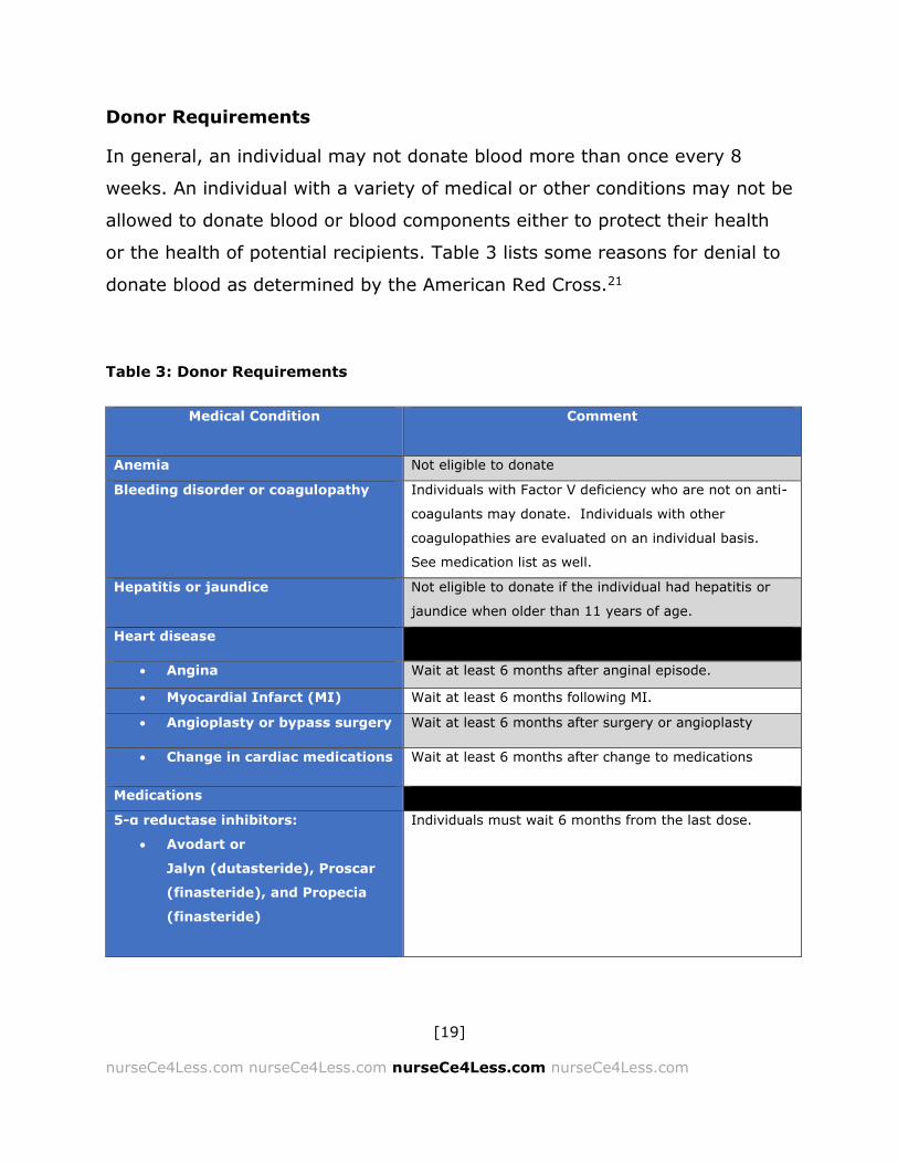
Donor Requirements
In general, an individual may not donate blood more than once every 8
weeks. An individual with a variety of medical or other conditions may not be
allowed to donate blood or blood components either to protect their health
or the health of potential recipients. Table 3 lists some reasons for denial to
donate blood as determined by the American Red Cross.21
Table 3: Donor Requirements
Medical Condition Comment
Anemia Not eligible to donate
Bleeding disorder or coagulopathy Individuals with Factor V deficiency who are not on anti-
coagulants may donate. Individuals with other
coagulopathies are evaluated on an individual basis.
See medication list as well.
Hepatitis or jaundice Not eligible to donate if the individual had hepatitis or
jaundice when older than 11 years of age.
Heart disease
Angina Wait at least 6 months after anginal episode.
Myocardial Infarct (MI) Wait at least 6 months following MI.
Angioplasty or bypass surgery Wait at least 6 months after surgery or angioplasty
Change in cardiac medications Wait at least 6 months after change to medications
Medications
5-α reductase inhibitors:
Avodart or
Jalyn (dutasteride), Proscar
(finasteride), and Propecia
(finasteride)
Individuals must wait 6 months from the last dose.

[20]
nurseCe4Less.com nurseCe4Less.com nurseCe4Less.com nurseCe4Less.com
Retinoids
Accutane, Amnesteem,
Claravis or Sotret
(isoretinoin),Soriatane
(acitretin)
3 years post treatment
Tegison (etretinate) Not eligible to donate
Aspirin, anticoagulants, anti-platelet
medications
Aspirin or aspirin-containing
medications
No waiting period for donating whole blood. Individuals
must wait 48 hours after taking aspirin or any
medication containing aspirin before donating platelets
by apheresis.
Coumadin (warfarin), heparin,
Pradaxa (dabigatran), Xarelto
(rivaoxaban) or Lovenox
(enoxaparin) or other
prescription anti-clotting
agents.
Plavix (clopidogrel)
Ticlid (ticlopidine)
Effient (prasugrel)
Feldene (piroxicam)
Not eligible to donate.
If medication is discontinued, the individual must wait 7
days before donating whole blood and 14 days before
donating platelets by apheresis.
For Feldene/piroxicam, if medication is discontinued, the
individual must wait 48 hours before donating platelets
by apheresis.
Hepatitis B Immune Globulin Individuals must wait for 12 months post-treatment
Hormone Treatments
Bovine hormone treatments Not eligible to donate
Human pituitary - derived
growth hormone
Not eligible to donate
Hormone replacement therapy
or oral contraceptives
Eligible to donate
Pregnancy Wait until 6 weeks post-partum.
Low weight Weight ≥110 pound
High or low blood pressure BP≤180/100
BP≥80/50
Age Donor must be at least 17 years old or if 16 years old,
must have parental consent.
Hemoglobin levels ≥12.5g/dL
[21]
nurseCe4Less.com nurseCe4Less.com nurseCe4Less.com nurseCe4Less.com
Whole blood and blood components, including fresh frozen plasma (FFP),
cryoprecipitate, white blood cells (WBCs, buffy coat), immune globulins,
platelet concentrates are routinely tested for surface antigens, plasma
antibodies and infectious diseases. Testing for antigens and antibodies is
referred to as compatibility testing and is done pre-transfusion, unless there
is an emergency need.
Electronic crossmatches are currently available at a growing number of
transfusion centers and standard operating procedures (SOPs) developed for
electronic crossmatching (EXM).22,23 The use of bar codes and automated
systems have facilitated safety measures as well as the movement and
tracking of blood and blood components across national and international
borders.
The implementation of ration frequency identification devices is also being
investigated and implemented in some areas to further improve the safety
and availability of blood components.22 Flow cytometry, dynamic
fluorescence imaging and patch-clamp measurements are increasingly used
to detect cell surface antigens, incompatibilities and specific antibodies to
various patient-specific and non-specific antigens.24-27
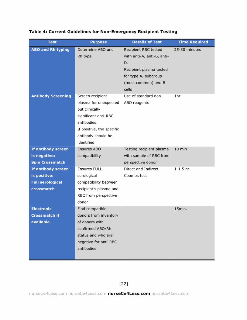
Current Guidelines for Pre-Transfusion Testing
The following guidelines are obtained from the American Society of
Hematology, shown in Table 4 below.9-12
[22]
nurseCe4Less.com nurseCe4Less.com nurseCe4Less.com nurseCe4Less.com
Table 4: Current Guidelines for Non-Emergency Recipient Testing
Test Purpose Details of Test Time Required
ABO and Rh typing Determine ABO and
Rh type
Recipient RBC tested
with anti-A, anti-B, anti-
D.
Recipient plasma tested
for type A1 subgroup
(most common) and B
cells
25-30 minutes
Antibody Screening Screen recipient
plasma for unexpected
but clinically
significant anti-RBC
antibodies.
If positive, the specific
antibody should be
identified
Use of standard non-
ABO reagents
1hr
If antibody screen
is negative:
Spin Crossmatch
Ensures ABO
compatibility
Testing recipient plasma
with sample of RBC from
perspective donor
10 min
If antibody screen
is positive:
Full serological
crossmatch
Ensures FULL
serological
compatibility between
recipient’s plasma and
RBC from perspective
donor
Direct and Indirect
Coombs test
1-1.5 hr
Electronic
Crossmatch if
available
Find compatible
donors from inventory
of donors with
confirmed ABO/Rh
status and who are
negative for anti-RBC
antibodies
15min.
[23]
nurseCe4Less.com nurseCe4Less.com nurseCe4Less.com nurseCe4Less.com
Blood and Blood Component Pre-Testing
Compatibility Testing
Karl Landsteiner described the ABO blood types in 1900. The ABO system
describes antigens located on the membranes of the RBCs. These antigenic
differences are based on different sugar residues — N-acetylgalactosamine
for A-type antigens and galactose for B-type antigens. These isoantigens
form the basis of blood compatibility testing and adverse transfusion
reactions.
ABO and Rh0 (D) testing is carried out
on donor blood. Other blood group
systems are also checked. These
other blood group systems include the
MNS system, the Kell system and the
Lewis system. In general, these
systems are checked for specific,
clinical purposes. For example,
antibodies to the MNS system are
occasionally seen in hemolytic
transfusion reactions and in cases of
hemolytic disease of the newborn.28
Maternal antibodies to Kell antigens
can be very important in hemolytic
disease of the newborn and critical in
intrauterine transfusions.29-31
It is believed that the ABO system,
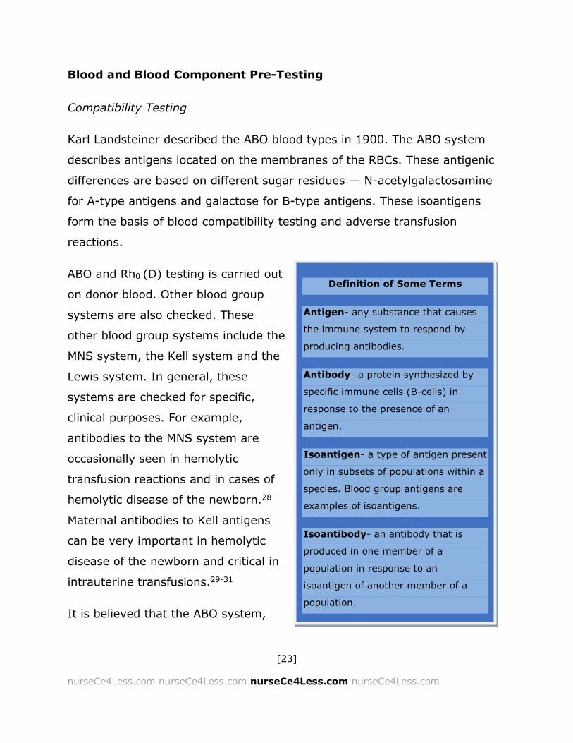
Definition of Some Terms
Antigen- any substance that causes
the immune system to respond by
producing antibodies.
Antibody- a protein synthesized by
specific immune cells (B-cells) in
response to the presence of an
antigen.
Isoantigen- a type of antigen present
only in subsets of populations within a
species. Blood group antigens are
examples of isoantigens.
Isoantibody- an antibody that is
produced in one member of a
population in response to an
isoantigen of another member of a
population.

[24]
nurseCe4Less.com nurseCe4Less.com nurseCe4Less.com nurseCe4Less.com
along with the parallel system in WBCs (the Human Leukocyte Antigen (HLA)
system) evolved in large part as an adaptation to selective pressures placed
by various infections, such as the malarial parasite, Plasmodium,32-34
bacteria and viruses.35 Alternatively, it is possible that these antibodies
arose from a network of interactions between environmental antigens,
microbes and the immune system.36
Overview Of ABO Inheritance
Red blood cell membranes express a number of surface antigens. During
immune development, these antigens induce an immune tolerance to “self”
through “education” in the thymus and by various means, but antibodies
against non-self antigens, isoantibodies or simply antibodies are also
produced. Many of these antibodies are hemagglutinins, causing the RBCs
which carry the isoantigens to clump together and, if complement binds, to
hemolyze the RBC. The antibodies tend to be of the IgM class. IgM
antibodies are pentameric and are the most efficient antibodies at inducing
cross-linking and hemolysis.
ABO System
There are four blood groups in the ABO system, A, B, AB and O. Inheritance
of the ABO blood group is controlled by a single gene, with three types of
alleles. These alleles are i, IA and IB; IA and IB are inherited in a co-
dominant manner with both IA and IB dominant over i.

[25]
nurseCe4Less.com nurseCe4Less.com nurseCe4Less.com nurseCe4Less.com
Type A RBCs have “A” antigen on their surface. Type B RBCs have “B”
antigen and Type AB RBCs have both “A” and “B” antigens on their surface.
Type O RBCs have neither “A” nor “B” on their surface but do have an
antigen expressed — this is usually called the “H” antigen and can be
thought of as common to the ABO system. Rare individuals who lack the “H”
antigen have the “Bombay” blood group. This phenotype appears in less
than 0.0004% of the population; and, these individuals, because they lack
the “A”, “B” and “H” antigens, can donate RBCs to any individual but cannot
receive any RBCs from any ABO donor. Type O blood is designated “ii”; type
A is either IAIA or IAi; type B is either IBIB or IBi; type AB is IAIB.
Table 5: Inheritance of the ABO Blood Types
Blood Type
of Mother
Blood Type
of Father
O (ii) A
B
AB
IAIA IAi IBIB IBi IAIB
O (ii) Ii
Type O
IAi
Type A
IAi or ii
Type A or
Type O
IBi
Type B
IBi or ii
Type B or
Type O
IAi or IBi
Type A or Type
B
A IAIA IAi Type A
IAIA
Type A IAIA or IAi
Type A IAIB
Type AB IAIB or IAi Type AB or Type A
IAIA or IAIB
Type A or Type AB
IAi IAi or ii Type A or Type O
IAIA or IAi
Type A IAIA,IAi or ii
Type A or Type O
IAIB or IBi Type AB or B
IAIB or IBi Type AB or Type B
IAIA, IAIB, IAi or IBi Type A, AB or B
B IBIB IBi Type B
IAIB
Type AB IBIAor IBi Type AB or Type B
IBIB
Type B IBIB or IBi Type B
IBIB, IBIA
Type B or Type AB
IBi IBi or ii
Type B or Type O
IBIA or IAi
Type AB or Type A
IBIAorIBi, IAi
or ii Type AB,B, A
or Type O
IBIB or IBi
Type B
IBIBor ii
Type B or Type O
IBIA, IBIBor IBi or
IAi Types AB,B or A
AB IAIB IAi or IBi Type A or Type B
IAIAor IAIB, IAi or IBi Types A, AB or B
IAIA,IAi,IBIA or IBi Types A, AB or B
IAIB, IBIB
Types AB or B
IAIB,IAi, IBIBor IBi Types AB, A or B
IAIA, IAIB, IBIA, IBIB Types A, AB or B

[26]
nurseCe4Less.com nurseCe4Less.com nurseCe4Less.com nurseCe4Less.com
Rh System
The Rh (for the Rhesus monkey in which it was first described) system is the
other main blood grouping system important in transfusions. There are five
antigens, but only the D antigen or RhoD is considered in blood typing.
The designation Rh-positive or Rh-negative refers to the presence or
absence of the D-antigen, respectively. Immunization against the D-antigen
occurs either through previous blood transfusions or in utero, that is if the
mother is Rh-negative and the fetus is Rh-positive. Under these conditions,
the mother responds to the RhoD antigen as non-self and makes antibodies
(usually IgG antibodies) to the RhoD antigen. These antibodies then cross
the placenta and can cause a hemolytic condition known as erythroblastosis
fetalis or hemolytic disease of the newborn. Intrauterine transfusions (see
below) may be necessary if these incompatibilities are not treated early.
Pregnant women are routinely given anti-RhoD antibodies (i.e., RhoGam™,
BayRHo-D™, Gamulin Rh™) at 28 weeks gestation to suppress the response
of an Rh-negative mother to the antigen expressed on the developing fetus.
Overview Of Whole Blood Compatibility
An individual with type A blood has the “A” antigen on the surface of the
RBCs and antibody to the “B” antigen in their serum or plasma. Conversely,
an individual with type B blood has the “B” antigen on the surface of the
RBCs and antibody to the “A” antigen in their serum or plasma. An individual
with type AB blood has both the “A” antigen and the “B” antigen on the
surface of the RBCs and no antibodies to either the “A” and the “B” antigen
in their serum or plasma. An individual with type O blood has neither antigen

[27]
nurseCe4Less.com nurseCe4Less.com nurseCe4Less.com nurseCe4Less.com
on the surface of the RBCs and antibodies to both “A” or “B” in the serum or
plasma. In addition, the Rh status is considered; the designation “Rh
positive” indicates the presence of the RhoD antigen and is indicated as “+”.
The “Rh negative” designation indicates there is an absence of the RhoD
antigen and is indicated as “-”. Rh+ blood should only be given to Rh+
individuals, but Rh- blood can be given to any patient.
In whole blood or packed RBC transfusions, only the antigens on the surface
of the RBC are important for transfusion reactions because the cells are
packed and washed and are essentially free of plasma (and therefore
antibodies). So, an individual with Type O- blood is considered a universal
blood donor and type AB+ is considered the universal blood recipient. Type
AB+ plasma, because it has no antibodies, is considered the universal
plasma donor.
People with type O blood do have surface antigen (the “H” antigen) but since
this antigen is common to all blood groups, most people have no antibodies;
these people have the rare Bombay blood type (hh) and can produce
antibodies to the H antigen. These individuals can donate blood but can only
receive transfusions from other hh donors.
Table 6: Donor and Recipient Table.
Note: Where there is a “", the transfusion is considered compatible
Donor
O A B AB
Recip
ien
t
AB
B
A
O

[28]
nurseCe4Less.com nurseCe4Less.com nurseCe4Less.com nurseCe4Less.com
Pre-Transfusion Testing
Donor Blood
Donor blood is pre-tested for ABO and RhoD antigens, the presence of
antibodies and for markers of infectious disease. ABO typing determines
whether the donor has A, B, O or AB type blood. RhoD testing determines
the presence or absence of the RhoD antigen.
A number of tests are also performed to determine if the donor is infected
with the Hepatitis B or C viruses, HIV-1 or HIV-2, human T-cell lymphotropic
viruses 1 and 2 (HTLV-1 or -2), Treponema pallidum (the causative
organism for syphilis) and, more recently, for West Nile Virus.
Table 7: Donor Testing
Infectious Agent Testing
HIV-1 and HIV-2 (If antibody +, infection confirmed
by Western blot or immunoblotting)
HTLV-1 and HTLV-2
Hepatitis B- core antigen
Hepatitis C (if nucleic acid sequence test is +)
Presence of antibody
HIV
West Nile Virus
Presence of specific nucleic
acid sequences
Treponema pallidum
Hepatitis B surface antigen
Presence of specific antigen

[29]
nurseCe4Less.com nurseCe4Less.com nurseCe4Less.com nurseCe4Less.com
Recipient Testing
The recipient is tested for ABO type, RhoD type and for rare or unexpected
antibodies to RBC surface antigens. This is considered essential because, the
presence of antibodies in recipient serum, for example, to antibodies to
other blood group antigens in the Kell(K) and Duffy (Fy) systems, can cause
severe hemolytic transfusion reactions in the recipients. Maternal blood is
also tested for Kell and Duffy antigens to prevent hemolytic disease of the
newborn.
Screening of unexpected plasma or serum anti-RBC antibodies is through
indirect antiglobulin testing (the indirect Coombs test is more commonly
known as the Indirect Antibody Test or IAT). The direct Coombs test, or the
Direct Antibody Test (DAT), screens for anti-RBC antibodies that are coating
the RBCs. Both tests involve the use of anti-human globulin and checks for
the presence of RBC agglutination. A full antibody titration may be done if a
significant anti-RBC antibody is detected in the plasma of a pregnant woman
or in the case of a patient with cold autoimmune hemolytic anemia (see
below).
In acute situations, there may not be enough time to type the patient and
cross-match the donor blood. Spin crossmatches, which are a shortened
version of the complete test, may be used in emergency situations as can
electronic crossmatching. In emergency situations, O- blood may be used as
RBCs and AB- plasma may be used.
Crossmatching only provides minimal additional protection against
incompatibility. After the major blood group antigens are screened, there are
a large number of possible antigens that may cause incompatibility
problems; and, the issue is which ones to screen. Without additional

[30]
nurseCe4Less.com nurseCe4Less.com nurseCe4Less.com nurseCe4Less.com
information to narrow down the search for potentially problematic antigens
and antibodies, it would be like looking for the proverbial needle in the
haystack.
Many hospitals are performing electronic crossmatches rather than the
physical lab testing in patients who have a negative antibody screen. If the
screening indicates significant levels of antibody, donor blood, which is
negative for that antigen, is used. Also, in many cases a spin cross-match is
used where the patient's serum is mixed with the donor cells and
immediately placed into a centrifuge and spun. The absence of agglutination
indicates ABO compatibility.
Current Guidelines For Pre-Transfusion Testing Of Recipient Samples
The following guidelines are obtained from the American Society of
Hematology.9-12
Pre-transfusion Blood Samples from the Intended Recipient
The following are usually obtained from the recipient, if time and
circumstances allow.
Usually EDTA tube (plasma and red cells)
Proper labeling of the sample includes:
o 2 independent patient identifiers
o Identity of the phlebotomist
o Date and time of sample collection
(Note: the blood sample is to be rejected without proper labeling as
specified above).

[31]
nurseCe4Less.com nurseCe4Less.com nurseCe4Less.com nurseCe4Less.com
If the individual is a hospital inpatient or, if in past 3 months, the
recipient has been pregnant, transfused or there is an uncertain
history of either, then the sample used can be up to 3 days old.
The sample tested can be older if the intended recipient has had
outpatient pre-op testing as long as there is a negative history within
the past 3 months; this may be significantly shorter, depending on
hospital or institutional policy.
Whole Blood And Blood Components For Transfusion
Whole blood is generally no longer used in the U.S. because blood
component therapy is equally useful and is believed to be more efficient
particularly since one unit of whole blood can benefit more than one patient.
In recent decades, a number of procedures have been developed where an
automated centrifuge device can separate out one or more types of cells
from whole blood, returning the unneeded cells and fluids to the donor. The
umbrella term is apheresis or cytapheresis — both may be used
interchangeably.
Cytapheresis can include:
Leukocytapheresis
Plateletpheresis
Erythrocytapheresis
Thrombocytapheresis
Each of these removes the specific cellular component.
[32]
nurseCe4Less.com nurseCe4Less.com nurseCe4Less.com nurseCe4Less.com
Apheresis is the removal of the acellular component of blood, the plasma,
whereas cytapheresis is the removal of a cellular component.
Packed Red Blood Cells (RBCs)
Whole blood is spun in a large centrifuge and the platelet-rich plasma and
buffy coat layer (WBC) is removed. The packed RBCs may be washed and
filtered and are then essentially free of WBCs, plasma or platelets.
Packed RBCs are the component of choice to increase a patient’s hemoglobin
levels. One unit of packed RBCs increases the Hb by approximately 1g/dL
and increases the hematocrit (HCT) by approximately 3%.9
Washed RBCs are commonly used for patients who may have been
sensitized to plasma. This would include patients who have severe allergies,
suffer from paroxysmal nocturnal hemoglobinuria or have received an
immunization with IgA.
Washed and filtered packed RBCs are also used for patients with a history of
non-hemolytic febrile transfusion reactions (see below), require CMV-
negative blood and for an exchange transfusion, a cyclic procedure where
the patient’s blood is slowly removed and replaced with an equal volume
donor blood until the patient’s blood has been completely replaced. This
procedure may be required in cases of sickle cell anemia crisis, Rh-induced
hemolytic disease of the newborn, neonatal polycythemia, toxic drug
overdose and severe jaundice.

[33]
nurseCe4Less.com nurseCe4Less.com nurseCe4Less.com nurseCe4Less.com
White Blood Cells (WBCs)
White blood cells may be used in cases of persistent neutropenia
(neutrophils <500/μL), particularly for sepsis that is resistant to antibiotic
treatment. WBC transfusions are much less frequent as advances in drugs
that stimulate granulocyte production have reduced the need.
Platelet Rich Plasma (PRP)
The plasma removed by centrifugation from the whole blood is platelet
enriched and is used to prevent bleeding for situations where:
Platelets <10,000/μL or severe thrombocytopenia
Platelets < 50,000/μL or moderate thrombocytopenia with active
bleeding
Drug-induced platelets dysfunction with active bleeding
Dilutional thrombocytopenia after transfusion
Surgery if the circulation will be extracorporeal for ≥ 2 hours.
Each unit of PRP increases the platelet count by approximately 10,000/μL
and is usually sufficient to control bleeding in patients with
thrombocytopenia without complicating factors or conditions.
Approximately 50,000/μL is the goal in surgery to prevent excessive
bleeding.
Fresh Frozen Plasma (FFP)
Fresh frozen plasma, or FFP, is deficient in platelets, but is a source of all
clotting factors. FFP is not recommended for volume expansion, but is
recommended for:
[34]
nurseCe4Less.com nurseCe4Less.com nurseCe4Less.com nurseCe4Less.com
Bleeding that is secondary to
clotting factor deficiencies,
particularly if the preparations of
the specific factor are unavailable
or if the specific deficiency is
unknown.
Multiple factor deficiency states —
i.e., disseminated intravascular
coagulation [DIC], liver failure, or
after a massive transfusion.
Acute warfarin overuse or
overdose to reverse the effects.
This is a 2nd line option —
Prothrombin Complex Concentrate
(PCC) is the 1st line choice.
In addition to packed RBCs if
whole blood is not available for
neonatal exchange transfusions.
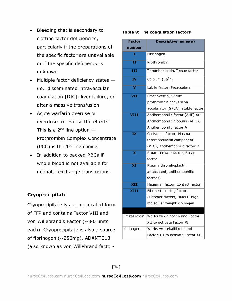
Cryoprecipitate
Cryoprecipitate is a concentrated form
of FFP and contains Factor VIII and
von Willebrand’s Factor (~ 80 units
each). Cryoprecipitate is also a source
of fibrinogen (~250mg), ADAMTS13
(also known as von Willebrand factor-
Factor
number
Descriptive name(s)
I Fibrinogen
II Prothrombin
III Thromboplastin, Tissue factor
IV Calcium (Ca2+)
V Labile factor, Proaccelerin
VII Proconvertin, Serum
prothrombin conversion
accelerator (SPCA), stable factor
VIII Antihemophilic factor (AHF) or
Antihemophilic globulin (AHG),
Antihemophilic factor A
IX Christmas factor, Plasma
thromboplastin component
(PTC), Antihemophilic factor B
X Stuart–Prower factor, Stuart
factor
XI Plasma thromboplastin
antecedent, antihemophilic
factor C
XII Hageman factor, contact factor
XIII Fibrin-stabilizing factor,
(Fletcher factor), HMWK, high
molecular weight kininogen
Prekallikrein Works w/kininogen and Factor
XII to activate Factor XI.
Kininogen Works w/prekallikrein and
Factor XII to activate Factor XI.
Table 8: The coagulation factors

[35]
nurseCe4Less.com nurseCe4Less.com nurseCe4Less.com nurseCe4Less.com
cleaving protease (VWFCP) which is deficient in congenital TTP), fibronectin
and Factor XIII (fibrin stabilizing factor).
Immune Globulins
Immune globulins (immunoglobulins specific for the antigen in question) can
be purified from collected sera and is used to produce concentrated supplies
of anti-RhoD antibody.
Other specific immune globulins are available for cytomegalovirus, rabies,
tetanus, hepatitis A and B, measles, respiratory syncytial virus, rubella,
smallpox, and varicella virus. They are generally given intravenously (IV)
and as such are referred to as IVIG. The use of immune globulins provides a
passive protection against the virus or bacteria as opposed to a normal,
active immune response where the individual’s immune system mounts a
cellular, humoral (antibody) or both a cellular and humoral response to the
invading pathogen. It is the treatment of choice for individuals who lack
specific antibody and is used increasingly to treat autoimmune diseases such
as:20,37,38
Idiopathic thrombocytopenic purpura (ITP)
Kawasaki disease
Guillain–Barré syndrome
Myasthenia gravis
Dermatomyositis
A recent Cochrane review has, however, recommended against using IVIG in
neonates for either proven or suspected infections.39

[36]
nurseCe4Less.com nurseCe4Less.com nurseCe4Less.com nurseCe4Less.com
Other Components
Recent research has been focused on the development of blood substitutes
to eliminate potential transfusion reactions. These are generally chemically
inert substances, which have oxygen-binding (and releasing) capacity. Even
more recently, the emphasis has shifted to oxygen (O2) therapeutics,
focusing on restoring tissue oxygenation in specific tissues.
Currently, there are alternatives including hemoglobin-based O2 carriers
(HBOCs) and perfluorocarbon-based O2 carriers (PFCOCs). Most of the
current products have the disadvantage of short half-lives and significant
adverse effects such as hypertension, gastrointestinal (GI) distress and
higher than expected death rates in trauma patients. The PFCOCs appear to
have fewer adverse effects.40
Other alternatives include autologous transfusions (pre-depositing one’s own
blood for transfusion for scheduled surgeries — this is known as preoperative
autologous donation (PAD)), acute normovolemic hemodilution (ANH), where
the patient donates their own blood immediately before surgery. Normal
fluid volumes are maintained by the transfusion of colloids, such as albumin,
dextrans or various starches or gelatins and crystalloids, such as normal
saline and Ringer's lactate.41-43 Free hemoglobin cannot be used because it is
renotoxic.
Hematopoietic stem cells may be obtained from autologous (the patient) or
from allogeneic (donor) sources for hematopoietic stem cell transplants.
Colloids including albumin can be purified from blood and are transfused to
help maintain fluid volumes, often in patients with sepsis.44
[37]
nurseCe4Less.com nurseCe4Less.com nurseCe4Less.com nurseCe4Less.com
Finally, coagulation factors may be transfused. These may involve the
specific factors if these are known, or may involve the use of cryoprecipitate,
a rich source of fibrinogen and many of the required coagulation factors in
high concentrations.45-49
Transfusion Techniques
For red blood cell transfusion, at least a standard 18-guage needle should be
used to prevent mechanical damage to the RBC from shearing or other
physical forces. Needles larger than 18-gauge may be used if needed.
Standard tubing filters should also be in place.
Other than sterile, isotonic saline (0.9%), no other fluid or drugs should be
added to the infusion of any blood component. Ringer’s solution contains
calcium ions (Ca2+), which can cause spontaneous clotting, and either
hypotonic or hypertonic solutions can damage and lyse the cells.
One unit of blood should take no longer than 4 hours for complete
transfusion. If there is a clinical concern with too-rapid transfusion (such as
concerns over heart failure, or hypervolemia) then single-donor blood may
be divided into smaller aliquots (portions). This should be done under sterile
conditions in the blood bank.
The procedure for dividing blood components into smaller aliquots is done
routinely for transfusions in children, minimizing the risk of hypervolemia
and transfusion reactions. If at all possible, these aliquots should come from
a single donor, again, to minimize transfusion reactions and the exposure to
multiple sources of new antigens and antigen sensitization reactions.

[38]
nurseCe4Less.com nurseCe4Less.com nurseCe4Less.com nurseCe4Less.com
Before ANY transfusion procedure is
undertaken, informed consent must
be obtained if the patient is conscious.
In addition, the following MUST be
checked and double-checked.
The patient's wristband
The blood unit label
The compatibility test
report
It is of primary importance that the
correct blood product is given to the
correct recipient.
Observation of the Transfusion Patient
The transfusion patient should be closely
observed, particularly during the
transfusion itself and the first 15 -60
minutes after the infusion of the blood
components. Pulse, respiratory rate,
heart rate, blood pressure and
temperature should be observed and
recorded and any significant
observations regarding mental status,
pain levels and fluid status (i.e., urine volume) should also be recorded and
reported as needed.
The patient should be kept warm and covered to prevent chill from the
environment, as these may be incorrectly interpreted as a potential
transfusion reaction. Acute reactions to transfusion of RBCs and blood
components may occur within the first 24 hours (acute) or over the following
1-14 days (delayed).
Specific Indications For Blood Transfusions
The specific indications for blood transfusions are discussed in this section
relative to a medical condition or mechanisms of injury. Guidelines for
transfusions under specific conditions are also reviewed.

[39]
nurseCe4Less.com nurseCe4Less.com nurseCe4Less.com nurseCe4Less.com
Injury
Rapid Massive Hemorrhage
Massive acute hemorrhages, which may result from injury and/or the
rupture of a major blood vessel, can result in anemia. Sudden loss of
approximately 30% of the total blood volume can result in a fatal outcome,
but with a less acute loss, approximately 60% of the total blood volume can
be lost without fatality.
Treatment for acute blood loss consists of returning the patient to
hemostasis, replacing blood volumes and the treatment of shock. Injury by
trauma of some form is the most common cause of death in those aged 1-44
years old. In general, trauma can be described as either blunt, involving a
forceful impact to the body or or penetrating, involving a piercing or
breakage of the skin. As in all emergency situations, the ABCDE survey is
the first priority. The ABCDE survey includes:
Airway
Breathing
Circulation
Disability
Exposure/environmental control
Regarding circulation issues and trauma, it should be noted that while
external hemorrhage is always readily apparent, internal and potentially life-
threatening hemorrhage is often not as obvious. The body compartments
with sufficient volume to allow significant internal bleeding are the chest,
abdomen and the soft tissues of the pelvis.
[40]
nurseCe4Less.com nurseCe4Less.com nurseCe4Less.com nurseCe4Less.com
External hemorrhage may be controlled by direct pressure. Internal
hemorrhage may have to be treated surgically with the potential for
autologous (autotransfusion) volume replacement using recovered blood.
Internal hemorrhage can be detected if signs of shock are present, including
tachypnea (decreased respiratory rate), intense sweating (diaphoresis),
change in color or an altered mental alertness or status. Pulse and blood
pressure measurements can also be key in alerting personnel to the
potential for internal blood loss. Imaging studies can also be critical in
detecting and determining the extent of the internal trauma and
hemorrhage. These imaging studies include chest X-rays, CTs of the chest,
abdomen, pelvis, spine, or head, and can be revealing.
Diagnostic peritoneal lavage (DPL) has been largely replaced as a diagnostic
tool with a Focused Assessment with Sonography in Trauma (FAST),
particularly for unstable patients. An Extended FAST (E-FAST) can also be
performed. FAST and E-FAST is able to detect significant volumes of
intraperitoneal blood.50,51
Abdominal injury can constitute damage to the abdominal wall, the
abdominal vasculature and either a solid organ such as the liver, spleen,
kidney(s) or pancreas or to the stomach, the small or large intestines, colon,
ureters or bladder. Grading scales may be used, ranging from Grade 1
(minimal damage) to Grade 5/6 for massive damage.
Hemorrhages in abdominal injuries may be immediate, especially in
penetrating trauma where an abdominal vessel is directed impacted, or
hemorrhages in abdominal injuries may be subtle, with low-volume blood
loss and few signs and symptoms of physiologic distress. Other causes of
hemorrhage requiring transfusions are post-partum hemorrhages and

[41]
nurseCe4Less.com nurseCe4Less.com nurseCe4Less.com nurseCe4Less.com
trauma due to both blunt and penetrating injuries (i.e., explosions with
shrapnel).
Current Guidelines for Emergency Release of Blood Products
The following guidelines are obtained from the American Society of
Hematology.9-12 If an emergency situation exists, such as severe, ongoing
and potentially life threatening hemorrhage or anemia, emergency release of
blood products may be necessary; and, the recommendations are to:
Notify blood bank of need for emergency release of RBC
Complete hospital’s “emergency release” form
o Document declaration of a transfusion emergency
o U.S. federal regulations require 2 specific items on the form
Statement of the nature of the emergency (i.e.,
“massive GI hemorrhage”)
Signature of MD or “equivalent”; (PA, NP, RN, etc.,
cannot sign)
Send patient blood sample to blood bank as soon as possible (before
emergency transfusion begins, if possible).
What the blood bank will provide (depending on how much testing has
already been performed):
Un-crossmatched RBC (ABO group-specific if determined on a
current blood specimen)
Group O RBC if blood bank has not documented patient’s ABO
group on a fresh blood sample
o Rh negative depending on availability and hospital policy, if
patient’s Rh status is unknown
[42]
nurseCe4Less.com nurseCe4Less.com nurseCe4Less.com nurseCe4Less.com
The blood bank will retrospectively crossmatch all emergently issued
units when it receives the patient’s testing sample and will begin
issuing type-specific and crossmatched products when testing is
complete.
Chronic Hemorrhage
Chronic blood loss or chronic hemorrhage may be due to bleeding tumors,
including benign tumors such as rectal polyps or malignant tumors; heavy
menstrual bleeding or gastrointestinal bleeds. Chronic bleeds can also occur
in the brain, the eye or within any solid or hollow organ. Chronic blood loss
can, over time, result in anemia, most often in an iron-deficiency anemia.
Chronic bleeding can also occur with certain medications including
anticoagulants (warfarin [Coumadin], heparin, rivaroxaban [Xarelto],
dabigatran [Pradaxa]) and anti-platelet medications (aspirin, clopidogrel
[Plavix], prasugrel [Effient]).
Bleeding and platelet disorders can also result in chronic hemorrhage — the
most common include hemophilia, von Willebrand disease, idiopathic
thrombocytopenia purpura (ITP), thrombotic thrombocytopenic purpura
(TTP), HELLP syndrome and Heparin-Induced Thrombocytopenia (HIT).
Liver disease with inadequate production of coagulation factors can also
result in chronic bleeding. Hypercoagulation syndromes such as
disseminated intravascular coagulation (DIC) may result in the anemia and
thrombocytopenia without blood loss.
[43]
nurseCe4Less.com nurseCe4Less.com nurseCe4Less.com nurseCe4Less.com
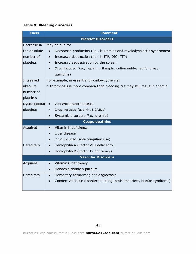
Table 9: Bleeding disorders
Class Comment
Platelet Disorders
Decrease in
the absolute
number of
platelets
May be due to:
Decreased production (i.e., leukemias and myelodysplastic syndromes)
Increased destruction (i.e., in ITP, DIC, TTP)
Increased sequestration by the spleen
Drug induced (i.e., heparin, rifampin, sulfonamides, sulfonureas,
quinidine)
Increased
absolute
number of
platelets
For example, in essential thromboycythemia.
* thrombosis is more common than bleeding but may still result in anemia
Dysfunctional
platelets
von Willebrand’s disease
Drug induced (aspirin, NSAIDs)
Systemic disorders (i.e., uremia)
Coagulopathies
Acquired Vitamin K deficiency
Liver disease
Drug induced (anti-coagulant use)
Hereditary Hemophilia A (Factor VIII deficiency)
Hemophilia B (Factor IX deficiency)
Vascular Disorders
Acquired Vitamin C deficiency
Henoch-Schönlein purpura
Hereditary Hereditary hemorrhagic telangiectasia
Connective tissue disorders (osteogenesis imperfect, Marfan syndrome)

[44]
nurseCe4Less.com nurseCe4Less.com nurseCe4Less.com nurseCe4Less.com
Traumatic Hemolytic Anemia
Traumatic hemolytic anemia can occur extravascularly, within the heart and
intravascularly. Mechanical shear forces or turbulence causes hemolysis. The
presence of schistocytes or abnormally shaped RBCs is diagnostic.
Illness Due to Deficient Erythropoiesis
Hypochromic-Microcytic Anemias
Hypochromic-microcytic anemias where the RBCs are smaller and paler than
normal may result from deficient erythropoiesis, the deficient production of
RBCs in the bone marrow. This may result from deficient heme or globin
synthesis. The microcytic anemias included in a differential diagnosis are
iron deficiency, iron-transport deficiency and iron-utilization deficiencies as
well as the anemia of chronic disease (iron re-utilization anemia) and the
thalassemias.
Normochromic–normocytic Anemias
Bone marrow failure results in a normochromic-normocytic anemia where
the cells contain normal amounts of hemoglobin and therefore are normally
pigmented (normochromic) and are normally sized (normocytic). Bone
marrow failure may result from a lack of response to erythropoietin (EPO), a
glycoprotein cytokine produced by the kidney.
Erythropoietin is believed to be regulated by a feedback mechanism based
on the levels of oxygen in the blood and is produced by interstitial fibroblasts
and peritubular cells of the renal cortex. Under hypoxic conditions, EPO is
secreted and targets burst-forming unit-erythroid (BFU-E) cells expressing

[45]
nurseCe4Less.com nurseCe4Less.com nurseCe4Less.com nurseCe4Less.com
the EPO receptor, the colony-forming-unit-erythroid (CFU-E) cells, which
maximally express the EPO receptor and proerythroblasts.
Erythropoietin cooperates with various other cytokines including
interleukins-3 and -6 (IL-3, IL-6) glucocorticoids and others to stimulate
pleuripotential stem cells.52 EPO has other non-hematopoietic roles such as
stimulating angiogenesis and inducing the proliferation of smooth muscle
fibers — the actions for which EPO is well known as a “doping agent” in a
number of endurance and professional sports.52,53
Normochromic-normocytic anemias include the anemia associated with
kidney disease, protein depletion and anemias associated with
hypometabolic states (i.e., hypothyroidism and hypopituitarism). Aplastic
anemia is a panhypoplasia where the precursors of the RBCs are affected
along with platelet and white blood cell precursors resulting in a “triad” of
anemia, thrombocytopenia and leukopenia. A normocytic-normochromic
anemia is also seen in myelodysplastic syndromes, a group of proliferative
disorders where blood cells are poorly formed, ineffective and abnormal
cells, and in myelophthisic anemias where the bone marrow is replaced by
non-hematopoietic cells.
Macrocytic Anemias
The macrocytic anemias, defined as mean corpuscular volume (MCV) of over
100fL/cell, can be megaloblastic or non-megaloblastic. Non-megaloblastic
anemias form a diverse group of clinical states including anemias associated
with chronic alcohol use and those associated with aberrant or abnormal
cholesterol esterification by the liver. Megaloblasts are nucleated, large RBCs
precursors — the nuclei of the megaloblasts show uncondensed chromatin.
[46]
nurseCe4Less.com nurseCe4Less.com nurseCe4Less.com nurseCe4Less.com
Megaloblastic anemias result from defective DNA synthesis and the most
common are caused by nutritional deficiencies of Vitamin B12 and folate. B12
and folate deficiencies can be due to poor diet, an increased utilization,
inadequate utilization, increased excretion or inadequate absorption.
Vitamin B12 for example, requires the presence of intrinsic factor (IF).
Pernicious anemia or the presence of peptic ulcers can decrease B12 uptake.
In addition, the use of proton pump inhibitors (PPIs) used to treat
gastroesophageal reflux disease (GERD) and peptic ulcer disease (PUD) have
recently been shown to be significantly associated with the risk of B12
deficiency.54 Similarly, folate antagonists such as methotrexate (used to
treat various cancers or as an immunosuppressant) or triamterene (a K+ -
sparing diuretic used to treat hypertension) can induce a folate-deficiency
associate megaloblastic anemia.
Hemolytic Anemias
Removal of senescent cells normally occurs at the end of the normal RBC
lifespan of approximately 120 days. This is primarily done by the spleen,
liver, bone marrow and by the reticuloendothelial system (RES). Hemolysis
is the premature destruction of RBCs and can result from either extrinsic or
intrinsic factors. The hemolysis may be extravascular, occurring primarily in
the liver and spleen, or intravascular.
Extrinsic factors include hypersplenism (hyperactivity of the RES),
autoimmune hemolytic anemia (cold, warm antibody or paroxysmal cold
antibody, resulting in increased urinary hemoglobin levels) or by trauma and
other mechanical injuries. Infections such as Clostridium perfringens, α- or
β-hemolytic streptococci cause an extrinsic hemolysis due to toxin
[47]
nurseCe4Less.com nurseCe4Less.com nurseCe4Less.com nurseCe4Less.com
production where other infectious agents such as malaria (Plasmodium)
species lyse the RBCs as part of the life cycle.
Intrinsic RBC membrane abnormalities may essentially induce an immune
response resulting in hemolysis. Other membrane abnormalities may result
in the destruction of the RBCs because of the resulting shape abnormalities
(poikilocytosis) or size abnormalities (anisocytosis) cause a mechanical
hemolysis within the smaller vessels (i.e., sheer stress). In addition to the
membrane abnormalities that may result in hemolysis, disorders of RBC
metabolism (Glucose-6-phosphate deficiency [G6PD]) and the
hemoglobinopathies including sickle cell anemia (SCA) and thalassemias (α,
β, and β-δ) may occur. Finally, either quantitative or functional abnormalities
in membrane proteins including spectrin, ankyrin and actin can result in RBC
hemolysis.
Autoimmune Hemolytic Anemias
In autoimmune hemolytic anemia (AHA), antibodies react with the RBCs with
the subsequent binding of complement. The most common form of AHA is
warm antibody hemolytic anemia. In warm antibody hemolytic anemia, the
autoantibodies react with RBCs at 37o C or higher. These antibodies may
occur spontaneously, associated with disorders such as systemic lupus
erythematosus (SLE), lymphoma or with leukemias or they may appear as
an adverse effect of drugs such as levodopa, cephalosporins, diclofenac and
alpha interferon.55
Warm antibody hemolytic anemia is particularly important in the context of
blood transfusion medicine because the presence of these warm antibodies
makes the crossmatching of blood technically more difficult and increases

[48]
nurseCe4Less.com nurseCe4Less.com nurseCe4Less.com nurseCe4Less.com
the risk of a transfusion reaction (see below). In addition, the transfusion
recipient may develop, in addition to the autoantibodies already present,
agglutinating (clumping) antibodies directed against the donor RBCs.56,57
Paroxysmal Nocturnal Hemoglobinuria
Paroxysmal nocturnal hemoglobinuria (PNH) is a relatively rare genetic
disorder where a stem cell membrane defect results in hemolysis and
decreased WBCs and platelets. The RBCs are especially sensitive to
complement and for reasons that are not well understood, hemolysis is
increased during sleep. Anemia, leukopenia, thrombocytopenia and
thromboses (both arterial and venous) are common and these events may
occur episodically.
Hereditary Spherocytosis and Elliptocytosis
Poikilocytosis refers to variations in RBC shape and anisocytosis refers to
variation in RBC size. Both may be seen in hereditary spherocytosis (HS)
and elliptocytosis (HE), congenital RBC membrane disorders. HS and HE are
autosomal dominant disorders with varying degrees of hemolysis (HS tends
to have more hemolysis) and splenomegaly (more prominent in HE).
Metabolic Disorders with Hemolytic Anemia
Embden-Meyerhof Pathway Defects:
These are all autosomal recessive metabolic disorders in glycolysis
pathways. Because they are autosomal recessive disorders, they only
cause anemias in homozygous individuals, i.e., those carrying two
copies of the defective gene. Pyruvate kinase deficiency is an example.

[49]
nurseCe4Less.com nurseCe4Less.com nurseCe4Less.com nurseCe4Less.com
Glucose-6-Phosphate Dehydrogenase (G6PD) Deficiency:
This is an X-linked defect more common in African Americans but
occurs also in people of Italian, Greek, Arabic and Sephardic Jewish
ancestry. Hemolysis often occurs after an acute illness or taking
oxidant drugs such as salicylates, some anti-malarials like primaquine,
dapsone (used in the treatment of leprosy and as a 2nd line of
treatment for pneumocystis in HIV+ patients) and sulfonamides.
Hemoglobinopathies
Normal adult hemoglobin (abbreviated HbA or α2β2) consists of four globular
subunits with a central iron-binding heme group. The four subunits consist of
2 α chains and 2 β chains. Normal infant Hb (HbF) has 2 α chains and 2 γ
chains (α2 γ2). As the infant grows and RBCs are replaced, the γ chains are
gradually replaced with adult-type β chains.
Another form of hemoglobin contains 2 δ chains along with 2 α chains and is
known as HbA2 or α2δ2. HbA constitutes 97% of normal adult hemoglobin
while HbF and HbA-2 constitute approximately 3%. Hemoglobin carries
oxygen from the lungs to the rest of the body tissues. It can also carry
carbon dioxide (CO2) and besides the RBC is found in macrophages, the
substantia nigra (in dopaminergic neurons), astrocytes, oligodendrocytes
and in alveolar cells. The different hemoglobinopathies represent abnormal
variations in Hb structure.

[50]
nurseCe4Less.com nurseCe4Less.com nurseCe4Less.com nurseCe4Less.com
Sickle Cell Anemia
Sickle Cell Anemia (SCA) is an autosomal recessive defect where a single
nucleotide polymorphism (SNP) causes an A T alteration in the DNA
sequence. This results in the substitution of a glutamic acid by a valine,
producing an HbS. The HbS, under hypoxic (low oxygen) conditions induces
polymerization by the Hb with subsequent crystallization and the
deformation and eventual hemolysis of the RBC.
Acute exacerbations, known as crises, can be precipitated by acute illness,
fever, viral infection or trauma, though often, they appear spontaneously or
for no known reason. These crises can be painful, with severe oxygen
deprivation (infarction) occurring in the bones, spleen, lungs and/or kidneys.
A common cause of mortality in SCA is acute chest syndrome with
microvascular occlusions accumulating in the lung tissue. Aplastic crises can
occur as well. Signs and symptoms include anemia, jaundice, pallor,
hepatosplenomegaly, cardiomegaly, systolic ejection (flow) murmurs,
cholelithiasis (gallstones) and chronic ulcers around the ankles (punched-out
ulcers).
Thalassemias
The thalassemias are characterized by defective hemoglobin synthesis and
produce a microcytic hemolytic anemia, and are an inherited disorder.
Thalassemias are the most common hemoglobinopathy and results from the
decreased or abnormal production of one of the four hemoglobin subunits —
α, β, γ, or δ. The types of anemias are most common in individuals of
Mediterranean, African and Asian ancestry. Iron overload can be the result of

[51]
nurseCe4Less.com nurseCe4Less.com nurseCe4Less.com nurseCe4Less.com
multiple transfusions. Signs and symptoms include anemia, hemolysis,
splenomegaly and bone marrow hyperplasia.
The condition β-thalassemia is the result of decreased production of the β
chain. Homozygotes (β thalassemia major) can be severely anemic and
show signs of bone marrow hypertrophy. Heterozygotes (β thalassemia
minor) are much less severely affected and are either asymptomatic or
present showing signs of mild or moderate anemia.
The other condition of α-thalassemia results from a defect in the production
of the α chain, but since 4 genes are involved in α chain production, there
are 4 sub-types, depending on the number of genes affected.
Single-gene defect: α-thalassemia-2. These individuals are usually
asymptomatic.
Two-gene defect: α-thalassemia-1. These individuals carry the train
and may have a mild to moderate microcytic anemia.
Three-gene defect: A three-gene defect results in hemoglobin with
excess β chains (HbH). In infants, the excess β chains are paired with
γ chains to produce Bart’s Hb. Individuals with the three-gene defect
have a hemolytic anemia often accompanied by splenomegaly.
Four-gene defects are fatal in utero.
Other Hemoglobinopathies
Other hemoglobinopathies include the following:
Hemoglobin C is more common in African Americans and has similar
but milder features as SCA.

[52]
nurseCe4Less.com nurseCe4Less.com nurseCe4Less.com nurseCe4Less.com
Hemoglobin S-C combines the S- and the C- Hb defect and is again
more common in African Americans. Symptoms tend to be less severe
than in SCA, but hematuria, retinal hemorrhages and aseptic necrosis
of the femoral head are common.
Hemoglobin E occurs primarily in those of African or Southeast Asian
descent and is an autosomal recessive disorder. Homozygotes for HbE
are most often treated with chronic transfusions.
Complications Of Transfusions: Part I
This section covers current guidelines for principles for the transfusion of
incompatible RBC in an emergency release of blood products or in the case
of mismatch. The following guidelines are obtained from the American
Society of Hematology.9-12
Clinically, the scenario of severe warm or cold autoimmune hemolytic
anemia can occur where the patient has auto-antibodies which react with all
of the blood bank’s reagent cells. Alternatively, the blood bank may not be
able to determine if alloantibodies are present or none of the currently
available RBC unit is compatible. In this type of scenario, a clinical decision
must be made if the risk of an incompatible RBC transfusion outweighs the
likelihood of death. In these types of situations, the American Society of
Hematology has produced the following principles:
Communication between bedside clinician and transfusion service
physician is essential.
Obtain careful history of prior transfusion or pregnancy:

[53]
nurseCe4Less.com nurseCe4Less.com nurseCe4Less.com nurseCe4Less.com
o If history negative, probably safe to transfuse ABO-
compatible RBC
o If history positive or uncertain, assess risk: benefit of
delaying transfusion to complete testing
Assess how long it may take for blood bank or reference lab to
complete pre-transfusion testing
Agree on best approach to choosing among incompatible RBC units
(transfusion physician will advise)
Attempt to mitigate need for immediate transfusion: bed rest, oxygen.
Ultimately, do not deprive a patient with autoimmune hemolytic
anemia of a needed, lifesaving transfusion.
Autoantibody will shorten survival of transfused RBC and patient’s
endogenous RBC to a similar extent.
Most undetected alloantibodies will cause delayed hemolytic
transfusion reactions:
o May be misdiagnosed as worsening of autoimmune hemolysis
o Not usually life-threatening
Bedside team must be hypervigilant for acute intravascular hemolytic
reaction during transfusion.
The most serious transfusion reactions include:
o Acute hemolytic transfusion reactions
o Acute respiratory distress and lung damage
o Circulatory (volume) overload
Early recognition of the signs and symptoms of serious transfusion reactions
is critical. These should not be confused with the more common and much
less severe reactions, which include febrile but non-hemolytic reactions and

[54]
nurseCe4Less.com nurseCe4Less.com nurseCe4Less.com nurseCe4Less.com
chills alternating with rigor (chill-rigor reactions). However, if the febrile or
chill-rigor reactions occur during the transfusion, the transfusion should be
halted immediately, with the IV line kept open with isotonic saline. The blood
component should be immediately returned to the blood bank along with a
sample of the patient’s anti-coagulated blood for the blood bank to examine.
Unless there is an emergency situation (and O- RBCs may then be used),
the transfusion should be halted until the cause of the transfusion reaction
can be determined.
Other reactions that can signal a transfusion reaction include:
Dyspnea or any difficulty breathing
Light-headedness
Urticaria (unless localized)
Significant itching (unless localized)
Flank or lumbar pain
Facial flushing
Acute Hemolytic Transfusion Reactions (AHTR)
While acute hemolytic transfusion
reactions (AHTR) are relatively rare, they
are serious and the nursing and hospital
staff should be alert to the possibilities.
The most common cause of AHTR is
human error; the mislabeling of the
patient’s pre-transfusion sample or
failing to check and confirm that the
blood component is intended for a
particular patient.
The most common cause of
AHTR is human error.
Mislabeling of the patient’s
pre-transfusion sample
Failure to check and
confirm that the blood
component is intended for
a particular patient.
AHTR is not generally a laboratory
or blood bank error.

[55]
nurseCe4Less.com nurseCe4Less.com nurseCe4Less.com nurseCe4Less.com
In most AHTRs, it is usually the donor cells that are hemolyzed by the
recipient’s immune system. This can result from ABO/Rh incompatibilities,
antibodies passively transfused by plasma that has not been fully removed
from the packed RBCs, the presence of fragile RBCs or fragments of RBCs
resulting from overheating, mechanical forces or by the inadvertent
introduction of hypotonic solutions.
In AHTR, the hemolysis is intravascular. It may be accompanied by
hematuria, depending on the condition of the patient’s kidneys. AHTR may
also be accompanied by disseminated intravascular coagulation (DIC). The
severity of the AHTR depends in large part on the patient’s overall condition,
condition of their heart, liver and kidneys, the degree of ABO/Rh
incompatibility, whether that patient has been sensitized previously, the
amount of blood component transfused and the administration rate of the
blood component.
If there is an acute reaction, it most commonly happens within 60 minutes
of the initiation of the transfusion. Onset of the reaction is usually abrupt
with the patient (if conscious) complaining of discomfort or anxiety. Other
symptoms include dyspnea, fever, chills, chill-rigor reaction, and flushing of
the face and flank or lumbar (low back) pain.
If the patient is unconscious or under anesthesia, the primary sign may be
hypotension (low blood pressure), uncontrolled bleeding from the incision or
site of injury or gross hematuria. If DIC is present, bleeding from the
mucous membranes may be observed as well. Shock may ensue, with a
rapid, feeble pulse, hypotension, cold and clammy skin and nausea or
vomiting. Jaundice may also appear, but is often difficult to detect in the
early stages.
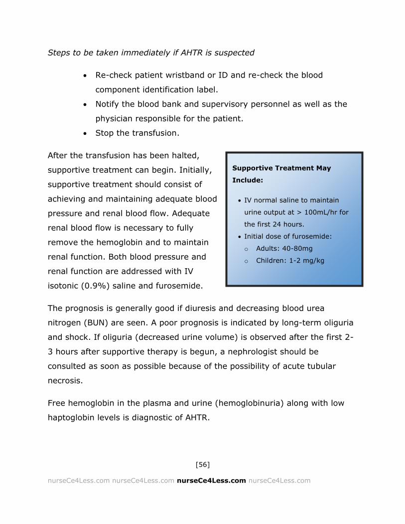
[56]
nurseCe4Less.com nurseCe4Less.com nurseCe4Less.com nurseCe4Less.com
Steps to be taken immediately if AHTR is suspected
Re-check patient wristband or ID and re-check the blood
component identification label.
Notify the blood bank and supervisory personnel as well as the
physician responsible for the patient.
Stop the transfusion.
After the transfusion has been halted,
supportive treatment can begin. Initially,
supportive treatment should consist of
achieving and maintaining adequate blood
pressure and renal blood flow. Adequate
renal blood flow is necessary to fully
remove the hemoglobin and to maintain
renal function. Both blood pressure and
renal function are addressed with IV
isotonic (0.9%) saline and furosemide.
The prognosis is generally good if diuresis and decreasing blood urea
nitrogen (BUN) are seen. A poor prognosis is indicated by long-term oliguria
and shock. If oliguria (decreased urine volume) is observed after the first 2-
3 hours after supportive therapy is begun, a nephrologist should be
consulted as soon as possible because of the possibility of acute tubular
necrosis.
Free hemoglobin in the plasma and urine (hemoglobinuria) along with low
haptoglobin levels is diagnostic of AHTR.
Supportive Treatment May
Include:
IV normal saline to maintain
urine output at > 100mL/hr for
the first 24 hours.
Initial dose of furosemide:
o Adults: 40-80mg
o Children: 1-2 mg/kg
[57]
nurseCe4Less.com nurseCe4Less.com nurseCe4Less.com nurseCe4Less.com
Delayed Hemolytic Transfusion Reactions
Delayed hemolytic transfusion reactions may be quite common, but are
often not clinically identified because the reactions are usually mild and in
general, appear to be self-limiting. These delayed reactions may be the
result of pre-sensitization to RBC antigens. The only symptoms may be a
slight fever. Lab tests may reveal a lower hematocrit and possibly an
increase in bilirubin and LDH. Gross hematuria is rare.
Acute Respiratory Distress and Lung Damage
Acute lung damage is relatively rare, but accounts for the most common
cause of death in transfusion reactions. The pathophysiology of the damage
is due to the presence of anti-HLA or anti-granulocyte antibodies in the
donor plasma; these antibodies attack the WBC (which carry the HLA
antigens) and both agglutinate and cause degranulation of recipient
granulocytes in the lung tissue. Acute respiratory distress may result. Chest
X-ray shows a characteristic non-cardiogenic pulmonary edema. However,
most cases of acute lung damage show mild symptoms and are likely often
missed, though all suspected cases should be reported.
The use of blood products donated by never-transfused men or never-
transfused women who have never been pregnant decreases the risk of any
acute transfusion reaction because it is unlikely that these individuals have
been sensitized to other human antigens, such as RBC or HLA antigens. (See
section on TRALI below).
Circulatory (Volume) Overload

[58]
nurseCe4Less.com nurseCe4Less.com nurseCe4Less.com nurseCe4Less.com
Volume overload, especially if the transfusion occurs too rapidly, is a real
and present danger, though historically, it has been under-recognized and
under-reported. Recent studies have indicated that volume overload is the
second most common cause of death in transfusion reactions reported to the
FDA.
Patients at risk for volume overload are those with pre-existing (though not
necessarily recognized) cardiac, respiratory and renal insufficiency. Those at
risk include the elderly and neonates. Blood components are highly
concentrated and can provide a high osmotic load, forcing fluids (by osmotic
pressure gradients) into intravascular spaces.58-61
Signs and symptoms of volume overload include dyspnea, the presence of
respiratory crackles, coughing, edema in the extremities, tachycardia, and
irregular heart rate and increased blood pressure. (See sections on TRALI
and TACO below).
If any of these symptoms are noted, the transfusion should be stopped
immediately and the supervisory staff and medical personnel should be
notified. Treatment is supportive, as used for heart failure, including the use
of 20 – 40 mg of furosemide (IV). If the patient was undergoing a plasma
transfusion (using fresh frozen plasma, for example) to reverse a warfarin
overdose, furosemide at a lower dose may be given, but the preferred
treatment is with a prothrombin complex concentrate (PCC). If pre-existing
heart failure or renal failure is known in a transfusion patient, furosemide is
given prophylactically at 20 – 40 mg (IV).
Aggressive volume expansion — essentially a volume overload — is
recommended in kidney transplantation, whether it is for chronic renal
failure of for other indications such as hypertension, ischemic heart disease,

[59]
nurseCe4Less.com nurseCe4Less.com nurseCe4Less.com nurseCe4Less.com
and chronic heart failure.62 However, more recent results indicate that it
may not be necessary to maximize the function of the transplanted
kidney(s). Recent results indicate that a less aggressive approach, with
lower volumes resulted in similar good results.62
Other Transfusion Reactions
Altered Oxygen Affinity
The affinity for oxygen in RBC is to a large part dependent on the
hemoglobin, but levels of RBC 2,3-diphosphoglycerate (DPG) play a role as
well. RBC 2,3-diphosphoglycerate decreases in RBC blood products stored
for over 7 days and is at negligible levels after 10 days of storage. This
decrease in DPG has the effect of increasing hemoglobin affinity for oxygen,
and this increase in affinity or attraction for oxygen has the effect of
decreasing the amount of oxygen eventually released to the tissues. This
can be an important factor for exchange transfusions in infants, and patients
in sudden cardiac arrest in acute crisis (acute chest syndrome, stroke) and
with severe heart failure. Using fresh packed RBC in these cases is
recommended.
Graft-Versus-Host Disease
Graft versus Host disease (GVHD) occurs when immune cells from the donor
(the “graft”) recognized the recipient (host) as being foreign, or carrying
foreign antigens on the surface of host cells. The grafted cells then attack
the host cells.

[60]
nurseCe4Less.com nurseCe4Less.com nurseCe4Less.com nurseCe4Less.com
In general, for GVHD to occur in a transfusion or in any transplantation,
three criteria known as the Billingham criteria63 must be met:
The donor cells must be immune-competent. This occurs when
functional WBCs containing T-cells and possibly B-cells are transferred.
The recipient shows immunological differences from the host and is
histo-incompatible (major or minor histocompatibility mismatches can
occur).
The recipient is immunocompromised and incapable of rejecting the
transplanted cells.
Acute GVHD can occur anytime before 100 days post transfusion (or
transplant) while chronic GVHD occurs after the 100-day period, those these
definitions may be altered by the current work of the National Institutes of
Health (NIH) chronic GVHD consensus project working group. Commonly,
GVHD occurs between 4 - 30 days after transfusion of blood components.
The severity of the GVHD is graded by the modified Glucksberg criteria.
These criteria are based on the appearance of fever, a centrifugally
spreading maculopapular erythematous skin rash (sometimes with bullae),
gastrointestinal symptoms (usually vomiting and/or a bloody diarrhea),
lymphadenopathy, pancytopenia and changes in liver function as measured
by the appearance of jaundice and increased bilirubin levels. Acute damage
in GVHD is the result of donor T-cell activation followed by the production of
a variety of inflammatory cytokines (a cytokine storm) including gamma
interferon (IFN-γ), Tumor Necrosis Factor-α (TNF-α) and Interleukins 1, 2, 6
and 12 (IL-1, IL-2, IL-6, IL-12). These inflammatory cytokines along with
the presence of natural killer cells (NK cells) and cytotoxic T lymphocytes
(CTLs) directly and indirectly induce host cell damage and death.64

[61]
nurseCe4Less.com nurseCe4Less.com nurseCe4Less.com nurseCe4Less.com
Graft versus host disease is rare in transfusion medicine, particularly if blood
components are irradiated to irreversibly damage the graft (donor)
lymphocytes. Irradiation of transfused blood components is strongly
recommended if the recipient is:
Immunocompromised:
This can include neonates or patients with:
o Congenital immunodeficiency syndromes
o Hematologic cancers
o Hematopoietic stem cell transplants
A 1st degree relative of the donor
When HLA- bearing blood components are transfused. In practice, this
means WBCs, but it should be remembered that WBC could
contaminate the packed RBCs or PRP components.
While rare in transfusions, GVHD has a >90% mortality because there is no
specific treatment available. Prevention by careful screening, checking to
ensure that the correct blood components are being transfused to the
correct individual and using irradiated blood components as recommended
has reduced the incidence of GVHD. However rare the incidence, vigilance is
required because GVHD as an adverse reaction to a transfusion is nearly
always fatal.
Complications of Massive Transfusions
Massive transfusion can be defined as the transfusion of greater than or
equal to an individuals’ total blood volume within a 24-hour period.65-69 This
represents approximately 10 units of blood. Recent advances have been
made in battlefield hospitals. The use of rapid infusers allow the transfusion

[62]
nurseCe4Less.com nurseCe4Less.com nurseCe4Less.com nurseCe4Less.com
of pre-warmed 250 mL and 500 mL boluses (rapid infusion) of blood and can
be mixed with FFP; this has resulted in great clinical advantage for severely
traumatized soldiers.69 These battlefield advances also involve the
concomitant use of hemostatic agents such as Tranexamic acid (TXA).69
The main issue in massive transfusions is that during the process of
transfusing a large volume of colloidal RBCs along with crystalloid fluids such
as Ringer’s lactate or isotonic saline, the clotting factors within the plasma
and the platelets transfused in the FFP, cryoprecipitate and/or PRP are
diluted and a dilutional coagulopathy is induced. In addition, under
conditions where the massive transfusion is given for massive accidental
blood loss, there is a “consumptive” coagulopathy occurring as well — that
is, the coagulation process is consuming the coagulation factors at a rate
faster than can be replaced. All this can lead to what has been termed a
“lethal triad” of acidosis, hypothermia, and bleeding.65 Research over the
past few years has indicated that the administration of high ratios of plasma
to packed RBC can improve mortality and is known as “damage control
resuscitation”.65,68 Overall, the data is indicating improved mortality by using
RBC, plasma and platelets in a 1:1:1 ratio.6
Hypothermia can be avoided by using heat-exchange devices to gently warm
the blood components before it enters the recipient. Alternative warming
devices such as microwave ovens or heating pads are not recommended
because of the potential for damage to the RBCs, hemolysis and uneven
warming.65-68
Citrate toxicity is a concern for patients with poor liver function. Patients
with poor renal function may be at risk for hyperkalemia (high potassium
levels) if the blood components used have been stored for longer than one
[63]
nurseCe4Less.com nurseCe4Less.com nurseCe4Less.com nurseCe4Less.com
week. Hypocalcemia (low calcium levels) has also been observed, and must
be adjusted accordingly. All these potential toxicities are increased with
hypothermia, increasing the importance of using appropriate warming
devices and transfusing blood components as slowly as is clinically feasible.
Protocols are currently being developed where the FFP and PRPs are given
earlier in the transfusion procedure before any coagulopathy develops. Early
results are indicative of decreased mortality and morbidity, but
recommendations have not yet been announced. Current recommendations
are that for every unit of blood given, one unit each of plasma and platelet
concentrates should be given.6
Infections Resulting from Transfusions
While blood and blood components are screened for a variety of pathogens
and while every effort is made to minimize contamination, there is an
inherent risk of infections arising from transfusions. The list of pathogens for
which all blood in the U.S. is screened is given in Table 7. They include HIV-
1 and HIV-2, HTLV-1 and HTLV-2, Hepatitis B and C, West Nile Virus and
Treponema pallidum (the causative spirochete in syphilis). However, this is
only a partial list and does not include new and evolving pathogens or more
rare pathogens. Part of this potential for infection is screened out at the
donor stage using the general health questionnaires and part is screened by
pre-testing donor samples. Clearly, if a pathogen is not tested for or not
screened or asked about during the patient evaluation, the risk of infection
remains. The best approach to infections in the transplant patient is to
maintain proper protocols and follow proper sterile procedures in addition to

[64]
nurseCe4Less.com nurseCe4Less.com nurseCe4Less.com nurseCe4Less.com
being aware of the possibilities and remaining vigilant in the constant
monitoring of transfusion patients.
Bacterial Infections
Current protocols for blood collection, storage and transfusion have resulted
in a decrease in the risk of bacterial contamination and infection from
packed RBC. Nonetheless, packed RBC should be inspected for any color
change, indicative of bacterial growth. Cryophilic (cold-loving) organisms
such as Yersinia spp. may contaminate packed RBCs and produce significant
levels of endotoxin. Rarely, syphilis has been transmitted in fresh blood
products and platelets. Pre-testing will not detect antibodies early in the
infection, and it is presumed that these cases occurred because the donors
were seronegative at the time of donation.
Viral Infections
Hepatitis:
The risk of hepatitis (A,B,C,E) transmission has been reduced by the
combination of pre-screening, pre-testing and by the inactivation (heat
treatment) of blood components. Recombinant technologies have been
used to produce purified and sterile coagulation factors; in the past,
treatment of coagulopathies was an important cause of hepatitis
infections.
Human Immunodeficiency Virus (HIV):
Pre-testing and screening for both HIV-1 (more common in the U.S.)
and HIV-2 is done on all donor blood products. Testing for both
antibodies to HIV and for the genetic material (nucleic acid testing) for

[65]
nurseCe4Less.com nurseCe4Less.com nurseCe4Less.com nurseCe4Less.com
HIV-1 and -2 are done to minimize the risk. Finally, potential blood
donors are screened for high-risk behaviors before they are accepted
as blood donors. Since the late 1980s, this combination of approaches
has greatly reduced the risk of HIV transmission via blood products.
Cytomegalovirus (CMV):
Cytomeglaovirus can be transmitted in WBC-containing components.
CMV is a ubiquitous virus and will not, in general, cause disease in an
immunocompromised recipient. However, CMV can be a serious
problem for immunocompromised patients. In these cases, WBC-
depleted blood components can be given. Alternatively, CMV-negative
donors should be used. This alternative is somewhat more difficult to
achieve given the widespread nature of CMV.
Human T-cell lymphotropic virus 1 and 2(HTLV-1 and HTLV-2):
HTLV-1 (also known as adult T-cell lymphoma virus type 1 or adult T-
cell leukemia lymphoma (ATLL)) is associated with adult T-cell
lymphoma/leukemia, HTLV-1–associated myelopathy, and tropical
spastic paraparesis. It was originally thought that HTLV-2 was
associated with Hairy Cell Leukemia, but this has not been proven
valid. Nevertheless, all donor blood is tested for both HTLV-1 and
HTLV-2 antibodies.
Prion Disease and Malaria
Prion Disease:
Concern for the transmission of the prion disease, Creutzfeldt-Jakob
disease (mad cow disease), is the reason that individuals who have

[66]
nurseCe4Less.com nurseCe4Less.com nurseCe4Less.com nurseCe4Less.com
lived for significant time periods in the United Kingdom (UK) and some
other parts of Europe where mad cow disease were detected are not
allowed to donate blood in the U.S. This denial stands even though
Creutzfeldt-Jakob disease has never been reported as transmitted by
transfusion, or the prions detected in blood components. Prevention of
potential prion transmission is also the rationale behind not allowing
blood donations from individuals who have in the past received
human-derived growth hormone, a dura mater transplant or anyone
with a family member with Creutzfeldt-Jakob disease.
Malaria:
Prospective donors are, by requirement, asked about malarial
symptoms and travel to areas where malaria is prevalent because the
malarial parasite can be easily transmitted through RBCs. Malaria can
be latent and many people are unaware that they are infected. Donors
with a previous diagnosis of malaria are asked to wait for 3 years after
successful treatment while those who have visited areas of the world
where malaria is endemic must wait for one year and be symptom-
free.
Newer or rarer pathogens do appear in blood products and
components. Chikungunya virus has appeared in Europe and North
America recently70 and the parvovirus, human erythrovirus B19 has
been reported in increasing numbers as well.71,72 In addition, septic
infections may be associated with other serious complications of
transfusions.73 Finally, tropical and parasitic diseases can appear in
blood, blood products and result in infections and sepsis.74

[67]
nurseCe4Less.com nurseCe4Less.com nurseCe4Less.com nurseCe4Less.com
Therapeutic Apheresis
Therapeutic apheresis may be defined as “the separation of whole blood into
its major components and removal of the abnormal, pathogenic
component.”75 There are a number of different types of therapeutic
apheresis including:
Plasmapheresis (Plasma exchange)
Leukocytapheresis (removal of white blood cells)
Thrombocytapheresis (removal of platelets)
Erythrocytapheresis (removal of red blood cells)
The terms apheresis, plasmapheresis, therapeutic plasma exchange and cytapheresis
are used interchangeably by many even though they describe different procedures.
Apheresis: Technically, this term refers only to the removal of whole blood for
extracorporeal processing.
Cytapheresis: The removal of whole blood, separation into components, removal of
cell fractions and the re-infusion of remaining cells.
Therapeutic Plasma Exchange: The removal of whole blood, separation into
components, removal of plasma and the re-infusion of cells. Therapeutic plasma
exchange (TPE) is also referred to a Therapeutic apheresis. Plasma is not re-infused,
but substituted with a purified blood component such as FFP to prevent hypovolemia.
Plasmapheresis: The removal of whole blood, separation into components, removal
of plasma and the re-infusion of cells. Plasma is not re-infused, but substituted with a
purified blood component such as FFP to prevent hypovolemia.

[68]
nurseCe4Less.com nurseCe4Less.com nurseCe4Less.com nurseCe4Less.com
Therapeutic apheresis is considered very safe, though there are some risks
to using large bore IV catheters and citrate anticoagulants, which may
negatively impact Ca2+ levels and risks associated with the movement of
fluids into intravascular spaces. Close observation of the patient and rapid
response if adverse reactions are noted is normally all that is required.
Modalities Used in Therapeutic Apheresis
Cytapheresis
o Cell removal and/or replacement
Erythrocytes (sickle cells, etc.)
Platelets (thrombocytosis)
Leukocytes (leukemia, ECP)
o Leukocyte collection
Stem cells
ex vivo (extracorporeal) immune modulation
Hemoperfusion:
Hemoperfusion refers to the passage of anti-coagulated blood through
material that has particles of specific properties adsorbed to it.
Specific components of the blood will adsorb to the particles, removing
those components from the blood. Examples include charcoal (to
remove poisons, drugs or toxins), resins to remove lipid-soluble drugs,
antigen- or antibody-coated particles to remove specific antibodies or
antigens, respectively, and, antibiotics (i.e., polymyxin B) to remove
specific endotoxins. Adsorption to the particles can be:
o Hydrostatic

[69]
nurseCe4Less.com nurseCe4Less.com nurseCe4Less.com nurseCe4Less.com
o Ionic
o Antibody mediated
o Combinations/other
Leukocytapheresis:
o Selective filtration
o Fcγ adhesion
Plasmapheresis:
o Superflux Hemofiltration - primarily used in nephrology. Similar
to hemodialysis
Albumin is used as substitute to replace protein
o Single filtration/centrifugation - blood is passed through one
filter and one round of centrifugation
Plasma removal
Albumin is used as substitute to replace protein
FFP may be used as a substitute
Combinations
o Cascade filtration - blood is passed through a series of filters
Albumin is used as substitute to replace protein
o Filtration with Adsorption
Albumin is used as substitute to replace protein
Precipitation of drugs, toxins, poisons
Combinations
o Biological devices
Albumin is used as substitute to replace protein

[70]
nurseCe4Less.com nurseCe4Less.com nurseCe4Less.com nurseCe4Less.com
Apheresis techniques may be manual or automated. Centrifugation is based
on the separation of blood components based in mass, size and specific
gravity while elutriation techniques are based on differences in cellular size.
Plasma Exchange
Therapeutic plasma exchange (TPE) replaces patient plasma with,
preferably, a 5% albumin solution and returns RBCs, WBCs and platelets.
The 5% albumin solution is preferred because it carries a lower risk of
infection and adverse reactions. Fresh frozen plasma (FFP) should be used in
place of the albumin solution for patients with thrombotic thrombocytopenic
purpura.
Therapeutic plasma exchange can be compared to dialysis, but, it can also
accomplish what dialysis cannot, which is the removal of protein-bound
drugs and toxins. A single-volume exchange can remove up to 66% of
protein-bound toxin such as toxic shock syndrome toxin-1 and shiga-
toxin.76-78 The American Society for Apheresis currently recommends plasma
exchange as 1st line therapy for:12
ANCA-mediated rapidly progressive glomerulonephritis (Wegener's
granulomatosis)
o On dialysis
o With diffuse alveolar hemorrhage
Antiglomerular basement membrane antibody disease (Goodpasture's
syndrome)
o On dialysis
o With diffuse alveolar hemorrhage
Chronic inflammatory demyelinating polyradiculoneuropathy

[71]
nurseCe4Less.com nurseCe4Less.com nurseCe4Less.com nurseCe4Less.com
Cryoglobulinemia (severe)
Focal, segmental glomerulosclerosis (recurrent)
Guillain-Barré syndrome
Hemolytic-uremic syndrome (HUS)
o Atypical: due to autoantibody to factor H
Hyperviscosity in monoclonal gammopathies
Myasthenia gravis
PANDAS (Pediatric Autoimmune Neuropsychiatric Disorders
Associated with Streptococcal infections)
Paraproteinemic polyneuropathy with IgG/IgA, IgM
o With or without Waldenström's macroglobulinemia
Renal transplantation, antibody-mediated rejection
Sydenham's chorea
Thrombotic thrombocytopenia purpura
Fulminant Wilson's disease
The American Society for Apheresis currently recommends plasma exchange
as 2nd line therapy, either alone or in combination with other treatment(s)
for:12
ABO incompatible stem cell or kidney transplant
o The recipient may be treated with therapeutic plasma exchange
Anemia, pure red cell aplasia
Catastrophic antiphospholipid syndrome
Autoimmune hemolytic anemia
o Cold agglutinin disease, if life threatening
Hemolytic-uremic syndrome, due to complement factor gene mutation
Lambert-Eaton syndrome
Multiple sclerosis

[72]
nurseCe4Less.com nurseCe4Less.com nurseCe4Less.com nurseCe4Less.com
o If acute and unresponsive to corticosteroids
Myeloma (cast nephropathy)
Neuromyelitis optica
Phytanic acid storage disease (Refsum's disease)
Rasmussen's encephalitis
Red cell alloimmunization in pregnancy
o If intrauterine transfusion is not available
Renal transplantation
o Desensitization to a living donor
o Due to donor-specific HLA (Human Leukocyte Antigen antibody)
System Lupus Erythematosus (SLE)
o Severe (i.e., cerebritis, diffuse alveolar hemorrhage)
The American Society for Apheresis currently has determined that the role of
therapeutic plasma exchange has not yet been defined and any decision to
treat with TPE should be made on an individual basis. Institutional Review
Board (IRB) approval should be obtained.12
Acute hepatic failure
Aplastic anemia
Autoimmune hemolytic anemia
o Warm
Cardiomyopathy, dilated
o New York Heart Association class II to IV
Antibody mediated Heart transplant rejection
Hemochromatosis, hereditary
Multiple myeloma with polyneuropathy
Multiple sclerosis (progressive)
Paraneoplastic neurologic syndromes

[73]
nurseCe4Less.com nurseCe4Less.com nurseCe4Less.com nurseCe4Less.com
Post-transfusion purpura
Progressive systemic sclerosis
Thyroid storm
Finally, the American Society for Apheresis currently has determined that the
role of therapeutic plasma exchange is either ineffective or may cause harm
in the following conditions.12
Systemic amyloidosis
Amyotrophic lateral sclerosis
Burn shock resuscitation
Coagulation factor inhibitors
Dermatomyositis or polymyositis
Hemolytic-uremic syndrome
o Typical
o Diarrhea associated
Immune thrombocytopenic purpura
Inclusion-body myositis
Pemphigus vulgaris
POEMS (Plasma cell dyscrasia with polyneuropathy, organomegaly,
endocrinopathy, monoclonal protein, and skin changes) syndrome
Psoriasis
Rheumatoid arthritis
Stiff-person syndrome
For all these conditions, the specifics of the plasma exchange must be
individualized. These can include the frequency of the procedure, the volume
to be exchanged at each procedure and the replacement fluid to be used
(i.e., 5% albumin versus fresh frozen plasma).
[74]
nurseCe4Less.com nurseCe4Less.com nurseCe4Less.com nurseCe4Less.com
More recently, special filters have been included that can remove specific
blood proteins such as Low Density Lipoprotein (LDL), other apolipoprotein
B100-containing lipoproteins or specific immune proteins (immunoadsorption
procedures).79-81
Cytapheresis
Cytapheresis is the removal of cells from blood; the plasma and the non-
selected cells are then re-infused into the donor. The most common use for
cytapheresis is erythrocytapheresis, where damaged or defective RBCs are
removed and normal RBCs are substituted. Erythrocytapheresis is commonly
used in sickle cell anemia (SCA) patients with acute chest syndrome, stroke
or frequent and/or severe sickle cell crises. It can also be used in pregnant
patients with SCA. Additionally, the hepatitis B surface (HBs) levels can be
decreased to less than 35% of pre-treatment levels without risking the
increased viscosity (due to an increased hematocrit) that can occur using
packed RBC.82-84
In acute and chronic leukemias, cytapheresis, specifically leukocytapheresis,
has been used, treating leukostasis (severe leukocytosis) and concomitant
splenomegaly. The reduction in leukocyte burden is temporary, but has been
recommended as therapeutically useful.85,86 Platelets are not replaced as
quickly as white blood cells and thrombocytapheresis (platelet-specific
cytapheresis) has been effective in patients with thrombocytosis
(thrombocythemia) and myeloproliferative disorders.87-89
Cytapheresis may also be used to harvest autologous or allogeneic
peripheral stem cells as a potential alternative to bone marrow
transplantation procedures.90 Finally, cytapheresis is being investigated to

[75]
nurseCe4Less.com nurseCe4Less.com nurseCe4Less.com nurseCe4Less.com
collect and purify antigen-specific lymphocytes for use in adoptive
immunotherapy.91,92
Photopheresis is a specialized procedure where lymphocytes are treated with
a photosensitizing agent such as 8-methoxypsoralen. On irradiation with
ultraviolet light (UV light), the lymphocytes become activated. They are then
re-infused into the body to treat a number of conditions, including graft
versus host disease (GVHD) and transplant rejections.81,93,94
The American Society for Apheresis currently recommends cytapheresis as
1st line therapy for:12
Severe babesiosis
o RBC exchange
Cutaneous T-cell lymphoma
o Erythrodermic
o Photopheresis
Familial hypercholesterolemia
o Homozygous
o Lipid absorption
Heart transplant rejection
o Prophylactic
o Photopheresis
Hyperleukocytosis with leukostasis syndrome
o Leukodepletion or Leukoreduction
Sickle cell disease with acute stroke
o RBC exchange
The American Society for Apheresis currently recommends cytapheresis as
2nd line therapy, either alone or in combination with other treatment(s) for:12

[76]
nurseCe4Less.com nurseCe4Less.com nurseCe4Less.com nurseCe4Less.com
Cryoglobulinemia due to hepatitis C
o Immune-adsorption
Familial hypercholesterolemia
o Heterozygous
o Lipid absorption
Graft versus host disease
o Skin involvement
o Photopheresis
Heart or lung transplant rejection
o Treatment
o Photopheresis
Inflammatory bowel disease
o Adsorptive cytapheresis
Malaria, severe
o RBC exchange
Rheumatoid arthritis
o Refractory
o Immune-adsorption
Sickle cell disease with acute chest syndrome
o RBC exchange
Thrombocytosis, symptomatic
o Platelet depletion
The American Society for Apheresis currently has determined that the role of
therapeutic plasma exchange has not yet been defined and any decision to
treat with cytapheresis should be made on an individual basis. In addition,
Institutional Review Board (IRB) approval should be obtained.12
[77]
nurseCe4Less.com nurseCe4Less.com nurseCe4Less.com nurseCe4Less.com
Coagulation factor inhibitors
o Immune-adsorption
Cutaneous T-cell lymphoma, non-erythrodermic
o Leukodepletion
Graft versus host disease
o Systemic or non-skin involvement
o Photopheresis
Pemphigus vulgaris
o Photophoresis
Polycythemia vera or erythrocytosis
o RBC depletion
Finally, the American Society for Apheresis currently has determined that the
role of therapeutic plasma exchange is either ineffective or may cause harm
in the following conditions.12
Dermatomyositis or polymyositis
o Leukocytapheresis may be ineffective or cause harm
Inclusion-body myositis
o Leukocytapheresis may be ineffective or cause harm
Intrauterine Fetal Blood Transfusion:
Rh Disease (Erythroblastosis Fetalis)
RhoD negative mothers carrying a RhoD positive fetus are at risk of
sensitization to the RhoD antigen. In a sensitized mother carrying an RhoD
positive fetus, the fetus is at risk for Rh disease or erythrobastosis fetalis,
also known as hemolytic disease of the newborn.95,96

[78]
nurseCe4Less.com nurseCe4Less.com nurseCe4Less.com nurseCe4Less.com
Rh Blood Group System
The Rh (for the Rhesus monkey in which it was first described) system is one
of the more clinically important blood group systems. There are over 50
known Rh antigens. The “D” antigen is coded for by a region on chromosome
1 and appears to code for a RBC membrane transport protein. The
inheritance of the D antigen can be best understood as a single dominant
gene with a positive (D) and negative (d) allele. In other words, an
individual either has the allele or is therefore RhoD+ or, the individual does
not, and he or she is RhoD-.
In the table below, the red shading indicates RhoD+ and yellow shading
indicates RhoD—.
Table 10
If Mother is DD Dd Dd
If Father is
DD DD DD or Dd Dd
Dd DD or Dd Dd Dd
Dd
Dd Dd Dd Dd
dd
Expectant mothers are screened for potential RhoD incompatibility between
the mother (RhoD-) and the fetus (RhoD+). At 28 weeks of gestation the
mothers are given an injection like RhoGam™ or MICROhoGam™, which
contains human anti-D IgG-class antibody. IgG can cross the placenta and
can suppress the immune response to the RhoD antigen. The injection is also

[79]
nurseCe4Less.com nurseCe4Less.com nurseCe4Less.com nurseCe4Less.com
given post-partum to protect against a response to any RhoD+ fetal cells
that leaked into the maternal circulation during birth. This treatment
prevents the majority of cases of Rh disease (erythroblastosis fetalis,
hemolytic disease of the newborn).
Signs of Rh disease in the fetus include hepatomegaly, splenomegaly and
cardiomegaly. Abdominal ascites in the fetus is also a symptom of Rh
disease. The older term for ascites and edema in the fetus is fetal hydrops.
Signs and symptoms of Rh disease in the newborn are:
Hemolytic anemia
Jaundice
Hepatomegaly
Splenomegaly
Edema
Ascites
Dyspnea
If Rh disease is detected in the fetus, an intrauterine fetal blood transfusion
may be performed. This involves the guided introduction of a compatible
blood type (usually O-) via a needle into the fetal umbilical cord vein or the
fetal abdomen.
There is no universally recognized protocols for performing an intrauterine
blood transfusion, though there have been a number of studies indicating
that overall survival of the infant after an intrauterine transfusion exceeds
90%.97 A recent Cochrane review concluded that more studies are needed.98

[80]
nurseCe4Less.com nurseCe4Less.com nurseCe4Less.com nurseCe4Less.com
Complications Involved In Blood Transfusions: Part II
There are three main categories of risk in blood transfusions:
1. Immune reactions
2. Non-immune reactions
3. Infections
These reactions may be immediate or delayed by 2 - 14 days.
Immediate Reactions
Immune reactions may be immediate. These include the following
conditions.
Direct allergic reactions characterized by urticaria (hives), itching
along with respiratory symptoms such as wheezing or stridor in severe
cases may occur. The incidence is 1-3%.7
Febrile, non-hemolytic transfusion reactions are induced by an immune
response to antigens present on the white blood cells or platelets.
These reactions are characterized by an increase in body temperature
of greater than 10 C above baseline (pre-transfusion). Chills or rigors
may accompany the febrile response, and is most frequently seen
within 2 hours post-transfusion. The incidence is 0.1-1%.7
Frank hemolysis usually due to a mismatch of blood or blood
components. Incidence is 1/6000 and is commonly caused by human
error at the bedside such as mis-labeling or mis-reading a label or
patient mis-identification, but may be caused by errors in
crossmatching and/or typing. Ranges from 1 in 40,000 to 1 in 76,000
[81]
nurseCe4Less.com nurseCe4Less.com nurseCe4Less.com nurseCe4Less.com
depending on whether the reaction is due to histoincompatibility or to
pre-formed antibodies respectively. It may be fatal (incidence 1 in 1.8
million).7
Transfusion-Related Acute Lung Injury (TRALI) is another immune
reaction that can occur. The pathologic mechanisms underlying TRALI
are not well understood, but are believed to be due to either
alloreactive leukocyte (human leukocyte antigen [HLA] or human
nuclear antigen [HNA]) antibodies. However, there are a significant
number of cases where TRALI has occurred in the absence of any
detectable anti-HLA or anti-HNA antibodies. Risk of TRALI may be
associated with the use of buffy-coat (WBC)-derived platelets that
have been stored for longer periods of time, but tests of such samples
have not revealed any significant levels of antigens nor have these
samples been shown to increase the risk of TRALI.
TRALI occurs within 6 hours of transfusion in patients with no pre-
existing lung conditions and is characterized by pulmonary edema and
hypoxia. The incidence is 1/10,000.7
Transfusion Associated Circulatory Overload (TACO) results from fluid
overload that overwhelms the patient’s capacity to handle the volume.
TACO was the second most reported adverse fatal event relating to
transfusions (TRALI was associated with the most common cause of
death). In fact, the incidence of TACO is increasing, and may be
under-reported, while the incidence of TRALI is decreasing.99 In a
recent study of TACO, 6% of patients in the intensive care unit (ICU)
were diagnosed and the risk factors included positive fluid balance,

[82]
nurseCe4Less.com nurseCe4Less.com nurseCe4Less.com nurseCe4Less.com
larger volume of transfusion, greater plasma transfusion volume, and
a faster transfusion rate.99
Anaphylaxis occurs in 1/20,000 to 1/50,000 patients and is due to
immediate type hypersensitivity responses. For example, it can occur
in IgA-deficient patients with pre-existing anti-IgA antibodies that
react with IgA in the donor plasma. The response results in mast cell
degranulation in tissues. Symptoms include hypotension (low blood
pressure), urticaria (hives), bronchospasm (asthma attacks),
angioedema (hive-like lesions under the skin) and anxiety.7
Non-immune reactions may occur as well, such as, volume overload. Volume
overload is most commonly due to a too-rapid transfusion, especially in
patients who have limited cardiovascular reserve. The major risk factors for
volume overload are age (the elderly and infants) and either pre-existing
cardiovascular or renal disease.
Delayed Reactions
Delayed transfusion reactions tend to be immune-based. Some delayed
hemolytic reactions appear to be anamnestic or secondary memory immune
responses to past pregnancies or a previous transfusion history. Hemolysis
and jaundice appear several days after transfusion. Other delayed hemolytic
reactions appear to be due to mechanical disruption of the RBCs that may be
due to centrifugation problems, temperature changes or the use of non-
isotonic fluids.
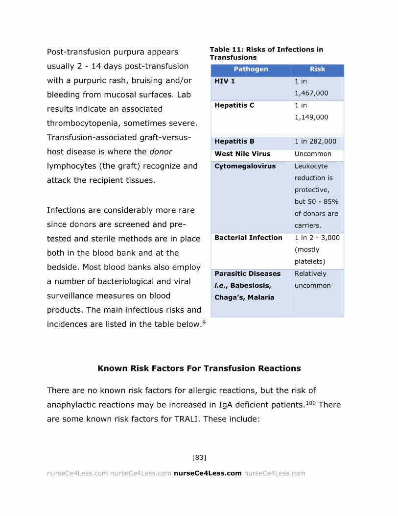
[83]
nurseCe4Less.com nurseCe4Less.com nurseCe4Less.com nurseCe4Less.com
Post-transfusion purpura appears
usually 2 - 14 days post-transfusion
with a purpuric rash, bruising and/or
bleeding from mucosal surfaces. Lab
results indicate an associated
thrombocytopenia, sometimes severe.
Transfusion-associated graft-versus-
host disease is where the donor
lymphocytes (the graft) recognize and
attack the recipient tissues.
Infections are considerably more rare
since donors are screened and pre-
tested and sterile methods are in place
both in the blood bank and at the
bedside. Most blood banks also employ
a number of bacteriological and viral
surveillance measures on blood
products. The main infectious risks and
incidences are listed in the table below.9
Known Risk Factors For Transfusion Reactions
There are no known risk factors for allergic reactions, but the risk of
anaphylactic reactions may be increased in IgA deficient patients.100 There
are some known risk factors for TRALI. These include:
Pathogen Risk
HIV 1 1 in
1,467,000
Hepatitis C 1 in
1,149,000
Hepatitis B 1 in 282,000
West Nile Virus Uncommon
Cytomegalovirus Leukocyte
reduction is
protective,
but 50 - 85%
of donors are
carriers.
Bacterial Infection 1 in 2 - 3,000
(mostly
platelets)
Parasitic Diseases
i.e., Babesiosis,
Chaga’s, Malaria
Relatively
uncommon
Table 11: Risks of Infections in
Transfusions

[84]
nurseCe4Less.com nurseCe4Less.com nurseCe4Less.com nurseCe4Less.com
A history of chronic alcohol abuse
Sepsis
Female donors, especially those with a higher number of pregnancies.
Donor units that test positive for anti-granulocyte antibodies and anti-HLA
class II antibodies.
The level of lysophosphatidylcholine in the transfused blood component.
Risk factors for volume overload include:
Age
o The elderly and infants are at higher risk
Patients with pre-existing cardiovascular disease
Patients with pre-existing renal disease
Risk factors for GVHD include:
An immunocompromised host
The donor is a related (biological) family member
Donor has a human leukocyte antigen (HLA) homozygous haplotype
which is identical with the patient’s HLA haplotype
Recipient is recognized as foreign by donor lymphocytes and the
graft/donor lymphocytes. The skin, liver, immune system, and bone
marrow are preferentially attacked by the donor lymphocytes.
Risk factors for post-transfusion purpura include:
Women who have been pregnant
History of transfusion and sensitization to the platelet antigen, HPA-1a.
[85]
nurseCe4Less.com nurseCe4Less.com nurseCe4Less.com nurseCe4Less.com
Transfusion-Related Acute Lung Injury (TRALI)
Transfusion-Related Acute Lung Injury (TRALI) is the most common cause of
transfusion-related deaths in the U.S.101 TRALI is an acute lung injury,
defined as “acute hypoxemia with a PaO2/FIO2 ratio of less than or equal to
300 mm Hg with bilateral pulmonary edema on frontal chest radiograph and
either pulmonary artery occlusion pressure less than or equal to 18 mm Hg
or no clinical evidence of left atrial hypertension.”102 Some of the risk factors
associated with acute lung injury include severe sepsis, pneumonia,
aspiration of gastric contents, drug overdose, burns, disseminated
intravascular coagulation, near drowning, shock, cardiopulmonary bypass,
and massive transfusions.103
The National Heart, Lung and Blood Institute (NHLBI) defines TRALI as “new
onset acute lung injury (ALI) with onset of symptoms within 6 hours of
transfusion with no preexisting ALI before the transfusion and confirmation
through assessment of the patient's clinical course.”104 TRALI is primarily a
diagnosis of exclusion, and since it often has a delayed appearance, it is
important that the nursing staff and other health professionals maintain their
awareness of and vigilance for the occurrence of TRALI. There is currently no
lab test that is diagnostic for TRALI.
Transfusion related acute lung injury is most commonly associated with the
transfusion of plasma-containing products, usually fresh frozen plasma.
Symptoms include dyspnea, fever (1°–2° C increase), hypotension,
tachycardia (rapid heart rate), tachypnea (rapid respiratory rate), frothy
endotracheal aspirate, cyanosis (bluish lips, skin), and severe hypoxemia
(lack of oxygen).101 If there are no symptoms of heart disease or dysfunction
or signs of acute volume overload, TRALI should be considered and a chest

[86]
nurseCe4Less.com nurseCe4Less.com nurseCe4Less.com nurseCe4Less.com
X-ray should be ordered. X-rays may show a “whiteout” appearance similar
to acute respiratory distress syndromes (ARDS).105 Transfusion-associated
circulatory overload (TACO), bacterial contamination, and anaphylactic
transfusion reactions need to be ruled out as well.
The most common association with TRALI is the presence of antibodies
against the human leukocyte antigen (HLA) system. The HLA system
describes the major histocompatibility complex (MHC) for humans and
functions in infectious diseases, graft rejection, autoimmunity and in cancer
surveillance and metastasis. The genes for the human MHC complex are
found on chromosome 6 and encode cell-surface antigen-presenting proteins
(found on antigen-presenting cells (APC) like macrophages and dendritic
cells). HLA antigens may be Class I (those that are found on APCs) or Class
II, which play a role in the production of antibodies and autoantibodies by B
cells. Women who have had one or more pregnancies often have detectable
anti-HLA antibodies in their sera or plasma.
The proposed mechanism of TRALI involves HLA antibodies that are present
in donor plasma. These antibodies are presumed to react with the patient’s
(recipient’s) immune system, specifically affecting the pulmonary
vasculature and increasing permeability.101,103-107 The increased permeability
allows for the entrance of polymorphonuclear neutrophils [PMN] into the
lung. PMN activation then can result in endothelial damage, capillary
leakage, and eventually, massive bilateral pulmonary edema, as seen in
TRALI.108,109 There have also been a number of reports of the association of
TRALI with antibodies to human neutrophil antigens (HNAs). For example,
HNA-3a is present in ~ 95% of humans and antibodies to HNA-3a can
activate neutrophils. In animal models of TRALI, these activated neutrophils
damage the pulmonary microvasculature in ways that strongly resemble

[87]
nurseCe4Less.com nurseCe4Less.com nurseCe4Less.com nurseCe4Less.com
those seen in TRALI.110 Lab results sometimes show a transient decrease in
peripheral WBC, which, if noted, can be a useful sign. The storage time of
blood products may also be a concern with TRALI, with longer storage of
buffy-coat associated platelets linked to an increased risk of TRALI.111
Supportive treatment should occur as soon as TRALI is suspected.101,103-107
The transfusion should be stopped immediately and the blood bank should
be notified right away. Supportive treatment constitutes respiratory support
and maintenance of hemodynamic status. Donor blood should be tested for
HLA and HNA antibodies. If these are negative, the donor blood should be
tested for WBC antibodies. If any antibodies are found in the donor samples,
the donor should be notified and advised that they should not donate any
plasma or blood components that contain plasma. The patient, once
recovered, should be counseled regarding avoiding unnecessary transfusions
in the future, even though the likelihood of a second TRALI episode is
extremely rare.
In the United Kingdom, as part of the Serious Hazards of Transfusion or
SHOT study,112 using plasma preferentially from male donors was instituted
on the basis of the rationale that pregnancy can induce the formation of
anti-HLA and anti-HNA antibodies and that males were less likely to produce
these types of antibodies. The number of cases of TRALI did drop as did the
mortality rate.105,107,112 The American Red Cross has also reported that in the
majority of fatalities due to TRALI, the recipient received plasma products
from female donors with WBC antibodies.113 The American Red Cross is
currently in the implementation phase of diverting donated female plasma to
alternative uses such as protein fractionation and preferentially using male
plasma for transfusions.

[88]
nurseCe4Less.com nurseCe4Less.com nurseCe4Less.com nurseCe4Less.com
Transfusion Associated Circulatory Overload (TACO)
Transfusion Associated Circulatory Overload (TACO) results from the inability
of the patient to compensate adequately for the volume of blood or blood
components transfused. This may occur if the patient has an impairment of
cardiac function or if the transfusion occurs too rapidly.58,61 According to the
FDA, TACO is the second most frequent cause of mortality post-
transfusion.114-118
Transfusion Associated Circulatory Overload is a relatively common
transfusion reaction, occurring most often in the elderly or the very young.
As little as 1-2 units may induce TACO, causing a cardiogenic pulmonary
edema. This can occur during the transfusion or up to 6 hours post-
transfusion. Symptoms include those of congestive heart failure:
Dyspnea and/or orthopnea (dyspnea when lying flat)
Tachycardia
Hypertension
Increased venous pressure (increased distention of the jugular vein
may be noted)
Pulmonary auscultation may reveal lung crackles and/or rales and an
elevated jugular venous pressure (JVP). Chest X-ray may reveal alveolar and
interstitial edema, distended pulmonary artery and cardiomegaly (an
enlarged heart). Lab studies indicate that brain natriuretic peptide (BNP) is
81% sensitive and 89% specific for TACO.58,117 BNP, also known as
ventricular natriuretic peptide, is secreted by cardiomyocytes in the
ventricles as a response to distention. Ca2+ levels are important in regulating
the release of BNP. BNP functions to increase the excretion of sodium and to
decrease vascular resistance, resulting in a decrease in blood pressure.
[89]
nurseCe4Less.com nurseCe4Less.com nurseCe4Less.com nurseCe4Less.com
If TACO is suspected and the transfusion is still being administered, the
transfusion should be stopped immediately and the blood bank should be
notified. Standard treatment is with diuretics and respiratory support
measures. Ultrafiltration is currently being investigated to replace
diuresis.119
Acute Kidney Injury (AKI)
The phrase “Acute kidney failure” (ARF) has been replaced with more
commonly used phrase Acute Kidney Injury (AKI). AKI is the abrupt
decrease in kidney function that may be partial or complete. The net result
is the retention of waste products. In transfusion patients, AKI is caused by
high levels of hemoglobin (hemoglobinuria) induced by the widespread
hemolysis of the RBC.
The diagnostic criteria for acute kidney failure are the presence of any of the
following:
An increase in serum creatinine level of 0.5 mg/dL
A 50% increase in creatinine level above baseline value
A 50% decrease in baseline glomerular filtration rate (GFR)
Oliguria, which is defined as urine output less than 400 mL/24 hours
Anuria, which defined as urine output less than 100 mL/24 hours

[90]
nurseCe4Less.com nurseCe4Less.com nurseCe4Less.com nurseCe4Less.com
Rifle and AKIN Criteria for Acute Kidney Injury
RIFLE System
The RIFLE classification for acute kidney injury in critical care was developed
after a systematic review and an expert consensus conference.120,121 The
classification is divided into categories of Risk, Injury, Failure, Loss of
function and End-stage kidney disease. The risk, injury and failure
categories are based on the glomerular filtrations rates (GFR) and urine
outputs.
Risk of renal dysfunction:
GFR criteria
o Increased serum creatinine 1.5-fold, or
o GFR decrease by greater than 25%
Urine output criteria
o Urine output of less than 0.5 mL/kg/hour for 6 hours
Injury to kidney:
GFR criteria
o Increased serum creatinine 2-fold, or
o GFR decrease by greater than 50%
Urine output criteria - urine output of less than 0.5 mL/kg/hour for 12
hours
Failure of kidney function:
GFR criteria
o Increased serum creatinine 3-fold, or

[91]
nurseCe4Less.com nurseCe4Less.com nurseCe4Less.com nurseCe4Less.com
o GFR decrease of greater than 75%, or
o Serum creatinine of greater 4 mg/dL in a setting of an acute
increase of at least 0.5 mg/dL
Urine output criteria
o Urine output of less than 0.3 mL/kg/hour for 24 hours (oliguria),
or
o Anuria for 12 hours
Loss of kidney function (persistent AKI) is defined as the complete loss of
kidney function for more than four weeks.
End Stage Kidney/Renal Disease (ESK/RD)
End stage kidney or renal disease is defined as the complete loss of kidney
function for more than three months.
AKIN System
The Acute Kidney Injury Network (AKIN) also has proposed diagnostic and
classification criteria.122,123 These two systems have obvious similarities, but
also have some important differences — a third classification system, the
Kidney Disease Improving Global Outcomes (KDIGO) clinical practice
guidelines attempts to reconcile the two systems. (See below).
Diagnostic Criteria:123
One of the following is required with a 48-hour period:
An absolute increase in serum creatinine of greater than or equal to
0.3 mg/dL percentage increase in serum creatinine of greater than or
equal to 50% (1.5-fold from baseline)

[92]
nurseCe4Less.com nurseCe4Less.com nurseCe4Less.com nurseCe4Less.com
A reduction in urine output with documented oliguria of less than 0.5
mL/kg/hour for more than 6 hours
Classification system:123
Stage 1
o Serum creatinine criteria
An increase of greater than or equal to 0.3 mg/dL, or
An increase to 150% - 200% from baseline
o Urine output criteria
Less than 0.5 mL/kg/hour for more than 6 hours
Stage 2
o Serum creatinine criteria
An increase to 201%-300% from baseline
o Urine output criteria
Less than 0.5 mL/kg/hour for for more than 12 hours
Stage 3
o Serum creatinine criteria
An increase to greater than 300% from baseline, or
greater than or equal to 4 mg/dL with an acute increase of
at least 0.5 mg/dL.
o Urine output criteria
Less than 0.3 mL/kg/hour for 24 hours, or
Anuria for 12 hours

[93]
nurseCe4Less.com nurseCe4Less.com nurseCe4Less.com nurseCe4Less.com
Kidney Disease Improving Global Outcomes (KDIGO) Clinical Practice
Guidelines
As mentioned, there are differences between the RIFLE and the AKIN
systems. The Kidney Disease Improving Global Outcomes (KDIGO) system is
an effort to reconcile the two systems.124-126
Staging:
Stage 1: With any one of the following -
o Serum creatinine 1.5 - 1.9 times baseline
o Serum creatinine greater than or equal to a 0.3 mg/dL increase.
o Urine output of less than 0.5 mL/kg/hour for 6-12 hours
Stage 2: With either of the following -
o Serum creatinine 2 - 2.9 times baseline
o Urine output of less than 0.5 mL/kg/hour for 12 hours or more
Stage 3: With any one of the following -
o Serum creatinine ≥ 3 times baseline
o Increase in serum creatinine to greater than or equal to 4 mg/dL
This criteria holds after the definition of acute kidney injury
is met
An increase in serum creatinine by
greater than or equal to 0.3 mg/dL
within 48 hours or an increase of
greater than or equal to 1.5 times
baseline.
o Initiation of renal replacement therapy
o Anuria for 12 hours or more
Note: If urine output and
serum creatinine lead to
different stages, patient
should be assigned the
higher stage.
[94]
nurseCe4Less.com nurseCe4Less.com nurseCe4Less.com nurseCe4Less.com
o Urine output of less than 0.3 mL/kg/hour for 24 hours or more
In patients less than 18 years old, a decrease in estimate
glomerular filtration rate (eGFR) to less than35 mL/minute
per 1.73 m2
Disseminated Intravascular Coagulation (DIC)
Disseminated Intravascular Coagulation (DIC) complicates many illnesses
and is a rare but significant complication of transfusion procedures. It is also
known variously as consumptive coagulopathy, defibrination syndrome and
defibrinogenation syndrome. DIC is characterized by the systemic activation
of the regulatory and functional coagulation pathways and results in the
generation of fibrin clots (causing organ failure due to the resultant
ischemia) and the consumption of platelets and coagulation factors that may
promote bleeding — a particularly significant problem in transfusion patients.
There are two forms of DIC:
Fulminant or overt:
This is the acute form of DIC resulting from a decompensated
hemostatic system. Overt DIC may be controlled or uncontrolled.
o Controlled:
Controlled DIC represents “temporary override” of the regulatory
network of the endothelium. An example would be the activation
of Protein C by thrombomodulin. Controlled DIC reverses rapidly
when the predisposing factor is removed. In transfusion
reactions, this translates to the immediate cessation of the
transfusion.

[95]
nurseCe4Less.com nurseCe4Less.com nurseCe4Less.com nurseCe4Less.com
o Uncontrolled:
Uncontrolled DIC represents the degradation of the endothelial
regulatory network in addition to an override of the network.
The degradation of the regulatory network can be in response to
trauma or sepsis and can therefore occur in patients undergoing
emergency transfusion for acute injury. Uncontrolled DIC may
not cease when the transfusion process is halted and the clinical
decision must often be made to continue the transfusion and to
add, for example, cryoprecipitate (containing concentrated
coagulation factors) and/or platelet rich plasma (PRP) in an
attempt to replace the consumed coagulation factors and
platelets.
Non-overt or “low grade” DIC:
This is the chronic form of DIC resulting from a stressed but still
functional and compensating hemostatic system.
Many of the understood and presumed pathogenic mechanisms of DIC can
occur pre-transfusion, i.e., traumatic tissue damage or RBC damage, during
the circulation or deposition of immune complexes (IC) composed of antigen
bound to specific antibody or endotoxin release; and, post-transfusion, i.e.,
IC deposition causing endothelial damage, RBC damage, endotoxemia,
tissue damage or platelet damage.
Regardless of the specific pathogenesis, the resulting process is the same
with, sometimes, massive activation of plasmin and thrombin by both the
intrinsic and extrinsic coagulation pathways (see above) with resultant
vasodilation, loss of endothelial tight junctions leading to leakage and
possible shock. Additionally, the consumption of platelets and coagulation
factors can worsen the anemia, bleeding and cause additional hemorrhage
[96]
nurseCe4Less.com nurseCe4Less.com nurseCe4Less.com nurseCe4Less.com
into tissues. Besides the activation of the coagulation pathways, the normal,
physiologic anticoagulant pathways are simultaneously suppressed because
anti-thrombin levels are reduced by the increased utilization of antithrombin,
increased enzymatic degradation, impaired neosynthesis by the liver and by
leakage into extravascular spaces, due in part to the leakage across the
weakened tight junctions, the vasodilation and common compensatory
increase in blood pressure as the body strives to maintain blood flow.127-130
Massive Transfusions
The most recent studies, sometimes based on research and data
accumulated in combat hospitals, involve the ideal ratio of units of packed
RBC to units of plasma or platelets that can increase survival and decrease
the nature and the degree of complications.10,127,131-135 Current data
indicates that early transfusion of units of plasma, platelets and coagulation
factors while at the same time transfusing packed RBCs can decrease both
mortality and morbidity.10,131-135 This, in fact, runs counter to many of the
official guidelines which do not recommend using crystalloid or colloidal
fluids (i.e., FFP, PRP, cryoprecipitates) in conjunction with the use of packed
RBC’s or whole blood.11,136-139
RBC Transfusion and Sepsis
Sepsis is the systemic inflammatory response to infection and affects
pediatric, neonatal and adult critical care. Many of the critically ill patients
who are thought to require and are transfused with packed RBC or blood
components are septic. A small number of those who receive packed RBC or

[97]
nurseCe4Less.com nurseCe4Less.com nurseCe4Less.com nurseCe4Less.com
blood components will become septic. The greatest risk of bacterial infection
is after the transfusion of platelets.
It has become increasingly accepted in the medical community that RBC
transfusion can lead to great morbidity and mortality, particularly when
transfused with older RBC.140-144 So, those septic patients who require RBC
transfusions must be monitored closely. In addition, those patients who
become septic after platelet or other blood component transfusion must also
be monitored closely.
Sepsis is an inflammatory response and stimulates and activates both the
innate and acquired immune systems. Inflammatory pathways are activated
and various cytokines and inflammatory mediators are secreted causing a
number of potentially detrimental effects including vasodilation, increased
capillary permeability, increased expression of adhesion molecules and an
activation of the clotting pathways evidenced by increased clot formation
and decreased clot dissolution (fibrinolysis). All these events can lead to
serious hypotension and multiple organ failures.142,143,145,146
As with any transfusion reaction, the prompt identification, recognition and
treatment of sepsis are critical. In addition, the recognition that the addition
of RBC to the treatment of septic patients may have adverse effects on the
outcomes, must be recognized as part of the responsibilities of the nursing
staff and part of what the nursing staff must be aware of as they monitor
their septic patients after transfusion and as they monitor their transfusion
patients for sepsis.
The “Surviving Sepsis Campaign”143,147 provides guidelines for nursing staff
in “bundles”. The benefits of routine screening of patients at risk for sepsis is

[98]
nurseCe4Less.com nurseCe4Less.com nurseCe4Less.com nurseCe4Less.com
emphasized and acknowledges that care of the septic patient necessitates a
multidisciplinary team approach and included infection prevention.
Infection, Prevention and Source Control
Hand hygiene, catheter care, comprehensive oral care and barrier
precautions should be followed. Both selective oral decontamination and
selective digestive decontamination approaches should be used to reduce
the incidence of ventilator-associated pneumonia.
Every effort should be made to identify the source of infection with
appropriate interventions taken as soon as possible. This can include
debridement, assessment and subsequent removal of suspicious access
devices and tubing and the observation of the patient for striae, tender
areas or other signs of inflammation and infection.
Initial Nursing Response to Sepsis
Within three hours of the initial diagnosis of sepsis, patients should have
lactate levels measured and blood cultures drawn for both aerobic and
anaerobic cultures. These must be drawn before the administration of broad
spectrum antibiotics. One sample should be obtained through venipuncture
and at least one sample should be obtained through each vascular access
device. Other samples for culture may be obtained (i.e., respiratory
secretions, other body fluids) depending on the individual patient. Sepsis
induced hypoperfusion is defined as hypotension that is refractory to the
administration of 30 mL/kg crystalloids (albumin) for hypotension or a
lactate level of greater than or equal to 36mg/dL (4 mmol/L). If the
[99]
nurseCe4Less.com nurseCe4Less.com nurseCe4Less.com nurseCe4Less.com
hypotension is refractory, the guidelines recommend the use of vasopressors
(norepinephrine/epinephrine) to maintain mean arterial pressure greater
than or equal to 65 mm Hg.
Additional targets include a central venous pressure of at least 8 mm Hg,
and central venous O2 saturation of at least 70 %. Urine output should be at
least 0.5 mL/kg per hour.143,147 It should be noted that the use of steroids is
no longer universally recommended and studies have failed to show any
benefit for steroid use except potentially in those patients who are
hemodynamically unstable after both fluid challenge and administration of a
vasopressor.143,147,148
Setting the goals for care and discussing these goals with the patient and
patient’s family are high priorities with the Surviving Sepsis Campaign
guidelines. These discussions should occur within 72 hours of admission to
the ICU and should include discussions about palliative care and end-of-life
care.143,147
Transfusion-Associated Graft-Versus-Host-Disease
The use of irradiated blood products and reduction in the number of donor
WBCs transfused (leukoreduction) is the current standard of care to prevent
transfusion-associated graft-versus-host disease (TA-GVHD) that has a
greater than 90% mortality rate. The initial stages of GVHD may be
confused with allergic reactions. It is vital to differentiate between allergic
reactions and TA-GVHD because the combined effects of the donor immune
cell response to the recipients tissues and the resulting bone marrow aplasia
and neutropenia place the recipient at a very high risk of sepsis.149

[100]
nurseCe4Less.com nurseCe4Less.com nurseCe4Less.com nurseCe4Less.com
As mentioned in the section on infections, donors and recipients are tested
and screened for a very small fraction of the number of potential pathogens.
Because of this, and to even further reduce the risk of TA-GVHD, recent
research has focused on pathogen reduction protocols.149 A number of these
protocols are used in Europe and it is possible that some or most of these
approaches may be adopted in the U.S. The protocols include the use of
combinations of agents along with ultraviolet (UV) light, visible light or
detergents to kill pathogens and decrease the proliferations of immune
lymphocytes.149
Graft versus host disease is a significant concern in allogeneic stem cell
transplants to treat hematological cancers. Current and experimental
approaches to the treatment of TA-GVHD include supportive care,
mesenchymal stem cell salvage techniques, the use of short-term
immunosuppressive agents and monoclonal antibodies against antigens
expressed on T and B cells.149-154
Hypotension: ACE Inhibitors
Acute hypotension after transfusion appears to be related to the use of
angiotensin-converting enzyme (ACE) inhibitors because of the role that ACE
plays in bradykinin metabolism. Bradykinin is a vasodilatory peptide whose
level is increased in patients taking ACE inhibitors to lower blood pressure.
These hypotensive reactions have also been associated with the use of
bedside leukocyte-reduction filtered blood products.155-157

[101]
nurseCe4Less.com nurseCe4Less.com nurseCe4Less.com nurseCe4Less.com
Step-by-Step Procedure For A Suspected Transfusion Reaction
These steps have been derived by the AABB and follow generally accepted
guidelines. Nursing staff must know, in addition, the specific policies and
protocols for their individual hospital and institution and follow those as
required when a transfusion reaction is suspected.
1. STOP the transfusion IMMEDIATELY
2. Disconnect the IV line FROM THE NEEDLE.
a. Do NOT disconnect the (blood component) unit from the IV set.
b. Attach a new IV set
c. Prime the new IV set with saline
d. Reconnect and open the line with a slow drip.
3. Notify the medical staff (physician on duty, the patient’s physician,
physician on call, etc.).
a. If necessary, call a code
4. CHECK the patient’s wrist ID and the unit label to ensure that the unit
was meant for the patient.
a. DO NOT ASSUME ANYTHING. CHECK AND DOUBLE CHECK!!
5. Do not discard the unit of blood or blood component. The blood bank
will need it to determine the underlying cause of the transfusion
reaction.
6. Notify the blood bank. Be prepared to briefly summarize and describe
the nature of the transfusion reaction you observed.
7. After the blood bank has been notified, begin filling out the Transfusion
Reaction Report Form. This may be a paper form or may be an online
form, depending on the institution or hospital.
8. If a hemolytic transfusion reaction is suspected, the blood bank will
need:
[102]
nurseCe4Less.com nurseCe4Less.com nurseCe4Less.com nurseCe4Less.com
a. A completed Transfusion Reaction Report
b. Post-transfusion blood specimens
i. Obtain a pink-top blood bank
tube with EDTA.
ii. Make certain to invert the sample 8-10x to ensure proper
mixing and the prevention of clotting. DO NOT SHAKE.
iii. Use a lavender- top tube if a pink-top is not available.
Lavender-top tubes also contain EDTA.
iv. Use the opposite arm to draw specimens (preferably at the
opposite antecubital fossa). Hand or wrist veins on the
opposite side may also be used.
v. If it is necessary to draw a specimen from the same IV line
that was used for the transfusion (because of collapsed
veins, rollers, etc.):
1. Collect a tube of blood and discard it, as it will be
contaminated.
2. Collect and label a pink-top tube as required for
testing.
c. The unit(s) of blood or blood components suspected to be
causing the transfusion reaction. Clamp the IV tubing used and
sent to the blood bank. Do not send the tubing with the needle.
Dispose of the needle in the proper container.
d. Be aware that a urine sample may also be requested
9. If the reaction is less severe and non-life threatening, the physician in
charge may determine that the benefit of continuing the transfusion
outweigh the risk. There may be more specimens requested later.
[103]
nurseCe4Less.com nurseCe4Less.com nurseCe4Less.com nurseCe4Less.com
General Guidelines For The Transfusion Procedure
Nurses may be considered to be at the frontline of safety for transfusion
procedures. It will be up to the nursing staff at the patient’s bedside to be
responsible for the proper and accurate identification of the patient and the
proper and accurate identification of the blood product, ensuring that the
correct patient receives the correct blood component. It is absolutely
essential to completely and consistently verify the identity of the patient
during every step of the procedure. It is also absolutely essential to verify
that the blood component sent from the blood bank is meant for that specific
patient. The general guidelines for performing transfusions of any blood
components include:
Pay attention to identification -
This includes checking the patient’s wristband with the patient’s chart.
Make sure, if possible, that you engage with the patient, asking
questions like “Is this how you spell your name”, “I see your first
name is Elizabeth. Do you prefer Elizabeth or a nickname like Liz or
Beth?”, “Can I double check the spelling of your last name”, “What is
your date of birth?” or ”Can you verify your date of birth for me?”
Check blood product label carefully against wristband to ensure that
the blood product or blood products is/are intended for that specific
patient.
o Mislabeling of samples, blood, blood products, is a significant but
totally avoidable cause of transfusion reactions.
ALWAYS check the expiry date on any blood or blood products to be
transfused.
Blood and blood products must NEVER be stored in a medication
refrigerator. If for some reason, the blood or blood product is not
[104]
nurseCe4Less.com nurseCe4Less.com nurseCe4Less.com nurseCe4Less.com
immediately used, send the blood or blood product back to the blood
bank for proper storage.
Check against the patient’s chart to ensure that whatever blood
products were ordered are the same ones that were delivered by the
blood bank.
Ensure that the blood or blood product is not damaged in any obvious
or clear way. Check blood for clots or discoloration.
The transfusion should be slow for the first 10-15 minutes. This is
meant to protect the patient against any acute reaction that may
occur. The signs and symptoms of acute reactions can often be
detected during this first phase of the transfusion. Check the hospital
or institutional policy for the time frame recommended for this initial
phase.
Always monitor the patient before, during and after the transfusion
process.
After the initial phase, ensure that the prescribed rate of infusion is
appropriate for the patient.
It is strongly recommended that the transfusion ends within 4-6 hours
of the removal of the blood or blood components from storage in the
blood bank. Check the hospital or institutional policy for the time
frame recommended for the transfusion.
Know the hospital or institutional policies on blood tubing. Change the
blood tubing according to that policy and discard properly in the
appropriate biohazards container when the transfusion is completed.
Know how to recognize and treat transfusion reactions.
If a suspected transfusion reaction is observed, stop the transfusion
IMMEDIATELY.

[105]
nurseCe4Less.com nurseCe4Less.com nurseCe4Less.com nurseCe4Less.com
Report reactions to the appropriate medical staff, nursing staff,
supervisory staff and to the blood bank immediately.
Evidence-Based Medicine Guidelines for the Transfusion Procedure
The following are guidelines derived from the American Association of Blood
Banks for nurses and nursing staff. They were developed in accordance with
FDA guidelines and the Agency for Healthcare Research and Quality
(AHRQ).158 The essential recommendations from AHRQ are the following:158
Donated blood is not transfused to the recipient until it has been
processed into its separate blood components.
Red cells are transfused in anemia to provide adequate oxygen
delivery.
Plasma (Octaplas®) is transfused in hemorrhage to provide
coagulation factors when the hemorrhage is not caused by the
deficiency of a single coagulation factor.
Platelets (i.e., thrombocytes) are indicated for the management of
bleeding or in thrombocytopenia to promote hemostasis when the
platelet number or function is insufficient.
In primary care, the use of blood products is limited to the treatment
of symptomatic chronic anemia when no alternative treatment is
available.
Platelet transfusions may also occasionally be administered outside
hospital, for example to patients with hematological diseases.
[106]
nurseCe4Less.com nurseCe4Less.com nurseCe4Less.com nurseCe4Less.com
The following are the AHRQ guidelines to the treating physician for blood or
blood component transfusion. It is important for nurses to understand the
guidelines under which the prescribing physician is functioning.
Guidelines to the Treating Doctor Regarding Blood Transfusion
1. A medical practitioner must prescribe a blood transfusion.
The prescription must state the type of blood component
(red cells, platelets, plasma), the volume to be transfused
and any special requirements (irradiation, washing,
phenotyping).
If necessary, instructions must be given regarding the
speed of transfusion, the possible need to warm the
product or any other factors to be considered due to, for
example, the patient's condition, unless the staff have
been issued relevant protocols.
If the patient has significant cold agglutinins, red blood cell
products should be warmed during transfusion using an
approved commercial blood warmer.
Note: Emphasis added for Guidelines, which directly impact the
transfusion nursing staff.
2. The person carrying out the transfusion must check the prescription
and ensure that the red cell packs intended for transfusion have
been found by the laboratory compatibility testing to be
compatible with the recipient.
Before a transfusion of red cells, check the result of the
compatibility testing and verify that the correct product
and patient was used for the test: the donation number on
the blood bag label must exactly match the number on the
[107]
nurseCe4Less.com nurseCe4Less.com nurseCe4Less.com nurseCe4Less.com
accompanying documentation and they must indicate the
compatibility of the unit.
If the patient has red cell antibodies, ensure that the label
on the red cell pack states the absence of the antigens
corresponding to the antibodies detected in the patient
(the label will state, for example, Jka — indicating the
absence of antigen Jka).
3. The blood product must be checked for defects (integrity and
cleanliness of the bag; the presence of clots, aggregates, gas or
a black-red color of a red blood cell product is suggestive of
bacterial contamination; when inspecting platelet concentrates
against light the presence of swirling should be observed).
4. Confirmation that the checks have been carried out is done by
signing the transfusion form.
5. A blood administration set with an integral 150–200 µm filter,
appropriately attached to the pack to be transfused, should be
used to transfuse all blood products.
6. Verification of the patient's identity: the patient's identity data must
be checked against the data on the blood product.
The patient is asked to state his/her identification (ID)
details or
A person familiar with the patient verifies the identity of
the patient (at the bedside); the identification details must
also match with the patient's wristband
7. Before starting a transfusion, the patient's vital signs (heart rate,
blood pressure, temperature) are checked and recorded. The
start time of the transfusion must be recorded. The sticker on
the blood product label indicating the unit number should be

[108]
nurseCe4Less.com nurseCe4Less.com nurseCe4Less.com nurseCe4Less.com
detached and reattached to the patient notes, or the transfusion
is entered in the electronic patient record system.
The recording is carried out to fulfill the statutory
obligation to ensure the traceability of the blood product
from the recipient to the donor and vice versa.
8. If possible, a transfusion is started slowly with a biological pre-
check.
During the first 10 minutes red cells are infused slowly
(10–15 drops/min) whilst carefully observing the patient.
9. The same administration set may be used to transfuse several
packs of red cells without interruption (according to the capacity
of the administration set filter), but it is recommended that the
administration set is changed after six hours in order to reduce
the risk of bacterial contamination.
It is recommended that platelets are administered via a
separate administration set or they may be transfused
first, if transfusion is to continue with red cells. If the
tubing has red cells, retention of platelets may occur.
10. Even though transfusion adverse effects often emerge at the
beginning of transfusion, the patient must be monitored
throughout the entire transfusion.
11. Red cell transfusion should be completed within six hours of
removing the pack from the refrigerator to room air. The end
time of the transfusion must be recorded.
12. The blood component pack with the administration set is
recommended to be stored after transfusion for 24 hours in case
adverse reaction investigations need to be carried out.”
[109]
nurseCe4Less.com nurseCe4Less.com nurseCe4Less.com nurseCe4Less.com
Step-by-Step Guidelines
Pre-Transfusion and Informed Consent
The nursing staff must ensure that informed consent is obtained from the
patient. This is an absolute requirement unless an emergency situation
exists or if the patient is unable to consent and no substitute individual with
a legal right to consent is available.
If it is at all possible, nurses must also determine that there is no evidence
of the patient’s prior wishes to avoid transfusion for either personal or for
religious reasons. Informed consent includes allowing the patient, if possible,
time to ask questions and time to fully understand the answers. The
following position statement from the New York State Nurses Association
presents the highest of legal and ethical standards in professional nursing.159
For patients undergoing a transfusion, informed consent implies that they
have an understanding of the risks versus the benefits of a transfusion,
receive an understandable (by the patient) description of the blood product
and that they understand why their physician has ordered a transfusion.
Nursing staff must document the informed consent process as well as the
questions asked and how they were answered.

[110]
nurseCe4Less.com nurseCe4Less.com nurseCe4Less.com nurseCe4Less.com
The nursing staff also needs to determine if pre-transfusion samples from
the patient have been ordered and obtain those as well, always following the
guidelines regarding venipuncture and sample acquisition. A pre-transfusion
sample may be required for ABO/Rh testing, antibody screening or for a type
and crossmatch. Before drawing a blood sample, always check the patient’s
arm or wristband ID. Also, remember to engage with the patient as much as
possible. The patient may be experiencing anxiety, pain, confusion or any
number of other emotions and it is important for their overall wellbeing that
they are made to feel as comfortable as possible under the circumstances.
“The New York State Nurses Association acknowledges that informed consent seeks to establish the rights of the
individual through self-determination. Inherent in the employee status of the registered professional nurse are
multiple instances of role conflict that arise through the occasionally conflicting responsibilities to the patient, the
family, the agency, care-providing colleagues, society, and to the advancement of knowledge.
The Nurses Association believes that the registered professional nurse’s role and responsibility in informed
consent is to:
Advocate for the patient’s human rights, including the right to autonomous decision making, assume
primacy.
Advocate that each patient (or their designated health care proxy agent(s)) is given the information
necessary for making informed judgments. This information should include, but is not limited to: risks,
benefits and alternatives and expected outcomes to any proposed test or treatments.
Advocate for each patient’s right to freedom from the imposition of another’s beliefs or judgmental
attitudes.
Advocate that the patient or designated healthcare proxy agent(s) have had sufficient communication
with the provider providing or performing the treatment and /or procedure.
Maintain the registered nurses role as a witness to the patient’s signature to the consent.
Ensure patients are fully involved in planning, implementing and evaluating their personal health care
as they are able. If the patient is declared incapacitated, or unable to make decisions about their health
care, a designated health care proxy agent should assume the patient’s role in planning, implementing
and evaluating care.
Ensure information received from patients regarding their treatment remains confidential and “such
data is used only in ways that are authorized by the patient” (American Nurses Association (ANA),
2008, p. 26) and in compliance with the law.
Facilitate patients (or their designated healthcare proxy agent(s)) right to determine what will be done
with their person. Each patient or health care proxy agent must be permitted to accept, refuse or
terminate treatment without duress.”

[111]
nurseCe4Less.com nurseCe4Less.com nurseCe4Less.com nurseCe4Less.com
Nurses should review the patient’s chart, review their most recent labs,
evaluate and monitor pre-transfusion symptoms. Vitals, including pulse,
blood pressure, respiratory rate, temperature and auscultate the heart and
lungs. Only a qualified MD or DO can order a transfusion. The transfusion
order must include the following:
The name of the ordering physician
The patient’s name and at least one additional unique identifier (i.e.,
date of birth, admission number, patient ID number, etc.)
The specific blood component or product
The amount to be transfused
The rate of administration
Any special requirements such as
irradiation, additional filtration, pre-
warming
If any pre-medication is required
The order may include medications
for minor allergic reactions (i.e.,
antihistamines) or for febrile
responses (i.e., a non-aspirin anti-
pyretic)
If at all possible, the blood or blood
component should be transfused during the
daytime hours.
Principles for an Accurate
Blood Draw
Take sample labels to the
patient’s bedside
Verify that the labels match
both the wrist band/ID AND
the paperwork and chart IDs.
Label at the bedside AFTER the
blood draw
Document that you drew
blood—and never sign for
someone else.
[112]
nurseCe4Less.com nurseCe4Less.com nurseCe4Less.com nurseCe4Less.com
Obtaining Components from the Blood Bank
When blood is obtained from the blood bank, the nursing staff should check
to ensure that the container is properly labeled with the correct patient
name along with a unique identifier. The nursing staff is also responsible for
making certain that the type of blood component delivered (i.e., packed
RBC, cryoprecipitate, fresh frozen plasma) is the one that has been ordered
for the patient. They should also make certain that an adequate amount of
the blood product has been delivered.
Some blood banks may require a diagnosis or an indication is given for each
transfusion. Some blood banks may also require that there be a rationale or
reason given for pre-warming a blood product or for the irradiation of a
blood product. Your hospital or institution may have specific policies
regarding these matters and it is the nurse’s responsibility to be aware of
these policies and adhere to them. In emergency situations, other policies
may be in force and the nursing staff must be aware of these as well.
Mislabeling
Missing a mislabeled sample or component, whether it is a sample that you
have drawn or has been delivered by the blood bank, is completely
avoidable. Also, remember that if something is mislabeled, another patient
may be harmed as well, especially in a busy hospital. The box outlined in red
has rules to follow that can help minimize these sorts of errors.
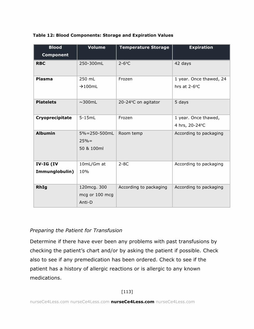
[113]
nurseCe4Less.com nurseCe4Less.com nurseCe4Less.com nurseCe4Less.com
Preparing the Patient for Transfusion
Determine if there have ever been any problems with past transfusions by
checking the patient’s chart and/or by asking the patient if possible. Check
also to see if any premedication has been ordered. Check to see if the
patient has a history of allergic reactions or is allergic to any known
medications.
Blood
Component
Volume Temperature Storage Expiration
RBC 250-300mL
2-6oC 42 days
Plasma 250 mL
100mL
Frozen 1 year. Once thawed, 24
hrs at 2-6oC
Platelets ~300mL
20-24oC on agitator 5 days
Cryoprecipitate 5-15mL Frozen 1 year. Once thawed,
4 hrs, 20-24oC
Albumin 5%=250-500mL
25%=
50 & 100ml
Room temp According to packaging
IV-IG (IV
Immunglobulin)
10mL/Gm at
10%
2-8C According to packaging
RhIg 120mcg. 300
mcg or 100 mcg
Anti-D
According to packaging According to packaging
Table 12: Blood Components: Storage and Expiration Values

[114]
nurseCe4Less.com nurseCe4Less.com nurseCe4Less.com nurseCe4Less.com
Table 13: IV Access for Transfusion Patients
Transfusion
IV Access: USE
RBC rapid transfusions for adults 16-18 gauge needle
RBC routine transfusions for adults 20-22 gauge needle
Other components Any adequate size
Pediatrics 23-25 gauge needle
All components/product for adults and
pediatric patients may be infused
through a CVAD
Central venous access devices (CVAD or
ports) may be ordered and inserted.
*** transfusing rapidly under pressure
and through too small gauge needle can
cause hemolysis
Blood or blood components cannot come into contact with any
medications or with either hypotonic or hypertonic solutions. This type
of contact can cause clotting, hemolysis and/or blood component
contamination.
All blood components must be infused through a filter. Most filters are
170 μ but can range to 260 μ.
Ensure that the IV access site is dedicated. With CVAD in place,
medication in solutions may be infused without damaging blood.
Make sure that IV pumps, blood warmers and rapid infusers are
suitable for the specific transfusion and will not damage the blood or
blood components.

[115]
nurseCe4Less.com nurseCe4Less.com nurseCe4Less.com nurseCe4Less.com
Blood Tubing
Blood tubing is fitted with a 170 - 260 micron (μ) filter to catch fibrin debris.
In preparation for the transfusion, flush tubing with normal saline (0.9%
saline), completely wetting the filter. Blood tubing must be changed at least
every 4 units or within the number of hours specified by hospital or
institutional policy. Blood tubing must be changed according to hospital
policy to prevent bacterial growth.
Platelets should not be transfused though tubing that has been used for
RBC. Platelets will adhere to any fibrin caught in the filter.
Immunoglobulin products (IVIg or IVIG) and albumin do not require blood
tubing, but most products are not compatible with normal saline. Blood
products for IV infusion that are delivered in glass bottles require vented IV
tubing. At the end of the transfusion, the tubing should be disconnected
from patient.
Before picking up blood, make sure patient is ready by connecting the
primed tubing to the patient IV site and verifying consent again. Arrange for
pickup from the blood bank, ensuring the appropriate paperwork is delivered
to the blood bank. Depending on the hospital or institution policy, the
transfusion should be completed within 4 - 6 hour. If the transfusion is
delayed for any reason, return the unused blood or blood components to the
blood bank immediately.

[116]
nurseCe4Less.com nurseCe4Less.com nurseCe4Less.com nurseCe4Less.com
Blood or Components: Five Rights of Transfusion
The following steps support the nurse to ensure the blood or components are
correctly and safely administered to the patient.
Confirm the order and that informed
consent has been obtained.
Check the blood or blood components
for ID numbers and compatibility
information.
Check for the “Right Product”, i.e.,
that the correct blood component has
been delivered.
Always check at the patient’s bedside.
Visually check the unit for clots,
discoloration, leaks, or punctures in the bag or cracks in the glass.
Double-check the blood group compatibility with the patient’s chart
information.
Check the patient’s wristband – spell the patient’s name, checking for
any potential mislabeling or errors. Check the patient’s date of birth.
Check for patient’s name and unique identifiers. Match the patient ID to
the label attached to the component from the blood bank.
Check that the blood unit number and group matches that of the patient
(or, if in an emergency it is O - blood or AB - plasma).
If there are any discrepancies, STOP and call the blood bank.
Some hospitals have 2 people independently checking that all the
information is correct and the “5 Rights” have been met. Enter the blood
bank label into the patient’s chart. Before starting, get vital signs, i.e.,
The 5 Rights of Transfusion
Right patient
Right product
Right amount
Right rate
Right time

[117]
nurseCe4Less.com nurseCe4Less.com nurseCe4Less.com nurseCe4Less.com
temperature, blood pressure, pulse, and respiratory rate. Oxygen saturation
is commonly obtained as well. The blood bank label should remain attached
throughout the transfusion process. Repeat auscultation for patients who
might be at risk for volume overload (elderly, children, those with pre-
existing cardiovascular or pulmonary disease).
Starting the Transfusion
Spiking the Blood component
Separate port cover until port is just exposed
Position port covers away from opening to prevent contamination
Hold bag in one hand and the exposed tubing spike in the other — do not
hang from an IV pole
Insert the blood tubing spike into the port by turning clockwise ¼ turn —
do not over-spike.
To remove the bag, pull gently while turning counterclockwise (¼ turns).
Spiking the blood component properly will ensure easy removal as
needed.
Transfusion
For the first 10 - 15 min, depending on local policies, infuse at a slow
rate unless an emergency situation exists.
After this initial 10 - 15 minute phase, reassess the patient.
Take and record vital signs.
If there are no reactions, and increase to prescribed rate.
Monitor

[118]
nurseCe4Less.com nurseCe4Less.com nurseCe4Less.com nurseCe4Less.com
o Local hospital or institutional policies may require specific
intervals for monitoring the patient and the vital signs.
Observe, monitor and record if there is suspected reaction.
Repeat with more units as needed.
If the patient is at risk for volume overload, unstable or with a history of
reactions, repeat vital signs more often.
Ask the patient, if conscious, if they are experiencing:
o Hives
o Chills
o Rigor/Chills
o Fever
o Shortness of breath (SOB, dyspnea)
o Back pain (may be an early indicator of hemolysis)
o Pain at infusion site
o If they are experiencing and difference in how they are feeling,
or if they are experiencing any anxiety
Complete the transfusion within the time frames indicated by policy.
Disconnect tubing when complete.
Check and document vital signs.
Repeat vital signs according to hospital policy.
Do not leave the blood tubing attached to patient — this increases the
risk of bacterial contamination.
Do not re-use any of the blood tubing.
Assess for any delayed reactions.
The patient should be given a prepared document regarding delayed
transfusion reactions. This information should include the signs and
symptoms of a delayed reaction as well as information on what to do and
who to contact. Delayed reactions to a transfusion include:

[119]
nurseCe4Less.com nurseCe4Less.com nurseCe4Less.com nurseCe4Less.com
A delayed hemolytic reaction (usually about 4-8 days post-transfusion)
o Symptoms may include fever, fatigue, hematuria (red urine), jaundice
Iron overload (rare unless there is a history of transfusions)
Graft Versus Host Disease (very rare; 3-4 weeks post-transfusion)
o Fever, rash, diarrhea and abnormal liver function tests (LFT)
Sepsis (rare)
o Hypotension (low blood pressure), fever, chills, nausea and vomiting,
respiratory distress, shock
Required Documentation
Copy of blood bank label should be entered into the patient’s chart.
Date, start and finish times for the transfusion.
Type and amounts of each component transfused.
Blood/component unit number(s).
The names of the persons starting the transfusion, checking and
monitoring the patient.
Document vitals and any assessments made during the transfusion.
There may be a transfusion form to be turned into the blood bank at
some hospitals or institutions.
Follow up testing may be required for some patients. This may include:
o CBCs
o INR
o PT/PTT
o Fibrinogen levels

[120]
nurseCe4Less.com nurseCe4Less.com nurseCe4Less.com nurseCe4Less.com
Acute Transfusion Reactions
Acute reactions occur usually within 6 hours of the beginning of transfusion.
If any transfusion reactions are noted, stop the transfusion immediately and:
Maintain IV access
Check all vital signs
Re-Verify patient ID
Re-Verify that the blood or component unit matches the label on the
bag or glass from the blood bank
Notify the patient’s physician and/or the physician on call
Remain with the patient
Notify the blood bank
Treat the patient as ordered by the physician
Signs and Symptoms of an Acute Transfusion Reaction
Fever
Chills with or without rigor
Hives, urticaria or angioedema
Rash
Pruritis
Edema or swelling
Respiratory distress: dyspnea, SOB, wheezing
Hypotension or hypertension
Hematuria (Red urine)
Diffuse bleeding or oozing at the site of the IV or at CVAD site.
Bleeding from any site, including mucous membranes
Lumbar pain

[121]
nurseCe4Less.com nurseCe4Less.com nurseCe4Less.com nurseCe4Less.com
Anxiety
Pain at IV site
Nausea and vomiting
Headache
Table 14: Summary of the Symptoms/Signs of Acute Transfusion Reactions
Symptom/Sign Differential Diagnosis
Fever chills rigor Sepsis, Acute hemolytic transfusion reaction, TRALI, febrile
non-hemolytic reaction transfusion reaction
Urticaria/hives/Pruritis Anaphylaxis, minor allergic reaction
Dyspnea TRALI, TACO, anaphylaxis
Hypertension TACO
Hypotension Bradykinin mediated hypotension, Sepsis, Acute hemolytic
transfusion reaction
Hematuria/Red urine Acute hemolytic transfusion reaction
Pain Acute hemolytic transfusion reaction lumbar, iv site
TACO chest
Nausea/Vomiting Acute hemolytic transfusion reaction, Anaphylaxis
Febrile non-hemolytic transfusion reaction
[122]
nurseCe4Less.com nurseCe4Less.com nurseCe4Less.com nurseCe4Less.com
Guidelines for Continuing Transfusion if Reactions Occurs
The nurse caring for a patient with a blood transfusion reaction should follow
facility policy; however, the following are generally accepted steps for the
nurse to observe.
Consult with the physician first. The blood bank physician may be called.
Medicate as ordered.
Proceed cautiously as ordered with more frequent checks on vital signs.
Document every vital sign and every observation.
Report to blood bank may be required
o Send to blood bank (if required)
Blood or component bag with attached tubing for possible
culture, hemolysis check or clerical check
Previous completed blood/component bags if available.
Transfusion reaction reporting document with symptoms, signs,
pre- and post- vitals, time of onset, blood bank unit number
Post-transfusion blood sample may be required for:
Repeat typing/ direct antibody test (DAT/Coombs)
Hemolysis check
Re-crossmatching
May also need:
Urine samples
Chest x-ray (TRALI/TACO)
Blood culture (use a different vein/site for venipuncture)
Other samples may be needed to rule out TRALI,
anaphylaxis, acute hemolytic transfusion reactions

[123]
nurseCe4Less.com nurseCe4Less.com nurseCe4Less.com nurseCe4Less.com
Table 15: Summary Chart for the Treatment of Transfusion Reactions
Reaction Adult Recommended
Treatment
Pediatric
Recommended
Treatment
Comments
Acute
Hemolytic
Reactions
Diuretic therapy:
Treatment:
40-80 mg Furosemide
(Lasix) IV.
Dose can be repeated once.
Note: No diuretic response
within 2-3 hours may indicate
acute renal failure (ARF).
Hydration:
Hydrate to a urinary output
of at least 100 mL/hr until
urine is free of hemoglobin.
Infuse a loading dose of
0.9% sodium chloride OR
5% dextrose in 0.45%
sodium chloride.
Chart hourly urine output.
Administering intravenous
fluid at 100 mL/hour until
urine is free of hemoglobin.
Note: If urinary output does
not increase, any additional
fluids should be infused
with caution.
Diuretic therapy:
Treatment:
1-2 mg/kg/dose.
Dose can be
repeated once at 2-
4 mg/kg.
Hydration:
Pediatric patients
should receive a
smaller volume of fluid
proportional to their
body surface area.
Observe the
patient for
shock, DIC,
volume
overload or
any other
adverse
reactions.
Allergic/
Anaphylactic
Reactions
Urticaria/Hives
If urticaria develops slowly,
antihistamines may be given
orally.
Wheezing
Aminophylline (125-250
mg) IV. Administer slowly~
5 minutes.
Severe allergic
reactions/Anaphylaxis
i.e., laryngeal edema or
bronchospasm
Epinephrine (0.1-0.5 mg or
0.1-0.5 mL of a 1:1000
solution) subcutaneously
(SC).
SC dose may be repeated at
10-15 minute intervals.
Urticaria/Hives
Pediatric dosing
For Benedryl, 1-2
mg/kg IM
o OR
IV for 25-50 mg per
average dose.
Wheezing
Aminophylline: 3
mg/kg/dose in IV
drip over ~20
minutes
Severe allergic
reactions/
Anaphylaxis
i.e., laryngeal
edema or
bronchospasm

[124]
nurseCe4Less.com nurseCe4Less.com nurseCe4Less.com nurseCe4Less.com
Total SC dose should not
exceed 5 mg in a 24-hour
period.
Epinephrine 0.03
mL/M2 (0.03 mg/M2
of a 1:1000
solution) given SC.
A single pediatric
dose should not
exceed 0.3 mg.
Febrile
Transfusion
Reactions
Treat with non-aspirin
containing NSAIDs or other
anti-pyretics
Treat with non-aspirin
containing NSAIDs or
other anti-pyretics
Chills Consider sedation Consider sedation
TRALI No formal treatment
approaches are generally
accepted.
Supportive care including:
Intubation and mechanical
ventilation with small tidal
volumes.
O2 support
Occasionally pressor agents
may be required but iv fluids
are generally sufficient to
maintain bp.
Diuretics if needed for
volume overload
Labs: Chest X-ray
No data available.
Supportive care.
TACO Supportive care including:
Place patient in an upright
position
O2 as needed
Diuretics as needed
Labs: Chest X-ray
No data available.
Supportive care.

[125]
nurseCe4Less.com nurseCe4Less.com nurseCe4Less.com nurseCe4Less.com
Summary
The transfusion of blood and blood components continues to be a critical
aspect of medical care. As an integral part of the multi-disciplinary team,
nurses are at the frontline of transfusion medicine and should be aware of
indications, contraindications and potential reactions to transfusions of any
type. The guidelines for nursing care are undergoing constant evolution in
order to provide patients and families with up-to-date information and the
best evidence-based practices available.
Please take time to help NurseCe4Less.com course planners evaluate
the nursing knowledge needs met by completing the self-assessment of Knowledge Questions after reading the article, and providing
feedback in the online course evaluation.
Completing the study questions is optional and is NOT a course
requirement.

[126]
nurseCe4Less.com nurseCe4Less.com nurseCe4Less.com nurseCe4Less.com
1. GM is a 32-year-old AA female who is typed with B+ blood. As a
donor, GM can MOST SAFELY provide packed RBC for which of
the following individuals?
a. TS, a 21 year old Asian female with AB+ blood
b. KT, a 56 year old European male with A - blood
c. PL, a 76 year old white female with AB - blood
d. MN, a 5-year-old Hispanic male with B+ blood
Answer: D
2. A patient is an otherwise healthy 46-year-old male with β-
thalassemia and scheduled for a transfusion of packed RBC. You
are with him and explaining some of the risks and benefits of his
upcoming transfusion. Which of the following would be true?
a. He runs a high risk of graft-versus-host disease along with
allergic reactions, a fever, a hemolytic reaction and some very
rare reactions but that he will be observed and monitored
throughout the transfusion.
b. He runs a high risk of Transfusion-Related Acute Lung Injury
(TRALI) or Transfusion Associated Circulatory Overload (TACO)
but that he will be observed and monitored throughout the
transfusion.
c. You should counsel him not to get a transfusion, but to get a
shot of iron for his iron deficiency anemia.
d. The transfusion should be completed within 4 hours and there
are some risks associated with an RBC transfusion, including
allergic reactions, a fever, a hemolytic reaction and some very
rare reactions but that he will be observed and monitored
throughout the transfusion.
Answer: D

[127]
nurseCe4Less.com nurseCe4Less.com nurseCe4Less.com nurseCe4Less.com
3. Your patient is typed AB — and is scheduled for an infusion of
plasma. Which of the following plasma types is most suitable for
your patient?
a. AB+
b. O -
c. A+
d. B -
Answer: B
4. A patient who is thrombocytopenic is scheduled to receive two
units of irradiated platelets from which the leukocytes (WBC)
have been removed. You spike the unit with a standard infusion
set and get ready to connect directly to her right arm.
Something makes you stop — what question do you need to ask
yourself?
a. Don’t I need to use a standard blood filter?
b. Shouldn’t I use another leukocyte reduction filter just to be sure
her chances of GVHD are reduced?
c. I should probably ask the blood bank if these platelets have been
tested for ABO compatibility, shouldn’t I?
d. I wonder if her platelet level is low enough to get two units of
platelets?
Answer: B
[128]
nurseCe4Less.com nurseCe4Less.com nurseCe4Less.com nurseCe4Less.com
5. Which of the following statements is FALSE?
a. All blood components must be infused through a filter.
b. The first 15 minutes of a transfusion should be at a slow rate to
ensure there are no acute reactions to the transfusion
c. Nurses can store blood products in the medication refrigerator as
long as the temperature is monitored.
d. If a patient has a history of cold agglutinins, a blood-warming
unit may be ordered, but it is not absolutely required.
Answer: C
6. A transfusion is started at 9:30 am. The packed RBCs were
signed out of the blood bank at 8:30 am, but the patient’s IV has
blown. It is now 10:30 am and the transfusion is not complete.
At what time should the transfusion be stopped, whether or not
it is complete?
a. By 12:30pm—4 hours after the blood was signed out.
b. By the end of the shift—at 11am
c. By 4:30 pm. Blood should be used within 8 hours of signing out
of the blood bank.
d. It doesn’t matter because the packed RBCs have been irradiated,
so there is little risk of infection.
Answer: A

[129]
nurseCe4Less.com nurseCe4Less.com nurseCe4Less.com nurseCe4Less.com
7. You notice that your patient’s temperature has gone up 0.50 in
the last 15 minutes. She is receiving one unit of packed RBC, one
unit of apheresis platelets and one unit of fresh frozen plasma.
She is also complaining of cold and chills. What should you do?
a. Give her a warming blanket
b. Slow down the transfusion rate
c. Stop the transfusion immediately
d. Nothing—everything seems normal
Answer: C
8. Which of the following should NOT be infused in the same line
with any blood component.
a. 5% dextrose in water (D5W)
b. 0.2% saline
c. Normal saline
d. Ringer’s solution
Answer: B
9. A first-time donor comes into the blood bank and has a few
questions. She asks whether her blood will be tested for
bacteria and viruses. Which of the following pathogens is not
routinely tested for?
a. Hepatitis
b. Treponema
c. Enterovirus B
d. CMV
Answer: C
[130]
nurseCe4Less.com nurseCe4Less.com nurseCe4Less.com nurseCe4Less.com
10. An acute hemolytic reaction during a transfusion for acute blood
loss is most likely due to:
a. ABO incompatibility
b. Too rapid infusion
c. Rh0D incompatibility
d. Massive sepsis
Answer: A
11. True/False. Intrinsic RBC membrane abnormalities may
essentially induce an immune response resulting in hemolysis
whereas other membrane abnormalities may result in the
destruction of the RBCs because of the resulting shape or size
abnormalities cause a mechanical hemolysis within the smaller
vessels.
a. True
b. False
Answer: A
12. True/False. A delayed hemolytic reaction usually occurs within
12-24 hours post-transfusion, and includes symptoms of fever,
fatigue, hematuria and jaundice.
a. True
b. False
Answer: B
[131]
nurseCe4Less.com nurseCe4Less.com nurseCe4Less.com nurseCe4Less.com
References Section
The reference section of in-text citations include published works intended as
helpful material for further reading. Unpublished works and personal
communications are not included in this section, although may appear within
the study text.
1. Pfutner, A., L. Wier, and C. Stocks. Most Frequent Procedures
Performed in U.S. Hospitals, 2010. Statistical Brief 2013 [cited 2013
December]; Available from: http://www.hcup-
us.ahrq.gov/reports/statbriefs/sb149.pdf
2. AABB. Blood FAQ. 2013 [cited 2013 December]; Available from:
http://www.aabb.org/resources/bct/Pages/bloodfaq.aspx#a1.
3. Goodnough, L.T., J.H. Levy, and M.F. Murphy, Concepts of blood
transfusion in adults. Lancet, 2013. 381(9880): p. 1845-1854.
4. FDA. Fatalities reported to FDA following blood collection and
transfusion. Annual summary for fiscal year 2011. 2013 [cited 2013
December]; Available from:
http://www.fda.gov/downloads/BiologicsBloodVaccines/SafetyAvailabili
ty/ReportaProblem/TransfusionDonationFatalities/UCM300764.pdf
5. Seeber, P. and A. Shander, Basics of Blood Management. 2nd ed. ed.
2013, Chichester, UK: Wiley-Blackwell.
6. Becker, J. and B. Shaz, Guidelines for Patient Blood Management and
Blood Utilization 2011, AABB.

[132]
nurseCe4Less.com nurseCe4Less.com nurseCe4Less.com nurseCe4Less.com
7. Spahn, D. and L. Goodnough, Alternatives to blood transfusion.
Lancet, 2013. 381(9880): p. 1855-65.
8. Shander, A., Gross, I., Hill, S., Javidroozi, M., Sledge, S. A new
perspective on best transfusion practices. Blood Transfusion 2013
[cited 2013 December]; Available from:
http://www.bloodtransfusion.it/ArticoliSing/195-12_pre-publishing.pdf.
9. Weinstein, R. 2012 Clinical Practice Guide on Red Blood Cell
Transfusion. 2012 [cited 2013 December]; Available from:
www.hematology.org/Practice/Guidelines/9138.aspx.
10. DeLoughery, T.G., Logistics of massive transfusions. Hematology / The
Education Program Of The American Society Of Hematology. American
Society Of Hematology. Education Program, 2010. 2010: p. 470-473.
11. Szczepiorkowski, Z.M. and N.M. Dunbar, Transfusion guidelines: when
to transfuse. Hematology / The Education Program Of The American
Society Of Hematology. American Society Of Hematology. Education
Program, 2013. 2013: p. 638-644.
12. Szczepiorkowski, Z., J. Winters, and N. Bandarenko, Guidelines on the
use of therapeutic apheresis in clinical practice: evidence-based
approach from the apheresis applications committee of the American
Society for Apheresis. Journal of Clinical Apheresis 2010. 25:p.83-177.
13. Dollard, S., et al., Measurements of human herpesvirus 8 viral load in
blood before and after leukoreduction filtration. Transfusion., 2013.
53(10): p. 2164-7.

[133]
nurseCe4Less.com nurseCe4Less.com nurseCe4Less.com nurseCe4Less.com
14. Bilgin, Y., L. van de Watering, and A. Brand, Clinical effects of
leucoreduction of blood transfusions. Neth J Med., 2011. 69(10): p.
441-50.
15. Feng, M., et al., Prevalence of immunoglobulin A deficiency in Chinese
blood donors and evaluation of anaphylactic transfusion reaction risk.
Transfus Med., 2011. 21(5): p. 338-43.
16. Nydegger, U., W. Fierz, and L. Risch, Benefits and risks of IgA in
immunoglobulin preparations. Transfus Apher Sci., 2012. 46(1): p.
97-102.
17. Kiani-Alikhan, S., et al., Successful desensitization to immunoglobulin
A in a case of transfusion-related anaphylaxis. Transfusion., 2010.
50(9): p. 1897-901.
18. Sloan, S., Neonatal transfusion review. Paediatr Anaesth., 2011.
21(1): p. 25-30.
19. Slichter, S., Evidence-Based Platelet Transfusion Guidelines.
Hematology / The Education Program Of The American Society Of
Hematology. American Society Of Hematology. Education Program,
2007. 2007(1): p. 172-178.
20. Liumbruno, G., et al., Recommendations for the transfusion of plasma
and platelets. Blood Transfusion, 2009. 7(2): p. 132-150.
21. Topic, E.C.b. 2013 [cited 2013 December]; Available from:
http://www.redcrossblood.org/donating-blood/eligibility-
requirements/eligibility-criteria-topic.
[134]
nurseCe4Less.com nurseCe4Less.com nurseCe4Less.com nurseCe4Less.com
22. Sharma, G., et al., Contemporary issues in transfusion medicine
informatics. J Pathol Inform., 2011. 7(2): p. 3.
23. Arslan, O., Electronic crossmatching. Transfus Med Rev, 2006. 20(1):
p. 75-9.
24. Panzer, S. and P. Jilma, Methods for testing platelet function for
transfusion medicine. Vox Sang., 2011. 101(1): p. 1-9.
25. Kim, Y. and R. Makar, Detection of fetomaternal hemorrhage. Am J
Hematol., 2012. 87(4): p. 417-23.
26. Bontadini, A., HLA techniques: typing and antibody detection in the
laboratory of immunogenetics. Methods., 2012 56(4): p. 471-6.
27. Minetti, G., et al., Red cell investigations: art and artefacts. Blood
Rev., 2013 27(2): p. 91-101.
28. Heathcote, D., T. Carroll, and R. Flower, Sixty years of antibodies to
MNS system hybrid glycophorins: what have we learned? Transfus Med
Rev., 2011. 25(2): p. 111-24.
29. Elhence, P., et al., Late onset neonatal anaemia due to maternal anti-
Kp(b) induced haemolytic disease of the newborn. Transfus Apher Sci.,
2012. 47(3): p. 247-50.
30. Lindenburg, I., et al., Increased perinatal loss after intrauterine
transfusion for alloimmune anaemia before 20 weeks of gestation.
BJOG, 2013. 120(7): p. 847-52.
31. Westhoff, C. and M. Reid, Review: the Kell, Duffy, and Kidd blood
group systems. Immunohematology, 2004. 20(1): p. 37-49.
[135]
nurseCe4Less.com nurseCe4Less.com nurseCe4Less.com nurseCe4Less.com
32. Ghosh, K., Evolution and selection of human leukocyte antigen alleles
by Plasmodium falciparum infection. Hum Immunol, 2008. 69(12): p.
856-60.
33. Cserti-Gazdewich, C., et al., Plasmodium falciparum malaria and the
immunogenetics of ABO, HLA, and CD36 (platelet glycoprotein IV).
Vox Sang., 2011. 100(1): p. 99-111.
34. Yamamoto, F., et al., ABO research in the modern era of genomics.
Transfus Med Rev. , 2012. 26(2): p. 103-18.
35. Springer, G., Importance of blood-group substances in interactions
between man and microbes. Ann N Y Acad Sci., 1970. 169(1): p. 134-
52.
36. Neil, S., et al., HIV-1 incorporates ABO histo-blood group antigens that
sensitize virions to complement-mediated inactivation. Blood 2005.
105(12): p. 4693–9.
37. Hartung, A.-P., et al., Clinical applications of intravenous
immunoglobulins (IVIg) – beyond immunodeficiencies and neurology.
Clin Exp Immunol., 2009. 158 (Suppl 1): p. 23–33.
38. Heidt, S., et al., Intravenous immunoglobulin preparations have no
direct effect on B cell proliferation and immunoglobulin production. Clin
Exp Immunol., 2009. 158(1): p. 99-105.
39. Ohlsson, A. and J. Lacy, Intravenous immunoglobulin for suspected or
proven infection in neonates. Cochrane Database Syst Rev (2013):
2(7).
[136]
nurseCe4Less.com nurseCe4Less.com nurseCe4Less.com nurseCe4Less.com
40. Kirschman, R.A., Finding alternatives to blood transfusion. Holistic
Nursing Practice, 2004. 18(6): p. 277-283.
41. Spahn, D.R. and L.T. Goodnough, Alternatives to blood transfusion.
Lancet, 2013. 381(9880): p. 1855-1865.
42. Massop, J., Alternatives to blood trasnfusions. Surgical Technologist,
2012. 44(1): p. 14-20.
43. Boulton, F.E. and V. James, Guidelines for policies on alternatives to
allogeneic blood transfusion. 1. Predeposit autologous blood donation
and transfusion. Transfusion Medicine (Oxford, England), 2007. 17(5):
p. 354-365.
44. Centre for, R. and Dissemination, Hydroxyethyl starch 130/0.38‐ 0.45
versus crystalloid or albumin in patients with sepsis: systematic review
with meta‐ analysis and trial sequential analysis (Structured abstract),
N. Haase, et al., Editors. 2013.
45. Joseph, B., et al., Factor IX complex for the correction of traumatic
coagulopathy. Journal of Trauma & Acute Care Surgery, 2012. 72(4):
p. 828-834.
46. Holcomb, J.B., et al., Cryoprecipitate use in the PROMMTT study.
Journal of Trauma & Acute Care Surgery, 2013. 75(1): p. S31-9.
47. Görlinger, K., et al., First-line therapy with coagulation factor
concentrates combined with point-of-care coagulation testing is
associated with decreased allogeneic blood transfusion in

[137]
nurseCe4Less.com nurseCe4Less.com nurseCe4Less.com nurseCe4Less.com
cardiovascular surgery: a retrospective, single-center cohort study.
Anesthesiology, 2011. 115(6): p. 1179-1191.
48. Eldin, K.W. and J. Teruya, Blood Components for Hemostasis.
Laboratory Medicine, 2012. 43(6): p. 237-244.
49. Dressler, D.K., Coagulopathy in the Intensive Care Unit. Critical Care
Nurse, 2012. 32(5): p. 48-60.
50. Schonfeld, D. and L. Lee, Blunt abdominal trauma in children. Curr
Opin Pediatr., 2012. 24(3): p. 314-8.
51. Harris, T., et al., Improving outcome in severe trauma: what's new in
ABC? Imaging, bleeding and brain injury. Postgrad Med J. , 2012
88(1044): p. 595-603.
52. Jelkmann, W., Physiology and Pharmacology of Erythropoietin.
Transfus Med Hemother., 2013. 40(5): p. 302-309.
53. Mørkeberg, J., Blood manipulation: current challenges from an anti-
doping perspective. Hematology Am Soc Hematol Educ Program.,
2013. 2013: p. 627-31.
54. Lam, J., et al., Proton pump inhibitor and histamine 2 receptor
antagonist use and vitamin B12 deficiency. JAMA, 2013. 310(22): p.
2435-42.
55. Garratty, G., Immune hemolytic anemia associated with drug therapy.
Blood Rev., 2010. 24(4-5): p. 143-50.

[138]
nurseCe4Less.com nurseCe4Less.com nurseCe4Less.com nurseCe4Less.com
56. El Kenz, H., et al., Transfusion support of autoimmune hemolytic
anemia: how could the blood group genotyping help? Transl Res.,
2013.
57. Liu, C. and B. Grossman, Antibody of undetermined specificity:
frequency, laboratory features, and natural history. Transfusion 2013.
53(5): p. 931-8.
58. Zhou, L., et al., Use of B-natriuretic peptide as a diagnostic marker in
the differential diagnosis of transfusion-associated circulatory
overload. Transfusion, 2005. 45(7): p. 1056-1063.
59. Triulzi, D.J., Transfusion-related acute lung injury: current concepts for
the clinician. Anesthesia And Analgesia, 2009. 108(3): p. 770-776.
60. Stubbs, J.R., Transfusion-related acute lung injury, an evolving
syndrome: the road of discovery, with emphasis on the role of the
Mayo Clinic. Transfusion Medicine Reviews, 2011. 25(1): p. 66-75.
61. Holland, P.V., TRALI and transfusion associated volume overload (also
called TACO for transfusion associated cardiac overload). Lijec̆nic̆ki
Vjesnik, 2007. 129 Suppl 3: p. 13-13.
62. De Gasperi, A., et al., Perioperative fluid management in kidney
transplantation: is volume overload still mandatory for graft function?
Transplantation Proceedings, 2006. 38(3): p. 807-809.
63. Billingham, R., The biology of graft-versus-host reactions. Harvey
Lecture Series., 1966-1967. 62: p. 21–78.

[139]
nurseCe4Less.com nurseCe4Less.com nurseCe4Less.com nurseCe4Less.com
64. Garnett, C., J. Apperley, and J. Pavlů, Treatment and management of
graft-versus-host disease: improving response and survival. Ther Adv
Hematol., 2013 4(6): p. 366-378.
65. Davis, D.T., J.A. Johannigman, and T.A. Pritts, New Strategies for
Massive Transfusion in the Bleeding Trauma Patient. Journal of Trauma
Nursing, 2012. 19(2): p. 69-75.
66. Mina, M.J., A.M. Winkler, and C.J. Dente, Let technology do the work:
Improving prediction of massive transfusion with the aid of a
smartphone application. Journal of Trauma & Acute Care Surgery,
2013. 75(4): p. 669-675.
67. Milligan, C., I. Higginson, and J.E. Smith, Emergency department staff
knowledge of massive transfusion for trauma: the need for an
evidence based protocol. Emergency Medicine Journal, 2011. 28(10):
p. 870-872.
68. Lal, D.S. and B.H. Shaz, Massive transfusion: blood component ratios.
Current Opinion in Hematology, 2013. 20(6): p. 521-525.
69. Bird, J., Massive blood transfusion for trauma patients. Emergency
Nurse, 2012. 20(4): p. 18-20.
70. Leydold, S.M., et al., Chikungunya virus and the safety of plasma
products. Transfusion, 2012. 52(10): p. 2122-2130.
71. Parsyan, A., et al., Effects of transfusion on human erythrovirus B19-
susceptible or -infected pediatric recipients in a genotype 3-endemic
area. Transfusion, 2006. 46(9): p. 1593-1600.

[140]
nurseCe4Less.com nurseCe4Less.com nurseCe4Less.com nurseCe4Less.com
72. de Mendoza, C., et al., Emerging viral infections-a potential threat for
blood supply in the 21st century. AIDS Rev., 2012. 14(4): p. 279-89.
73. Rollins, M.D., et al., Two septic transfusion reactions presenting as
transfusion-related acute lung injury from a split plateletpheresis unit.
Critical Care Medicine, 2012. 40(8): p. 2488-2491.
74. Machado, C.M. and J.E. Levi, Transplant-associated and blood
transfusion-associated tropical and parasitic infections. Infectious
Disease Clinics, 2012. 26(2): p. 225-241.
75. Anon. Apheresis. 2013 [cited 2013 December]; Available from:
http://medical-dictionary.thefreedictionary.com/therapeutic+apheresis.
76. Brummelhuis, W., et al., Biological and technical considerations
regarding the removal of bacteriotoxins in sepsis with emphasis on
toxic shock syndrome toxin 1. Shock. , 2012. 37(3): p. 247-52.
77. Schutt, R., C. Ronco, and M. Rosner, The role of therapeutic plasma
exchange in poisonings and intoxications. Semin Dial. , 2012. 25(2):
p. 201-6.
78. Trachtman, H., et al., Renal and neurological involvement in typical
Shiga toxin-associated HUS. Nat Rev Nephrol., 2012. 8(11): p. 658-
69.
79. Borberg, H., The lower the better: target values after LDL-Apheresis
and semi-selective LDL-elimination therapies. Transfus Apher Sci,
2013. 48(2): p. 203-6.
80. Stefanutti, C. and U. Julius, Lipoprotein apheresis: state of the art and
novelties. Atheroscler Suppl. 2013. 14(1): p. 19-27.

[141]
nurseCe4Less.com nurseCe4Less.com nurseCe4Less.com nurseCe4Less.com
81. Sanchez, A., R. Cunard, and D. Ward, The selective therapeutic
apheresis procedures. J Clin Apher., 2013. 28(1): p. 20-9.
82. Velasquez, M., et al., Erythrocytapheresis in children with sickle cell
disease and acute chest syndrome. Pediatr Blood Cancer., 2009.
53(6): p. 1060-3.
83. Driss, F., et al., Regular automated erythrocytapheresis in sickle cell
patients. Br J Haematol, 2011. 154(5): p. 656-9.
84. Savage, W., et al., Partial manual exchange reduces iron accumulation
during chronic red cell transfusions for sickle cell disease. J Pediatr
Hematol Oncol., 2013. 35(6): p. 434-6.
85. Garban, F., et al., Extracorporeal photopheresis as a curative
treatment strategy in non epidermotropic T-cell lymphoma and large
granular lymphocyte leukemia. Ann Oncol., 2012. 23(9): p. 2386-90.
86. Veljković, D., et al., Leukapheresis in management hyperleucocytosis
induced complications in two pediatric patients with chronic
myelogenous leukemia. Transfus Apher Sci., 2012. 46(3): p. 263-7.
87. Duvall, D., Therapeutic cytapheresis: too many platelets, too many
white blood cells. J Clin Apher., 2011. 26(1): p. 47-52.
88. Das, S., et al., Thrombocytapheresis: managing essential
thrombocythemia in a surgical patient. Ann Thorac Surg., 2011.
92(1): p. e5-6.
89. Natelson, E., Extreme thrombocytosis and cardiovascular surgery:
risks and management. Tex Heart Inst J., 2012. 39(6): p. 792-8.

[142]
nurseCe4Less.com nurseCe4Less.com nurseCe4Less.com nurseCe4Less.com
90. Guest, J., High efficiency stem cell collection collecting from children
and adolescents. Transfus Apher Sci., 2013. 48(2): p. 173.
91. Powell, D., et al., Efficient clinical-scale enrichment of lymphocytes for
use in adoptive immunotherapy using a modified counterflow
centrifugal elutriation program. Cytotherapy., 2009. 11(7): p. 923-35.
92. Rutella, S., et al., Adoptive immunotherapy with cytokine-induced
killer cells generated with a new good manufacturing practice-grade
protocol. Cytotherapy., 2012. 14(7): p. 841-50.
93. Trautinger, F., U. Just, and R. Knobler, Photopheresis (extracorporeal
photochemotherapy). Photochem Photobiol Sci., 2013. 12(1): p. 22-8.
94. Sung, A. and N. Chao, Concise review: acute graft-versus-host
disease: immunobiology, prevention, and treatment. Stem Cells Transl
Med, 2013. 2(1): p. 25-32.
95. Karanth, L., et al., Anti-D administration after spontaneous
miscarriage for preventing Rhesus alloimmunisation. Cochrane
Database Syst Rev., 2013. 28 (3): p. CD009617.
96. Moise, K. and P. Argoti, Management and prevention of red cell
alloimmunization in pregnancy: a systematic review. Obstet Gynecol.,
2012. 120(5): p. 1132-9.
97. H, J.E., D. Oepkes, and E. Lopriore, Long-term neurodevelopmental
and cardiovascular outcome after intrauterine transfusions for fetal
anaemia: a review. Prenatal Diagnosis, 2013. 33(9): p. 815-822.

[143]
nurseCe4Less.com nurseCe4Less.com nurseCe4Less.com nurseCe4Less.com
98. Dodd, J.M., Techniques of intrauterine fetal transfusion for women with
red-cell isoimmunisation for improving health outcomes. Cochrane
Database of Systematic Reviews, 2012(9).
99. Li, G., et al., Incidence and transfusion risk factors for transfusion-
associated circulatory overload among medical intensive care unit
patients. Transfusion, 2011. 51(2): p. 338-343.
100. Nydegger, U.E., W. Fierz, and L. Risch, Benefits and risks of IgA in
immunoglobulin preparations. Transfusion And Apheresis Science:
Official Journal Of The World Apheresis Association: Official Journal Of
The European Society For Haemapheresis, 2012. 46(1): p. 97-102.
101. Clark, C.T., Transfusion-related acute lung injury: clinical features and
diagnostic dilemmas. Journal of Infusion Nursing, 2009. 32(3): p. 132-
136.
102. Bernard, G., A. Artigas, and K. Brigham, The American-European
Consensus Conference on ARDS: definitions, mechanisms, relevant
outcomes, and clinical trial coordination. Am J Respir Crit Care Med.,
1994. 149: p. 818–824.
103. Kleinman, S., T. Caulfield, and P. Chan, Toward an understanding of
transfusion-related acute lung injury: statement of a consensus panel.
. Transfusion., 2004. 44: p. 1774–1789.
104. Toy, P., M. Popovsky, and E. Abraham, Transfusion-related acute lung
injury: definition and review. Crit Care Med., 2005. 33: p. 721–726.
105. Hughes, V.C., TRALI Revisited. Laboratory Medicine, 2013. 44(1): p.
e33-6.
[144]
nurseCe4Less.com nurseCe4Less.com nurseCe4Less.com nurseCe4Less.com
106. Middelburg, R.A., et al., The role of donor antibodies in the
pathogenesis of transfusion-related acute lung injury: a systematic
review. Transfusion, 2008. 48(10): p. 2167-2176.
107. Arinsburg, S.A., et al., Conversion to low transfusion-related acute
lung injury (TRALI)-risk plasma significantly reduces TRALI.
Transfusion, 2012. 52(5): p. 946-952.
108. Nathens, A.B., Massive transfusion as a risk factor for acute lung
injury: association or causation? Critical Care Medicine, 2006. 34(5):
p. S144-50.
109. Looney, M.R., M.A. Gropper, and M.A. Matthay, Critical care review.
Transfusion-related acute lung injury: a review. Chest, 2004. 126(1):
p. 249-258.
110. Silliman, C., B. Curtis, and P. Kopko, Donor antibodies to HNA-3a
implicated in TRALI reactions prime neutrophils and cause PMN-
mediated damage to human pulmonary microvascular endothelial cells
in a two-event in vitro model. Blood., 2007. 109: p. 1752–1755.
111. Middelburg, R., et al., Storage time of blood products and transfusion-
related acute lung injury. Transfusion., 2012. 52(3): p. 658-67.
112. Serious Hazards of Transfusion (SHOT) Accessed November 3, 2008.
[cited 2013 December]; Available from:
http://www.shotuk.org/home/.
113. Eder, A., R. Herron, and A. Strupp, Transfusion-related acute lung
injury surveillance (2003–2005) and the potential impact of the
[145]
nurseCe4Less.com nurseCe4Less.com nurseCe4Less.com nurseCe4Less.com
selective use of plasma from male donors in the American Red Cross.
Transfusion., 2007. 47: p. 599–607.
114. Narick, C., D.J. Triulzi, and M.H. Yazer, Transfusion-associated
circulatory overload after plasma transfusion. Transfusion, 2012.
52(1): p. 160-165.
115. Menis, M., et al., Transfusion-associated circulatory overload (TACO)
and potential risk factors among the inpatient US elderly as recorded
in Medicare administrative databases during 2011. Vox Sanguinis,
2013.
116. Lieberman, L., et al., A Retrospective Review of Patient Factors,
Transfusion Practices, and Outcomes in Patients With Transfusion-
Associated Circulatory Overload. Transfusion Medicine Reviews, 2013.
27(4): p. 206-212.
117. Li, G., et al., The accuracy of natriuretic peptides (brain natriuretic
peptide and N-terminal pro-brain natriuretic) in the differentiation
between transfusion-related acute lung injury and transfusion-related
circulatory overload in the critically ill. Transfusion, 2009. 49(1): p.
13-20.
118. Alam, A., et al., The Prevention of Transfusion-Associated Circulatory
Overload. Transfusion Medicine Reviews, 2013. 27(2): p. 105-112.
119. Riha, H., et al., Major themes for 2010 in cardiothoracic and vascular
anesthesia. HSR Proceedings In Intensive Care & Cardiovascular
Anesthesia, 2011. 3(1): p. 33-43.
[146]
nurseCe4Less.com nurseCe4Less.com nurseCe4Less.com nurseCe4Less.com
120. Nadim, M., et al., Hepatorenal syndrome: the 8th International
Consensus Conference of the Acute Dialysis Quality Initiative (ADQI)
Group. Crit Care., 2012. 16(1): p. R23.
121. Del Giudice, A. and F. Aucella, Acute renal failure in the elderly:
epidemiology and clinical features. J Nephrol., 2012. 25(Suppl 19): p.
S48-57.
122. Cruz, D., Z. Ricci, and C. Ronco, Clinical review: RIFLE and AKIN--time
for reappraisal. Crit Care., 2009. 13(3): p. 211.
123. Mehta, R., et al., Acute Kidney Injury Network: report of an initiative
to improve outcomes in acute kidney injury. Crit Care. , 2007. 11(2):
p. R31.
124. Lameire, N. and J. Kellum, Contrast-induced acute kidney injury and
renal support for acute kidney injury: a KDIGO summary (Part 2).
2013. Crit Care.(17): p. 1.
125. Kellum, J. and N. Lameire, Diagnosis, evaluation, and management of
acute kidney injury: a KDIGO summary (Part 1). Crit Care., 2013.
17(1): p. 204.
126. Stevens, P. and A. Levin, Evaluation and management of chronic
kidney disease: synopsis of the kidney disease: improving global
outcomes 2012 clinical practice guideline. Ann Intern Med., 2013.
158(11): p. 825-30.
127. Brown, J., et al., Debunking the survival bias myth: characterization of
mortality during the initial 24 hours for patients requiring massive
transfusion. J Trauma Acute Care Surg., 2012. 73(2): p. 358-64.

[147]
nurseCe4Less.com nurseCe4Less.com nurseCe4Less.com nurseCe4Less.com
128. Mitra, B., et al., Fresh frozen plasma (FFP) use during massive blood
transfusion in trauma resuscitation. Injury, 2010. 41(1): p. 35-9.
129. Clark, W., Thrombotic microangiopathy: current knowledge and
outcomes with plasma exchange. Semin Dial., 2012. 25(2): p. 214-9.
130. Ledgerwood, A. and W. Blaisdell, Coagulation challenges after severe
injury with hemorrhagic shock. J Trauma Acute Care Surg., 2012.
72(6): p. 1714-8.
131. Stinger, H.K., et al., The ratio of fibrinogen to red cells transfused
affects survival in casualties receiving massive transfusions at an army
combat support hospital. The Journal Of Trauma, 2008. 64(2 Suppl):
p. S79.
132. Sharpe, J.P., et al., Accounting for differences in transfusion volume:
Are all massive transfusions created equal? Journal of Trauma & Acute
Care Surgery, 2012. 72(6): p. 1536-1540.
133. Levi, M., et al., Prevention and treatment of coagulopathy in patients
receiving massive transfusions. Vox Sanguinis, 2011. 101(2): p. 154-
174.
134. Fukes, D., Blood Component Ratios in Massive Transfusions.
International Student Journal of Nurse Anesthesia, 2011. 10(1): p. 39-
42.
135. Borgman, M.A., et al., The ratio of blood products transfused affects
mortality in patients receiving massive transfusions at a combat
support hospital. Journal of Trauma, 2007. 63(4): p. 805-813.

[148]
nurseCe4Less.com nurseCe4Less.com nurseCe4Less.com nurseCe4Less.com
136. Westerman, R.W., K.L. Davey, and K. Porter, Assessing the potential
for major trauma transfusion guidelines in the UK. Emergency
Medicine Journal: EMJ, 2008. 25(3): p. 134-135.
137. Rowan, M.S. and A. Carter, Evaluation of the red blood cell and plasma
transfusion guidelines. International Journal For Quality In Health
Care: Journal Of The International Society For Quality In Health Care /
Isqua, 2000. 12(1): p. 11-17.
138. Grafstein, E. and G. Innes, Guidelines for red blood cell and plasma
transfusion for adults and children: an emergency physician's overview
of the 1997 Canadian Blood Transfusion Guidelines. Part 2: Plasma
transfusion and infectious risk. The Journal Of Emergency Medicine,
1998. 16(2): p. 239-241.
139. Roback, J.D., et al., Evidence-based practice guidelines for plasma
transfusion. Transfusion, 2010. 50(6): p. 1227-1239.
140. Lelubre, C., M. Piagnerelli, and J. Vincent, Association between
duration of storage of transfused red blood cells and morbidity and
mortality in adult patients: myth or reality? Transfusion, 2009. 49: p.
1384–1394.
141. Watkins, T.C. and C.T. Clark, Critically Ill and Septic Patient: Is Red
Blood Cell Transfusion Adding to the Domino Effect? Journal of Infusion
Nursing, 2013. 36(2): p. 116-121.
142. Karam, O., et al., Red blood cell transfusion thresholds in pediatric
patients with sepsis. Pediatric Critical Care Medicine, 2011. 12(5): p.
512-518.

[149]
nurseCe4Less.com nurseCe4Less.com nurseCe4Less.com nurseCe4Less.com
143. Aitkin, L., C.A. Schorr, and R. Kleinpell, IMPLICATIONS OF THE NEW
INTERNATIONAL SEPSIS GUIDELINES FOR NURSING CARE. American
Journal of Critical Care, 2013. 22(3): p. 212-222.
144. Nacionales, D.C., et al., The acute immunological response to blood
transfusion is influenced by polymicrobial sepsis. Shock (Augusta,
Ga.), 2012. 38(6): p. 598-606.
145. Carrico, C.J., et al., Multiple-organ-failure syndrome. Archives Of
Surgery (Chicago, Ill.: 1960), 1986. 121(2): p. 196-208.
146. Mizock, B.A., The multiple organ dysfunction syndrome. Disease-A-
Month: DM, 2009. 55(8): p. 476-526.
147. 2013 [cited 2013 December]; Available from:
http://www.survivingsepsis.org/Pages/default.aspx.
148. Sprung, C., D. Annane, and D. Keh, Hydrocortisone therapy for
patients with septic shock. N Engl J Med., 2008. 358(111-124).
149. Fast, L.D., Developments in the prevention of transfusion-associated
graft-versus-host disease. British Journal Of Haematology, 2012.
158(5): p. 563-568.
150. Weng, J.Y., et al., Mesenchymal stem cell as salvage treatment for
refractory chronic GVHD. Bone Marrow Transplantation, 2010. 45(12):
p. 1732-1740.
151. von Bonin, M., et al., Treatment of refractory acute GVHD with third-
party MSC expanded in platelet lysate-containing medium. Bone
Marrow Transplantation, 2009. 43(3): p. 245-251.

[150]
nurseCe4Less.com nurseCe4Less.com nurseCe4Less.com nurseCe4Less.com
152. Orti, G., et al., Phase I study of high-stringency CD8 depletion of
donor leukocyte infusions after allogeneic hematopoietic stem cell
transplantation. Transplantation, 2009. 88(11): p. 1312-1318.
153. Kanda, J., R.D. Lopez, and D.A. Rizzieri, Alemtuzumab for the
prevention and treatment of graft-versus-host disease. International
Journal Of Hematology, 2011. 93(5): p. 586-593.
154. Huang, X.J., et al., Administration of short-term immunosuppressive
agents after DLI reduces the incidence of DLI-associated acute GVHD
without influencing the GVL effect. Bone Marrow Transplantation,
2009. 44(5): p. 309-316.
155. Bruno, D.S. and J.H. Herman, Acute hypotensive transfusion reactions.
Laboratory Medicine, 2006. 37(9): p. 542-545.
156. Hui, Y., Y. Wu, and C.A. Tormey, The development of a novel
molecular assay examining the role of aminopeptidase P
polymorphisms in acute hypotensive transfusion reactions. Archives Of
Pathology & Laboratory Medicine, 2013. 137(1): p. 96-99.
157. Lavee, J. and Y. Paz, Hypotensive reactions associated with transfusion
of bedside leukocyte-reduction filtered blood products in heart
transplanted patients. The Journal Of Heart And Lung Transplantation:
The Official Publication Of The International Society For Heart
Transplantation, 2001. 20(7): p. 759-761.
158. AHRQ. Blood transfusion: indications, administration and adverse
reactions. 2012; Available from:
http://www.guideline.gov/content.aspx?id=34955&search=transfusion
[151]
nurseCe4Less.com nurseCe4Less.com nurseCe4Less.com nurseCe4Less.com
159. NYSNA. Position Statement on the Role and Responsibility of the
Registered Professional Nurse in Informed Consent. 2010; Available
from: http://www.nysna.org/practice/positions/informedConsent.htm.
160. Moore, S., Transfusion-related acute lung injury (TRALI): Clinical
presentation, treatment, and prognosis. Crit Care Med Vol. 34, No. 5
(Suppl.), 2006 34(5 (Suppl)): p. S114-117.
The information presented in this course is intended solely for the use of healthcare professionals taking this course, for credit, from NurseCe4Less.com. The information is designed to assist healthcare professionals, including nurses, in addressing issues associated with healthcare.
The information provided in this course is general in nature, and is not designed to address any specific situation.
This publication in no way absolves facilities of their responsibility for the appropriate orientation of healthcare professionals. Hospitals or other organizations using this publication as a part of their own orientation processes should review the contents of this publication to ensure accuracy and compliance before using this publication.
Hospitals and facilities that use this publication agree to defend and indemnify, and shall hold NurseCe4Less.com, including its parent(s), subsidiaries, affiliates, officers/directors, and employees from liability resulting from the use of this publication.
The contents of this publication may not be reproduced without written permission from NurseCe4Less.com.