Embed Size (px)
Citation preview

1
Wound Care:
Part I
Jassin M. Jouria, MD
Dr. Jassin M. Jouria is a medical doctor, professor of academic medicine, and medical
author. He graduated from Ross University School of Medicine and has completed his clinical
clerkship training in various teaching hospitals throughout New York, including King’s
County Hospital Center and Brookdale Medical Center, among others. Dr. Jouria has passed
all USMLE medical board exams, and has served as a test prep tutor and instructor for
Kaplan. He has developed several medical courses and curricula for a variety of educational
institutions. Dr. Jouria has also served on multiple levels in the academic field including
faculty member and Department Chair. Dr. Jouria continues to serves as a Subject Matter
Expert for several continuing education organizations covering multiple basic medical
sciences. He has also developed several continuing medical education courses covering
various topics in clinical medicine. Recently, Dr. Jouria has been contracted by the
University of Miami/Jackson Memorial Hospital’s Department of Surgery to develop an e-
module training series for trauma patient management. Dr. Jouria is currently authoring an
academic textbook on Human Anatomy & Physiology.
Abstract
Although many types of wounds are easily treated, some require specialized
expertise in order to resolve or treat the primary cause and to prevent
additional wounds. Registered nurses and advanced RNs who opt to
specialize in wound care provide an important skillset to patients suffering
from chronic or acute injury, disease, or medical treatment. Most of these
nurses adopt a holistic approach, coordinating efforts from the medical team
to ensure that all aspects of a patient's health are considered in the
treatment plan. These nurses provide both initial and ongoing wound care
and serve as a resource to prepare the patient to continue care at home. As
wound care is a rapidly advancing field, continuing education is necessary to

2
ensure that nurses stay on top of the latest techniques and strategies.
Nurses also have several options for certification in the field of wound care.
Continuing Nursing Education Course Director & Planners
William A. Cook, PhD, Director, Douglas Lawrence, MS, Webmaster,
Susan DePasquale, CGRN, MSN, FPMHNP-BC, Lead Nurse Planner
Accreditation Statement
This activity has been planned and implemented in accordance with the
policies of NurseCe4Less.com and the continuing nursing education
requirements of the American Nurses Credentialing Center's Commission on
Accreditation for registered nurses.
Credit Designation
This educational activity is credited for 4.5 hours. Nurses may only claim
credit commensurate with the credit awarded for completion of this course
activity.
Course Author & Planner Disclosure Policy Statements
It is the policy of NurseCe4Less.com to ensure objectivity, transparency, and
best practice in clinical education for all continuing nursing education (CNE)
activities. All authors and course planners participating in the planning or
implementation of a CNE activity are expected to disclose to course
participants any relevant conflict of interest that may arise.
Statement of Need
Nurses need to understand causes of skin breakdown, and, importantly, of
wound prevention, types of wounds, and the treatments of acute and chronic
wounds to allow healing.
Course Purpose

3
To provide nursing professionals with knowledge of wound risk, phases of
development and healing.
Learning Objectives
1. Identify the three main causes of wounds.
2. List the six types of wounds.
3. Describe a typical diabetic ulcer.
4. Identify common surgical wounds.
5. List common wound risk factors.
Target Audience
Advanced Practice Registered Nurses, Registered Nurses, Licensed Practical
Nurses, and Associates
Course Author & Director Disclosures
Jassin M. Jouria, MD, William S. Cook, PhD, Douglas Lawrence,
Susan DePasquale, CGRN, MSN, FPMHNP-BC – all have no disclosures
Acknowledgement of Commercial Support
There is no commercial support for this course.
Activity Review Information
Reviewed by Susan DePasquale, CGRN, MSN, FPMHNP-BC
Release Date: 1/17/2015 Termination Date: 1/17/2018

4
1. Tertiary intention involves the process of:
a. wound closure done by applying physical measures to close a wound.
b. initially leaving the wound open to partial healing.
c. leaving a wound open to heal through production of new granulation tissue to
fill in the wound base.
d. None of the above.
2. A nurse must continue to monitor a surgical wound
a. regardless of the type of surgical wound.
b. when a wound is closed with sutures.
c. only if there is an open wound
d. Answers a and b.
3. True or False: venous insufficiency is a condition that develops when
the veins are unable to return blood to the heart at a normal rate and the
blood collects in the lower extremities.
a. True.
b. False.
4. Surgical wound drains
a. must be emptied on a regular basis to ensure that they work properly.
b. may cause damage to wound tissues.
c. prevent wound infections caused by drainage accumulation.
d. All of the above.
5. When a nurse must administer medications intravenously, he or
she should be aware that
a. medications known as vesicants are safe because they do not cause
tissue damage to the skin.
b. it is preferable to administer medications intravenously because they
cannot accidentally infiltrate into the skin tissues.
Please take time to complete the self-assessment Knowledge Questions before
reading the article. Opportunity to complete a self-assessment of knowledge
learned will be provided at the end of the course

5
c. some medications, when administered intravenously, can cause significant
wounds if their solutions are accidentally infiltrated into the skin and tissues.
d. extravasation does not develop if medications are administered
intravenously.
Introduction
Wound care is a specialized form of nursing that requires thorough
knowledge of the skin and its potential for breakdown and ulceration. A
nurse who provides wound care on a regular basis must have the skills to
perform various procedures and to provide treatment to patients of different
backgrounds. Wounds can develop through a number of sources, including
chronic disease, injury or trauma, cancer, or through surgical procedures.
Appropriate care and treatment of wounds requires experience and
understanding of the complex needs of the diverse patient populations who
develop wounds, as well as those methods that will support the best
outcomes for these patients.
Causes Of Wounds
The skin is the largest organ of the body and provides a significant amount
of protection for internal structures from damaging pathogens and
environmental factors that can cause internal injuries. There are times,
though, when the skin breaks down and its damaged areas are unable to
perform their normal functions. When a skin wound develops, the skin
requires time and extra care for healing, particularly when the wound is
deep or extensive. There are multiple causes of wounds, which can occur on
any area of the body covered by skin.
Injury

6
An injury is an event that causes damage to the body. Injuries may cause
various types of wounds, from small and minor tears in the skin to large
openings that expose underlying tissue and organs. The type of wound that
results from an injury depends on the mechanism of injury that incurred the
trauma. Wounds caused by injuries may include incisions, lacerations,
abrasions, bites, penetrating wounds, and burns.
A nurse may care for a wound caused by an injury at different stages of its
healing. A trauma nurse may care for a patient who is being seen in the
emergency department just after a gunshot wound and the nurse must work
to stabilize the patient and the wound to prevent hemorrhage. Alternatively,
a nurse who works in a rehabilitation unit may care for a patient who is
slowly recovering from a significant burn wound, providing debridement and
dressing changes on a regular basis. Both types of wounds occurred because
of injuries, but they are cared for in different stages.
Management of wounds caused by injuries involves assessment of the size
and depth of the wound, understanding the mechanism of injury, ensuring
there are no other factors involved that would complicate the wound, such
as the presence of foreign objects or other injuries around the wound,
managing the patient’s pain, and preventing other complications associated
with the wound, such as bleeding or infection.
Disease
Chronic disease can impair skin and tissue integrity, causing wounds that
may be slow to heal. Certain diseases impact the circulatory system, which
causes skin breakdown when the peripheral tissues do not receive enough
oxygen. Examples of diseases that can cause wounds include venous
insufficiency and diabetes.

7
Other types of wounds may occur from diseases that cause skin breakdown
after exposure to substances or environmental stimuli. Some diseases cause
wounds when they impact patient mobility and activity levels, increasing the
risk of skin breakdown from pressure sores and poor circulation. Finally,
some diseases cause growths within the body that ultimately lead to
external wounds. An example of this situation might be a cancerous tumor
that grows under the skin and then causes a wound on the skin surface,
known as a fungating wound.
In addition to treating the wound caused by a disease process, a significant
part of the nurse’s work when caring for a wound patient is to also manage
the underlying disease. This includes administration of medications,
providing treatment or therapy for the disease, and educating the client
about his or her condition. When a wound has developed as a result of a
disease, the nurse must work to help the patient control the disease
symptoms to prevent the wound from occurring again in the future.
Medical Treatment
Medical treatments and procedures can
cause wounds. The wound may heal
rapidly or it may need more time to heal.
Surgical incisions are one of the most
common types of wounds that occur
because of medical treatments, although
other procedures, such as radiation
therapy or the administration of certain
kinds of medications, can also cause sores
or burns on the skin that must be monitored and treated.

8
The process of wound healing may vary depending on the method of
intention used to close the wound. Wounds are healed by intention, which is
categorized into three different stages and is based on the type of wound,
the amount of debris present or if the wound is contaminated, and the
mechanisms of the cause of the wound.
Primary intention is a method of wound
closure that is done by applying physical
measures to close a wound. A wound may
be closed by primary intention by applying
sutures, staples, or medical-grade glue to
approximate the wound edges and bring
them together for healing. Primary
intention is most often used with linear
wounds, such as when closing a surgical
incision. As the wound edges grow
together to form a scar, the resulting tissue is typically as strong as the
surrounding, undamaged tissue.
Secondary intention involves leaving a wound open to heal through
production of new granulation tissue to fill in the wound base. Eventually,
the wound edges will heal and result in a scar, although this process
typically takes months longer than a wound healed by primary intention.
When the wound has completely healed, the scar tissue covering the wound
is not as strong as the surrounding tissue; it is thought that it reaches
approximately 80 percent of its previous strength as that of surrounding,
undamaged tissue once a wound has healed by secondary intention.21
Wound closure by primary intention

9
Examples of wounds that may heal by secondary intention include wounds
that develop from pressure ulcers, venous ulcers, and diabetic ulcers.
Tertiary intention involves the process of initially leaving the wound open to
partial healing. The application of sutures, staples, or glue, to bring the
edges together, closes the wound after a period of time. These types of
wounds initially develop some scar tissue as they heal. After the wound
edges are brought together, the scar may become stronger than when the
wound was healing through secondary intention.21 Tertiary intention may be
performed in a case when there is an extensive wound that is contaminated
and needs to be cleaned and debrided for a period before surgically closing
the wound.
As a wound heals, it goes through a series of stages in which the tissue that
was broken down comes back together to form a scar. A wound that is small
may heal relatively quickly and without complications. Alternatively, a very
deep wound, one that is contaminated, or a wound in a patient who has an
underlying chronic disease that is poorly controlled, may take much longer
to heal. The stages of wound healing include the following.
Inflammatory phase
Bleeding that may be present initially stops when the blood starts to clot. As
the blood clots dry, they form a scab, which is a combination of old blood
and wound exudate. The body’s immune system responds to the wound by
causing inflammation. In the first hours or days after the wound has
developed, it may become red, swollen, and tender to the touch. White
blood cells are sent to the wound site and there is increased blood flow to
provide oxygen. There may be exudate production at this stage.

10
Proliferative phase
Granulation tissue begins to form in the wound bed and angiogenesis, the
process of creating new blood vessels, takes place under the skin. The
wound edges begin to come together as the cells migrate during
epithelialization. This stage lasts anywhere from a few days to several weeks
after the wound has developed.
Remodeling phase
Collagen formation builds strength in the wound bed; the wound has “filled
in” with epithelial tissue, although it is not as strong as the surrounding
tissue. The remodeling phase may occur for months or years after a wound
has developed.102
Wound Types
Wounds are typically classified as being either chronic or acute wounds,
depending on how the wound has formed and the mechanism of injury
causing the wound. Chronic wounds are those that develop after tissue
damage has been ongoing. Examples of chronic wounds include wounds that
develop due to arterial insufficiency, diabetic ulcers, pressure sores, and
wounds that occur from venous insufficiency. The period of time that it takes
to develop a chronic wound may be weeks to months, but the point that
differentiates chronic wounds from acute wounds is that chronic wounds
develop over some period of time.
Alternatively, acute wounds are those that occur after injury to the skin
leads to damage and bleeding. Examples of types of acute wounds include
wounds from burns or trauma, and surgical wounds in which an incision is
made and the surgeon closes the wound with sutures or staples. The type of
wound that occurs, whether it is acute or chronic, typically affects one or
11
more layers of the skin, and may extend enough to impact the subcutaneous
fat, underlying tendons and ligaments, or may even affect the bones and
organs under the skin.
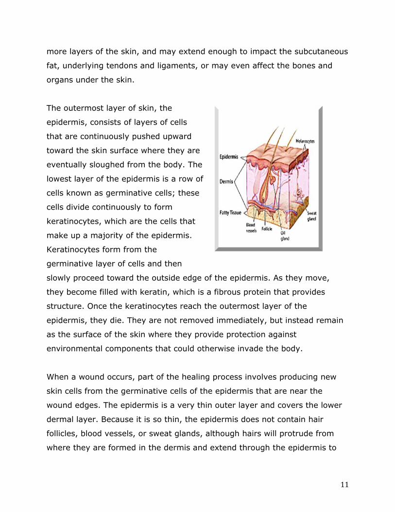
The outermost layer of skin, the
epidermis, consists of layers of cells
that are continuously pushed upward
toward the skin surface where they are
eventually sloughed from the body. The
lowest layer of the epidermis is a row of
cells known as germinative cells; these
cells divide continuously to form
keratinocytes, which are the cells that
make up a majority of the epidermis.
Keratinocytes form from the
germinative layer of cells and then
slowly proceed toward the outside edge of the epidermis. As they move,
they become filled with keratin, which is a fibrous protein that provides
structure. Once the keratinocytes reach the outermost layer of the
epidermis, they die. They are not removed immediately, but instead remain
as the surface of the skin where they provide protection against
environmental components that could otherwise invade the body.
When a wound occurs, part of the healing process involves producing new
skin cells from the germinative cells of the epidermis that are near the
wound edges. The epidermis is a very thin outer layer and covers the lower
dermal layer. Because it is so thin, the epidermis does not contain hair
follicles, blood vessels, or sweat glands, although hairs will protrude from
where they are formed in the dermis and extend through the epidermis to

12
the skin surface. When a wound occurs that is very superficial and only
affects the epidermis, the wound typically heals quickly and with little
scarring, as the body is able to produce new skin cells from nearby
germinative cells.
Deeper wounds may affect the dermis, the skin layer that lies below the
epidermis. The dermis is thicker than the epidermis and it mostly consists of
connective tissue. The dermal layer contains many structures, including
blood vessels, hair follicles and nerve endings, as well as other cells that
take part in inflammatory processes when a wound occurs. The dermis is
much tougher than the epidermis because of its composition. The lower
levels of this layer contain collagen fibers that provide strength for the skin
and that take part in wound healing and scar formation.
Below the dermis is the subcutaneous tissue, which consists of fat and other
components, including blood vessels, nerves, and lymph channels. The
subcutaneous tissue is covered by fascia, a membrane of connective tissue
that provides protection. The subcutaneous tissue covers underlying
structures such as bone and muscle, however, the thickness of
subcutaneous tissue layers varies between locations. Some areas, such as
those of the abdomen or upper thigh, naturally contain more fat tissue when
compared to other areas. The organs and muscles underneath the
subcutaneous tissue also have their own protective membranes. Depending
on the wound and the mechanism of injury, the wound can extend down into
the subcutaneous tissue and can expose underlying muscles or bone. There
are many different mechanisms that can produce wounds, whether by
disease processes, through acute injury to the tissue, or through ongoing
factors that contribute to skin breakdown over time.
Pressure Ulcers

13
A pressure ulcer develops in an area that becomes ischemic because
increased pressure on the skin and underlying tissues prevents adequate
blood flow to the area. Pressure ulcers can develop almost anywhere on the
body where excessive pressure impairs blood flow, but they are most
common on areas that cover bony prominences. The most likely areas where
pressure ulcers develop include the sacrum, the heels, the ear, and the
coccyx.3 Pressure ulcers used to be referred to as decubitus ulcers or
bedsores; however, these terms do not necessarily reflect a comprehensive
mechanism of injury. For instance, a person who is not confined to bed may
still develop a pressure ulcer. The term “pressure ulcer” is more consistent in
defining the means of injury that occurs with this type of wound.
Increased pressure over an area of skin causes compression of the blood
vessels that normally supply oxygenated blood to the skin, subcutaneous
tissue, and underlying fascia. When the blood vessels are constricted in this
manner, blood flow to these sites slows and the distal areas do not receive
adequate oxygen or nutrients that are so needed to maintain healthy skin.
Further, venous return is also slowed, and blood is unable to adequately flow
away from these areas and back toward the heart because of vessel
compression. As a result, metabolic wastes, which are normally carried away
from the area as part of venous return, instead accumulate in the affected
area. This causes a negative cycle as the increased build up of metabolic
wastes causes vasodilation of surrounding blood vessels, followed by edema,
and further compression of the blood vessels supplying the area.
Pressuresores101.com. (n.d.). Pressure sores & bony prominences. Retrieved
from http://pressuresores101.weebly.com/bony-prominences.html
14
After a period of time in which blood flow is restricted, tissue ischemia
develops whereby the tissues fed by the compressed blood vessels no longer
have enough oxygen to survive and cell death occurs. This cell death then
contributes to skin breakdown and the affected person develops a pressure
ulcer.
Regular wound assessment is required to determine the depth and extent of
the wound, as well as whether treatment measures are being effective in
healing the wound. The nurse should note the location, size, and appearance
of the wound to better determine the degree of damage. The National
Pressure Ulcer Advisory Panel (NPUAP) has defined several classifications of
pressure ulcers according to the depth of tissue involvement and the extent
of damage. By understanding the stages of pressure ulcers, the nurse can
assess a wound and better understand how it is staged. By staging the
wound, the provider then has a guide for the best form of wound
management.
Stage I
In stage I the skin is still intact but does not blanch when pressed. The skin
appears red, which does not resolve with time or position changes. It may
more likely appear over a bony prominence. In a person with dark skin, the
area may not be red or even noticeable except that the affected skin
appears as a different color when compared to the surrounding skin.
Normally, an area of skin may turn red after a short period of pressure; this
situation is known as reactive hyperemia. The process occurs when the body
increases blood flow to the compressed area to make up for temporary
15
oxygen deprivation.8 With reactive hyperemia, the skin becomes red and
appears flushed. However, this typically resolves quickly with position
changes and as blood flow resumes to its normal pattern.3 An area of
redness can be considered a stage I ulcer if the redness does not resolve
and the area does not blanch.
Stage II
The skin is broken in Stage II but the wound is typically confined to the
epidermis. The skin appears red and blisters filled with serous fluid may be
present; blisters may have broken, resulting in shallow wounds that ooze.
The base of the wound may appear pink or red and slough may or may not
be present.
Stage III
In state III the wound is deep enough that it extends through the epidermis
and into the dermis. A stage III pressure wound is considered a full-
thickness wound; however, this stage of wound does not affect the
underlying muscle, tendons, or bone. The subcutaneous fat under the dermis
may be seen in some areas where there are greater amounts of fat. Slough
may or may not be present at the base of the wound, which may make it
difficult to determine depth. Stage III wounds can have tunneling, in which a
hole or tunnel progresses deeper into surrounding tissue. If a second wound
is nearby, tunneling may connect the two wounds.8
Undermining may also be present at this stage, which occurs when the
edges of the wound at the surface cover more of the wound than is present
at the base. When undermining is present, the wound is actually larger than
it appears at the surface.

16
Stage IV
Stage IV pressure ulcer is a full-thickness wound that extends from the
epidermis into the dermis and subcutaneous tissue and exposes underlying
bones, muscles, or tendons. In areas where there is less subcutaneous fat
and cartilage is present instead, such as on the nose or the ear, the
exposure of underlying cartilage classifies the wound as a stage IV pressure
ulcer.3 Tunneling and undermining may also be present with this stage of
wound.
In addition to the standard categories of wounds based on the depth of the
affected tissue and the exposure of underlying structures, there are other
classifications of pressure wounds that consider those injuries whose
measurements or depth are not obvious and apparent.
Deep Tissue Injury
In a deep tissue injury (DTI) the skin may or may not be broken but there is
significant bruising that appears as blue or purplish skin with bruising that
extends down into lower layers of skin. The appearance of the wound may
also look like that of a blood-filled blister. The texture of the skin with a deep
tissue injury can vary; some patients have skin that feels warmer than
surrounding tissue, while others have cooler skin. The skin texture may feel
firm or it may be mushy. Some patients have intense pain while others do
not.
Deep tissue injury occurs in an area that has been injured by shear forces. It
can be difficult to determine how deep the injury is and if it extends down
past the dermal layer. A DTI can be difficult to assess in a patient with dark

17
skin. As the injury heals, it may become an ulcer with open skin on the
surface or it may resolve under the skin.
Kennedy Terminal Ulcer
Another type of ulcer, termed the Kennedy Terminal Ulcer, is a specific type
of skin breakdown that may occur hours, weeks, or months before death in a
terminal patient. This type of terminal ulcer typically develops among
patients who are nearing death and who are cared for in long-term care
situations. The skin takes on a purple, red, or yellow appearance and the
wound may be shaped like a pear or a butterfly.3,4 The most common
location where the ulcer develops is on the sacrum, although it can show up
on any part of the skin. This type of ulcer may be more commonly seen by
caregivers who work in long-term care facilities or among those who work in
critical care units who receive patient transfers from long-term care facilities.
A Kennedy terminal ulcer may
develop rapidly through the process
of skin breakdown as the patient
nears death. As Margaret Falconio-
West of Medline best explained,
when a person nears death, organ
failure becomes an issue and is often
a cause of death. It stands to
reason, then, that the skin, the
largest organ of the body, may also
fail, leading to skin breakdown
associated with a Kennedy terminal
ulcer.4
Unstageable Wound
Rosenfield Injury Lawyers. (2014). Bedsore FAQ. Retrieved from
http://www.bedsorefaq.com/
18
The unstageable type of wound is not obvious as far as its depth is
concerned. The clinician may not be able to classify the wound based on its
appearance and further measurements are often required. The base of the
wound is usually covered with slough or eschar, which makes the depth of
the wound difficult to determine.
Several other terms that describe wound tissue may be identified as
characteristics of wounds; these elements may be present in pressure ulcers
or in wounds that have developed as a result of other reasons. Eschar is
used to describe necrotic tissue that has developed within a wound. Eschar
is dead skin that is often tough and thick; it may have a similar appearance
to a scab but it is not the same. Eschar is what must be removed with
debridement. Without removal of eschar, would healing can be significantly
delayed.
Slough is another component of the wound that may develop alongside
eschar, but it has an appearance that is different. Slough is also a collection
of necrotic tissue, but unlike eschar, it is typically moist, crusty, or crumbly.
It is typically white, yellow, or cream colored and it is thought to contain
dead leukocytes, bacteria, dead skin, fibrin, and wound exudate.22 Slough
must be removed during debridement in order to promote wound healing, as
the body typically cannot get rid of slough on its own and it may accumulate,
harboring bacteria and preventing growth of normal, new skin tissue.
Factors contributing to pressure ulcers
Other factors may contribute to development of pressure ulcers, putting
some populations of patients at higher risk. Immobility is a prime cause of
development, as the inability to move or change positions to relieve pressure
on an affected area results in sustained periods of time in which affected

19
blood vessels are compressed. Patients who have excess moisture on their
skin, whether from such factors as sweating or poor hygiene, are at
increased risk, particularly when the skin has become ischemic from too
much pressure. The excess moisture on the skin causes the surface skin to
become softer and more prone to breakdown.
Older adults are a population at high risk, not only because of their
increased instances of immobility, but also because of body changes
associated with aging. Many older adults have less subcutaneous fat under
the surface of the skin, which results in less protection from epidermal
injury. Older adults also have thinner skin as a result of aging, which often
becomes dry and less elastic due to decreased action of collagen and elastin
within the skin’s structure. These effects of aging cause the skin to heal
much more slowly when a wound occurs. Further, some older adults suffer
from sensory changes that result in diminished sensation in the extremities
and distal tissues. These older adults may be less likely to perceive when
tissue damage is happening because they cannot feel it immediately.
Obese patients are also at higher risk of skin breakdown due to pressure
ulcers and tissue ischemia. Patients who are obese have more weight
applied to certain areas when lying in different positions. A person who is
obese may have extra skin folds that can retain moisture and can be difficult
to clean. The skin in these folds may break down more easily when moisture
remains between the folds or skin folds rub on bed sheets or linens, causing
small abrasions on the surface of the skin.
Various other factors, both extrinsic and intrinsic, can impact the risk of
developing pressure ulcers. Extrinsic factors include such elements as:
Friction and shear
20
Level of moisture
Irritating substances on the skin
The environment that prevents movement or turning to relieve
pressure
Alternatively, intrinsic factors are part of the patient; some intrinsic factors
can be changed, while others cannot. Intrinsic factors that affect wound
development include:
Age
Circulation status
Personal habits that affect skin integrity (smoking, diet, alcohol
consumption)
Body temperature
Use of some medications (steroids, vasoactive drugs)
Weight
History of injury or disability
Assessment tools and rating scales
Multiple rating scales are available to assess patient risk for development of
pressure ulcers. In the United States, the Braden scale is one of the most
common tools used to assess whether particular patients are at risk of skin
breakdown or if the skin is no longer intact. The nurse may use the Braden
scale when performing a patient assessment. The results are given scores
based on factors such as the patient’s levels of sensory perception, moisture
content of the skin, nutrition levels, and mobility. The lower the score, given
with the Braden scale, the higher the risk for skin breakdown.
Assessment tools may be used on any patient who may be at risk of
pressure ulcers; although, not all patients may need intervention for

21
pressure ulcer prevention, it is always better to provide more care to prevent
ulcers than to avoid interventions because a patient is believed to be at low
risk. Increased nursing care and interventions for prevention of pressure
ulcers has been shown to decrease pressure ulcer development, regardless
of the patient’s level of risk.3
Arterial Insufficiency
Arterial insufficiency refers to decreased and inadequate blood flow to
tissues and organs. When a patient has arterial insufficiency, he or she is at
increased risk of developing ulcers when the skin and underlying tissues
become ischemic from lack of blood flow. Arterial insufficiency ulcers most
commonly affect the lower extremities, including the legs and feet. As blood
flow diminishes, the cells are starved for oxygen and tissue ischemia
develops. Without correction of adequate blood flow, the skin becomes
necrotic and starts to break down, forming a wound.
Arterial insufficiency can develop
through various causes and it may
occur suddenly or it can develop
gradually. A sudden cause of arterial
insufficiency may result from trauma or
injury to a part of the body that
disrupts blood flow to the extremities.
Alternatively, chronic arterial
insufficiency may develop over time
due to atherosclerosis.9
According to Hess, author of an arterial
ulcer checklist in the journal Advances

22
in Skin and Wound Care, several conditions are associated with arterial
insufficiency and patients with these illnesses are more likely to suffer from
blood flow abnormalities and wounds that develop from arterial insufficiency.
Examples include thrombosis of any cause, vasculitis, rheumatoid arthritis,
systemic lupus erythematosus, sickle cell disease, polycythemia, and
Raynaud’s phenomenon. These conditions affect blood circulation through
such factors as abnormalities in blood vessel structure or anomalies within
blood cells, resulting in decreased circulation to peripheral tissues. Despite
underlying abnormalities in blood flow associated with certain diseases, the
most common cause of arterial insufficiency is atherosclerosis.9
Frequent sites of ulcers in the lower extremity include the lateral malleolus
of the ankle, the foot, and the toes. Wounds that develop from arterial
insufficiency are often small and round without granulation tissue in the
wound base. They often cause significant pain for the affected patient.
Arterial insufficiency causes symptoms similar to that of peripheral arterial
disease (PAD) and is often affiliated with the condition. PAD develops as a
result of atherosclerosis in the large vessels that supply blood to the lower
extremities; the plaques found in the walls of the blood vessels disrupt blood
flow and decrease circulation. Older adults are at increased risk of PAD and,
ultimately, an increased risk of arterial insufficiency wounds. Older adults
are more likely to develop atherosclerosis, as the incidence increases with
advancing age. The Clinical Guide to Skin and Wound Care states that the
incidence of arterial ulcers is 2.2 percent in patients ages 50 to 59 years and
7.7 percent among patients ages 70 to 74 years.10
When a wound does develop as a result of decreased blood flow, the healing
process can be slow and difficult. Because oxygen is needed not only to

23
prevent wounds from forming, but also for wounds to heal properly,
decreased oxygenation from arterial insufficiency results in wounds that heal
poorly and that do not close properly. When a wound develops because of
trauma, the wound is more likely to close slowly and have difficulties with
healing in a person with arterial insufficiency when compared to someone
with normal circulation. For example, a person with PAD if injured by
stepping on a sharp object, may have a wound at the site of injury.
Decreased circulation to the site may further potentiate spreading of the
wound or it may limit the pace at which the wound heals.
Arterial insufficiency often is paired with other illnesses that all contribute to
ulcers and wounds as a result of impaired circulation. A patient may not only
have arterial insufficiency due to peripheral artery disease or vasculitis, but
may also have other conditions that contribute to wound development, such
as diabetes. The risk of ulceration and tissue necrosis is often increased
when more than one condition affecting circulation is present.
Diabetic Ulcers
Diabetes is becoming a global epidemic, affecting more than 150 million
people throughout the world, over 5 percent of the world’s population.
Approximately 16 million people in the United States alone suffer from
diabetes, of which, over 33 percent are not even aware that they have the
disease.13 Based on these statistics, the implications for treatment and
management of diabetes, as well as prevention of its complications, are of
immense proportions.
Ulcers of the foot and lower leg are a complication of diabetes, with
approximately 5 percent of diabetic patients developing foot ulcers each year
and 1 percent requiring amputation.12 A diabetic ulcer occurs when a wound

24
develops in a person who has diabetes; the wound is typically on one of the
lower extremities, such as the foot. The wound may initially be superficial
and affect only the upper layers of skin, but without treatment, it can spread
to the underlying soft tissue structures below the skin and cause further
tissue breakdown in the tendons, muscles, and bones.
Diabetic ulcers commonly occur as a
result of atherosclerosis, peripheral
vascular disease, or neuropathy in
the diabetic patient. Persons with
diabetes are at higher risk of
developing atherosclerosis because
of an increased production of free
radicals that affect the ability of the
blood vessels to relax. Diabetes also
increases chronic inflammation and
slows blood flow through the vessels, which significantly contributes to
atherosclerosis.18 Additionally, people with diabetes are at increased risk of
other factors that contribute to atherosclerosis, including increased levels of
low-density lipoproteins and increased platelet adhesiveness.12
Peripheral vascular disease develops in conjunction with diabetes when
poorly controlled blood glucose levels cause changes in the vascular system
that result in narrowing of the blood vessels. With narrowing, the blood
vessels become smaller in diameter, which limits blood flow to less than
normal levels and reduces the amount of oxygenated blood that reaches the
peripheral tissues. Decreased blood flow, in particular to peripheral tissues
such as the extremities, can result in tissue ischemia followed by skin
breakdown when the tissue becomes necrotic.
Diabetic foot wound

25
Diabetic peripheral neuropathy develops as a result of nerve damage to the
peripheral sensory nerves because of elevated glucose levels in the
bloodstream. It may also be related to other factors, such as nerve injury,
chronic inflammation, and reduced blood flow due to peripheral vascular
disease.12 The patient with diabetic peripheral neuropathy typically has
reduced sensation to the peripheral extremities — most often the lower legs
and feet — which affects how well he or she is able to determine if an injury
has occurred or to feel if a wound is developing. For instance, a diabetic
patient with neuropathy may have such diminished feeling in the feet that he
or she is unaware when shoes do not fit normally and consequently rub the
skin off in one area. This can cause further skin breakdown and infection
because of blood flow abnormalities as a result of the diabetes. If a wound
does develop and grow larger on the foot, it may become extensive before
the affected person notices it, particularly if he or she does not perform
routine foot self-exams.
Diabetic peripheral neuropathy may be present to some extent in up to 60
percent of patients who have diabetes.12 It is one of the most common
causes of diabetic ulcers in this population. A structural complication that
can develop as a result of diabetic peripheral neuropathy and that
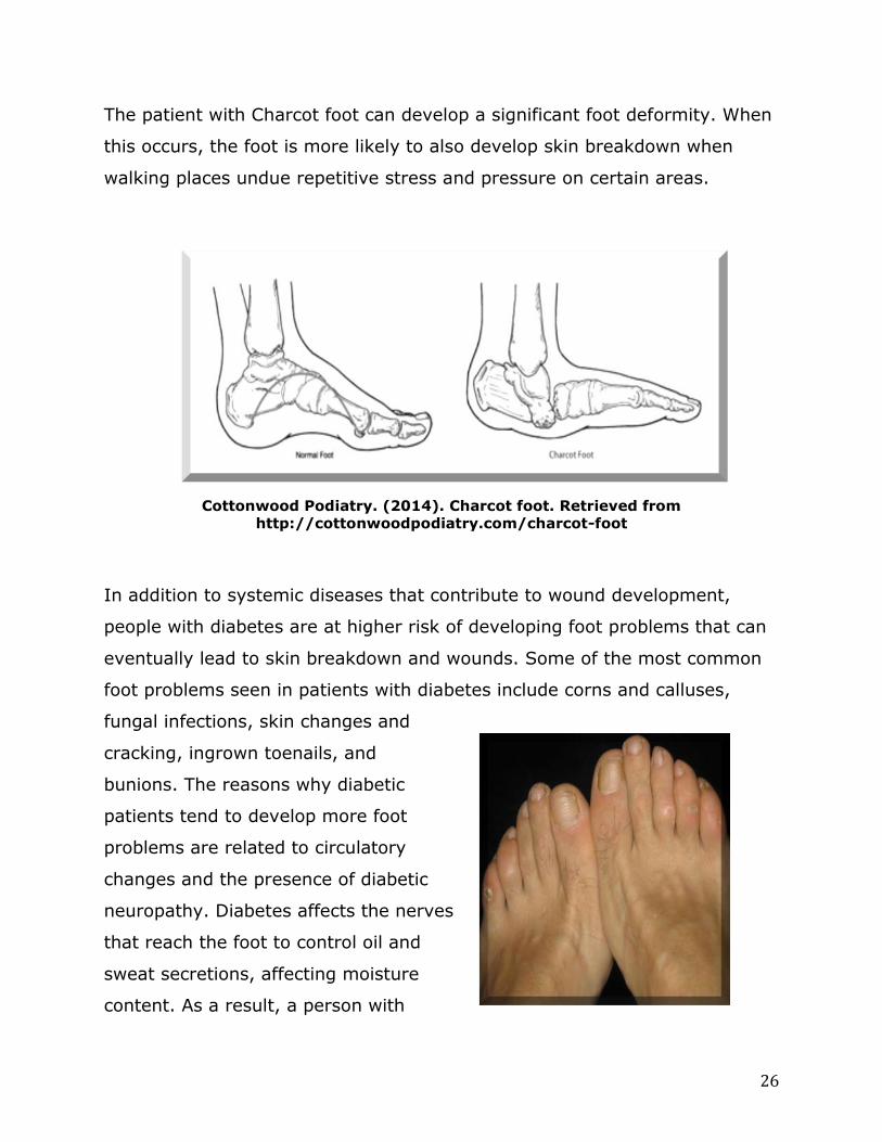
contributes to wound development is the Charcot foot. Rogers, et al., in the
journal Diabetes Care, describe this condition as an inflammatory syndrome
that results in changes to the bones and connective tissue of the feet,
altering the structure and causing a “rocker bottom” appearance, in which
the sole of the foot is rounded.19 The muscles of the foot are weakened and
small fractures may occur in the bones of the foot.
26
The patient with Charcot foot can develop a significant foot deformity. When
this occurs, the foot is more likely to also develop skin breakdown when
walking places undue repetitive stress and pressure on certain areas.
Cottonwood Podiatry. (2014). Charcot foot. Retrieved from
http://cottonwoodpodiatry.com/charcot-foot
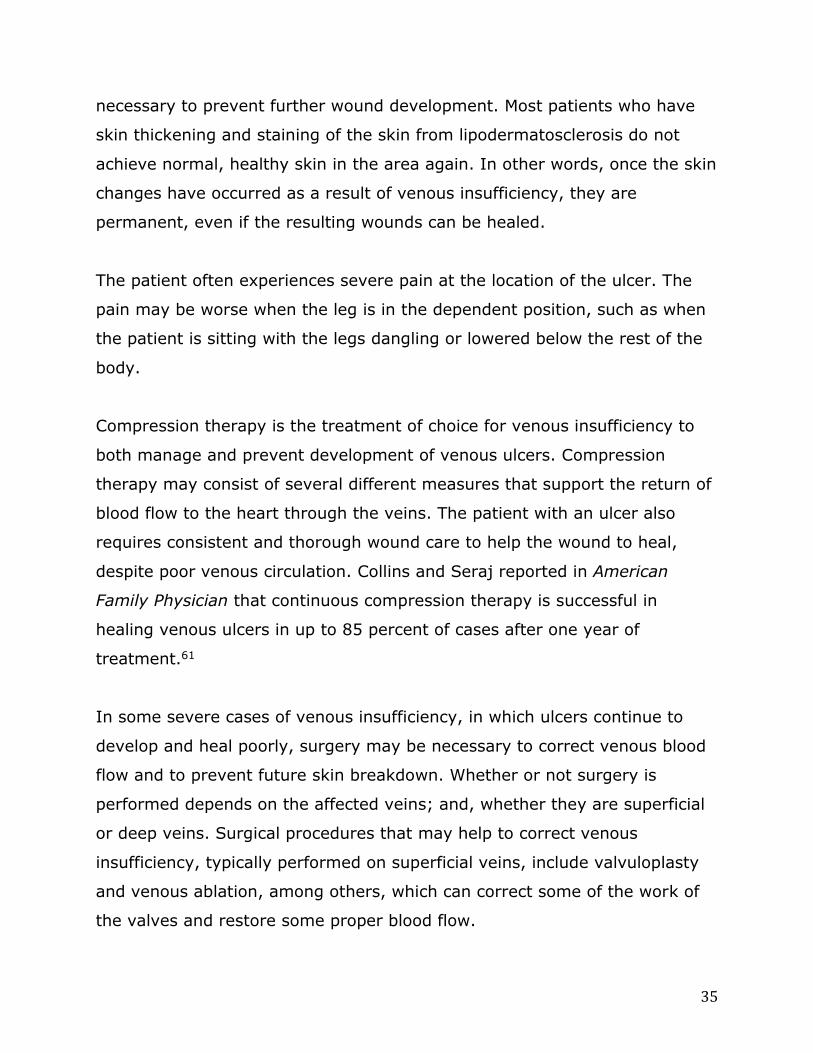
In addition to systemic diseases that contribute to wound development,
people with diabetes are at higher risk of developing foot problems that can
eventually lead to skin breakdown and wounds. Some of the most common
foot problems seen in patients with diabetes include corns and calluses,
fungal infections, skin changes and
cracking, ingrown toenails, and
bunions. The reasons why diabetic
patients tend to develop more foot
problems are related to circulatory
changes and the presence of diabetic
neuropathy. Diabetes affects the nerves
that reach the foot to control oil and
sweat secretions, affecting moisture
content. As a result, a person with

27
diabetes may have very dry skin on the feet and lower legs, which can crack
and bleed, increasing the risk of wound development.
Calluses form as thickened areas of skin on the feet and toes; at times, a
person with diabetes may try to trim calluses, but any minor cut outside of
the calloused area can cause an increase in infection risk. Further, chemicals
applied to corns and calluses can also cause skin breakdown, skin burns, and
foot damage that can perpetuate wound development. Alternatively, calluses
can become quite thick and then the skin can break down when they are not
cared for, which can cause a diabetic ulcer.
A diabetic patient who does not care for his or her feet regularly may
develop an ulcer if unaware of the damage occurring. Poorly fitting shoes,
minor injuries from walking barefoot, and small objects in the shoe that rub
on the foot can all lead to skin breakdown. The most common locations of
diabetic ulcers are on the ball of the foot and on the great toe; however,
diabetic ulcers can develop anywhere on the feet, ankles, toes, and lower
legs.
There is not one particular classification system that has been universally
accepted for staging and grading diabetic wounds. Some of the most well
known systems include the Meggitt-Wagner staging system and the
University of Texas system. A thorough and straightforward staging system
can better help practitioners to discern effective treatment methods and to
evaluate the effectiveness of those methods on the diabetic wounds they are
used for.
The Meggitt-Wagner system classifies the diabetic wound on the foot based
on its depth and/or extent of tissue damage caused by gangrene, if the

28
wound is significant. According to the Meggitt-Wagner system, the stages of
the diabetic wound are classified as follows:
Stage 0: No external symptoms of the wound; the presence of pain
indicates that damage has occurred
Stage 1: Superficial skin wound affecting the uppermost layers of
skin
Stage 2: Deep ulcers
Stage 3: Wounds that affect the underlying bone
Stage 4: Gangrene present on a partial area of the foot
Stage 5: Gangrene present on the entire foot
The Journal of Diabetic Foot Complications performed a review of the
common types of diabetic wound classification systems and found several
limitations with this staging system. First, the system is used for classifying
the foot, and although it is the most common area of diabetic wound
development, there may be other areas where a wound can develop that has
been caused by complications of diabetes but that cannot be staged using
this system. Secondly, this system does not consider other problems
associated with wounds, such as the presence of infection, nor does it
quantify the significance of vascular disease as the cause of this type of
wound.15 However, when a patient presents with a diabetic wound on the
foot, the Meggitt-Wagner system can be implemented in many cases to
properly stage the wound according to its depth and the presence of necrotic
tissue.
The University of Texas has also provided a staging system for classifying
diabetic wounds. This system classifies wounds from Grade 0 to Grade 3,
and then further breaks each stage down into stages that range from Stage

29
A to Stage D, which represent the presence of infection, ischemia, or both as
part of the wound. A Grade 0 wound may represent very little tissue damage
when classified at Grade 0, Stage A of the system; alternatively, a wound
classified as Grade 3, Stage D of the system is the most extensive with
infection and ischemia present.15 The classification system is demonstrated
as follows:
Grade 0 Grade 1 Grade 2 Grade 3
Stage
A
Pre-ulcerative
lesion with no
obvious skin
breakdown
Superficial wound Wound that
penetrates to
underlying tendons
Wound
penetrating to
bone or joint
Stage
B
Infection Infection Infection Infection
Stage
C
Ischemia Ischemia Ischemia Ischemia
Stage
D
Infection and
ischemia
Infection and
ischemia
Infection and
ischemia
Infection and
ischemia
Jain, A. K. C. (2012). A new classification of diabetic foot complications: A simple
and effective teaching tool. The Journal of Diabetic Foot Complications 4(1): 1-5.
Retrieved from http://jdfc.org/2012/volume-4-issue-1/a-new-classification-of-
diabetic-foot-complications-a-simple-and-effective-teaching-tool/#hide
Diabetes affects both the development of wounds and their ability to heal
properly. When a patient does not control diabetes well, he or she may be
more likely to suffer nerve damage from neuropathy; decreased sensation,
particularly in the extremities, decreased immune function, poor circulation,
and an increased risk of infections.2 When an infection is present, the
condition requires strict management and prevention of complications, as
the diabetic patient may have delayed wound healing that could cause an

30
infection to spread to surrounding tissues or to the underlying bone. If a
patient has neuropathy, he or she may not notice the skin breakdown right
away and may not feel significant pain. It is still important for the patient to
seek care and to manage the diabetic wound before it worsens or becomes
infected. All diabetic patients should be taught of the importance of caring
for their feet and the possible consequences of untreated diabetic wounds.
Venous Insufficiency
Formerly called venous stasis, venous insufficiency is a condition that
develops when the veins are unable to return blood to the heart at a normal
rate. For various reasons, the valves inside the veins do not propel blood
toward the heart, leading to changes in the lower extremities, including the
appearance of the skin and the increased risk of skin breakdown that causes
wounds. Approximately 500,000 people develop wounds from venous
insufficiency each year.
The arteries deliver blood throughout the circulatory system at relatively
high pressures; the amount of this pressure is measured when taking a
patient’s blood pressure. Once the blood has been sent to pertinent organs
and tissues and has been filtered through the capillary system, the pressure
at which the veins return the blood to the heart is comparatively much
lower.57 Veins are classified as being superficial, deep, or communicating
veins. An example of a superficial vein is one of the saphenous veins in the
legs, while the popliteal or iliac veins would be considered deep veins.58
When a person is standing up, the veins must return blood to the heart at a
force that works against hydrostatic pressure, which is the pressure of
gravity that pushes the blood down toward the feet. In order to get the
blood to return to the heart and to push against the hydrostatic pressure,

31
the internal lumens of the veins contain valves, which support blood flow in
the proper direction and prevent the backflow of blood toward the feet.
Venous insufficiency develops when the veins are unable to effectively pump
blood toward the heart and the blood collects in the lower extremities. The
backup of blood in the veins may cause them to become larger and
distorted; the condition also leads to venous hypertension, which is
increased blood pressure inside the veins. Venous hypertension causes the
skin changes associated with venous insufficiency, as well as edema and
pain in the lower extremities.
The calf muscle pump, found in the
lower leg and consisting of the calf
muscle and the veins that run through
it, is important for promoting blood
return to the heart. When the calf
muscle contracts, it pushes blood
against hydrostatic pressure to promote
venous return and prevent blood
pooling. Some patients with venous
insufficiency have calf muscle pump
dysfunction, in which blood does not
always flow in the proper direction and
instead pools in the lower extremities.57 Calf muscle pump dysfunction may
be more prone to develop when a patient sits with the legs in the dependent
position for long periods of time. This position does not require use of the
pump as often and the blood tends to pool in the lower legs.
Deep vein thrombosis (DVT) develops as a blood clot in one of the deep
veins that feeds into the vena cava. A patient with venous insufficiency is at

32
higher risk of developing DVT when blood flow is poor or blood pools in the
lower extremities. DVT can lead to other complications, such as a pulmonary
embolism; it is dangerous because almost 50 percent of cases do not
demonstrate any symptoms.62 When DVT does cause symptoms, the patient
may experience pain in the legs while walking. DVT symptoms also include
swelling and warm, tender skin.
Patients with immobility and those with a history of varicose veins are at
higher risk of developing a DVT, as well as patients who are obese, those
who have had major surgery, and people with blood clotting disorders. Once
inflammation develops as a result of DVT, the patient is at increased risk of
developing more blood clots again in the future.
Up to one-half of patients who
develop DVT will have post-
thrombotic syndrome, a condition
that causes chronic pain, a feeling of
heaviness in the legs, swelling, and
skin ulcers.62 When DVT develops, it
causes damage to the vein and its
valves, further perpetuating poor
venous return. The risk of post-
thrombotic syndrome (PTS) is
increased if the patient has had
more than one blood clot. Other signs and symptoms of PTS are similar to
venous insufficiency and may cause changes in skin appearance and texture,
as well as swelling and eczema in the lower extremities.

33
A patient with post-thrombotic syndrome after DVT is more likely to develop
a venous ulcer, which will then require assessment and management of the
condition as long as the wound is healing. Further prevention of PTS is
required to prevent future blood clots and skin wounds. Prevention of PTS is
similar to management of venous insufficiency and venous ulcers; the
patient should use compression therapy to promote blood flow and
medications such as anticoagulants may be necessary to reduce the chance
of further blood clots.
Venous insufficiency tends to develop more commonly among people who
live sedentary lifestyles and those who are overweight or obese. It is also
more common among women and its incidence increases with pregnancy.
Other populations who are at higher risk of venous insufficiency include men
who smoke, and people who must stand for long periods of time, such as
required standing to perform a job. In addition to those who have calf
muscle pump dysfunction or a history of DVT, some people are born with
incompetent valves in some of the veins of the lower extremities, and a
small population of people who are born with Klippel-Trénaunay-Weber
(KTW) syndrome will also have venous insufficiency. KTW syndrome causes
skin changes such as port-wine stains and an increased incidence of varicose
veins; both conditions have been shown to cause an increase in skin
breakdown and ulceration among affected patients.58
The skin changes that occur as a result of venous insufficiency develop when
blood leaks out of the capillaries and the veins and is deposited in the
surrounding tissues. The tissue becomes edematous and the skin may turn a
patchy brown color. This occurs from the creation of hemosiderin, a yellow-
brown skin pigment that is formed by the breakdown of hemoglobin.
Eventually, the skin increasingly becomes more fibrous in appearance and

34
the texture is firm and thickened.59 The fibrous, thickened skin condition is
known as lipodermatosclerosis. The patient may also develop skin eczema,
known as stasis dermatitis.
As blood leaks from the capillaries into the tissues, the inflammatory process
is activated and cell damage ensues. This sets off a series of events that
includes release of leukocytes, platelet aggregation, and cellular edema that
contribute to tissue breakdown and wound development.61 If a patient has
an ulcer that may be caused by venous insufficiency, diagnostic tests should
be performed to confirm that venous problems are the cause of the ulcer
and that it is not caused by other factors, such as diabetic neuropathy or
arterial insufficiency. Diagnostic tests such as the ankle-brachial index or
venous ultrasonography may be used to pinpoint the cause. Identifying the
cause as venous insufficiency is important because the treatment modalities
will differ for this type of ulcer when compared to some other causes.
The most common location of venous ulcers is just above the lateral
malleolus of the ankle.59 Venous ulcers also tend to develop over other bony
prominences. They are usually shallow wounds with irregular borders. It
may take months to years for an ulcer of this type to heal; many patients
who originally develop one venous ulcer as a result of venous insufficiency
will have recurring ulcers form. The patient most commonly has pain, edema
that is reduced when the affected extremity is elevated, and itching and
signs of eczema around the ulcerated site.
When a venous ulcer develops, a major component of managing the wound
is the correction or management of venous insufficiency as well. Although
venous insufficiency does not need to be corrected completely for a wound
to heal, management of the condition and reduction of symptoms is
35
necessary to prevent further wound development. Most patients who have
skin thickening and staining of the skin from lipodermatosclerosis do not
achieve normal, healthy skin in the area again. In other words, once the skin
changes have occurred as a result of venous insufficiency, they are
permanent, even if the resulting wounds can be healed.
The patient often experiences severe pain at the location of the ulcer. The
pain may be worse when the leg is in the dependent position, such as when
the patient is sitting with the legs dangling or lowered below the rest of the
body.
Compression therapy is the treatment of choice for venous insufficiency to
both manage and prevent development of venous ulcers. Compression
therapy may consist of several different measures that support the return of
blood flow to the heart through the veins. The patient with an ulcer also
requires consistent and thorough wound care to help the wound to heal,
despite poor venous circulation. Collins and Seraj reported in American
Family Physician that continuous compression therapy is successful in
healing venous ulcers in up to 85 percent of cases after one year of
treatment.61
In some severe cases of venous insufficiency, in which ulcers continue to
develop and heal poorly, surgery may be necessary to correct venous blood
flow and to prevent future skin breakdown. Whether or not surgery is
performed depends on the affected veins; and, whether they are superficial
or deep veins. Surgical procedures that may help to correct venous
insufficiency, typically performed on superficial veins, include valvuloplasty
and venous ablation, among others, which can correct some of the work of
the valves and restore some proper blood flow.

36
Anticoagulant medications may be used for a patient who has developed a
DVT and has an increased risk of developing more blood clots. However,
other medications, such as antibiotics, are not useful for treating venous
insufficiency and are not typically prescribed unless the resulting ulcer
becomes infected.
Surgical Wounds
A surgical wound is one that is created by a surgical intervention, rather
than developing as a result of an injury or disease process. Most surgical
wounds are made when a surgeon cuts the skin with a scalpel as part of a
surgical process. The wound may also be closed with sutures or staples —
through primary intention — but the creation of a surgical wound does not
necessarily mean that it will be closed in this method. Some surgical wounds
are left open for healing by secondary intention. Surgical wounds, regardless
of how they are closed, are at risk of infection and complications such as
dehiscence.
According to the World Health Organization (WHO), there are several basic
types of surgical wounds that are classified according to their levels of
contamination:63
1. Clean wounds that show no sign of infection or inflammation and are
free of debris.
2. Clean contaminated wounds that have normal tissue that is colonized
with chronic bacterial colonization.
3. Contaminated wounds that contain debris or foreign materials that can
cause an infection.
4. Infected wounds that demonstrate signs of infection and where pus is
present.
37
The classification of wound contamination helps the provider to understand
how best to close the wound, whether by primary or secondary intention or
by delayed closure. Wounds that are clean should be closed by primary
intention, with the wound site monitored regularly; this includes checking for
signs of redness or drainage and ensuring that the sutures or staples are
intact. An example would be when a patient has a surgical procedure, such
as an appendectomy, and the surgeon opens the skin for the purpose of
removing tissue. There is no infection present and the skin is not inflamed,
so the surgeon uses sutures to close the wound. The nurse checks the
incision during the patient assessment to ensure that it is healing well.
Depending on the patient’s health and nutrition status, the wound is likely to
heal and leave a minor scar.
When a surgical wound is closed by primary intention, the physician may use
sutures, staples, adhesive skin tape, glue, or a combination thereof. Sutures
are available in different materials. Some sutures are absorbable and will be
broken down by enzymes in the skin.66 Alternatively, non-absorbable sutures
will later need to be removed. Non-absorbable sutures may remain in place
in the skin for varied periods of time; the physician will prescribe removal of
sutures based on the strength of the surgical wound and the process of
healing. Sutures are removed at the right time that demonstrates that the
wound has approximated enough that it will hold together without the
sutures in place. Alternatively, keeping sutures in place too long can lead to
infection at the incision site.
A wound that is contaminated or infected should not be closed by primary
intention. Instead, it should be left open to heal by secondary intention,
which involves regular wound maintenance and monitoring for signs of
delayed healing or of the wound infection spreading to nearby tissues. For
38
example, a patient with a lymph node infection that has developed into a
seroma under the skin may need surgery to remove the tissue. Based on the
infection, the surgeon leaves the wound open to heal by secondary intention.
The nurse checks the wound on a regular basis, controls exudate and keeps
a moist wound bed, performs dressing changes, and provides antibiotics.
Delayed primary closure involves leaving a wound open for a period of time
and then closing the wound later with sutures or staples. Delayed primary
closure is often done on surgical wounds that are contaminated or those that
are clean but that are more than 6 hours old. The surgeon may delay closing
the wound by primary intention in order to give the wound time to heal, and
to prevent infection. When debris is contaminating the wound bed, the
surgeon will not close the wound over the debris, as this process would most
likely lead to infection. Delayed closure gives time for the debris to be
removed and the wound to partially heal from the base before it is closed.
Regardless of the type of surgical wound, the nurse must continue to
monitor the wound frequently for signs and symptoms of infection. When a
wound is closed with sutures, the nurse assesses for red skin around the
incision, wound edges that are not well approximated, drainage, swelling,
tenderness, and skin that is hot to the touch. Because a surgical wound
involves a break in the skin, there is always the potential for microorganisms
to enter the space and multiply to cause an infection.
In some situations, a surgeon may place a surgical wound drain, which
allows excess fluid and blood to drain from the wound instead of building up
within the tissue. The surgical drain may be sutured in place to prevent
being pulled out. Some drains are passive drains, in which fluid leaks directly
out of the opening. An example of a passive drain that may be placed in a
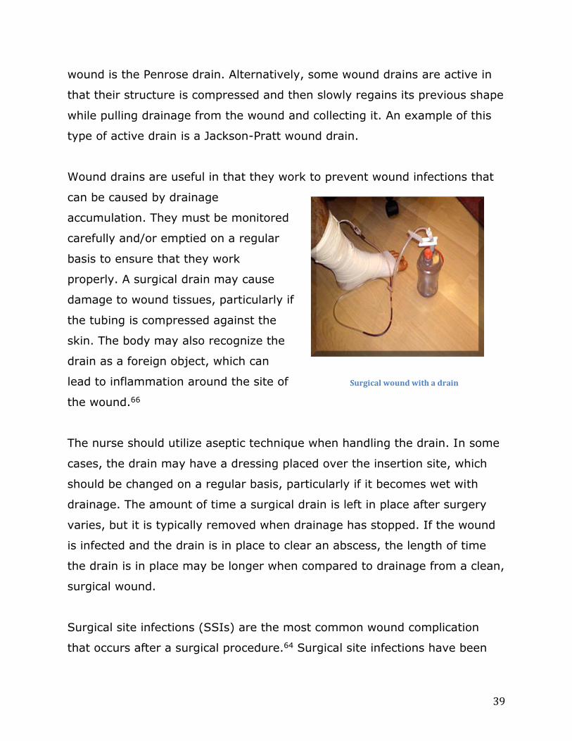
39
wound is the Penrose drain. Alternatively, some wound drains are active in
that their structure is compressed and then slowly regains its previous shape
while pulling drainage from the wound and collecting it. An example of this
type of active drain is a Jackson-Pratt wound drain.
Wound drains are useful in that they work to prevent wound infections that
can be caused by drainage
accumulation. They must be monitored
carefully and/or emptied on a regular
basis to ensure that they work
properly. A surgical drain may cause
damage to wound tissues, particularly if
the tubing is compressed against the
skin. The body may also recognize the
drain as a foreign object, which can
lead to inflammation around the site of
the wound.66
The nurse should utilize aseptic technique when handling the drain. In some
cases, the drain may have a dressing placed over the insertion site, which
should be changed on a regular basis, particularly if it becomes wet with
drainage. The amount of time a surgical drain is left in place after surgery
varies, but it is typically removed when drainage has stopped. If the wound
is infected and the drain is in place to clear an abscess, the length of time
the drain is in place may be longer when compared to drainage from a clean,
surgical wound.
Surgical site infections (SSIs) are the most common wound complication
that occurs after a surgical procedure.64 Surgical site infections have been
Surgical wound with a drain

40
shown to occur in between 3 and 11 percent of postoperative surgical
incisions; the variation exists depending on the type of surgery performed.65
The severity of an SSI can range from purulent drainage, redness, and
incisional swelling to full-blown septicemia that rapidly develops into a life-
threatening situation.
Milne, et al., in an article found in Wounds UK, described an approach that
can reduce the risk of surgical site infection by performing assessments
before, during, and after the operative session. Prior to surgery, during the
preoperative phase, the nurse must assess for those patient factors that will
increase the risk of skin breakdown and wound formation, such as advancing
age, obesity, immobility, or malnutrition. During the surgical procedure, the
healthcare team provides care using aseptic technique to reduce the risk of
infection. This includes cleansing the operative site appropriately, ensuring
sterile technique throughout the procedure, and ensuring instruments have
been properly sterilized before use. Postoperatively, the nurse must continue
to use aseptic technique when caring for the wound, clean the wound with
saline and perform dressing changes as ordered, and monitor for signs of
skin breakdown.64
The risk of surgical site infection is increased with advancing age, longer
length of time of the surgical operation, a longer stay in the hospital prior to
the surgical procedure, inadequate bathing or showering before the
procedure, lack of appropriate removal of body hair in the operative area;
and, other patient factors, including a history of smoking, increased BMI,
and a history of certain chronic diseases, such as chronic obstructive
pulmonary disease, diabetes, and malnutrition.66 Antibiotics that are ordered
prophylactically must be given at the appropriate time surrounding the

41
surgery. Antibiotics are effective when given on time and not too far before
the procedure or well after the surgery has started.
The initial process of inflammation that develops at the beginning of the
healing process may be confused with a surgical site infection.
Administration of antibiotics occurs after confirmation of infection through a
wound culture, as well as other physical manifestations of wound infection,
such as increased pain, patient fever, surrounding skin maceration, and
purulent drainage.
Wound dehiscence is another potential complication of a surgical wound.
Dehiscence occurs when the edges of the surgically closed wound come
apart. It may occur as partial or complete separation of the wound edges.
The most common area of wound dehiscence is a surgical wound on the
abdomen; however, the condition has also developed with surgical wounds
in other locations, such as cesarean section wounds, episiotomies, and
sternotomy wounds.66 Normally, the healing surgical wound develops a ridge
of tissue along the incision line. When this ridge is missing or has never
developed, dehiscence is more likely to occur.
When wound dehiscence occurs, the nurse should cover the wound and get
help. If evisceration has also occurred with dehiscence, a condition in which
the organs behind the wound spill out through the wound opening, the nurse
should quickly place a saline-soaked gauze over the organs for protection
and prepare for surgical intervention. Depending on the extent of
dehiscence, surgery may be necessary to clean out debris and anything that
is contaminating the wound, as well as infected materials that may have
collected within the wound. The wound should stay covered with gauze
soaked in normal saline until the patient goes back to surgery.

42
Similar to management of wounds caused by other factors, such as venous
insufficiency or diabetic neuropathy, surgical wounds require an appropriate
healing environment that promotes tissue growth and prevention of
infection. Surgical wounds may have complications that are specific to this
type of opening, particularly when affected tissues under the incision
become infected or develop other problems because of underlying medical
issues, such as chronic disease. The nurse should frequently monitor the
surgical wound and perform routine care measures to promote healing,
support nutrition and health, and to educate the patient about his or her
condition.
Fungating Cancer or Malignant Wound
A wound that develops as a result of a tumor, sometimes called a fungating
cancer wound or malignant wound, occurs when a tumor that is growing
under the skin reaches the skin surface and breaks through to create a
wound. This type of wound can be very difficult to manage, through physical
or psychological patient care, as the wound can be painful and debilitating,
but the patient may also need emotional support to handle a potential
malignant diagnosis associated with the wound.
As it grows, a fungating tumor wound may cause damage under the skin
when it presses toward the skin surface. The affected tissues may become
deprived of oxygen after capillary damage from the growing tumor, leading
to ischemia and tissue necrosis. The wound may have an odor and typically
it may release fluid, blood, or other discharge that must be managed. The
patient may also experience severe pain or itching at the site.
The most common types of cancer that cause tumor wounds are breast
cancer, melanoma, and head and neck cancer. Approximately 62 percent of

43
fungating wounds develop on the breast, with another 24 percent occurring
as lesions on the head or neck.67,68 The wound may grow through the skin at
the site of the cancer or it may appear in an area where the cancer has
metastasized. A fungating wound typically develops within the last few
months of a patient’s life. Confirmation that the wound has actually
developed from metastatic disease and not from another infectious process
or chronic illness is important to guide treatment measures for the wound.
The initial appearance of the tumor may look like a hard nodule that is the
same color as the outer skin or it may be blue, violet, or black in color. The
nodule(s) that develop are typically not painful at first, but as they grow,
they become more prominent and the ulceration can also lead to formation
of sinus tracts or tunneling around the wound.68 When the tissue becomes
necrotic as a result of the wound, anaerobic organisms collect in the area,
since it has been deprived of oxygen. Because of this, the fungating wound
is more likely to have a strong odor that is usually embarrassing and
unpleasant for the affected patient.
The nurse who cares for a patient with a fungating wound must not only
provide wound care and management, but also should assess and provide
treatments for the patient’s malignant condition. This may involve
administering medications for cancer treatment or drugs to increase the
patient’s comfort levels and reduce distress associated with cancer
symptoms. An additional aspect of care is continually assessing the patient’s
psychological and emotional health as the wound and the cancer diagnosis
relates to care. Other factors, such as the patient’s nutritional status,
presence of other chronic conditions, mobility, and ability to perform
activities of daily living should also be incorporated as part of wound care
management in these situations.
44
Management of wound odor is one of the most prominent concerns when
working with a patient who has a fungating wound. The patient may suffer
much embarrassment because of the smell of the wound, which is attributed
to growth of anaerobic bacteria in the wound bed. Administration of
antibiotics may be necessary, but systemic antibiotics are typically not
administered on a long-term basis in order to avoid the potential for
development of resistant organisms. Topical metronidazole, an antifungal
medication, has been shown to reduce wound odor after 2 to 3 days of
application.68 The nurse may also consider other measures that can help
with wound odor, such as by applying dressings infused with charcoal, which
absorb some of the wound odor, controlling exudate, and performing regular
debridement.
A fungating wound often causes pain for the patient, which is controlled
through medications given for pain management, including both opioids and
non-opioid analgesics, as well as other adjuvant medications. Other
considerations that the nurse must give for management of a tumor wound
include control of exudate and bleeding, as this type of wound tends to
produce greater amounts of fluid and/or is more likely to bleed. These
aspects are controlled by appropriate dressings and regular wound care and
dressing changes.
Depending on the patient’s condition, the nurse may need to provide
palliative treatment as part of wound care, particularly if the wound has
developed at the end of the patient’s life. When this occurs, comfort
measures and psychosocial support are essential, in addition to regular
wound care to minimize discomfort from a spreading infection or skin
breakdown from a poorly managed wound. Alternatively, some patients with
fungating wounds may choose more aggressive treatments and may opt for
45
surgical removal or treatment with other elements, such as chemotherapy or
hormone therapy. The nurse must provide support and help for the patient
with a wound caused by a tumor regardless of the treatment decision that is
made. Each situation is different and depending on the choice for treatment
or wound management, the nurse continues to provide gentle and
compassionate care that is therapeutic for this very difficult situation.
Wound Risk Factors
Although there are various causes of wounds that result from differing states
of health or disease, there are some risk factors that are more common to
wound development in general. By and large, a poor state of health, whether
because of chronic disease, malnutrition, lack of activity, or poor self-care,
typically contributes to an increased risk of wound development and poor
wound healing when a wound does happen.
Insufficient Oxygen
Poor oxygen perfusion contributes to wound development when the tissues
do not receive enough oxygenated blood. This may more likely occur in a
condition in which blood flow is reduced or blocked due to an occlusion, such
as in the case of arterial insufficiency or peripheral arterial occlusive disease.
The skin and underlying tissues need oxygen from the blood in order to stay
healthy and to prevent the growth of anaerobic bacteria, which are
microorganisms that can grow in the absence of oxygen. When the skin and
peripheral tissues do not have enough oxygen, the skin is more likely to
break down, causing a wound, and the resultant wound could become
infected more easily.

46
Malnutrition
Malnutrition impacts wound healing due to changes in protein sufficiency and
lack of vitamins that normally act as healing factors in the body. Malnutrition
can develop in some people because of a lack of intake due to a number of
situations, including socioeconomic factors that affect accessibility of food,
stressful events or periods of severe illness, difficulties with feeding,
chewing, or swallowing food, malabsorption syndromes that affect how the
body digests and absorbs nutrients, or mental health diagnoses of eating
disorders, such as anorexia. Alternatively, malnutrition can also occur among
some patients who are overweight or obese. An obese patient may have
malnutrition even when food intake is excessive because he or she may only
be eating certain types of foods that add to weight gain but that do not
provide important nutrients.
When a person is malnourished, he or she may use protein for energy
instead of glucose. To get this protein, the body breaks down its own
sources, such as protein found within skeletal muscle tissue. Because wound
healing requires protein to form a healing matrix through collagen, wounds
may heal slower when the body is focusing its protein sources instead on
gaining energy.
Malnutrition also contributes to wound development through other methods.
Poor nutrition depletes lean body mass and the patient has less muscle
tissue for activities of daily living; he or she may be more likely to develop
greater degrees of immobility, which can further impair other body
processes and contribute to skin breakdown. Further, decreased protein
intake impairs the immune system and can increase the risk of infection if a
wound develops.20

47
A patient who develops an illness or goes through a surgical procedure loses
a certain amount of protein each day. This protein loss then contributes to
further effects that can lead to wound development. Additionally, certain
procedures, periods of hospitalization, or general lack of intake can affect
how well a wound heals when it does develop. Malnutrition has been shown
to impact function of both B and T lymphocytes and prevents proper
functioning of leukocytes in the body, increasing the risk of infection.
Further, if a wound starts to develop, skin breakdown may be perpetuated
by loss of protein and malnutrition; as a malnourished state also increases
the length of the inflammation stage of wound healing, it decreases collagen
synthesis and decreases overall strength of the skin.53
A patient who is malnourished may also be underweight and may have less
fat tissue to protect bony prominences. Consequently, more bony
prominences increase the risk of pressure ulcers without the extra padding
under the skin. A patient who has decreased muscle mass and more bony
prominences as a result of malnutrition may have less activity when
compared to another person who is not malnourished. The increase in
immobility, decrease in muscle mass, and greater number of pressure points
can all contribute to skin breakdown associated with pressure ulcers.
Diabetes mellitus can cause a form of malnutrition because the patient has
abnormal carbohydrate metabolism and is therefore unable to adequately
use this type of macronutrient in a normal manner needed for the body.
Diabetes, or even a severe state of stress or illness that leads to changes in
blood glucose concentrations, can disrupt the functions of the cells of the
immune system, thereby increasing the risk of infection. Hyperglycemia has
also been shown to reduce the body’s ability to absorb vitamin C into
leukocytes and fibroblasts in the skin cells.20
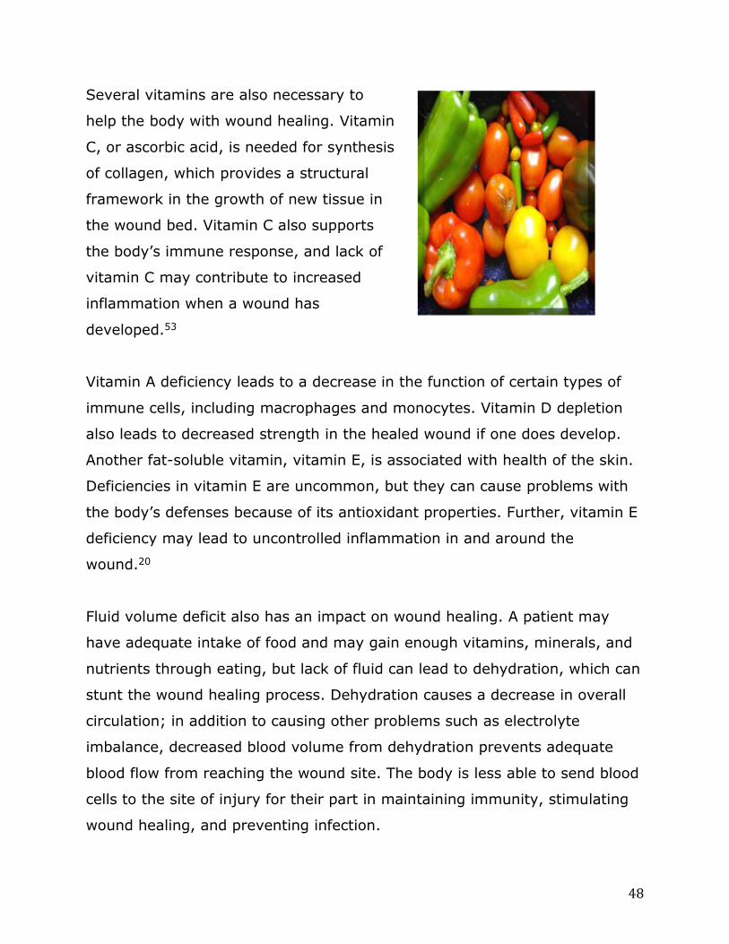
48
Several vitamins are also necessary to
help the body with wound healing. Vitamin
C, or ascorbic acid, is needed for synthesis
of collagen, which provides a structural
framework in the growth of new tissue in
the wound bed. Vitamin C also supports
the body’s immune response, and lack of
vitamin C may contribute to increased
inflammation when a wound has
developed.53
Vitamin A deficiency leads to a decrease in the function of certain types of
immune cells, including macrophages and monocytes. Vitamin D depletion
also leads to decreased strength in the healed wound if one does develop.
Another fat-soluble vitamin, vitamin E, is associated with health of the skin.
Deficiencies in vitamin E are uncommon, but they can cause problems with
the body’s defenses because of its antioxidant properties. Further, vitamin E
deficiency may lead to uncontrolled inflammation in and around the
wound.20
Fluid volume deficit also has an impact on wound healing. A patient may
have adequate intake of food and may gain enough vitamins, minerals, and
nutrients through eating, but lack of fluid can lead to dehydration, which can
stunt the wound healing process. Dehydration causes a decrease in overall
circulation; in addition to causing other problems such as electrolyte
imbalance, decreased blood volume from dehydration prevents adequate
blood flow from reaching the wound site. The body is less able to send blood
cells to the site of injury for their part in maintaining immunity, stimulating
wound healing, and preventing infection.

49
Immobility
Immobility is one of the most common factors associated with pressure ulcer
development, but it also plays a contributing role in wound development for
patients with other chronic diseases, such as venous insufficiency and
diabetes. Lack of movement from immobility decreases overall circulation
and can cause wounds related to incontinence or an inability to perform self-
care measures.
When a patient is immobile and must rely on caregivers for movement or
repositioning, he or she is at greater risk of skin breakdown because of an
inability to shift positions to take pressure off of certain areas of the skin.
Wounds may be more likely to occur in an immobile patient who must spend
a significant amount of time in bed or sitting in a chair, and who otherwise
can do little to increase circulation in the extremities and support or
maintain proper blood flow.
Many immobile patients have difficulties with getting up to use the bathroom
and are often forced to rely on bedpans, catheters, and/or bedside
commodes for elimination. Immobile patients may be at increased risk of
incontinence if they are unable to access these devices quickly enough or if
they must rely on a caregiver for help. Urinary and fecal incontinence
contribute to skin breakdown because of the components of these wastes.
Urine is a fluid that, when left on the skin, increases moisture content and
causes skin softening and maceration. The enzymes found in stool, as well
as its pH content can also cause skin breakdown, particularly after times
when fecal matter is left on the skin without being cleaned in a timely
manner.

50
Many people think of immobility as affecting older adults who are living in
long-term care facilities and who are dependent on caregivers for help with
turning or with getting out of bed. While this is true in many cases, another
population of patients are at risk of skin breakdown because of immobility in
the healthcare environment are those who receive care in the intensive care
unit.
Patients in intensive care environments are at risk of wound development,
often because of the increased amounts of medical equipment used because
of their fragile medical states.3 A patient in the intensive care unit may be at
higher risk of pressure ulcers because he or she is typically immobile
because of illness, rather than a chronic disability or advanced age, as is
seen among some other immobile patients. The patient in the intensive care
unit often also needs more medical equipment as part of his or her care,
which may include a ventilator, urinary catheter, sequential compression
device, intravenous line or central catheter, and hemodynamic monitoring
systems. Some of the medications administered to a patient may also
increase the risk for skin breakdown.
Based on the amount of equipment required and the clinical status of acute
illness, the intensive care unit patient is actually quite immobile. Depending
on the level of care required, he or she may not be able to get up or move
out of bed in any way, whether because of illness and level of consciousness
or because of the presence of so many pieces of medical equipment needed
to provide care. For example, a patient who requires mechanical ventilation
typically requires sedation, which places him or her in an altered state of
consciousness and, most likely, restricted to bed rest rather than chair
activity to facilitate position changes. Prevention of pressure ulcers,
therefore, is centered on turning and repositioning the patient frequently and

51
preventing medical equipment from applying too much pressure at a
particular site.
In addition to limiting how much a patient in the intensive care unit is able
to get up or move, medical equipment also places pressure on certain parts
of the skin, which can lead to skin breakdown. An endotracheal tube that is
positioned in the same area or that presses against the lip for too long can
cause tissue breakdown in that area. A patient who is turned and who is
accidentally positioned so that the catheter hub of the central line is under
the body can suffer from skin breakdown in a short time due to the
intravenous tubing continuously pressing into the skin until such time the
patient is moved again. The administration of some vesicant solutions as
part of treatment for complex medical conditions often seen in the intensive
care unit can cause significant skin and tissue damage if extravasation
occurs. For instance, administration of cisplatin can cause tissue damage and
necrosis if it leaks from the intravenous site into the surrounding tissue.
Comorbidities
Certain factors impact wound development and affect healing when a wound
does develop. Comorbidities are diseases that are present in the patient that
may directly cause wound development or may result in a wound becoming
chronic and difficult to heal. Some patients have several comorbidities,
making their risks for wound development much higher than the general
population.
As a wound goes through the phases of healing, a comorbid condition that is
already present in the patient may interrupt the process and either slow or
stop wound healing altogether or cause complications that require further
intervention. For example, a patient who has diabetes and has developed a

52
foot wound may be on track with wound healing through proper care and
wound management. However, time spent with uncontrolled blood glucose
levels and improper foot care — both of which are factors associated with
diabetes as a comorbidity in this case — can lead to a wound infection and
skin breakdown on the wound periphery, further complicating the healing
process.
There are a number of comorbid conditions that can impair and delay wound
healing. These factors may contribute to the cause of the wound or they
may be elements that affect the wound’s healing progress. Examples of
comorbid conditions that can impact wound development and healing include
chronic conditions such as diabetes, vasculitis, systemic lupus
erythematosus, renal failure, various forms of cancer, rheumatoid arthritis,
and scleroderma.
When comorbidities are present in the wound care patient, the nurse
performs more than one role. Part of wound care management is controlling
the health of the wound, providing supportive care and treatment through
dressing changes and medication administration, and ensuring that the
patient knows how to care for the wound. Additionally, the nurse must
provide interventions that deal with the comorbid conditions that are present
in the patient in order to prevent these conditions from harming the wound
care process. As part of comprehensive care, the nurse provides medications
and patient education, performs or assists with certain procedures designed
to improve the patient’s health, and provides psychosocial support for the
patient’s condition.
As an example, a patient who has renal failure and who has a lower leg ulcer
not only needs ongoing wound care through assessment and treatment, but

53
also needs supportive treatment for renal disease. The nurse is often also
responsible for providing other care measures that may seem unrelated to
the wound because they do not directly affect the wounded area. However,
care of comorbid conditions will impact wound healing and is a necessary
part of treatment. In this example, in addition to providing wound care, the
nurse might also administer medications, arrange a referral for a dietary
consult, and/or assist with the patient’s dialysis.
Any patient who has a chronic disease and a healing wound must be
educated about how the condition affects wound repair and healing. Because
the patient should be an active participant in the wound care process, he or
she needs to know what factors could possibly delay wound healing. The
patient may or may not understand the correlation between a chronic
disease and a wound involving the skin. It is the nurse’s responsibility to
educate the patient about how each condition is related to the other. By
educating the patient about the factors that affect wound healing, the
patient can become more involved in his or her treatment regimens and may
take steps to assist not only with wound care, but with care of his or her
chronic disease as well.
Medications
Certain medications can cause skin breakdown when they cause changes in
the skin because of side effects or when they are inadvertently administered
in a method that the medication interacts with the skin when it is not
supposed to. There are a number of medications that cause rash or eczema
as side effects. While this may not initially cause skin breakdown, the skin
can become more sensitive when a patient takes medications with these
effects. The patient may also scratch the skin in an attempt to soothe the

54
itching; excessive scratching can eventually lead to skin breakdown, sores,
or lesions.
Some medications, when administered intravenously, can cause significant
wounds if their solutions are accidentally infiltrated into the skin and tissues.
This can happen when, upon administration of the medication into the
intravenous catheter, the medication leaks out into the tissues and causes
damage. Medications known as vesicants can cause tissue damage and
necrosis of the skin when extravasation occurs during administration.
Extravasation can lead to such a significant wound that the patient requires
debridement and regular dressing changes while the wound heals,
sometimes over a period of weeks or months.
When a nurse must administer medications intravenously, he or she should
be aware of the potential affects on the skin. When giving vesicant
medications, the nurse must routinely check the intravenous catheter and
monitor the intravenous site and the tubing for changes to ensure that
extravasation does not develop and cause severe tissue damage to the skin.
Summary
Management of wounds involves a number of considerations, which include
the mechanism of injury and the assessment of the wound. Assessment of
the wound involves the wound size and depth. Additionally, its important to
consider other complicating factors, such as the presence of foreign objects
or other injuries around the wound, pain management, and the prevention
of bleeding or infection.
The process of wound healing may vary depending on the method of
intention used to close the wound. Wounds are healed by intention, which is

55
categorized into three different stages, primary, secondary and tertiary;
and, is based on the type of wound, the amount of debris present or if the
wound is contaminated, and the mechanisms of the cause of the wound.
Wounds are typically classified as being either chronic or acute wounds.
Chronic wounds are those that develop after tissue damage has been
ongoing, such as, wounds due to arterial insufficiency, diabetic ulcers,
pressure sores, and venous insufficiency. The point that differentiates
chronic wounds from acute wounds is that chronic wounds develop over
some period of time.
Risk factors more common to wound development generally include a poor
state of health due to chronic disease or poor self-care. Immobility, the most
common factors associated with pressure ulcer development, also plays a
contributing role in wound development for patients with chronic diseases,
such as venous insufficiency and diabetes. A decrease in overall circulation
due to immobilization can cause wounds related to incontinence or poor self-
care performance. Patients at risk of skin breakdown rely on caregivers to
help with mobility and position change to prevent skin pressure areas.
Wound assessment, the first step in the management of a wound, is
discussed in Wound Care Part II and is important throughout the entire
diagnostic and treatment process. In this study, wound care risks and stages
of development have been discussed. The next level of care involves
evaluation of wound healing and treatment efficacy.
Please take time to help the NURSECE4LESS.COM course planners evaluate nursing
knowledge needs met following completion of this course by completing the self-
assessment Knowledge Questions after reading the article.
Correct Answers, page 57.

56
1. Tertiary intention involves the process of:
a. wound closure done by applying physical measures to close a wound.
b. initially leaving the wound open to partial healing.
c. leaving a wound open to heal through production of new granulation tissue to
fill in the wound base.
d. None of the above.
2. A nurse must continue to monitor a surgical wound
a. regardless of the type of surgical wound.
b. when a wound is closed with sutures.
c. only if there is an open wound
d. Answers a and b.
3. True or False: venous insufficiency is a condition that develops when
the veins are unable to return blood to the heart at a normal rate and the
blood collects in the lower extremities.
a. True.
b. False.
4. Surgical wound drains
a. must be emptied on a regular basis to ensure that they work properly.
b. may cause damage to wound tissues.
c. prevent wound infections caused by drainage accumulation.
d. all of the above.
5. When a nurse must administer medications intravenously, he or
she should be aware that
a. medications known as vesicants are safe because they do not cause
tissue damage to the skin.
b. it is preferable to administer medications intravenously because they
cannot accidentally infiltrate into the skin tissues.
c. some medications, when administered intravenously, can cause significant
wounds if their solutions are accidentally infiltrated into the skin and tissues.
d. extravasation does not develop if medications are administered
intravenously.

57
Correct Answers:
1. b
2. d
3. a
4. d
5. c
Wound Care Series
Footnotes:
1. Katz, M. J., Kirr, C. A. (2012). Wound care. Retrieved from
http://www.nursingceu.com/courses/395/index_nceu.html
2. Kifer, Z. A. (2012). Fast facts for wound care nursing: Practical wound management in
a nutshell. New York, NY: Springer Publishing Company, LLC
3. Cooper, K. L. (2013, Dec.). Evidence-based prevention of pressure ulcers the intensive
care unit. Critical Care Nurse 33(6): 57-66. Retrieved from
http://www.aacn.org/wd/Cetests/media/C1363.pdf
4. Falconio-West, M. (2013, Sep.). Kennedy Terminal Ulcer (KTU) is now recognized by
CMS for long-term acute care hospitals (LTAC or LTCH). Retrieved from
http://mkt.medline.com/clinical-blog/channels/clinical-solutions/kennedy-terminal-
ulcer-ktu-is-now-recognized-by-cms-for-long-term-acute-care-hospitals-ltac-or-ltch/
5. Covidien AG. (2008, Jan.). Support services and the prevention of pressure ulcers.
Retrieved from http://www.patientcare-
edu.com/imageServer.aspx?contentID=20368&contenttype=application/pdf
6. Brunner, M., Droegemueller, C., Rivers, S., Deuser, W. E. (2012). Prevention of
incontinence-related skin breakdown for acute and critical care patients. Urology
Nurse 32(4): 214-219. Retrieved from
http://www.medscape.com/viewarticle/769850_2
7. DeMarco, S. (n.d.). Wound and pressure ulcer management. Retrieved from
http://www.hopkinsmedicine.org/gec/series/wound_care.html
8. Lippincott Nursing Center.com. (2009). Wound watch: Assessing pressure ulcers.
LPN2009 5(1): 20-23. Retrieved from
http://www.nursingcenter.com/lnc/static?pageid=844487
58
9. Hess, C. T. (2010, Sep.). Arterial ulcer checklist. Advances in Skin and Wound Care
23(9): 432. Retrieved from
http://journals.lww.com/aswcjournal/Fulltext/2010/09000/Arterial_Ulcer_Checklist.11.
aspx
10. Hess, C. (2012). Clinical guide to skin and wound care (7th ed.). Ambler, PA: Lippincott
Williams & Wilkins
11. Bhutani, S., Vishwanath, G. (2012, Sep.). Hyperbaric oxygen and wound healing.
Indian Journal of Plastic Surgery 45(2): 316-324. Retrieved from
http://www.ijps.org/article.asp?issn=0970-
0358;year=2012;volume=45;issue=2;spage=316;epage=324;aulast=Bhutani
12. Lopez Rowe, V. (2014, Jul.). Diabetic ulcers. Retrieved from
http://emedicine.medscape.com/article/460282-overview
13. Medfocus guidebook on: Diabetic foot ulcers. (2011). Princeton, NJ: Medfocus.com,
Inc.
14. American Diabetes Association. (2014, Oct.). Foot complications. Retrieved from
http://www.diabetes.org/living-with-diabetes/complications/foot-complications/
15. Jain, A. K. C. (2012). A new classification of diabetic foot complications: A simple and
effective teaching tool. The Journal of Diabetic Foot Complications 4(1): 1-5. Retrieved
from http://jdfc.org/wp-content/uploads/2012/01/v4-i1-a1.pdf
16. Cruciani, M., Lipsky, B. A., Mengoli, C., de Lalla, F. (2013). Granulocyte-colony
stimulating factors as adjunctive therapy for diabetic foot infections (review).
Hoboken, NJ: John Wiley & Sons, Ltd.
17. Beldon, P. (2007). What you need to know about skin grafts and donor site wounds.
Wound Essentials, Vol. 2: 149-155. Retrieved from
http://www.woundsinternational.com/pdf/content_196.pdf
18. University of Rochester Medical Center. (2008, Mar.). How diabetes drives
atherosclerosis. Science Daily. Retrieved from
http://www.sciencedaily.com/releases/2008/03/080313124430.htm
19. Rogers, L. C., et al. (2011, Sep.). The Charcot foot in diabetes. Diabetes Care 34(9):
2123-2129. Retrieved from http://care.diabetesjournals.org/content/34/9/2123.full
20. McCullogh, J. M., Kloth, L. C. (2010). Wound healing: Evidence-based management
(4th ed.). Philadelphia, PA: F. A. Davis Company
21. Cowan, L. (2013). Wound series part 2: Approaches to treating chronic wounds.
Retrieved from
http://www.ceufast.com/courses/viewcourse.asp?id=269#Wound_Cleansing

59
22. Medline Industries, Inc. (2007). The wound care handbook [Chapter 8]. Mundelein, IL:
Medline
23. Foster, C. (2010, Apr.). Non-traumatic wound debridement. Ostomy Wound
Management 56(4): 8. Retrieved from
http://www.polymem.com/pearls/pearls4practice0410.pdf?line_id=410
24. Sussman, C., Bates-Jensen, B. M. (1998). Wound care collaborative practice manual
for physical therapists and nurses. [Excerpt]. New York, NY: Aspen Publishers.
Retrieved from http://www.medicaledu.com/whirlpoo.htm
25. Dale, B. A., Wright, D. H. (2011). Say good-bye to wet-to-dry wound care dressings:
Changing the culture of wound care management within your agency. Home
Healthcare Nurse 29(7): 429-440. Retrieved from
http://journals.lww.com/homehealthcarenurseonline/Fulltext/2011/07000/Say_Goodb
ye_to_Wet_to_Dry_Wound_Care_Dressings_.8.aspx
26. Ramundo, J., Gray, M. (2008, Jun.). Enzymatic wound debridement. Journal of
Wound, Ostomy, and Continence Nursing 35(3): 273-280. Retrieved from
http://www.nursingcenter.com/lnc/journalarticle?Article_ID=794501
27. Swezey, L. (2012, Jul.). Wound debridement techniques 6: Biological debridement.
Retrieved from http://woundeducators.com/wound-debridement-techniques-6-
biological-debridement/
28. Dowsett, C., Newton, H. (2005). Wound bed preparation: TIME in practice. Retrieved
from http://woundsinternational.com/pdf/content_86.pdf
29. Martin, B. (2011, Apr.). Moist wound healing. Ostomy Wound Management 57(4): 10.
Retrieved from http://www.polymem.com/pearls/pearls4practice0411.pdf?line_id=411
30. ATI Nursing Education. (n.d.). Dressing and bandage types. Retrieved from
http://www.atitesting.com/ati_next_gen/skillsmodules/content/wound-
care/equipment/dressing_and_bandage_types.html
31. Bjarnsholt, T. (2011). Biofilm infections. New York, NY: Springer Science+Business
Media, LLC
32. Southwesthealthline.ca. (2011, Dec.). Levine method for wound stab for culture &
sensitivity. Retrieved from
http://www.southwesthealthline.ca/healthlibrary_docs/B.7.3.LevineWoundSwabMetho
d.pdf
33. Romanelli, M., Vowden, K., Weir, D. (2010). Exudate management made easy.
Wounds International 1(2): 1-6. Retrieved from
http://www.woundsinternational.com/pdf/content_8812.pdf

60
34. Organogenesis, Inc. (2010). What is Apligraf? Retrieved from
http://www.apligraf.com/professional/what_is_apligraf/index.html
35. DermNetNZ. (2013, Dec.). Bioengineered skin. Retrieved from
http://www.dermnetnz.org/procedures/bioengineered-skin.html
36. Troy, J., Karlnoski, R., Payne, W. G. (2013). The use of EZ Derm® in partial-thickness
burns: An institutional review of 157 patients. Eplasty 13(4). Retrieved from
http://www.ncbi.nlm.nih.gov/pmc/articles/PMC3593337/
37. Organogenesis, Inc. (2013). Proven DFU results and extensive DFU experience.
Retrieved from http://www.dermagraft.com/proven-results/
38. KCI. (2013). Science behind wound therapy. Retrieved from
http://www.kci1.com/KCI1/sciencebehindwoundtherapy
39. Martindell, D. (2012, Jun.). Safety monitor: The safe use of negative-pressure wound
therapy. American Journal of Nursing 112(6): 59-63. Retrieved from
http://www.nursingcenter.com/lnc/JournalArticle?Article_ID=1353037
40. Alumia, R. (2013, Sep.). Improving outcomes with non-contact low-frequency
ultrasound. Retrieved from http://woundcareadvisor.com/improving-outcomes-with-
noncontact-low-frequency-ultrasound/
41. Bryant, R. A., Nix, D. P. (2012). Acute & chronic wounds: Current management
concepts (4th ed.). St. Louis, MO: Elsevier Mosby
42. Westgate, S., Cutting, K. F., DeLuca, G., Asaad, K. (2012, Mar.). Collagen dressings
made easy. Wounds UK 8(1): 1-4. Retrieved from http://www.wounds-uk.com/made-
easy/collagen-dressings-made-easy/page-1
43. DermNetNZ. (2013, Dec.). Keratin-based dressings for chronic wounds. Retrieved
from http://www.dermnetnz.org/procedures/keratin-dressings.html
44. Keraplast Technologies, LLC. (n.d.). A new paradigm in wound care. Retrieved from
http://www.keraplast.com/wound-care#Kerasorb
45. Keraplast Technologies, LLC. (2014, Jul.). User’s guide for treatment of chronic
wounds with Keraplast’s range of Replicine™ Functional Keratin® advanced wound
healing products. Retrieved from
http://www.keraplast.com/images/stories/pdfs/users_guide_for_all_products_for_chro
nic_wounds.pdf
46. Parsons, D., Bowler, P. G., Phil, M., Myles, V., Jones, S. (2005). Silver antimicrobial
dressings in wound management: A comparison of antibacterial, physical, and
chemical characteristics. Wounds 17(8): 222-232. Retrieved from
http://www.medscape.com/viewarticle/513362

61
47. Adkins, C. L. (2013, May). Wound care dressings and choices for care of wounds in the
home. Home Healthcare Now 31(5): 259-267. Retrieved from
http://www.nursingcenter.com/lnc/CEArticle?an=00004045-201305000-
00006&Journal_ID=54023&Issue_ID=1547910
48. Rawe, I. (2012). Technology update: Pulsed radio-frequency electromagnetic field
(PEMF) therapy as an adjunct wound healing therapy. Wounds International 3(4).
Retrieved from http://www.woundsinternational.com/product-reviews/pulsed-radio-
frequency-electromagnetic-field-pemf-therapy-as-an-adjunct-wound-healing-therapy
49. Schwartz, A. (2012). Ozone therapy and its scientific foundations. Revista Española de
Ozonoterapia 2(1): 199-232. Retrieved from http://www.xn--
revistaespaoladeozonoterapia-7xc.es/index.php/reo/article/view/27/30
50. U. S. Food and Drug Administration. (2014, Sep.). CFR-Code of Federal Regulations
Title 21. Retrieved from
http://www.accessdata.fda.gov/scripts/cdrh/cfdocs/cfcfr/CFRsearch.cfm?fr=801.415
51. Sarabahi, S., Tiwari, V. K. (Eds.). (2012). Principles and practice of wound care. New
Dehli, India: Jaypee Brothers Medical Publishers, Ltd.
52. European Wound Management Association. (EWMA). (2008). Position document:
Hard-to-heal wounds: A holistic approach. London, UK: MEP, Ltd.
53. Wild, T., Rahbarnia, A., Kellner, M., Sobotka, L. (2010, May). Basics in nutrition and
wound healing. Nutrition 26: 862-866.
54. Wounds International. (2012). International consensus: Optimising wellbeing in
people living with a wound. An expert working group review. London, UK: Wounds
International
55. Wound Care Centers. (n.d.). Living with a wound: Psychological considerations.
Retrieved from http://www.woundcarecenters.org/article/living-with-wounds/living-
with-a-wound-psychological-considerations
56. The Wound Healing Society. (2009). Chronic wound prevention guidelines. Bethesda,
MD: The Wound Healing Society
57. Vein Center of North Texas. (2012). About venous disease. Retrieved from
http://www.veincenternorthtexas.com/avd-calf-muscle-pump.html
58. Weiss, R. (2014, Oct.). Venous insufficiency. Retrieved from
http://emedicine.medscape.com/article/1085412-overview
59. Lewis, S. L., Dirksen, S. R., Heitkemper, M. M., Bucher, L. (2014). Medical-surgical
nursing: Assessment and management of clinical problems (9th ed.). St. Louis, MO:
Elsevier Mosby

62
60. Abreu, A. M., Baptista de Oliveira, B. R., Manarte, J. J. (2013, Apr.). Treatment of
venous ulcers with an Unna boot: A case study. Online Brazilian Journal of Nursing
12(1): 198-208.
61. Collins, L., Seraj, S. (2010, Apr.). Diagnosis and treatment of venous ulcers. Am Fam
Physician 81(8): 989-996. Retrieved from
http://www.aafp.org/afp/2010/0415/p989.html
62. Vazquez, S. R., Kahn, S. R. (2010). Postthrombotic syndrome. Circulation 121: e217-
e219. Retrieved from http://circ.ahajournals.org/content/121/8/e217.full
63. World Health Organization. (2005). Wound management. Retrieved from
http://www.who.int/surgery/publications/WoundManagement.pdf
64. Milne, J., Vowden, P., Fumarola, S., Leaper, D. (2012, Nov.). Postoperative incision
management. Wounds UK 8(4). Retrieved from http://www.wounds-uk.com/made-
easy/postoperative-incision-management
65. Johns Hopkins Medicine. (n.d.). Surgical site infections. Retrieved from
http://www.hopkinsmedicine.org/innovation_quality_patient_care/areas_expertise/inf
ections_complications/SSI.html
66. Pudner, R. (Ed.). (2010). Nursing the surgical patient (3rd ed.). New York, NY: Elsevier
67. Macmillan Cancer Support. (2013, Jan.). Fungating cancer wounds (malignant
wounds). Retrieved from
http://www.macmillan.org.uk/Cancerinformation/Livingwithandaftercancer/Symptoms
sideeffects/Othersymptomssideeffects/Fungatingwounds.aspx
68. Bergstrom, K. J. (2011). Assessment and management of fungating wounds. Journal
of Wound, Ostomy, and Continence Nursing 38(1): 31-37
69. Winland-Brown, J. E., Allen, S. (2010, Jun.). Wound care: Foreign bodies in the skin.
The Nurse Practitioner: The American Journal of Primary Healthcare 35(6): 43-47.
Retrieved from http://www.nursingcenter.com/lnc/static?pageid=1037067
70. Mudge, E., Orsted, H. (2010, May). Wound infection and pain management. Wounds
International 1(3): 1-6. Retrieved from
http://www.woundsinternational.com/pdf/content_8902.pdf
71. Kent Hospital. (2011). Hyperbaric oxygen therapy fact sheet. Retrieved from
http://www.kentri.org/woundcare/hyperbaric-oxygen-therapy-facts.cfm
72. Bjork, R. (2013, Jan.). Bedside ankle-brachial index testing: Time-saving tips.
Retrieved from http://woundcareadvisor.com/best-practices_abi_vol2_no1/

63
73. Wound, Ostomy, and Continence Nursing Certification Board (WOCNCB). (n.d.).
Wound, ostomy, and continence certification. Retrieved from
https://www.wocncb.org/certification/wound-ostomy-continence
74. American Board of Wound Management. (2015). How to apply: CWCA, CWS, and
CWSP. Retrieved from http://www.abwmcertified.org/abwm-certified/how-to-apply/
75. Wikipedia Commons. (2013, Apr.). Peripheral arterial disease (PAD). Retrieved from
http://en.wikipedia.org/wiki/Peripheral_artery_disease#mediaviewer/File:Peripheral_A
rterial_Disease.gif
76. National Heart, Lung, and Blood Institute. (n.d.). Varicose veins. Retrieved from
http://commons.wikimedia.org/wiki/File:Varicose_veins.jpg
77. Wikipedia Commons. (2012, Jan.). Diabetic foot ulcer. Retrieved from
http://commons.wikimedia.org/wiki/File:Diabetic_Wound_121.jpg
78. Wikipedia Commons. (2008, Aug.). Complete suturing of low uterine incision for c-
section. Retrieved from http://commons.wikimedia.org/wiki/File:C-sec_suture.jpg
79. Moore, J. (2008, Sep.). Neuropathic heel ulcer diabetic. Retrieved from
http://commons.wikimedia.org/wiki/File:Neuropathic_heel_ulcer_diabetic.jpg
80. Wikipedia Commons. (2008, Sep.). Skin graft on leg trauma. Retrieved from
http://commons.wikimedia.org/wiki/File:Skin_graft_treated_with_vacuum_assisted_cl
osure_for_five_days.jpg
81. Wikipedia Commons. (2014, Feb.). Ankle-brachial index test. Retrieved from
http://commons.wikimedia.org/wiki/File:Pad_abi_ENG.svg
82. Kazur, C. (2007, Sep.). Drainage. Retrieved from
http://commons.wikimedia.org/wiki/File:Drainage.JPG
83. Wikipedia Commons. (2001, Feb.). Application of a KCI Wound Vac dressing to a large
wound. Retrieved from
http://commons.wikimedia.org/wiki/File:KCI_Wound_Vac01.jpg
84. Wikipedia Commons. (2010, Nov.). Ulcus cruris in secondary Sjögren's syndrome.
Retrieved from
http://commons.wikimedia.org/wiki/File:Ulcus_cruris_in_secondary_Sj%C3%B6gren
%27s_syndrome.JPG
85. Moore, J. (2008, Sep.). Venous ulcer dorsal leg. Retrieved from
http://commons.wikimedia.org/wiki/File:Venous_ulcer_dorsal_leg.jpg
86. Wikipedia Commons. (2006, Jul.). Maggots. Retrieved from
http://commons.wikimedia.org/wiki/File:Maggots.jpg

64
87. Wound Source.com. (2014). Unna boots. Retrieved from
http://www.woundsource.com/product/unna-boots
88. Hartmann USA. (2013). Debridement procedure for wound cleansing. Retrieved from
http://us.hartmann.info/Debridement_procedure_for_wound_cleansing.php
89. Jerry’s Drug and Surgical Supply. (n.d.). Hydrogels. Retrieved from
http://jerryssurgicalnj.com/wound_care.htm
90. Medical Supplies & Equipment Co. (2015). Calcium alginate dressings. Retrieved from
http://skin-wound-care.medical-supplies-equipment-company.com/product/calcium-
alginate-dressings_533.htm
91. Heilman, J. (2010, Aug.). Vascular ischemia of the toes with characteristic cyanosis.
Retrieved from http://commons.wikimedia.org/wiki/File:Ischemia.JPG
92. Wikipedia Commons. (n.d.). Corns. Retrieved from
http://commons.wikimedia.org/wiki/File:Corns.jpg
93. Heilman, J. (2008, Jul). HBO chamber. Retrieved from
http://commons.wikimedia.org/wiki/File:HBO_Chamber2008.JPG
94. Heilman, J. (2011, May). Arterial thrombosis. Retrieved from
http://commons.wikimedia.org/wiki/File:Aterialthrombosis.jpg
95. National Cancer Institute. (n.d.). Anatomy of the skin. Retrieved from
http://commons.wikimedia.org/wiki/File:Anatomy_The_Skin_-
_NCI_Visuals_Online.jpg
96. Pressuresores101.com. (n.d.). Pressure sores & bony prominences. Retrieved from
http://pressuresores101.weebly.com/bony-prominences.html
97. Rosenfield Injury Lawyers. (2014). Bedsore FAQ. Retrieved from
http://www.bedsorefaq.com/
98. Wikipedia Commons. (2011, Jul.). Wound closed with surgical sutures. Retrieved from
http://commons.wikimedia.org/wiki/File:Wound_closed_with_surgical_sutures.jpg
99. Fresco, J. (2005, Feb.). Colorfull. Retrieved from
http://commons.wikimedia.org/wiki/File:Colorfull.jpg
100. Wikipedia Commons. (2008, Jan.). Compression stockings. Retrieved from
http://commons.wikimedia.org/wiki/File:Kompressionsstr%C3%BCmpfe.jpg
101. Wikipedia Commons. (2010, Feb.). Adhesive dressing. Retrieved from
http://commons.wikimedia.org/wiki/File:Adhesive_dressing-20100202.jpg
102. Medline Plus. (2014, Jun.). How wounds heal. Retrieved from
http://www.nlm.nih.gov/medlineplus/ency/patientinstructions/000741.htm

65
103. Cottonwood Podiatry. (2014). Charcot foot. Retrieved from
http://cottonwoodpodiatry.com/charcot-foot
104. Morgan, N. (2013, Jan.). How to do a Semmes-Weinstein monofilament exam.
Retrieved from http://woundcareadvisor.com/apple-bites-vol2-no1/
The information presented in this course is intended solely for the use of healthcare professionals taking this course, for credit, from NurseCe4Less.com. The information is designed to assist healthcare professionals, including nurses, in addressing issues associated with healthcare. The information provided in this course is general in nature, and is not designed to address any specific situation. This publication in no way absolves facilities of their responsibility for the appropriate orientation of healthcare professionals. Hospitals or other organizations using this publication as a part of their own orientation processes should review the contents of this publication to ensure accuracy and compliance before using this publication. Hospitals and facilities that use this publication agree to defend and indemnify, and shall hold NurseCe4Less.com, including its parent(s), subsidiaries, affiliates, officers/directors, and employees from liability resulting from the use of this publication. The contents of this publication may not be reproduced without written permission from NurseCe4Less.com.