Embed Size (px)
Citation preview

BLOOD COMPATIBILITY ASSESSMENT OF METALLOCENE
POLYETHYLENE FOLLOWING NITRIC ACID TREATMENT
MUTHU VIGNESH VELLAYAPPAN
A thesis submitted in fulfilment of the
requirements for the award of the degree of
Master of Philosophy (Biomedical Engineering)
Faculty of Biosciences and Medical Engineering
Universiti Teknologi Malaysia
JULY 2016

I dedicate this thesis to my beloved family:
My dearest parents, Mr. Vellayappan, Mrs. Meenakshi & fiancée Dr.Kamala.

ii
ACKNOWLEDGEMENT
First of all, I wish to give my highest praise to God for giving me blessings
and strength to complete this research. My deepest gratitude to my research
supervisor, Dr. Saravana Kumar Jaganathan, Universiti Teknologi Malaysia (Faculty
of Biosciences and Medical Engineering, UTM) for his continuous encouragement,
guidance and support in the tenure of my study. I would like to thank my co-
supervisors Prof. Ida Idayu Muhammad (Faculty of Chemical Engineering, UTM)
and Dr. Ahmad Zahran Md. Khudzari (Faculty of Biosciences and Medical
Engineering, UTM) for their guidance.
I would like to acknowledge Mr. Yong Lee Ming and Miss. Farah Nadiya
binti Muhamad Sobri, MSI Technologies, Malaysia for their support to use Hirox 3D
digital microscope KH-8700, and the Lab technicians of UTM for their assistance.
I am very much thankful to my lab-mates: Arunpandian, Aruna
Priyadharshni, amd Agnes Aruna John for their continued support. I am grateful to
all faculties and non-teaching staffs of UTM.
This thesis would have been impossible without the unconditional love and
support from my parents, Mr. M. Vellayappan and Mrs. V. Meenakshi. I would like
to thank my brother Mr. V. Periakarruppan, sister Mrs. V. Umayal and my fiancée
Dr. Kamala Swarnamani for being a constant source of motivation and
encouragement.
Ultimately, my sincere appreciation goes to dean of Faculty of Biosciences &
Medical Engineering, Prof. Dr. Jasmy bin Yunus and the IJN-UTM cardiovascular
engineering center director Prof. Dr. -Ing. Eko Supriyanto for their constant support
in helping me to complete my research.

iii
ABSTRACT
Metallocene polyethylene (mPE) has been known for its excellent physical
and mechanical properties, but its poor hemocompatibility limits its clinical
application. Objective of this study was to analyze the physicochemical properties
and blood compatibility of mPE following nitric acid (HNO3) treatment.
Characterization tests were performed using 3D Hirox, SEM, AFM, contact angle
and FTIR. Blood compatibility of the sample was studied by conducting blood
coagulation assays; hemolysis assay, PT, APTT, platelet adhesion and protein
adsorption test. Result shows that the contact angle of the mPE treated with HNO3
decreased from 86º to 69.7º. Surface of the mPE and the HNO3 treated mPE
investigated with FTIR revealed no major changes in its functional groups. 3D Hirox
digital microscopy, SEM and AFM images show increased porosity and surface
roughness. The protein adsorption studies show that the adsorbed albumin increased
and adsorbed fibrinogen decreased in 60 minutes HNO3 treated sample. Blood
coagulation assays prothrombin time (PT) and activated partial thromboplastin time
(APTT) were delayed significantly (P < 0.05) for the 60 minutes HNO3 treated
sample. Hemolysis assay and platelet adhesion of the treated surface resulted in
reduced lysis of red blood cells and platelet adherence indicating improved
hemocompatibility of HNO3 treated mPE. To determine that HNO3 does not
deteriorate elastic modulus of mPE, the elastic modulus, tensile strength and tensile
strength at break of mPE and HNO3 treated mPE was compared and the result shows
that HNO3 treatment does not deteriorate the mechanical properties of mPE. To
conclude, the overall observation suggests that the novel HNO3 treated mPE may
hold great potential to be exploited for various temporary blood contacting devices
like catheters, endoscopy tip and etc.
iv
ABSTRAK
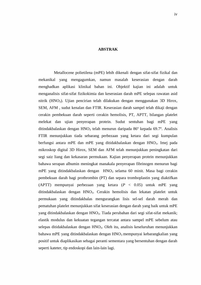
Metallocene polietilena (mPE) lebih dikenali dengan sifat-sifat fizikal dan
mekanikal yang mengagumkan, namun masalah keserasian dengan darah
menghadkan aplikasi klinikal bahan ini. Objektif kajian ini adalah untuk
menganalisis sifat-sifat fizikokimia dan keserasian darah mPE selepas rawatan asid
nitrik (HNO3). Ujian pencirian telah dilakukan dengan menggunakan 3D Hirox,
SEM, AFM , sudut kenalan dan FTIR. Keserasian darah sampel telah dikaji dengan
cerakin pembekuan darah seperti cerakin hemolisis, PT, APTT, bilangan platelet
melekat dan ujian penyerapan protein. Sudut sentuhan bagi mPE yang
ditindakbalaskan dengan HNO3 telah menurun daripada 86º kepada 69.7º. Analisis
FTIR menunjukkan tiada sebarang perbezaan yang ketara dari segi kumpulan
berfungsi antara mPE dan mPE yang ditidakbalaskan dengan HNO3. Imej pada
mikroskop digital 3D Hirox, SEM dan AFM telah menunjukkan peningkatan dari
segi saiz liang dan kekasaran permukaan. Kajian penyerapan protein menunjukkan
bahawa serapan albumin meningkat manakala penyerapan fibrinogen menurun bagi
mPE yang ditindakbalaskan dengan HNO3 selama 60 minit. Masa bagi cerakin
pembekuan darah bagi prothrombin (PT) dan separa tromboplastin yang diaktifkan
(APTT) mempunyai perbezaan yang ketara (P < 0.05) untuk mPE yang
ditindakbalaskan dengan HNO3. Cerakin hemolisis dan lekatan platelet untuk
permukaan yang ditindakbalas mengurangkan lisis sel-sel darah merah dan
pematuhan platelet menunjukkan sifat keserasian dengan darah yang baik untuk mPE
yang ditindakbalaskan dengan HNO3. Tiada perubahan dari segi sifat-sifat mekanik;
elastik modulus dan kekuatan tegangan tercatat antara sampel mPE sebelum atau
selepas ditidakbalaskan dengan HNO3. Oleh itu, analisis keseluruhan menunjukkan
bahawa mPE yang ditindakbalaskan dengan HNO3 mempunyai kebarangkalian yang
positif untuk diaplikasikan sebagai peranti sementara yang bersentuhan dengan darah
seperti kateter, tip endoskopi dan lain-lain lagi.

v
TABLE OF CONTENTS
CHAPTER TITLE PAGE
DECLARATION ii
DEDICATION iv
ACKNOWLEDGEMENTS v
ABSTRACT vi
ABSTRAK vii
TABLE OF CONTENTS viii
LIST OF TABLES xi
LIST OF FIGURES xii
LIST OF ABBREVIATIONS xiv
LIST OF SYMBOLS xvi
LIST OF APPENDICES
xvii
1 INTRODUCTION
1.1 Background
1.2 Statement of Problem
1.3 Objectives
1.4 Scope of Study
1.5 Significance of Study
1.6 Thesis Outline
1
5
5
6
6
7
2 LITERATURE REVIEW
2.1 Biomaterials
2.1.1 Blood Contact Activation
2.1.2 Platelet Activation
2.1.3 Protein Adsorption on Biomaterials
2.2 Surface Treatment of Biomaterials
8
15
16
18
20

vi
2.2.1 Types of Surface Modification
Techniques
2.2.2 Chemical Surface Modification
Technique
2.3 Nitric Acid Treatment
24
26
28
2.4 Summary
31
3 METHODOLOGY
3.1 Chemicals and Instruments
3.2 Blood Procurement and Ethical Approval
3.3 Sample Preparation and Optimization of
HNO3 Treatment Duration
3.4 Physical Characterization
3.4.1 Contact Angle Measurement
3.4.2 Attenuated Total Reflectance Fourier
Transform Infrared Spectroscopy
(ATR-FTIR)
3.4.3 3D-Hirox Digital Microscope
3.4.4 Scanning Electron Microscope (SEM)
3.4.5 Atomic Force Microscopy (AFM)
3.4.6 Tensile Testing
3.5 Blood Coagulation Assays
3.5.1 Protein Adsorption Test
3.5.2 Prothrombin Time (PT)
3.5.3 Activated Partial Thromboplastin Time
(APTT)
3.5.4 Hemolysis Assay
3.5.5 Platelet Adhesion Assay
3.6 Statistical Analyses
33
34
34
37
37
37
38
40
40
42
42
42
43
43
44
45
45
4 RESULTS AND DISCUSSION
4.1 Optimization of HNO3 Treatment Duration
4.2 .Characterization of the Samples
4.2.1 Contact Angle Measurement
46
48
48

vii
4.2.2 Attenuated Total Reflectance Fourier
Transfer Infrared Spectroscopy (ATR-
FTIR)
4.2.3 3D-Hirox Digital Microscopy
4.2.4 Scanning Electron Microscopy
4.2.5 Atomic Force Microscopy
4.2.6 Tensile Testing
4.3 Blood Coagulation Assays
4.3.1 Protein Adsorption Test
4.3.2 Prothrombin Time (PT)
4.3.3 Activated Partial Thromboplastin Time
(APTT)
4.3.4 Hemolysis Assay
4.3.5 Platelet Adhesion Assay
4.3.6 Mechanism of Improved Blood
Compatibility
49
50
55
56
58
60
61
64
65
67
69
71
5 CONCLUSION AND RECOMMENDATIONS
5.1 Conclusion
5.2 Recommendations
73
74
REFERENCES 76
APPENDICES 93

viii
LIST OF TABLES
TABLE NO. TITLE PAGE
4.1 Percentage weight loss of the HNO3 treated samples
for optimization of acid treatment duration
46
4.2 Contact angle of the HNO3 treated samples for
optimization of acid treatment duration
47
4.3 Tensile testing result of mPE before and after 30
minutes and 60 minutes HCl and HNO3 treatment
60

ix
LIST OF FIGURES
FIGURE NO. TITLE PAGE
2.1 Biomaterials used for different heart valve
components
10
2.2 Circumferential direction 11
2.3 Structure of metallocene compound 14
2.4 Structure of polyethylene 14
2.5 Activation of the blood coagulation system initiated
by biomaterial-protein interaction
17
2.6 Activation of platelets by artificial surfaces 17
2.7 Physico-chemical modification strategies 25
3.1 Schematic representation of series of
characterization and blood compatibility
experiments done
36
3.2 3D Hirox Microscope 39
3.3 Scanning Electron Microscopy 40
3.4 Atomic Force Microscopy 41
4.1 A representative FTIR spectra of untreated, 30
minutes and 60 minutes HNO3 treated metallocene
polyethylene
50
4.2 Different three-dimensional representations using
3D Hirox digital microscope
52

x
4.3 Different three-dimensional representations using
3D Hirox digital microscopy with profiling line
52
4.4 The representative height of the pores of different
samples measured using 3D-profiling of 3D Hirox
digital microscopy
53
4.5 The average pore density of untreated, 30 minutes
and 60 minutes HNO3 acid treated mPE.
54
4.6 Representative SEM micrographs of untreated, 30
minutes and 60 minutes HNO3 treated metallocene
polyethylene.
56
4.7 Representative AFM images of untreated
metallocene polyethylene, 30 minutes and 60
minutes HNO3 treated metallocene polyethylene
58
4.8 Comparison of normalized albumin adsorption of
control and HNO3 treated metallocene polyethylene
63
4.9 Comparison of normalized fibrinogen adsorption of
control and HNO3 treated metallocene polyethylene
63
4.10 Comparison of prothrombin time (PT) of control and
HNO3-treated metallocene polyethylene
65
4.11 Comparison Activated Partial Thromboplastin Time
graph of control and HNO3 treated metallocene
polyethylene
66
4.12 Comparison of hemolysis of control and HNO3
treated metallocene polyethylene
68
4.13 Comparison of platelet adhered on untreated and
HNO3 treated metallocene polyethylene
70
4.14 Photomicrograph of platelets adhered on
untreated,30 minutes and 60 minutes HNO3 treated
mPE at 40x magnification
70
4.15 Mechanism of improved hemocompatibility by
HNO3 treatment
72

xi
LIST OF ABBREVIATIONS
AFM - Atomic force microscopy
APTT - Activated partial thromboplastin time
ATR-FTIR - Attenuated total reflectance Fourier transformed infrared
spectroscopy
BHV - Bioprosthetic heart valves
BMS - Bare-metal stents
DES - Drug-eluting stents
Fg - Fibrinogen
HA - Haemolysis assay
HCL - Hydrochloric acid
HI - Hemolytic index
HNO3 - Nitric acid
IH - Intimal hyperplasia
ISR - In-stent restenosis
MHV - Mechanical heart valves
mPE - Metallocene polyethylene
NaOH - Sodium hydroxide
PANCMA - Poly(acrylonitrile-co-maleic acid)s
PCL - Poly-3-caprolactone

xii
PES - Polyethersulfone
PRP - Platelet rich plasma
PT - Prothrombin time
PTFE - Polytetrafluoroethylene
PU - Polyurethane
PVA - Poly vinyl alcohol
PVC - Poly vinyl chloride
RBC - Red blood cells
SD - Standard deviation
SEM - Scanning electron microscopy
ST - Stent thrombosis
SWCNT - Single-walled carbon nanotubes
THF - Tetrahydrofuran
VAD - Ventricular assist devices
vWF - Von willebrand factor
BCA - Bicinchoninic acid assay

xiii
LIST OF SYMBOLS
cm-1
-
Per centimetre
mm-1
-
Per millimetre
cm2
-
Square centimetre
g/mol - Grams per mole
Hz - Hertz
MPa - Megapascal
mL - Millilitre
M - Molar
nm - Nanometre
mm - Millimeter
Ra - Average roughness
W - Watt
N - Newton
w/v - Weight per volume
L - Microlitre
m - Micrometre
µ2
-
Micrometre square
oC - Degree celsius
% - Percentage
± - Plus-minus sign

xiv
LIST OF APPENDICES
APPENDIX TITLE PAGE
A Biomechanical properties of native and tissue
engineered heart valve constructs
93
B Properties of conventional blood contacting
materials
95
C Important human blood serum proteins and their key
biological functions
102
D Surface water contact angle and the cell types
supported
104
E Intensity of the FTIR peaks 106
F Publications 107

CHAPTER 1
INTRODUCTION
1.1 Background
Materials which have been playing a vital role in replacing and mirroring the
functions of various organs in human system [1] are collectively known as
biomaterials. Biomaterial is the combination of substances originating from natural,
inorganic or organic materials. These materials are biocompatible exactly or partially
when it comes in contact with the body during the healing time. They involve
complete or part of a living organism or biomedical device which perform, augments
or replaces any natural functions. Biomaterials are commonly used in various
medical devices and systems like synthetic skin [2], drug delivery systems [3], tissue
cultures [4], hybrid organs [5], synthetic blood vessels [6], artificial heart valves,
cardiac pacemakers [7], screws, plates, wires and pins for bone treatments, total
artificial joint implants, skull reconstruction [8], dental and maxillofacial applications
[9].
Biomaterials broadly fall into the four main types namely metals, ceramics,
polymers and biological substances [10]. Metals have unique atomic structure which
confers them characteristic strength and properties which enable them specifically
for load-bearing applications like orthopaedics. However, the corrosion associated
with the use of metals limits their utility. Ceramics have evolved as better
biomaterials because of their bio-inertness and compatibility. However, due to
brittleness and low impact strength [11], ceramics are losing popularity. Polymers

2
have widespread applications in the field of biomaterials. Properties of polymers are
dependent on the unit macromolecule present in the long chain of the polymer.
Among all four types, the polymers have widespread application in the field
of biomaterials because of excellent physico-chemical and mechanical properties.
Moreover, polymers can be feasibly molded into desired shapes with desired
mechanical characteristics. The most important application of polymers is
cardiovascular based implants [7] and blood contacting devices [1]. The use of
polymers in medical application ranges from vascular grafts, stents, prosthetic heart
valves, catheters, heart assist devices, hemodialyser.
Modern revolution in polymer technology like metallocene single-site
catalyst introduced a new class of polyolefins with improved performance properties
like enhanced toughness, sealability, clarity, and elasticity [12]. The metallocene
consists of two cyclopentadienyl anions (Cp,) which are bound to a metal center (M)
[12] which has an oxidation state II, thereby resulting in a general formula M(C5H5)2.
One among the polymers developed through metallocene technology is metallocene
polyethylene (mPE). mPE typically finds applications in disposable bags, storage
bottles, blood bags, and syringe tubes. Even though mPE has outstanding
permeability to oxygen and acts as a barrier towards ammonia and water, mPE lacks
blood compatibility [13] to be used for blood contacting biomedical implants.
Biocompatibility is a prime factor which determines the quality of a
biomaterial and its application in various arenas. There are different definitions for
biocompatibility. It may be defined as the ability of the material to perform at a
specific body site with an appropriate host reaction. Biocompatibility may also be
defined as the ability of a biomaterial to perform its desired function with respect to a
medical therapy, without eliciting any undesirable local or systemic effects in the
recipient or beneficiary of that therapy, but generating the most appropriate
beneficial cellular or tissue response to that specific situation and optimizing the
clinically relevant performance of that therapy [14].

3
Biocompatibility has been mentioned in many works with increasing interest
in evaluating the characteristics of medical materials and devices and also the
responses caused by its components. The ideal pattern for determining these
properties has not yet been determined; but, various methods have been suggested for
this purpose. Biocompatibility encompasses many aspects of the material, including
its physical, mechanical and chemical properties, and potential cytotoxic, mutagenic
and allergenic effects [15], so that no significant injuries or toxic effects on the
biological function of cells and individuals arise. Until the biocompatibility of a
material is proven, it must be subjected to various studies ranging from in vitro
assays to clinical trials, involving distinct areas such as pharmaceutics, biology,
chemistry and toxicology to justify its use as a biomaterial.
The term biocompatibility has been defined by consensus, but not blood
compatibility. The interactions between blood and a surface depend on the blood
composition, the blood flow and the surface of the material defined by its
physicochemical feature. The design of bloodcompatible materials is clearly a
challenge to increase success in all medical devices that come in contact with blood
and to answer unsolved problems in vascular reconstruction.
To explain blood compatibility from a different perspective, consider a
material that is not blood compatible, i.e. a thrombogenic material. Such material
would produce specific adverse reactions when placed in contact with blood:
formation of clot or thrombus composed of various blood elements; shedding or
nucleation of emboli (detached thrombus); the destruction of circulating blood
components and activation of the complement system and other immunologic
pathways [10]. Thus, we can define blood compatibility as the ability of the material
to work in a particular place without eliciting any of the above mentioned blood
related complications. Indeed, biocompatibility of blood contacting devices relates
mainly to the thrombotic response induced by the materials.
Several distinct but interrelated thrombotic and antithrombotic systems exist
to prevent the formation of intravascular clots expected in response to vascular

4
trauma. Haemostasis is the sum of these mechanisms and serves to limit blood loss
following injury. Once regulation is initiated, these same mechanisms combine first
to localize the clot at the site of injury, then to terminate coagulation and finally to
remove the clot once it has served its purpose. These haemostatic mechanisms
include platelet activation, coagulation, fibrinolysis and local vascular effects. Blood
clotting, platelet adhesion and giant cell formations are major problems associated
with blood clotting devices. These problems frequently arise in cardiovascular
implants since the material is always in contact with blood and its components [7].
The hypothesis of this thesis was that the HNO3 treatment on mPE may
improve the blood compatibility of the mPE polymer, to be utilized for different
temporary blood contacting devices application like endscope tip, catheters and etc.
The rationale for this hypothesis was that, when the mPE surface is subjected to
HNO3 treatment it was expected to etch the mPE surface which may improve the
wetabbility or hydrophilicity of mPE. It may be expected that improved
hydrophilicity may alter the protein adsorption and blood coagulation time resulting
in enhanced blood compatibility. Besides that, it is also hypothesized that the HNO3
treatment on mPE will not affect the mechanical strength of mPE.
1.2 Statement of Problem
Although mPE has excellent physico-chemical and mechanical properties, it
fails as a promising biomaterial because of its poor blood compatibility.
Biocompatibility is a vital factor which determines the quality of a biomaterial and
its application in various arenas. It may be defined as the ability of the material to
perform at a specific region with the appropriate host reaction. The events occur
when the blood comes in contact with the implant is collectively called as blood
mediated reactions or blood compatibility. Whenever the blood comes in contact
with the implants (biomaterial) it will lead to following complications:

5
1. Blood components interaction with surfaces resulting in protein and water
adsorption.
2. Blood cells interfere with the surface of biomaterial.
3. These actions lead to the hemostasis and coagulation.
To solve these issues, different surface modification techniques have been
studied yet most of them are complex and limited to certain family of polymers. In
recent times, millions of dollars was invested in advanced biomaterial research which
includes discovery of new alternatives. However, in order to cater the high demand,
more research needs to be encouraged to enhance the properties of the existing
medical materials using feasible, eco-friendly and affordable modification technique.
Hence, nitric acid surface modification technique on mPE was performed for
improving its blood compatibility and it is the research gaph which will be addressed
in this research.
1.3 Objectives
This research was carried out to determine the potential of nitric acid (HNO3)
treated metallocene polyethylene (mPE) as a biomaterial, for blood contacting device
application. The following are the objectives of this study:
1. To investigate the physico-chemical changes induced on the surface of the
mPE after HNO3 treatment.
2. To determine the changes in the blood compatibility of the mPE subjected to
acid treatment.

6
1.4 Scope of Study
This study consists of two parts. The first part of the study is focused mainly
on the sample preparation, optimization of the nitric acid treatment on the mPE, and
the characterization of mPE and HNO3 treated mPE surfaces. Various methods were
utilized; contact angle, attenuated total reflectance Fourier Transform Infrared
Spectroscopy (ATR-FTIR), 3D Hirox digital microscopy, scanning electron
microscopy (SEM), and atomic force microscopy (AFM) for the determination of the
surface characteristic of the mPE and HNO3 treated sample. To examine whether the
HNO3 treatment affect the tensile strength, elastic modulus and break strength of
mPE, mechanical testing of mPE and HNO3 treated mPE was compared.
In the second part, blood coagulation assays were carried out. The reason for
this step is to ascertain the thromboresistance property of mPE and HNO3 treated
mPE surfaces when they are utilized as biomaterials, particularly for blood
contacting devices. The blood coagulation assays like prothrombin test (PT),
activated partial thromboplastin time (APTT), hemolysis assay (HA), and platelet
adherence test is performed to investigate the blood compatibility of the mPE and
HNO3 treated mPE. Protein adsorption assay was carried out for determining the
specific proteins albumin and fibrinogen adsorption on mPE and HNO3 treated mPE.
The mPE and the HNO3 treated mPE blood compatibility results were compared with
the conventional blood contacting materials for positioning this research with related
studies in this field.
1.5 Significance of Study
The result of this study will provide an account on the improved blood
compatibility of mPE by nitric acid treatment. In addition to that, the effect of the
HNO3 on the mPE polymer which was performed for the first time may kindle the
enthusiasm of the other researchers to further explore the alternative acids available,
to enhance the blood compatibility of polymers. Besides that, as nitric acid is low in

7
cost, a cost effective method for the blood compatibility enhancement of polymers
can be introduced.
1.6 Thesis outline
This thesis is divided into five chapters. In Chapter 1, a brief explanation
about the biomaterials and the research background of this study is given. Further,
the objectives of this study have been presented in the context of solving the clinical
complications mentioned. Finally, the importance of the proposed method and its
influence in encouraging future researches is also projected.
In Chapter 2, a brief explanation about polymers and the problems observed
during its contact with biological environment is summarized. In addition, the
importance of surface modification techniques in solving those issues and some of
the previous researches reported in that viewpoint is also discussed.
In Chapter 3, the research methodology and characterization studies followed
in this research are given in detail. The discussions mainly cover the particulars,
procedures and the need for characterization studies.
In Chapter 4, the results obtained from characterization and blood
compatibility studies have been discussed and compared with the previous study’s
results. This section is the heart of the thesis, since it evidently reflects the
achievement and the effectiveness of proposed idea.
In Chapter 5, a short summary of the whole work and its effectiveness in
approaching blood compatibility problems are projected. Moreover, some suggestion
about future research in the proposed perspective is also presented.

REFERENCES
1. John, A., Subramanian, A., Vellayappan, M., Balaji, A., Jaganathan, S.,
Mohandas, H., Paramalinggam, T., Supriyanto, E. and Yusof, M. Review:
physico-chemical modification as a versatile strategy for the biocompatibility
enhancement of biomaterials. RSC Advances. 2015. 5(49): 39232-39244.
2. MacNeil, S. Biomaterials for tissue engineering of skin. Materials Today. 2008.
11(5): 26-35.
3. Buckles, R.G. Biomaterials for drug delivery systems. J Biomed Mater Res.
1983. 17(1): 109-28.
4. Stark, Y., Bruns, S., Stahl, F., Kasper, C., Wesemann, M., Grothe, C. and
Scheper T. A study on polysialic acid as a biomaterial for cell culture
applications. J Biomed Mater Res A. 2008. 85(1): 1-13.
5. Woerly, S., Plant, G. W. and Harvey, A.R. Neural tissue engineering: from
polymer to biohybrid organs. Biomaterials. 1996. 17(3): 301-10.
6. Ravi, S. and Chaikof, E.L. Biomaterials for vascular tissue engineering.
Regenerative medicine. 2010. 5(1): 107.
7. Jaganathan, S.K., Eko, S., Selvakumar, M., Arunpandian, B. and Manjesh, A.
Biomaterials in Cardiovascular Research: Applications and Clinical Implications.
BioMed Research International. 2014. 2014:11.
8. Stevens, M.M., Biomaterials for bone tissue engineering. Materials Today. 2008.
11(5): 18-25.
9. Galler, K.M., D'Souza, R.N. and Hartgerink, J.D.Biomaterials and their potential
applications for dental tissue engineering. Journal of Materials Chemistry. 2010.
20(40): 8730-8746.

77
10. Jaganathan, S.K., Balaji, A., Vellayappan, M.V., Subramanian, A.P., John A.A.,
Manjesh, A. and Eko, S. Review: Radiation-induced surface modification of
polymers for biomaterial application. Journal of Materials Science. 2014. 1-12.
11. Niinomi, M. Recent metallic materials for biomedical applications. Metallurgical
and Materials Transactions A. 2002. 33(3): 477-486.
12. Kealy, T.J. and Pauson P. L. A. New Type of Organo-Iron Compound. Nature.
1951. 168(4285): 1039-1040.
13. Mohandas, H., Sivakumar, G., Kasi, P., Jaganathan, S.K. and Eko, S.
Microwave-Assisted Surface Modification of Metallocene Polyethylene for
Improving Blood Compatibility. BioMed Research International. 2013. 2013: 7.
14. Williams, D.F. On the mechanisms of biocompatibility. Biomaterials. 2008.
29(20): 2941-53.
15. Lemons, J. and Natiella, J. Biomaterials, biocompatibility, and peri-implant
considerations. Dent Clin North Am. 1986. 30(1): 3-23.
16. Schieker, M., Hermann, S., Inga, D., Sebastian, S. and Wolf. M.Biomaterials as
Scaffold for Bone Tissue Engineering. European Journal of Trauma. 2006.
32(2): 114-124.
17. Hutmacher, D.W., Sittinger, M. and Risbud, M.V. Scaffold-based tissue
engineering: rationale for computer-aided design and solid free-form fabrication
systems. Trends Biotechnol. 2004. 22(7): 354-62.
18. Gomes, M.E., Godinho, J.S., Tchalamov, D., Cunha, A.M. and Reis R.L.
Alternative tissue engineering scaffolds based on starch: processing
methodologies, morphology, degradation and mechanical properties. Materials
Science and Engineering: C. 2002. 20(1–2): 19-26.
19. Anselme, K. Osteoblast adhesion on biomaterials. Biomaterials. 2000. 21(7):
667-81.
20. Vo-Dinh, T. and Cullum, B. Biosensors and biochips: advances in biological and
medical diagnostics. Fresenius J Anal Chem. 2000. 366(6-7): 540-51.
21. Rossen, L., Pernille, N., Kim, H. and Ole, F. R. Inhibition of PCR by
components of food samples, microbial diagnostic assays and DNA-extraction
solutions. International Journal of Food Microbiology. 1992. 17(1): 37-45.

78
22. Ryou, M. and Thompson, C.C. Tissue Adhesives: A Review. Techniques in
Gastrointestinal Endoscopy. 2006. 8(1): 33-37.
23. Vellayappan, M.V., Balaji, A., Subramanian, A.P., John A.A., Jaganathan, S.K.,
Murugesan, S., Supriyanto, E. and Yusof, M. Multifaceted prospects of
nanocomposites for cardiovascular grafts and stents. Int J Nanomedicine. 2015.
10: 2785-803.
24. Formanek, G., Frech, R, S. and Amplatz, K. Arterial thrombus formation during
clinical percutaneous catheterization. Circulation. 1970. 41(5): 833-9.
25. Roată, C.E. Clinical practice guidelines for vascular access. Am J Kidney Dis.
2006. 48 (1): 176-247.
26. Vellayappan, M.V., Balaji, A., Subramanian, A.P., John A.A., Jaganathan, S.K.,
Murugesan, S., Hemanth, M., Supriyanto, E. and Yusof, M. Tangible
nanocomposites with diverse properties for heart valve application. Science and
Technology of Advanced Materials. 2015. 16(3): 033504.
27. Mohammadi, H. and Mequanint, K. Prosthetic aortic heart valves: Modeling and
design. Medical Engineering & Physics. 2011. 33(2): 131-147.
28. Anwarul, H., Kim, R., Wojciech, S., Šeila, S., Arghya, P., Gulden, C.,
Mohammad R.K. and Ali .K .Biomechanical properties of native and tissue
engineered heart valve constructs. Journal of Biomechanics. 2014. 47(9): 1949-
1963.
29. Balguid, A., Rubbens M.P., Mol, A., Bank R.A., Bogers A.J., Kats J.P., Mol
B.A., Baaijens, F.P. and Bouten, C.V. The role of collagen cross-links in
biomechanical behavior of human aortic heart valve leaflets--relevance for tissue
engineering. Tissue Eng. 2007. 13(7): 1501-11.
30. Mavrilas, D. and Missirlis, Y. An approach to the optimization of preparation of
bioprosthetic heart valves. Journal of Biomechanics. 1991. 24(5): 331-339.
31. Conte, M.S. The ideal small arterial substitute: a search for the Holy Grail?
Faseb Journal. 1998. 12(1): 43-5.
32. Kannan, R.Y., Salacinski, H.J., Butler, P.E., Hamilton, G. and Seifalian,
A.M.Current status of prosthetic bypass grafts: a review. J Biomed Mater Res B
Appl Biomater. 2005. 74(1): 570-81.

79
33. Desai, N.P. and Hubbell, J.A. Biological responses to polyethylene oxide
modified polyethylene terephthalate surfaces. J Biomed Mater Res. 1991. 25(7):
829-43.
34. Schmedlen, R.H., Elbjeirami, W.M., Gobin, A.S., West, J.L.Tissue engineered
small-diameter vascular grafts. Clin Plast Surg. 2003. 30(4): 507-17.
35. Balasubramanian, V., Grusin, N.K., Bucher, R.W., Turitto, V.T. and Slack, S.M.
Residence-time dependent changes in fibrinogen adsorbed to polymeric
biomaterials. J Biomed Mater Res. 1999. 44(3): 253-60.
36. Ballyk P.D., Walsh, C., Butany, J. and Ojha, M. Compliance mismatch may
promote graft-artery intimal hyperplasia by altering suture-line stresses. J
Biomech. 1998. 31(3): 229-37.
37. Fager, G. Thrombin and proliferation of vascular smooth muscle cells. Circ Res.
1995. 77(4): 645-50.
38. Roqué, M., Reis, E.D., Fuster, V., Padurean, A., Fallon, J.T, Taubman, M.B.,
Chesebro, J.H. and Badimon, J.J. Inhibition of tissue factor reduces thrombus
formation and intimal hyperplasia after porcine coronary angioplasty. J Am Coll
Cardiol. 2000. 36(7): 2303-10.
39. Hansson, G.K. and Libby.P. The immune response in atherosclerosis: a double-
edged sword. Nat Rev Immunol. 2006. 6(7): 508-19.
40. Stone, G.W., Ellis, S.G., Cannon, L., Mann, J.T., Greenberg, J.D., Spriggs, D.,
DeMaio, S., Hall, P., Popma, J.J., Koglin, J. and Russell, M.E. Comparison of a
polymer-based paclitaxel-eluting stent with a bare metal stent in patients with
complex coronary artery disease: a randomized controlled trial. Jama. 2005.
294(10): 1215-23.
41. Daemen, J., Wenaweser, P., Tsuchida, K., Abrecht, L., Vaina, S., Morger, C.,
Kukreja, N., Jüni, P., Sianos, G., Hellige, G., vanDomburg, R.T., Hess, O.M.,
Boersma, E., Meier, B., Windecker, S. and Serruys, P.W. Early and late coronary
stent thrombosis of sirolimus-eluting and paclitaxel-eluting stents in routine
clinical practice: data from a large two-institutional cohort study. Lancet. 2007.
369(9562): 667-78.
80
42. Niccoli, G., Montone, R.A., Ferrante, G. and Crea, F. The evolving role of
inflammatory biomarkers in risk assessment after stent implantation. J Am Coll
Cardiol. 2010. 56(22): 1783-93.
43. Byrne, R.A., Joner, M., Tada, T. and Kastrati, A. Restenosis in bare metal and
drug-eluting stents: distinct mechanistic insights from histopathology and optical
intravascular imaging. Minerva Cardioangiol. 2012. 60(5): 473-89.
44. Bonaventura, K., Leber, A.W., Sohns, C., Roser, M., Boldt, L.H., Kleber, F.X.,
Haverkamp, W. and Dorenkamp, M. Cost-effectiveness of paclitaxel-coated
balloon angioplasty and paclitaxel-eluting stent implantation for treatment of
coronary in-stent restenosis in patients with stable coronary artery disease. Clin
Res Cardiol. 2012. 101(7): 573-84.
45. Liistro, F., Angioli, P., Porto, I., Ricci, L., Ducci, K., Grotti, S., Falsini, G.,
Ventoruzzo, G., Turini, F., Bellandi, G. and Bolognese, L. Paclitaxel-eluting
balloon vs. standard angioplasty to reduce recurrent restenosis in diabetic
patients with in-stent restenosis of the superficial femoral and proximal popliteal
arteries: the debate-isr study. J Endovasc Ther. 2014. 21(1): 1-8.
46. Thierry, B., Winnik, F.M., Merhi, Y., Silver, J. and Tabrizian, M. Bioactive
coatings of endovascular stents based on polyelectrolyte multilayers.
Biomacromolecules. 2003. 4(6): 1564-71.
47. Pokasermsong, P. and Praserthdam, P. Comparison of Activity of Ziegler-Natta
Catalysts Prepared by Recrystallization and Chemical Reaction Methods towards
Polymerization of Ethylene. 2009. 13(1): 1-8.
48. Global Polyolefin Industry Development [http://blogs.eci99.com/global-
polyolefin-industry-development/].Retrieved date: 4, April, 2016.
49. Ahmad Shamiri, H.C., Shah, J., Mohd, H., Walter, K., Purushothaman, V. and
Wageeh, A. The Influence of Ziegler-Natta and Metallocene Catalysts on
Polyolefin Structure, Properties, and Processing Ability. Materials. 2014.
2014(7): 5069-108.
50. Harrington, B.A.W. and Glenn, M. Comparison of Ziegler-Natta and
Metallocene ethylene elstomer-products. EBSCO Host. 2004. 230(2): 20.

81
51. Kaewarsa, P. Polymerization of ethylene over the supported Ziegler-Natta and
metallocene catalysts on magnesium hydroxide and magnesium hydroxychloride:
PhD Thesis. Khonkaen University; 2005.
52. Nitta, K.H. and Tanaka, A. Dynamic mechanical properties of metallocene
catalyzed linear polyethylenes. Polymer. 2001. 42(3): 1219-26.
53. Hay, M.R. Thermal and dynamic mechanical properties of metallocene
polyethylene polymer. Polymer. 2001. 42(21): 8621-7.
54. Bubeck, R.A. Structure–property relationships in metallocene polyethylenes.
Materials Science and Engineering: R: Reports. 2002. 39(1): 1-28.
55. Sunny, K.E. Studies on Metallocene Polyolefin and Polyvinyl Chloride for Blood
and Blood Component Storage Applications. PhD Thesis. Cochin University of
Science and Technology; 2006.
56. Vogler, E.A. and Siedlecki, C.A.Contact activation of blood-plasma coagulation.
Biomaterials. 2009. 30(10): 1857-69.
57. Gorbet, M.B. and M.V. Sefton, Biomaterial-associated thrombosis: roles of
coagulation factors, complement, platelets and leukocytes. Biomaterials. 2004.
25(26): 5681-703.
58. Furie, B. and Furie, B.C. Mechanisms of Thrombus Formation. New England
Journal of Medicine. 2008. 359(9): 938-949.
59. Godo, M.N. and Sefton, M.V. Characterization of transient platelet contacts on a
polyvinyl alcohol hydrogel by video microscopy. Biomaterials. 1999. 20(12):
1117-26.
60. Sheppard, J.I., McClung, W.G. and Feuerstein, I.A. Adherent platelet
morphology on adsorbed fibrinogen: effects of protein incubation time and
albumin addition. J Biomed Mater Res. 1994. 28(10): 1175-86.
61. Wachtfogel, Y.T., Hack, C.E., Nuijens, J.H., Kettner, C., Reilly, T.M., Knabb,
R.M., Bischoff, R., Tschesche, H., Wenzel, H. and Kucich, U. Selective
kallikrein inhibitors alter human neutrophil elastase release during extracorporeal
circulation. Am J Physiol. 1995. 268(3 2): 1352-57.
62. Hakim, R.M. and Schafer, A.I. Hemodialysis-associated platelet activation and
thrombocytopenia. Am J Med. 1985. 78(4): 575-80.

82
63. Dee, K.C., Puleo, D.A. and Bizios, R. Protein-Surface Interactions. An
Introduction To Tissue-Biomaterial Interactions. John Wiley & Sons. 2003.
64. Anderson, J.M., Rodriguez, A. and Chang D.T. Foreign body reaction to
biomaterials. Seminars in immunology. 2008. 20(2): 86-100.
65. Kim, M.S., Khang, G. and Lee, H.B. Gradient polymer surfaces for biomedical
applications. Progress in Polymer Science. 2008. 33(1): 138-64.
66. Sun, S., Yue, Y., Huang, X. and Meng, D. Protein adsorption on blood-contact
membranes. Journal of Membrane Science. 2003. 222(1–2): 3-18.
67. Kinnari, T.J., Salonen, E.M. and Jero, J. Durability of the binding inhibition of
albumin coating on tympanostomy tubes. International Journal of Pediatric
Otorhinolaryngology. 2003. 67(2): 157-64.
68. Jung, F., Braune, S. and Lendlein, A. Haemocompatibility testing of biomaterials
using human platelets. Clinical hemorheology and microcirculation. 2013. 53(1-
2): 97-115.
69. Kottke, K., Anderson, J.M., Umemura, Y. and Marchant R.E. Effect of albumin
coating on the in vitro blood compatibility of Dacron® arterial prostheses.
Biomaterials. 1989. 10(3): 147-55.
70. DeQueiroz, A.A., Barrak, E.R, Gil, H.A. and Higa, O.Z. Surface studies of
albumin immobilized onto PE and PVC films. Journal of biomaterials science
Polymer edition. 1997. 8(9): 667-81.
71. Baican, M., Pâslaru, E., Hitruc, E.G. and Vasile, C. Albumin immobilization on
polyvinylidene fluoride surfaces. Dig J Nanometer Bios. 2011. 6(3): 1053-64.
72. Ratner, B.D.H., Allan, S., Frederick, J. and Lemons, J. Biomaterials Science: An
Introduction to Materials in Medicine. 2 ed. San Diego / London: Elsevier
Academic Press; 2004.
73. Turci, F., Ghibaudi, E., Colonna, M., Boscolo, B., Fenoglio, I. and Fubini, B. An
Integrated Approach to the Study of the Interaction between Proteins and
Nanoparticles. Langmuir : the ACS journal of surfaces and colloids. 2010.
26(11): 8336-46.

83
74. Amarnath, L.P., Srinivas, A. and Ramamurthi, A. In vitro hemocompatibility
testing of UV-modified hyaluronan hydrogels. Biomaterials. 2006. 27(8): 1416-
24.
75. VanKooten, T.G., Schakenraad, J.M., VanderMei, H.C. and Busscher, H.J.
Influence of substratum wettability on the strength of adhesion of human
fibroblasts. Biomaterials. 1992. 13(13): 897-904.
76. VanWachem, P.B., Beugeling, T., Feijen, J., Bantjes, A., Detmers, J.P. and
Aken, W.G. Interaction of cultured human endothelial cells with polymeric
surfaces of different wettabilities. Biomaterials. 1985. 6(6): 403-8.
77. Dalby M.J. Cellular response to low adhesion nanotopographies. Int J Nanomed.
2007. 2(3): 373-81.
78. Wachem, P.B., Hogt, A.H., Beugeling, T., Feijen, J., Bantjes, A. and Detmers,
J.P. Adhesion of cultured human endothelial cells onto methacrylate polymers
with varying surface wettability and charge. Biomaterials. 1987. 8(5): 323-8.
79. Nakaoka, R., Yamakoshi, Y., Isama, K. and Tsuchiya, T. Effects of surface
chemistry prepared by self-assembled monolayers on osteoblast behavior.
Journal of Biomedical Materials Research Part A. 2010. 94(2): 524-32.
80. Xu, L.C. and Siedlecki, C.A. Effects of surface wettability and contact time on
protein adhesion to biomaterial surfaces. Biomaterials. 2007. 28(22): 3273-83.
81. Joseph, C., Dongwoo, K. and Thomas, J.W. Nanometer polymer surface
features: the influence on surface energy, protein adsorption and endothelial cell
adhesion. Nanotechnology. 2008. 19(50): 505103.
82. Deligianni, D.D., Katsala, N., Ladas, S., Sotiropoulou, D., Amedee, J. and
Missirlis Y.F. Effect of surface roughness of the titanium alloy Ti-6Al-4V on
human bone marrow cell response and on protein adsorption. Biomaterials. 2001.
22(11): 1241-51.
83. Ma, M., and Hill, R.M. Superhydrophobic surfaces. Current Opinion in Colloid
& Interface Science. 2006. 11(4): 193-202.
84. Sigal, G.B., Mrksich, M. and Whitesides, G.M. Effect of Surface Wettability on
the Adsorption of Proteins and Detergents. Journal of the American Chemical
Society. 1998. 120(14): 3464-73.

84
85. Jaganathan, S.K.., Mohandas, H., Sivakumar, G., Kasi, P., Sudheer, T., and
Avineri S. Enhanced Blood Compatibility of Metallocene Polyethylene
Subjected to Hydrochloric Acid Treatment for Cardiovascular Implants. BioMed
Research International. 2014. 2014: 7.
86. Arima, Y. and Iwata, H. Effect of wettability and surface functional groups on
protein adsorption and cell adhesion using well-defined mixed self-assembled
monolayers. Biomaterials. 2007. 28(20): 3074-82.
87. Chang, H.I. and Wang, Y. Cell Responses to Surface and Architecture of Tissue
Engineering Scaffolds. Intech. 2011.
88. Bahram, S.J., Julien, P., Rong, Q., Shuling, G., Eric, H., Gschweng, B., Stiles,
K., Tzy, M., Owen, N., Witte, X., Bruce, D. and Hong, W. Hydrophobic surfaces
for enhanced differentiation of embryonic stem cell-derived embryoid bodies.
Proceedings of the National Academy of Sciences of the United States of
America. 2008. 105(38): 14459-64.
89. Ranella, A., Barberoglou, M., Bakogianni, S., Fotakis, C. and Stratakis, E.
Tuning cell adhesion by controlling the roughness and wettability of 3D
micro/nano silicon structures. Acta biomaterialia. 2010. 6(7): 2711-20.
90. Ishizaki, T., Saito, N. and Takai, O. Correlation of Cell Adhesive Behaviors on
Superhydrophobic, Superhydrophilic, and Micropatterned
Superhydrophobic/Superhydrophilic Surfaces to Their Surface Chemistry.
Langmuir : the ACS journal of surfaces and colloids. 2010. 26(11): 8147-54.
91. Neto, A.I., Custodio, C.A., Song, W. and Mano, J.F. High-throughput evaluation
of interactions between biomaterials, proteins and cells using patterned
superhydrophobic substrates. Soft Matter. 2011. 7(9): 4147-51.
92. Song, W., Veiga, D.D., Custódio, C.A. and Mano, J.F. Bioinspired Degradable
Substrates with Extreme Wettability Properties. Advanced Materials. 2009.
21(18): 1830-34.
93. Zheng, J., Li, L., Tsao, H.K., Sheng, Y.J., Chen, S. and Jiang S. Strong Repulsive
Forces between Protein and Oligo (Ethylene Glycol) Self-Assembled
Monolayers: A Molecular Simulation Study. Biophysical Journal. 2005. 89(1):
158-66.
85
94. Arima, Y. and Iwata, H. Effects of surface functional groups on protein
adsorption and subsequent cell adhesion using self-assembled monolayers.
Journal of Materials Chemistry. 2007. 17(38): 4079-87.
95. Flemming, R.G., Murphy, C.J., Abrams, G.A., Goodman, S.L and Nealey, P.F.
Effects of synthetic micro- and nano-structured surfaces on cell behavior.
Biomaterials. 1999. 20(6): 573-88.
96. Curtis, A. and Wilkinson, C. Nantotechniques and approaches in biotechnology.
Trends in biotechnology. 2001. 19(3): 97-101.
97. Chou, L., Firth, J.D., Uitto, V.J. and Brunette D.M. Substratum surface
topography alters cell shape and regulates fibronectin mRNA level, mRNA
stability, secretion and assembly in human fibroblasts. Journal of cell science.
1995. 108 (4): 1563-73.
98. Rosales , J.I., Rodríguez, M.A., Mazzaglia, G., Ramón, P.J., Díaz, L., García, M.,
Vallecillo, M., Ruizc, C. and Cabrerizo, M.A. Effect of roughness, wettability
and morphology of engineered titanium surfaces on osteoblast-like cell adhesion.
Colloids and Surfaces A: Physicochemical and Engineering Aspects. 2010.
365(1–3): 222-29.
99. Curtis, A. and Wilkinson, C. Topographical control of cells. Biomaterials. 1997.
18(24): 1573-83.
100. Clark, P., Connolly, P., Curtis, A.S., Dow, J.A. and Wilkinson, C.D.
Topographical control of cell behaviour: II. Multiple grooved substrata.
Development. 1990. 108(4): 635-44.
101. Tiaw K.S., Goh S.W., Hong, M., Wang, Z., Lan, B. and Teoh, S.H. Laser surface
modification of poly(epsilon-caprolactone) (PCL) membrane for tissue
engineering applications. Biomaterials. 2005. 26(7): 763-9.
102. Lampin, M., Warocquier, C., Legris, C., Degrange, M. and Sigot, M.F.
Correlation between substratum roughness and wettability, cell adhesion, and
cell migration. Journal of biomedical materials research. 1997. 36(1): 99-108.
103. Wan, Y., Wang, Y., Liu, Z., Qu, X., Han, B., Bei, J. and Wang, S. Adhesion and
proliferation of OCT-1 osteoblast-like cells on micro- and nano-scale topography
structured poly(L-lactide). Biomaterials. 2005. 26(21): 4453-59.

86
104. Puckett, S., Pareta, R. and Webster, T.J. Nano rough micron patterned titanium
for directing osteoblast morphology and adhesion. Int J Nanomed. 2008. 3(2):
229-41.
105. Yim, E.K., Pang, S.W. and Leong, K.W. Synthetic nanostructures inducing
differentiation of human mesenchymal stem cells into neuronal lineage.
Experimental cell research. 2007. 313(9): 1820-9.
106. Dalby, M.J., Riehle, M.O., Yarwood, S.J., Wilkinson, C.D. and Curtis, A.S.
Nucleus alignment and cell signaling in fibroblasts: response to a micro-grooved
topography. Experimental cell research. 2003. 284(2): 274-82.
107. Mirzadeh, H., Dadsetan, M. and Sharifi, N. Platelet adhesion on laser-induced
acrylic acid–grafted polyethylene terephthalate. Journal of Applied Polymer
Science. 2002. 86(13): 3191-3196.
108. Mittal, K.L. Polymer International.Taylor and Francis Group, 2009.
109. Vladkova, T.G. Surface Engineered Polymeric Biomaterials with Improved
Biocontact Properties. International Journal of Polymer Science. 2010. 1-22.
110. Frederick, M.F. Contact Angle, Wettability, and Adhesion. American Chemical
Society. 1964.
111. Lim, J.Y. and Donahue, H.J. Cell sensing and response to micro- and
nanostructured surfaces produced by chemical and topographic patterning. Tissue
Eng. 2007. 13(8): 1879-91.
112. Miller, D.C., Thapa, A., Haberstroh, K.M. and Webster, T.J. Endothelial and
vascular smooth muscle cell function on poly(lactic-co-glycolic acid) with nano-
structured surface features. Biomaterials. 2004. 25(1): 53-61.
113. Balakrishnan, B., Kumar, D.S., Yoshida, Y., and Jayakrishnan, A. Chemical
modification of poly(vinyl chloride) resin using poly(ethylene glycol) to improve
blood compatibility. Biomaterials. 2005. 26(17): 3495-3502.
114. Aldenhoff, Y.B. and Koole, L.H.. Platelet adhesion studies on dipyridamole
coated polyurethane surfaces. Eur Cell Mater. 2003. 5: 61-67.
115. Xiang, T., Wen, Y., Rui, W., Su, L., Shu-Dong, S. and Chang-Sheng, Z. Surface
hydrophilic modification of polyethersulfone membranes by surface-initiated

87
ATRP with enhanced blood compatibility. Colloids Surf B Biointerfaces. 2013.
110: 15-21.
116. Mona, J., Kuo, C.J., Perevedentseva, E., Priezzhev, A.V. and Cheng, C.L.
Adsorption of human blood plasma on nanodiamond and its influence on
activated partial thromboplastin time. Diamond and Related Materials. 2013. 39:
73-7.
117. Park, H.D., Lee, W.K., Ooya, T., Park, K.D., Kim, Y.H. and Yui, N. J Biomed
Mater Res. 2003. 66(3): 596-604.
118. Xu, Z.K., Nie, F.Q., Qu, C., Wan, L.S., Wu, J. and Yao, K. Tethering
poly(ethylene glycol)s to improve the surface biocompatibility of
poly(acrylonitrile-co-maleic acid) asymmetric membranes. Biomaterials. 2005.
26(6): 589-598.
119. Akelah, A. Polymeric Materials: Preparation and Properties, in Functionalized
Polymeric Materials in Agriculture and the Food Industry. Springer. 2013.
120. Nie, W.Z. and Li, J. Effects of plasma and nitric acid treatment of carbon fibers
on the mechanical properties of thermoplastic polymer composites. Mechanics of
Composite Materials. 2010. 46(3): 251-256.
121. Cagiao, M.E., Rueda, D.R. and Baltá-Calleja, F.J. Degradation of nitric acid-
treated bulk polyethylene. Polymer Bulletin. 1980. 3(5): 305-310.
122. Cagiao, M.E., Rueda, D.R. and Baltá Calleja, F.J. Hardness of nitric acid treated
polyethylene followed by recrystallization. Colloid and Polymer Science. 1987.
265(1): 37-41.
123. Hock, C.W. Selective oxidation with nitric acid reveals the microstructure of
polypropylene. Journal of Polymer Science Part B: Polymer Letters. 1965. 3(7):
573-576.
124. Hay, I.L. and Keller, A. Morphology of Synthetic Fibres; A Study on Drawn
Polyethylene. Nature. 1964. 204(4961): 862-864.
125. Maxim, N., Tchoul, W.T.F., Giulio, L., Daniel, E. and Sivaram, A. Effect of
Mild Nitric Acid Oxidation on Dispersability, Size, and Structure of Single-
Walled Carbon Nanotubes. Chemistry of Materials. 2007. 19(23): 5765-5772.

88
126. Cagiao, M.E., Rueda, D. R. and Baltá, F. J. Degradation of Nitric Acid-Treated
Bulk Polyethylene. III. Melting Behavior. Colloid and Polymer Science. 1983.
261: 626-630.
127. Ferro-Garcia, M. A., Joly, J.P., Bautista-Toledo, I., Carrasco-Marin, F. and
Rivera-Utrilla, J. Activated Carbon Surface Modifications by Nitric Acid,
Hydrogen Peroxide, and Ammonium Peroxydisulfate Treatments. Langmuir.
1995. 11(11): 4386-4392.
128. ChenboDong, A.S.C., Reem, E., Gabriela, P., Yon, R. and Cerasela, Z. Effects of
acid treatment on structure, properties and biocompatibility of carbon nanotubes.
Applied Surface Science. 2013. 264: 261–268.
129. Hanford. W.E. Oxidized polymers and process for their preparation.
US2360673A. 1944.
130. Fu, Y.C. and Gunnell, T.J. Nitric acid treatment of carbon black. US3336148A.
1967.
131. Gomathi, N., Mishra, D., Maiti, T.K. and Neogi, S. Helium Plasma Treatment to
Improve Biocompatibility and Blood Compatibility of Polycarbonate. Journal of
Adhesion Science and Technology. 2010. 24(13-14): 2237-55.
132. Qi, P., Maitz, M.F., and Huang, N. Surface modification of cardiovascular
materials and implants. Surface and Coatings Technology. 2013. 233: 80-90.
133. Pereira, C. Nondestructive characterization and enzyme cleaning of painted
surfaces: assessment from the macro to nano level. Microsc Microanal. 2013.
19(6): 1632-44.
134. Roozbahani, F., Sultana, N., Almasi, D. and Naghizadeh, F. Effects of Chitosan
Concentration on the Protein Release Behaviour of Electrospun Poly(-
caprolactone)/Chitosan Nanofibers. Journal of Nanomaterials. 2015. 2015: 11.
135. Wagner, A., Poursorkhabi, V., Mohanty, A.K. and Misra, M. Analysis of Porous
Electrospun Fibers from Poly(l-lactic acid)/Poly(3-hydroxybutyrate-co-3-
hydroxyvalerate) Blends. ACS Sustainable Chemistry & Engineering. 2014. 2(8):
1976-82.
136. Pelagade, S.M., Rane, R.S., Mukherjee, S., Deshpande, U.P., Ganesan, V. and
Shripathi, T. Investigation of Surface Free Energy for PTFE Polymer by Bipolar

89
Argon Plasma Treatment. Journal of Surface Engineered Materials and
Advanced Technology. 2012. 2(2): 132-136.
137. Roy, R.K., Choi, H.W., Yi, J.W., Moon, M.W., Lee, K.R. and Han, D.K.
Hemocompatibility of surface-modified silicon-incorporated diamond-like
carbon films. Acta biomaterialia. 2009. 5(1): 249-56.
138. Kwok, S.C.H., Wang, J. and Chu, P.K. Surface energy, wettability, and blood
compatibility phosphorus doped diamond-like carbon films. Diamond and
Related Materials. 2005. 14(1): 78-85.
139. Gomathi, N., Rajasekar, R., Rajesh, R., Debasish, M. and Neogi. S. Development
of bio/blood compatible polypropylene through low pressure nitrogen plasma
surface modification. Materials Science and Engineering: C. 2012. 32(7): 1767-
1778.
140. Chau, T.T., Bruckard, W.J., Koh, P.T.L. and Nguyen, A.V. A review of factors
that affect contact angle and implications for flotation practice. Advances in
Colloid and Interface Science. 2009. 150(2): 106-115.
141. Hardy Paul W. Nitric acid treatment of steel. US2915420A. 1959.
142. Mirzadeh, H. and Dadsetan, M. Influence of laser surface modifying of
polyethylene terephthalate on fibroblast cell adhesion. Radiation Physics and
Chemistry. 2003. 67(3–4): 381-5.
143. Makphon, P., Ratanatongchai, W., Chongkum, S., Tantayanon, S. and Supaphol,
P. Polycarbonate microfilters by nuclear tracking and chemical etching (track-
etching) technique: Preparation and characterization. Journal of Applied Polymer
Science. 2006. 101(2): 982-90.
144. Gui-qiu, M., Ben, L., Chen, L., Dinghai, H. and Jing, S. Plasma modification of
polypropylene surfaces and its alloying with styrene in situ. Applied Surface
Science. 2012. 258(7): 2424-2432.
145. Cesca, F., Limongi, T., Accardo, A., Rocchi, A., Orlando, M., Shalabaev, V., Di
Fabrizio, E. and Benfenati, F. Fabrication of biocompatible free-standing
nanopatterned films for primary neuronal cultures. RSC Advances. 2014. 4(86):
45696-45702.

90
146. Tverdokhlebov, S.I., Bolbasov, E.N., Shesterikov, E.V., Antonova, L.V.,
Golovkin, A.S., Matveeva, V.G., Petlin, D.G.and Anissimov, Y.G. Modification
of polylactic acid surface using RF plasma discharge with sputter deposition of a
hydroxyapatite target for increased biocompatibility. Applied Surface Science.
2015. 329(0): 32-39.
147. Wanke, C.H. Tuning of polypropylene wettability by plasma and polyhedral
oligomeric silsesquioxane modifications. Polymer. 2011. 52(8): 1797-1802.
148. Slepička, P. Surface characterization of plasma treated polymers for applications
as biocompatible carriers. Express Polymer Letters. 2013. 7(6): 535-545.
149. Poletti, G., Orsini, F., Lenardi, C., and Barborini, E. A comparative study
between AFM and SEM imaging on human scalp hair. Journal of Microscopy.
2003. 211: 249–255.
150. Josep, F.B., Stefan, G. G., Notni, J.S., Jean, M. B. and Angela, D. Surface
characterization techniques for determining the root-mean-square roughness and
power spectral densities of optical components. Applied Optics. 2002. 41 (1). 1-
23.
151. Jolly, W.L. Modern Inorganic Chemistry: McGraw-Hill; 1984.
152. Bell, R.P. The Proton in Chemistry: Cornell University Press; 1973.
153. Schopka, S. Current Strategies in Cardiovascular Biomaterial Functionalization.
Materials. 2010. 3(1): 638-655.
154. Ma, W.J., Ruys, A.J., Mason, R.S., Martin, P.J., Bendavid, A. and Liu, Z. DLC
coatings: Effects of physical and chemical properties on biological response.
Biomaterials. 2007. 28(9): 1620-8.
155. Bohnert, J.L. Changes in adsorbed fibrinogen and albumin interactions with
polymers indicated by decreases in detergent elutability. J Coll Interf Sci. 1986.
111(2): 363-78.
156. Jeyachandran, Y.L., Mielczarski, E. Rai, B. and Mielczarski. J.A. Quantitative
and Qualitative Evaluation of Adsorption/Desorption of Bovine Serum Albumin
on Hydrophilic and Hydrophobic Surfaces. Langmuir : the ACS journal of
surfaces and colloids. 2009. 25(19): 11614-20.

91
157. Seeger, J.M., Ingegno, M.D., Bigatan, E., Klingman, N. Amery, D. and
Widenhouse, C. Hydrophilic surface modification of metallic endoluminal stents.
Journal of vascular surgery. 1995. 22(3): 327-36.
158. Wu, Y., Simonovsky, F.I., Ratner, B.D. and Horbett, T.A. The role of adsorbed
fibrinogen in platelet adhesion to polyurethane surfaces: a comparison of surface
hydrophobicity. protein adsorption. monoclonal antibody binding. and platelet
adhesion. Journal of biomedical materials research Part A. 2005. 74(4): 722-38.
159. Vroman, L. and Adams, A.L. Findings with the recording ellipsometer
suggesting rapid exchange of specific plasma proteins at liquid/solid interfaces.
Surface Science. 1969. 16: 438-46.
160. Hemmerlé, J., Altmann, S.M., Maaloum, M., Hörber, J.K.H., Heinrich, L. and
Voegel, J.C. Direct observation of the anchoring process during the adsorption of
fibrinogen on a solid surface by force-spectroscopy mode atomic force
microscopy. Proceedings of the National Academy of Sciences of the United
States of America. 1999. 96(12): 6705-10.
161. Lu, D.R. and Park, K. Effect of surface hydrophobicity on the conformational
changes of adsorbed fibrinogen. Journal of colloid and interface science. 1991.
144(1): 271-81.
162. Iwamoto, G.K., Winterton, L.C., Stoker, R.S., VanWagenen, R.A., Andrade, J.D.
and Mosher, D.F. Fibronectin adsorption detected by interfacial fluorescence.
Journal of colloid and interface science. 1985. 106(2): 459-64.
163. Lin, W.C., Liu, T.Y. and Yang, M.C. Hemocompatibility of polyacrylonitrile
dialysis membrane immobilized with chitosan and heparin conjugate.
Biomaterials. 2004. 25(10): 1947-57.
164. Wen, C.L., Da-Guang, Y. and Ming-Chien, Y. Blood compatibility of
thermoplastic polyurethane membrane immobilized with water-soluble
chitosan/dextran sulfate. Colloids and Surfaces B: Biointerfaces. 2005. 44: 82–
92.
165. Gomathi, N. and Sudarsan, N. Investigation on Argon–Oxygen Plasma Induced
Blood Compatibility of Polycarbonate and Polypropylene. Journal of Adhesion
Science and Technology. 2009. 23: 1811-1826.

92
166. Wang, J., Pan, C.J., Huang, N., Sun, H., Yang, P., Leng, Y.X., Chen, J.Y., Wan,
G.J. and Chu, P.K. Surface characterization and blood compatibility of
poly(ethylene terephthalate) modified by plasma surface grafting. Surface &
Coatings Technology. 2005. 196: 307– 311.
167. Zhengbao, Z., YanMa, X., YueMeng, L. and Zhifei, D. Self-assembled
hemocompatible coating on poly (vinyl chloride) surface. Applied Surface
Science. 2009. 256: 805–814.
168. Fazley, M. Hemocompatibility of surface modified silk fibroin materials: a
review. Reviews on Advanced Materials Science. 2014. 38:148-159.
169. Bailly, A.L., Lautier, A., Guiffant, G., Dufaux, J., Houdart, E. and Labarre, D.
M. Thrombosis of angiographic catheters in humans: experimental study. The
International journal of Artificial Organs. 1999. 22: 690-700.
170. Wenzhong, S., Li, Z. and Liu, Y. Surface chemical functional groups
modification of porous carbon. Recent Patents on Chemical Engineering 2008.
1(1): 27-40.
171. Habibzadeh, S., Li, L., Shum-Tim, D., and Davis, E.C. Omanovic. S.
Electrochemical polishing as a 316L stainless steel surface treatment method:
Towards the improvement of biocompatibility. Corrosion Science. 2014. 87(0):
89-100.
172. Lee, J.H. and Lee, H.B. Platelet adhesion onto wettability gradient surfaces in the
absence and presence of plasma proteins. Journal of biomedical materials
research. 1998. 41(2): 304-11.
173. Zhao, T., Li, Y., Gao, Y., Xiang, Y., Chen, H. and Zhang, T. Hemocompatibility
investigation of the NiTi alloy implanted with tantalum. Journal of materials
science Materials in medicine. 2011. 22(10): 2311-18.
174. Zingg, W., Neumann, A.W., Strong, A.B. Hum, O.S. and Absolom, D.R. Platelet
adhesion to smooth and rough hydrophobic and hydrophilic surfaces under
conditions of static exposure and laminar flow. Biomaterials. 1981. 2(3): 156-68.
175. Zingg, W., Neumann, A.W., Strong, A.B. and Hum, O.S. Absolom. D.R. Effect
of surface roughness on platelet adhesion under static and under flow conditions.
Canadian journal of surgery. 1982. 25(1): 16-9.