Embed Size (px)
Citation preview
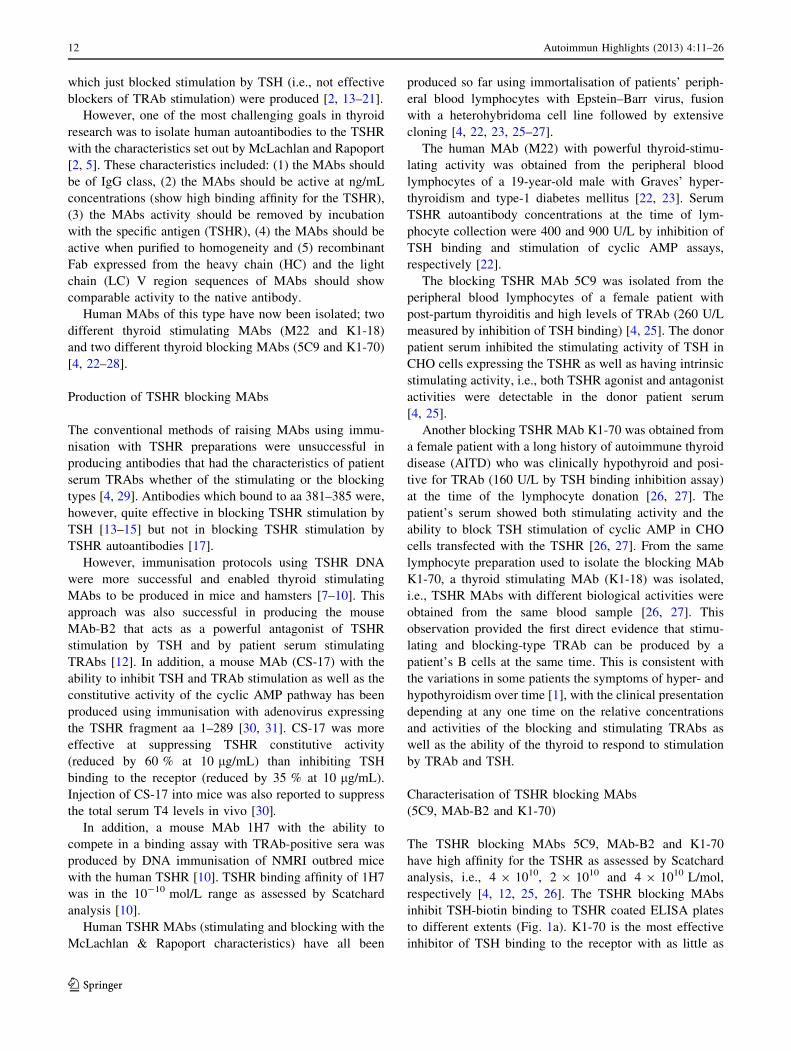
REVIEW ARTICLE
Blocking type TSH receptor antibodies
Jadwiga Furmaniak • Jane Sanders •
Bernard Rees Smith
Received: 18 January 2012 / Accepted: 29 February 2012 / Published online: 21 March 2012
� Springer-Verlag 2012
Abstract TSH receptor (TSHR) autoantibodies (TRAbs)
play a key role in the pathogenesis of Graves’ disease. In the
majority of patients, TRAbs stimulate thyroid hormone
synthesis via activation of the TSHR (stimulating TRAbs,
TSHR agonists). In some patients, TRAbs bind to the
receptor but do not cause activation (blocking TRAbs, TSHR
antagonists). Isolation of human TSHR monoclonal anti-
bodies (MAbs) with either stimulating (M22 and K1-18) or
blocking activities (5C9 and K1-70) has been a major
advance in studies on the TSHR. The binding characteristics
of the blocking MAbs, their interaction with the TSHR and
their effect on TSHR constitutive activity are summarised in
this review. In addition, the binding arrangement in the
crystal structures of the TSHR in complex with the blocking
MAb K1-70 and with the stimulating MAb M22 (2.55 A and
1.9 A resolution, respectively) are compared. The stimulat-
ing effect of M22 and the inhibiting effect of K1-70 on
thyroid hormone secretion in vivo is discussed. Furthermore
the ability of K1-70 to inhibit the thyroid stimulating activity
of M22 in vivo is shown. Human MAbs which act as TSHR
antagonists are potentially important new therapeutics. For
example, in Graves’ disease, K1-70 may well be effective in
controlling hyperthyroidism and the eye signs caused by
stimulating TRAb. In addition, hyperthyroidism caused by
autonomous TSH secretion should be treatable by K1-70,
and 5C9 has the potential to control hyperthyroidism asso-
ciated with TSHR activating mutations. Furthermore, K1-70
has potential applications in thyroid imaging as well as
targeted drug delivery to TSHR expressing tissues.
Keywords TSH receptor � Thyroid stimulating
autoantibody � Thyroid blocking autoantibody �Graves’ disease � Autoimmunity
Introduction
TSH receptor (TSHR) autoantibodies (TRAbs) play a key
role in the pathogenesis of Graves’ disease and are
detectable in almost all patients when measured using
sensitive assays [1–4]. There are two types of TRAbs; in
the majority of patients TRAbs bind to the TSHR, mimic
the biological activity of TSH and stimulate the cyclic
AMP pathway and thyroid hormone synthesis (stimulating
TRAbs, TSHR agonists). In some patients, TRAbs bind to
the receptor but do not activate the cyclic AMP pathway,
and act as TSHR antagonists (blocking TRAbs) [1–4]. The
main feature of TRAbs with thyroid stimulating or thyroid
blocking activity is their high affinity binding to the TSHR
and their ability to inhibit TSH binding to the receptor
[1–4]. To understand the key role of TRAbs in the patho-
genesis of Graves’ disease, different laboratories tried to
isolate human TSHR monoclonal autoantibodies (MAbs)
or to produce animal MAbs that would have at least some
of the characteristics of human TRAbs [2, 5, 6]. MAbs with
strong thyroid stimulating and TSH binding inhibiting
activities were eventually produced in mice and hamsters
[7–11]. At least, some of these MAbs showed high binding
affinity for the TSHR and competed effectively in a dose-
dependent manner with TSH and each other for binding to
the TSHR. In addition, patient TRAbs with stimulating or
blocking activities inhibited MAb binding to the TSHR [7].
Mouse TSHR MAbs with the ability to block the stimu-
lating activity of TRAbs and TSH have also been produced
and characterised [10, 12]. Earlier, mouse TSHR MAbs
J. Furmaniak � J. Sanders � B. Rees Smith (&)
FIRS Laboratories, RSR Ltd, Parc Ty Glas, Llanishen,
Cardiff CF14 5DU, UK
e-mail: [email protected]
123
Autoimmun Highlights (2013) 4:11–26
DOI 10.1007/s13317-012-0028-1
which just blocked stimulation by TSH (i.e., not effective
blockers of TRAb stimulation) were produced [2, 13–21].
However, one of the most challenging goals in thyroid
research was to isolate human autoantibodies to the TSHR
with the characteristics set out by McLachlan and Rapoport
[2, 5]. These characteristics included: (1) the MAbs should
be of IgG class, (2) the MAbs should be active at ng/mL
concentrations (show high binding affinity for the TSHR),
(3) the MAbs activity should be removed by incubation
with the specific antigen (TSHR), (4) the MAbs should be
active when purified to homogeneity and (5) recombinant
Fab expressed from the heavy chain (HC) and the light
chain (LC) V region sequences of MAbs should show
comparable activity to the native antibody.
Human MAbs of this type have now been isolated; two
different thyroid stimulating MAbs (M22 and K1-18)
and two different thyroid blocking MAbs (5C9 and K1-70)
[4, 22–28].
Production of TSHR blocking MAbs
The conventional methods of raising MAbs using immu-
nisation with TSHR preparations were unsuccessful in
producing antibodies that had the characteristics of patient
serum TRAbs whether of the stimulating or the blocking
types [4, 29]. Antibodies which bound to aa 381–385 were,
however, quite effective in blocking TSHR stimulation by
TSH [13–15] but not in blocking TSHR stimulation by
TSHR autoantibodies [17].
However, immunisation protocols using TSHR DNA
were more successful and enabled thyroid stimulating
MAbs to be produced in mice and hamsters [7–10]. This
approach was also successful in producing the mouse
MAb-B2 that acts as a powerful antagonist of TSHR
stimulation by TSH and by patient serum stimulating
TRAbs [12]. In addition, a mouse MAb (CS-17) with the
ability to inhibit TSH and TRAb stimulation as well as the
constitutive activity of the cyclic AMP pathway has been
produced using immunisation with adenovirus expressing
the TSHR fragment aa 1–289 [30, 31]. CS-17 was more
effective at suppressing TSHR constitutive activity
(reduced by 60 % at 10 lg/mL) than inhibiting TSH
binding to the receptor (reduced by 35 % at 10 lg/mL).
Injection of CS-17 into mice was also reported to suppress
the total serum T4 levels in vivo [30].
In addition, a mouse MAb 1H7 with the ability to
compete in a binding assay with TRAb-positive sera was
produced by DNA immunisation of NMRI outbred mice
with the human TSHR [10]. TSHR binding affinity of 1H7
was in the 10-10 mol/L range as assessed by Scatchard
analysis [10].
Human TSHR MAbs (stimulating and blocking with the
McLachlan & Rapoport characteristics) have all been
produced so far using immortalisation of patients’ periph-
eral blood lymphocytes with Epstein–Barr virus, fusion
with a heterohybridoma cell line followed by extensive
cloning [4, 22, 23, 25–27].
The human MAb (M22) with powerful thyroid-stimu-
lating activity was obtained from the peripheral blood
lymphocytes of a 19-year-old male with Graves’ hyper-
thyroidism and type-1 diabetes mellitus [22, 23]. Serum
TSHR autoantibody concentrations at the time of lym-
phocyte collection were 400 and 900 U/L by inhibition of
TSH binding and stimulation of cyclic AMP assays,
respectively [22].
The blocking TSHR MAb 5C9 was isolated from the
peripheral blood lymphocytes of a female patient with
post-partum thyroiditis and high levels of TRAb (260 U/L
measured by inhibition of TSH binding) [4, 25]. The donor
patient serum inhibited the stimulating activity of TSH in
CHO cells expressing the TSHR as well as having intrinsic
stimulating activity, i.e., both TSHR agonist and antagonist
activities were detectable in the donor patient serum
[4, 25].
Another blocking TSHR MAb K1-70 was obtained from
a female patient with a long history of autoimmune thyroid
disease (AITD) who was clinically hypothyroid and posi-
tive for TRAb (160 U/L by TSH binding inhibition assay)
at the time of the lymphocyte donation [26, 27]. The
patient’s serum showed both stimulating activity and the
ability to block TSH stimulation of cyclic AMP in CHO
cells transfected with the TSHR [26, 27]. From the same
lymphocyte preparation used to isolate the blocking MAb
K1-70, a thyroid stimulating MAb (K1-18) was isolated,
i.e., TSHR MAbs with different biological activities were
obtained from the same blood sample [26, 27]. This
observation provided the first direct evidence that stimu-
lating and blocking-type TRAb can be produced by a
patient’s B cells at the same time. This is consistent with
the variations in some patients the symptoms of hyper- and
hypothyroidism over time [1], with the clinical presentation
depending at any one time on the relative concentrations
and activities of the blocking and stimulating TRAbs as
well as the ability of the thyroid to respond to stimulation
by TRAb and TSH.
Characterisation of TSHR blocking MAbs
(5C9, MAb-B2 and K1-70)
The TSHR blocking MAbs 5C9, MAb-B2 and K1-70
have high affinity for the TSHR as assessed by Scatchard
analysis, i.e., 4 9 1010, 2 9 1010 and 4 9 1010 L/mol,
respectively [4, 12, 25, 26]. The TSHR blocking MAbs
inhibit TSH-biotin binding to TSHR coated ELISA plates
to different extents (Fig. 1a). K1-70 is the most effective
inhibitor of TSH binding to the receptor with as little as
12 Autoimmun Highlights (2013) 4:11–26
123
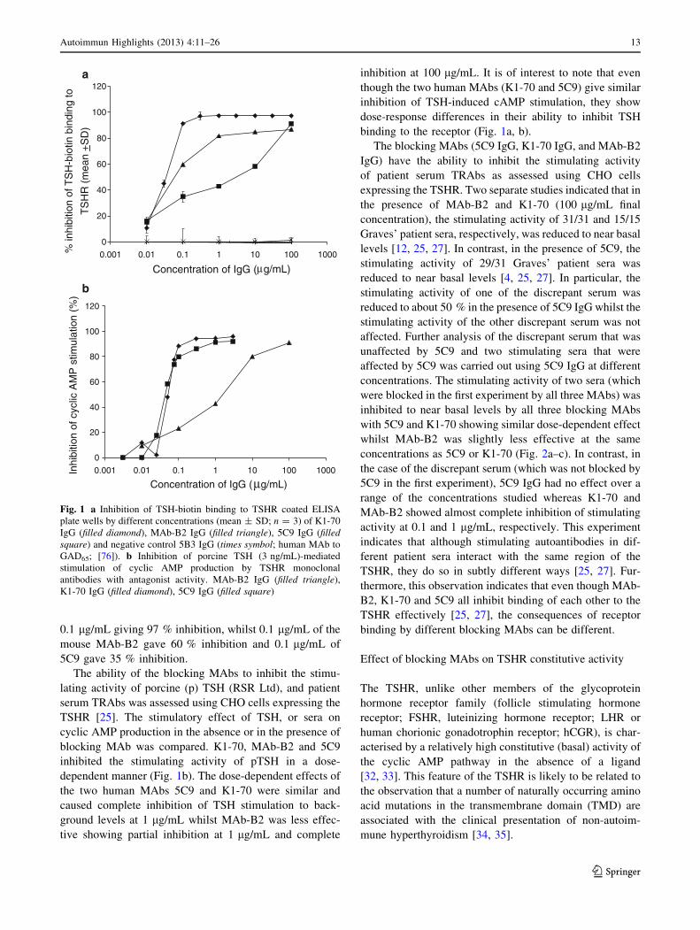
0.1 lg/mL giving 97 % inhibition, whilst 0.1 lg/mL of the
mouse MAb-B2 gave 60 % inhibition and 0.1 lg/mL of
5C9 gave 35 % inhibition.
The ability of the blocking MAbs to inhibit the stimu-
lating activity of porcine (p) TSH (RSR Ltd), and patient
serum TRAbs was assessed using CHO cells expressing the
TSHR [25]. The stimulatory effect of TSH, or sera on
cyclic AMP production in the absence or in the presence of
blocking MAb was compared. K1-70, MAb-B2 and 5C9
inhibited the stimulating activity of pTSH in a dose-
dependent manner (Fig. 1b). The dose-dependent effects of
the two human MAbs 5C9 and K1-70 were similar and
caused complete inhibition of TSH stimulation to back-
ground levels at 1 lg/mL whilst MAb-B2 was less effec-
tive showing partial inhibition at 1 lg/mL and complete
inhibition at 100 lg/mL. It is of interest to note that even
though the two human MAbs (K1-70 and 5C9) give similar
inhibition of TSH-induced cAMP stimulation, they show
dose-response differences in their ability to inhibit TSH
binding to the receptor (Fig. 1a, b).
The blocking MAbs (5C9 IgG, K1-70 IgG, and MAb-B2
IgG) have the ability to inhibit the stimulating activity
of patient serum TRAbs as assessed using CHO cells
expressing the TSHR. Two separate studies indicated that in
the presence of MAb-B2 and K1-70 (100 lg/mL final
concentration), the stimulating activity of 31/31 and 15/15
Graves’ patient sera, respectively, was reduced to near basal
levels [12, 25, 27]. In contrast, in the presence of 5C9, the
stimulating activity of 29/31 Graves’ patient sera was
reduced to near basal levels [4, 25, 27]. In particular, the
stimulating activity of one of the discrepant serum was
reduced to about 50 % in the presence of 5C9 IgG whilst the
stimulating activity of the other discrepant serum was not
affected. Further analysis of the discrepant serum that was
unaffected by 5C9 and two stimulating sera that were
affected by 5C9 was carried out using 5C9 IgG at different
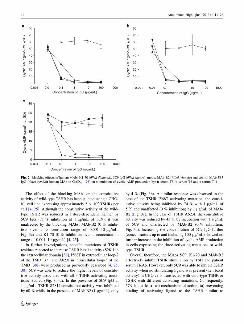
concentrations. The stimulating activity of two sera (which
were blocked in the first experiment by all three MAbs) was
inhibited to near basal levels by all three blocking MAbs
with 5C9 and K1-70 showing similar dose-dependent effect
whilst MAb-B2 was slightly less effective at the same
concentrations as 5C9 or K1-70 (Fig. 2a–c). In contrast, in
the case of the discrepant serum (which was not blocked by
5C9 in the first experiment), 5C9 IgG had no effect over a
range of the concentrations studied whereas K1-70 and
MAb-B2 showed almost complete inhibition of stimulating
activity at 0.1 and 1 lg/mL, respectively. This experiment
indicates that although stimulating autoantibodies in dif-
ferent patient sera interact with the same region of the
TSHR, they do so in subtly different ways [25, 27]. Fur-
thermore, this observation indicates that even though MAb-
B2, K1-70 and 5C9 all inhibit binding of each other to the
TSHR effectively [25, 27], the consequences of receptor
binding by different blocking MAbs can be different.
Effect of blocking MAbs on TSHR constitutive activity
The TSHR, unlike other members of the glycoprotein
hormone receptor family (follicle stimulating hormone
receptor; FSHR, luteinizing hormone receptor; LHR or
human chorionic gonadotrophin receptor; hCGR), is char-
acterised by a relatively high constitutive (basal) activity of
the cyclic AMP pathway in the absence of a ligand
[32, 33]. This feature of the TSHR is likely to be related to
the observation that a number of naturally occurring amino
acid mutations in the transmembrane domain (TMD) are
associated with the clinical presentation of non-autoim-
mune hyperthyroidism [34, 35].
b
0
20
40
60
80
100
120
0.001 1000% in
hibi
tion
of T
SH
-bio
tin b
indi
ng to
TS
HR
(m
ean
+S
D)
Concentration of IgG ( g/mL)
0
20
40
60
80
100
120
0.001 0.01 0.1 1 10 100 1000Inhi
bitio
n of
cyc
lic A
MP
stim
ulat
ion
(%)
Concentration of IgG (µg/mL)
a
µ0.01 0.1 1 10 100
Fig. 1 a Inhibition of TSH-biotin binding to TSHR coated ELISA
plate wells by different concentrations (mean ± SD; n = 3) of K1-70
IgG (filled diamond), MAb-B2 IgG (filled triangle), 5C9 IgG (filledsquare) and negative control 5B3 IgG (times symbol; human MAb to
GAD65; [76]). b Inhibition of porcine TSH (3 ng/mL)-mediated
stimulation of cyclic AMP production by TSHR monoclonal
antibodies with antagonist activity. MAb-B2 IgG (filled triangle),
K1-70 IgG (filled diamond), 5C9 IgG (filled square)
Autoimmun Highlights (2013) 4:11–26 13
123
The effect of the blocking MAbs on the constitutive
activity of wild-type TSHR has been studied using a CHO-
K1 cell line expressing approximately 5 9 105 TSHRs per
cell [4, 25]. Although the constitutive activity of the wild-
type TSHR was reduced in a dose-dependent manner by
5C9 IgG (71 % inhibition at 1 lg/mL of 5C9), it was
unaffected by the blocking MAbs: MAb-B2 (0 % inhibi-
tion over a concentration range of 0.001–10 lg/mL;
Fig. 3a) and K1-70 (0 % inhibition over a concentration
range of 0.001–10 lg/mL) [4, 25].
In further investigations, specific mutations of TSHR
residues reported to increase TSHR basal activity (S281I in
the extracellular domain [36], I568T in extracellular loop-2
of the TMD [37], and A623I in intracellular loop-3 of the
TMD [38]) were produced as previously described [4, 25,
30]. 5C9 was able to reduce the higher levels of constitu-
tive activity associated with all 3 TSHR activating muta-
tions studied (Fig. 3b–d). In the presence of 5C9 IgG at
1 lg/mL, TSHR S281I constitutive activity was inhibited
by 60 % whilst in the presence of MAb B2 (1 lg/mL), only
by 4 % (Fig. 3b). A similar response was observed in the
case of the TSHR I568T activating mutation, the consti-
tutive activity being inhibited by 74 % with 1 lg/mL of
5C9 and unaffected (0 % inhibition) by 1 lg/mL of MAb-
B2 (Fig. 3c). In the case of TSHR A623I, the constitutive
activity was reduced by 43 % by incubation with 1 lg/mL
of 5C9 and unaffected by MAb-B2 (0 % inhibition;
Fig. 3d). Increasing the concentration of 5C9 IgG further
(concentrations up to and including 100 lg/mL) showed no
further increase in the inhibition of cyclic AMP production
in cells expressing the three activating mutations or wild-
type TSHR.
Overall therefore, the MAbs 5C9, K1-70 and MAb-B2
effectively inhibit TSHR stimulation by TSH and patient
serum TRAb. However, only 5C9 was able to inhibit TSHR
activity when no stimulating ligand was present (i.e., basal
activity) in CHO cells transfected with wild-type TSHR or
TSHR with different activating mutations. Consequently,
5C9 has at least two mechanisms of action: (a) preventing
binding of activating ligand to the TSHR similar to
b
0
10
20
30
40
50
60
70
80
0.001 0.01 0.1 1 10 100 1000
Cyc
lic A
MP
(pm
ol/m
L +
SD
)
Concentration of IgG (µg/mL)
0
10
20
30
40
50
60
70
80
0.001 0.01 0.1 1 10 100 1000
Cyc
lic A
MP
(pm
ol/m
L +
SD
)
Concentration of IgG (µg/mL)
a
0
5
10
15
20
25
30
0.001 0.01 0.1 1 10 100 1000
Cyc
lic A
MP
(pm
ol/m
L +
SD
)
Concentration of IgG (µg/mL)
c
Fig. 2 Blocking effects of human MAbs K1-70 (filled diamond), 5C9 IgG (filled square), mouse MAb-B2 (filled triangle) and control MAb 5B3
IgG (times symbol; human MAb to GAD65; [76] on stimulation of cyclic AMP production by: a serum T5, b serum T8 and c serum T11
14 Autoimmun Highlights (2013) 4:11–26
123

MAb-B2 and K1-70 and (b) an effect on TSHR activity not
dependent on activating ligand binding.
A mouse MAb to the TSHR (CS-17) has also been
reported to block both TSH-stimulated cyclic AMP activity
and basal cyclic AMP activity in COS-7 cells expressing
wild-type TSHR and TSHRs with activating mutations [30,
31]. Both 5C9 and CS-17 were able to inhibit the consti-
tutive activity of TSHR S281I, TSHR I568T, and TSHR
A623I at 1 lg/mL; however, 5C9 appears to be a more
effective inhibitor than CS-17 at low IgG concentrations.
The effect of TSHR amino acid mutations
on the biological activity of TSHR blocking MAbs
The effect of various TSHR amino acid mutations on the
biological activity of TSHR MAbs with blocking activity
has been studied using CHO cells expressing wild-type or
mutated TSHRs as described in detail previously [25, 28,
39]. Flp-In-CHO cells expressing either wild-type or
mutated TSHRs were seeded into 96-well plates and used
to test the ability of 5C9 IgG, MAb-B2 IgG or K1-70 IgG
to block the stimulating activity of pTSH. In the case of the
mouse MAb-B2, mutation of TSHR residues K58, R80,
Y82 and K129 to alanine and TSHR mutations R80D,
E107R, R109D and K129D resulted in a complete loss of
the ability of MAb-B2 to block TSH stimulation of cyclic
AMP production [39] (Table 1 and Fig. 4b) whilst MAb-
B2 blocking activity was reduced when TSHR was mutated
at R109 and F134 to alanine. Furthermore mutation of
TSHR residues K58, I60, E61, Y82, R109 and K183 to
alanine all caused reduction of K1-70 blocking activity
[28] (Table 1 and Fig. 4c). Out of the TSHR mutations
investigated, mutation of K129, D203, and F153 to alanine
resulted in a complete loss of the ability of 5C9 IgG to
block TSH stimulation of cyclic AMP production [25]
(Table 1 and Fig. 4a) and TSHR mutations K183, E178,
and E251 to alanine were associated with reduction of 5C9
blocking activity.
In a different study, a mouse MAb 1H7 [10] with
TSH binding inhibiting activity and reactive with a
a
c
0
2000
4000
6000
8000
10000
12000
14000
16000
5C9 IgG 2G4 IgG MAb-B2 IgG Buffer only
Cyc
lic A
MP
(fm
ol/c
ell w
ell)
0
500
1000
1500
2000
2500
5C9 IgG 2G4 IgG MAb-B2 IgG Buffer only
Cyc
lic A
MP
(fm
ol/c
ell w
ell)
0
1000
2000
3000
4000
5000
6000
5C9 IgG 2G4 IgG MAb-B2 IgG Buffer only
Cyc
lic A
MP
(fm
ol/c
ell w
ell)
0
2000
4000
6000
8000
10000
12000
5C9 IgG 2G4 IgG MAb-B2 IgG Buffer only
Cyc
lic A
MP
(fm
ol/c
ell w
ell)
wild type TSHR TSHR S281I
TSHR I568T TSHR A623I
b
d
Fig. 3 Effect of 1 lg/mL of 5C9 IgG or MAb-B2 IgG on constitutive
(i.e., basal) activity of wild-type TSHR and TSHRs containing
activating mutations: a wild-type TSHR. b S281I (TSHR extracellular
domain). c I568T (TSHR transmembrane domain extracellular loop-
2). d A623I (TSHR transmembrane domain intracellular loop-3). 2G4
is a negative control human autoantibody to thyroid peroxidase (TPO;
[54]). Reprinted from reference [77]. The publisher for this
copyrighted material is Mary Ann Liebert, Inc publishers
Autoimmun Highlights (2013) 4:11–26 15
123

conformational epitope on the TSHR was affected by sin-
gle TSHR mutations at T56, K58, R80, Y82 and R109 and
a triple mutation F130, G132 and F134 (mutations to the
equivalent amino acid in the LH/CG receptor). These
observations indicate that these MAbs with TSH antagonist
activity (K1-70, MAb-B2 and 1H7) have overlapping
binding sites on the TSHR and all three interact with TSHR
amino acids K58, Y82 and R109 whilst the two mouse
MAbs (MAb-B2 and 1H7) also interact with R80 and
F134. In contrast, the TSHR binding region for human
MAb 5C9, which has both antagonist and inverse agonist
activity, overlaps with the binding sites of the other
Table 1 TSHR residues important for biological activity of 5C9, MAb-B2, K1-70 and M22
TSHR residues important
for blocking activity of 5C9
(mutation experiments)
TSHR residues important for
blocking activity of MAb-B2
(mutation experiments)
TSHR residues that interact with K1-70
(from the K1-70-TSHR crystal
structure and mutation experiments)
TSHR residues that interact with M22
(from the M22-TSHR crystal structure
and mutation experiments)
D36 D36 D36
R38 R38 R38
K42
Q55
T56
K58 K58a K58
I60a
E61
R80 R80 R80a
Y82 Y82a Y82a
S84
T104
H105 H105a
E107 E107a
R109 R109a R109a
N110 N110
K129 K129 K129 K129a
F130 F130a
F134
D151 D151a
I152
F153 F153 F153a
I155 I155
E157 E157
E178
T181
K183 K183a K183a
Y185a
D203 D203
N208
K209a
Q235
E251 E251
R255a
N256
The residues shown in bold are important for more than one antibody. Interactions between TSHR residues and K1-70 or M22 residues in their
respective crystal structures include hydrogen bonds, salt bridges, non-hydrogen bonding polar interactions, hydrophobic contacts, van der Waals
interactions (showing variation of accessible surface area on complexation [60 A2) and charge–charge interactions (strength [ 6.0e-10 N by
residues or [2.5e-10 N by atoms)a Amino acids, which when mutated, affected the biological activity of K1-70 or M22
16 Autoimmun Highlights (2013) 4:11–26
123

blocking MAbs but interacts with only one TSHR residue
(TSHR K129) identified as important for MAb-B2 activity
in the mutation studies. Furthermore 5C9 appears to
interact with the residues located more C-terminally on the
TSHR leucine rich domain compared to MAb B2 or K1-70.
All the amino acid mutations that were found to affect the
activity of the TSHR blocking MAbs are on the concave
surface of the TSHR extracellular domain. This is in con-
trast to reports that the mouse MAb CS-17 [30, 31] which
has both TSH antagonist activity and inverse agonist
activity (similar to 5C9) interacts with amino acids on the
convex surface of the TSHR (Y195 and S243) and Q235 on
the concave surface.
A series of TSHR mutations in the TSHR cleavage
domain (CD; amino acids 282–409), in particular, E297A,
E297 K, E303A, E303 K, R312A, K313A, E325A, D382A,
D382 K, H384A, Y385A, and D386A some of which have
been implicated in TSHR activation by TSH [15, 40–42]
and TSHR mutations Y195, S243, Y148, K201, Y225, and
E247 to alanine on the convex surface of the TSHR LRD
have been produced in our laboratory. However, the ability
of 5C9 to inhibit pTSH-induced stimulation of cyclic AMP
production was not affected by any of the mutations in the
TSHR CD or on the convex surface of the TSHR that were
investigated. Consequently the mutation studies carried out
to date indicate that the human blocking-type MAb 5C9
which has inverse agonist activity forms strong interactions
with amino acids on the concave surface of the TSHR only.
In addition, as mutations in other parts of the TSHR (i.e.,
the CD and convex surface of the LRD) had no effect on
the activity of 5C9 it is likely that 5C9 forms either no or
only weak interactions with amino acids in the TSHR CD
implicated in TSH binding [29] or on the convex surface of
the TSHR, implicated in CS-17 binding [31]. However,
it should be noted that the absence of an effect of
TSHR mutations on 5C9 activity cannot discount entirely
interactions with these residues [25]. Overall, our
experiments suggest that the interacting region for 5C9
on the TSHR is different to that of the CS-17 MAb
although extensive mutation studies of amino acids on
the TSHR concave surface or CD on CS-17 activity have
not been reported.
Interaction of the TSHR with blocking and stimulating
MAbs at the molecular level
Recently, the crystal structure of a stable complex of a part
of the TSHR extracellular domain (aa 22–260; TSHR260)
Fig. 4 Spacefill representation of the TSHR leucine-rich domain
interactive surface (based on the crystal structure of the TSHR in
complex with K1-70 solved at 1.9 A resolution). Single amino acid
mutations which affect inhibition of TSH-mediated stimulation of
cyclic AMP production in TSHR transfected CHO cells in the case of:
a MAb 5C9 shown in red (Table 1) (antagonist and inverse agonist);
b MAb-B2 shown in blue (Table 1) (antagonist); c TSHR residues
that interact with MAb K1-70 (antagonist) in the TSHR–K1-70
crystal structure are shown in yellow see Table 1); d TSHR residues
that interact with MAb M22 (agonist) in the TSHR–M22 crystal
structure are shown in green see Table 1). Figure is reproduced from
Fig. 22.4 in [29] [doi:10.1016/B978-0-12-381296-4.00022-1]. The
publisher for this copyrighted material is Elsevier and a license
number 2831340216726 has been granted
Autoimmun Highlights (2013) 4:11–26 17
123

with the antagonist MAb (K1-70) was solved at 1.9 A
resolution [28]. The binding arrangement of K1-70 with
the TSHR LRD can be compared to the binding arrange-
ments of the thyroid stimulating human MAb M22 with
TSHR260 in the crystal structure solved at 2.55 A resolu-
tion [24] (Fig. 5). These solved structures also provide
atomic level detail of the structure of the LRD of the
TSHR [24, 28] and many of the interactions observed in
the crystal structures have been confirmed by experi-
ments involving mutation of TSHR or MAb amino acids
[28, 39, 43].
TSHR260 has the shape of a slightly curved helical tube
constructed from leucine-rich repeat motifs. It has opposed
concave and convex surfaces, with an 11-stranded b-sheet
located on the concave surface (10 parallel strands, 1 per
repeat and an antiparallel strand at the N terminus; Fig. 5).
The structure of the convex surface of the TSHR LRD
presents eight small strands (two residues each) forming
two 3-stranded b-sheets and one 2-stranded b-sheet
(Fig. 5). The inner surface of the tube is lined with
hydrophobic residues and there are no a helices in the
TSHR LRD structure. All five glycosylation sites (N77,
N99, N113, N177, and N198) are located on the convex
surface of the TSHR LRD and are glycosylated.
The FSHR is the closest homologue to the TSHR with
40.9 % amino acid sequence identity [34]. The crystal
structure of the FSHR LRD in complex with FSH has been
solved at 2.9 A resolution [44]. The root mean square
deviation (rmsd) on Ca core atoms between the TSHR260
and FSHR LRD structures is 1.1 A providing the first
detailed evidence that the LRDs of the two receptors have
very similar structures [24]. Furthermore, a comparison of
the structures of TSHR260 in complex with K1-70 or with
M22 shows an rmsd on all Ca atoms between the structures
of 0.51 A thus confirming the solved structure of the
TSHR260 originally reported [24, 28]. In addition, the
higher resolution TSHR260–K1-70 structure provided
details of disulphide bond arrangements at the N terminus
of the TSHR (Fig. 5). Disulphide bonds are present
between the first and second cysteines (C24 and C29) and
between the third and fourth cysteines (C31 and C41). In
the original TSHR260-M22 structure, only the C31 and
C41 disulphide bond was visible due to disorder in the
structure of the extreme N terminus of the receptor. The
TSHR N-terminal disulphide bonding arrangements are
different to those observed in the FSHR crystal structure,
where the first and third cysteines (C18 and C25) and the
second and fourth cysteines (C23 and C32) are disulphide
bonded [44]. The TSHR has a three-amino acid insertion
between TSHR C31 and C41 which is not present in the
equivalent position in the FSHR sequence, consequently
bonding between C29 and C41 of the TSHR would be
entropically less favourable than between the equivalent
FSHR cysteines. Earlier studies based on mutagenesis
proposed that TSHR C41 was paired with either C29 or
C31 [45], however, the most recent crystal structure of the
TSHR [28] now shows the actual molecular details of
cysteine pairing at the TSHR N terminus. In the crystal
structure, the blocking antibody K1-70 shows no interac-
tion with the extreme N terminus of the TSHR LRD
Fig. 5 Crystal structures of
TSHR—K1-70 Fab and TSHR–
M22 Fab. Diagram of the TSH
receptor leucine-rich repeat
domain (TSHR LRD) in
complex with the human
monoclonal autoantibodies
K1-70 and M22. TSHR is in
cyan, antibody light chains are
in green, and heavy chains in
blue. Disulphide-bonded
cysteines are shown as ball andstick, disulphide bonds are in
yellow, and cysteine residues
are labelled in orange. The
positions of amino (N)- and
carboxy (C)-termini are
indicated. The complex of
TSHR–K1-70 Fab is reproduced
from Fig. 2 in [28], with
permission of the copyright
holder (Society for
Endocrinology)
18 Autoimmun Highlights (2013) 4:11–26
123

(aa 22–34), but forms contacts with residues D36 and R38
(distance \ 4 A) of the first repeat of the TSHR LRD
(aa 35–52); [28]. Molecular replacement analysis of the
interaction of M22 with the TSHR (using the TSHR LRD
structure from the complex with K1-70 solved at 1.9 A)
also showed that there were no M22 interactions involving
the extreme N-terminal region of the TSHR LRD with the
first contact amino acid residue being R38 (dis-
tance \ 4 A) of the first repeat. K1-70 binds slightly more
N-terminally to the first TSHR repeat than M22 and this
may account for the improved order of the extreme
N-terminus in the TSHR260—K1-70 crystal structure.
These observations suggest that the TSHR region con-
taining the four disulphide-bonded cysteines does not have
a major role in TSHR autoantibody binding. For example,
it has been proposed [45, 46] that the TSHR N-terminal
region (aa 22–41) is part of a highly conformational epi-
tope for thyroid stimulating autoantibodies. However, a
more recent study using a mutated TSHR, with the first 30
amino acids removed, confirmed the binding arrangements
observed in the TSHR260-M22 crystal structure, i.e., that
the stimulating antibody M22 does not interact with the
extreme N terminus (aa 22–34) of the TSHR [47]. The N
terminus of the TSHR most likely acts as a protective
N-terminal cap aiding stability, preventing degradation and
keeping the LRD in its correct conformation [48, 49].
The crystal structures of the two antibodies in complex
with the TSHR show numerous strong interactions
(hydrogen bonds, salt bridges, polar or hydrophobic con-
tacts, Van der Waals interactions) between the antibody
HCs and LCs and residues on the concave surface of
TSHR260. This strong network of interactions is consistent
with the high binding affinity of M22 and K1-70 to the
TSHR.
Table 1 and Fig. 4 show a comparison of the TSHR
residues that were identified as important for the activity
of 5C9 and MAb-B2 by TSHR mutation experiments and
the K1-70 and M22 TSHR contact residues confirmed by
analysis of crystal-structure data (residues that were
identified as important for the activity of M22 and K1-70
by TSHR mutation experiments are marked with an
asterisk in Table 1) [39]. The residues of TSH that interact
with the TSHR LRD were obtained by comparative
modelling [50–53] and are shown in Table 1 and Fig. 4
for comparison with the MAb–TSHR interactions. As
shown in Table 1, there are a number of the same TSHR
residues that are important for the activity of more than
one MAb. In particular, TSHR K129 is important for
interaction of all four MAbs (5C9, MAb-B2, K1-70, and
M22) whether they have antagonist, inverse agonist, or
agonist activity and this is consistent with the observation
that even though the antibodies have different functional
activities, their binding sites on the concave surface of the
TSHR overlap [24, 25, 28, 39]. However, the antagonist
MAb-B2 contact residues are located more N-terminal on
the TSHR concave surface. Furthermore, seven of the
eight TSHR residues identified as important for the
activity of MAb-B2 also interact with the antagonist
K1-70 MAb (observed in the crystal structure). Remarkably,
the crystal structures of the TSHR complexed with the
antagonist MAb K1-70 and the agonist MAb M22 show
that 16 TSHR residues (D36, R38, K58, R80, Y82, H105,
E107, R109, N110, K129, F130, D151, F153, I155, E157,
and K183) interact with both MAbs. An extensive overlap
is also evident between the TSHR residues predicted to be
involved in TSH interaction with the receptor and those
involved in binding of the four MAbs to the TSHR. Iden-
tification of TSHR interacting residues from crystal struc-
tures of TSHR complexed with the MAbs provides more
accurate information compared to TSHR mutation experi-
ments. For example, a mutation may not show any effect
on the biological activity of an antibody if other interac-
tions with the TSHR are not disrupted and are strong
enough to compensate for the mutation. However, in most
cases, the experimental mutation results are in good
agreement with the binding arrangements observed in the
crystal structures [24, 28, 39, 43]. Overall, the data
obtained from the crystal structures and mutation experi-
ments suggest that although the binding sites of the MAbs
on the concave surface of the TSHR overlap with each
other, their functional effects are likely to be defined by
subtle differences in the actual amino acids contacted.
Effect of K1-70 on thyroid function in vivo
In vivo effects of K1-70 on thyroid function were studied
in rats which have naturally high serum TSH and conse-
quently high serum T4 levels (unsuppressed rats). These
high levels of serum TSH and T4 can be suppressed by
providing rats with T3 in their drinking water for a few
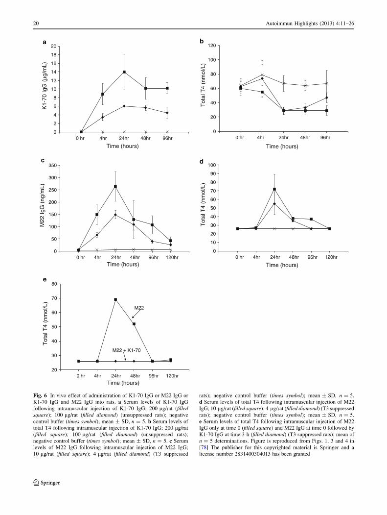
days (T3 suppressed rats). Intramuscular injection of dif-
ferent concentrations of the blocking MAb K1-70 into
unsuppressed rats caused a dose-dependent decrease of
total and free T4 levels. The total and free T4 concentra-
tions decreased to their lowest levels at 24 h post injection
(Fig. 6a) and this coincided with the maximum levels of
K1-70 IgG measured in the same samples. Doses of 100 or
200 lg per animal of K1-70 IgG caused a decrease of the
serum total and free T4 to the low levels observed in T3
suppressed rats (Fig. 6b). In contrast, intramuscular injec-
tion of different amounts of M22 IgG into T3 suppressed
rats caused a dose-dependent increase in total and free T4
concentrations. The maximum concentrations of total and
free T4 in the rat sera were detected at 24 h post injection
and coincided with the peak in the levels of M22 IgG
measured in the same rat-serum samples (Fig. 6c, d).
Autoimmun Highlights (2013) 4:11–26 19
123
b
0
2
4
6
8
10
12
14
16
18
20K
1-70
IgG
(µg
/mL)
Time (hours)
0
20
40
60
80
100
120
Tot
al T
4 (n
mol
/L)
Tot
al T
4 (n
mol
/L)
Tot
al T
4 (n
mol
/L)
Time (hours)
0 hr 4hr 24hr 48hr 96hr 0 hr 4hr 24hr 48hr 96hr
c d
0
50
100
150
200
250
300
350
M22
IgG
(ng
/mL)
Time (hours)
0
10
20
30
40
50
60
70
80
90
100
Time (hours)0 hr 4hr 24hr 48hr 96hr 120hr 0 hr 4hr 24hr 48hr 96hr 120hr
e
20
30
40
50
60
70
80
Time (hours)0 hr 4hr 24hr 48hr 96hr 120hr
M22
M22 + K1-70
a
Fig. 6 In vivo effect of administration of K1-70 IgG or M22 IgG or
K1-70 IgG and M22 IgG into rats. a Serum levels of K1-70 IgG
following intramuscular injection of K1-70 IgG; 200 lg/rat (filledsquare); 100 lg/rat (filled diamond) (unsuppressed rats); negative
control buffer (times symbol); mean ± SD, n = 5. b Serum levels of
total T4 following intramuscular injection of K1-70 IgG; 200 lg/rat
(filled square); 100 lg/rat (filled diamond) (unsuppressed rats);
negative control buffer (times symbol); mean ± SD, n = 5. c Serum
levels of M22 IgG following intramuscular injection of M22 IgG;
10 lg/rat (filled square); 4 lg/rat (filled diamond) (T3 suppressed
rats); negative control buffer (times symbol); mean ± SD, n = 5.
d Serum levels of total T4 following intramuscular injection of M22
IgG; 10 lg/rat (filled square); 4 lg/rat (filled diamond) (T3 suppressed
rats); negative control buffer (times symbol); mean ± SD, n = 5.
e Serum levels of total T4 following intramuscular injection of M22
IgG only at time 0 (filled square) and M22 IgG at time 0 followed by
K1-70 IgG at time 3 h (filled diamond) (T3 suppressed rats); mean of
n = 5 determinations. Figure is reproduced from Figs. 1, 3 and 4 in
[78] The publisher for this copyrighted material is Springer and a
license number 2831400304013 has been granted
20 Autoimmun Highlights (2013) 4:11–26
123
Furthermore, administration of K1-70 IgG at 200 lg per
animal caused complete inhibition of the stimulation of the
rat thyroid in vivo by M22 IgG (4 lg per animal) when
injected 3 h before the M22 or 3 h after the M22 or at the
same time as the M22 (Fig. 6e). These experiments showed
for the first time that a blocking-type MAb can act in vivo
as an effective inhibitor of thyroid stimulation by TSH and
TRAb. The ability of K1-70 to inhibit autoantibody stim-
ulation of the thyroid in vivo emphasizes its potential as a
new type of anti-thyroid drug.
Binding to the TSHRs in thyroid tissue sections
in immunohistochemistry studies
Biotinylated TSHR MAbs (K1-70 and M22), a human
MAb to thyroid peroxidase (2G4, positive control [54]) and
a negative isotype control antibody (human IgG1 lambda)
were analysed for binding to cryo-sections of human thy-
roid fixed in acetone and methanol using standard proce-
dures. In addition, cryo-sections of human prostate,
cerebellum, kidney and liver were used in the experiments
as negative control tissues.
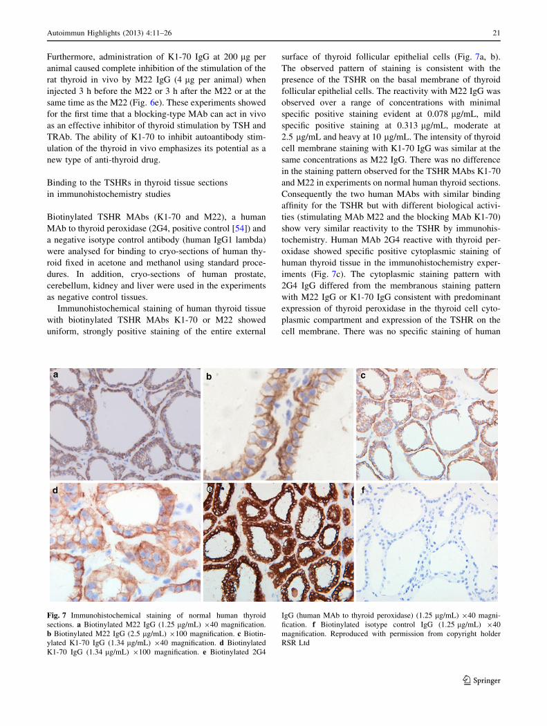
Immunohistochemical staining of human thyroid tissue
with biotinylated TSHR MAbs K1-70 or M22 showed
uniform, strongly positive staining of the entire external
surface of thyroid follicular epithelial cells (Fig. 7a, b).
The observed pattern of staining is consistent with the
presence of the TSHR on the basal membrane of thyroid
follicular epithelial cells. The reactivity with M22 IgG was
observed over a range of concentrations with minimal
specific positive staining evident at 0.078 lg/mL, mild
specific positive staining at 0.313 lg/mL, moderate at
2.5 lg/mL and heavy at 10 lg/mL. The intensity of thyroid
cell membrane staining with K1-70 IgG was similar at the
same concentrations as M22 IgG. There was no difference
in the staining pattern observed for the TSHR MAbs K1-70
and M22 in experiments on normal human thyroid sections.
Consequently the two human MAbs with similar binding
affinity for the TSHR but with different biological activi-
ties (stimulating MAb M22 and the blocking MAb K1-70)
show very similar reactivity to the TSHR by immunohis-
tochemistry. Human MAb 2G4 reactive with thyroid per-
oxidase showed specific positive cytoplasmic staining of
human thyroid tissue in the immunohistochemistry exper-
iments (Fig. 7c). The cytoplasmic staining pattern with
2G4 IgG differed from the membranous staining pattern
with M22 IgG or K1-70 IgG consistent with predominant
expression of thyroid peroxidase in the thyroid cell cyto-
plasmic compartment and expression of the TSHR on the
cell membrane. There was no specific staining of human
Fig. 7 Immunohistochemical staining of normal human thyroid
sections. a Biotinylated M22 IgG (1.25 lg/mL) 940 magnification.
b Biotinylated M22 IgG (2.5 lg/mL) 9100 magnification. c Biotin-
ylated K1-70 IgG (1.34 lg/mL) 940 magnification. d Biotinylated
K1-70 IgG (1.34 lg/mL) 9100 magnification. e Biotinylated 2G4
IgG (human MAb to thyroid peroxidase) (1.25 lg/mL) 940 magni-
fication. f Biotinylated isotype control IgG (1.25 lg/mL) 940
magnification. Reproduced with permission from copyright holder
RSR Ltd
Autoimmun Highlights (2013) 4:11–26 21
123
thyroid tissue with the biotinylated isotype control MAb
(Fig. 7d). Biotinylated M22 IgG, K1-70 IgG or 2G4 IgG
showed no specific staining with a selected panel of normal
human tissues (prostate, kidney, liver and cerebellum) in
control experiments.
Specific TSHR staining on normal human thyroid sec-
tions at the basal pole of the thyroid cells was reported
previously using a mouse MAb reactive with a conforma-
tional epitope on the extracellular domain of the TSHR
[55]. In a different study, more cytoplasmic rather than
membranous staining was observed in sections from 2/3
human thyroid glands using a mouse MAb reactive with
the N terminus of the TSHR [56] whilst a MAb binding to
the TMD of the TSHR showed predominant reactivity with
the basolateral surface of thyroid cell membrane [56]. In
addition, some mouse MAbs have been reported to bind to
the TSHR in sections of orbital fat from patients with
Graves’ associated ophthalmopathy but not normal control
orbital fat [57].
The ability of both K1-70 and M22 to bind specifically
to the TSHR on thyroid tissue sections suggest potential
applications of these MAbs in detection and visualisation
of the TSHR in thyroid sections from different species as
well as other tissues such as orbital fat or bone [58, 59].
Furthermore, the ability of our human MAbs to bind
strongly and specifically to the TSHR on thyroid epithelial
cells indicates that appropriately labelled M22 and K1-70
are likely to have potentially important applications in in
vivo imaging.
Potential clinical applications of TSHR human MAbs
For many years, the control of thyroid over-activity has
relied on inhibiting thyroid hormone synthesis with anti-
thyroid drugs (ATDs), reduction of thyroid volume by
surgery (subtotal thyreoidectomy) or destruction of the
thyroid gland with radioactive iodine [60–62]. In particu-
lar, the pharmacological approach for controlling hyper-
thyroidism has not changed since the introduction of ATDs
in the 1940s [63]. Overall current treatment options provide
effective control of the symptoms of the pathogenic effects
of TRAbs in the majority of patients. However, treatment
strategies that act at the beginning of the pathogenic
pathway preventing the binding of TRAbs to the TSHR
and/or acting directly on the TSHR to inhibit the unwanted
biological activity of TRAbs seem more appropriate but are
not currently available for clinical use.
TRAbs with TSHR antagonist activities are ‘‘natural’’
inhibitors of TSHR stimulation by both stimulating TRAbs
and TSH. Consequently antibodies such as human MAbs
K1-70 and 5C9 or the mouse MAb-B2 could be used to
control thyroid over-activity in Graves’ disease. K1-70,
5C9 and MAb-B2 have the ability to inhibit the stimulating
activity of TSH and patient-serum-stimulating TRAbs in
CHO cells expressing the TSHR (Figs. 1 and 2). Further-
more, the potent inhibiting effect of K1-70 IgG on thyroid
hormone secretion has been shown in vivo (Fig. 6b). In
addition, in vivo studies have indicated that K1-70 IgG
effectively inhibits M22 thyroid stimulating activity
(Fig. 6e). The effects of K1-70 on endogenous TSH and
M22 stimulated thyroid hormone levels were evident
within a relatively short time after administration. These
observations open new opportunities for the management
of Graves’ disease. In particular, relatively quick effects of
K1-70 in vivo suggests that the euthyroid state may be
achieved earlier in patients than with currently used ther-
apies particularly ATD. Consequently, using a powerful
TSHR blocking-type MAb as a first-line treatment should
lead to an improvement in the ability to control the course
of Graves’ disease [64, 65].
Furthermore, blocking-type TSHR MAbs would have a
particular application when quick control of thyroid over-
activity is essential as in a thyroid crisis [60]. In addition,
patients who do not tolerate or do not respond well to
ATDs may well benefit from treatment with blocking-type
MAbs [60–62]. Another potential application of blocking-
type TSHR MAbs would be in controlling thyroid over-
activity in pregnant women. In this application, K1-70 Fab
that has similar TSHR antagonist activity as intact K1-70
IgG [27], and does not cross the placenta would act on the
thyroid of a mother without affecting the baby’s thyroid.
Almost 90 % of patients with Graves’ disease present
with eye signs, and in approximately 5 %, the symptoms
are serious and require special interventions [66]. TSHR
expression has been demonstrated in orbital fibroblasts and
preadipocytes, and furthermore, evidence of humoral and
cellular autoimmune responses to the TSHR in the orbit has
now been shown [58, 59, 66, 67]. Consequently, it is likely
that autoimmune responses against the TSHR expressed in
the orbital tissues are involved in the pathogenesis of
Graves’ ophthalmopathy [58, 59, 66, 67]. In most cases, the
symptoms of ophthalmopathy resolve with the control of
thyroid over-activity. In severe cases, the treatment is
based on controlling the symptoms and preventing optic
nerve damage by use of glucocorticoids, orbital irradiation
or surgical decompression. More recently, trials with rit-
uximab, a MAb against the CD-20 antigen on B cells have
shown some encouraging results [68]. However, treatment
with rituximab is not targeted specifically at the mecha-
nisms involved in the pathogenesis of Graves’ ophthal-
mopathy and may have harmful side effects [68, 69].
Blocking-type TSHR MAbs, however, are potentially
useful therapeutics for the management of Graves’ oph-
thalmopathy. It may well be that preventing TSHR acti-
vation in orbital fibroblasts early in the disease process
would have an effect on the complex autoimmune and
22 Autoimmun Highlights (2013) 4:11–26
123

inflammatory responses in the orbit [66–69]. If successful,
blocking-type TSHR MAbs would provide for the first time
a specific treatment for patients with this debilitating
condition.
It has been suggested that the autoimmune/inflammatory
responses to the TSHR on fibroblasts may have a role in the
pathogenesis of pretibial myxedema and it has been pro-
posed that the mechanisms involved in the development of
pretibial myxedema and ophthalmopathy are similar, i.e.,
not dependent on site-specific properties of the fibroblasts
[70]. It may be that blocking-type TSHR MAbs would be
helpful in the management of pretibial myxoedema.
In most cases, clinical hyperthyroidism is caused by the
stimulating effect of TRAbs. However in some cases,
thyroid overactivity is the result of autonomous secretion
of TSH by pituitary adenomas [71]. Blocking-type TSHR
MAbs such as K1-70, 5C9 or MAb-B2 would be expected
to be effective in preventing thyroid hormone production in
response to the excess of autonomously produced TSH by
specifically inhibiting TSH binding to the receptor. 5C9
MAb with its ability to inhibit TSHR constitutive activity
would also be expected to be effective in controlling
thyroid over-activity associated with at least some TSHR-
activating mutations [34, 35]. The mouse MAb CS-17
appears to have similar properties to 5C9, but use of CS-17
in patients would depend on its successful ‘‘humanising’’
[72, 73]. Treatment options with 5C9 (or humanised
CS-17) should be valuable in patients with familial non-
autoimmune hyperthyroidism. In addition, patients with
toxic nodular goitre awaiting the effects of radical treatment
(surgery or radioiodine) would likely benefit from blocking-
MAb reduction of TSHR-activity and toxic effects of thyroid
hormones on the cardiovascular system [60–62].
Furthermore, suppression of TSHR constitutive activity
with 5C9 (or humanised CS-17) would be expected to be of
benefit in patients with thyroid cancer [74]. The important
goal in the treatment of thyroid cancer is complete TSH
suppression to eliminate potentially cancer-growth-pro-
moting TSHR activity. However, the means to control
constitutive TSHR activity in the absence of TSH are not
currently available and these MAbs with TSHR inverse
agonist activity would be good candidates.
The observation that human MAbs to the TSHR bind
specifically to TSHRs on thyroid epithelial cells in
immunohistochemistry studies provides a rational basis for
using M22 or K1-70 for in vivo imaging applications.
These applications may be particularly useful in thyroid
cancer and thyroid-cancer metastases localisations in vivo.
K1-70 which has equal ability to bind to the TSHRs on the
thyroid cells as M22 without causing stimulation of cyclic
AMP would be an appropriate choice for imaging pur-
poses. Furthermore, TSHR MAbs such as K1-70 could be
used to deliver drugs directly to the site of thyroid tumours
expressing the TSHR. For example, an active compound
selected from drugs known to have chemotherapeutic
effects could be linked to the MAb or the MAb could be
linked to a metal chelator carrying a radionuclide for tar-
geted therapy [75].
Consequently, availability of blocking-type TSHR MAbs
should provide new in vivo diagnostic and therapeutic tools
for clinicians caring for patients with Graves’ disease,
patients with thyroid cancer and patients with other thyroid
diseases who would benefit from the control of TSHR
activity.
Conflict of interest All authors are employees of RSR Ltd. RSR
Ltd is a developer of medical diagnostics including kits for measuring
thyroid autoantibodies.
References
1. Rees Smith B, McLachlan SM, Furmaniak J (1988) Autoanti-
bodies to the thyrotropin receptor. Endocr Rev 9:106–121. doi:
10.1210/edrv-9-1-106
2. Rapoport B, Chazenbalk GD, Jaume JC, McLachlan SM (1998)
The thyrotropin (TSH) receptor: interaction with TSH and auto-
antibodies. Endocr Rev 19:673–716. doi:10.1210/er.19.6.673
3. Rees Smith B, Bolton J, Young S, Collyer A, Weeden A, Brad-
bury J, Weightman D, Perros P, Sanders J, Furmaniak J (2004) A
new assay for thyrotropin receptor autoantibodies. Thyroid
14:830–835. doi:10.1089/thy.2004.14.830
4. Rees Smith B, Sanders J, Furmaniak J (2007) TSH receptor
antibodies. Thyroid 17:923–938. doi:10.1089/thy.2007.0239
5. McLachlan SM, Rapoport B (1996) Monoclonal, human auto-
antibodies to the TSH receptor—the holy grail and why are we
looking for it? J Clin Endocrinol Metab 81:3152–3154
6. McLachlan SM, Rapoport B (2004) Thyroid stimulating mono-
clonal antibodies: overcoming the road blocks and the way for-
ward. Clin Endocrinol 61:10–18. doi:10.1111/j.1365-2265.2004.
02028.x
7. Sanders J, Jeffreys J, Depraetere H, Richards T, Evans M, Kiddie
A, Brereton K, Groenen M, Oda Y, Furmaniak J, Rees Smith B
(2002) Thyroid stimulating monoclonal antibodies. Thyroid
12:1043–1050. doi:10.1089/105072502321085135
8. Ando T, Latif R, Pritsker A, Moran T, Nagayama Y, Davies TF
(2002) A monoclonal thyroid-stimulating antibody. J Clin
Investig 110:1667–1674. doi:10.1172/JCI16991
9. Costagliola S, Franssen JDF, Bonomi M, Urizar E, Willnich M,
Bergmann A, Vassart G (2002) Generation of a mouse monoclonal
TSH receptor antibody with stimulating activity. Biochem Biophys
Res Commun 299:891–896. doi:10.1016/S0006-291X(02)02762-6
10. Costagliola S, Bonomi M, Morganthaler NG, Van Durme J,
Panneels V, Refetoff S, Vassart G (2004) Delineation of the
discontinuous-conformational epitope of a monoclonal antibody
displaying full in vitro and in vivo thyrotropin activity. Mol
Endocrinol 18:3020–3034. doi:10.1210/me.2004-0231
11. Gilbert J, Gianoukakis A, Salehi S, Moorhead J, Rao P, Kahn
MZ, McGregor A, Smith T, Banga JP (2006) Monoclonal path-
ogenic antibodies to the thyroid stimulating hormone receptor in
Graves’ disease with potent thyroid-stimulating activity but dif-
ferential blocking activity activate multiple signalling pathways.
J Immunol 176:5084–5092
12. Sanders J, Allen F, Jeffreys J, Bolton J, Richards T, Depraetere H,
Nakatake N, Evans M, Kiddie A, Premawardhana LDKE,
Autoimmun Highlights (2013) 4:11–26 23
123

Chirgadze DY, Miguel RN, Blundell TL, Furmaniak J, Rees Smith
B (2005) Characteristics of a monoclonal antibody to the thyrot-
ropin receptor that acts as a powerful thyroid-stimulating autoan-
tibody antagonist. Thyroid 15:672–682. doi:10.1089/thy.2005.15.
672
13. Shepherd PS, Da Costa CR, Cridland JC, Gilmore KS, Johnstone
AP (1999) Identification of an important thyrotrophin binding site
on the human thyrotrophin receptor using monoclonal antibodies.
Mol Cell Endocrinol 149:197–206
14. Jeffreys J, Depraetere H, Sanders J, Oda Y, Evans M, Kiddie A,
Richards T, Furmaniak J, Rees Smith B (2002) Characterization
of the thyrotropin binding pocket. Thyroid 12:1051–1061. doi:
10.1089/105072502321085144
15. Costagliola S, Panneels V, Bonomi M, Koch J, Many MC, Smits
G, Vassart G (2002) Tyrosine sulfation is required for agonist
recognition by glycoprotein hormone receptors. EMBO J 21:504–
513. doi:10.1093/emboj/21.4.504
16. Davies TF, Bobovnikova Y, Weiss M, Vlase H, Moran T, Graves
PN (1998) Development and characterization of monoclonal
antibodies specific for the murine thyrotropin receptor. Thyroid
8:693–701
17. Lenzner C, Morgenthaler NG (2003) The effect of thyrotropin-
receptor blocking antibodies on stimulating autoantibodies
from patients with Graves’ disease. Thyroid 13:1153–1161. doi:
10.1089/10507250360731569
18. Minich WB, Lenzner C, Morgenthaler NG (2004) Antibodies to
TSH-receptor in thyroid autoimmune disease interact with
monoclonal antibodies whose epitopes are broadly distributed on
the receptor. Clin Exp Immunol 136:129–136. doi:10.1111/j.1365-
2249.2004.02417.x
19. Oda Y, Sanders J, Roberts S, Maruyama M, Kato R, Perez M,
Petersen VB, Wedlock N, Furmaniak J, Rees Smith B (1998)
Binding characteristics of antibodies to the TSH receptor. J Mol
Endocrinol 20:233–244. doi:10.1677/jme.0.0200233
20. Oda Y, Sanders J, Evans M, Kiddie A, Munkley A, James C,
Richards T, Wills J, Furmaniak J, Rees Smith B (2000) Epitope
analysis of the human thyrotropin (TSH) receptor using monoclonal
antibodies. Thyroid 10:1051–1059. doi:10.1089/thy.2000.10.1051
21. Vlase H, Weiss M, Graves PN, Davies TF (1998) Characteriza-
tion of the murine immune response to the murine TSH receptor
ectodomain: induction of hypothyroidism and TSH receptor
antibodies. Clin Exp Immunol 113:111–118. doi:10.1046/j.1365-
2249.1998.00622.x
22. Sanders J, Evans M, Premawardhana LDKE, Depraetere H,
Jeffreys J, Richards T, Furmaniak J, Rees Smith B (2003) Human
monoclonal thyroid stimulating autoantibody. Lancet 362:126–
128. doi:10.1016/S0140-6736(03)13866-4
23. Sanders J, Jeffreys J, Depraetere H, Evans M, Richards T, Kiddie
A, Brereton K, Premawardhana LD, Chirgadze DY, Nunez
Miguel R, Blundell TL, Furmaniak J, Rees Smith B (2004)
Characteristics of a human monoclonal autoantibody to the
thyrotropin receptor: sequence structure and function. Thyroid
14:560–570. doi:10.1089/1050725041692918
24. Sanders J, Chirgadze DY, Sanders P, Baker S, Sullivan A,
Bhardwaja A, Bolton J, Reeve M, Nakatake N, Evans M, Rich-
ards T, Powell M, Nunez Miguel R, Blundell TL, Furmaniak J,
Rees Smith B (2007) Crystal structure of the TSH receptor
in complex with a thyroid-stimulating autoantibody. Thyroid
17:395–410. doi:10.1089/thy.2007.0034
25. Sanders J, Evans M, Betterle C, Sanders P, Bhardwaja A, Young
S, Roberts E, Wilmot J, Richards T, Kiddie A, Small K, Platt H,
Summerhayes S, Harris R, Reeve M, Coco G, Zanchetta R,
Chen S, Furmaniak J, Rees Smith B (2008) A human monoclonal
autoantibody to the thyrotropin receptor with thyroid-stimulating
blocking activity. Thyroid 18:735–746. doi:10.1089/thy.2007.
0327
26. Rees Smith B, Sanders J, Evans M, Tagami T, Furmaniak J
(2009) TSH receptor—autoantibody interactions. Hormon Metab
Res 41:448–455. doi:10.1055/s-0029-1220913
27. Evans M, Sanders J, Tagami T, Sanders P, Young S, Roberts E,
Wilmot J, Hu X, Kabelis K, Clark J, Holl S, Richards T, Collyer
A, Furmaniak J, Rees Smith B (2010) Monoclonal autoantibodies
to the TSH receptor, one with stimulating activity and one with
blocking activity, obtained from the same blood sample. Clin
Endocrinol 73:404–412. doi:10.1111/j.1365-2265.2010.03831.x
28. Sanders P, Young S, Sanders J, Kabelis K, Baker S, Sullivan A,
Evans M, Clark J, Wilmot J, Hu X, Roberts E, Powell M, Nunez
Miguel R, Furmaniak J, Rees Smith B (2011) Crystal structure of
the TSH receptor (TSHR) bound to a blocking-type TSHR auto-
antibody. J Mol Endocrinol 46:81–99. doi:10.1530/JME-10-0127
29. Sanders J, Nunez Miguel R, Furmaniak J, Rees Smith B (2010)
TSH receptor monoclonal antibodies with agonist, antagonist and
inverse agonist activities. Methods Enzymol 485:393–420. doi:
10.1016/B978-0-12-381296-4.00022-1
30. Chen CR, McLachlan SM, Rapoport B (2007) Suppression
of thyrotropin receptor constitutive activity by a monoclonal
antibody with inverse agonist activity. Endocrinology 148:2375–
2382. doi:10.1210/en.2006-1754
31. Chen CR, McLachlan SM, Rapoport B (2008) Identification of
key amino acid residues in a thyrotropin receptor monoclonal
antibody epitope provides insight into its inverse agonist and
antagonist properties. Endocrinology 149:3427–3434. doi:10.1210/
en.2008-0207
32. Zhang M, Phuong K, Tong T, Fremont V, Chen J, Narayan P,
Puett D, Weintraub BD, Szkudlinski MW (2000) The extracel-
lular domain suppresses constitutive activity of the transmem-
brane domain of the human TSH receptor: implications for
hormone-receptor interaction and antagonist design. Endocrinol-
ogy 141:3514–3517. doi:10.1210/en.141.9.3514
33. Vlaeminck-Guillem VK, Ho SC, Rodien P, Vassart G, Costa-
gliola S (2002) Activation of the cAMP pathway by the TSH
receptor involves switching of the ectodomain from a tethered
inverse agonist to an agonist. Mol Endocrinol 16:736–746. doi:
10.1210/me.16.4.736
34. Sanders J, Oda Y, Roberts S, Maruyama M, Furmaniak J, Rees
Smith B (1997) Understanding the thyrotropin receptor function-
structure relationship. Bailliere’s Clin Endocr Metab 11:451–479.
doi:10.1016/S0950-351X(97)80693-3
35. Krohn K, Fuhrer D, Bayer Y, Eszlinger M, Brauer V, Neumann
S, Paschke R (2005) Molecular pathogenesis of euthyroid and
toxic multinodulargoiter. Endocr Rev 26:504–524. doi:10.1210/
er.2004-0005
36. Kopp P, Muirhead S, Jourdain N, Gu W-X, Jameson JL, Rodd C
(1997) Congenital hyperthyroidism caused by solitary toxic
adenoma harboring a novel somatic mutation (serine281 to iso-
leucine) in the extracellular domain of the thyrotropin receptor.
J Clin Investig 100:1634–1639. doi:10.1172/JCI119687
37. Parma J, Van Sande J, Swillens S, Tonacchera M, Dumont J,
Vassart G (1995) Somatic mutations causing constitutive activity
of the thyrotropin receptor are the major cause of hyperfunc-
tioning thyroid adenomas: identification of additional mutations
activating both the cyclic adenosine 30,50-monophosphate and
inositol phosphate-Ca2 ? cascades. Mol Endocrinol 9:725–733.
doi:10.1210/me.9.6.725
38. Parma J, Duprez L, Van Sande J, Cochaux P, Gervy C, Mockel J,
Dumont J, Vassart G (1993) Somatic mutations in the thyrotropin
receptor gene cause hyperfunctioning thyroid adenomas. Nature
365:649–651. doi:10.1038/365649a0
39. Sanders J, Bolton J, Sanders P, Jeffreys J, Nakatake N, Richards
T, Evans M, Kiddie A, Summerhayes S, Roberts E, Nunez
Miguel R, Furmaniak J, Rees Smith B (2006) Effects of TSH
receptor mutations on binding and biological activity of
24 Autoimmun Highlights (2013) 4:11–26
123

monoclonal antibodies and TSH. Thyroid 16:1195–1206. doi:
10.1089/thy.2006.16.1195
40. Kosugi S, Ban T, Akamizu T, Kohn LD (1991) Site-directed
mutagenesis of a portion of the extracellular domain of the rat
thyrotropin receptor important in autoimmune thyroid disease and
nonhomologous with gonadotropin receptors: relationship of
functional and immunogenic domains. J Biol Chem 266:19413–
19418
41. Mueller S, Kleinau G, Jaeschke H, Paschke R, Krause G (2008)
Extended hormone binding site of the human thyroid stimulating
hormone receptor: distinctive acidic residues in the hinge region
are involved in bovine thyroid stimulating hormone binding and
receptor activation. J Biol Chem 283:18048–18055. doi:10.1074/
jbc.M800449200
42. Mueller S, Kleinau G, Szkudlinski MW, Jaeschke H, Krause G,
Paschke R (2009) The superagonistic activity of bovine thyroid-
stimulating hormone (TSH) and the human TR1401 TSH analog
is determined by specific amino acids in the hinge region of
the human TSH receptor. J Biol Chem 284:16317–16324. doi:
10.1074/jbc.M109.005710
43. Sanders J, Nunez Miguel R, Bolton J, Bhardwaja A, Sanders P,
Nakatake N, Evans M, Furmaniak J, Rees Smith B (2007)
Molecular interactions between the TSH receptor and a thyroid-
stimulating monoclonal autoantibody. Thyroid 17:699–706. doi:
10.1089/thy.2007.0041
44. Fan QR, Hendrickson WA (2005) Structure of human follicle-
stimulating hormone in complex with its receptor. Nature
433:269–277. doi:10.1038/nature03206
45. Chen C-R, Tanaka K, Chazenbalk GD, McLachlan SM, Rapoport
B (2001) A full biological response to autoantibodies in Graves’
disease requires a disulfide-bond loop in the thyrotropin N-ter-
minus homologous to a laminin EGF-like domain. J Biol Chem
276:14767–14772. doi:10.1074/jbc.M008001200
46. Chazenbalk GD, Latrofa F, McLachlan SM, Rapoport B (2004)
Thyroid stimulation does not require antibodies with identical
epitopes but does involve recognition of a critical conformation at
the N terminus of the thyrotropin receptor A-subunit. J Clin
Endocrinol Metab 89:1788–1793. doi:10.1210/jc.2003-031554
47. Hamidi S, Chen C-R, McLachlan SM, Rapoport B (2011) Insight
into thyroid-stimulating autoantibody interaction with the thy-
rotropin receptor N-terminus based on mutagenesis and re-eval-
uation of ambiguity in this region of the receptor crystal structure.
Thyroid 21:1013–1020. doi:10.1089/thy.2011.0147
48. Kobe B, Deisenhofer J (1994) The leucine-rich repeat: a versatile
binding motif. Trends Biochem Sci 19:415–421. doi:10.1016/
0968-0004(94)90090-6
49. Buchanan SG, Gay NJ (1996) Structural and functional diversity
in the leucine rich repeat family of proteins. Prog Biophys Mol
Biol 65:1–44. doi:10.1016/S0079-6107(96)00003-X
50. Nunez Miguel R, Sanders J, Jeffreys J, Depraetere H, Evans M,
Richards T, Blundell TL, Rees Smith B, Furmaniak J (2004)
Analysis of the thyrotropin receptor-thyrotropin interaction by
comparative modeling. Thyroid 14:991–1011. doi:10.1089/thy.
2004.14.991
51. Nunez Miguel R, Sanders J, Blundell TL, Rees Smith B,
Furmaniak J (2005) Comparative modeling of the thyrotropin
receptor. Thyroid 15:746–747. doi:10.1089/thy.2005.15.746
52. Nunez Miguel R, Sanders J, Chirgadze DY, Blundell TL, Fur-
maniak J, Rees Smith B (2008) FSH and TSH binding to their
respective receptors: similarities, differences and implication for
glycoprotein hormone specificity. J Mol Endocrinol 41:145–164.
doi:10.1677/JME-08-0040
53. Nunez Miguel R, Sanders J, Chirgadze DY, Furmaniak J, Rees
Smith B (2009) Thyroid stimulating autoantibody M22 mimics
TSH binding to the TSH receptor leucine rich domain: a
comparative structural study of protein–protein interactions.
J Mol Endocrinol 42:381–395. doi:10.1677/JME-08-0152
54. Horimoto M, Petersen VB, Pegg CAS, Fukuma N, Wabayashi N,
Kiso Y, Furmaniak J, Rees Smith B (1993) Production and
characterization of a human monoclonal thyroid peroxidase
autoantibody. Autoimmunity 14:1–7. doi:10.3109/0891693930
9077350
55. Costagliola S, Rodien P, Many MC, Ludgate M, Vassart G (1998)
Genetic immunisation against the human thyrotropin receptor
causes thyroiditis and allows production of monoclonal antibodies
recognising the native receptor. J Immunol 160:1458–1465
56. Sequeira M, Jasani B, Fuhrer D, Wheeler M, Ludgate M (2002)
Demonstration of reduced in vivo surface expression of activating
mutant thyrotropin receptors in thyroid sections. Eur J Endocrinol
146:163–171. doi:10.1530/eje.0.1460163
57. Boschi A, Daumerie Ch, Spiritus M, Beguin C, Senou M, Yuksel
D, Duplicy M, Costagliola S, Ludgate M, Many MC (2005)
Quantification of cells expressing the thyrotropin receptor in
extraocular muscles in thyroid associated orbitopathy. Br J
Ophthalmol 89:724–729. doi:10.1136/bjo.2004.050807
58. de Lloyd A, Bursell J, Gregory JW, Rees DA, Ludgate M (2010)
TSH receptor activation and body composition. J Endocrinol
204:13–20. doi:10.1677/JOE-09-0262
59. Williams GR (2011) Extrathyroidal expression of TSH receptor.
Ann Endocrinol 72:68–73. doi:10.1016/j.ando.2011.03.006
60. Cooper DS (2005) Antithyroid drugs. N Engl J Med 352:905–
917. doi:10.1056/NEJMra042972
61. Pearce EN (2006) Diagnosis and management of thyrotoxicosis.
Br Med J 332:1369–1373. doi:10.1136/bmj.332.7554.1369
62. Brent GA (2008) Graves’ disease. N Engl J Med 358:2594–2605.
doi:10.1056/NEJMcp0801880
63. Astwood EB (1943) Treatment of hyperthyroidism with thiourea
and thiouracil. J Am Med Assoc 122:78–86. doi:10.1001/jama.
1943.02840190008003
64. Volpe R (1978) The pathogenesis of Graves’ disease: an over-
view. Clin Endocr Metab 7:3–29. doi:10.1016/S0300-595X(78)
80033-4
65. Laurberg P (2006) Remission of Graves’ disease during anti-
thyroid drug therapy: time to reconsider the mechanism? Eur J
Endocrinol 155:783–786. doi:10.1530/eje.1.02295
66. Prabhakar BS, Bahn RS, Smith TJ (2003) Current perspective on
the pathogenesis of Graves’ disease and ophthalmopathy. Endocr
Rev 24:802–835. doi:10.1210/er.2002-0020
67. Kumar S, Schiefer R, Coenen MJ, Bahn RS (2010) A stimulatory
thyrotropin receptor antibody (M22) and thyrotropin increase
interleukin-6 expression and secretion in Graves’ orbital prea-
dipocyte fibroblasts. Thyroid 20:59–65. doi:10.1089/thy.2009.
0278
68. El Fassi D, Banga JP, Gilbert JA, Padoa C, Hegedus L, Nielsen
CH (2009) Treatment of Graves’ disease with rituximab specifi-
cally reduces the production of thyroid stimulating antibodies.
Clin Immunol 130:252–258. doi:10.1016/j.clim.2008.09.007
69. Ueki I, Abiru N, Kobayashi M, Nakahara M, Ichikawa T, Egchi
K, Nagayama Y (2011) B cell-targeted therapy with anti-CD20
monoclonal antibody in a mouse model of Graves’ hyperthy-
roidism. Clin Exp Immunol 163:309–317. doi:10.1111/j.1365-
2249.2010.04301.x
70. Rapoport B, Alsabeh R, Aftergood D, McLachlan SM (2000)
Elephantiasic pretibial myedema: insight into and a hypothesis
regarding the pathogenesis of the extrathyroidal manifestations of
Graves’ disease. Thyroid 10:685–692. doi:10.1089/105072500
50137761
71. Beck-Peccoz P, Brucker-Davis F, Persani L, Smallridge RC,
Weintraub BD (1996) Thyrotropin-secreting pituitary tumors.
Endocr Rev 17:610–638. doi:10.1210/edrv-17-6-610
Autoimmun Highlights (2013) 4:11–26 25
123
72. Brekke OH, Sandlie I (2003) Therapeutic antibodies for human
diseases at the dawn of the twenty-first century. Nat Rev Drug
Discov 2:52–62. doi:10.1038/nrd984
73. Roskos LK, Davis CG, Schwab GM (2004) The clinical phar-
macology of therapeutic monoclonal antibodies. Drug Dev Res
61:108–120. doi:10.1002/ddr.10346
74. Luster M (2006) Present status of the use of recombinant human
TSH in thyroid cancer management. Acta Oncol 45:1018–1030.
doi:10.1080/02841860600979013
75. Alley SC, Okeley NM, Senter PD (2010) Antibody-drug conju-
gates: targeted drug delivery for cancer. Curr Opin Chem Biol
14:529–537. doi:10.1016/j.cbpa.2010.06.170
76. Hayakawa N, Premawardhana LDKE, Powell M, Masuda M,
Arnold C, Sanders J, Evans M, Chen S, Jaume JC, Baekkeskov S,
Rees Smith B, Furmaniak J (2002) Isolation and characterization
of human monoclonal autoantibodies to glutamic acid decar-
boxylase. Autoimmunity 35:343–355. doi:10.1080/089169302
1000003206
77. Sanders J, Evans M, Betterle C, Sanders P, Bhardwaja A, Young
S, Roberts E, Wilmot J, Richards T, Kiddie A, Small K, Platt H,
Summerhayes S, Harris R, Reeve M, Coco G, Zanchetta R, Chen
S, Furmaniak J, Rees Smith B (2008) A human monoclonal
autoantibody to the thyrotropin receptor with thyroid-stimulating
blocking activity. Thyroid 18(7):735–746. doi:10.1089/thy.2007.
0327
78. Furmaniak J, Sanders J, Young S, Kabelis K, Sanders P, Evans M
et al (2012) In vivo effects of a human thyroid timulating
monoclonal autoantibody (M22) and a human thyroid blocking
autoantibody (K1-70). Autoimmun Highlights (in press) doi:
10.1007/s13317-011-0025-9
26 Autoimmun Highlights (2013) 4:11–26
123