Embed Size (px)
Citation preview
Available online at www.sciencedirect.com
008) 743–746www.elsevier.com/locate/matlet
Materials Letters 62 (2
Biosurfactant mediated synthesis of NiO nanorods
Prakash Palanisamy ⁎
Advanced Materials Processing and Analysis Center (AMPAC), Department of Mechanical, Materials and Aerospace Engineering, Engineering Building I,Room 381, University of Central Florida, Orlando, FL 32816-2455, United States
Received 22 May 2007; accepted 18 June 2007Available online 27 June 2007
Abstract
Nickel Oxide (NiO) nanorods were synthesized using a solution based water-in-oil microemulsion technique, which uses biosurfactantdispersed in n-heptane hydrocarbon phase. The synthesized nickel hydroxide particles were found to have a flaky morphology at lower pH andchanged to a mixture of flaky and spherical particles as the pH of the solution was increased. Calcination treatment of the nickel hydroxideparticles gave rise to nanorods of NiO. X-ray diffraction (XRD) was used for the phase identification. The morphology of the synthesized nickelhydroxide particles and NiO nanorods was evaluated using Scanning Electron Microscopy (SEM) and Transmission Electron Microscopy (TEM)analysis. The nanorods were found to be approximately 22 nm in diameter and 150–250 nm in length. This environment friendly approach fornanomaterial synthesis is simpler compared to other conventional methods.© 2007 Elsevier B.V. All rights reserved.
Keywords: Nanomaterials; Synthesis; Biosurfactant; X-ray techniques; Electron microscopy
1. Introduction
NiO in the form of nanoparticles and nanorods has beenextensively studied in the past due to their excellent catalyticactivity [1], insulating behavior [2] and finds its applications inbattery cathode [3], electrochromic devices [4] and gas sensors [5].A wide variety of techniques have been developed to synthesizeNiO in the form of nanoparticles and nanorods, such as sol–gel [6],pyrolysis [7], organic solvent method [8], electrospinning [9] andwater-in-oil microemulsion technique [10,11]. Among varioustechniques used for synthesis, water-in-oil microemulsion tech-nique plays an important role as the particle size can be controlledby varying the surfactant to water ratio. The surfactants used inmicroemulsion systems are usually toxic and have an adverseeffect on the environment [12]. In the present study, an attempt hasbeen made using rhamnolipids as a biosurfactant for theenvironment friendly approach for the synthesis of nanomaterials.
Microbially derived surfactant generally called biosurfactantsare increasingly used in heavymetal removal such as Copper, Zincand Nickel [13], oil cleaning and oil recovery, emulsifiers and
⁎ Tel.: +1 407 882 1503; fax: +1 407 882 1462.E-mail address: [email protected].
0167-577X/$ - see front matter © 2007 Elsevier B.V. All rights reserved.doi:10.1016/j.matlet.2007.06.053
dispersants, wetting agents [14]. Recently, Xei et al. [15]synthesized silver nanoparticles in a reverse micelle techniquestabilized by rhamnolipid. They found that the phase diagram ofrhamnolipid/n-butanol/n-heptane/water pseudoternary system hasa wide region for synthesizing nanoparticles. The synthesizedsilver nanoparticles were found to have a spherical nature. Kamiyaet al. [16] prepared barium titanate nanoparticles in the size rangeof 20–30 nmusing a lowmolecular weight biosurfactant extractedfrom the metabolites of microorganisms. It was found that theaggregation of these particles was minimized at an appropriateamount of the biosurfactant.
2. Experimental procedure
2.1. Preparation
Nickel (II) chloride hexahydrate (99% pure) was purchasedfrom S-D fine-chem. Ltd, India, ammonia solution (25 wt.%)from Ranbaxy fine chemicals Ltd, India and n-heptane fromQualigens fine chemicals Ltd, India. Rhamnolipids (25%) werepurchased from Jeneil biosurfactant Co. Wisconsin, USA. Allchemicals were used without further purification.
In all the cases 1 M of NiCl2 6H2O and 10 M of NH4OH wereused. Three different samples were synthesized by varying the pH
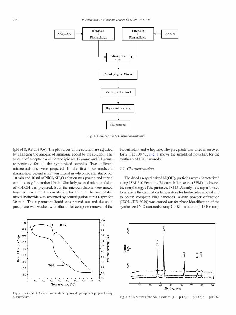
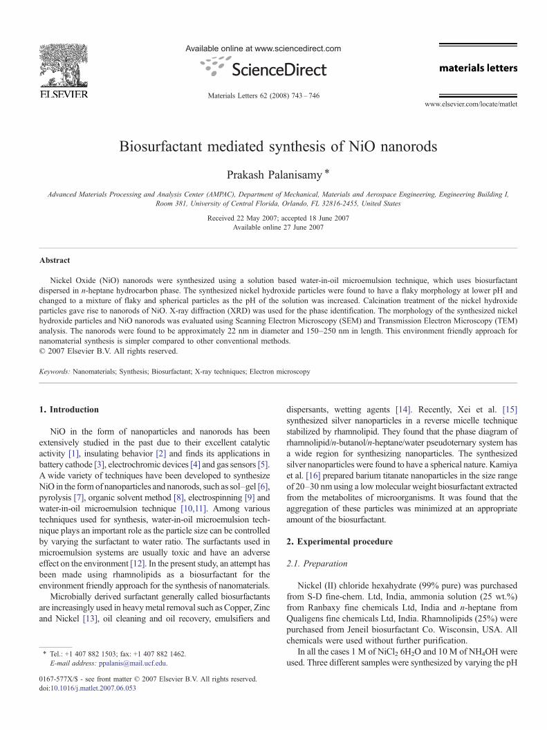
Fig. 1. Flowchart for NiO nanorod synthesis.
744 P. Palanisamy / Materials Letters 62 (2008) 743–746
(pH of 8, 9.3 and 9.6). The pH values of the solution are adjustedby changing the amount of ammonia added to the solution. Theamount of n-heptane and rhamnolipid are 17 grams and 0.1 gramsrespectively for all the synthesized samples. Two differentmicroemulsions were prepared. In the first microemulsion,rhamnolipid biosurfactant was mixed in n-heptane and stirred for10 min and 10 ml of NiCl2 6H2O solution was poured and stirredcontinuously for another 10 min. Similarly, secondmicroemulsionof NH4OH was prepared. Both the microemulsions were mixedtogether in with continuous stirring for 15 min. The precipitatednickel hydroxide was separated by centrifugation at 5000 rpm for30 min. The supernatant liquid was poured out and the solidprecipitate was washed with ethanol for complete removal of the
Fig. 2. TGA and DTA curve for the dried hydroxide precipitates prepared usingbiosurfactant.
biosurfactant and n-heptane. The precipitate was dried in an ovenfor 2 h at 100 °C. Fig. 1 shows the simplified flowchart for thesynthesis of NiO nanorods.
2.2. Characterization
The dried as-synthesized Ni(OH)2 particles were characterizedusing JSM-840 Scanning ElectronMicroscope (SEM) to observethemorphology of the particles. TG-DTA analysis was performedto estimate the calcination temperature for hydroxide removal andto obtain complete NiO nanorods. X-Ray powder diffraction(JEOL-JDX 8030) was carried out for phase identification of thesynthesized NiO nanorods using Cu-Kα radiation (0.15406 nm).
Fig. 3. XRD pattern of the NiO nanorods. (1— pH 8, 2— pH 9.3, 3— pH 9.6).
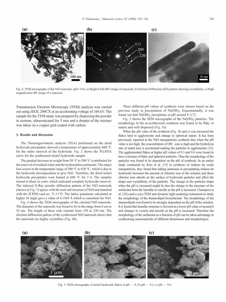
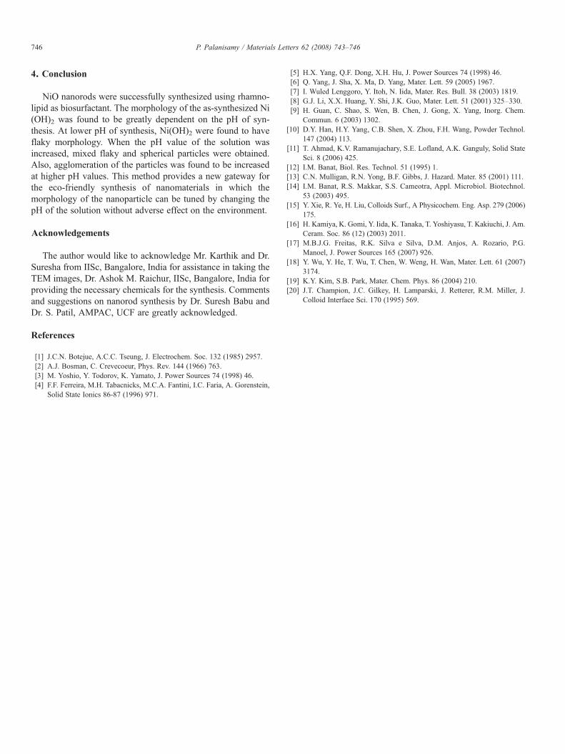
Fig. 4. TEM micrographs of the NiO nanorods. (pH=9.6). a) Bright Field (BF) image of nanorods, b) Electron Diffraction (ED) pattern showing crystallinity, c) Highmagnification BF image of a nanorod.
745P. Palanisamy / Materials Letters 62 (2008) 743–746
Transmission Electron Microscopy (TEM) analysis was carriedout using JEOL 200CX at an accelerating voltage of 160 kV. Thesample for the TEM study was prepared by dispersing the powderin acetone, ultrasonicated for 5 min and a droplet of the mixturewas taken on a copper grid coated with carbon.
3. Results and discussion
The Thermogravimetric analysis (TGA) performed on the driedhydroxide precipitates showed a temperature of approximately 600 °Cfor the entire removal of the hydroxide. Fig. 2 shows the TG-DTAcurve for the synthesized nickel hydroxide sample.
The gradual decrease in weight from 50 °C to 300 °C is attributed forthe removal of residual water and the hydrocarbon surfactant. Themajorloss occurs in the temperature range of 300 °C to 650 °C, which is due tothe hydroxide decomposition to give NiO. Therefore, the dried nickelhydroxide precipitates were heated at 600 °C for 3 h. The samplesturned to black in color, which indicated complete hydroxide removal.The indexed X-Ray powder diffraction pattern of the NiO nanorods(shown in Fig. 3) agrees with the rock salt structure of NiO and matchedwith the JCPDS card no. 71-1179. The lattice parameter calculated athigher 2θ angle gave a value of 4.1569 Å which is consistent for NiO.
Fig. 4 shows the TEM micrographs of the calcined NiO nanorods.The diameter of the nanorods was found to be in the range from 8 nm to35 nm. The length of these rods extends from 150 to 250 nm. Theelectron diffraction pattern of the synthesized NiO nanorods shows thatthe nanorods are highly crystalline (Fig. 4b).
Fig. 5. SEM micrographs of nickel hydroxide fla
Three different pH values of synthesis were chosen based on theprevious study in precipitation of Ni(OH)2. Experimentally, it wasfound out that Ni(OH)2 precipitates at pH around 8 [17].
Fig. 5 shows the SEM micrographs of the Ni(OH)2 particles. Themorphology in the as-synthesized condition was found to be flaky innature and well dispersed (Fig. 5a).
When the pH value of the synthesis (Fig. 5b and c) was increased theflakes tend to agglomerate and change to spherical nature. It has beenpreviously reported in the NiO nanoparticles synthesis that when the pHvalue is too high, the concentration of OH− ions is high and the hydrolysisrate of metal ions is accelerated making the particles to agglomerate [18].The agglomerated flakes at higher pH values of 9.3 and 9.6 were found tohave a mixture of flaky and spherical particles. Thus the morphology of theparticles was found to be dependent on the pH of synthesis. In an earlierstudy conducted by Kim et al. [19] in synthesis of indium tin oxidenanoparticles, they found that adding ammonia in precipitating indium tinhydroxide increases the amount of chlorine ions in the solution and thesechlorine ions adsorb on the surface of hydroxide particles and affect theshape and crystallinity of the particles. The change in the particles shapewhen the pH is increased might be due the change in the structure of thesurfactant from the lamellar to micelle as the pH is increased. Champion etal. [20] used a cryo-TEM and dynamic light scattering instrument to studythe morphology of the rhamnolipid biosurfactant. The morphology of therhamnolipids was found to be strongly dependent on the pH of the solution.It is found that lamellar structure is favored at a lower pH value of around 6and changes to vesicle and micelle as the pH is increased. Therefore themorphology of the surfactant as a function of pH can be taken advantage insynthesizing nanomaterials of different dimensions and morphologies.
kes a) pH — 8, b) pH — 9.3, c) pH — 9.6.
746 P. Palanisamy / Materials Letters 62 (2008) 743–746
4. Conclusion
NiO nanorods were successfully synthesized using rhamno-lipid as biosurfactant. The morphology of the as-synthesized Ni(OH)2 was found to be greatly dependent on the pH of syn-thesis. At lower pH of synthesis, Ni(OH)2 were found to haveflaky morphology. When the pH value of the solution wasincreased, mixed flaky and spherical particles were obtained.Also, agglomeration of the particles was found to be increasedat higher pH values. This method provides a new gateway forthe eco-friendly synthesis of nanomaterials in which themorphology of the nanoparticle can be tuned by changing thepH of the solution without adverse effect on the environment.
Acknowledgements
The author would like to acknowledge Mr. Karthik and Dr.Suresha from IISc, Bangalore, India for assistance in taking theTEM images, Dr. Ashok M. Raichur, IISc, Bangalore, India forproviding the necessary chemicals for the synthesis. Commentsand suggestions on nanorod synthesis by Dr. Suresh Babu andDr. S. Patil, AMPAC, UCF are greatly acknowledged.
References
[1] J.C.N. Botejue, A.C.C. Tseung, J. Electrochem. Soc. 132 (1985) 2957.[2] A.J. Bosman, C. Crevecoeur, Phys. Rev. 144 (1966) 763.[3] M. Yoshio, Y. Todorov, K. Yamato, J. Power Sources 74 (1998) 46.[4] F.F. Ferreira, M.H. Tabacnicks, M.C.A. Fantini, I.C. Faria, A. Gorenstein,
Solid State Ionics 86-87 (1996) 971.
[5] H.X. Yang, Q.F. Dong, X.H. Hu, J. Power Sources 74 (1998) 46.[6] Q. Yang, J. Sha, X. Ma, D. Yang, Mater. Lett. 59 (2005) 1967.[7] I. Wuled Lenggoro, Y. Itoh, N. Iida, Mater. Res. Bull. 38 (2003) 1819.[8] G.J. Li, X.X. Huang, Y. Shi, J.K. Guo, Mater. Lett. 51 (2001) 325–330.[9] H. Guan, C. Shao, S. Wen, B. Chen, J. Gong, X. Yang, Inorg. Chem.
Commun. 6 (2003) 1302.[10] D.Y. Han, H.Y. Yang, C.B. Shen, X. Zhou, F.H. Wang, Powder Technol.
147 (2004) 113.[11] T. Ahmad, K.V. Ramanujachary, S.E. Lofland, A.K. Ganguly, Solid State
Sci. 8 (2006) 425.[12] I.M. Banat, Biol. Res. Technol. 51 (1995) 1.[13] C.N. Mulligan, R.N. Yong, B.F. Gibbs, J. Hazard. Mater. 85 (2001) 111.[14] I.M. Banat, R.S. Makkar, S.S. Cameotra, Appl. Microbiol. Biotechnol.
53 (2003) 495.[15] Y. Xie, R. Ye, H. Liu, Colloids Surf., A Physicochem. Eng. Asp. 279 (2006)
175.[16] H. Kamiya, K. Gomi, Y. Iida, K. Tanaka, T. Yoshiyasu, T. Kakiuchi, J. Am.
Ceram. Soc. 86 (12) (2003) 2011.[17] M.B.J.G. Freitas, R.K. Silva e Silva, D.M. Anjos, A. Rozario, P.G.
Manoel, J. Power Sources 165 (2007) 926.[18] Y. Wu, Y. He, T. Wu, T. Chen, W. Weng, H. Wan, Mater. Lett. 61 (2007)
3174.[19] K.Y. Kim, S.B. Park, Mater. Chem. Phys. 86 (2004) 210.[20] J.T. Champion, J.C. Gilkey, H. Lamparski, J. Retterer, R.M. Miller, J.
Colloid Interface Sci. 170 (1995) 569.