Embed Size (px)
Citation preview
Biophysics of Sensory Perception,
Receptors,
Biophysics of Vision
Ján Jakuš
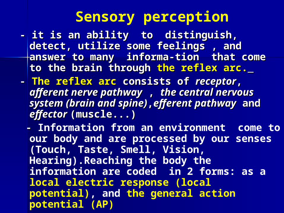
Sensory perception- - it is an ability to distinguish, detect, utilize it is an ability to distinguish, detect, utilize
some feelings , and answer to many informa-some feelings , and answer to many informa-tion that come to the brain through tion that come to the brain through the reflexthe reflex arc.arc.
- - The reflex arcThe reflex arc consists of consists of receptorreceptor, , afferent afferent nerve pathwaynerve pathway , , the central nervous system the central nervous system (brain and spine(brain and spine)),,efferent pathwayefferent pathway and and effector effector (muscle...)(muscle...)
- - Information from an environment come to our body and are processed by our senses (Touch, Taste, Smell, Vision, Hearing).Reaching the body the information are coded in 2 forms: as a local electric response (local potential), and the general action potential (AP)

Reflex arc
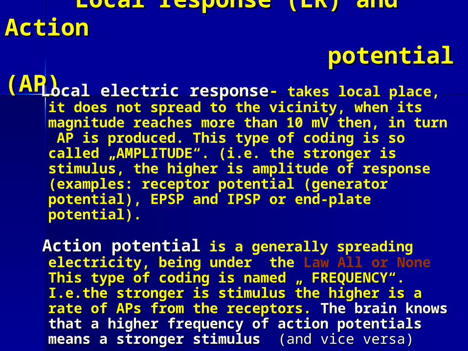
Local response (LRLocal response (LR)) and Action and Action potential (AP potential (AP))
Local electric responseLocal electric response-- takes local place, it does not spread to the vicinity, when its magnitude reaches more than 10 mV then, in turn AP is produced. This type of coding is so called „AMPLITUDE“. (i.e. the stronger is stimulus, the higher is amplitude of response (examples: receptor potential (generator potential), EPSP and IPSP or end-plate potential).
Action potentialAction potential is a generally spreading electricity, being under the Law All or None This type of coding is This type of coding is named „ FREQUENCY“. I.e.the stronger is stimulus the named „ FREQUENCY“. I.e.the stronger is stimulus the higher is a rate of APs from the receptors. higher is a rate of APs from the receptors. The brain The brain knows that a higher frequency of action potentials knows that a higher frequency of action potentials means a stronger stimulusmeans a stronger stimulus (and vice versa (and vice versa))

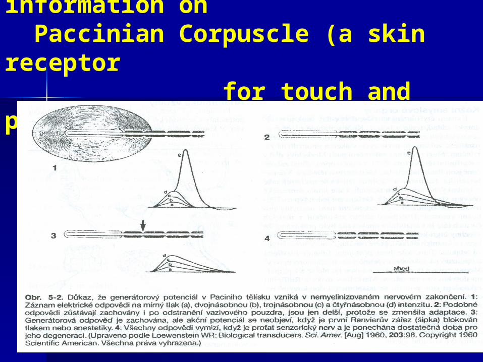
Coding of sensory information on Paccinian Corpuscle (a skin receptor for touch and pressure)
Coding of Stimulus on Coding of Stimulus on Paccinian Paccinian corpuscle corpuscle
Mechanical touch-pressure energy affects Mechanical touch-pressure energy affects the the receptor nerve membranereceptor nerve membrane (without (without myelin) causing its myelin) causing its local depolarizationlocal depolarization, , that results in an appearance of that results in an appearance of RECEPTOR RECEPTOR or GENERATOR POTENTIALor GENERATOR POTENTIAL (GP).(GP). When When another touch pres-sure stimuli come on another touch pres-sure stimuli come on the receptor it causes creation of many the receptor it causes creation of many local potentials and their local potentials and their summation . summation . When the amplitude of GP is aboveWhen the amplitude of GP is above 10mV 10mV thenthen the series of the series of ACTION POTENTIALS ACTION POTENTIALS rise uprise up on the afferent nerve fibre (which is on the afferent nerve fibre (which is covered with myelincovered with myelin)),,that leaves the that leaves the Paccinian corpuscle.Paccinian corpuscle.
Receptors- Definition and Properties - - Sensory Receptors Sensory Receptors are special nerve endings, are special nerve endings,
distributed throughout the body ( in the skin, distributed throughout the body ( in the skin, muscles, vessels, bones and joints, in lungs, muscles, vessels, bones and joints, in lungs, heart, and another organs).heart, and another organs).
- They Convert Different Forms of Energy into - They Convert Different Forms of Energy into Electrical SignalsElectrical Signals. . Thus they serve asserve as transducerstransducers,, changing the particular form of changing the particular form of energy ( e.g. me-chanical, chemical, thermal, or energy ( e.g. me-chanical, chemical, thermal, or electromagnetic) into the electromagnetic) into the electrical signal.electrical signal.
- Our body contains 20 types of receptors that can detect e.g.heat, pressure, stretch, acceleration, sound, light, smells, taste, partial pressure, concentration of salts, hormons...and other forms of stimuli (Only receptors for ionizing radiation are missing)

Receptors - ClassificationReceptors - Classification
I.According to localityI.According to locality: : Exteroreceptors- are placed within the skin, like receptors for touch, pressure, heat, cold or pain
Proprioreceptors- are placed in muscles, in bones and joints -they inform about the lengt of muscles and ligaments
Interoreceptors – receptors within the organs (heart, lungs, kidney) They detect plasma osmolarity, partial pressure of O2 blood pressure..
II.According to type of energyII.According to type of energy: : Mechanoreceptors- they transform mechanic energy into electric signal.E.g. exte-roreceptorś, baroreceptors, pulmonary stretch receptors).
Fotoreceptors- receptors containing photopigments (rods and cones at retina
Chemoreceptors – taste receptors in the tongue, smell receptors within a nose, osmoreceptors in hypothalamus,..
Nociceptors- pain receptors - in skin, in organs ...
III.According to complexityIII.According to complexity: simple receptors (skin) and complex ones (eye, ear)

The Flight ReflexThe Flight Reflex
bireflexarc.swf
Laws of Sensory Perception:
Weber-FechnerWeber-Fechner’’s Law of Perceptions Law of Perception: : is a is a basic psy-chophysical Law. Tbasic psy-chophysical Law. The bigger is the intensity of sti-mulation, the higher is the magnitude of sensation. Magnitude of sensation Magnitude of sensation E = log E = log SS , ( S - intensity of stimulation , ( S - intensity of stimulation ))or in a or in a modified formmodified form : :
StevensonStevenson’’s Laws Law: : FFAPAP= k . S= k . Snn ( (FAP is rate of APs from a receptor, k- constant, n=1 is valid for mechanoreceptors, n 1, for fotoreceptors, n 1, for pain receptors.
The Law of Projection:The Law of Projection:Each Each sense sense ococcucuppiesies the the uniquniq--ue and separated site within the brain ue and separated site within the brain cortex. Therefore, we are able to distinguish cortex. Therefore, we are able to distinguish the individual stimuli the individual stimuli -- like touch, pressure like touch, pressure,, pain sensationspain sensations, light or sound., light or sound.

The Law of Adaptation Adaptation - -is an internal electric property(caused by
membrane properties of the receptor) to respond when the long-term stimulus of a constant intensity is applied. Actualy, it is a drop of the receptor excitabili-ty to give rise the GP and then the APs
Receptors with Rapid Adaptation of their Burst Activity - - their fire just for a short time, during the constant (maintained ) stimulation ( as typically seen in touch, pressure, taste and smell receptors.)
Receptors with Low Adaptation of their Burst Activity -they fire for a long time with only a low drop of their firing activity (as seen in the pain, cold, heat receptors, baroreceptors, in pulmonary stretch recepors, the chemoreceptors, carotid baroreceptors or in the pulmonary stretch receptors).
The receptors with Low Adaptation are are involved in involved in a control a control of blood pressure, in of blood pressure, in control of breathing ,control of breathing , in in responses of body to responses of body to the pain, etc.the pain, etc.

Receptor Adaptation

Biophysics of Biophysics of VisionVision
is the most important human sense taking 80% of information from an environment.The stimulus for vision is electromagnetic waving of photons ( for Visible Light λ = 380 – 780 nm). For Ultraviolet Light λ is below 380 nm, for Infrared Light λ is above 780 nm. The velocity of visible light in vaccum is approx. 186 000 miles/s = 300 000 km/s.
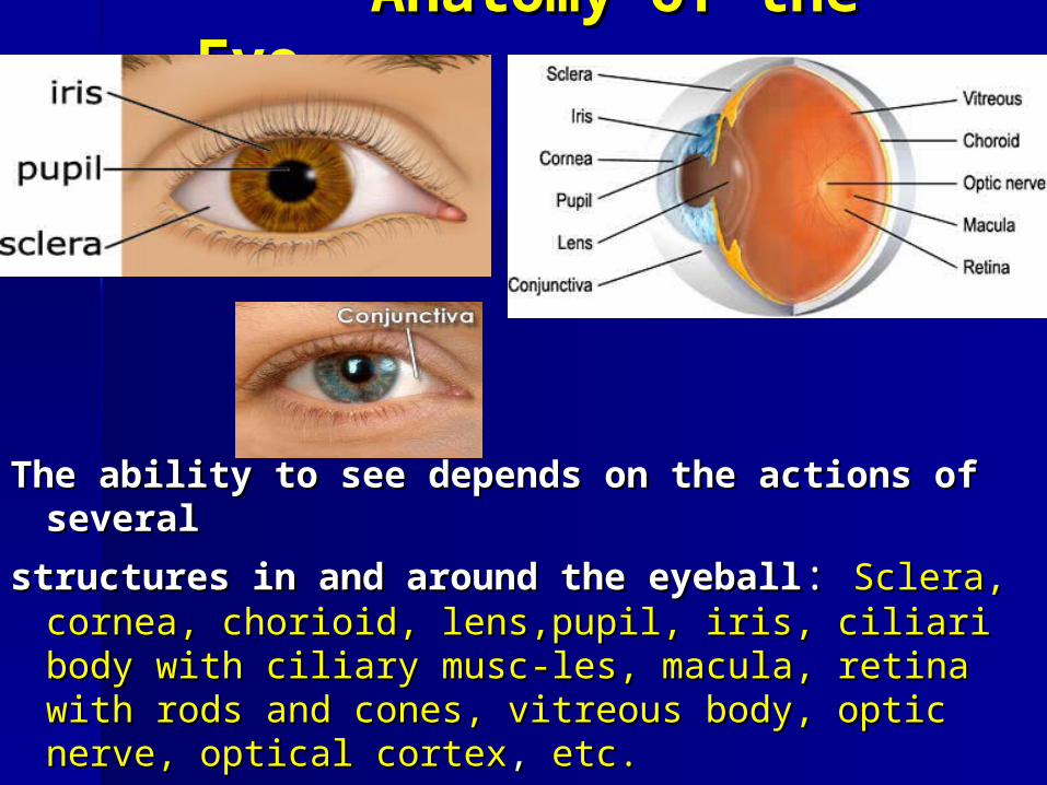
Anatomy of the EyeAnatomy of the Eye
The ability to see dependThe ability to see depends s on the actions of several on the actions of several
structures in and around the eyeballstructures in and around the eyeball:: Sclera, cornea, Sclera, cornea, chorioid, lens,pupil, iris, ciliari body with ciliary muscchorioid, lens,pupil, iris, ciliari body with ciliary musc--les, macula, retina with rods and cones, vitreous body, les, macula, retina with rods and cones, vitreous body, optic nerve, optical cortexoptic nerve, optical cortex, , etc.etc.

Parts of the Parts of the EyeEye
Anatomy.swf
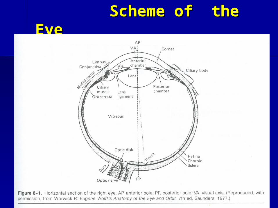
Scheme of the EyeScheme of the Eye
Optic analyzer consists of three main parts: Eyeball, Optic nerves and pathways, Occipital cortex.
The EyeballThe Eyeball ( is spherically shaped with d = 2.5 cm), consists of three special tissue covers( from outside to inside ): 1. the sclera, 2. the chorioid, 3. the retina, and with a two liquids( the humor aquens and the vitrous humor)
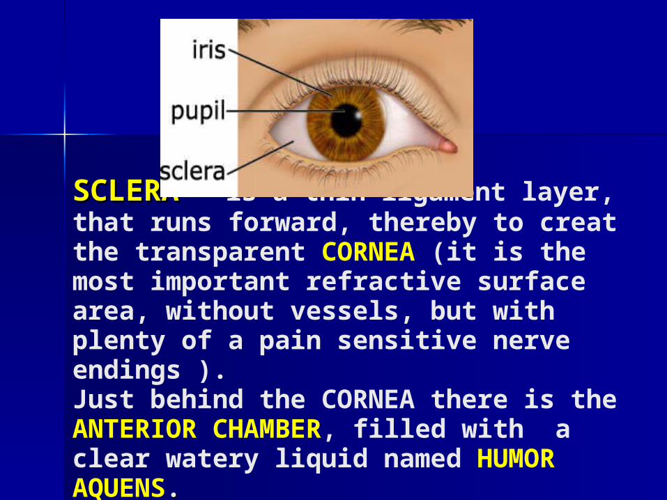
SCLERASCLERA – – is a thin ligament layer, that runs forward, thereby to creat the transparent CORNEA (it is the most important refractive surface area, without vessels, but with plenty of a pain sensitive nerve endings ). Just behind the CORNEA there is the ANTERIOR CHAMBER, filled with a clear watery liquid named HUMOR AQUENS. .
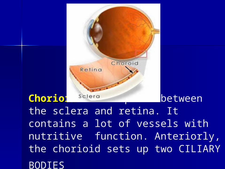
ChorioidChorioid takes place between the sclera and retina. It contains a lot of vessels with nutritive function. Anteriorly, the chorioid
sets up two CILIARY BODIES
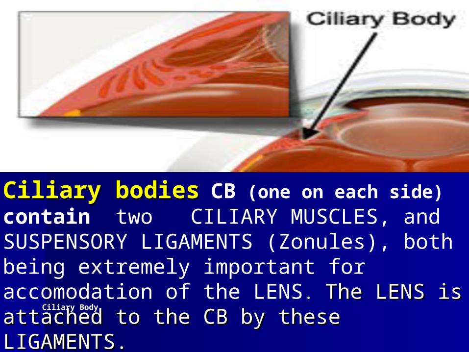
Ciliary Body
Ciliary bodCiliary bodiesies CB (one on each side) contain two CILIARY MUSCLES, and SUSPENSORY LIGAMENTS (Zonules), both being extremely important for accomodation of the LENS. The The LENS is attached to the CB by theseLENS is attached to the CB by these
LIGAMENTS. LIGAMENTS.

Lens
LENSLENS is transparent and placed just behind the iris. Its role is to focus (refract) the light rays onto the retina. When the lens is patologically changed (as a result of injury or diabetes mellitus), the lens is dimmed and this pathology is named as Cataract
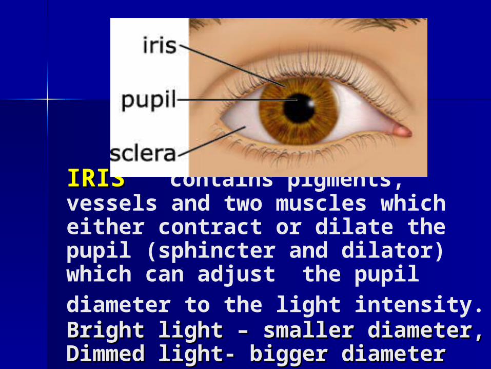
IRISIRIS contains pigments, vessels and two muscles which either contract or dilate the pupil (sphincter and dilator) which can adjust the pupil diameter to
the light intensity. Bright light – smaller Bright light – smaller diameter, Dimmed light- bigger diameterdiameter, Dimmed light- bigger diameter
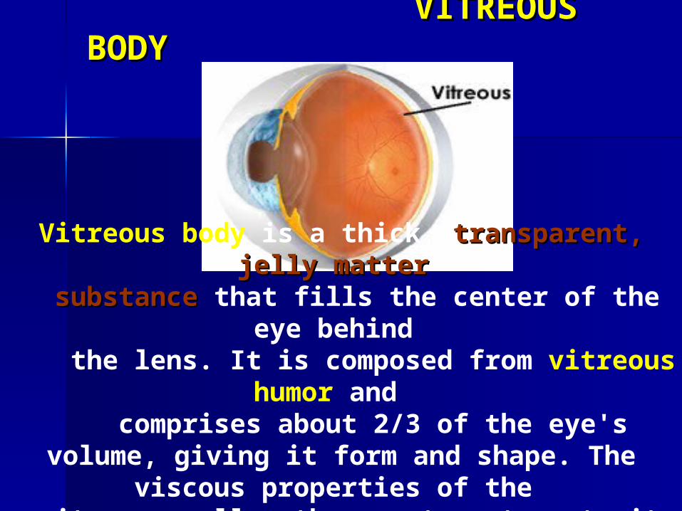
VITREOUS BODYVITREOUS BODY
Vitreous body is a thick, transparent,transparent, jelly matter jelly matter substancesubstance that fills the center of the eye behind
the lens. It is composed from vitreous humor and comprises about 2/3 of the eye's volume, giving it
form and shape. The viscous properties of the vitreous allow the eye to return to its normal shape
if compressed.
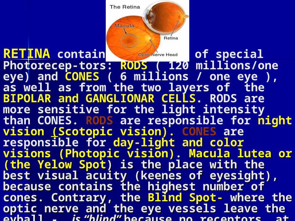
RETINA contains two kinds of special Photorecep-tors: RODS ( 120 millions/one eye) and CONES ( 6 millions / one eye ), as well as from the two layers of the BIPOLAR and GANGLIONAR CELLS. RODS are more sensitive for the light intensity than CONES. RODS are responsible for night vision (Scotopic vision). CONES are responsible for day-light and color visions (Photopic vision). Macula lutea or (the Macula lutea or (the Yelow SpotYelow Spot) is the place with the best visual acuity ) is the place with the best visual acuity (keenes of eyesight), because contains the highest (keenes of eyesight), because contains the highest numbernumber of cones. Contrary, the of cones. Contrary, the Blind Spot-Blind Spot- where where the optic nerve and the eye vessels leave the eyballthe optic nerve and the eye vessels leave the eyball - - is “blind” is “blind” because no receptors at this because no receptors at this site.site.

RETINAL RETINAL BACKGROUND BACKGROUND ((searched by Ophtalmoscopysearched by Ophtalmoscopy))
RememberRemember: The light rays first pass through the : The light rays first pass through the
layer of the Ganglionar Cells, then the Bipolar cells layer of the Ganglionar Cells, then the Bipolar cells
and finally strike the Rods and Cones. and finally strike the Rods and Cones.
PhotopigmentsPhotopigments within the rods and cones are bro- within the rods and cones are bro-
ken by light and electrons are released.The electro-ken by light and electrons are released.The electro-
nes create nes create Generator PotentialGenerator Potentialss inside th inside the Ganglio-e Ganglio-
arar Cells, Cells, but but not inside the RODES and CONES not inside the RODES and CONES
(because they are hyperpolarized at that time). (because they are hyperpolarized at that time).
Action PotentialAction Potentialss areare produced on the efferent produced on the efferent
axons that leave the Ganglionar Cells.axons that leave the Ganglionar Cells.
Chemistry of PhotopigmentsChemistry of PhotopigmentsRODSRODS contain pigment rhodopsine ( 11 cis-retinal-
opsine) that undergos the fotochemic reaction. Light chemically changes the Rhodposine into the Opsine ( all-trans - retinal opsine) + 1 electron. This electrone from the photoreceptor induces the production of Generator Potential and the AP. At night, OPSINE ( all- trans-form ) is reniewed into the original pigment RHODOPSINE ( 11-cis form) under the catalytic effect of Vitamin A. Thus, the VITAMIN A is important for synthesis of photo-pigments . When there is vitamin A deficiency it results in a disorder- HEMERALOPYHEMERALOPY (Dark-blind-ness syndrom)
3 types of CONES3 types of CONES contain photopigments :contain photopigments :Erytrolab, Erytrolab, Chlorolab, Cyanolab,Chlorolab, Cyanolab,(sensitive for red,green,blue(sensitive for red,green,blue))

COLOR COLOR VISION VISION (( HELMHOLTZ HELMHOLTZ --YYUUANG ANG THEORYTHEORY of of CColor olor VVisionision))..
Humans are able to perceive Humans are able to perceive 3 basic colors3 basic colors , green, , green, red, blue and a variety of mixing colors, because the red, blue and a variety of mixing colors, because the existency of existency of 3 kinds of special pigments in 3 3 kinds of special pigments in 3 different types of conesdifferent types of cones, within the retina. Normal , within the retina. Normal color vision is a typical feature for color vision is a typical feature for TRICHROMATS.TRICHROMATS. When one type of cone is missing or disabled, then When one type of cone is missing or disabled, then patient is patient is DICHROMAT DICHROMAT ,suffering from the particu ,suffering from the particu--lar type of a color blindnesslar type of a color blindness e.g. e.g. deuteroanopy, deuteroanopy, protanopy or tritanopyprotanopy or tritanopy.. ( see practicals for details ( see practicals for details))When all three types of CONES are disabled one is When all three types of CONES are disabled one is MONOCHROMATMONOCHROMAT. Color blindness is hereditary . Color blindness is hereditary disorderdisorder (For more details see Nave and Nave and (For more details see Nave and Nave and Handouts).Handouts).

AccomodationAccomodation – is a process – is a process whenwhen the refrac-tory power of the eye rises of the eye rises upup. Accomodation enab. Accomodation enab--les to focus our eye from the les to focus our eye from the FAR POINTFAR POINT ( approx. ( approx. above above 6 m, to the point named 6 m, to the point named NEAR POINTNEAR POINT of of vision (the closest point on which one can focus vision (the closest point on which one can focus sharply) sharply) Durind the accomodationDurind the accomodation , , contraction of ciliary musclesciliary muscles causes causes the relaxation of the suspe-suspe-nsory ligamentsnsory ligaments. Because the lens own elasticity, . Because the lens own elasticity, the lens will be thicker - obtaining more spherical the lens will be thicker - obtaining more spherical shape.shape. It looks like the lens „ It looks like the lens „moves forward“.moves forward“. The The shorter is the distance between the subject and the shorter is the distance between the subject and the eye, the greater has to be the accomodation. eye, the greater has to be the accomodation. The ref-ractory power of the eye ( degree of accomodation) is measured in unit named DIOPTRIA (diopterDIOPTRIA (diopter) D= 1 / focal distance (mD= 1 / focal distance (m) For whole eye the optical power is approx. 59 D, for cornea is 43 D, for lens around 16 D. !!

Eye.swf
FFailures of the Image Forming ailures of the Image Forming MechanismMechanism Refraction Refraction FFailuresailures :: MYOPIA, HYPEROPIA, PRESBYOPIA, and ASTIGMATISM
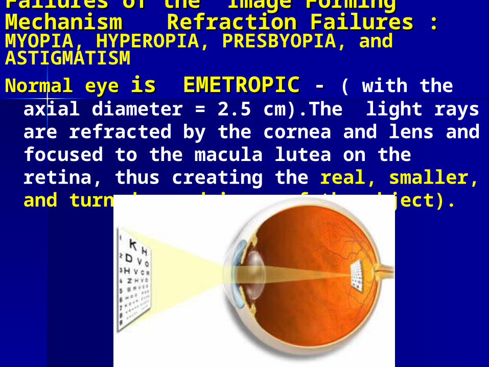
Normal Normal eye eye is EMETROPICis EMETROPIC - - ( with the axial diameter = 2.5 cm).The light rays are refracted by the cornea and lens and focused to the macula lutea on the retina, thus creating the real, smaller, and turned round image of the object).
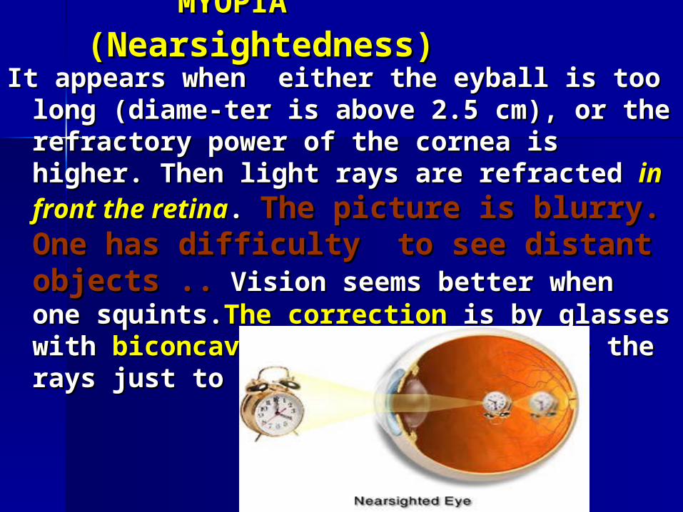
MYOPIA MYOPIA (N(Nearsightednessearsightedness)) It It appears when appears when either either the eyball is too long (dthe eyball is too long (diame-iame-
terter is above 2 is above 2..5 cm), 5 cm), or the refractory power of the or the refractory power of the cornea is higher. Tcornea is higher. Then hen light light rays are refracted rays are refracted in in front the retinafront the retina. . The picture is blurry.The picture is blurry. One hasOne has difficulty to see distant objects ..difficulty to see distant objects .. Vision Vision seems better when one squints.seems better when one squints.The correctThe correctiionon is is by glasses with by glasses with biconcave lenses,biconcave lenses, that diverge the that diverge the rays just to the retina. rays just to the retina.
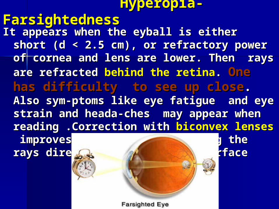
Hyperopia- FarsightednessHyperopia- Farsightedness
It It appears when the eyball is appears when the eyball is either either short (d short (d << 2.5 2.5 cm)cm), or refractory power of cornea and lens are , or refractory power of cornea and lens are lower.lower. Then Then rays are refracted rays are refracted behind the retinabehind the retina. . One hasOne has difficulty to see up closedifficulty to see up close. . Also sym-Also sym-ptoms like eye fatigue and eye strain and heada-ptoms like eye fatigue and eye strain and heada-ches may appear when reading .ches may appear when reading .Correction with Correction with biconvex lensesbiconvex lenses improves this failure, focusing improves this failure, focusing the rays directly on the retinal surfacethe rays directly on the retinal surface
PRESBYOPIA- PRESBYOPIA- OLDSIGHTEDNEESOLDSIGHTEDNEES (is(is
kind of Hyperopia i.e. farsightedness). Presbyopia is also known as the “short arm
syndrome” The elasticity of LENS is age
depended. The persons above 45 years, loose progressively the lens elasticity, therefore their refractive eye power and accomodation
are steping down,are steping down, Their NEAR POINT of vision reaches the distance more than 45 cm. In order to improve the sharpness of vision
one has to take glasses with biconvex lensesbiconvex lenses.


Refraction Failures- schemeRefraction Failures- scheme
All of mentioned above failures can be All of mentioned above failures can be treated by wearing of glasses with bicon-treated by wearing of glasses with bicon-cave or biconvex lenses , or using the cave or biconvex lenses , or using the
conact lenses , or even by laser surgeryconact lenses , or even by laser surgery. .
See Handouts for Practicals. Tasks: See Handouts for Practicals. Tasks: Determination ofDetermination of Visual AquityVisual Aquity by by Snellens TypesSnellens Types , , Determination of Near and Far Points of VisionDetermination of Near and Far Points of Vision

Vision.swf

Thanks for Comming Thanks for Comming
and and
Attention !Attention !