Embed Size (px)
Citation preview

0
CECE 2011 “… bringing
people and ideas
together …”
8th International Interdisciplinary
Meeting on Bioanalysis
November 3-4, 2011
Hotel Continental, Brno, Czech Republic
www.ce-ce.org
1
ISBN 978-80-904959-0-6
Organized by:
Institute of Analytical Chemistry AS CR, v. v. i., Veveří 97, 602 00 Brno
Organizing committee: František Foret, Ludmila Křivánková, Karel Klepárník, Iveta Drobníková
Webmaster: František Matulík
Find the meeting history and more at www.ce-ce.org
2
Foreword
Welcome to CECE 2011. After two years in Pecs, Hungary we are back to Brno. I
want to thank Professor Ferenc Kilár for his hospitality and excellent organization of
the meeting in 2009 and 2010 and hope we will find new international venues in the
future again. Since 2004, when a one day seminar was given by Dusan Kaniansky,
Ernst Kenndler and Bob Gas in the conference room of the Institute of Analytical
Chemistry, we are now in the 8th
year of CECE. Today, the two day meeting with
invited lectures and poster sessions is still unique. Our goal is bringing together
scientists who may not meet at specialized meetings, promote informal
communication of researchers from different disciplines and map the current status of
the fields shaping the bioanalytical science. The organizers want to thank the invited
speakers and all the participants and hope that you will enjoy the scientific
presentations as well as personal contacts and informal discussions.
Franta Foret

3
Program
Thursday, November 3
9:00 – 9:15 Conference opening
9:15 – 9:45
Detlev Belder, Universität Leipzig, Germany
Enantioselective catalysis and analysis in single microfluidic devices
9:45 - 10:15
Christian Klampfl, Johannes Kepler-University Linz, Austria
Coupling CE to MS: new interfaces, new ion sources new applications
10:15 – 10:45
Oliver Trapp, Ruprecht-Karls-Universität Heidelberg, Germany
CE as Tool to Investigate the Stereodynamics and Catalytic Reactions
10:45 – 11:15
Ziad El Rassi, Oklahoma State University, Stillwater, USA
Immuno-Monoliths at Reduced Nonspecific Interactions
11:15 – 11:45
Lloyd M. Smith, University of Wisconsin-Madison, USA
New Technologies for the Genome Age
11:45 – 14:00 Lunch break – poster session
14:00 – 14:30
Jiri Fajkus, Masaryk University Brno, Czech Republic
Analysis of telomeres and telomerases
14:30 – 15:00
Rui Vitorino, University of Aveiro, Portugal
What is saliva? A perspective of salivary proteomics at University of
Aveiro
15:00 – 15:30
Françoise Nepveu, University of Toulouse 3, France
Indolone-n-oxides: relation between redox properties and antimalarial
activities.
4
15:30 – 16:00
Pier Giorgio Righetti, Politecnico di Milano, Italy
The proteome Argonauts: conquering the “golden fleece” of alcoholic
beverages and soft drinks via combinatorial peptide ligands
19:00
Conference dinner with traditional Moravian music-Hotel
Continental
Friday, November 4
9:00 – 9:30
Huan-Tsung Chang, National Taiwan University, Taiwan
Separation of DNA and DNA-templated metal clusters by CE-LIF
9:30 – 10:00
Andras Guttman, University of Debrecen, Hungary
Capillary electrophoresis analysis of the altered glycosylation of
immunoglobulins in autoimmune diseases
10:00 – 10:30
Aran Paulus, Bio-Rad Laboratories, USA
Microfabricated and Lab-on-a-chip Devices in the Life Science Industry:
Promises, Products and Problems
10:30 – 11:00 coffee break
11:00 – 11:30
Steven Soper, University of North Carolina, USA
Single-Molecule Electrochromatography in Nano-columns with
Conductivity Readout: A Novel Approach for High Throughput DNA
Sequencing
11:30 - 12:00
Andreas Manz, KIST Europe, Saarbrücken, Germany
Microfluidics for applications in chemistry
12:00 – 14:00 Lunch break – poster session
14:00 – 14:30
Ferenc Kilar, University of Pecs, Hungary
5
Isoelectric focusing coupled to mass spectrometry for bioanalysis
14:30 – 15:00
Gyula Vigh, Texas A&M University, USA
Design, Synthesis and Analytical Characterization of a New Family of pI
Markers
15:00 – 15:30
Vaclav Kasicka Institute of Organic Chemistry and Biochemistry,
Prague, Czech Republic
Investigation of biopeptide interactions by capillary electrophoresis
15:30 Closing remarks
6
About the Invited Speakers
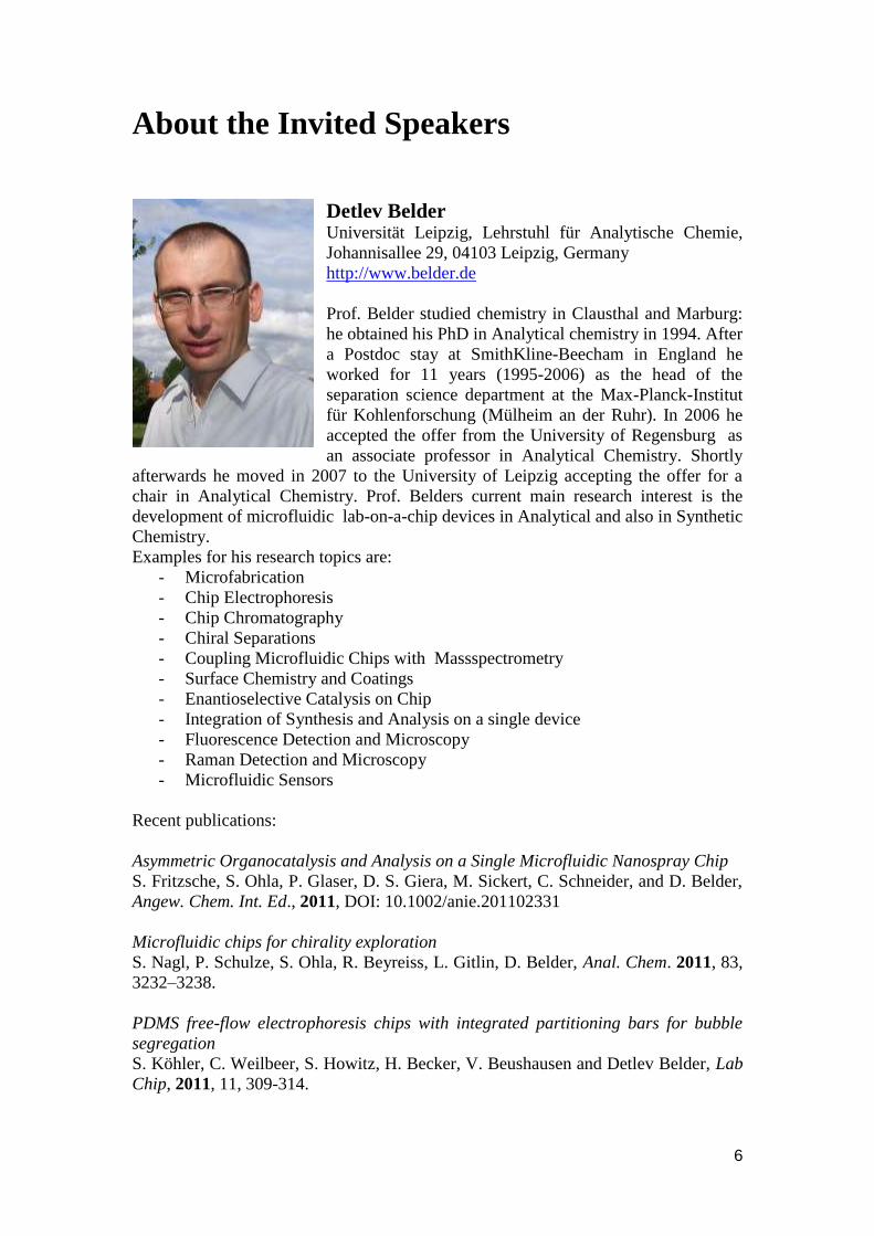
Detlev Belder Universität Leipzig, Lehrstuhl für Analytische Chemie,
Johannisallee 29, 04103 Leipzig, Germany
http://www.belder.de
Prof. Belder studied chemistry in Clausthal and Marburg:
he obtained his PhD in Analytical chemistry in 1994. After
a Postdoc stay at SmithKline-Beecham in England he
worked for 11 years (1995-2006) as the head of the
separation science department at the Max-Planck-Institut
für Kohlenforschung (Mülheim an der Ruhr). In 2006 he
accepted the offer from the University of Regensburg as
an associate professor in Analytical Chemistry. Shortly
afterwards he moved in 2007 to the University of Leipzig accepting the offer for a
chair in Analytical Chemistry. Prof. Belders current main research interest is the
development of microfluidic lab-on-a-chip devices in Analytical and also in Synthetic
Chemistry.
Examples for his research topics are:
- Microfabrication
- Chip Electrophoresis
- Chip Chromatography
- Chiral Separations
- Coupling Microfluidic Chips with Massspectrometry
- Surface Chemistry and Coatings
- Enantioselective Catalysis on Chip
- Integration of Synthesis and Analysis on a single device
- Fluorescence Detection and Microscopy
- Raman Detection and Microscopy
- Microfluidic Sensors
Recent publications:
Asymmetric Organocatalysis and Analysis on a Single Microfluidic Nanospray Chip
S. Fritzsche, S. Ohla, P. Glaser, D. S. Giera, M. Sickert, C. Schneider, and D. Belder,
Angew. Chem. Int. Ed., 2011, DOI: 10.1002/anie.201102331
Microfluidic chips for chirality exploration
S. Nagl, P. Schulze, S. Ohla, R. Beyreiss, L. Gitlin, D. Belder, Anal. Chem. 2011, 83,
3232–3238.
PDMS free-flow electrophoresis chips with integrated partitioning bars for bubble
segregation
S. Köhler, C. Weilbeer, S. Howitz, H. Becker, V. Beushausen and Detlev Belder, Lab
Chip, 2011, 11, 309-314.

7
Huan-Tsung Chang (Department of Chemistry National Taiwan University,
http://www.ch.ntu.edu.tw/~htchang/) focuses on
development of micro-nano techniques and green
approaches. He has demonstrated a number of new
concepts and methods, including (1) On-line
concentration and separation capillary electrophoresis
(CE); (2) Nanoparticles (NPs) filled capillary
electrophoresis (NFCE); (3) Green syntheses of
anisotropic structures of metallic NPs, quantum dots
(QDs), and Te/Au nanowires in aqueous solution; (4)
NPs as concentration and laser desorption/ionization
matrices in mass spectrometry; (5) Aptamers
functionalized NPs for the detection of metal ions, small
molecules, and proteins; (6) Preparation and applications of fluorescent gold nanodots
(Au NDs); (7) Sensitive and selective DNA probes for the detection of DNA and
metal ions; (8) Preparation of fluorescent DNA-templated metal clusters for the
detection of metal ions and DNA; (9) DNA-functional nanomaterials for control of
enzyme activity; (10) Fabrication of quantum dot sensitized solar cells; (11) Pt
nanomaterials for highly electrocatalytic methanol oxidation; and (12) New
nanomaterials (Te/Au nanowires) as substrates for surface enhanced Raman
scattering. He has published more than 180 papers.
Recognition: Young Chemists Award of the Chemical Society Located in Taipei
(2000); Young Scholar Award, College of Science, National Taiwan University
(2000); Fu Szu-Nien Award, National Taiwan University (2005); Outstanding
Research Award, National Science Council, Taiwan (2007); Y. Z. Hsu Scientific
Paper Award (Green Technology Category) (2008); Distinguished Professor of
National Taiwan University (2008-present)
Jiří Fajkus (http://www.ceitec.eu/contact/research-
programmes/genomics-and-proteomics-of-plant-
systems/) has started research during his PhD studies
at the Institute of Biophysics, ASCR (1988-1992) with
analysis of structure and function of plant DNA
repeats and their chromatin structure at the level of
nucleosomes and chromatin loops. After his
postdoctoral stay in the laboratory of Prof. Ronald
Hancock, Laval University Cancer Research Centre,
Québec, Canada, where he has been performing
mapping of topoII-associated chromatin regions in the
human hprt gene (1992-1993,1995), he started his
own research group at the Institute of Biophysics. He became interested in biology of
telomeres, and plant telomeres in particular. In 1995, he has described a specific
nucleosome structure of plant telomeres, later on developed into a columnar model of
telomeric chromatin (in collaboration with Prof. Edward N. Trifonov, 2001). In 1996
he was the first to detect telomerase in plant cells. In 1998, his group described the
maintenance of telomere length stability during plant development and a reversible
8
control of plant telomerase activity, the features distinguishing telomere biology of
plants from that of humans. Since 2000, he has published a number of studies on
plant telomere binding proteins and their interactions, molecular evolution of plant
telomeres and telomerases, and also on applications of telomere and telomerase
analysis in oncology diagnostics. For the purpose of diagnostic applications, he has
developed a two-color quantitative real-time version of TRAP assay for telomerase
activity (2003). His recent achievements include detection of telomerase-independent
telomere lengthening in plants and its role in plant development (2008), description of
the selective loss of telomeres and rDNA genes in Arabidopsis thaliana CAF1
(chromatin-assembly factor 1) mutants (2010), or studies on the epigenetic regulation
of telomere maintenance (2011).
Recognitions
Prize of the Rector of the Masaryk University and J. E. Purkinje Medal - 1987;
Award of the Czech Ministry of Education "Talent 98" - 1999;
Prize of the Minister of Education for Research – 2000,
Otto Wichterle Premium (Czech Academy of Sciences) - 2002.
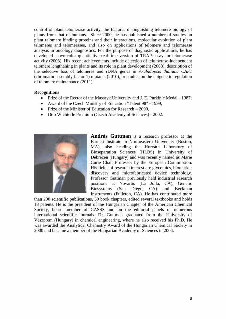
András Guttman is a research professor at the
Barnett Institute in Northeastern University (Boston,
MA), also heading the Horváth Laboratory of
Bioseparation Sciences (HLBS) in University of
Debrecen (Hungary) and was recently named as Marie
Curie Chair Professor by the European Commission.
His fields of research interest are glycomics, biomarker
discovery and microfabricated device technology.
Professor Guttman previously held industrial research
positions at Novartis (La Jolla, CA), Genetic
Biosystems (San Diego, CA) and Beckman
Instruments (Fulleton, CA). He has contributed more
than 200 scientific publications, 30 book chapters, edited several textbooks and holds
18 patents. He is the president of the Hungarian Chapter of the American Chemical
Society, board member of CASSS and on the editorial panels of numerous
international scientific journals. Dr. Guttman graduated from the University of
Veszprem (Hungary) in chemical engineering, where he also received his Ph.D. He
was awarded the Analytical Chemistry Award of the Hungarian Chemical Society in
2000 and became a member of the Hungarian Academy of Sciences in 2004.
9
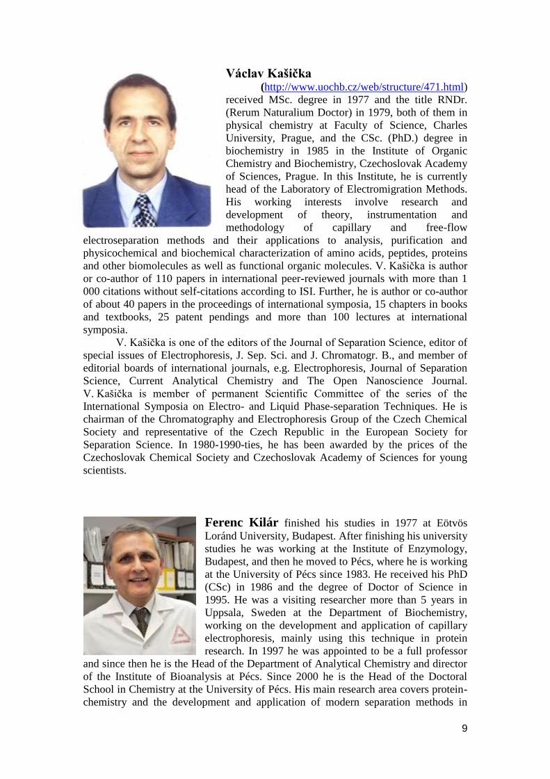
Václav Kašička (http://www.uochb.cz/web/structure/471.html)
received MSc. degree in 1977 and the title RNDr.
(Rerum Naturalium Doctor) in 1979, both of them in
physical chemistry at Faculty of Science, Charles
University, Prague, and the CSc. (PhD.) degree in
biochemistry in 1985 in the Institute of Organic
Chemistry and Biochemistry, Czechoslovak Academy
of Sciences, Prague. In this Institute, he is currently
head of the Laboratory of Electromigration Methods.
His working interests involve research and
development of theory, instrumentation and
methodology of capillary and free-flow
electroseparation methods and their applications to analysis, purification and
physicochemical and biochemical characterization of amino acids, peptides, proteins
and other biomolecules as well as functional organic molecules. V. Kašička is author
or co-author of 110 papers in international peer-reviewed journals with more than 1
000 citations without self-citations according to ISI. Further, he is author or co-author
of about 40 papers in the proceedings of international symposia, 15 chapters in books
and textbooks, 25 patent pendings and more than 100 lectures at international
symposia. V. Kašička is one of the editors of the Journal of Separation Science, editor of
special issues of Electrophoresis, J. Sep. Sci. and J. Chromatogr. B., and member of
editorial boards of international journals, e.g. Electrophoresis, Journal of Separation
Science, Current Analytical Chemistry and The Open Nanoscience Journal.
V. Kašička is member of permanent Scientific Committee of the series of the
International Symposia on Electro- and Liquid Phase-separation Techniques. He is
chairman of the Chromatography and Electrophoresis Group of the Czech Chemical
Society and representative of the Czech Republic in the European Society for
Separation Science. In 1980-1990-ties, he has been awarded by the prices of the
Czechoslovak Chemical Society and Czechoslovak Academy of Sciences for young
scientists.
Ferenc Kilár finished his studies in 1977 at Eötvös
Loránd University, Budapest. After finishing his university
studies he was working at the Institute of Enzymology,
Budapest, and then he moved to Pécs, where he is working
at the University of Pécs since 1983. He received his PhD
(CSc) in 1986 and the degree of Doctor of Science in
1995. He was a visiting researcher more than 5 years in
Uppsala, Sweden at the Department of Biochemistry,
working on the development and application of capillary
electrophoresis, mainly using this technique in protein
research. In 1997 he was appointed to be a full professor
and since then he is the Head of the Department of Analytical Chemistry and director
of the Institute of Bioanalysis at Pécs. Since 2000 he is the Head of the Doctoral
School in Chemistry at the University of Pécs. His main research area covers protein-
chemistry and the development and application of modern separation methods in
10
bioanalysis. He is a co-author of more then 100 scientific publications and 3 books.
He is a member of several national and international research consortia and received
several national and European grants for his research. He was a visiting professor at
Universitá "La Sapienza" and Istituto di Cromatografia, Rome, Italy, University of
Bern, Switzerland and L'Institut Pasteur, Paris, France. He is the member of the
editorial boards of Journal of Biochemical and Biophysical Methods (2001-2008),
Hungarian Chemical Journal (2001-2007), Studia Universitatis Babes-Bolyai Chemia
(since 2007), Electrophoresis (since 2008), Journal of Proteomics (since 2008).
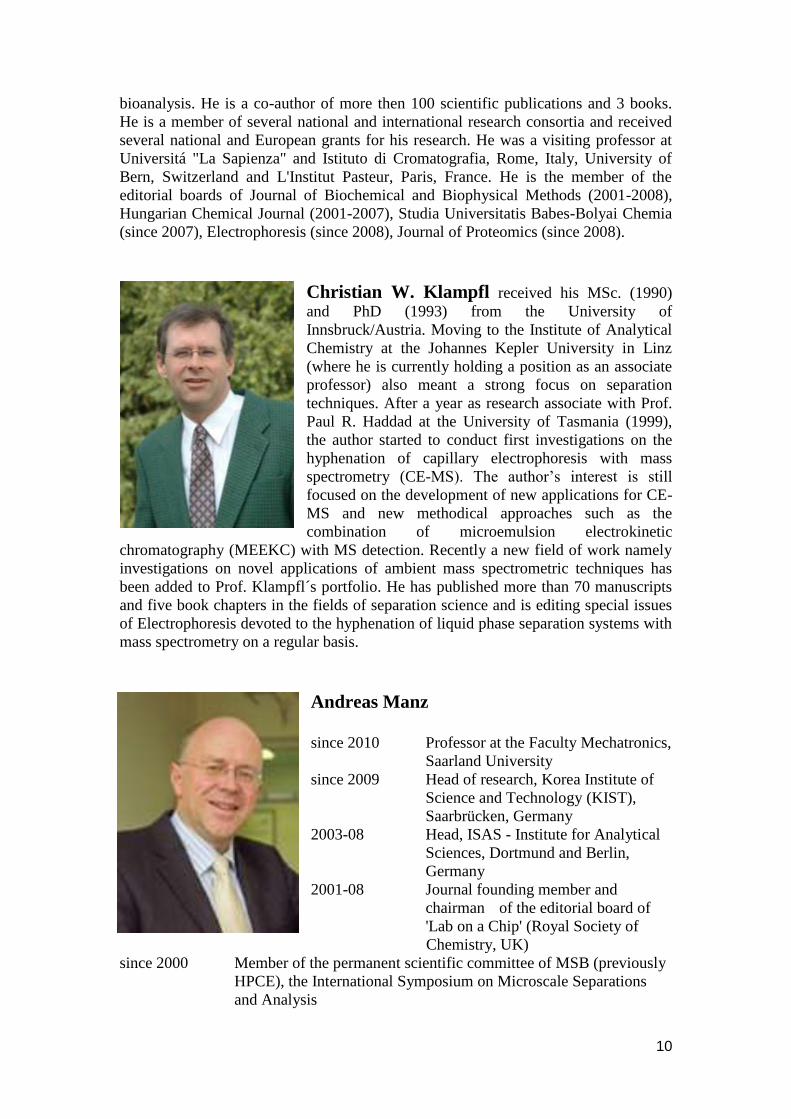
Christian W. Klampfl received his MSc. (1990)
and PhD (1993) from the University of
Innsbruck/Austria. Moving to the Institute of Analytical
Chemistry at the Johannes Kepler University in Linz
(where he is currently holding a position as an associate
professor) also meant a strong focus on separation
techniques. After a year as research associate with Prof.
Paul R. Haddad at the University of Tasmania (1999),
the author started to conduct first investigations on the
hyphenation of capillary electrophoresis with mass
spectrometry (CE-MS). The author‟s interest is still
focused on the development of new applications for CE-
MS and new methodical approaches such as the
combination of microemulsion electrokinetic
chromatography (MEEKC) with MS detection. Recently a new field of work namely
investigations on novel applications of ambient mass spectrometric techniques has
been added to Prof. Klampfl´s portfolio. He has published more than 70 manuscripts
and five book chapters in the fields of separation science and is editing special issues
of Electrophoresis devoted to the hyphenation of liquid phase separation systems with
mass spectrometry on a regular basis.
Andreas Manz
since 2010 Professor at the Faculty Mechatronics,
Saarland University
since 2009 Head of research, Korea Institute of
Science and Technology (KIST),
Saarbrücken, Germany
2003-08 Head, ISAS - Institute for Analytical
Sciences, Dortmund and Berlin,
Germany
2001-08 Journal founding member and
chairman of the editorial board of
'Lab on a Chip' (Royal Society of
Chemistry, UK)
since 2000 Member of the permanent scientific committee of MSB (previously
HPCE), the International Symposium on Microscale Separations
and Analysis
11
since 1997 Fellow of the Royal Society of Chemistry (CChem FRSC), U.K.
1995 - 2004 SmithKline Beecham Professor of Analytical Chemistry at Imperial
College, Dept. Chemistry, London, U.K.
1995 - 2000 Scientific advisor and consultant of Caliper Technologies, Mountain
View, California. The company is active in "lab on chip"
technology.
1995 Habilitation thesis (supervisor: Prof. Dr. Grasserbauer) accepted at
the Technical University (TU) Vienna, Austria. Topic: 'Micro
System Technology for Use in Analytical Chemistry'.
1994 - 2008 Member of the scientific committee and the board of of µTAS, the
International Symposium on Micro Total Analysis Systems
1991 - 1995 Group leader at Ciba-Geigy Corporate Analytical Research, Basel,
Switzerland.
1988 - 1991 Researcher at Ciba-Geigy Central Research Lab., Basel,
Switzerland
1987 - 1988 Postdoctoral fellow at Hitachi Central Research Lab., Hitachi Ltd.,
Tokyo, Japan.
1983 - 1986 Dr. sc. tech. thesis (PhD) under the guidance of Prof. Dr. W. Simon
(Dept. for Organic Chemistry, Swiss Federal Institute of
Technology, ETH, Zürich, Switzerland).
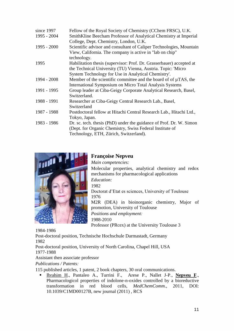
Françoise Nepveu Main competencies:
Molecular properties, analytical chemistry and redox
mechanisms for pharmacological applications
Education:
1982
Doctorat d‟Etat es sciences, University of Toulouse
1976
M2R (DEA) in bioinorganic chemistry, Major of
promotion, University of Toulouse
Positions and employment:
1988-2010
Professor (PRcex) at the University Toulouse 3
1984-1986
Post-doctoral position, Technische Hochschule Darmastadt, Germany
1982
Post-doctoral position, University of North Carolina, Chapel Hill, USA
1977-1988
Assistant then associate professor
Publications / Patents:
115 published articles, 1 patent, 2 book chapters, 30 oral communications.
Ibrahim H., Pantaleo A., Turrini F., Arese P., Nallet J-P., Nepveu F.,
Pharmacologicol properties of indolone-n-oxides controlled by a bioreductive
transformation in red blood cells, MedChemComm., 2011, DOI:
10.1039/C1MD00127B, new journal (2011) , RCS
12
Reybier K., Perio P, Ferry G., Bouajila J, Delagrange Ph;, Boutin J. A., Nepveu
F. Insights into the redox cycle of human quinone reductase 2. Free Rad Res.,
2011, 45(10), 1184-1195
Nepveu F., Kim S., Boyer J., Chatriant O., Ibrahim H., Reybier K., Monje M-C,
Chevalley S., Perio P., Lajoie B., Bouajila J., Deharo E., Sauvain M., Tahar R.,
Basco L., Pantaleo A., Turini F., Arese P., Valentin A., Thompson E., Vivas L.,
Petit S., Nallet J-P, Synthesis and antiplasmodial activity of new indolon N-
oxide derivatives, J. Med. Chem, 2010, 53, 699-714.
Ibrahim N., Ibrahim H., Kim S., Nallet J.-P., Nepveu F., Interactions
between Antimalarial Indolone-N-oxide Derivatives and Human Serum
Albumin. Biomacromolecules, 2010, 11, 3341-3351.
Reybier K., Ribaut C., Coste A., Launay J., Fabre P.L., Nepveu F.,
Characterization of oxidative stress in Leishmaniasis-infected or LPS-stimulated
macrophages using electrochemical impedance spectroscopy, Biosensors and
Bioelectronics, 2010, 25(12), 2566-2572.
Awards, activities, memberships:
Awards: Chevalier dans l‟Ordre de la Légion d‟Honneur (14/12/2010). Alexander
Von Humboldt fellowhip ; PEDR 1990-2010. Activities: Head of the laboratory UMR
152 IRD-UT3 2003-2010; teaching activities: 192 h/year in physical chemistry and
instrumentation for biological media, drug and food analysis; antioxidants, pro-
oxidants; supervision of 36 post-graduate students (1986-2005), 13 PhD 1991-2010;
23 grants (1991-2010); Head of the Department of analytical chemistry (1996-2001);
President of the local CNU section 39eme (2007-2008): head of laboratories (1991-
2002). Memberships: Office Member of the Food-Health Consortium of the Region
Midi-Pyrenees (2000-2010); member of the Doctoral School in Sciences of Matter;
member of scientific societies (SFC, SCT, SFRR, ACS). Expert (AERES, ANR),
Reviewer of scientific journals.
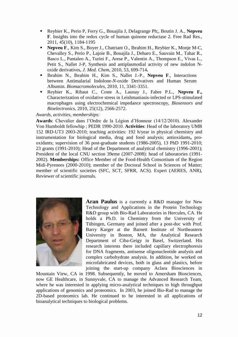
Aran Paulus is a currently a R&D manager for New
Technology and Applications in the Protein Technology
R&D group with Bio-Rad Laboratories in Hercules, CA. He
holds a Ph.D. in Chemistry from the University of
Tübingen, Germany and joined after a post-doc with Prof.
Barry Karger at the Barnett Institute of Northeastern
University in Boston, MA, the Analytical Research
Department of Ciba-Geigy in Basel, Switzerland. His
research interests there included capillary electrophoresis
for DNA fragments, antisense oligonucleotide analysis and
complex carbohydrate analysis. In addition, he worked on
microfabricated devices, both in glass and plastics, before
joining the start-up company Aclara Biosciences in
Mountain View, CA in 1998. Subsequently, he moved to Amersham Biosciences,
now GE Healthcare, in Sunnyvale, CA to manage the Advanced Research Team,
where he was interested in applying micro-analytical techniques to high throughput
applications of genomics and proteomics. In 2003, he joined Bio-Rad to manage the
2D-based proteomics lab. He continued to be interested in all applications of
bioanalytical techniques to biological problems.

13
Ziad El Rassi was born in Lebanon (land of Cedars of
God http://www.youtube.com/watch?v=NMfvmSdriVM)
and received his early education in the school system of
Lebanon. The B.S. degree in chemistry was earned at the
Lebanese University, Beirut, in 1972, followed by an
Education Degree at the same University in 1973. He
matriculated to Claude-Bernard University in Lyon,
France, for M.S. and Ph.D. degrees in analytical chemistry
in 1974 and 1978, respectively. A visiting Assistant
Professorship followed at Ecole Centrale de Lyon
(Engineering School), France (1978-1980). He then joined
the Chemical Engineering Department at Yale University,
New Haven, CT, as an Associate Research Scientist (1980-1985) and was appointed
to Research Scientist over the period of 1985-1988. An Assistant Professorship was
accepted in 1988 at Oklahoma State University (OSU) in the Department of
Chemistry. He rose through the ranks to become Full professor in 1998.
El Rassi‟s current research in separation science is focused on furthering the
development of modern liquid phase separation techniques such as high performance
liquid chromatography, capillary electrophoresis and capillary electrochromatography
by (i) introducing novel separation schemes and principles of high resolving power
for biological substances and natural products (ii) investigating the underlying
physico-chemical phenomena, (iii) improving the methodology of the three separation
techniques, (iv) developing column-based separation platforms for proteomics and
(iv) introducing novel applications of general use in the life sciences.
http://casb.okstate.edu/testas/chem2010/index.php?option=com_content&task=view&
id=39&Itemid=123
During El Rassi‟s tenure at OSU, he has directed the research of 10
undergraduates, 10 M.S. students, 17 Ph.D. students and 5 Postdoctoral fellows. The
research has resulted in 190 papers in peer-reviewed journals, 200 presentations
including 140 invited lectures at various locations around the world.
Prof. El Rassi is a member of the permanent scientific committees of The Asia
Pacific International Symposium on Microscale Separations, The International
Symposium on Capillary Electroseparation Techniques (ITP), and The Latin-
American Symposium on Biotechnology, Biomedical, Biopharmaceutical and
Industrial Applications of Capillary Electrophoresis and Microchip Technology
Besides serving on the editorial boards of several international journals, Prof.
El Rassi is the Editor-In-Chief of the journal ELECTROPHORESIS. He also edited
two books on carbohydrate analysis by chromatography and electrophoresis.
Prof. El Rassi has received the following research awards: Sigma Xi
Lectureship Award in 2004, Regents Distinguished Research Award in 2005,
Oklahoma Scientist of the Year Award in 2006 and Oklahoma Chemist of the Year
Award in 2007.
14
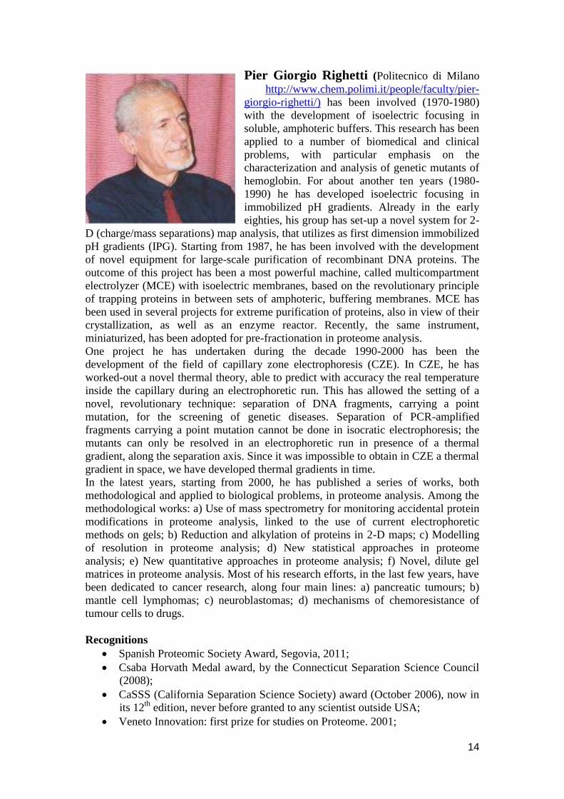
Pier Giorgio Righetti (Politecnico di Milano
http://www.chem.polimi.it/people/faculty/pier-
giorgio-righetti/) has been involved (1970-1980)
with the development of isoelectric focusing in
soluble, amphoteric buffers. This research has been
applied to a number of biomedical and clinical
problems, with particular emphasis on the
characterization and analysis of genetic mutants of
hemoglobin. For about another ten years (1980-
1990) he has developed isoelectric focusing in
immobilized pH gradients. Already in the early
eighties, his group has set-up a novel system for 2-
D (charge/mass separations) map analysis, that utilizes as first dimension immobilized
pH gradients (IPG). Starting from 1987, he has been involved with the development
of novel equipment for large-scale purification of recombinant DNA proteins. The
outcome of this project has been a most powerful machine, called multicompartment
electrolyzer (MCE) with isoelectric membranes, based on the revolutionary principle
of trapping proteins in between sets of amphoteric, buffering membranes. MCE has
been used in several projects for extreme purification of proteins, also in view of their
crystallization, as well as an enzyme reactor. Recently, the same instrument,
miniaturized, has been adopted for pre-fractionation in proteome analysis.
One project he has undertaken during the decade 1990-2000 has been the
development of the field of capillary zone electrophoresis (CZE). In CZE, he has
worked-out a novel thermal theory, able to predict with accuracy the real temperature
inside the capillary during an electrophoretic run. This has allowed the setting of a
novel, revolutionary technique: separation of DNA fragments, carrying a point
mutation, for the screening of genetic diseases. Separation of PCR-amplified
fragments carrying a point mutation cannot be done in isocratic electrophoresis; the
mutants can only be resolved in an electrophoretic run in presence of a thermal
gradient, along the separation axis. Since it was impossible to obtain in CZE a thermal
gradient in space, we have developed thermal gradients in time.
In the latest years, starting from 2000, he has published a series of works, both
methodological and applied to biological problems, in proteome analysis. Among the
methodological works: a) Use of mass spectrometry for monitoring accidental protein
modifications in proteome analysis, linked to the use of current electrophoretic
methods on gels; b) Reduction and alkylation of proteins in 2-D maps; c) Modelling
of resolution in proteome analysis; d) New statistical approaches in proteome
analysis; e) New quantitative approaches in proteome analysis; f) Novel, dilute gel
matrices in proteome analysis. Most of his research efforts, in the last few years, have
been dedicated to cancer research, along four main lines: a) pancreatic tumours; b)
mantle cell lymphomas; c) neuroblastomas; d) mechanisms of chemoresistance of
tumour cells to drugs.
Recognitions
Spanish Proteomic Society Award, Segovia, 2011;
Csaba Horvath Medal award, by the Connecticut Separation Science Council
(2008);
CaSSS (California Separation Science Society) award (October 2006), now in
its 12th
edition, never before granted to any scientist outside USA;
Veneto Innovation: first prize for studies on Proteome. 2001;
15
Hirai Prize award for outstanding research in Separation Science (Tokyo,
Japan). 1999;
Milano Award: price awarded for advanced genetic research in mutational
analysis, offered at the ATB'97 Conference, Milano, November-1997;
Prize of the English Electrophoresis Society, awarded at the Seattle Meeting of
the International Electrophoresis Society, March 1997;
Prize C.I.B. (Consorzio Italiano di Biotecnologie) for the "Biotechnologist of
the year, 1995.
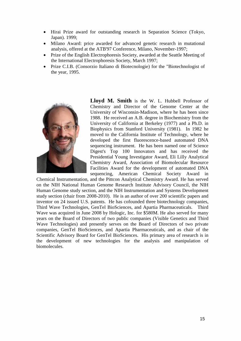
Lloyd M. Smith is the W. L. Hubbell Professor of
Chemistry and Director of the Genome Center at the
University of Wisconsin-Madison, where he has been since
1988. He received an A.B. degree in Biochemistry from the
University of California at Berkeley (1977) and a Ph.D. in
Biophysics from Stanford University (1981). In 1982 he
moved to the California Institute of Technology, where he
developed the first fluorescence-based automated DNA
sequencing instrument. He has been named one of Science
Digest's Top 100 Innovators and has received the
Presidential Young Investigator Award, Eli Lilly Analytical
Chemistry Award, Association of Biomolecular Resource
Facilities Award for the development of automated DNA
sequencing, American Chemical Society Award in
Chemical Instrumentation, and the Pittcon Analytical Chemistry Award. He has served
on the NIH National Human Genome Research Institute Advisory Council, the NIH
Human Genome study section, and the NIH Instrumentation and Systems Development
study section (chair from 2008-2010). He is an author of over 200 scientific papers and
inventor on 24 issued U.S. patents. He has cofounded three biotechnology companies,
Third Wave Technologies, GenTel BioSciences, and Apartia Pharmaceuticals. Third
Wave was acquired in June 2008 by Hologic, Inc. for $580M. He also served for many
years on the Board of Directors of two public companies (Visible Genetics and Third
Wave Technologies) and presently serves on the Board of Directors of two private
companies, GenTel BioSciences, and Apartia Pharmaceuticals, and as chair of the
Scientific Advisory Board for GenTel BioSciences. His primary area of research is in
the development of new technologies for the analysis and manipulation of
biomolecules.
16
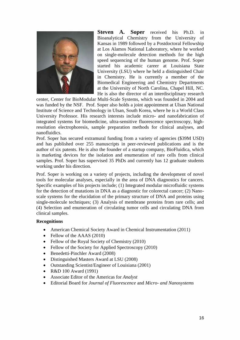
Steven A. Soper received his Ph.D. in
Bioanalytical Chemistry from the University of
Kansas in 1989 followed by a Postdoctoral Fellowship
at Los Alamos National Laboratory, where he worked
on single-molecule detection methods for the high
speed sequencing of the human genome. Prof. Soper
started his academic career at Louisiana State
University (LSU) where he held a distinguished Chair
in Chemistry. He is currently a member of the
Biomedical Engineering and Chemistry Departments
at the University of North Carolina, Chapel Hill, NC.
He is also the director of an interdisciplinary research
center, Center for BioModular Multi-Scale Systems, which was founded in 2004 and
was funded by the NSF. Prof. Soper also holds a joint appointment at Ulsan National
Institute of Science and Technology in Ulsan, South Korea, where he is a World Class
University Professor. His research interests include micro- and nanofabrication of
integrated systems for biomedicine, ultra-sensitive fluorescence spectroscopy, high-
resolution electrophoresis, sample preparation methods for clinical analyses, and
nanofluidics.
Prof. Soper has secured extramural funding from a variety of agencies ($39M USD)
and has published over 255 manuscripts in peer-reviewed publications and is the
author of six patents. He is also the founder of a startup company, BioFluidica, which
is marketing devices for the isolation and enumeration of rare cells from clinical
samples. Prof. Soper has supervised 35 PhDs and currently has 12 graduate students
working under his direction.
Prof. Soper is working on a variety of projects, including the development of novel
tools for molecular analyses, especially in the area of DNA diagnostics for cancers.
Specific examples of his projects include; (1) Integrated modular microfluidic systems
for the detection of mutations in DNA as a diagnostic for colorectal cancer; (2) Nano-
scale systems for the elucidation of the primary structure of DNA and proteins using
single-molecule techniques; (3) Analysis of membrane proteins from rare cells; and
(4) Selection and enumeration of circulating tumor cells and circulating DNA from
clinical samples.
Recognitions
American Chemical Society Award in Chemical Instrumentation (2011)
Fellow of the AAAS (2010)
Fellow of the Royal Society of Chemistry (2010)
Fellow of the Society for Applied Spectroscopy (2010)
Benedetti-Pinchler Award (2008)
Distinguished Masters Award at LSU (2008)
Outstanding Scientist/Engineer of Louisiana (2001)
R&D 100 Award (1991)
Associate Editor of the Americas for Analyst
Editorial Board for Journal of Fluorescence and Micro- and Nanosystems

17
Oliver Trapp
Ruprecht-Karls-Universität Heidelberg
Organisch-Chemisches Institut
Im Neuenheimer Feld 270
69120 Heidelberg
1993-1998 Undergraduate and graduate studies in
Chemistry at the Eberhard-Karls-University Tübingen,
Germany
1998 Diploma in Chemistry and Diploma-Thesis
with Prof. Dr. V. Schurig, Institute of Organic Chemistry,
Eberhard-Karls-University Tübingen, Germany
1998-2001 Fellow in the DFG Graduate College „Chemistry in Interphases‟ at the
University of Tübingen, Germany
2001 PhD and Dissertation with Prof. Dr. V. Schurig, Institute of Organic
Chemistry, Eberhard-Karls-University Tübingen, Germany
2002-2004 Postdoctoral Fellow in the Group of Prof. Dr. R.N. Zare, Department of
Chemistry, Stanford University, California, USA
2004-2008 Assistant Professor at the Max-Planck-Institut für Kohlenforschung in
Mülheim an der Ruhr, Germany
2005-2008 Emmy Noether Research Group Leader at the Max-Planck-Institut für
Kohlenforschung
Since 10/2008 Full Professor of Organic Chemistry at the University of Heidelberg
Awards
1999-2001 Doctorate scholarship of the Funds of the Chemical Industry (FCI)
2001 Attempto University Prize of the Eberhard-Karls-University Tübingen
2001 Procter&Gamble Innovation Prize
2002 Emmy Noether Postdoctoral Fellowship of the DFG
2003 Prize of the Analytical Chemistry Division of the GDCh
2005 Emmy Noether Research Group of the DFG
2007 Thieme Journal Award
2007 Research Grant Award of the Merck Research Laboratories
2008 Member of the Young College of the Northrhine Westphalian
Academy of Sciences
2008 ADUC-Prize of the German Chemical Society (GDCh) for the Best
Habilitation Thesis
2008 Heinz Maier-Leibnitz Award of the German Research Foundation
(DFG)
2008 Innovation Award 2008 of the Northrhine-Westphalian Ministry of
Innovations
2009 Annual Award of the Ruprecht-Karls-Universität Heidelberg for
Outstanding Research Achievements
2010 HTC-11 Award, Brugge, Belgium
2010 ERC Starting Grant
18
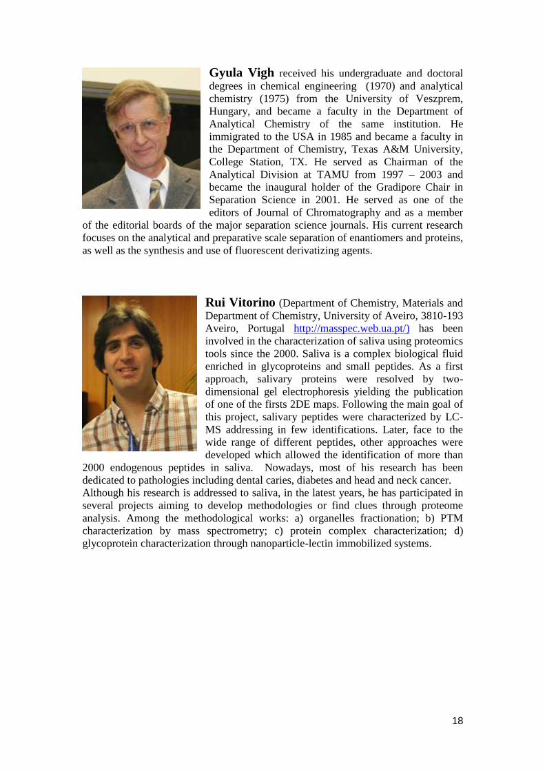
Gyula Vigh received his undergraduate and doctoral
degrees in chemical engineering (1970) and analytical
chemistry (1975) from the University of Veszprem,
Hungary, and became a faculty in the Department of
Analytical Chemistry of the same institution. He
immigrated to the USA in 1985 and became a faculty in
the Department of Chemistry, Texas A&M University,
College Station, TX. He served as Chairman of the
Analytical Division at TAMU from 1997 – 2003 and
became the inaugural holder of the Gradipore Chair in
Separation Science in 2001. He served as one of the
editors of Journal of Chromatography and as a member
of the editorial boards of the major separation science journals. His current research
focuses on the analytical and preparative scale separation of enantiomers and proteins,
as well as the synthesis and use of fluorescent derivatizing agents.
Rui Vitorino (Department of Chemistry, Materials and
Department of Chemistry, University of Aveiro, 3810-193
Aveiro, Portugal http://masspec.web.ua.pt/) has been
involved in the characterization of saliva using proteomics
tools since the 2000. Saliva is a complex biological fluid
enriched in glycoproteins and small peptides. As a first
approach, salivary proteins were resolved by two-
dimensional gel electrophoresis yielding the publication
of one of the firsts 2DE maps. Following the main goal of
this project, salivary peptides were characterized by LC-
MS addressing in few identifications. Later, face to the
wide range of different peptides, other approaches were
developed which allowed the identification of more than
2000 endogenous peptides in saliva. Nowadays, most of his research has been
dedicated to pathologies including dental caries, diabetes and head and neck cancer.
Although his research is addressed to saliva, in the latest years, he has participated in
several projects aiming to develop methodologies or find clues through proteome
analysis. Among the methodological works: a) organelles fractionation; b) PTM
characterization by mass spectrometry; c) protein complex characterization; d)
glycoprotein characterization through nanoparticle-lectin immobilized systems.
19
Abstracts
Lectures
Enantioselective catalysis and analysis in single microfluidic
devices
Detlev Belder, Institut für Analytische Chemie, Universität Leipzig, 04103 Leipzig,
Germany
There is tremendous progress in the miniaturisation of chemical laboratories to the
chip-scale either by downsizing chemical reactions or by chip based analytical
devices. In order to exploit the ultimate potential of chip-technology in chemistry the
different chemical processes should be integrated on a single microfluidic device.
Such a microfluidic chip integrating chemical reaction and analyses on a single device
is highly attractive, e.g. for high throughput screening and in combinatorial chemistry.
In this talk some of the main challenges for the development of an integrated chip
based reaction and analysis device are discussed, with a focus on the detection issue
(MS and life time fluorescence). Recent results on the integration of on-chip analysis
and synthesis for the screening of organo catalysts and bio catalysts are presented.
Coupling CE to MS: new interfaces, new ion sources
new applications
Christian W. Klampfl, Institute of Analytical Chemistry, Johannes Kepler-University
Linz, Altenbergerstr. 69, A-4040 Linz, Austria
It is more than 30 years since capillary electrophoresis (CE) was coupled to mass
spectrometry (MS) for the first time. Since than it has evolved from a quite exotic
combination, primarily performed on lab-made equipment, to a well established
method within the group of hyphenated techniques [1]. A major driving force for
the increased acceptance of CE-MS was the development of suitable interfaces,
mainly involving electrospray ionization (ESI), allowing the simple and robust
coupling of CE to MS.
Scanning the literature of these last 30 years a series of different interfaces (with
respect to their physical design) as well as several different ionization modes have
been employed for interfacing CE to MS. These comprise sheathless interfaces,
sheath flow interfaces or interfaces including a liquid junction [2]. Among those
the co-axial sheath flow interface still plays the most important role. This type of
interface is also used in the majority of commercially available devices for CE-
20
MS. Focusing on ionization, ESI still makes up for almost 99% of all CE-MS
applications. Nevertheless alternative approaches employing other ionization
techniques have been exploited. These include both, designs using commercially
available instrumentation (i.e. adapting HPLC-MS hardware for CE-MS) and also
completely lab-made ion sources. A nice example for the latter type is the
recently presented atmospheric pressure afterglow ion source for CE-MS [3]. In
the present paper different approaches towards the realization of sheath-flow
interfaces for CE-ESI-MS are discussed; including investigations using fully
commercially available instrumentation as well as interfaces based on
commercially available equipment that has been slightly modified and adapted to
the specific needs encountered in CE-MS in the laboratory [4]. Thereby the
influence of geometry changes (distances and angles) on sensitivity as well as
robustness has been tested. The lab-modified interface design provided better
results than commercially available devices that often represent a compromise as
they often should be usable for both, ultra low flow techniques such as CE and
(relatively) high flow separation techniques like HPLC. Regarding ionization,
different modes such as ESI, atmospheric pressure chemical ionization,
atmospheric pressure photoionization and others are discussed. With respect to
parameters like robustness, ease of use and their “unique selling property”
regarding the classes of anayltes amenable as well as compatibility of different
electroseparation techniques.
References
[1] Klampfl CW, Electrophoresis 30 (2009) S83
[2] Schmitt-Kopplin P, Frommberger M, (2003) Electrophoresis 24:3837.
[3] Jecklin MC, Schmid S, Urban PL, Amantonico A, Zenobi R,
Electrophoresis 31 (2010) 3597.
[4] S.M. Reiter, W. Buchberger, C.W. Klampfl, Chromatographia 71 (2010)
715.
CE as tool to investigate the stereodynamics and catalytic
reactions
Oliver Trapp, Organisch-Chemisches Institut, Ruprecht-Karls-Universität
Heidelberg, Im Neuenheimer Feld 270, 69120 Heidelberg, Germany
The investigation of the molecular dynamics of stereoisomers and the study of the
kinetics of reactions, in particular of catalyzed reactions, is of fundamental interest in
chemistry, biochemistry, and medicine. This presentation focuses on the recent
advances to study the stereodynamics of molecules and reaction kinetics of catalyzed
processes by means of capillary electrophoresis. Models and algorithms to evaluate
interconversion profiles obtained by electrophoretic separation techniques are
discussed with respect to the challenging demands of high separation efficiencies
typical for electrophoretic techniques. Models used for evaluation are based on
iterative computer simulation algorithms using the theoretical plate model or
stochastic model of chromatography, empirical calculation methods, and direct access
with the approximation function and more recently with the unified equation, which
21
can be applied to all kinds of first order reactions taking place during a
chromatographic or electrophoretic separation. Studies of enantioselective
sulfoxidations using immobilized salen ligands in CE will be presented and discussed
[6].
References
[1] O. Trapp, Electrophoresis 2010, 31, 786-813.
[2] S. Bremer, O. Trapp, Electrophoresis 2009, 30, 329-336.
[3] O. Trapp, S.K. Weber, S. Bauch, W. Hofstadt, Angew. Chem. Int. Ed. 2007, 46,
7307-7310 (Highlighted in Nature 2007, 448, 730-731).
[4] O. Trapp, Electrophoresis 2007, 28, 691-696.
[5] O. Trapp, Anal. Chem. 2006, 78, 189-198.
[6] S. Sandel, S.K. Weber, O. Trapp 2011, submitted.
Immuno-monoliths at reduced nonspecific interactions
Ziad El Rassi, Gunasena, D.N.
Department of Chemistry, Oklahoma State University, Stillwater, OK 74078-3071,
USA
A polar organic polymer monolith was introduced for performing immuno affinity
chromatography (IAC) at reduced non-specific interactions. The hydrophilic monolith
was prepared by the in situ polymerization of glyceryl methacrylate (GMM) as the
functional monomer and pentaerythritol triacrylate (PETA) as the crosslinker in the
presence of cyclohexanol, dodecanol and water as the porogenic solvent. The polar
monolith through its diol groups provides the opportunity to achieve readily the
immobilization of antibodies and other affinity ligands for nano liquid affinity
chromatography (nano-LAC). In this investigation, anti-haptoglobin antibody was
used as the model antibody to study the overall behavior of the immuno-monolith thus
obtained in terms of its binding to the antigen (haptoglobin) and to evaluate its non-
specific binding with other proteins, especially the high abundant serum proteins such
as human serum albumin, transferrin and 1-antitrypsin etc. Due to the presence of
hydroxyl groups in the crosslinker and the functional monomer as well, the
hydrophilic monolith exhibited negligible non-specific hydrophobic interactions with
proteins.
As an extra evaluation of its hydrophilicity, the novel monolith carrying diol
functionalities was exploited for its full potentials in normal phase or hydrophilic
interaction capillary electrochromatography (HI-CEC). Although the monolith is
neutral and void of a fixed charge on the surface, a relatively strong cathodal EOF
was observed due to the electric double layer formed by the adsorption of ions from
the mobile phase producing a bulk mobile phase flow. The novel monolith can be
used to separate polar compounds such as phenol derivatives, small aliphatic amides,
nucleic acid bases and nucleosides etc.
22
New technologies for the genome age
Lloyd M. Smith, Department of Chemistry & Genome Center of Wisconsin
University of Wisconsin-Madison, USA
The successful sequencing of the human genome has provided us with the blueprints
of life.... but now the likely greater challenge of understanding those blueprints lies
squarely before us. Again, information is needed. The molecules that are encoded by
the genome come in many different types and forms. We need to know what
molecules are where, when, and in what form, how they change with time and in
response to internal and external signals, and how they interact with one another.
This information is a first step towards developing an understanding of the complex
web of networks and pathways that comprise functioning biological systems. As was
true for the Genome Project, new technologies are needed to provide this information.
This talk will present challenges, opportunities, and progress in the development of
such new technologies, with a particular emphasis on surface science, biological mass
spectrometry, and the conjunction of the two.
Analysis of telomeres and telomerases
Jiří Fajkus, CEITEC MU and Faculty of Science, Masaryk University and
Institute of Biophysics ASCR, v.v.i., Brno, Czech Republic
Biology of telomeres has undergone a widespread development since the initial
findings on the molecular nature of chromosome ends and mechanisms of their
replenishment by the ribonucleoprotein complex of telomerase, or by alternative
pathways. Although the interest in telomeres has been initiated by a pure scientific
curiosity, later progress has shown that telomeres and telomerase are intimately
connected with the processes of cell proliferation, aging and cancer. In addition,
recent papers report on a number of non-telomeric roles of telomerase e.g., in
regulation of gene expression or DNA damage response.
In my talk, I would like to explain current approaches to analysis of telomeres and
telomerases, together with motivation to perform such analysis, and example results
from our research. These will include analyses of telomeric sequences in various
groups of organisms, measurement of telomere length, semi-quantitative and
quantitative assays for telomerase activity and expression, or evaluation of telomere
RNA transcript levels. Moreover, since telomeres are nucleoprotein (chromatin)
structures, their functions are mediated and modulated by a number of telomere-
associated proteins, and the analysis of DNA-protein and protein-protein interactions
thus represents an important input towards understanding telomere functions.
Acknowledgements
Our research is supported by the Czech Ministry of Education (MSM0021622415),
Czech Science Foundation (P501/11/0289), ASCR (M200040903) and the project
“CEITEC - Central European Institute of Technology” (CZ.1.05/1.1.00/02.0068) from
the European Regional Development Fund.

23
What is saliva? A perspective of salivary proteomics at
University of Aveiro
Rui Vitorino, Francisco Amado, University of Aveiro, Portugal
Traditionally, saliva is defined as a complex mixture of the secretion of major and
minor glandular secretions, in addition to the crevicular fluid, bacteria and epithelial
cells. To date, using multiple methods such as polyacrylamide gel electrophoresis
(mono and two-dimensional) and liquid chromatography combined with mass
spectrometry, over 3000 proteins from different sources have been identified in saliva.
Variations in saliva composition have been associated with salivary disorders such as
Sjogren‟s syndrome or dental caries. Saliva, often called “the body mirror”, has
emerged as an attractive diagnostic fluid facing its simplicity, non-invasive collection
and the cost-effective applicability for screening large populations collection.
Moreover, it possesses advantageous for various biochemical tests since saliva reflects
the concentrations of many blood components. With this presentation, we intend to
give a perspective of our approaches and simultaneously present the most relevant
findings about salivary proteomics.
Indolone-n-oxides: relation between redox properties and
antimalarial activities
Françoise Nepveu, University of Toulouse 3, UPS and IRD; UMR 152 PHARMA-
DEV, 118 route de Narbonne, F-31062 Toulouse cedex 9, France
Although several antimicrobial agents are currently available there is clearly a critical
need for the development of new pharmacophores due to the emergence of the
resistance of numerous microbes to several drugs. Microbes have a high metabolite
rate producing oxidative by-products, which is amplified by the oxidative attack of the
host immune system in which reactive oxygen and nitrogen species contribute. The
detoxification of reactive oxygen and nitrogen species is a challenge for tissues
infected with microbes. Redox metabolism is thus an attractive target for
antimicrobial drug development. In the search for anti-infective molecules using their
redox properties, so as to disrupt the antioxidant defence systems of the pathogen, we
recently show that indolone-N-oxide derivatives have antimalarial properties. The
pharmacomodulation studies confirmed the central role played by the indolone redox
pharmacophore and gave hits with antiplasmodial malarial activities at the nanomolar
level. The indolone-N-oxide scaffold is original, was not investigated for its
antimalarial potentialities before and gives the possibility to build new chemical series
comparatively to the artemisinine-like and chloroquine-like ones, and potentially new
drug classes.
N
O
R3
O
R1
R2
Structure of indolone N-oxide derivatives
24
The unique ability of nitrones to form persistent radical adducts and to be involved in
oxidation-reduction reactions, lead us to study how the peculiar redox properties of
indolone-N-oxides might exert a critical action towards the erythrocytic development
of Plasmodium falciparum, the parasite responsible for malaria. Herein, we report
some redox properties of indolone-N-oxides in relation with their antimalarial
properties.
References 1Nepveu F., Kim S., Boyer J., Chatriant O., Ibrahim H., Reybier K., Monje MC.,
Chevalley S., Perio P., Lajoie BH., Bouajila J., Deharo E., Sauvain M., Tahar R.,
Basco L., Pantaleo A., Turini F., Arese P., Valentin A, Thompson E., Vivas L., Petit
S., Nallet J-P., Synthesis and antiplasmodial activity of new indolone N-oxide
derivatives, J. Med. Chem. 2010, 44, 699-714.
The proteome Argonauts: conquering the “golden fleece” of
alcoholic beverages and soft drinks via combinatorial
peptide ligands
Pier Giorgio Righetti, Elisa Fasoli, Alfonsina D‟Amato, Politecnico di Milano,
Department of Chemistry, Materials and Chemical Engineering "Giulio Natta", Via
Mancinelli 7, Milano 20131, Italy.
Proteomic science has been vastly exploited in the past ten years for biomarker
discovery in sera, in search of panels of proteins able to warn about the onset of
various diseases. According to Mitchell (Nature Biotech. 28, 2010, 665-670), this has
been the biggest “fiasco” in this arena, with billions of dollars wasted. Completely
different results have been obtained by us when analyzing a “fiasco” (a 1.5 liter jug)
of white or red wine, with the combinatorial peptide ligand library (CPLL)
technology. It turns out that most wine producers treat white wines with casein (and
red wines with egg albumen) in order to eliminate residual grape proteins that would
flocculate upon long term storage. Although required by EC rulers, no producer has
ever stated the residual amount of these allergenic additives in their product. With the
CPLL technology, we were able to detect as little as 1 µg casein/L, an extremely high
detection sensitivity, unreported up to the present (the official ELISA test of the EC
reached barely down to 200 µg/L). However, if untreated wines are analyzed, we can
detect well over 100 residual grape proteins present in wines, this suggesting the
possibility of proteo-typing grand crus against counterfeited products invading the
market. We will additionally report proteo-typing of beers as well as different
carbonated soft beverages. One could thus easily distinguish among artificial
beverages, made only with synthetic additives and flavours (Coca Cola being a
classical example) vs. genuine products made with plant extracts. Regulatory agencies
and customers would thus have a new, formidable tool for protection against
adulterated and counterfeited foodstuff and beverages.
References
Cereda A, Kravchuk AV, D'Amato A, Bachi A, Righetti PG. J. Proteomics 73 (2010)
1732-1739.
25
D'Amato A, Kravchuk AV, Bachi A, Righetti PG. J. Proteomics 73 (2010) 2370-
2377.
Fasoli E, Aldini G, Regazzoni L, Kravchuk AV, Citterio A, Righetti PG. J. Proteome
Res. 9 (2010) 5262-5269.
Fasoli E, D'Amato A, Kravchuk AV, Righetti PG. J. Proteome Res. 2011, in press.
Separation of DNA and DNA-templated metal clusters by
CE-LIF
Ming-Feng Huang, Cheng-Kang Chiang, Huan-Tsung Chang
Department of Chemistry, National Taiwan University, 1, Sec. 4, Roosevelt Road,
Taipei 106, Taiwan
Capillary electrophoresis with laser induced fluorescence (CE-LIF) detection is a
powerful technique for the study of DNA conformational changes and
characterization of nanomaterials. We have studied the changes in the electrophoretic
mobility of the complexes of 5´ end labeled 6-FAM-T33 in the presence of Hg2+
using
CE-LIF. Upon increasing the concentration of Hg2+
, the electrophoretic mobility for
6-FAM-T33 increases, due to its conformation change from a random coil to a folded
structure and a decrease in its charge-to-mass ratio. CE-LIF using 2% poly(ethylene
oxide) solutions containing OliGreen (fluorophore) and 0.3 mM Hg2+
has been
applied to the separation of T33, T5C28, T5C5T23, and T15C5T13. We have employed
CE-LIF to confirm the purity of fluorescent DNA-templated metal clusters that are
interesting and sensitive probes for many analytes such as Cu(II) ions and DNA. Our
studies have shown that CE-LIF is useful for the characterization of nanomaterials
and can provide information to support the sensing mechanisms of many DNA based
probes.
Capillary electrophoresis analysis of the altered
glycosylation of immunoglobulins in autoimmune diseases
András Guttman (1), Csaba Váradi (1), Bertalan Meskó (2) and Laszlo Nagy (2)
(1) Horváth Laboratory of Bioseparation Sciences, and (2) Department of
Biochemistry and Molecular Biology, University of Debrecen, Hungary
Glycosylation is one of the most important post-translational modifications, which
holds the promise to provide numerous biological markers in clinical diagnostics for
various diseases. The N-glycosylation on human immunoglobulins, especially on
IgG1, plays a critical role in the bioactivity of this group of very important proteins.
Significant differences have been reported in IgG1 glycosylation in pregnancy, aging,
various autoimmune diseases and multiple myeloma. The four highest abundant
glycans of IgG1 are biantennary-agalacto (G0), biantennary-mongalacto (G1 and G1‟)
and biantennary–digalacto (G2) structures. Among these structures, G0 shows the
highest variability in the above mentioned normal and pathological conditions. The
26
aim of this study was to investigate the changes in the relative amount of G0 glycan of
IgG1 in such autoimmune diseases as reumatoid arthritis (RA) and Crohn‟s disease,
especially in view of transcriptomics data on galactosyl transferase experession. IgG1
was isolated from human blood samples using Protein A affinity partitioning and the
N-glycans were released by peptide-N-glycanase F (PNGase F). The free glycans
were then fluorescently labeled with aminopyrene-trisulfonate (APTS) and analyzed
by capillary electrophoresis with laser induced fluorescence detection. Glycosylation
patterns of sex and age matched samples were compared and analyzed to reveal any
possible correlation between IgG1 N-glycan profile, galactosyl transferase expression
and the pathogenesis of the disease.
Microfabricated and Lab-on-a-chip devices in the life
science industry: promises, products and problems
Aran Paulus, Bio-Rad Laboratories, Laboratory Separation Division, 6000 James
Watson Drive, Hercules, CA 94547, USA
Microfabrication on glass and plastic devices set out about 20 years ago to
revolutionize analytical workflows in the life science field by making them more
automated, with higher throughput through both speed and multiplexing and more
integrated by combining multiple steps onto a single chip. Despite some early
successes with glass-based chips for RNA, DNA and protein separations, the
promised market potential has not yet been reached. What happened and how far are
we from finding the killer application for a lab-on-the-chip? This presentation will
look at common biological workflows with an emphasis on protein applications and
compare traditional techniques such as slab gel electrophoresis and Western blotting
with manual sample processing steps to chip-based approaches. The comparison will
include a look at the practical aspects of the complete workflows, critical time-
dependent steps and analyte concentration and absolute amounts for each step in the
protocol.
Single-molecule electrochromatography in nano-columns
with conductivity readout: a novel approach for high
throughput DNA sequencing
Steven A. Soper,1,3
Sunggook Park,2 Dimitris Nikitopoulos,
2 Yoonkyoung Cho,
3
Heungjoo Shin,3 Dorel Moldovan,
2 Franklin Uba,
1 Dong-Kyu Park,
3 Jiahao Wu
2 and
Mateusz Hupert1
1Department of Biomedical Engineering, Department of Chemistry, University of
North Carolina, Chapel Hill 27599, USA 2Department of Chemistry and Department of Mechanical Engineering, Louisiana
State University, Baton Rouge, LA 70803, USA 3Ulsan National Institute of Science and Technology (UNIST), Ulsan, South Korea
Complete integration of the sample processing pipeline into a single fluidic system is
of interest for potential point-of-care applications, especially in the area of in vitro
27
diagnostics. Microfluidic systems are a promising technology platform as they can
provide automated sample handling and reagent delivery as well as timely results with
minimal operator intervention. While microfluidics offers some compelling
advantages for molecular processing, systems using nanofluidics offers new
opportunities that are not accessible via microfluidics. In particular, unique physics
occur at the nanometer scale. In this presentation, we will discuss the development of
a nanofluidic system for biopolymer analysis, such as DNAs, RNAs and proteins. The
microfluidic components used for sample preparation prior to entering the nanofluidic
system, were made from conventional thermoplastics using high production rate
replication technologies, such as injection molding. The microfluidic system could be
interfaced to a nanofluidic system fabricated via nano-replication technologies, such
as nanoimprint lithography (NIL). One of the nanofluidic elements consisted of a
column with dimensions on the order of 50 x 50 nm (depth x width) with a length of
60 µm. With the addition of an applied longitudinal electric field, single molecules
could travel through this nano-column experiencing wall interactions predicated on
the nature of the molecule and the surface structure of the column (nano-capillary
electrochromatography). Thus, single molecules could be identified through their
unique mobility through the nano-column. We will discuss the ability to fabricate
nano-scale molds in Si using focused ion beam milling with the masters used to create
polymer stamps for the high scale production of these nano-scale chromatography
columns. Molecular Dynamic (MD) simulation results have been secured as well to
demonstrate proof-of-concept for moving single molecules through nano-columns
with the migration time used for their molecular identification.
Microfluidics for applications in chemistry
Andreas Manz, Jörg Ingo Baumbach, Pavel Neuzil
KIST Europe, Campus E71, 66123 Saarbrücken, Germany
Microfluidics and miniaturization of detectors are important components for future
clinical diagnostics instrumentation. The field has developed now for many years, and
has its niche products in genomics, proteomics and in combination with sensors.
Initially, the driving forces were curiosity and the drug discovery application.
However, interests have shifted towards clinical applications in recent years.
Several aspects will be highlighted, like non-invasive diagnostics applications in
breath analysis for infectious diseases, "generic" product development in the
electrophoresis & polymerase chain reaction area, and novel approaches to
micro/nano multiphase systems using droplet generators for pseudo-crystalline
emulsions.

28
Isoelectric focusing coupled to mass spectrometry for
bioanalysis
Kilár Ferenc1,2
, Páger Cs1, Dörnyei Á
1, Vargová A
2, Takácsi-Nagy A
1, Thormann
W3
1Institute of Bioanalysis, Faculty of Medicine, University of Pécs, Szigeti út 12, H-
7624 Pécs, Hungary 2Department of Analytical and Environmental Chemistry, Faculty of Sciences,
University of Pécs, Ifjúság útja 6, H-7624 Pécs, Hungary 3Department of Clinical Pharmacology and Visceral Research, University of Bern,
Murtenstrasse 35, CH-3010 Bern, Switzerland
Isoelectric focusing is one of the most important separation techniques in proteomics.
Its application is inevitable in obtaining information on the appearance, as well as up
and down regulation of proteins determined by the commonly used 2D
polyacrylamide gel-electrophoresis and combined with off-line mass-spectrometry.
The introduction of capillary isoelectric focusing leads, however, to a new opportunity
in those analytical studies. An on-line combination of CIEF with an appropriate mass
spectrometric detector, is able to replace 2D-PAGE, with high throughput and a much
less laborious manner. The coupling of isoelectric focusing with mass spectrometry is,
however, still a challenge in bioanalysis. The lecture will summarize the steps and
future opportunities in this topic and discuss the various methodological aspects.
CIEF in presence of electroosmosis with sequential injection of carrier ampholytes
and sample was found to be suitable for mass spectrometry detection. The separate
injection of the sample and the ampholytes provides good condition to suppress and
overcome the undesirable effect of the presence of ampholytes in mass spectrometry.
By the appropriate selection of ampholyte solutions, whose pH range not necessarily
covers the pI values of the analytes, the migration of the components can be
controlled, and the impact of the ampholytes on mass spectrometric detection is
decreased. The unique applicability of this setup is shown by testing several
parameters, such as, the application of volatile electrolyte solutions, the type of the
ampholytes, the order and the number of the ampholyte and sample zones. Broad and
narrow pH range ampholytes were applied in experiments using uncoated capillaries
with different length for the analyses of substituted nitrophenol dyes to achieve
optimal conditions for the mass spectrometric detection [1]. Although, the sample
components are not leaving the pH gradient, due to the decrease in the ampholyte
concentration at the position of the components, and because the sample components
migrate in charged state, the ionization is more effective for mass spectrometry
detection. Results with model calculations confirm the mechanism of the process in
the isoelectric focusing using one or two ampholyte zones before and/or after the
sample zone [2].
Acknowledgements
This work was supported by the grants SROP-4.2.2/08/1/2008-0011 and TÁMOP-
4.2.1. B-10/2/KONV-2010-0002, and by the Swiss NSF.
References: 1. Páger, C. et al. (2011) Electrophoresis 32, 1875-1884.
2. Takácsi-Nagy, A. et al. Electrophoresis (submitted for publication)

29
Design, synthesis and analytical characterization of a new
family of pI markers
Gyula Vigh, Department of Chemistry, Texas A&M University, College Station, TX
77842-3012, USA
In continuation of our efforts to introduce fluorescent derivatizing agents optimized
for the various operation modes of capillary electrophoresis, we have designed a new
family of structurally and spectrally homogeneous pI markers for use in cIEF, and
synthesized and analytically characterized the first seven members of the family. The
new pI markers can be detected by both absorbance and fluorescence detectors. Their
excitation
max is at 495 nm, emission
max is at 525 nm. The excitation spectra are broad
enough for excitation with any laser in the 440 < excitation
< 510 nm range. Their
fluorescence spectra do not change when the pH of their solution is varied in the 2.2 <
pH < 11 range, or when the buffering species in the background electrolytes are
changed or when the pI markers are dissolved in different narrow pI range carrier
ampholyte fractions (3 < pI < 4; 4 < pI < 6; 5 < pI < 8 and 8 < pI < 10).
Since the new pI markers contain multiple protic groups with closely spaced pKa
values, their tentative pI values were characterized by both capillary zone
electrophoresis in a series of background electrolytes whose pH values bracketed the
pI values of the markers as described in Ref. 1, and by cIEF using broad-range carrier
ampholytes, UV-absorbing primary pI markes (ElphoMarks) and pressure
mobilization. The tentative pI values of the first seven members of the new family are
3.07; 4.16, 5.74; 7.45; 8.34; 9.42 and 10.50, respectively.
References
[1] P. Glukhovskiy, G. Vigh, Electrophoresis, 19, 3166- 3170 (1998).
Investigation of biopeptide interactions by capillary
electrophoresis
Kašička Václav, Ehala S., Šolínová V., Schimperková T., Sázelová P., Koval D.
Institute of Organic Chemistry and Biochemistry, v. v. i., Academy of Sciences of the
Czech Republic, Flemingovo 2, 166 10 Prague 6, Czech Republic
Identification and quantification of biopeptide interactions with other (bio)molecules
is of extraordinary importance since most of peptide biological functions are
implemented via these interactions. The strength of these interactions is quantitatively
characterized by the binding (stability, association) constant, Kb, of peptide
complexes. Among the methods employed for Kb determination, affinity capillary
electrophoresis (ACE) possesses several advantages – small sample amount (typically
picomole of analyte in nanoliter applied volumes), simultaneous Kb determination of
complexes of different analytes, the analytes need not be quite pure and their
concentration need not be exactly known [1, 2].
30
In this contribution, application of ACE to investigation of non-covalent interactions
of valinomycin, macrocyclic dodecadepsipeptide antibiotic ionophore, with
ammonium and alkali metal ions in methanol will be presented. The apparent binding
constants of the valinomycin-cation complexes were obtained from the ACE
measurements of the dependence of valinomycin effective mobility on the cation
concentration in the background electrolyte (BGE) using a non-linear regression
analysis. The determined Kb values of the above complexes confirmed a considerably
higher selectivity of valinomycin for Rb+, K
+ and Cs
+ ions (with log Kb in the range
4.63 – 3.81) over NH4+, Li
+ and Na
+ ions with log Kb between 1.45 and 1.54 [3, 4]. In
addition, binding constants of the complexes of enantiomers of an antimicrobial
dipeptide H--Ala-D,L-Tyr-OH and its derivatives (N-Ac--Ala-D,L-Tyr-OH, and H-
-Ala-D,L-Tyr-NH2) with a chiral selector, 2-hydroxypropyl--cyclodextrin, have
been evaluated by nonlinear fitting of the mobility data to regression model describing
the dependence of the effective mobilities of these peptides on cyclodextrin
concentration in the BGE. Prior to Kb calculation, the effective mobilities have been
corrected to reference temperature and constant ionic strength and constant viscosity
of the applied BGEs. ACE proved to be a suitable method for investigation of both
weak and medium-strong interactions of charged as well as non-charged biopeptides
with ligands of different character and size.
References
[1] Chen, Z., Weber, S. G. Trends Anal. Chem. 2008, 27/9, 738-748.
[2] Winzor, D. J. Anal. Biochem. 2008, 383/1, 1-17.
[3] Ehala, S., Kašička, V., Makrlík, E. Electrophoresis 2008, 29, 652-657.
[4] Ehala, S., Dybal, J., Makrlík, E., Kašička, V.: Electrophoresis 2009, 30, 883-889.
Acknowledgements
The work was supported by the Czech Science Foundation, grants nos. 203/08/1428
and 203/09/0675, and by the Research Project AV0Z40550506 of the Acad. Sci. of
CR.

31
Posters
1 Investigation of matrix deposition techniques for high
throughput MALDI mass spectrometry imaging
Bednařík A.
a, Ertl L.
a, Ráţová I.
a, Tomalová I.
a, Moskovets E.
b, Preisler J.
a
a) Department of Chemistry, Faculty of Science and Central European Institute of
Technology, CEITEC, Masaryk University, Brno, Czech Republic
b) MassTech, Inc. 6992 Columbia Gateway Drive, Suite #160, Columbia, MD 21046,
USA
Matrix assisted laser desorption/ionization mass spectrometry imaging (MALDI-MSI)
is an emerging technique with an immense biochemical and pharmaceutical potential,
which possesses ability to show spatial distribution of specific compounds in tissues.
Here, we will present a comparison of common MALDI-MSI sample preparation
techniques. The MALDI matrix together with rhodamine B labeled angiotensin I was
deposited on sample MALDI plate by three different approaches, namely by
electrospray, spin-coating and piezoelectric nano-spotter. The homogeneity of
deposited samples was studied under microscope either by observing simple optical
image or in the case of piezo-deposited nano-spots by observing fluorescence image
after irradiating the sample with 532 nm laser. Optical image was compared with the
MSI generated map of peptide distribution along the samples. To measure spatial
distribution of selected peptides, a laboratory-built high-throughput axial time-of-
flight (TOF) mass spectrometer with 2 kHz 355 nm Nd:YAG laser was used.
Acknowledgements This work was supported by The Czech Science Foundation (P206/10/J012), by The
Ministry of Education, Youth and Sports (MSM0021622415) and the project
“CEITEC - Central European Institute of Technology” (CZ.1.05/1.1.00/02.0068) from
European Regional Development Fund.

32
2 Stability constants of charged cyclodextrins with neutral
analytes - determination by CE
Beneš M., Svobodová J., Zusková I., Gaš B.
Department of Physical and Macromolecular Chemistry, Faculty of Science, Charles
University in Prague, Hlavova 8, 128 43, Prague 2, Czech Republic
E-mail: [email protected]
Stability constant characterizes binding interaction between an analyte and
complexation agent. These interactions play very important role in separation
processes of, in other way undistinguishable, compounds, e.g. enantiomers. The most
widely used complexation agents are cyclodextrins (CD). Charged cyclodextrins
significantly extend the applicability of the electrophoretic methods for neutral
analytes.
Affinity capillary electrophoresis (ACE) belongs to methods suitable for the
determination of stability constants(0,0)
. If charged CDs are used, the attention must be
paid not only to viscosity of the BGE and to the influence of Joule heating on the
temperature in the capillary but also to the increasing ionic strength.
The thermodynamic stability constants of R- and S-hydrobenzoin and R- and S-(3-
brom-2-methyl-1-propanol) with cationic modified β-cyclodextrin: 6-monodeoxy-6-
mono(3-hydroxy)propylamino-β-cyclodextrin hydrochlorid (PA-β-CD) were
determined by ACE. The average temperature (25°C) of the BGE in the capillary was
kept constant. This was achieved by decreasing of the cassette temperature (based on
the conductivity measurements). The viscosity correction was performed using the
viscosity ratio(3)
. The increase of ionic strength due to increasing PA-β-CD
concentration in the BGE was compensated by changing of the concentration of the
separation buffer. The nonlinear regression was used to analyze of the experimental
data. In the next step, the dependence of the analyte effective mobilities on PA-β-CD
concentration was measured without the ionic strength compensation to demonstrate
the influence of ionic strength on the stability constant. A new procedure was
established to estimate stability constants from such data, too. These stability
constants are in very good agreement with those obtained at the constant ionic
strength.
References
1. Ušelová-Včeláková K., Zusková I., Gaš B. Electrophoresis 2007, 28, 2145-2152
2. Tanaka Y., Terabe S. J. Chromatogr. B 2002, 768, 81-92
3. Bowser M. T., Sternberg E. D., Chen D. D. Y. Electrophoresis 1997, 18, 82-91
Acknowledgements
The support of the Grant Agency of the Czech Republic, grant no. P205/11/J043,
Grant Agency of Charles University, grant no. 323611, CEEPUS network, the long-
term research plan of the Ministry of Education of the Czech Republic
(MSM0021620857) and Cyclolab (Budapest, Hungary) for donation of the
cyclodextrin PA-β-CD are gratefully acknowledged.
33
3 The use of intrinsic time-resolved fluorescence detection in
miniaturized analysis systems
Beyreiss, R., Ohla, S., Nagl, S., Belder D.
University of Leipzig, Institute of Analytical Chemistry, Weissenfelser Str. 67, 04229
Leipzig, Germany
Lab-on-chip technology is an emerging and fast growing research field applicable in
many life science disciplines. The miniaturization and transfer of chemical procedures
into multifunctional microfluidic devices leads to intensification and acceleration of
the whole process. Due to the small microscale dimensions, miniaturized systems
demand highly sensitive detection techniques. In this respect, one commonly used
technique is fluorescence detection, which however requires suitable fluorophores.
Introducing extrinsic chromophores can be troublesome. One possibility to
circumvent labeling procedures is to shift the excitation wavelength in the deep UV
spectral region and thus inducing intrinsic fluorescence of various analytes [1, 2].
Additional molecular information about the analytes can be gathered by applying
time-resolved fluorescence detection [3, 4]. Since the parameter lifetime is a
molecular signature, lifetime determination enables peak assignment and distinction
between different analytes. Another feature of time-resolved detection is the
possibility to discriminate background contributions, resulting in enhancement of
sensitivity, which is especially crucial in miniaturized systems.
In this context, we established intrinsic, time-resolved fluorescence detection in
microchip electrophoresis. A mixture of the small aromatics serotonin, propranolol, 3-
phenoxy-1,2-propanediol and tryptophan was used for system validation [4]. In the
experimental setup a 20 MHz picosecond laser operating at 266 nm served as
excitation source and emitted fluorescence was detected using time-correlated single
photon counting (TCSPC). Furthermore, the detection technique in lab-on-a-chip
applications was demonstrated by investigation of on-chip Pictet-Spengler conversion.
The conversion of dopamine by acetaldehyde was carried out within an integrated
serpentine micromixer. Subsequently the residual substrate and the enantiomeric
product salsolinol were separated on the same microchip and detected online by
TCSPC. Fluorescence lifetime determination of the separated compounds allowed
peak identification [5].
References [1] Li, Q., Seeger, S. Appl. Spec. Rev. 2010, 45, 12–43.
[2] Suhling, K.; French, P. M. W.; Phillips, D. Photochem. Photobiol. Sci. 2005, 4,
13–22.
[3] Llopis, S. D.; Stryjewski, W.; Soper, S. A. Electrophoresis 2004, 25, 3810–3819.
[4] Beyreiss, R., Ohla, S., Nagl, S., Belder, D. Electrophoresis 2011, accepted.
[5] Ohla, S. ,Beyreiss, R., Fritzsche, S., Gläser, P., Nagl, S., Stockhausen, K.,
Schneider, C., Belder, D. Chem. Eur. J. 2011, accepted.
34
4 Polyethyleneimine coated capillary in nucleotide and short
oligonucleotide analysis
Bittová M.
1, Roblová V.
1, Majerová, E.
2, Trnková L.
1
1 Department of Chemistry, Faculty of Science, Masaryk University, Kotlarska 2,
611 33 Brno, Czech Republic. 2 Department of Experimental Biology, Faculty of Science, Masaryk University,
Kotlarska 2, 611 37 Brno, Czech Republic.
In last few decades, there have been increasing demands for fast and simple analysis
of nucleotides and short oligonucleotides from biological matrices. Their separation
and purification is fundamental in pharmaceutical and clinical studies as well as in
new biotechnology development.
Capillary zone electrophoresis as a powerful analytical technique provides a versatile
tool for nucleotide and oligonucleotide analysis. Inner surface modification of fused-
silica capillaries with appropriate chemical reagents along with electroosmotic flow
control allow targeted selection of separation system for analytes of interest.
Polyethyleneimine (PEI) is polycationic highly branched polymer with positively
charged primary, secondary and tertiary amino groups in the structure that can be used
as fused-silica capillary modifier. It was firstly introduced by Towns and Regnier [1].
Its irreversible adsorption on the silanol groups of the fused-silica capillary surface
eliminates negative charge and excess of charged amino groups cause reversion of
electroosmotic flow towards the anode. However, consequential interactions with
background electrolyte can alter the charge again, thus the electrolyte for analysis
must be chosen carefully [2].
In our work, we successfully applied polyethyleneimine coated capillaries in analysis
of nucleotides and synthetic pentamers. Also, we studied the influence of particular
component of Tris/boric acid/EDTA buffer (TBE) on the PEI coated capillary surface
[4]. Furthermore, the effect of hydroxyethylcellulose (HEC) and/or -cyclodextrin on
the nucleotide separation was followed up. Finally, we analyzed restricted DNA from
Nicotiana tabacum and the representation of nucleotides was evaluated.
References
[1] Towns, J., K., Regnier, F., E., J Chromatogr A 1990, 516, 69-78.
[2] Spanila, M., Pazourek, J., Havel, J., J Sep Sci 2006, 29, 2234-2240.
[3] Nutku, S., M., Erim Berker, F., B. Turk J Chem 2003, 27, 9-17.
[4] Wang, P., Ren, J. J Pharmaceut Biomed 2004, 34, 227-283.
Acknowledgements
This work was supported by a project of Ministry of Education, Youth and Sports of
the Czech Republic - LC06035.

35
5 On-line two-dimensional liquid chromatography-capillary
electrophoresis separations of phenolic compounds
Česla P., Dinisová P., Fischer J., Jandera P.
University of Pardubice, Faculty of Chemical Technology, Department of Analytical
Chemistry, Studentská 95, CZ-53210 Pardubice, Czech Republic
Separation of complex samples containing analytes with wide range of
physicochemical properties is often difficult in one-dimensional separation system.
The resolution can be significantly improved by subjecting the effluent
outcomingfrom first separation system to the second separation based on different
principle. The highest improvement of resolving power is usually achieved when the
separation principles used in both dimensions are completely independent such as
combination of liquid chromatography and capillary electrophoresis. The simpler off-
line approach is not strictly limited by the volume of transferred fractions and
differences in geometry of LC column and CE capillary [1]. On-line connection offers
shorter analysis time and higher sample throughput, but it requires special instrument
modification or even construction of new separation system [2,3].
In this work, new approach to on-line two-dimensional separation method combining
capillary liquid chromatography with capillary electrophoresis is presented [3].
Capillary columns with fused core C8 and C18 silica particles were applied in the first
separation dimension. In the second dimension, the short capillaries with extended
light path cells were applied for fast separation of compounds in fractions
transferredfrom LC separation. The transfer of the fractions between the two
dimensions is based on the application of electronically controlled switching valve.
The separation conditions in both LC and CE dimensions were optimized using a set
of representative standards of phenolic compounds. Further, optimized programmed
separation conditions in both dimensions were used to improve the sample resolution
and peak capacity in comparison to the isocratic system. The on-line two-dimensional
method under optimal conditions was applied on the separation of phenolic
compounds in environmental and food samples.
References [1] Česla, P., Fischer, J., Jandera, P. Electrophoresis 2010, 31, 2200-2210.
[2] Bushy, M.M., Jorgenson, J.W.Anal. Chem. 1990, 62, 978-984.
[3]Česla, P., Dinisová, P., Fischer, J., Jandera, P. Chromatographia, submitted.
Acknowledgements
The authors acknowledge the financial support by the Czech Science Foundation,
project No. 203/09/P199.
36
6 Development of a validated capillary electrophoresis method
for enantiomeric purity control and quality control of
levocetirizine in a pharmaceutical formulation
Deng X.
*, De Cock B., Adams E., Van Schepdael A.
Katholieke Universiteit Leuven, Faculteit Farmaceutische Wetenschappen,
Laboratorium voor Farmaceutische Analyse, O&N2, PB 923, Herestraat 49, B-3000
Leuven, Belgium
Levocetirizine, a single-enantiomer drug, is the active enantiomer of the second
generation antihistaminic drug cetirizine. It is an oral histamine H1 receptor antagonist
of the latest generation that is licensed for the symptomatic treatment of allergic
rhinitis, with twice the affinity for the H1 receptor compared with cetirizine and is a
potent antihistamine as demonstrated by inhibition of histamine-induced weal and
flare reactions and in clinical studies.
A chiral capillary electrophoresis method has been developed for the quantification of
0.1 % of the enantiomeric impurity (dextrocetirizine) in levocetirizine and
determination of both in pharmaceuticals using sulfated-β-cyclodextrins as chiral
selector. Several parameters affecting the separation were studied such as the type and
concentration of chiral selectors, buffer composition and pH, organic modifier,
mixtures of two cyclodextrins in a dual system, voltage and temperature. The optimal
separation conditions were obtained using a 50 mM tetraborate buffer (pH 8.2)
containing 1 % (w/v) sulfated-β-cyclodextrins on a fused-silica capillary. Under these
conditions, the resolution of racemic cetirizine was higher than 3 and the chiral
impurity migrates in front of levocetirizine, which is suitable to determine
enantiomeric impurity. In order to validate the method, the stability of the solutions,
robustness (two level half fraction factorial design for 5 factors using 19 experiments
(2n-1
+3)), precision of the method, linearity (dextrocetirizine 0.25-2.5 µg/ml,
R2=0.9994 y=0.0375x+0.0008; levocetirizine 15-100 µg/ml, R
2 =0.9996
y=0.0213x+0.0339), limit of detection (0.075 µg/ml, 0.03 % m/m), limit of
quantification (0.25 µg/ml, 0.1 % m/m), accuracy (dextrocetirizine 84-109 %,
levocetirizine 97.3-103.1 %), filter effect and different CD batches were examined.
The validated method was further applied to bulk drug and tablets of levocetirizine.
37
7 Characterization of lipid A acylation patterns in rough-type
bacterial strains using MALDI-TOF MS in reflectron mode
Dörnyei Á.
1,*, Kilár A.
2, Sándor V.
2, Kocsis B.
3, Kilár F.
1,2
1 Institute of Bioanalysis, Faculty of Medicine, University of Pécs, Szigeti út 12, H-
7624 Pécs, Hungary 2 Department of Analytical and Environmental Chemistry, Faculty of Sciences,
University of Pécs, Ifjúság útja 6, H-7624 Pécs, Hungary 3 Department of Medical Microbiology and Immunology, Faculty of Medicine,
University of Pécs, Szigeti út 12, H-7624 Pécs, Hungary
Most Gram-negative bacteria have lipopolysaccharide (LPS, also called endotoxin) on
their outer membrane. In general a LPS consists of a phosphoglycolipid part (termed
lipid A) and two variable oligosaccharide (OS) regions (the core OS and the O-
specific polysaccharide), meanwhile it is the lipid A domain that is held responsible
for the endotoxic activity. The rough (R)-type LPSs do not possess any O-specific
polysaccharide and sometimes lack portions of the core, as well. Lipid A has mostly
phosphorylated β-D-GlcpN-(1‟→6)-α-D-GlcpN (GlcpN: 2-amino-2-deoxy-
glucopyranose) disaccharide backbone decorated with multiple fatty acid side chains.
The acylation and phosphorylation patterns strongly influence the charge state and
secondary structure of lipid A and consequently, its endotoxic activity.
Lipid A samples are generally prepared from isolated LPS samples with mild acidic
hydrolysis to split the acid-labile ketosidic linkage between the lipid A and core OS
parts. The disadvantages of this chemical degradation step are the high risk of losing
acid-labile, but biologically important, functional groups and the enhancement of the
intrinsic heterogeneity of the product. Distinguishing laboratory artifacts from natural
heterogeneity is one of the major analytical challenges when dealing with LPS and
lipid A preparations.
In this study, we show that the negative-ion, reflectron mode MALDI-TOF MS
technique can be used to identify the lipid A components with different acylation
patterns directly from R-type LPS samples without performing the chemical isolation
of lipid A prior to the mass spectrometric measurement. Four R-type Shigella sonnei
strains (S. sonnei 4350, R41, 562H, and 4303) with different core OS parts were used
to demonstrate the usefulness of this method.
Acknowledgement
Á.D. acknowledges the support of the János Bolyai Research Scholarship (Hungarian
Academy of Sciences).
38
8 Divergent flow isoelectrofocusing of whey
caseinomacropeptide
Duša F.
1,2, Moravcová D.
2, Kahle V.
2, Šlais K.
2
1Department of Biochemistry, Faculty of Science, Masaryk University, Kotlářská 2,
611 37 Brno,Czech Republic 2Department of liquid phase separations,Institute of Analytical Chemistry AS CR,
v. v. i.,Veveří 97, 60200Brno, Czech Republic
Isoelectric focusing (IEF) is an efficient method for separation of macromolecules.
Classic IEF formats as capillary or gel are not suited for a processing of high volume
of a sample. This disadvantage is solved by free flow IEF. Thanks to continuous flow
of sample and ongoing separation the technique could be conveniently used for pre-
separation and possibly separation of analytes. Method of divergent flow IEF (DF-
IEF) invented in our department [1]further improves separation efficiency of
currentfree flow IEF instrument pattern. Based on our previous research [2-4] we used
the DF-IEF for concentration and purification of caseinomacropeptide (CMP). CMP
originates during cheese making process by cleavage ofκ-casein with chymosin. It is
heterogeneous group of differently glycated polypeptides based on the same primary
amino acid structure. We used sweet bovine whey as a source of CMP since it
composesabout 25 % of whole whey protein. Particular focused fractions originated
from sweet bovine whey were analyzed by capillary HPLC with C18 reverse
phasecolumn and UV detection.Presence of CMP in fractions was confirmed by
comparison of obtained chromatograms with results of HPLC analysis of CMP
standard. Results showed an ability of device to separate CMP from α-lactalbumin
and β-lactoglobulin. We proved that DF-IEF may be used for CMP purification as
atechniquealternativeto ultrafiltration and ion-exchange chromatography.
References
[1] Slais, K., Electrophoresis 2008, 29, 2451-2457.
[2] Stastna, M., Slais, K., Electrophoresis 2008, 29, 4503-4507.
[3] Mazanec, K., Bobalova, J., Slais, K., Analytical and Bioanalytical Chemistry
2009, 393, 1769-1778.;
[4] Stastna, M., Slais, K., Electrophoresis 2010, 31, 433-439.
Acknowledgements
This work was supported by the Grant Agency of the Academy of Sciences of the
Czech Republic No. IAAX00310701 and by the Institutional research plan AVO
Z40310501. The financial support of the Ministry of Education, Youth and Sports of
the Czech Republic by grant No. LC06023 and by the Grant of Ministry of Interior
No. VG20102015023is also gratefully acknowledged.

39
9 Laser diode thermal vaporization inductively-coupled
plasma mass spectrometry
Foltynová P.
b, Kanický V.
b, Preisler J.
a,b
aCentral European Institute of Technology, CEITEC MU, Masaryk University, Brno,
Czech Republic, [email protected], Kamenice 5, 625 00 Brno, Czech Republic bDepartment of Chemistry, Faculty of Science, Masaryk University, Brno, Czech
Republic
A new approach of sample introduction to inductively coupled plasma mass
spectrometry (ICP-MS), laser diode thermal vaporization (LDTV), is presented. The
method does not require an expensive high-energy pulse laser, because the technique
employs a low-cost NIR diode laser which makes it a promising alternative to
nebulizers. Defined submicroliter volumes of samples are deposited on a suitable
sample carrier with an absorber, such as common office paper with commercial
printer black ink. The diode laser energy is sufficient to induce burning and/or
pyrolysis of the carrier substrate sample which leads to aerosol generation. Selection of
optimal conditions, e.g., experimental arrangement, raster, scan speed, additives etc. will be discussed. The limits of detection of Co, Ni, Zn, Mo, Cd, Sn and Pb deposited on the preprinted
paper were found to be in the range of 4 – 300 pg. Applicability was demonstrated on
determination of lead in whole blood and tin in canned food. Using a multielemental
calibration set prearranged on the carrier, LDTV ICP MS provided rapid and
reproducible (10%) quantitative analysis of metals. The advantages of LDTV are also
easy sample archiving, very low consumption of sample solution, transportation and
simplicity of sample preparation.
Acknowledgements
Authors acknowledge support of the Czech Science Foundation (Grant No.
203/09/1025), the Ministry of Education, Youth and Sports of the Czech Republic
(MSM0021622411, MSM0021622412 and MSM0021622415) and by the project
"CEITEC - Central European Institute of Technology" (CZ.1.05/1.1.00/02.0068) from
European Regional Development Fund.
40
10 Effective pre-concentration and analysis of heavy metals by
use of ligand step gradient focusing
Glovinová, E., Pospíchal J.
Department of Chemistry and Biochemistry, Mendel University in Brno, Czech
Republic, [email protected]
The capillary electrophoretic method for the focusing and selective pre-concentration
of metal chelates with subsequent on-line ITP analysis was developed and verified.
The selected ions of heavy metals (Pb2+
, Cd2+
) were pre-concentrated from the
mixture and analyzed. Focusing of metals was carried out in ligand field step
gradient, which was created by an addition of a convenient ligand agent to the regular
stationary pH step gradient.
During the first step, the metal ions were continuously dosed into the column, where
they were selectively trapped on the stationary ligand step gradient in the form of non-
moving zones of citrate complexes with effective zero charge. After accumulation of
detectable amount of analyte, the dosing was stopped and accumulated zones were
mobilized to the analytical column, where they were analyzed by ITP method with
conductivity or photometric detection. The proper electrolyte systems for the dosing
(mode IEF), mobilizing (mode MBE) and analytical step (mode ITP) were developed
and tested.
For the focusing and ITP analysis, the following electrolytes were used:
Alcaline primary (focusing) electrolyte: LE/PE: 0.01M NH4Ac + 0.01M NH4OH +
0.002M ammonium hydrogencitrate + 1% polyethylenglycole, pH=9.24.
Acidic dosing (focusing) electrolyte: DE: 0.01M HAc + 0.01M NH4Ac + 1% tritone
X100, + sample metals. pH=4.75.
It was found out from the experiments outcomes, that we reached practical limits of
the LSGF method, nearly 103- fold accumulation, from the concentration terms a cca
106- fold increase in the concentration, which is similar to limits reached by CAF-IEF.
A continuous dosing technique was used for the lowering of detection limit, with the
dosing time of 2000-3000 sec. cLOD can be lowered by 739/686 fold for a Cd and Pb
respectivelly. For example, analysis of tape water is given, with results 1.0x10⁻⁸
M
Cd2+
and 0.9x10⁻⁸
M Pb2+
; such a sample cannot by analysed by routine ITP analysis.
Acknowledgements
This work was supported by the Grant Agency of the Czech Republic, Grant No.
206/10/1219.

41
11 Microfluidic liquid junction system for CE/MS
Grym J.,Křenková J., Foret F.
Institute of Analytical Chemistry ASCR v. v. i., Brno, Czech Republic
Mass spectrometry (MS) represents one of the most important tools for
bioanalysis. At present electrospray is the dominant method for on-line coupling of
microscale separations with MS. Commercially available MS instrumentation is, in
many respects, mature and provides robust MS coupling with chromatographic
separations. On the other hand capillary electrophoresis/MS coupling is still an
evolving technology. In this work we have designed experimental systems suitable for
electrospray coupling of microcolumn separations and especially capillary
electrophoresis. This system is based on hybrid microfluidics design where the
microfabricated part serves as a manifold for attachment of the separation capillary
and connection of the electrode reservoirs. The device is designed as a hybrid
capillary/microfluidics manifold system. The microfabricated part incorporates self-
aligning liquid junction for precise positioning of the CE separation capillary and
fused silica electrospray needle and connections for the external electrode reservoirs.
The interface is constructed as an external part attached to the sampling orifice of the
mass spectrometer via a miniature subatmospheric electrospray chamber. The system
performance was characterized with respect to the signal intensity and practical use
for analyses of peptides, proteins and oligosaccharides.
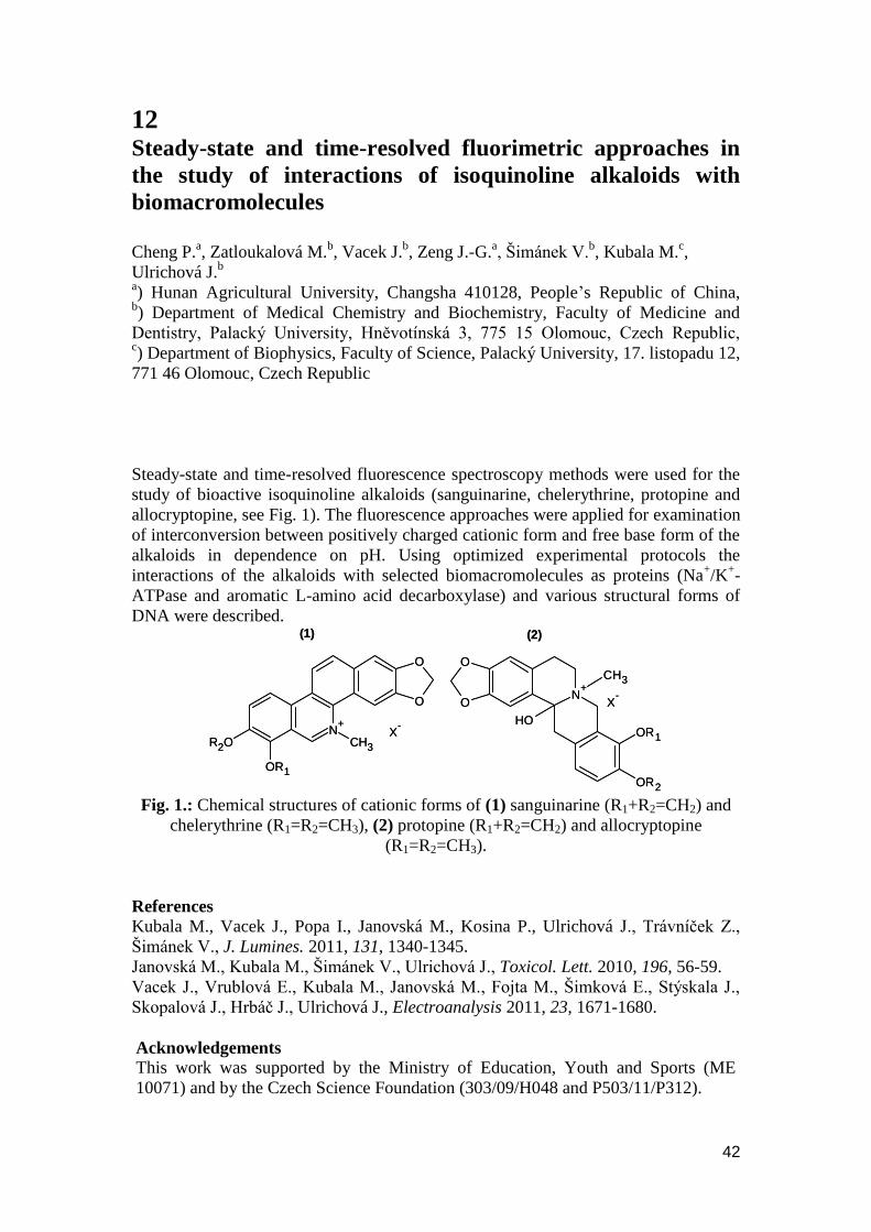
42
12 Steady-state and time-resolved fluorimetric approaches in
the study of interactions of isoquinoline alkaloids with
biomacromolecules
Cheng P.
a, Zatloukalová M.
b, Vacek J.
b, Zeng J.-G.
a, Šimánek V.
b, Kubala M.
c,
Ulrichová J.b
a) Hunan Agricultural University, Changsha 410128, People‟s Republic of China,
b) Department of Medical Chemistry and Biochemistry, Faculty of Medicine and
Dentistry, Palacký University, Hněvotínská 3, 775 15 Olomouc, Czech Republic, c) Department of Biophysics, Faculty of Science, Palacký University, 17. listopadu 12,
771 46 Olomouc, Czech Republic
Steady-state and time-resolved fluorescence spectroscopy methods were used for the
study of bioactive isoquinoline alkaloids (sanguinarine, chelerythrine, protopine and
allocryptopine, see Fig. 1). The fluorescence approaches were applied for examination
of interconversion between positively charged cationic form and free base form of the
alkaloids in dependence on pH. Using optimized experimental protocols the
interactions of the alkaloids with selected biomacromolecules as proteins (Na+/K
+-
ATPase and aromatic L-amino acid decarboxylase) and various structural forms of
DNA were described.
N+
CH3
OR1
R2O
O
O
X-
(1)
O
ON
+
OR1
OR2
CH3
HO
X-
(2)
N+
CH3
OR1
R2O
O
O
X-
(1)
O
ON
+
OR1
OR2
CH3
HO
X-
(2)
Fig. 1.: Chemical structures of cationic forms of (1) sanguinarine (R1+R2=CH2) and
chelerythrine (R1=R2=CH3), (2) protopine (R1+R2=CH2) and allocryptopine
(R1=R2=CH3).
References
Kubala M., Vacek J., Popa I., Janovská M., Kosina P., Ulrichová J., Trávníček Z.,
Šimánek V., J. Lumines. 2011, 131, 1340-1345.
Janovská M., Kubala M., Šimánek V., Ulrichová J., Toxicol. Lett. 2010, 196, 56-59.
Vacek J., Vrublová E., Kubala M., Janovská M., Fojta M., Šimková E., Stýskala J.,
Skopalová J., Hrbáč J., Ulrichová J., Electroanalysis 2011, 23, 1671-1680.
Acknowledgements
This work was supported by the Ministry of Education, Youth and Sports (ME
10071) and by the Czech Science Foundation (303/09/H048 and P503/11/P312).
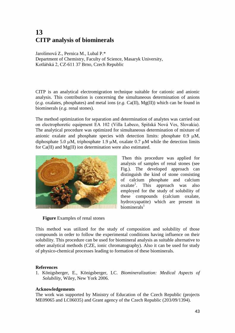
43
13 CITP analysis of biominerals Jarolímová Z., Pernica M., Lubal P.*
Department of Chemistry, Faculty of Science, Masaryk University,
Kotlářská 2, CZ-611 37 Brno, Czech Republic
CITP is an analytical electromigration technique suitable for cationic and anionic
analysis. This contribution is concerning the simultaneous determination of anions
(e.g. oxalates, phosphates) and metal ions (e.g. Ca(II), Mg(II)) which can be found in
biominerals (e.g. renal stones).
The method optimization for separation and determination of analytes was carried out
on electrophoretic equipment EA 102 (Villa Labeco, Spišská Nová Ves, Slovakia).
The analytical procedure was optimized for simultaneous determination of mixture of
anionic oxalate and phosphate species with detection limits: phosphate 0.9 M,
diphosphate 5.0 M, triphosphate 1.9 M, oxalate 0.7 M while the detection limits
for Ca(II) and Mg(II) ion determination were also estimated.
Then this procedure was applied for
analysis of samples of renal stones (see
Fig.). The developed approach can
distinguish the kind of stone consisting
of calcium phosphate and calcium
oxalate1. This approach was also
employed for the study of solubility of
these compounds (calcium oxalate,
hydroxyapatite) which are present in
biominerals1
Figure Examples of renal stones
This method was utilized for the study of composition and solubility of those
compounds in order to follow the experimental conditions having influence on their
solubility. This procedure can be used for biomineral analysis as suitable alternative to
other analytical methods (CZE, ionic chromatography). Also it can be used for study
of physico-chemical processes leading to formation of these biominerals.
References
1. Königsberger, E., Königsberger, LC. Biomineralization: Medical Aspects of
Solubility, Wiley, New York 2006.
Acknowledgements
The work was supported by Ministry of Education of the Czech Republic (projects
ME09065 and LC06035) and Grant agency of the Czech Republic (203/09/1394).
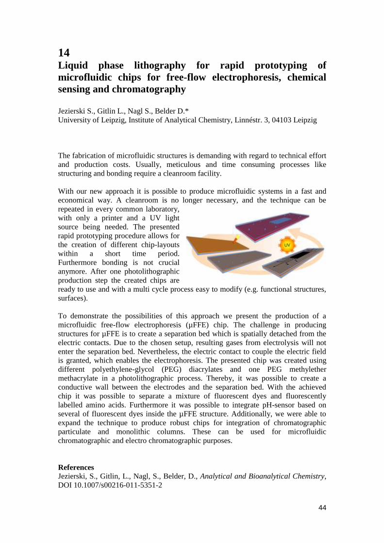
44
14 Liquid phase lithography for rapid prototyping of
microfluidic chips for free-flow electrophoresis, chemical
sensing and chromatography
Jezierski S., Gitlin L., Nagl S., Belder D.*
University of Leipzig, Institute of Analytical Chemistry, Linnéstr. 3, 04103 Leipzig
The fabrication of microfluidic structures is demanding with regard to technical effort
and production costs. Usually, meticulous and time consuming processes like
structuring and bonding require a cleanroom facility.
With our new approach it is possible to produce microfluidic systems in a fast and
economical way. A cleanroom is no longer necessary, and the technique can be
repeated in every common laboratory,
with only a printer and a UV light
source being needed. The presented
rapid prototyping procedure allows for
the creation of different chip-layouts
within a short time period.
Furthermore bonding is not crucial
anymore. After one photolithographic
production step the created chips are
ready to use and with a multi cycle process easy to modify (e.g. functional structures,
surfaces).
To demonstrate the possibilities of this approach we present the production of a
microfluidic free-flow electrophoresis (µFFE) chip. The challenge in producing
structures for µFFE is to create a separation bed which is spatially detached from the
electric contacts. Due to the chosen setup, resulting gases from electrolysis will not
enter the separation bed. Nevertheless, the electric contact to couple the electric field
is granted, which enables the electrophoresis. The presented chip was created using
different polyethylene-glycol (PEG) diacrylates and one PEG methylether
methacrylate in a photolithographic process. Thereby, it was possible to create a
conductive wall between the electrodes and the separation bed. With the achieved
chip it was possible to separate a mixture of fluorescent dyes and fluorescently
labelled amino acids. Furthermore it was possible to integrate pH-sensor based on
several of fluorescent dyes inside the µFFE structure. Additionally, we were able to
expand the technique to produce robust chips for integration of chromatographic
particulate and monolithic columns. These can be used for microfluidic
chromatographic and electro chromatographic purposes.
References
Jezierski, S., Gitlin, L., Nagl, S., Belder, D., Analytical and Bioanalytical Chemistry,
DOI 10.1007/s00216-011-5351-2
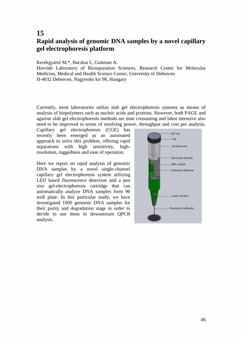
45
15 Rapid analysis of genomic DNA samples by a novel capillary
gel electrophoresis platform
Kerékgyártó M.*, Bacskai I., Guttman A.
Horváth Laboratory of Bioseparation Sciences, Research Centre for Molecular
Medicine, Medical and Health Science Center, University of Debrecen
H-4032 Debrecen, Nagyerdei krt 98, Hungary
Currently, most laboratories utilize slab gel electrophoresis systems as means of
analysis of biopolymers such as nucleic acids and proteins. However, both PAGE and
agarose slab gel electrophoresis methods are time consuming and labor intensive also
need to be improved in terms of resolving power, throughput and cost per analysis.
Capillary gel electrophoresis (CGE) has
recently been emerged as an automated
approach to solve this problem, offering rapid
separations with high sensitivity, high-
resolution, ruggedness and ease of operation.
Here we report on rapid analysis of genomic
DNA samples by a novel single-channel
capillary gel electrophoresis system utilizing
LED based fluorescence detection and a pen
size gel-electrophoresis cartridge that can
automatically analyze DNA samples form 96
well plate. In this particular study, we have
investigated 1000 genomic DNA samples for
their purity and degradation stage in order to
decide to use them in downstream QPCR
analysis.
46
16 Biomarker screening of the human plasma proteome with
disease specific monoclonalantibodies
Kovács A.
1*, Patai Z.
1, Sperling E.
1, Lázár J.
2, Balogh A.
2, Kádas J.
2,
Szekrényes Á.1, Takács L.
2, Kurucz I.
2 and Guttman A.
1,2
1Horváth Laboratory of Bioseparation Sciences, Research Centre for Molecular
Medicine,Medical and Health Science Center, University of Debrecen
H-4032 Debrecen, Nagyerdei krt 98, Hungary 2BioSystems International Kft., H-4032 Debrecen, Nagyerdeikrt 98, Hungary
Monoclonal antibody proteomics is a novel platform based approach to discover
proteins of biomarker potential.Starting from 500 mL normal pooled human plasma
we have generated a comprehensive analyte library representing the human plasma
proteome using specific chromatography based partitioning methods such as Blue
Sepharose and thiophilic interaction chromatography to deplete the albumin and
immunoglobulin fractions. This was followed by ammonium sulfate precipitation, gel
filtration, cation and anion exchange as well as hydrophobic interaction
chromatography fractionations steps. This process resulted in 783 fractions with the
average protein concentration of 1 mg/mL. All chromatography and precipitation
steps were carefully designed with the purpose of maintaining the native forms of the
intact proteins throughout the fractionation process.
As a first application of the analyte library, a dot-blot assay was used to investigate
and compare the separation routes of various disease specific acute phase proteins by
monoclonal antibodies raised against them. In the majority of the cases the
distribution of the antigens in the fractions matched the expectations predictable from
the different phenomena – hydrophilicity-hydrophobicity, molecular shape, size – of
the proteins.The library was also screened with mAbs raised against unknown protein
antigens with the aim of the identification of potential biomarkers following the
workflow of high throughput screening with Western blot verification and antigen
identification by mass spectrometry.
References
Kovács, A., Sperling, E., Lázár, J., Balogh, A., Kádas, J., Szekrényes, Á., Takács, L.,
Kurucz, I., Guttman, A., Electrophoresis 2011, 32, 1916-1925.

47
17 Iron oxide nanoparticle coating of organic polymer based
monolithic columns for phosphopeptide enrichmentand off-
line MALDI/MS or LC-ESI/MS/MS analysis
Křenková J.
1, Nováková K.
2, Zdráhal Z.
2, Foret F.
1
1Institute of Analytical Chemistry of the ASCR, v. v. i., Brno, Czech Republic
2Core Facility - Proteomics, CEITEC-MU and Faculty of Science, Masaryk
University, Brno, Czech Republic
Phosphoproteins are mostly characterized using mass spectrometry (MS) after
proteolysis. However, phosphopeptides may be difficult to analyze by MS due to
lower ionization efficiency of phosphopeptides in the presence of non-phosphorylated
peptides. Therefore, efficient capturing or enrichment of phosphopeptides using
selective affinity techniques prior to MS detection is necessary to increase ionization
efficiency and detection sensitivity of phosphopeptides from highly complex peptide
mixtures.
We have developed a new monolithic capillary column with an iron oxide
nanoparticle coating for selective and efficient enrichment of phosphopeptides. Iron
oxide nanoparticles were prepared by a co-precipitation method and stabilized by
citrate ions. A stable coating of nanoparticles was obtained via multivalent
electrostatic interactions of citrate ions on the surface of iron oxide nanoparticles with
a quaternary amine functionalized poly(glycidyl methacrylate-co-ethylene
dimethacrylate) monolith. A high dynamic binding capacity of 86 μmol per mL
column volume was measured with adenosine-5'-triphosphate. Performance of the
monolithic column was demonstrated with the efficient and selective enrichment of
phosphopeptides from casein digests and off-lineMALDI/MS or LC-ESI/MS/MS
analysis.
Acknowledgements
The Project is funded from the SoMoProprogramme. Research leading to these results
has received a financial contribution from the European Community within the
Seventh Framework Programme (FP/2007-2013) under Grant Agreement No. 229603.
The research is also co-financed by the South Moravian Region. This work was also
supported by the project “CEITEC - Central European Institute of Technology”
(CZ.1.05/1.1.00/02.0068) from European Regional Development Fund. Financial
support from the Grant Agency of the Czech Republic
(P301/11/2055,GD204/08/H054) and the institutional research plan (AV0Z40310501)
is also acknowledged.
48
18 Synthesis of a Caspase-3 sensor
Lišková M.*
1,2, Pazdera P.
2, Klepárník K.
1, Foret F.
1
1Institute of Analytical Chemistry ASCR, v. v. i., Brno, Czech Republic
2Masaryk University, Faculty of Science, Centre for Syntheses at Sustainable
Conditions and their Management, Brno, Czech Republic
Caspase 3, a cysteine-aspartic acid protease activated in the apoptotic pathway, plays
an essential role in the programmed cell death. Moreover, failure of apoptosis is one
of the main contributions to tumor development. Under normal circumstances,
caspases recognize tetra-peptide sequences Asp-Glu-Val-Asp (DEVD) on their
substrates and hydrolyze peptide bonds after aspartic acid residues. Activity of
Caspase-3 is usually assayed by a chemiluminescence reaction. The best limit of
detection by using commercial methods is higher than 1pg of Caspase 3.
We have constructed a special device for the determination of Caspase-3 activity in
free cells. The activity of Caspase 3 is determined by the system based on
Luciferin/Luciferase chemiluminescence reaction. The luciferin modified with
tetrapeptide sequence (DEVD) specific to the recognition for Caspase-3 is cleaved to
form free luciferin, which immediately reacts with luciferase to produce light. Our
detection device consists of a microfluidic chamber held inside a housing of
photomultiplier module for photon counting (Sens-Tech P25USB) with spectral
response 275-620 nm. The sensitivity of the device proved to be an order of
magnitude more sensitive than currently available commercial technologies.
We have designed the synthesis of Caspase-3 sensor. The sensor consists of a CdTe
quantum dot (QD), aderivative of Rhodamine B, caspase recognizing the tetra-
peptide(DEVD) and linkers, bonding the sensor together. Common organic reactions
are used for preparation of this sensor (substitution, double bond addition, reduction
etc.), but with new organic approach based on piperazine and polyethylene glycol
(PEG) chemistry. Optimization of the synthesis is investigated on several model
reactions, where model reactants with the same functional group as the original
component of the sensor are used. Products of synthesis are checked by NMR, FTIR,
MS and CZE-LIF. Our sensor operateson Förster resonance energy transfer
(FRET).QD as a donor chromophore transfers energy to an acceptor chromophore
(derivative of Rhodamine) through non-radiative dipole–dipole coupling. Successful
energy transfer changes the maximum emission wavelength before and after cleavage
of DEVD sequence in the sensor. This sensor can be used for a sensitive
determination of Caspase-3 concentration or activity.
Acknowledgments This work was supported by The Grant Agency of the Czech Republic
(GA203/08/1680, P301/11/2055 and P206/11/2377), Ministry of Industry and Trade
(2A-1TP1/090) and institute research plan AV0Z40310501.
49
19 A prospective non-invasive assessment of a human embryo
viability based on capillary electrophoresis
Mádr A.
1*, Foltová K.
1, Ţáková J.
2, Lousová E.
2, Crha I.
2, Glatz Z.
1
1Department of Biochemistry, Faculty of Science and CEITEC, Masaryk University,
Kamenice 5, 625 00 Brno, Czech Republic 2Department of Gynecology and Obstetrics, Medical Faculty of Masaryk University
and University Hospital, Obilní trh 11, 602 00, Brno, Czech Republic
The human embryo viability is mainly assessed by means of a light-microscopy in
assisted reproduction. However, this extensively used technique have been
confronted about its deficiencies and the world leading embryologist call for a novel
method for an embryo quality assessment to improve an accuracy of an embryo
viability determination. In general, in-vitro fertilization (IVF) procedures suffer from
a relatively low rate of pregnancy thus multiple embryos are implanted to gain the
higher pregnancy outcome. This strategy inevitably increases chances of a multiple
pregnancy with associated health risk issues thus neonatal survival chances could be
compromised and the mother is exposed to risks as well. Currently, elective single-
embryo transfer policy is being preferred by IVF clinics thus improved methods for an
embryo quality assessment are highly demanding. Besides invasive techniques like
preimplantation genetic screening including biopsy, non-invasive assessment based on
metabolome analysis of spent embryo culture media is very prospective and several
analytical techniques were already tested for such purpose. Namely, 1H-NMR, LC-
MS, LC-MS/MS and IR spectroscopy; however utilization of capillary electrophoresis
(CE) was left behind.
Our research group examines the suitability of CE for targeted and untargeted
metabolomics of spent embryo culture media. At first, applicability of CE for amino
acids determination using the contactless conductivity detection was tried. Led by
published hints [1-4], acetic acid in the mixture with neutral hydrophilic polymer
serves as background electrolyte. Bare fused-silica capillary with an inner diameter
50 μm and a total length 80.0 cm (65.0 cm to the detection cell), constant cassette
temperature 25°C and applied voltage +30 kV provide a complete amino acid profile
of commercially available G-1™ medium (Vitrolife, Göteborg, Sweden). Advantages
and drawbacks are discussed and possible improvements are outlined.
References
1. Coufal, P., Zuska, J., van de Goor, T., Smith, V., Gas, B., Electrophoresis 2003,
24, 671-677.
2. Tuma, P., Samcova, E., Andelova, K., J. Chromatogr. B 2006, 839, 12-18.
3. Tuma, P., Soukupova, M., Samcova, E., Stulik, K., Electrophoresis 2009, 30,
3436-3441.
4. Tuma, P., Samcova, E., Stulik, K., Anal. Chim. Acta 2011, 685, 84-90.
Acknowledgements
Authors would like to express thanks for the financial support provided by the Czech
Science Foundation (Project No. P206/11/0009) and by the Ministry of Education,
Youth and Sports of the Czech Republic (Project MSM 0021622413 and Research
Centre LC06023).
50
20 Important electromigration effects of carbon dioxide in
capillary electrophoresis at high pH
Malá Z., Gebauer P., Pantůčková P., Boček P.
Institute of Analytical Chemistry of the Academy of Sciences of the Czech Republic,
v.v.i., Veveří 97, CZ-602 00 Brno (Czech Republic)
This paper reveals the importance of omnipresent effects of carbonate in capillary
zone electrophoretic analyses of anions in alkaline background electrolytes and brings
the knowledge necessary to reach correct qualitative and quantitative results.
Computer simulations and experimental study of selected model systems with indirect
UV absorption and conductivity detection have shown that carbon dioxide absorbed
from air into BGEs and samples induce important electrophoretic effects like
formation of new additional zones and/or boundaries that may further induce strong
and pronounced temporary changes in the migration of analytes. Examples are
reduction of the pH of alkaline BGEs around pH 11 by up to 1 unit or formation of a
pronounced detectable carbon dioxide peak comparable with peaks of analytes at 1
mM level. The higher the pH of the BGE, the stronger these effects and the broader
their spectrum, involving (i) changes of effective mobilities and selectivity due to
changes in pH of the BGE, (ii) occurrence of additional system zones appearing in
form of peaks, dips or more complex disturbances in the detection signal, (iii)
temporary interactions with the sample components and subsequent modification of
the separation process and of its result.
Acknowledgements
We gratefully acknowledge support by the Grant Agency of the Academy of Sciences
of the Czech Republic (Grant No IAA 400310703), by the Grant Agency of the Czech
Republic (Grants No 203/08/1536 and P206/10/1219) and by the Institutional
Research Plan AV0Z40310501 of the Academy of Sciences of the Czech Republic.
51
21 Determination of disinfection by products in drinking water
on an electrophoresis chip
Masár M., Danč L.*, Ţurek R.
Department of Analytical Chemistry, Faculty of Natural Sciences, Comenius
University in Bratislava, Mlynska dolina CH-2, SK-84215 Bratislava, Slovakia
A development of capillary electrophoresis (CE) methods on the chip for separation
and determination of some nonvolatile disinfection by-products formed during the
chlorination of drinking waters was the aim of this work. Monochloroacetic,
dichloroacetic, trichloroacetic, monobromoacetic and dibromoacetic acids were
studied in this context. Halogenacetic acids (HAA) are toxic to humans, plants, and in
particular to algae. Some of them are carcinogens.
Separations were realized on the column-coupling (CC) chip with conductivity
detectors placed at the end of each separation channel. Zone electrophoresis (ZE)
separations carried out in the 2nd
separation channel were combined with electric field
sample stacking or isotachophoresis (ITP-ZE) pre-concentration of the analytes. ZE
separations of HAA were studied in different carrier electrolytes at pH interval 4-6. In
ITP-ZE separations of HAA, ITP as a sample injection technique for ZE on the CC
chip was used. ITP separations were carried out at low pH (pH 4) to remove potential
interfering constituents (weak acids) present in drinking water.
RSD values of migration times for studied analytes were in the range of 0.1-2.7%, and
for peaks areas they were between 1.2-9.9%. The limits of detection (LOD) for HAA
were in the range of 45-180 μg/l when a 900 nl volume of the sample was loaded onto
the chip. Recoveries of HAA in ZE and ITP-ZE determinations varied in 67-101%
and 59-103%, respectively. Lower recoveries of HAA have been mainly caused by
the analyte adsorption on the inner wall of the chip.
Different anions as potential co-migrating constituents present in drinking waters, e.g.
chloride, fluoride, bromide, iodide, sulphate, oxalate, nitrate, nitrite, acetate and
phosphate have been studied. Of these, only acetate and phosphate interfered in ITP-
ZE determinations of HAA. A negative impact of these constituents in real water
samples was eliminated by supported liquid membrane extraction (SLME) of HAA.
In addition, this sample pretreatment technique allowed selective extraction and pre-
concentration of the analytes.
Acknowledgments This work was supported by grants VEGA 1/0672/09, UK/37/2011, VVCE 0070-07
and OPVaV-2009/4.1/02-SORO (CE Green II).

52
22 Fast monitoring of cations and anions in water by chip
electrophoresis
Masár M., Kruk P., Luc M.*, Kaniansky D.
Department of Analytical Chemistry, Faculty of Natural Sciences, Comenius
University, Mlynská Dolina CH-2, SK-84215 Bratislava, Slovakia
E-mail: [email protected]
This work was focused on development of sequential CZE methods for determination
of inorganic anions (chloride, nitrate, sulfate) and cations (ammonium, calcium,
magnesium, potassium, sodium) in drinking waters. Two different background
electrolytes (BGE) with corresponding separation mechanisms for each group of
analytes (anionic and cationic BGE) were employed on the poly(methyl methacrylate)
chip with conductivity detection.
Separations of anions were carried out at pH 4.2 in the BGE containing zwitterionic
detergent. For cationic constituents, BGE with pH 3.2 containing wall adsorption
suppressor (negatively charged surface of the chip) and 18-crown-6-ether was
employed. In addition, water soluble high-molecular polymer
(methylhydroxyethylcellulose) was used to suppress electroosmotic flow in both
instances. Such conditions enabled separations of both groups of analytes. With
respect to the sample injection volume applied (900 nl), the concentration limits of
detection were estimated at 100 g/L and 10 g/L for anions and cations,
respectively. Due to different concentration levels of the ions studied in real samples,
maximum concentration ratios for the neighboring constituents were determined.
Under preferred separation conditions (suppressed hydrodynamic and electroosmotic
flow), the RSD values of migration times were below 0.8% for both groups of
analytes. The peak areas of anions were characterized by 0.7-2.7% RSD while for the
cations the RSD values of peak areas were in range 0.8-3.5 %.
A practical applicability of the developed analytical methods was demonstrated on the
analyses of tap and packed drinking waters. Degassing and/or a proper dilution of the
samples were the only pretreatment procedures before the analysis. The contents of
anions and cations in drinking waters determined by the proposed CZE methods were
in a good agreement with those claimed by water treatment companies. Total analysis
time was approx. 15 min, corresponding to one parameter (ion) per 2 min, chip
maintenance between the runs included.
Acknowledgements
This work was supported by the Slovak Grant Agency for Science (VEGA
1/0672/09), the Slovak Research and Development Agency (VVCE-0070-07),
Comenius University (UK/307/2009) and the CEEPUS program CII-HU-0010-05-
1011.
53
23 CE-LIF of green fluorescent fusion proteins
for investigation of phosphorylation
Michalcová L.
a, Dopitová R.
b, Hejátko J.
b, Válková M.
b, Preisler J.
a,b
aDepartment of Chemistry, Faculty of Science and CEITEC, Masaryk University
bResearch group Functional Genomics and Proteomics of Plants, Mendel Centre for
Plant Genomics and Proteomics, CEITEC, Masaryk University
A histidine-containing phosphotransmitters from A. thaliana (AHP1-5) mediate signal
transduction downstream from receptor histidine kinases to subsequent phospho-
accepting response regulators via so called multistep phosphorelay. Auto-
phosphorylated histidine of the histidine kinase becomes a substrate for the
subsequent phosphorelay, leading via AHPs-mediated nucleus signal delivery to the
activation of ARRs that are involved in transcription control or direct interaction with
cytokinin response effector proteins.1
In this study, CE-LIF with 405 nm laser diode was used to study AHP5 fused with
GFP. AHP5-GFP fusion proteins overexpressed in E. coli were used for initial
optimization of CE-LIF technique because of lower concentrations of the fusion
protein produced by plant. The stability of the AHP5-GFP sample was examined; the
complex was found to be unstable with GFP being released. The GFP release was not
affected by either a long-term storage at -80 °C and -20 °C or the addition of one of
the inhibitors (EDTA, PMSF). These results were confirmed by the western blotting.
The phosphorylation of the sample AHP5-GFP in vitro is under investigation.
Phosphorylation has not been successful yet, the reasons may be the sample
contamination with proteins or an inappropriate phosphorylation protocol. Gel
filtration was used to obtain mass fractions for better understanding
electrophoregrams. Interestingly, some fluorescent fractions were not detected with
the CE-LIF technique.
References
1. To, J.P.,Kieber, J.J. Trends Plant Sci. 2008, 13, 85-92.
Acknowledgements This work was supported by The Czech Science Foundation (Grant No. 203/09/1025),
by The Ministry of Education, Youth and Sports (MSM0021622415) and the project
“CEITEC - Central European Institute of Technology” (CZ.1.05/1.1.00/02.0068) from
European Regional Development Fund.

54
24 Characterization of rhizobia by capillary electrophoresis,
capillary isoelectric focusing, and matrix-assisted laser
desorption/ionization time-of-flight mass spectrometry
Moravcová D.*, Horká M., Šalplachta J., Kubesová A., Kahle V.
Institute of Analytical Chemistry of the AS CR, v. v. i., Veveří 97, 602 00 Brno,
Czech Republic
Fast and reliable identification of individual bacterial strains is crucial for many
branches of human activities as medicine, cosmetics, food industry as well as
microbial forensics, environmental studies, or agriculture. Phenotypic identification of
microorganisms using gram staining, culture, and biochemical methods is traditionally
used; however, these methods are time-consuming, tedious, and low throughput to be
applied to wide populations. In recent years, polymerase chain reaction has been used
for sensitive and specific identification of bacteria species. Nevertheless, molecular
techniques are expensive and technically demanding, therefore, there is still a request
for other sensitive and valid methods for bacterial identification.
Capillary electrophoresis, capillary isoelectric focusing, and matrix-assisted laser
desorption/ionization time-of-flight mass spectrometry were used for analysis of
bacterial cell suspensions of four strains of Rhizobiumand one strain of
Sinorhizobiumspecies - Rhizobium leguminosarumbv. trifolii, R. leguminosarumbv.
viciae, R. galegae, R. loti, and Sinorhizobiummeliloti. These bacteria belong to
beneficial group of bacteria associated with crop plants and they can enhance plant
growth directly by increasing nutrient availability.
All proposed techniques have shown differences in individual profiles of tested
bacterial strains and especially capillary electrophoresis and capillary isoelectric
focusing perform quick and effective bacteria characterization without any additional
sample handling which reduces time necessary for the analysis.
Acknowledgements
This work was supported by the Grant Agency of the Academy of Sciences of the
Czech Republic No. IAAX00310701, by the grant of the Ministry of the Interior
of the Czech Republic No.VG20112015021, by the Ministry of Education, Youth and
Sports of the Czech Republic by grant No. LC06023, and by the Institutional research
plan AVO Z40310501.
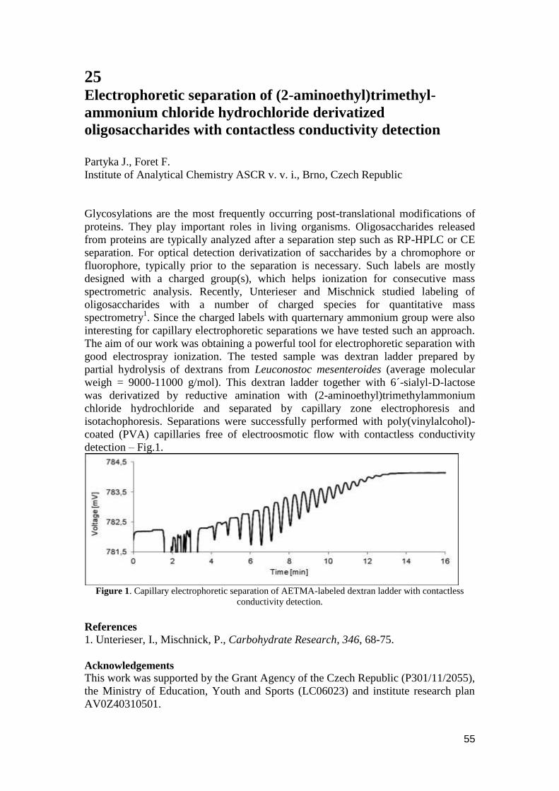
55
25 Electrophoretic separation of (2-aminoethyl)trimethyl-
ammonium chloride hydrochloride derivatized
oligosaccharides with contactless conductivity detection
Partyka J., Foret F.
Institute of Analytical Chemistry ASCR v. v. i., Brno, Czech Republic
Glycosylations are the most frequently occurring post-translational modifications of
proteins. They play important roles in living organisms. Oligosaccharides released
from proteins are typically analyzed after a separation step such as RP-HPLC or CE
separation. For optical detection derivatization of saccharides by a chromophore or
fluorophore, typically prior to the separation is necessary. Such labels are mostly
designed with a charged group(s), which helps ionization for consecutive mass
spectrometric analysis. Recently, Unterieser and Mischnick studied labeling of
oligosaccharides with a number of charged species for quantitative mass
spectrometry1. Since the charged labels with quarternary ammonium group were also
interesting for capillary electrophoretic separations we have tested such an approach.
The aim of our work was obtaining a powerful tool for electrophoretic separation with
good electrospray ionization. The tested sample was dextran ladder prepared by
partial hydrolysis of dextrans from Leuconostoc mesenteroides (average molecular
weigh = 9000-11000 g/mol). This dextran ladder together with 6´-sialyl-D-lactose
was derivatized by reductive amination with (2-aminoethyl)trimethylammonium
chloride hydrochloride and separated by capillary zone electrophoresis and
isotachophoresis. Separations were successfully performed with poly(vinylalcohol)-
coated (PVA) capillaries free of electroosmotic flow with contactless conductivity
detection – Fig.1.
Figure 1. Capillary electrophoretic separation of AETMA-labeled dextran ladder with contactless
conductivity detection.
References
1. Unterieser, I., Mischnick, P., Carbohydrate Research, 346, 68-75.
Acknowledgements
This work was supported by the Grant Agency of the Czech Republic (P301/11/2055),
the Ministry of Education, Youth and Sports (LC06023) and institute research plan
AV0Z40310501.

56
26 Separation of humic acids by zone electrophoresis and on-
line combination of isotachophoresis- zone electrophoresis
Pastierová A.*
a, Bodor R.
a, Tesařová E.
b, Kaniansky D.
† a
a Department of Analytical Chemistry, Faculty of Natural Sciences, Comenius
University, Mlynská dolina, 842 15 Bratislava, Slovakia b Department of Physical and Macromolecular Chemistry, Faculty of Natural
Sciences, Charles University in Prague, Albertov 6, 128 43 Praha 2, Czech Republic
Humic substances (HS) have an important role in the environment. These substances
are classified into three groups, humic acids (HAs), fulvic acids (FAs) and humins, on
the basis of their solubility in acidic and basic solutions. HAs and related fulvic acids
(FAs) occur in soils, natural waters, marine and lake sediments, peat, lignin, brown
coal and other natural deposits. Humic acids (HAs), natural compounds widely
distributed in nature still of unknown structure, are intensively studied.
Different methods have been proposed for the separation, characterization and
behavior of HAs, including capillary electromigration methods. Capillary zone
electrophoresis (CZE) is the most used from capillary electromigration methods for
the separation of HAs carried out in the open separation system. Effective
combination of CZE with another electromigration method usually requires to use
column coupling system. Column coupling system is provided by hydrodynamically
closed system in many cases. The primary difference between open and closed
hydrodynamic separation system is in the location of mechanical barriers at the end of
the capillary (semi-permeable membrane or gel layer). The movement of the
electrolyte solution in the capillary is mechanically limited by this way.
Electroosmotic flow suppression in hydrodynamically closed system is required.
In our work we compared the separation of HAs carried out by on-line coupling of
capillary isotachophoresis with zone electrophoresis (ITP-CZE) in a
hydrodynamically closed separation system and by single-column zone
electrophoresis with hydrodynamically opened separation systems. We used a
zwitterionic detergent as a carrier electrolyte additive in CZE stage of ITP-CZE. The
dodecyldimethylammoniopropanesulfonate (DDAPS) molecule, bearing the anionic
and cationic groups (having no net charge), and in turn, the DDAPS micelles have no
charges if they are present in the water. However, in an electrolyte, an imbalance
between the anion- and cation- partition, can induces surface charges and surface
potential. A critical micellar concentration of DDAPS is in the range 2-4 mmol/l.
A different electrophoretic pattern from the separation of HAs was reached by use of
online combination of capillary isotachophoresis with zone electrophoresis and zone
electrophoresis, respectively. A significant influence of DDAPS on the electrophoretic
mobility of HAs was observed in both used method, and its addition to the carrier
electrolyte leads to increased number of peaks in ITP-CZE.
Additionally, it was confirmed by spectrophotometry, that DDAPS increases the
solubility of HAs in the water.
Acknowledgements
This work was supported by grants UK/39/2011 VEGA 1/0882/09, VVCE 0070/07,
CII-HU-0010-05-1011
57
27 New approach of sample preparation for sensitive
determination of 8-hydroxy-2´-deoxyguanosine in human
urine coupled with capillary electrophoresis
Petrů K.
1, Plíšek J.
1,2, Krčmová L.
1,2, Široká J.
1, Polášek M.
1
1Department of Analytical Chemistry, Charles University in Prague, Faculty of
Pharmacy in Hradec Králové 2Department of Metabolic Care and Gerontology, Teaching Hospital, Sokolská 581,
500 05 Hradec Králové, Czech Republic
E-mail: [email protected]
8-hydroxy-2´-deoxyguanosine (8OHdG) belongs to the group DNA oxidative damage
markers. It is believed to be excreted in urine without further metabolism and it´s
urinary levels correlate with DNA damage. An extremely high concentrations were
recorded at oncological patients and patients treated with radiation or chemotherapy,
respectively. Thus determination of urinary 8OHdG has been proposed as a non-
invasive assay of in vivo oxidative DNA damage [1].
A novel sensitive capillary electrophoretic method conducted with bubble cell
capillary and applying large volume sample stacking was devised for on-line sample
pre-concentration. A 106-fold enhancement of the analytical signal was achieved
comparing with conventional 50 μm capillary.
Sample pre-treatment method Electromembraneextraction (EME) formedfrom Hollow
fibre-liquid-phase microextractionconnected with power source isn‟t so frequent
technique in clinical research. Application of the power source andtwoelectrodestothe
conventionalmethodallows attracting chargedanalytestoward electrode of the
oppositecharge throughsupportedliquidmembrane. In comparison with other
extraction methods additional electrical field utilizing EME, yields to reducing time of
the process.
EME will be applied to urine sample preparation andfurther analyte pre-concentration.
Optimizing procedure such a choice of suitable fibre, organic solvent, working
voltage and extraction time will be described. The results will be documented by an
application of the whole analytical procedure to a patients urine samples.
References
[1] Mei, S. R., Yao, Q. H., Wu, C. Y., Xu, G. W., Journal of Chromatography B-
Analytical Technologies in the Biomedical and Life Sciences 2005, 827, 83-87.
Acknowledgements
The study was supported by the Grant Agency of Charles University GAUK
263811/2011 and by the Project SVV 263 002.
58
28 Potential of surface-enhanced Raman scattering detector for
capillary electrophoresis
Přikryl J., Klepárník K., Foret F.
Institute of Analytical Chemistry of the ASCR, v. v. i.,
Veveří 97, 602 00 Brno, Czech Republic, [email protected]
Surface-Enhanced Raman Spectroscopy (SERS) is interdisciplinary method covering
vibrational spectroscopy, plasmonics and nanotechnologies. This method is based on
the enhancement of the weak Raman scattering of molecules in a close proximity of
metal nanomaterials, such as metal nanoparticles, roughened surfaces or metal tips.
The enhancement factor could reach a value up to 1014 [1,2], and thus SERS becomes
a high sensitive analytical method. In most of the applications, SERS was used as a
stand-alone method. However, relatively little attention was focused to on-line
coupling of SERS with separation methods such as liquid chromatography or capillary
electrophoresis[3,4]. Nirodeet. al. published a simple way to integrate the on-column
SERS detection in CE by usage of SERS-active silver nanoparticles suspended in the
background electrolyte [3]. Leopold et. al. prepared a spot of SERS substrate by laser-
induced citrate reduction of silver nitrate during the CE separation [4].
This poster is devoted to a new, simple photodeposition method of silver
nanoparticles inside a fused-silica capillary induced by laser. The photodeposited
compact spot of a size of ∼10 μm is temporary and spatially stable and resistant to a
hydrodynamic flow. The advantage of this approach is that neither the silver
nanoparticles nor the chemicals for their preparation are components of the
background electrolyte during the electrophoretic separation. Thus, the substrate
formation and separation of analytes are two independent processes and can be
performed under their optimum conditions. The zone broadening due to the sorption
of analytes on the immobilized nanoparticles can be significantly reduced by an
addition of 20% solution of methanol. The efficiency of capillary electrophoresis and
detection selectivity of surface-enhanced Raman scattering is demonstrated by the 3D
electropherograms of rhodamines 123 and B as model samples.
References
[1] Nie, S., Emory, S. R. Science 1997, 275, 1102-1106.
[2] Kneipp, K., Wang, Y.,Kneipp, H., Perelman, L. T.,Itzkan, I.,Dasari, R. R., Feld,
M. S. Phys. Re .Lett. 1997, 78, 1667-1670.
[3] Nirode, W.F., Devault, G.L., Sepaniak, M.J., Anal. Chem. 2000, 72, 1866-1871.
[4] Leopold, N., Lendl, B., Anal. Bioanal.Chem.2010, 396, 2341-2348.
Acknowledgements
The research was financially supported by Grants of the Grant Agency of the Czech
Republic GA203/08/1680, P301/11/2055, the Ministry of Education, Youth and
Sports LC06023 and Research Plan of the Institute of Analytical Chemistry
Z40310501.
59
29 On-line inhibition study of cytochrome P450 2C9 isoform
reaction with diclofenac
Řemínek R.*, Zeisbergerová M., Glatz Z.
Department of Biochemistry, Faculty of Science and CEITEC – Central European
Institute of Technology, Masaryk University, Kamenice 5, 625 00 Brno, Czech
Republic
During last two decades, capillary electrophoresis (CE) proved its position of a
powerful and versatile analytical technique for enzyme assays exhibiting a number of
advantages over conventional methods. These include fast, highly effective
separations, low sample and other chemicals consumption, and high throughput by
automation. In addition, the fuse-silica capillary can serve not only as a separation
column but also as a nanoscale reaction chamber integrating production, separation
and detection of analytes into single run. In-capillary enzymatic assays thus further
intensify the testing economy and enable fully automated analyses performed by
commercially available CE devices.
Development of effective way for reactants mixing inside very limited space of a
capillary constitutes a challenging task which prevents wider practical utilization of
this attractive concept, nevertheless. Couple of approaches dealing with this limitation
has been already presented; however, none of them is suitable for practical
implementation. For this reason, the goal of this study was to introduce a generic
methodology which enables interfusing of selectable reactants into homogenous
reaction mixture inside a nanoscale capillary reactor. Principle based on combination
of longitudinal and transverse diffusion was employed to fulfill these requirements.
Conceptually, the solutions of reaction mixture components are injected by relatively
low pressure as a series of consecutive narrow plugs having parabolic profiles due to
laminar flow inside the capillary. Resulting character of plugs with rather longitudinal
interfaces then enable creation of homogenous reaction mixture by both longitudinal
and transverse diffusions within minute period.
On-line inhibition study of cytochrome P450 2C9 reaction with diclofenac and
sulfaphenazole was performed in order to prove the applicability of proposed system.
Obtained values of Michaelis constant, 50 % inhibitory concentration and inhibition
constant were in agreement with literature data determined by other techniques.
Presented principle thus constitutes a promising tool for on-line inhibition studies of
cytochrome P450 2C9 requiring frequent changes in reaction mixture composition.
Acknowledgements
This work was supported by research project MSM0021622413 and research centre
LC06023 both from Czech Ministry of Education and by grant No. P206/10/0057
from Czech Science Foundation.
60
30 Electrophoretic and mass spectrometric analysis of the lipid
A part of bacterial lipopolysaccharides
Sándor V.
1,*, Scheffer D.
1, Dörnyei Á.
2, Kilár A.
1, Kocsis B.
3, Kilár F.
1,2
1 Department of Analytical and Environmental Chemistry, Faculty of Sciences,
University of Pécs, Ifjúság útja 6, H-7624 Pécs, Hungary 2 Institute of Bioanalysis, Faculty of Medicine, University of Pécs, Szigeti út 12, H-
7624 Pécs, Hungary 3 Department of Medical Microbiology and Immunology, Faculty of Medicine,
University of Pécs, Szigeti út 12, H-7624 Pécs, Hungary
Lipopolysaccharides (LPSs, or endotoxins) are the main components of the external
membrane of Gram-negative bacteria. LPS is composed of three distinct structural
regions: the O-chain polysaccharide, the core oligosaccharide and the lipid A moiety.
Lipid A serves as the hydrophobic anchor of LPS in the outer membrane and is
mainly responsible for the endotoxic activity. It generally consists of a β-1,6-linked
glucosamine disaccharide backbone that is acylated by up to seven C10-C18 fatty acids
or β-hydroxy-fatty acids linked as ester at C3 and C3‟ positions and as amides at C2
and C2‟ positions. The hydroxyl groups of these β-hydroxy-fatty acid chains can be
further esterified by additional fatty acids. Phosphates, with or without other
substituents, are linked at C1 and/or C4' positions. The number and type of the acyl
chains and the state of the phosphorylation of the glycolipid are fundamental
determinants of the toxicity of LPS.
The structure of lipid A is relatively conserved compared to the highly variable O-
chain polysaccharide. The lipid A samples from a single bacterial population may
contain more than one lipid A structural type. Alteration of lipid A structure (i.e.,
changes in acylation, phosphorylation, and glycosylation) could be affected also by
the media for the growth of the bacteria. The moderate variability of lipid A
molecules might serve as the basis for the rapid identification of bacterial strains.
The need for a fast and small-scale analysis of lipid A inspired us to develop a method
that combines the high resolution efficiency of capillary electrophoresis and the high
sensitivity and specificity of mass spectrometry. According to the chemical nature of
the lipid A molecules we applied capillary electrophoresis coupled to electrospray
ionization tandem mass spectrometry (CZE/ESI-MSn), which might enable the
separation and identification of these structurally very closely related amphiphilic
compounds.
References
Hübner, G., Lindner, B. Electrophoresis 2009, 30, 1808-1816.
Acknowledgements
Á.D. acknowledges the support of the János Bolyai Research Scholarship (Hungarian
Academy of Sciences).
61
31 SIMUL 5 COMPLEX - Simulation of the effects of complex-
formation equilibria in electrophoresis
Svobodová J., Beneš M., Hruška V., Zusková I., Gaš B.
Department of Physical and Macromolecular Chemistry, Faculty of Science, Charles
University in Prague, Hlavova 8, 128 43, Prague 2, Czech Republic,
E-mail: [email protected]
The simulation program Simul 5 [1, 2] that was developed in our laboratory enables to
simulate processes in electrophoresis. The implemented theoretical model is based on
principles of mass conservation, acid-base equilibria and electroneutrality. Simul 5
yields the complete picture of all the processes happening during separation and the
concentration profiles of all electrolytes in time. Thus, the simulated
electrophoregram (the detector record) can be obtained.
Currently, the theoretical model of electromigration was extended for systems
containing complexation agent. The complexation equilibria were implemented into
the previous model of electromigration. Simul 5 with complexation mode is especially
useful for simulation of chiral separations, when a chiral selector is used as the
complexation agent. It is applicable for any number of analytes at various degrees of
dissociation. The model was verified experimentally. Three systems that differ in
chiral selectors and/or experimental conditions were selected. The complexation
constants and mobilities of complexes, which are the necessary input data for the new
Simul 5 software, were determined. In the next step the simulations at different
concentrations of chiral selectors were performed and compared with the
experimental profiles obtained from UV and conductivity detections. The profiles
show very good agreement in the position and shape of the analyte peaks. As the
result, the newly developed Simul 5 with complexation mode can be used to predict
the results of chiral separations and to study the processes taking place in
electrophoresis systems, where complexation is involved in the separation.
References
[1] Hruska, V., Jaros, M., Gas, B., Electrophoresis 2006, 27, 984–991.
[2] Hruska, V., Gas, B., Vigh, G., Electrophoresis 2009, 30, 433–443.
Acknowledgements
Funding from the long term research plan of the Ministry of Education of the Czech
Republic MSM 0021620857, the Grant Agency of the Czech Republic, grant No.
203/08/1428, the Grant Agency of the Charles University, grant No. 101309 and grant
No. 323611 is gratefully acknowledged.

62
32 Rapid identification of microorganisms using
electromigration techniques and MALDI-TOF mass
spectrometry Šalplachta J.*
a, Horká M.
a, Kubesová A.
a, Moravcová D.
a, Růţička F.
b, Horký J.
c
a Institute of Analytical Chemistry of the ASCR, v. v. i., Veveří 97, 602 00 Brno,
Czech Republic, E-mail: [email protected] bDepartment of Microbiology, Faculty of Medicine, Masaryk University Brno, Czech
Republic cState Phytosanitary Administration, Division of Diagnostics, Šlechtitelů 23, 779 00
Olomouc, Czech Republic
Increasing demands request for rapid, reliable, and accurate analytical tool able to
identify various types of microorganisms and their products are apparent in different
fields of human activity. Especially, medicine and food industry have great interest in
such techniques to ensure a patient or consumer safety because microbial outbreaks
are spreading quickly. The conventional laboratory methods are still time-consuming
and often inadequate for identification of phenotypically similar species. Therefore,
the traditional phenotype systems are necessary to complement with multiple
analytical techniques.
This work was focused on methods able to identify biofilm-negative and biofilm-
positive strains of Candida „psilosis“ as well as Monilinia species, respectively. For
this purpose, isoelectric focusing (both capillary and gel format) and matrix-assisted
laser desorption/ionization time-of-flight mass spectrometry were used. These
techniques were applied to the analysis of either cell-surface proteins or whole cell
suspension of representative strains of Candida and Monilinia species. Our results
have revealed that proposed methods are suitable for rapid and reliable identification
of C. „psilosis“ strains in a common laboratory. Additionally, our results have shown
that these fast techniques could be possible way for rapid and reliable differentiation
of morphologically similar Monilinia species.
Acknowledgements
This work was supported by the Grant of Ministry of Interior of the Czech Republic
no. VG20102015023 and by the Institutional research plan AVO Z40310501.
63
33 Electromembrane extraction using stabilized constant d.c.
electric current – a simple tool for improvement of
extraction performance
Šlampová A., Kubáň P., Boček P.
Institute of Analytical Chemistry of the Academy of Sciences of the Czech Republic,
v. v. i., Brno, Czech Republic
Electromembrane extractions (EME) are normally performed at constant voltage in all
applications these days. This can be attributed to the fact that the only theoretical
paper considered electric field as one of the key factors for the ion transfer in EME
[1]. Another reason for the dominant use of constant voltage in EME may also be the
fact that voltages used are mostly units to hundreds volts and usual constant d.c.
voltage power supplies or even common 9V batteries [2] are used, which are readily
available. Note, however, that extraction repeatability is often compromised for EME
at constant voltage. Typical repeatability values (expressed as RSDs of peak areas) for
a set of independent EME measurements are around 10%, however, significantly
lower repeatability, with RSD values up to 30%, is often reported. This work presents
an experimental approach for improvement of analytical performance of EME, which
is based on the use of stabilized constant d.c. electric current. Extractions were
performed using a high voltage power supply, which provided stabilized constant d.c.
current down to 1 µA and facilitated current-controlled transfer of ions of interest
from a donor solution through a supported liquid membrane (SLM) into an acceptor
solution. Repeatability of the extraction process has significantly improved for EME
at constant electric current compared to EME at constant voltage. The improved
repeatability of the extraction process was demonstrated on EME-capillary
electrophoresis (EME-CE) analyses of selected basic drugs and amino acids in
standard solutions and in human urine and serum samples. RSD values of peak areas
of the analytes for EME-CE analyses were about two-fold better for EME at constant
electric current (2.8 – 8.9%) compared to EME at constant voltage (3.6 – 17.8%).
Other analytical parameters of the EME-CE methods, such as limits of detection,
linear ranges and correlation coefficients were not statistically different for the two
EME modes. Moreover, EME at constant electric current did not suffer from SLM
instabilities frequently observed for EME at constant voltage.
References
[1] Gjelstad, A., Rasmussen, K.E., Pedersen-Bjergaard, S. J. Chromatogr. A 2007,
1174, 104-111.
[2] Kjelsen, I.J.O., Gjelstad, A., Rasmussen, K.E., Pedersen-Bjergaard, S. J.
Chromatogr. A 2008, 1180, 1-9.
Acknowledgements
Financial support from the Academy of Sciences of the Czech Republic (Institute
Research Funding AV0Z40310501) and the Grant Agency of the Czech Republic
(Grant No. P206/10/1219) is gratefully acknowledged.
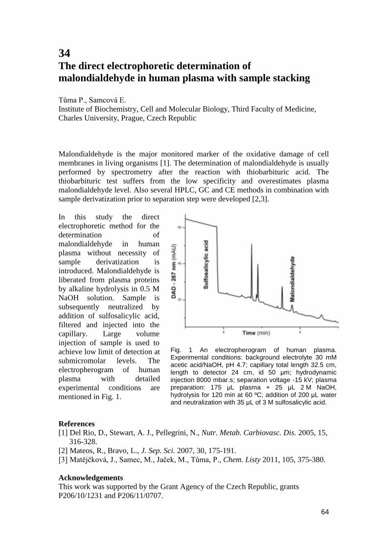
64
34 The direct electrophoretic determination of
malondialdehyde in human plasma with sample stacking
Tůma P., Samcová E.
Institute of Biochemistry, Cell and Molecular Biology, Third Faculty of Medicine,
Charles University, Prague, Czech Republic
Malondialdehyde is the major monitored marker of the oxidative damage of cell
membranes in living organisms [1]. The determination of malondialdehyde is usually
performed by spectrometry after the reaction with thiobarbituric acid. The
thiobarbituric test suffers from the low specificity and overestimates plasma
malondialdehyde level. Also several HPLC, GC and CE methods in combination with
sample derivatization prior to separation step were developed [2,3].
In this study the direct
electrophoretic method for the
determination of
malondialdehyde in human
plasma without necessity of
sample derivatization is
introduced. Malondialdehyde is
liberated from plasma proteins
by alkaline hydrolysis in 0.5 M
NaOH solution. Sample is
subsequently neutralized by
addition of sulfosalicylic acid,
filtered and injected into the
capillary. Large volume
injection of sample is used to
achieve low limit of detection at
submicromolar levels. The
electropherogram of human
plasma with detailed
experimental conditions are
mentioned in Fig. 1.
References
[1] Del Rio, D., Stewart, A. J., Pellegrini, N., Nutr. Metab. Carbiovasc. Dis. 2005, 15,
316-328.
[2] Mateos, R., Bravo, L., J. Sep. Sci. 2007, 30, 175-191.
[3] Matějčková, J., Samec, M., Jaček, M., Tůma, P., Chem. Listy 2011, 105, 375-380.
Acknowledgements
This work was supported by the Grant Agency of the Czech Republic, grants
P206/10/1231 and P206/11/0707.
Fig. 1 An electropherogram of human plasma. Experimental conditions: background electrolyte 30 mM acetic acid/NaOH, pH 4.7; capillary total length 32.5 cm, length to detector 24 cm, id 50 μm; hydrodynamic injection 8000 mbar.s; separation voltage -15 kV; plasma preparation: 175 μL plasma + 25 μL 2 M NaOH, hydrolysis for 120 min at 60 ºC; addition of 200 μL water and neutralization with 35 μL of 3 M sulfosalicylic acid.

65
35 Fast liquid chromatography as a tool for rapid screening of
explosives
Šesták J.1, 2
, Moravcová D.2, Večeřa Z.
2, Kahle V.
2
1 Institute of Analytical Chemistry of the ASCR, v. v. i., Veveří 97, 602 00 Brno,
Czech Republic 2 Faculty of Chemistry, Brno University of Technology, Purkyňova 464/118, 612 00
Brno, Czech Republic
Current devices for detection of explosives suffer by problems with selectivity and
contamination. Coupling of liquid chromatography with ultra-sensitive
chemiluminescence detection could be a right solution of these. Generally, explosive
detection systems should respond instantly and for this reason a rapid LC method has
to be developed and the use of very efficient separation columns [1] is necessary. In
addition, the gradient elution can, of course, significantly increase performance of the
system.
Submitted work assesses a suitability of columns filled with superficially porous
particles [2], Kinetex 2,6 µm C-18, Poroshell 120, 2,7 µm C-18, and monolithic
column Chromolith CapRod RP-18e to perform ultra-fast separation of target
explosives (hexogen, octogen, trinitrotoluene and pentrit). Column permeability and
efficiency in wide range of mobile phase velocities were evaluated. The results show
that the columns filled with superficially porous particles are more efficient than the
monolithic one but the difference is not dramatic and decrease of efficiency in higher
mobile phase velocities is equivalent. On the other hand, the monolithic column
reaches the best separation impedance [3].
Finally, columns filled with superficially porous particles as well as monolithic
column can provide fast and high performance separation of explosives. The
superficially porous particles are used when high efficiency and speed are a priority.
However, if the pressure drop is limiting factor a monolithic column is a right choice.
In this case, the tested monolithic column provided sufficient efficiency for resolution
of target explosives in less than 2 minutes.
References [1] Sýkora, D., Tesařová, E. Chem. Listy, 2007, 101, 190-199.
[2] Cunlife, J. M., Maloney, T. D. J. Sep. Sci. 2007, 30, 3104-3109.
[3] Bristow, P. A., Knox, J. H. Chromatografia, 1977, 10, 279-289.
Acknowledgements
This work was supported by the Institutional research plan AVO Z40310501. The
financial support of the Ministry of Education, Youth and Sports of the Czech
Republic by grant No. LC06023 is also gratefully acknowledged.
66
36 A High-Throughput Platform for Preparation of APTS-
Labeled N-Glycans: Improving the Accuracy,
Reproducibility and Time-to-Results of N-Glycan Profiling
Szekrenyes A., Guttman A.
Horvath Laboratory of Bioseparation Sciences, Debrecen, Hungary
Kimzey M., Pourkaveh S., Mast S., Sun B., Minalla A., Haxo T., Nishida C.,
Wegstein J.
ProZyme, Inc., Hayward, CA, USA
Szabo Z., Karger B.L.
Barnett Institute, Boston, MA, USA
Chapman J.
Beckman Coulter, Inc., Brea, CA, USA
A novel sample preparation protocol is introduced for optional purification of mAbs
including enzymatic deglycosylation, APTS labeling, sample cleanup, and
standardization of N-glycans optimized for downstream CE-LIF analysis. The
optimized labeling reagents provide complete derivatization with only one-hour
incubation time, without degradation of important labile groups, such as sialic acid
and core or outer-arm fucose residues. After labeling, the remaining unconjugated
APTS is efficiently (>99.9 %) removed and the sample eluted in water, enabling the
potential for mass spectrometry confirmation of atypical peaks. Spike-in, lower and
upper internal mobility standards are used to normalize glycan migration times,
further reducing variability between runs. Glucose unit (GU) assignment based on
normalized glycan migration times showed significant improvement in precision
compared to GU assignment without the use of internal mobility standards. It is
envisioned that this unique glycoanalysis solution will be important for the
standardization and expansion of N-glycan profiling in such large scale applications
as clone selection and cell-culture optimization.
67
37 Analysis of Altered IgG Galactosylation in Autoimmune
Diseases
Váradi C., Rezsu N., Bertalan Meskó B., Guttman A.
Horváth Laboratory of Bioseparation Sciences, University of Debrecen, Hungary
Immunglobulins are the major secretory products of the immune system. The N-
glycosylation on human immunoglobulins, especially on IgG1 (conserved site at Asn
297), plays critical roles in the bioactivity of these proteins. Significant differences
have been reported in IgG glycosylation in pregnancy, aging and various diseases.
The four characteristic glycans of IgG1 are biantennary-agalacto (G0), biantennary-
monogalacto (G1 and G1‟) and biantennary–digalacto (G2) structures, with the
relative amount of G0 showing the highest variability in the above mentioned
conditions. The aim of this study was to investigate the changes in the relative amount
of G0 glycans of IgG in a few prevalent autoimmun diseases and investigating the
quantitative changes of G0 with transcriptomics data on galactosyl transferase
expression. The glycosylation pattern in sex and age matched samples were analyzed
by capillary electrophoresis to see the correlation between IgG N-glycan profile,
galactosyl transferase expression and the pathogenesis of Rheumatoid arthritis and
Crohn disease. IgG was isolated by means of Protein A affinity pulldown from the
samples of relevant patients and the N-glycans were released by peptide-N-glycanase
F (PNGase F). The released glycans were then fluorescently labeled with
aminopyrene-trisulfonate (APTS) and analyzed by capillary electrophoreis with laser
induced fluorescence detection. Significant differences were found in the
galactosylation ratio after treatment between the responsive and non responsive
patients.
68
38 Capillary electrophoresis of CdTe quantum dots and its
probes
Voráčová I.
1, Klepárník K.
1, Lišková M.
1, 2, Patrmanová M.
1,2, Zeiselová M.,
1 Foret
F.,1
1Institute of Analytical Chemistry AS CR, v. v. i., Veveří 97, 602 00 Brno, Czech
Republic, 2Department of Chemistry, Faculty of Science, Masaryk University,
Kotlářská 2, 611 37 Brno, Czech Republic
Quantum dots (QDs) are semiconductor nanocrystals with unique optical properties
finding wide scope of applications. Water soluble CdTe QDs are prepared uncoated or
coated with inorganic salts (CdS, ZnS, SiO2) and with several organic ligands bonded
on its surface. The organic ligands usually contain thiol groups that bind on QD
surface and ionic groups (carboxyl or amino) that provide water solubility and serve
as linkers for conjugation of other molecules [1, 2].
The QDs with mercaptopropionic acid organic ligand on the surface were conjugated
via zero-length cross linkers 1-ethyl-3-(3-dimethyl-3-aminopropyl) carbodiimide
hydrochloride and N-hydroxysulfosuccinimide with macrocyclic ligand (1,4,7-
triacetyl-10-aminopenthyl- 1,4,7,10-tetraazacyclododecane, MPI) or its Zn(II) or
Eu(III) ion complexes. Conjugation conditions were optimized and formation of the
conjugate was confirmed by capillary electrophoresis with laser induced fluorescence
detection (CE-LIF). The ratio of conjugated QD:MPI and QD:metal ion complex were
also tested. The electrophoretic mobility increases from -4.1 × 10⁻⁸
to -2.1 × 10⁻⁸
m2V
-
1s
-1 for ratios QD: MPI 1:0 to 1:20. A further increase of this ratio has practically no
influence on the mobility of conjugate. The QDs conjugated with simple organic
molecules and its metal ion complexes should create luminescent probes for Förster
resonance energy transfer (lanthanide (III) complex) and DNA labeling (Zn(II)
complex). QDs with different sizes and organic ligands on the surface were
characterized by CE-LIF using various background electrolytes. Their zeta-potential
and charge were calculated from electrophoretic mobilities according to theory
derived by Ohshima [3]. The determined charges in pH=10 are -12 in the case of 3.1
and 3.5 nm QD with mercaptopropionic acid and -14 in the case of 3.9 nm QD with
thioglycolic acid on the surface. Providing the complete dissociation of organic
ligands, the number of charges should also indicate the number of organic ligands
bonded on QD surface.
References
[1] Kleparnik, K., Voracova, I., Liskova, M., Prikryl, J., et al., Electrophoresis 2011,
32, 1217-1223.
[2] Kleparnik, K., Liskova, M., Voracova, I., Hezinova, V., et al., Analytical and
Bioanalytical Chemistry 2011, 400, 369-379.
[3] Ohshima, H., Journal of Colloid and Interface Science 2001, 239, 587-590.
Acknowledgements
This work was supported by grants of the Grant Agency of the Czech Republic
(GA203/08/1680, P301/11/2055, P206/11/2377), Ministry of Education, Youth and
Sports (LC06023) and institute research plan AV0Z40310501.

69
39 The effect of different inactivation procedures on Candida
species confirmed by capillary HPLC and CIEF
Vykydalová M.*
1,2, Horká M.
1, Moravcová D.
1, Růţička F.
3, Kahle V.
1
1 Institute of Analytical Chemistry ofthe AS CR, v. v. i., Veveří 97, 602 00 Brno, Czech
Republic 2Department of Biochemistry, Faculty of Science, Masaryk University Brno, Kotlářská
267/2, 611 37 Brno, Czech Republic 3Department of Microbiology, Faculty of Medicine, Masaryk University, Pekařská 53,
602 00 Brno, Czech Republic
Three close species Candida parapsilosis, Candida methapsilosis and Candida
orthopsilosis have different pathogenicity to humans. C. parapsilosis and C.
methapsilosisexistin two forms biofilm-positive and biofilm-negative. C. orthopsilosis
does not form biofilm. C. “psilosis” occur on the skin, hands and mucous membranes
of healthy people; however, biofilm positive strains especially C. parapsilosis become
a significant problem for immuno-compromised patients leading even to death.
Therefore, it is necessary to check the presence of infectious pathogens and to
investigate possibilities of their inactivation. The subsequent control of an efficiency
of inactivation procedure is important part of research as well.
In this study the efficiency of inactivation procedures onCandida species was
investigated by capillary HPLC and CIEF. Biofilm-positivestrains of C.parapsilosis
and C.methapsilosis were used as model organisms. Inactivation of these yeasts was
achieved by different physical or chemical methods, e.g., boiling, using of 70% v/v
ethanol or 4% v/v formaldehyde, or 2% v/v peroxoacetic acid [1]. Suspensions of cell
pellets and supernatants after inactivation procedure were analyzed by CIEF and
capillary HPLC.
Isoelectric fingerprints of tested bacteria cells have changed in all cases of
inactivation compared to native cells. Significant differences in HPLC separation
profiles of bacterial supernatants were found as well. Detailed study of the
inactivation processes of different bacterial strains will be followed.
References
[1] Růţička, F., Horká, M., Holá, V., Kubesová, A., Pavlik, T., Votava, M., Journal
of Microbiological Methods 2010, 80, 299-301
Acknowledgements
This work was supported by the Grant Agency of the Academy of Sciences
of the Czech Republic No. IAAX00310701, by the Institutional research plan
AV0Z40310501, by the grant of Ministry of the Interior of the Czech Republic No.
VG20112015021 and by the Ministry of Education, Youth and Sports of the Czech
Republic by grant No. LC06023.
70
40 In-capillary screening of matrix metalloproteinase inhibitors
by capillary electrophoresis for coupling with ESI mass
spectrometry
Wang* X., Adams E., Van Schepdael A.
Laboratory for Pharmaceutical Analysis, Faculty of Pharmaceutical Sciences, K.U.
Leuven, Herestraat 49 PB 923, B-3000 Leuven, Belgium
Matrix metalloproteinases (MMPs) have been considered as a novel biomarker and
potential therapeutic target in human cancer. The MMP-2 and MMP-9 which have
been particularly implicated in tumor invasion and metastasis formation were selected
as model enzymes. A capillary electrophoresis (CE) based method with enzymatic
reaction inside the capillary for screening of MMP inhibitors with fluorescence
detector was firstly developed in our lab. In order to develop a label free method for
the screening of MMP inhibitors, we are trying to investigate the applicability of CE-
MS for the in-capillary screening of MMP inhibitors. In this project, a volatile system
was firstly developed with CE-fluorescence detector. The buffer concentration and pH
have been optimized in this system. Finally, 20 mM ammonium acetate (pH 7.0) was
selected for both enzyme reaction and electrophoretic separation. Then, the inline
incubation was performed using this method. The whole reaction, separation and
detection were achieved within one minute. For the mass spectrometry analysis, the
ion trap MS conditions were optimized using reaction product. The negative ESI
detection mode was selected. The sheath liquid composition was also optimized.
Acetonitrile-water (80:20) was finally selected for further CE-MS investigation.
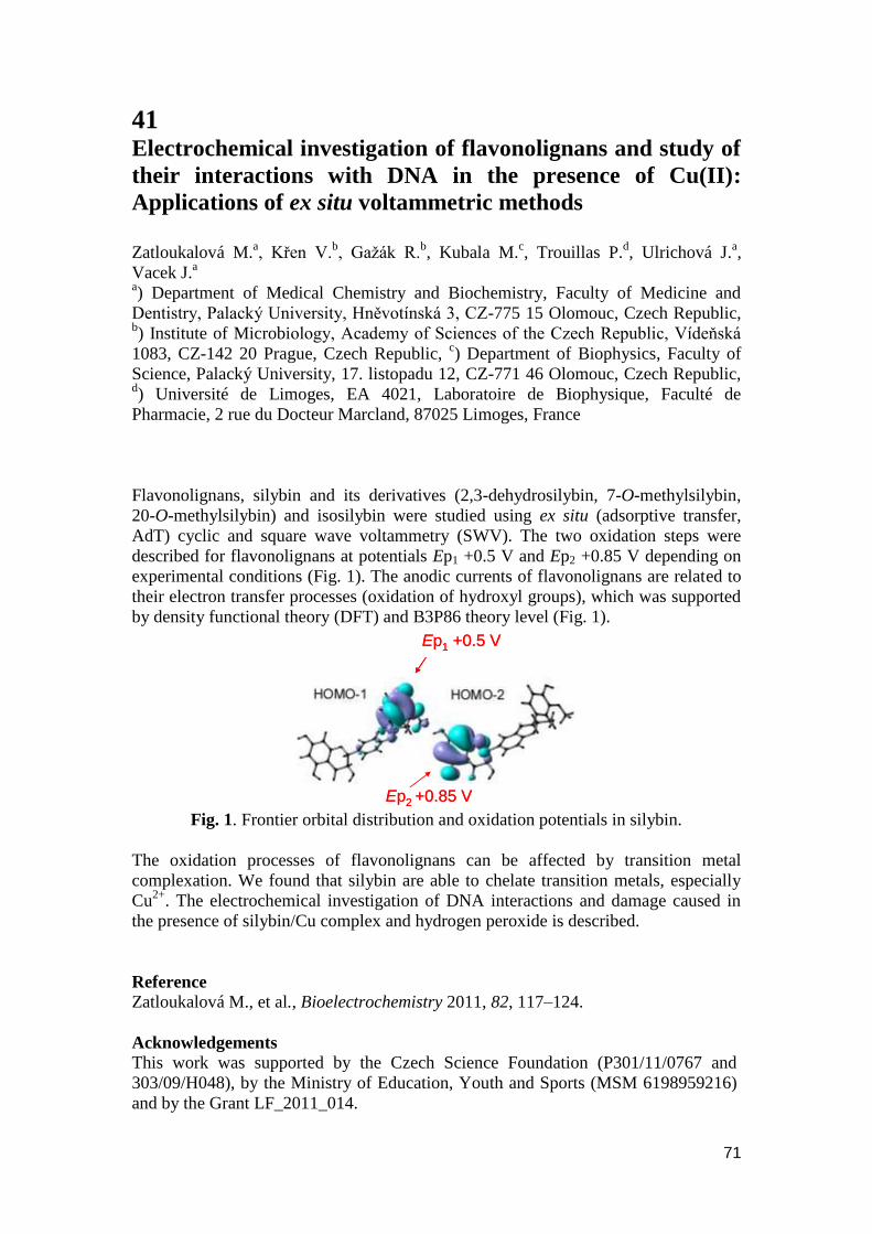
71
41 Electrochemical investigation of flavonolignans and study of
their interactions with DNA in the presence of Cu(II):
Applications of ex situ voltammetric methods
Zatloukalová M.
a, Křen V.
b, Gaţák R.
b, Kubala M.
c, Trouillas P.
d, Ulrichová J.
a,
Vacek J.a
a) Department of Medical Chemistry and Biochemistry, Faculty of Medicine and
Dentistry, Palacký University, Hněvotínská 3, CZ-775 15 Olomouc, Czech Republic, b) Institute of Microbiology, Academy of Sciences of the Czech Republic, Vídeňská
1083, CZ-142 20 Prague, Czech Republic, c) Department of Biophysics, Faculty of
Science, Palacký University, 17. listopadu 12, CZ-771 46 Olomouc, Czech Republic, d) Université de Limoges, EA 4021, Laboratoire de Biophysique, Faculté de
Pharmacie, 2 rue du Docteur Marcland, 87025 Limoges, France
Flavonolignans, silybin and its derivatives (2,3-dehydrosilybin, 7-O-methylsilybin,
20-O-methylsilybin) and isosilybin were studied using ex situ (adsorptive transfer,
AdT) cyclic and square wave voltammetry (SWV). The two oxidation steps were
described for flavonolignans at potentials Ep1 +0.5 V and Ep2 +0.85 V depending on
experimental conditions (Fig. 1). The anodic currents of flavonolignans are related to
their electron transfer processes (oxidation of hydroxyl groups), which was supported
by density functional theory (DFT) and B3P86 theory level (Fig. 1).
Ep1 +0.5 V
Ep2 +0.85 V
Ep1 +0.5 V
Ep2 +0.85 V Fig. 1. Frontier orbital distribution and oxidation potentials in silybin.
The oxidation processes of flavonolignans can be affected by transition metal
complexation. We found that silybin are able to chelate transition metals, especially
Cu2+
. The electrochemical investigation of DNA interactions and damage caused in
the presence of silybin/Cu complex and hydrogen peroxide is described.
Reference
Zatloukalová M., et al., Bioelectrochemistry 2011, 82, 117–124.
Acknowledgements
This work was supported by the Czech Science Foundation (P301/11/0767 and
303/09/H048), by the Ministry of Education, Youth and Sports (MSM 6198959216)
and by the Grant LF_2011_014.
72
42 Optimization of non-aqueous media for capillary
electrophoresis of dextromethorphan and its metabolites
Zeisbergerová M.
*, Cvingráfová E., Glatz Z.
Masaryk University, Faculty of Science, Department of Biochemistry, Kamenice 5,
625 00 Brno, Czech Republic.
The use of organic solvents as the basis of the electrophoretic media in CE has been
reported as a powerful technique for achieving highly selective and fast separations
[1]. The selectivity of the non-aqueous capillary electrophoresis (NACE) is based on
physicochemical properties of organic solvents, e.g. viscosity, electrical conductivity
and acid-base behavior which are different from those of water. Non-aqueous
background electrolytes (BGEs) also affect parameters of the CE system, namely
electrophoretic mobility, acid-base and complexation characteristics. One of the most
attractive features of NACE is the large panel of organic solvents to be used, in pure
form or in mixture. Other advantages include reduced electric conductivity which
results in low electric currents and Joule heat production. Notably, NACE extends the
applicability of CE to analytes with poor solubility in water.
NACE was found to be a good alternative for the analysis of pharmaceutical drugs
[2, 3]. In our work we have targeted the separation of dextromethorphan (DEX) and
its metabolites. DEX is primarily used as an over-counter antitusive drug for
temporary relief of cough. A variety of analytical methods have been developed to
determine DEX and its metabolites in biological matrices, mainly involving the use of
HPLC, GC, TLC and CE.
Our study has been focused on the influence of non-aqueous BGEs on the separation
of DEX and its metabolites. Three organic solvents of different characteristics have
been chosen for BGEs containing ammonium acetate; namely methanol, acetonitrile
and 2-propanol. Apparent pH of all involved solutions was measured as well as the
pH of aqueous electrolyte used for the comparison. Separations of the object analytes
with the addition of levallorphan (internal standard) were performed in a 75 μm I.D.
fused-silica capillary, 48.5/40 cm Ltot/Leff, respectively. The evaluation was based on
the resolution of all analytes, the efficiency of levallorphan separation, electroosmotic
mobility and the total analysis time.
References:
1. Steiner, F., Hassel, M., Electrophoresis 2000, 21, 3994-4016.
2. Geiser, L., Veuthey, J.-L., Electrophoresis 2007, 28, 45-57.
3. Suntornsuk, L., Anal. Bioanal. Chem. 2010, 398, 29-52.
Acknowledgments
Financial support from Grant Agency of Czech Republic (grant No. P206/10/0057)
and Czech Ministry of Education (research project No. MSM0021622413 and
research center LC06023) is gratefully acknowledged.
73
43 Comparison of Two Electrophoretic Methods Documented
by Separation of Dextromethorphan and its Metabolites
Zeisbergerová M., Langmajerová M
*., Cvingráfová E., Glatz Z.
Masaryk University, Faculty of Science, Department of Biochemistry., Kamenice 5,
625 00 Brno, Czech Republic.
Capillary electrophoresis (CE) belongs to powerful analytical techniques and its great
advantage is the existence of various modes. It allows analyses of different
compounds from inorganic particles to biopolymers. CE is applied to drug assays
because of its unique separation mechanism, speed, efficiency and versatility.
Although capillary zone electrophoresis (CZE) and micellar electrokinetic
chromatography are the most commonly used modes for drug analysis, the usage of
others (e.g. NACE, ITP, CEC) is increasing.
Non-aqueous capillary electrophoresis (NACE) is a term applied to describe a system
in which pure organic solvents or mixtures thereof are used for background
electrolytes. The application of organic solvents in CE has advantages over aqueous
media such as good solubility of hydrophobic analytes, low conductivity and
modification of separation selectivity. The organic solvent affects degrees of
electrolyte dissociation, forms ion-pairs and changes conductivity of ions.
In our work we focused on the comparison of CZE and NACE methods applied to
determination of a drug and its metabolites. As a model drug dextromethorphan was
chosen. Parameters for CZE method were taken from the literature [1] and conditions
for NACE were based on the publication [2]. Separations in both modes were carried
out under these conditions: a fused silica capillary with inner diameter of 75 µm,
48,5/40 cm Ltot/Leff, the positive separation voltage and UV detection at 200 nm. The
methods were compared based on the following parameters: repeatability of migration
times, normalized peak areas, peak resolution and total analysis time. Other
parameters such as background electrolyte, sample matrix and working conditions
will be discussed.
Literature:
1. Kristensen, H., J. Pharm. Biomed. Anal. 1998, 18, 827-838.
2. Dong, Y., Chen, X., Chen, Y., Chen, X., Hu, Z., J. Pharm. Biomed. Anal. 2005,
39, 285-289.
Acknowledgements:
The authors thank for financial support Grant Agency of Czech Republic (grant No.
P206/10/0057) and Czech Ministry of Education (research project No.
MSM0021622413 and research center LC06023).
74
44 Irreversible protein heat denaturation analyzed by Bayesian
approach
Ţídková J.
a, Matejková M.
b, Ţídek L.
b, Padrta P.
b, Bobálová J.
a, Sklenář V.
b
a Institute of Analytical Chemistry of the ASCR, v. v. i., Veveří 97, 602 00, Brno,
Czech Republic, [email protected]
bNational Centre for Biomolecular Research, Faculty of Science, and CEITEC,
Masaryk University, Kamenice 5, 625 00, Brno, Czech Republic
Detailed description of heat denaturation is important for understanding protein
function and for evaluating possibilities of industrial utilization of the protein. This
presentation describes analysis of heat denaturation data, obtained by various
experimental techniques, using Bayesian statistics. The first source of data was
differential scanning calorimetry. NMR spectroscopy was used as a complementary
approach to differential scanning calorimetry. One-dimensional NMR spectra reflect
presence of well-defined three-dimensional structure of the studied protein and offer
certain selectivity – analysis of the methyl and aromatic regions provides insight into
the packing of the hydrophobic core of the studied enzyme, while the amide region
reflects changes of backbone conformation. A denaturation curve can be obtained by
recording a series of spectra at gradually increasing temperature. We present a
protocol allowing to quantify the structural changes by a Bayesian analysis of the data
and of a simple but correct model of irreversible denaturation. The procedure allows
to evaluate enthalpic and entropic contributions in a case of sufficiently sampled
temperatures. The methodology is presented on an example of non-specific lipid
transfer protein 1 from barley. This protein denaturates irreversibly at extremely high
temperatures (approximately 110°C) and thus presents a considerable experimental
challenge. In order to monitor protein behaviour as such high temperatures, a special
set-up (sealed tubes and choice of a suitable probe-head) was used.
References
1. Matejková, M., Ţídková, J., Ţídek, L., et al., JAFC 2009, 57, 8444-8452
Acknowledgments This work was supported by the Grants 1M0570 (J.Ţ. and J.B.), MSM0021622413,
(M.M. and L.Ţ.) and LC06030 (V.S.) of the Ministry of Education, Youth, and
Physical Culture of the Czech Republic and by Grant AV0Z40310501 of the Academy
of Sciences of the Czech Republic (J.Ţ. and J.B.).

75
45 Analysis of cellular metabolites by HILIC UHPLC-MS/MS
Ţupková M., Janečková H., Fikarová I., Najdekr L., Wojtowicz P., Mičová K.,
Friedecký D., Adam T.
Laboratory of Inherited Metabolic Disorders and Institute of Molecular and
Translational Medicine, Faculty of Medicine and Dentistry, Palacký University,
Olomouc, Czech Republic
Background: Metabolomics became a valuable tool for diagnostics and research in
clinical biochemistry over the past few years. Analysis of human metabolome is still a
great challenge thanks to the variability of the compounds and due to the huge amount
of metabolites. Development of methods is necessary for better understanding of all
metabolic processes. With new more faster and precise methods we will be able to
analyse and find more metabolite markers.
Methods: Separations were performed on an Ultimate 3000 RS UHPLC (Dionex)
with aminopropyl column (Luna 3 μm NH2, 2 x 150 mm, Phenomenex). The mobile
phase consisted of 20mM ammonium acetate (pH 9.45) as eluent A and acetonitrile as
eluent B. For detection, a QTRAP 5500 tandem mass spectrometer (AB Sciex) with
electrospray ionization was used. The samples were measured in a multiple reaction
monitoring mode in positive and negative mode. Dwell time was 15 msec with
complete cycle of 3 sec. The method was tested on human leukocytes. Cells were
quenched by the chloroform/methanol/water mixture (4:4:2.8). Final extracts were
freeze-dried and re-dissolved in mobile phase. For accurate quantification 6
isotopically labeled internal standards were added to the samples before protein
precipitation.
Results: We developed method including 352 intermediates of metabolic pathways
based on Kegg and HMDB databases. The method covers aminoacids, organic acids,
bases, ribosides, nucleotides, sugars, and alcohols. By this method all compounds
were detected with the limit of detection in the range 10-8
- 10-5
mol/L. The analyses
were completed within 72 minutes including both ionisation modes.
Conclusion: The developed method can be successfully used for profiling of polar
metabolites and offers complementary approach to widely used separation systems
based on reversed phase columns.
Acknowledgments This work was supported by grants IGA MZČR NT12218 and LF UP 2011-011.
The infrastructural part of this project (Institute of Molecular and Translational
Medicine) was supported from the Operational programme Research and
Development for Innovations (project CZ.1.05/2.1.00/01.0030).
76
SPONSORS

77
NOTES

78

79
80