Embed Size (px)
Citation preview

April 2018 | ISSN: 0067-8856 04 54 1 | Volume 54 (1)
BIOMEDICAL SCIENCES INSTRUMENTATIONAn international journal for the study of biomedical engineering, technology, & education
Oldest biomedical engineering journal | Published since 1964 IAE PublishingWindsor, CanadaEditor, Michelle Tucci, PhDGuest Editor: Jeffrey Anderson, PhD and Amanda Brooks, PhD

Volume 54 (1) April 2018 Biomedical Sciences
Instrumentation
Aims and Scope
Biomedical Sciences Instrumentation publishes peer-reviewed scientific articles for the advancement of
biomedical engineering in relationship to patient safety, patient care, automated instrumentation for
clinical decision making, and rehabilitation. It is the oldest engineering journal that encompasses the
individual and collaborative efforts of scientists in clinical medicine, dentistry, basic and applied sciences,
engineering, and bioethics. The journal is dedicated to the publication of outstanding articles of interest
in the biomedical engineering research community.
Society Information
Beginning in 1963, the Rocky Mountain Bioengineering Symposium is the oldest, continuously held
biomedical engineering symposium in the United States. It was founded by a group of the most visionary
and historical individuals at the US Air Force Academy in the engineering field to promote dialog and the
exchange of ideas and experiences between attendees, including between professionals and students.
From its beginning as a regional meeting it has grown to a global event regularly attracting attendees
from across the world. Since 1970, it has merged with the International Society of Automation Biomedical
Sciences Instrumentation Symposium. Submitted papers are peer-reviewed, and those accepted for
presentation and publication appear in the yearly issue of Biomedical Sciences Instrumentation journal,
an internationally distributed publication by International Academic Express Company Ltd (iaexpress.ca).
Editorial Board
Editor –IN- Chief
Michelle A. Tucci, PhD, FAIMBE
Professor,
Department of Anesthesiology
University of Mississippi Medical Center
Special Edition -Guest Editors
Jeff Anderson, PhD, University of Wyoming
Amanda Brooks, PhD, University of North Dakota
Associate Editors
Hamed Benghuzzi, PhD, FAIMBE, FBSE, University of Mississippi Medical Center
Lynne Jones, PhD, FAIMBE, FBSE, Johns Hopkins University
Adel Mohamed, MD, University of Saskatchewan
Elena Oggero, PhD, University of Wyoming
Guido Pagnacco, PhD, University of Wyoming
Julian Thayer, PhD, The Ohio State University
Yoshiharu Yonezawa Hiroshima Institute of Technology
Volume 54 (1) April 2018 Biomedical Sciences
Instrumentation
Editorial Board
Jeff Anderson, PhD, University of Wyoming
Steve Barrett, PhD, University of Wyoming
Kenneth Butler, PhD, University of Mississippi Medical Center
Amanda Brooks, PhD, University of North Dakota
Joseph A. Cameron, PhD, Jackson State University
Ibrahim Farah, PhD, Jackson State University
Paul Frenger, MD, A Working Hypothesis, Inc.
Patrick Patterson, PhD, Texas Tech University
David Paulus, PhD, University of Arkansas
Brian Stemper, PhD, University of Wisconsin
John Sollers III, PhD, North Carolina Central University
Gabi Waite, PhD, Geisinger Commonwealth School of Medicine
Lee Waite, PhD, Engineering Consultant
Jennifer Wagner, PhD, University of Colorado
Cameron Wright, PhD, University of Wyoming
Publication Policy
Biomedical Sciences Instrumentation (ISSN 0067-8856) is published quarterly by International Academic Express (IAE) publishing at 747 Sarah Court, Windsor, ONT, N9G2Y7 Canada. Papers may not be reproduced in any form without written permission from IAE. Reprints of articles in this publication are available on a custom basis at reasonable prices at IAE (http://iaexpress.ca/ . For publication information contact [email protected] (Editor) and for technical or special request information contact [email protected]
ISBN: 9780107752422-1-3ISSN: 0067-8856
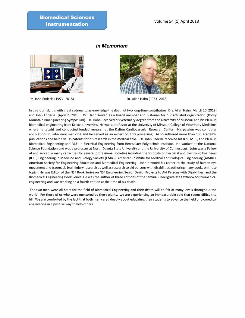
In Memoriam
Dr. John Enderle (1953 –2018) Dr. Allen Hahn (1933- 2018)
In this journal, it is with great sadness to acknowledge the death of two long-time contributors, Drs. Allen Hahn (March 20, 2018)
and John Enderle (April 2, 2018). Dr. Hahn served as a board member and historian for our affiliated organization (Rocky
Mountain Bioengineering Symposium). Dr. Hahn Received his veterinary degree from the University of Missouri and his Ph.D. in
biomedical engineering from Drexel University. He was a professor at the University of Missouri College of Veterinary Medicine,
where he taught and conducted funded research at the Dalton Cardiovascular Research Center. His passion was computer
applications in veterinary medicine and he served as an expert on ECG processing. Al co-authored more than 130 academic
publications and held five US patents for his research in the medical field. Dr. John Enderle received his B.S., M.E., and Ph.D. in
Biomedical Engineering and M.E. in Electrical Engineering from Rensselaer Polytechnic Institute. He worked at the National
Science Foundation and was a professor at North Dakota State University and the University of Connecticut. John was a Fellow
of and served in many capacities for several professional societies including the Institute of Electrical and Electronic Engineers
(IEEE) Engineering in Medicine and Biology Society (EMBS), American Institute for Medical and Biological Engineering (AIMBE),
American Society for Engineering Education and Biomedical Engineering. John devoted his career to the study of human eye
movement and traumatic brain injury research as well as research to aid persons with disabilities authoring many books on these
topics. He was Editor of the NSF Book Series on NSF Engineering Senior Design Projects to Aid Persons with Disabilities, and the
Biomedical Engineering Book Series. He was the author of three editions of the seminal undergraduate textbook for biomedical
engineering and was working on a fourth edition at the time of his death.
The two men were All-Stars for the field of Biomedical Engineering and their death will be felt at many levels throughout the
world. For those of us who were mentored by these giants, we are experiencing an immeasurable void that seems difficult to
fill. We are comforted by the fact that both men cared deeply about educating their students to advance the field of biomedical
engineering in a positive way to help others.
Volume 54 (1) April 2018 Biomedical Sciences
Instrumentation

Volume 54 (1) April 2018 Biomedical Sciences
Instrumentation
Table of Contents
EFFECTS OF A SEASON OF YOUTH FOOTBALL ON STATIC POSTURAL CONTROL 1
Eamon T. Campolettano and Steven Rowson
QUANTIFYING HEAD IMPACT DURATION: ANALYSIS OF LABORATORY HELMET EVALUATION SYSTEMS 9
Bethany Rowson, Megan L. Bland, Steven Rowson, and Stefan M. Duma
DEVELOPMENT OF A TIME-WEIGHTED HEAD IMPACT EXPOSURE METRIC 16
Brian Tomblin, Joel D. Stitzel, Jillian E. Urban
ASSESSING STATIC AND DYNAMIC POSTURAL CONTROL IN A HEALTHY POPULATION 24
Eamon T. Campolettano, Ryan A. Gellner, and Steven Rowson
METHOD FOR DETERMINING THE STRUCTURAL RESPONSE OF HELMET SHELLS DURING 32
DYNAMIC LOADING
Ryan A. Gellner, Tyler P. Morris, and Steven Rowson
ASSOCIATION BETWEEN TACKLING TECHNIQUE AND HEAD ACCELERATION MAGNITUDE 39
IN YOUTH FOOTBALL PLAYERS
Ryan A. Gellner, Eamon T. Campolettano, and Steven Rowson
HEAD INJURY RISK ASSOCIATED WITH BASEBALL STIFFNESS AS A FUNCTION OF PLAYER AGE 46
Tyler P. Morris, Ryan A. Gellner, Steven Rowson
CHARACTERIZING HEAD IMPACT EXPOSURE BY PLAYER POSITION IN HIGH SCHOOL FOOTBALL 54
Liam P. McNamara, Jillian E. Urban, Mireille E. Kelley, Logan E. Miller, Joel D. Stitzel
EVALUATION OF HEAD IMPACT EXPOSURE IN YOUTH FOOTBALL GAMES 61
William C. Flood, Mireille E. Kelley, Barret Zimmerman, Joel D. Stitzel, Jillian E. Urban
CEREBROSPINAL FLUID-SKULL INTERACTION ANALYSIS FOR A NON-INVASIVE 69
INTRACRANIAL MONITORING TECHNIQUE
Ashkan Eslaminejad, Mohammadreza Ramzanpour, Mohammad Hosseini-Farid,
Mariusz Ziejewski, and Ghodrat Karami
COMPARATIVE STUDY OF COUP AND CONTRECOUP BRAIN INJURY IN IMPACT INDUCED TBI 76
Mohammadreza Ramzanpour, Ashkan Eslaminejad, Mohammad Hosseini Farid,
Mariusz Ziejewski, Ghodrat Karami
Biomechanics
Volume 54 (1) April 2018 Biomedical Sciences
Instrumentation
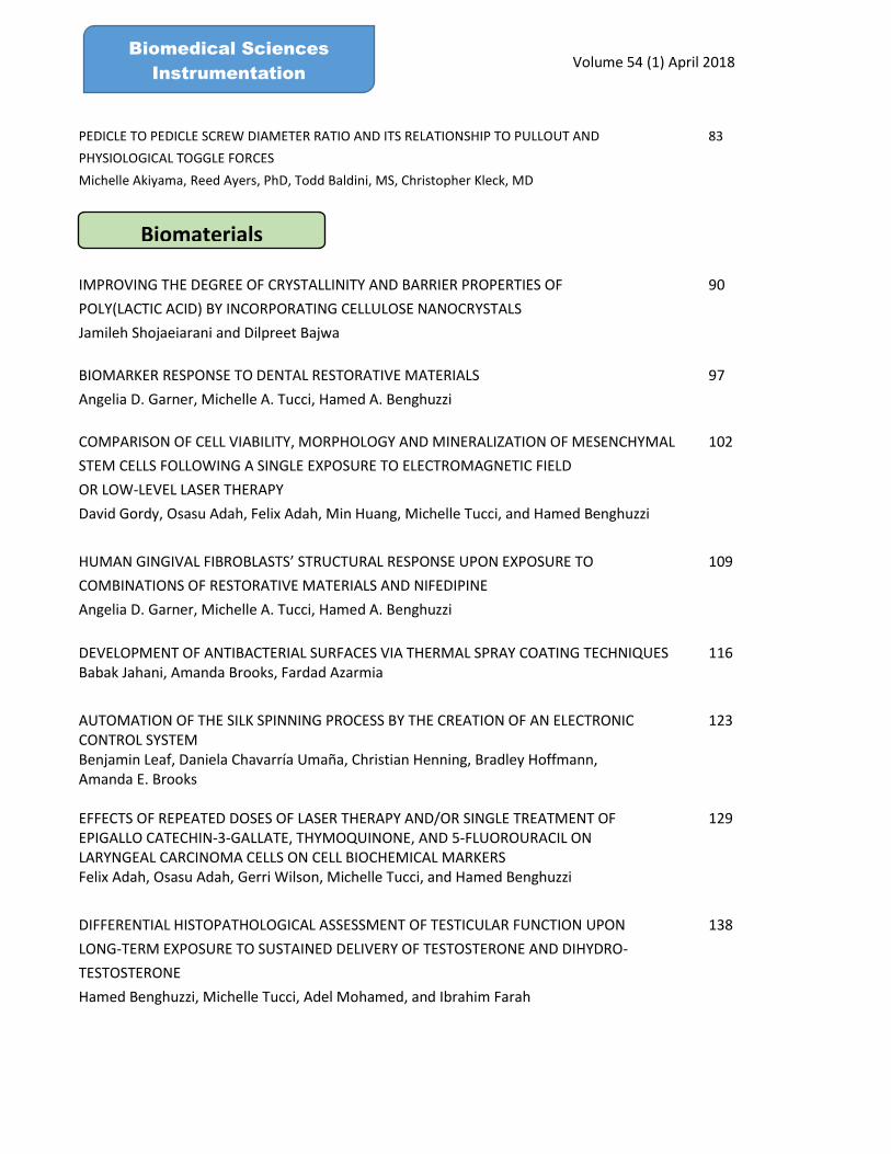
PEDICLE TO PEDICLE SCREW DIAMETER RATIO AND ITS RELATIONSHIP TO PULLOUT AND 83
PHYSIOLOGICAL TOGGLE FORCES
Michelle Akiyama, Reed Ayers, PhD, Todd Baldini, MS, Christopher Kleck, MD
90
97
102
109
116
123
129
138
IMPROVING THE DEGREE OF CRYSTALLINITY AND BARRIER PROPERTIES OF
POLY(LACTIC ACID) BY INCORPORATING CELLULOSE NANOCRYSTALS
Jamileh Shojaeiarani and Dilpreet Bajwa
BIOMARKER RESPONSE TO DENTAL RESTORATIVE MATERIALS
Angelia D. Garner, Michelle A. Tucci, Hamed A. Benghuzzi
COMPARISON OF CELL VIABILITY, MORPHOLOGY AND MINERALIZATION OF MESENCHYMAL
STEM CELLS FOLLOWING A SINGLE EXPOSURE TO ELECTROMAGNETIC FIELD
OR LOW-LEVEL LASER THERAPY
David Gordy, Osasu Adah, Felix Adah, Min Huang, Michelle Tucci, and Hamed Benghuzzi
HUMAN GINGIVAL FIBROBLASTS’ STRUCTURAL RESPONSE UPON EXPOSURE TO
COMBINATIONS OF RESTORATIVE MATERIALS AND NIFEDIPINE
Angelia D. Garner, Michelle A. Tucci, Hamed A. Benghuzzi
DEVELOPMENT OF ANTIBACTERIAL SURFACES VIA THERMAL SPRAY COATING TECHNIQUES Babak Jahani, Amanda Brooks, Fardad Azarmia
AUTOMATION OF THE SILK SPINNING PROCESS BY THE CREATION OF AN ELECTRONIC CONTROL SYSTEM Benjamin Leaf, Daniela Chavarría Umaña, Christian Henning, Bradley Hoffmann, Amanda E. Brooks
EFFECTS OF REPEATED DOSES OF LASER THERAPY AND/OR SINGLE TREATMENT OF EPIGALLO CATECHIN-3-GALLATE, THYMOQUINONE, AND 5-FLUOROURACIL ON LARYNGEAL CARCINOMA CELLS ON CELL BIOCHEMICAL MARKERSFelix Adah, Osasu Adah, Gerri Wilson, Michelle Tucci, and Hamed Benghuzzi
DIFFERENTIAL HISTOPATHOLOGICAL ASSESSMENT OF TESTICULAR FUNCTION UPON
LONG-TERM EXPOSURE TO SUSTAINED DELIVERY OF TESTOSTERONE AND DIHYDRO-
TESTOSTERONE
Hamed Benghuzzi, Michelle Tucci, Adel Mohamed, and Ibrahim Farah
Biomaterials

Volume 54 (1) April 2018 Biomedical Sciences
Instrumentation
DISCRETE WAVELET TRANSFORM BASED ERD/ERS PATTERNS FOR THE MOTOR IMAGERY 145
BRAIN COMPUTER INTERFACE
Niraj Bagh and M. Ramasubba Reddy
DISCRIMINATING SINGLE LEAD ECG’S WITH NORMAL SINUS RHYTHM AND SLEEP APNEA USING 153MULTISCALE FREQUENCY ANALYSIS
Suganti Shivaram, Zahara Z. Meghji, Susan Karki, Anjani Muthyala, Naveen Sundar Rameshkumar
and Shivaram P. Arunachalam
ANALYSIS ON THE STRUCTURAL DEFORMITY OF BRAINSTEM IN ALZHEIMER 159MR IMAGES USING p-LAPLACE BASED LEVEL SET AND ORTHOGONAL MOMENTS
M. Ramesh, K.R. Anandh, C.M. Sujatha
MULTIPLE MICROTUBULE TRACKING IN MICROSCOPY TIME-LAPSE IMAGES USING 167
PIECEWISE-STATIONARY MULTIPLE MOTION MODEL KALMAN SMOOTHER
S. Masoudi, C. H.G. Wright, N. Rahnavard, J. C. Gatlin and J. S. Oakey
EXPRESSION AND CELLULAR LOCALIZATION OF DOMAIN DELETION VARIANTS OF RAGE 176
Swetha Thiyagarajan, Estelle Leclerc and Stefan Vetter
DESIGN OF AN EXPERIMENTAL PLATFORM FOR FLOW VISUALIZATIONS 184
IN A MICROFLUIDIC CHIP
J. Singh, Y. Zhang, Y. Wang, B. Gerber, K. Stark, K. Yon, A. Brooks, B. Brooks
EFFECTS OF ANTIBIOTICS ON MURINE FECAL COLONIZATION 189
Jacob Shreffler, Jon Kauk, Zachariah Storey, Meredith Schroeder, Raquib Hasan,
Amanda Brooks Ph. D
ESTABLISHING ULTRASOUND AS AN EFFECTIVE METHOD FOR QUANTIFYING ADIPOSE GAIN 196
Codi Schaper, Jacob Shreffler, Hunter Schleske, Farnaz Fouladi, Amanda Brooks PhD
Biosignal Acquisition
Biotechnology

Volume 54 (1) April 2018 Biomedical Sciences
Instrumentation
INVESTIGATION ON VASCULATURE IN DIABETIC RETINOPATHY DIGITAL FUNDUS IMAGES IN 202
TERMS OF TEXTURE DESCRIPTORS
M Tamilnidhi and K Gunaseelan
COLOR CORRECTION AND ITS VALIDATION IN PRESSURE ULCER IMAGES FOR 209
CHRONIC WOUND ASSESSMENT
Kavitha I, Punitha N and Ramakrishnan Swaminathan.
SPECTRAL BOUNDARY ELEMENT ANALYSIS ON DROPLET BASED MICROFLUIDICS 217
USED IN CELL SEEDING
John-Luke Singh, Julie Melbye, Yechun Wang, Yan Zhang, Amanda E. Brooks,
and Benjamin D. Brooks
ANALYSIS OF SURFACE ELECTROMYOGRAPHY SIGNALS TO DISTINGUISH 222
NONFATIGUE AND FATIGUE CONDITIONS USING DEGREE CENTRALITY OF VISIBILITY GRAPHS
Navaneethakrishna Makaram and Ramakrishnan Swaminathan
DIFFERENTIATION OF TERM AND PRETERM CONDITIONS FROM UTERINE 228
SURFACE ELECTROMYOGRAPHY SIGNALS USING TIME-FREQUENCY IMAGES
N. Punitha and S. Ramakrishnan
ASSESSMENT OF ALZHEIMER DISEASE PROGRESSION USING TEXTURE ANALYSIS IN 236
MAGNETIC RESONANCE IMAGES
Rohini Palanisamy, Sundar S and Ramakrishnan Swaminathan
ANALYSIS OF FATIGUE IN BICEPS BRACHII MUSCLES USING SEMG SIGNALS AND LINEAR 244
CHIRPLET TRANSFORM
Diptasree Maitraghosh and Ramakrishnan Swaminathan.
A METHOD TO ANALYZE PLANTAR STIFFNESS USING MYOTONOMETRY AND ITS 252
COMPARISON WITH INFRARED IMAGING TECHNIQUE
Hariram S, S Lakshmi Lasya, Edward Jero and Ramakrishnan Swaminathan

Volume 54 (1) April 2018 Biomedical Sciences
Instrumentation
258
270
278
EFFECTIVENESS OF WIRELESS POWERED FILTERS IN THE THROMBOLYSIS OF BLOOD CLOTSNolan G. Schwartz, Ivan T. Lima, Jr.
INVESTIGATION OF SILK FIBROIN HEART VALVE CONSTRUCTS
Claire DeJong, Alec Halvorson, Mitchell Munar, Jonas Sahouani, Christopher Wreede,
Bradley Hoffmann, Yan Zhang, Long Jiang
TRACKING PIVOT POINT OF A NUMERICALLY SIMULATED MEANDERING ROTOR USING
KURTOSIS
Suganti Shivaram, Anjani Muthyala, Zahara Z. Meghji, Susan Karki, Naveen Sundar
Rameshkumar and Shivaram P. Arunachalam
AN EXPERIMENTAL STUDY OF PULSATILE FLOW PAST COMPLIANT AORTIC VALVE
USING PARTICLE IMAGE VELOCIMETRY
Ruihang Zhang and Yan Zhang
286
293
301
309
Cardiovascular Mechanics
Drug Delivery
INFUSING SYNTHETICALLY SPUN SPIDER SILK WITH RIFAMPICIN
Nasim Soufizadeh-Balaneji, Alec Staiger, Nathan Johnson, Adam Forness, Greg Fondong,
Oluwaseyi Ogundolani, Pranothi Mulinti, Bradley Hoffmann, and Amanda E. Brooks
HYPOXIA RESPONSIVE LIPID INCORPORATION INTO BOVINE MILK EXOSOMES
Jessica Pullan, Pooja Chemeiti, Matthew Confeld, Li Feng, James Froberg, Yongki Chio,
Sanku Mallik
Environmental and Clinical Engineering
ASSESSMENT OF ANIMAL MODELS AS SURROGATES FOR HUMAN TUMORS FROM THREE DIFFERENT ORGANS Ibrahim O. Farah, Zikri Arslan, Michelle Tucci, Hamed Benghuzzi, and Joseph A. Cameron
ACETIC ACID REMEDIATION OF ANTHROPOGENIC CONTAMINATION OF WATER
AT THE GBNERR IN MISSISSIPPI
Ibrahim O. Farah, Willis O. Lyons, Zikri Arslan, Michelle Tucci, and Paul B. Tchounwou
264

Volume 54 (1) April 2018 Biomedical Sciences
Instrumentation
TOMOGRAPHIC PIV OF LARGE INTRACRANIAL ANEURYSM MODELS 317
Timothy Rossman, M.S., Kendall Dennis, M.S., Adam Stolt, B.S., Yu-Ju Chen, B.S.,
David Kallmes, M.D., Jordi Estevadeordal, Ph.D., Dan Dragomir-Daescu, Ph.D.
EFFECT OF MR CONTRAST AGENTS ON MYOCARDIAL TISSUE ELASTICITY IMAGING: 325 A PILOT STUDY Suganti Shivaram, Susan Karki, Anjani Muthyala, Zahara Z. Meghji, Naveen Sundar Rameshkumar and Shivaram P. Arunachalam
THE EFFECT OF PLATELET-RICH PLASMA ON COMPLETE TEARS OF THE MEDIAL COLLATERAL 331
LIGAMENT: A CRITICALLY APPRAISED TOPIC
Bradley J. Conant and Shannon L. David PhD, ATC
ASSESSING THE ABILITY OF WOMEN’S LACROSSE HELMETS TO REDUCE RISK OF HEAD INJURY 340
Emily E. Kieffer, Megan L. Bland, Steven Rowson
EFFECT OF ANVIL ANGLE ON IMPACT KINEMATICS IN LABORATORY EVALUATION OF 347
BICYCLE HELMETS
Megan L. Bland, Craig McNally, Steven Rowson
EFFECT OF FACEMASK WEIGHT ON HELMET PERFORMANCE 355
Abigail M. Tyson, Emily E. Kieffer, Steven Rowson
ESTIMATING THE BRAIN STRAIN RATES DURING TRAUMATIC BRAIN INJURY 361
Mohammad Hosseini Farid, Mohammadreza Ramzanpour, Mariusz Ziejewski,
and Ghodrat Karami
COMPUTATIONAL SIMULATION OF BRAIN INJURY BY GOLF BALL IMPACTS IN 369
ADULT AND CHILDREN
Mohammad Hosseini Farid, Mohammadreza Ramzanpour, Ashkan Eslaminejad,
Mariusz Ziejewski, and Ghodrat Karami
Trauma and Impact
EFFECTS OF A SEASON OF YOUTH FOOTBALL ON
STATIC POSTURAL CONTROL
Eamon T. Campolettano and Steven Rowson
Biomedical Engineering and Mechanics
Virginia Tech, Blacksburg, VA 24061
ABSTRACT
Concussions occur in youth football with lower frequency than observed at higher levels of play, though the effect of repetitive
subconcussive head impacts resulting from participation in youth football is unknown at this point. One measure shown to be
affected by concussions is athlete postural control. The objective of this study was to compare performance on the Balance
Error Scoring System (BESS) and a force plate protocol at two time points within a cohort of healthy youth football players
and healthy non-contact youth track or baseball athletes. In absence of a clinically-diagnosed concussion, the hypothesis was
tested that a season of youth football would affect measures of static postural control and stability. Between time points, there
were no significant differences observed between either BESS scores or force plate metrics. Between athlete groups, there
were no significant differences observed for either the BESS or the force plate protocol. Particularly for pediatric males,
postural control is still developing and current assessments may not be sensitive enough to detect changes. Continued research
is necessary to determine what postural control testing may be most viable for use within an active, pediatric population.
Keywords: balance, pediatrics, concussion, subconcussive, BESS, force plate
INTRODUCTION
Athletes who have sustained a concussion have been shown to suffer from transient decreases in postural
control [1, 2]. Some research has shown that even non-concussed football players may experience balance
deficits [3, 4]. These studies have largely focused on collegiate athletes, despite the fact that youth players
comprise 70% of the football-playing population. Though concussions occur less frequently for these
youth athletes, potential adverse effects of subconcussive head impacts associated with playing football
remain unknown at present.
Two commonly implemented tools for assessing postural control changes in instances of athlete
concussion are the Balance Error Scoring System (BESS) and force plate testing. The BESS was
developed as a clinical, static balance assessment for sideline use and has been shown to measure
postconcussion balance changes in both youth and adult athletes [1, 5, 6]. Reliability of the BESS is
variable, and a known practice effect exists with repeated administration [7]. Instrumented force plates
have also been used to quantitatively assess postural control changes in athletes with and without
concussion [8-10]. Force plate testing typically involves tracking changes in the center of pressure (COP).
The BESS and force plate protocols have seen limited use with youth athletes, with most research
assessing postural control for healthy and concussed athletes [10-12]. The objective of this study was to
compare performance on the BESS and a force plate protocol at two time points within a cohort of healthy
youth football players and healthy non-contact youth baseball or track control athletes. The first time
point occurred before sports participation, while the second time point occurred after the conclusion of
the season, which meant completing testing after a season of head impact exposure for the football players.
Postural control is still developing for pediatric males, so current balance assessments may not be sensitive
enough to detect changes or suitable for use within this population [13, 14]. Further research investigating
Biomed Sci Instrum. Vol 54(1) April, 2018 ©2018 IAE All rights reserved
ISSN: 0067-8856 04 54 1 1 ISBN: 978-1-7752455-1-3
QUANTIFYING HEAD IMPACT DURATION: ANALYSIS OF
LABORATORY HELMET EVALUATION SYSTEMS
Bethany Rowson, Megan L. Bland, Steven Rowson, and Stefan M. Duma
Department of Biomedical Engineering and Mechanics
Virginia Tech, Blacksburg, VA 24061
ABSTRACT
Although head impact duration is thought to contribute to head injury severity, it is often not quantified. For laboratory
simulations of real-world impacts, it is important to ensure that the data are representative of what they are simulating in both
magnitude and duration. A number of different laboratory impact systems have been used to evaluate protective headgear
performance in sports. Some safety standards account for impact duration by evaluating the Severity Index (SI), while others
use only peak acceleration tolerances. The objective of this study was to determine if impact durations from commonly used
laboratory impact systems were different, and how they compare to previously reported durations in both sports and
automotive environments. Four different laboratory systems (a National Operating Committee on Standards for Athletic
Equipment drop tower, an International Organization for Standardization drop tower, a pneumatic ram, and a pendulum
impactor) were evaluated using 2 different helmet types (football and hockey) and 3 impact speeds. Differences in duration
were evaluated between helmet types and laboratory systems using 2-factor ANOVAs. Both helmet type and impact system
had a significant effect on impact duration (p < 0.0003). Although there were significant differences in duration, these
differences were small, and similar to previously reported values for helmeted head impacts in sports.
Keywords: biomechanics, concussion, acceleration, pendulum, pneumatic ram, drop tower
INTRODUCTION
Head impact duration has been shown to contribute to injury severity since the earliest experimental
work to determine human head injury tolerance. Cadaver drop tests and animal brain injury
studies demonstrated a decreasing tolerance to head acceleration or pressure with increasing impact
duration [1-3]. These studies along with human volunteer data were used to develop the Wayne State
Tolerance Curve (WSTC) [4]. This curve represented human tolerance for moderate to severe head
injury with acceleration magnitude as a function of time. The WSTC has been used as the basis for a
number of proposed head injury criteria [5-8]. The dependence of head impact tolerance on impact
duration has also been supported by experimental work with primates [9, 10].
Despite the theorized importance of impact duration on head injury tolerance, it is often not quantified.
Recent concerns regarding the long-term effects of repetitive head impacts and concussions have led to
an increase in helmet safety and efficacy research [11-19]. For laboratory simulations of real-
world impacts, it is important to ensure that the impacts are representative of what they are simulating in
both magnitude and duration. A number of different systems have been developed to evaluate
headgear performance for sports. Some safety standards account for impact duration by evaluating
the Severity Index (SI), while others use only peak acceleration tolerances [5]. Regardless of the
criterion used to evaluate headgear, the impact durations for these systems have not been quantified
and compared to impacts that occur on the field.
Four laboratory systems developed for helmet standards or evaluation methods were used in this study: a
National Operating Committee on Standards for Athletic Equipment (NOCSAE) drop tower, an
International Organization for Standardization (ISO) drop tower, a pneumatic linear impactor, and a
pendulum impactor. The NOCSAE drop tower is used to certify football helmets in accordance with the
Biomed Sci Instrum. Vol 54(1) April, 2018 ©2018 IAE All rights reserved
ISSN: 0067-8856 04 54 1 9 ISBN: 978-1-7752455-1-3
DEVELOPMENT OF A TIME-WEIGHTED HEAD
IMPACT EXPOSURE METRIC
Brian Tomblin, Joel D. Stitzel, Jillian E. Urban
Virginia Tech – Wake Forest University School of Biomedical Engineering and Sciences,
Wake Forest University Center for Injury Biomechanics
Winston Salem, NC 27101
ABSTRACT Head impact exposure (HIE) is often quantified from percentiles computed from the distribution of acceleration or
summation of the magnitude of hits. With increasing evidence demonstrating a potential link between brain changes
and HIE over time, there is a critical need to improve upon existing HIE metrics, accounting for temporal variations
in the accumulation of exposure. In this methodological study, the weighting of time on the effects of consecutive
HIE is addressed. In this approach, each impact prior to a given impact within a time window is weighted based
on a function describing a linear or exponential relationship comparing exposure weight and time between impacts.
This is iteratively computed for each head impact over any time. For this paper, a single example impact exposure
and two youth football players’ recorded head impact risk exposures from a full season were used with a time-decay
window of 36 hours to demonstrate the influence of time-weighting on cumulative exposure using this novel
approach. The proposed cumulative time-weighted exposure metric results in a single value representing the
time-weighted cumulative exposure of each impact. Due to the novelty of this metric, no large-scale data comparison
tests have been completed between this metric and previously accepted metrics. However, this approach qualitatively
improves upon prior HIE metrics, accounting for temporal variations that occur with HIE, and will lead to a better
understanding of the relationship between brain changes and HIE in sports.
Keywords: concussion, HIE, impact, metric, risk, time, weight
INTRODUCTION
Sports-related concussions occur between 1.6 million to 3.8 million times each year in numerous
sports and age groups [1, 2]. Repetitive head impacts that do not result in the signs and symptoms
of concussion, often termed subconcussive head impacts, are a rising concern as increasing
evidence has demonstrated a potential link between brain changes and repetitive head impacts over
time [3, 4]. While there is rising concern for subconcussive head impact exposure (HIE), the
development of comprehensive cumulative HIE metrics is lacking.
HIE is often quantified from percentiles computed from the distribution of acceleration or
summation of the severity of hits [5, 6]. Rowson et al. reported that solely reviewing the linear and
rotation acceleration associated with a concussive head impact did not result in a definitive
concussion threshold as tolerance may be specific to each individual [7]. A possible improvement
of existing HIE metrics is to account for temporal variations in the accumulation of exposure.
Concussions are shown to have lingering effects on the brain ranging from 1-10 days past the
impact, supporting the hypothesis that the brain is in a vulnerable state of repair for some time
following a concussive head impact; however, it is unknown how long the brain may need to
recover from a subconcussive head impact [8]. In a 2017 study, Broglio et al. reported that time
between impacts influenced the risk of concussion due to the brain’s temporary physiological
vulnerability after HIE [9]. Temporal accumulation of risk was described by Murray et al. in the
context of disease progression, such as heart and lung disease from smoking, explaining that
exposures to a risk factor and the subsequent health outcomes include a time dimension [10].
Murray et al. introduces several models describing the temporal dimensions of the risk factor-
Biomed Sci Instrum. Vol 54(1) April, 2018 ©2018 IAE All rights reserved
ISSN: 0067-8856 04 54 1 16 ISBN: 978-1-7752455-1-3
ASSESSING STATIC AND DYNAMIC POSTURAL CONTROL IN A HEALTHY POPULATION
Eamon T. Campolettano, Ryan A. Gellner, and Steven Rowson Department of Biomedical Engineering and Mechanics
Virginia Tech, Blacksburg, VA 24061
ABSTRACT
Static postural control testing is often conducted by clinicians and athletic trainers for use with athletes who have sustained a concussion. Dynamic postural control involves the body’s response to perturbation of the center of mass and may offer additional insight that static testing cannot capture. The objective of this study was to assess the reliability and feasibility of a balance protocol consisting of both static and dynamic postural control assessments with a healthy, adult population. Subjects stood in both unipedal and bipedal stances on a force plate to capture quantitative data regarding the center of pressure over time. Further, subjects completed the Balance Error Scoring System (BESS), a static measure, and a modified version of the Star Excursion Balance Test (SEBT), a dynamic measure. Reliability with the BESS was limited, while moderate to strong reliability was obtained for the modified SEBT. Unipedal stances were associated with a greater variance than bipedal stances for both the BESS and force plate protocol. These assessments will be applied within a pediatric populations to determine the validity of their use. Further postural control research is necessary to determine the most viable assessments for use within an active, pediatric population.
Keywords: balance, force plate, BESS, SEBT, concussion
INTRODUCTION After mild traumatic brain injury (mTBI), it is common for postural control deficits to be observed [1-3]. Many post-concussion assessments now include postural control tests as an evaluative tool to determine patient health [4-7]. Postural control represents the ability of a person to maintain balance naturally and when exposed to perturbation [8]. Postural control can be defined by assessing static and dynamic balance. Static balance involves an individual establishing a stable base and attempting to minimize movement while holding the particular posture. Dynamic balance, on the other hand, refers to the introduction of perturbations to this stable base of support. It can be assessed by having subjects establish a base of support and then requiring some level of movement away from that equilibrium. Static balance has been most commonly assessed in post-concussion situations, though dynamic balance assessments are gaining favor as they may involve movements similar to those experienced while playing sports [9-11].
Static balance is most commonly assessed using the Balance Error Scoring System (BESS) or force plates. The BESS is an easily administered, static balance assessment for sideline use in instances of suspected concussion that asks individuals to hold different static postures while an evaluator assesses deviations from this desired posture [12, 13]. Instrumented force plates are used to quantitatively track the center of pressure (COP) over time during a static stance.
Dynamic balance assessments are necessarily more involved than are static assessments, and have seen less use [14]. One of the most commonly employed assessments is the Star Excursion Balance Test (SEBT), which tasks individuals with maintaining balance with one foot while reaching out in prescribed directions with the other foot [9]. By more closely aligning concussion testing assessments with physical activity, it is hypothesized that the tools will be more relevant. The SEBT is traditionally used to assess
Biomed Sci Instrum. Vol 54(1) April, 2018 ©2018 IAE All rights reserved
ISSN: 0067-8856 04 54 1 24 ISBN: 978-1-7752455-1-3
METHOD FOR DETERMINING THE STRUCTURAL RESPONSE OF HELMET SHELLS DURING DYNAMIC
LOADING
Ryan A. Gellner, Tyler P. Morris, and Steven RowsonBiomedical Engineering and Mechanics Virginia Tech, Blacksburg, VA 24061
ABSTRACT Football helmet design and development involves changing a range of parameters including padding material and thickness, shell material and thickness, and padding location, all of which alter a helmet’s dynamic response to impact. All of these parameters can affect performance of the helmets in conventional standards and supplemental testing (ref NOCSAE and FB STAR paper). These parameters can be costly and time-consuming to change quickly during prototype development, and computational modeling of helmets helps to reduce both cost and time required. As one method of enabling helmet modeling for reduced prototyping time, full helmet models will need to be developed and validated with appropriate material characteristics. Most current material testing methods do not characterize response during real world loading conditions. We present a novel method for measuring the force-deflection characteristics of a football helmet shell using a pneumatic ram. This method involves a rigidly mounted helmet which is allowed to move along a single axis. Two accelerometers enabled the measurement of force and relative displacement, and tests were conducted in the range of 3 – 6 m/s input velocities for impacts to the front and side of the helmet. Data demonstrate repeatability at each impact configuration.
Keywords: force-deflection, football, helmet, method, dynamic loading
INTRODUCTION Tensile or compressive testing machines are often used to determine mechanical properties of materials. These machines typically load the specimens at rates in the quasi-static range below 1 cm/s [1]. Small loading rates such as these can be orders of magnitude lower than loading rates seen in everyday use of these products. Ideally, the loading rates used in tests would coincide with the rates at which these products are used, as some rate dependency may exist. In addition, appropriate modeling of these material properties can enable more accurate finite element modeling by validating model predictions against experimental results [2].
Finite element modeling (FEM) of products enables designers to prototype and iterate efficiently. Recently, the National Football League’s Engineering Roadmap spoke of FEM in football helmet development as one of the top priorities for driving new innovation in the field [3]. Force-deflection curves are commonly used in finite element model validation [4]. Previous studies have used FEM, validated by physical force-deflection tests, to characterize motorcycle helmet foam characteristics under both quasi-static and dynamic loading [2]. Each season, football helmets are subject to hundreds of impacts that occur to a number of different locations on the helmet and at a variety of severities [5]. Because football helmets are subject to dynamic loading events during their normal use, this study sought to present a novel experimental method for quantifying helmet shell force-deflection characteristics undergoing dynamic loading in two different orientations: front and side. It is expected these methods could be expanded to other loading orientations and severities across a range of helmet models.
Biomed Sci Instrum. Vol 54(1) April, 2018 ©2018 IAE All rights reserved
ISSN: 0067-8856 04 54 1 32 ISBN: 978-1-7752455-1-3
ASSOCIATION BETWEEN TACKLING TECHNIQUE AND HEAD ACCELERATION MAGNITUDE IN YOUTH
FOOTBALL PLAYERS
Ryan A. Gellner, Eamon T. Campolettano, and Steven Rowson
Biomedical Engineering and MechanicsVirginia Tech, Blacksburg, VA 24061
ABSTRACT In order to address concerns about head injury in youth sports, a number of youth football organizations have developed rules and recommendations surrounding the tackling form which should be used in order to reduce unnecessary head impact exposure. Reduction in injury has been suggested with these programs, but association between tackling form and head acceleration magnitude has not been studied previously. To address this knowledge gap, grading criteria were developed from multiple youth organizations’ recommendations for a collision. A total of 142 tackles from a youth football team were graded. Head acceleration data were collected from helmet-mounted accelerometer arrays. An association was found between poor form and resultant head acceleration being greater than 40 g for both the tackler and the ball carrier. This study demonstrates the potential usefulness of tackling technique coaching programs in youth football.
Keywords: concussion, grading, tackling form, high magnitude, impact exposure
INTRODUCTION Concussions continue to be a major health concern in American football. With a large majority of players of this contact sport at the youth level, the accumulation of head impact exposure over a lifetime has begun to be extensively studied as a potential risk factor for impairment later in life [1-5]. Specifically, Alosco et al. [4] found that exposure to football before age 12 resulted in a twofold increase in odds of having clinically impaired scores on self-reported measures of executive function and behavioral regulation, depression, and apathy in former amateur and professional football players. Montenigro et al. [5] suggested that this increase in odds may be more strongly related to repetitive head impact exposure than other metrics, including concussion history. Associations such as these have led a number of organizations to seek methods of reducing head impact exposure in athletes, rather than only addressing injuries. The three best strategies today are thought to be development of better equipment, rule changes prohibiting head contact, and teaching better technique when contact occurs [6, 7].
Recently, multiple organizations have created or prioritized rules which prohibit certain tackling techniques, and some have even started programs which teach what the organization considers to be proper tackling technique [8-11]. Previous studies have shown these types of programs have resulted in less injury overall. Kerr et al. [12] found that injury rates for all types of injuries in games were lower among teams implementing USA Football’s Heads Up Football program. Concussions were only found to be reduced in practice if the Heads Up Football program was implemented and Pop Warner’s practice rules were also followed, which limited time allowed for contact in practices and eliminated high-speed, head-on tackling drills. These findings scaled with age, with stronger effects from these tackling recommendations and rule changes seen in players aged 11-15 rather than those 5-10 years old. There has been disagreement as to the degree of effectiveness these programs truly have, as reported concussion reduction may have been skewed when initially reported [12, 13]. To date, tackling technique programs have only been studied in terms of concussion incidence numbers, but none have attempted to determine if individual impacts with proper technique actually result in lower head accelerations for the athletes involved.
Biomed Sci Instrum. Vol 54(1) April, 2018 ©2018 IAE All rights reserved
ISSN: 0067-8856 04 54 1 39 ISBN: 978-1-7752455-1-3

HEAD INJURY RISK ASSOCIATED WITH BASEBALL STIFFNESS AS A FUNCTION OF PLAYER AGE
Tyler P. Morris, Ryan A. Gellner, Steven RowsonBiomedical Engineering and Mechanics, Virginia Tech, Blacksburg, VA 24061
ABSTRACT
The majority of head injuries in baseball are due to ball impact. To reduce injury risk, standard baseball stiffness varies between age groups. The objective of this study was to compare head injury risk across a range of baseball stiffnesses (RIF1, RIF5, RIF10, Youth, HS/College and Pro) designed for different age groups. To simulate baseball impacts, a customized pitching machine was used to propel baseballs from 15 m/s to 30 m/s in 5 m/s increments. The balls impacted the center of the forehead of a 50th percentile Hybrid III headform. The headform was connected to a Hybrid III neck, mounted on a 16 kg sliding table, positioned vertically and instrumented with a nine accelerometer array in a 3-2-2-2 configuration. To account for head size differences between ages, acceleration data collected from the Hybrid III were transformed using geometric scaling laws. Skull fracture risk and concussion risk were compared between ball types at each impact velocity. Analysis of these data show that the youth ball, age 13-14, produced the highest skull fracture and concussion risk across the velocity range. However at age matched velocity, the professional level (Pro ball) yielded the greatest skull fracture and concussion risk and the safety balls used for 5-8 year olds (RIF 1) yielded the lowest skull fracture and concussion risk. This study provides framework for determining optimal age-specific ball stiffness.
Keywords: head impacts, baseball, head injury risk, linear, rotational, acceleration, biomechanics
INTRODUCTION It is estimated that in the United States there are more than 19 million children that participate in youth baseball annually [1]. Baseball players between the ages of 5-14 sustain the highest fatality rate of all sports, with approximately one in four annual deaths resulting from an impact from the ball to the head [1, 2]. Ball impact has been identified as the leading cause of injury in baseball, with the most common injury being from the ball to the head [3]. A pitcher throwing a ball toward the head of a batter and striking the head is one specific scenario that can result in head injury, and is the interest of this study. These impacts can lead to concussion, skull fracture, and in some instances death. Development of reduced injury factor (RIF) balls have provoked rule changes to specify certain ball stiffness to different age groups as a way to mitigate injury [4]. RIF baseballs range from levels 1-10, with 1 being the most compliant and 10 being the stiffest. Previous studies on the effect baseball stiffness has on injury risk have shown that a softer ball reduces the potential for head injury [3, 5, 6]. RIF 1, RIF 5, and RIF 10 balls have been specified for age groups 5-8, 7-10, and 9-12 respectively [4]. In addition, there is a youth style ball for ages 13-14, a high school and college style ball, and a professional ball all for ages 14 and higher.
A baseball must be certified by the National Operating Committee on Standards for Athletic Equipment (NOCSAE) prior to its use in the field of play. The requirements are as follows: weigh between 5.0 and 5.25 ounces, have a circumference within 9 to 9.25 inches, and a coefficient of restitution (COR) value between 0.45 and 0.55. Depending on the ball compression type (low, medium, and high) the compression deflection value at 0.25 inch displacement must not exceed 45 lbs., be within 75-150 lbs., or be within 200-350 lbs. respectively [7].
Few studies have investigated age specific head injury risk as a function of baseball stiffness. The objective of this study was to compare head injury risk across a range of baseball stiffnesses designed for
Biomed Sci Instrum. Vol 54(1) April, 2018 ©2018 IAE All rights reserved
ISSN: 0067-8856 04 54 1 46 ISBN: 978-1-7752455-1-3
CHARACTERIZING HEAD IMPACT EXPOSURE BY
PLAYER POSITION IN HIGH SCHOOL FOOTBALL
Liam P. McNamara, Jillian E. Urban, Mireille E. Kelley, Logan E. Miller, Joel D.
Stitzel
Wake Forest University Graduate School of Biomedical Engineering and
Sciences Winston-Salem, NC 27157, USA
ABSTRACT
Football has the highest participation rate of all high school sports in the United States and is
among the sports with the highest incidence of concussion. Therefore, it is imperative to
understand the characteristics that influence athletes’ exposure to head impacts at this level of
play. This study quantifies head impact exposure (HIE) by position type in a high school
football population. HIE was measured by equipping helmets of 114 players over 4 seasons
with an accelerometer array that records peak linear acceleration, estimated peak rotational
acceleration, and impact location. Players were grouped into four position types: linemen
(n=50), backers (tight ends, running backs and linebackers, n=27), secondary (receivers and
defensive backs, n=32), and quarterbacks (n=5). A total of 48,977 impacts were recorded.
Linemen received the highest average number of impacts per player-season (n=1080), while
backers sustained the largest average 95th percentile impacts (60.6 g). Quarterbacks received
both the lowest average number of impacts per player-season (n=173) and 95th percentile
impacts (53.5 g). The proportion of impacts for each general impact location also varied by
position type. These data will be useful in determining the role of position type in a players'
HIE measured over a season of high school football.
INTRODUCTION
Football leads all high school sports in both participation rate and incidence of concussion, with
over 1.08 million participating in the 2015-2016 season and a sports-related concussion rate of
9.21/10000 athlete-exposures [1], [2]. While concussion rates are higher at the collegiate level [3],
the high-school level of play represents a significantly larger population of athletes, often with
reduced access to certified medical care; with just over a third of high schools nationwide report
having a full-time athletic trainer staffed to support their athletic programs [4].
A growing concern for collision sports, such as football, is the possible effect of repetitive, sub-
concussive impacts—impacts to the head that do not result in any acute signs or symptoms of
concussion. These impacts have been suggested as a potential cause of chronic brain injury [5], as
well as adversely affecting cerebral function [5], [6], and may even cause changes in the brain over
the course of a single season [7], [8]; however, the relationship between participation in youth and
high school football and long term neurodegenerative diseases is not well understood [9], [10].
Characterizing head impact exposure (HIE) in the sport of football at the high school level is a
relatively new area of study, as most of the current body of research has focused on play at the
Biomed Sci Instrum. Vol 54(1) April, 2018 ©2018 IAE All rights reserved
ISSN: 0067-8856 04 54 1 54 ISBN: 978-1-7752455-1-3
EVALUATION OF HEAD IMPACT EXPOSURE IN
YOUTH FOOTBALL GAMES
William C. Flood, Mireille E. Kelley, Barret Zimmerman, Joel D. Stitzel, Jillian E. Urban
Virginia Tech - Wake Forest University, Center for Injury Biomechanics
Winston-Salem, NC 27101
ABSTRACT
Growing concern has led to some youth football organizations to implement rule changes for safety purposes;
however, there is a lack of biomechanical evidence in youth football to inform such changes. Therefore, the
objective of this study was to evaluate differences in HIE during youth football games. In this study, youth
football players, ages 9-13, were equipped with a helmet-mounted sensor. All games were filmed to verify each
head impact and assign each impact to a specific play type. A total of 3,003 impacts were evaluated from 29
athletes during 28 games. The median [95th percentile] linear acceleration measured on passing downs was
20.5g [50.7g] and 20.3g [52.6g] on running downs. Special Teams plays accounted for 10% of all plays, with
only 43% of the kicks being returned by the receiving team. In Special Teams scenarios, the median [95th]
linear acceleration measured was 22.5g [62.6g] when kicks were returned by the receiving team and 18.9g
[73.1g] were not attempted to be returned. The results of this study demonstrate that Special Teams scenarios
yielded slightly higher head impact exposure than running and passing downs; however, further research is
needed to investigate biomechanical exposure measured during game impact scenarios in youth football.
Keywords: head impact exposure, youth football, special teams, game impact exposure
INTRODUCTION
Football is among the sports with the highest incidence of sports-related concussions in male
youth, high school, collegiate, and professional sports [1], [2]. According to the Centers for
Disease Control (CDC), the number of annual emergency department visits due to concussions in
youths ages 11-13 has nearly doubled since 2001 [3]. There is increasing public awareness of
and concern for the potential effects of sub-concussive head impacts, those that do not result in
the signs and symptoms of concussion [4]. There is increasing evidence demonstrating that sub-
concussive head impact exposure (HIE) may affect player’s brains over their lifetime and may
even cause changes in the brain over course of a single season [4][5][6][7].
While many studies have examined HIE in high school, collegiate, and professional players,
there is still a lack of biomechanical evidence in youth football to help guide potential policies
related sport safety at this level of play. With an estimated 5 million participants in both youth
and high school football in the United States, effective regulation that helps protect athletes
while preserving the game’s integrity is paramount. From the NFL and NCAA’s alteration of the
initial kickoff point to the implementation and evaluation of the head targeting rule, it is evident
that more attention is being placed upon player head safety. While all three phases of the game
(Offense, Defense, and Special Teams) have been altered to improve player safety, Special
Teams scenarios have been highlighted as the most critical aspect of football that needs to be
addressed [8] .
Biomed Sci Instrum. Vol 54(1) April, 2018 ©2018 IAE All rights reserved
ISSN: 0067-8856 04 54 1 61 ISBN: 978-1-7752455-1-3
CEREBROSPINAL FLUID-SKULL INTERACTION
ANALYSIS FOR A NON-INVASIVE INTRACRANIAL
MONITORING TECHNIQUE
Ashkan Eslaminejad, Mohammadreza Ramzanpour, Mohammad Hosseini-Farid, Mariusz
Ziejewski, and Ghodrat Karami1
Department of Mechanical Engineering,
North Dakota State University Fargo, ND 58108-6050
ABSTRACT
Intracranial pressure (ICP) monitoring methods can be categorized into invasive and non-invasive. Invasive methods increase
the risks of bleeding and infection and need professional personnel; therefore, non-invasive methods are investigated more
often. One non-invasive method is based on monitoring transcranial signals, which can be captured and processed from the
skull. For this reason, the effects of cerebrospinal fluid (CSF) pressure increment on the natural frequencies of the skull have
been investigated. In this paper, we model the human skull as a hemispherical shell employing skull bone mechanical
characteristics. CSF will be considered as an incompressible and inviscid fluid with a pressure increase less than 2 kPa.
Employing Finite Element (FE) numerical techniques, the fluid-solid interaction (FSI) of CSF-skull is discretized, and the
eigenvalue problem is solved to obtain the first 50 natural frequencies and the associated skull vibrational mode-shapes. The
results illustrate that rising in CSF pressure causes slightly decrement in the unsymmetrical and symmetrical vibration
frequency modes. Moreover, the modes of skull vibration sensitivity with respect to CSF pressure variation are calculated.
The sensitivity graph demonstrates that the skull vibration in higher frequencies modes is sensitive to ICP variation in
comparison with the lower vibration modes.
Keywords: Non-invasive Intracranial Pressure Monitoring, Finite Element, Acoustics Modal Analysis, Fluid-Structure
Interaction.
INTRODUCTION
The pressure inside the cranium space is called intracranial pressure (ICP) and applied on the
intracranial organs such as the brain. Cerebrospinal fluid (CSF) is the fluid which fills up the inside of
the skull and spinal cord ventricles that protects the brain tissue from impact shocks. In addition, CSF
indicates the brain health statues form intracranial diseases such as brain tissue infection, swelling,
and intracranial tumors. CSF volume is about 130-150 milliliter and the normal value of ICP varies from
600 Pa to 2000 Pa for adults which is considered low (Figure 1) [1]. Since the skull is a rigid body that
contain the brain tissue and CSF, the intracranial tissues can be compressed by the small amount of
pressure. The ICP increment causes stopping the oxygen supply and hypoxia then brain death after few
minutes. Monitoring the ICP variation is an important task before and after neurosurgeries. However,
the common accurate ICP monitoring methods are invasive that can increase the risks of infection and
bleeding. Still, there is no clinical noninvasive technique.
There are several attempts to monitor the ICP noninvasively by using the novel method such as brain
imaging, optic nerve sheath size, indirect pressure transmission, Tympanic Membrane deformation.
However, none of the investigated techniques appear individually accurate enough to assess ICP
variation [2]. In all these noninvasive studies, the main purpose is to obtain approximately the ICP from
the data which can be measured from the extracranial region such as transcranial acoustic signals. For
Biomed Sci Instrum. Vol 54(1) April, 2018 ©2018 IAE All rights reserved
ISSN: 0067-8856 04 54 1 69 ISBN: 978-1-7752455-1-3

COMPARATIVE STUDY OF COUP AND CONTRECOUP
BRAIN INJURY IN IMPACT INDUCED TBI
Mohammadreza Ramzanpour, Ashkan Eslaminejad, Mohammad Hosseini Farid, Mariusz
Ziejewski, Ghodrat Karami
Department of Mechanical Engineering
North Dakota State University, Fargo, ND, 58108-6050
ABSTRACT
Traumatic brain injury (TBI) may happen due to impact, blast or rapid movement of the head. Among the many
categorizations, brain injuries can be divided into coup and contrecoup injuries. When impact happens to head, cerebrospinal
fluid (CSF) flows to the site of impact and therefore, cushions the brain. This causes positive pressure at coup site and negative
pressure at the contrecoup site. This study has examined a human head model under frontal impact with a rigid body. The finite
element analysis was conducted under different speeds of the impactor to obtain coup and contrecoup pressure for the brain.
As the speed of the impact increases, the ratio of the negative contrecoup pressure to the positive coup pressure increases. It
can be concluded that the increase in cushioning effect of CSF on the coup site of the impact comes with increasing negative
contrecoup pressure which will add to the severity of contrecoup injury as the intensity of the impact escalates.
Keywords: Traumatic Brain Injury, Head Impact, Coup and Contrecoup Pressure, CSF Cushioning Effect
INTRODUCTION
Traumatic brain injury (TBI) may happen due to impact, pressure waves (shock waves) and rapid head
movement. Numerous studies have applied finite element method for simulating head response for the
case of impact and blast [1-3]. TBI can be divided into coup and contrecoup brain injury. Contrecoup
brain injury is an injury caused at the areas far from the impact side due to the travelling shock waves
which cause stress or cavitation effect [4]. Dawson et al. insert that contrecoup brain injury can be more
significant compared to the coup injury [5]. Drew et al. stated that brain initial movement in the skull after
impact is toward the contrecoup side which will result in more severe contusions compared to the coup
injury [6]. There are several theories proposed for better understanding of contrecoup brain injury such
as positive pressure theory or CSF displacement [5], negative pressure theory also known as cavitation
theory [5] and rotational shear stress theory [7].
This study focuses on CSF displacement theory by characterizing the CSF and brain coup and contrecoup
pressure. When impact happens, CSF flows to the site of the impact (coup) and therefore, cushions the
brain when it hits to the skull. As a result, cushioning effect of CSF at the contrecoup site of the impact
decreases. Brain moves in the skull while the CSF is concentrated at the coup side. This may result in
more severe injury at the opposite side of the impact known as contrecoup brain injury.
Biomed Sci Instrum. Vol 54(1) April, 2018 ©2018 IAE All rights reserved
ISSN: 0067-8856 04 54 1 76 ISBN: 978-1-7752455-1-3
PEDICLE TO PEDICLE SCREW DIAMETER RATIO AND ITS RELATIONSHIP TO PULLOUT AND PHYSIOLOGICAL
TOGGLE FORCES Michelle Akiyama1, Reed Ayers, PhD2, Todd Baldini, MS2, Christopher Kleck, MD2
1College of Engineering and Applied Science, Department of Bioengineering, University of Colorado Denver Anschutz Medical Campus, Denver, CO
2School of Medicine, Department of Orthopedics, University of Colorado Denver | Anschutz Medical Campus, Denver, CO
ABSTRACT Spinal hardware improves fusion rates, yet failure still occurs. This may necessitate revision surgery adding cost and morbidity. A common failure is pedicle screw loosening, where the screw moves about a fulcrum point in the bone (toggle). The current standard for testing pedicle screws is pullout of bone. While this speaks to the screw/bone interface, it does not describe the screws effect on the bone. Current literature suggests the best way to increase pullout strength is to increase the diameter of the screw. However, this may lead to a breach in the pedicle, which may result in neurologic injury. Through biomechanics, our program evaluated screw loosening in a cadaver model by applying physiologic toggling forces and varying screw diameter. Also, we examined strain exerted on the bone from the screw. Screw sizes of 5.5 and 6.5 were placed in L2-L3.
Keywords: Pedicle screw, pedicle screw loosening, toggle, biomechanics
INTRODUCTION Pedicle screws are bone screws used in spine surgery to connect two or more vertebral bodies providing structural stability. This will enable fusion of the vertebrae to treat patients with back and/or nerve pain resulting from degenerative disc disease, scoliosis, spondyloses and other spinal conditions. The spinal construct mainly consists of pedicle screws, interbody cages, and rods that span. Unfortunately, there are some common risks associated with fusion surgery. These include hardware failure (pedicle screw loosening, rod breakage), pseudoarthosis, proximal junctional kyphosis, pain, and lack of fusion. All of these could lead to additional surgery adding cost and morbidity to the patient.
Pullout strength (POS) has been established by industry standards ASTM F2884-12 and F1717-15 for testing pedicle screws [1], [2]. Pullout is the tensile force required to pull the screw from the vertebrae. A greater POS can result in lower failure rates. It has been found that the best way to increase the POS is to increase the diameter of the screw to catch the cortical bone [3]. Unfortunately, from a clinical standpoint, increasing the diameter of the pedicle screw can cause bone breaches during surgery which can be detrimental to the patient [4], [5]. As screws do not fail in pure pullout in the physiologic environment, the value of increasing screw diameter may not provide a value comparable to the possible risks [6].
In a clinical setting, screw loosening is a common failure mechanism of the hardware. It can be referred to as screw toggling. In basic beam mechanics, screw toggling is a moment added to one end of a fixed beam. Figure 1 illustrates the difference between pullout and toggle in terms of simple free body diagrams. The physiological motion of the spine causes a “windshield wiper” effect within the vertebral body. Screw loosening can cause increased pain, lack of fusion, or even bone breach. However, screw
Biomed Sci Instrum. Vol 54(1) April, 2018 ©2018 IAE All rights reserved
ISSN: 0067-8856 04 54 1 83 ISBN: 978-1-7752455-1-3
IMPROVING THE DEGREE OF CRYSTALLINITY AND
BARRIER PROPERTIES OF POLY(LACTIC ACID) BY
INCORPORATING CELLULOSE NANOCRYSTALS
Jamileh Shojaeiarani* and Dilpreet Bajwa
Department of Mechanical Engineering
North Dakota State University, Fargo, ND 58102* Corresponding Author, Email address: [email protected]
ABSTRACT
Increasing concerns over the human health have motivated the scientific community to improve the potential application of
bio-based materials in different fields. Food packaging industry represents an important consumption of petroleum-based
materials with short-term applications. Bio-based polymers have been explored during recent decades as substitutes to non-
degradable polymers. Poly (lactic acid), (PLA) is one of the most promising bio-polymer in the food industry, owing to its
inherent biocompatibility and biodegradability. However, high gas permeability in PLA limits its application. The
incorporation of bio-based nanofillers into PLA to improve the barrier properties of nanocomposite films is of interest.
Cellulose nanocrystals (CNCs) with crystalline structure have the potential of improving the barrier properties of
nanocomposite films. Uniform dispersion of CNCs in PLA is essential in preparing nanocomposite films. In the current
study, a new method was introduced to enhance the dispersion of CNCs in PLA and to improve the barrier properties of PLA
thin films. The spin-coating technique was employed in this study to improve the barrier properties of PLA-based
nanocomposite films. In spin-coating technique, PLA-CNCs nanocomposite films get dried through a dynamic technique to
increase the solvent evaporation rate and decrease the possibility for CNCs to self-assemble to micro-sized aggregates. By
introducing spin-coating technique, a maximum improvement of 176.38% was observed in in the crystallinity of thin film as
compared to solvent cast films.
Keywords: Barrier properties; Poly (lactic acid); Cellulose nanocrystals; Spin-coating.
INTRODUCTION
Current development in the food packaging industry has focused on limiting the applications of petroleum-
based polymers owing to increasing the awareness over the human and environmental health.
Polypropylene (PP), polyvinylchloride (PVC), and polyethylene terephthalate (PET) are the most
common petroleum-based plastics in the food packaging industry [1]. However, the non-
biodegradable characteristics of conventional polymers and the corresponding environmental pollution
are considered as a challenge which needs to be addressed. The application of biopolymers as
alternative materials to petroleum-based materials is becoming increasingly important. Biopolymers are
defined as polymers with the renewable resources which can be biodegradable or compostable [2].
Poly(lactic acid) (PLA) with renewable and biodegradable characteristics has received a huge interest in
different areas of food packaging industry [3]. PLA with high production rate is economically
competitive and meets the needs in food packaging industry such as high transparency and decent
mechanical characteristics. However, PLA application suffers from low barrier properties against
small molecules such as water vapor and oxygen [4]. To improve the potential application of PLA in
food packaging industry, the introduction of nanofillers with higher mechanical and barrier
characteristics have been extensively explored. The application of nanocomposite materials in food
packaging industry is more favorable through using bio-based materials for both polymer matrix and
nanofillers [5].
Biomed Sci Instrum. Vol 54(1) April, 2018 ©2018 IAE All rights reserved
ISSN: 0067-8856 04 54 1 90 ISBN: 978-1-7752455-1-3
BIOMARKER RESPONSE TO DENTAL RESTORATIVE MATERIALS
Angelia D. Garner1, Michelle A. Tucci2, Hamed A. Benghuzzi1
1Clinical Health Sciences Graduate Program, University of Mississippi Medical Center, Jackson, MS 39216
2Department of Anesthesiology, University of Mississippi Medical Center, Jackson, MS 39216
ABSTRACT
Dental professionals are charged with the care and treatment of various bacterial, viral, and neoplastic conditions of the oral cavity. Dental caries are bacterial infections of the oral cavity that often requires both preventive and restorative measures [1]. Preventive restorations such as sealants and flowable composites are used to prevent or minimize the progression of incipient lesions. For carious lesions involving the enamel and underlying tooth structures or fixed and removable appliances; acrylic, composite, and porcelain are used. Standard practices of dentistry understand that healthy gingiva readily accepts and tolerates the various restorative materials. The purpose of this study was to analyze biomarker responses of gingival fibroblasts when exposed to restorative dental materials. Human gingival fibroblasts were exposed to Acrylic (0.1 g), Composite (0.1 g), Porcelain (0.1 g), and Sealant (0.1 g) materials at 24, 48, and 72 hour durations. When comparing the metabolic activity of the experimental groups consisting of a dental restorative material to the control group, there were no significant differences noted at 24 (P=0.299), 48 (P=0.170), and 72 Hours (P=0.081). The experimental group containing the restorative materials Acrylic (P=0.015) and Composite (P=0.023) demonstrated statistically significant differences when compared to the control at 48 Hours when evaluating the reduced glutathione levels. No other groups were statistically significant when compared to the control with regards to cellular membrane damage.
Keywords: Dental restorations, Gingival fibroblasts, Acrylic, Composite, Porcelain, Sealant
INTRODUCTION Dental professionals are charged with the care and treatment of various bacterial, viral, and neoplastic conditions of the oral cavity. Dental caries are the most common infectious disease found in children [1]. The prevalence of pediatric caries in the United States has remained constant for the past three decades. Caries in the primary dentition has only decreased from 42% to 35% [2]. Despite advances in restorative materials and the implementation of various preventive measures, more than 90% of adults in the United States have experienced dental caries before 30 years of age [3]. Dental caries, also known as cavities often require both preventive and restorative measures. Streptococcus mutans is the primary pathogen associated with dental caries. Dental caries may affect any surface of the tooth. The progression of the cavitation leads to break down of dental enamel and eventually underlying tissues such as dentin and pulp [4]. Preventive restorations such as sealants are used to avert caries development. Flowable composites are used to halt the progression of an incipient carious lesion. When a dental cavity compromises the integrity of hard dental tissues such as enamel, restorative materials such as composites and amalgams are utilized to fill the void in the tooth structure. When the dental cavity is breaks down an entire surface of a tooth, larger restorations such as crowns are used to help support the tooth in its mechanical function against occlusal forces. The loss of a tooth often requires replacement and acrylic restorations are used in full and partial dentures along with temporary restorative crowns.
Biomed Sci Instrum. Vol 54(1) April, 2018 ©2018 IAE All rights reserved
ISSN: 0067-8856 04 54 1 97 ISBN: 978-1-7752455-1-3
COMPARISON OF CELL VIABILITY, MORPHOLOGY AND
MINERALIZATION OF MESENCHYMAL STEM CELLS FOLLOWING A
SINGLE EXPOSURE TO ELECTROMAGNETIC FIELD OR LOW-LEVEL
LASER THERAPY
David Gordy, Osasu Adah, Felix Adah, Min Huang, Michelle Tucci, and Hamed Benghuzzi
University of Mississippi Medical Center, Jackson, MS
ABSTRACT
Mesenchymal stem cells are multipotential cells capable of differentiating into osteoblasts, adipose
cells or neural cells, but they differentiate slowly. Electromagnetic field (EMF) and low-level laser therapy
(LLLT) are methods that have been used in vitro and clinically to accelerate this process. Increases in cell
viability, differentiation and mineralization of mesenchymal stem cells grown in osteogenic medium and
exposed to either EMF or LLLT have been reported. The use of osteogenic medium has been shown to
enhance differentiation of MSCs into osteoblasts. The goals of this experiment were: (1) to determine the
effects of EMF at a distance of 3 inches for a period 30-minutes on cell viability, morphology and
mineralization of murine MSCs grown in osteogenic medium at 7, 14, and 21 days; and (2) to determine
the effects of a single dose of LLLT at 10 joules on cell viability, morphology and mineralization of murine
MSCs grown in osteogenic medium at 7, 14, and 21 days. At 7 and 14 days the EMF treated cells were
more numerous than controls while the LLLT treated cells were fewer in number than the controls. At 21
days, both treated cell groups were similar in size, shape and numbers as the control group. While neither
EMF nor LLLT exposure at recommended dosages caused a detrimental effect on the viability of the murine
MSCs used, both produced increases in proliferation and differentiation. However, at 7 and 14 days, the
cells treated with LLLT had a significant increase in mineralization.
INTRODUCTION
Management of the estimated half-million bone fractures in the United States each year that result
in healing abnormalities such as delayed union or nonunion may entail surgical intervention to debride
necrotic bone or placement of bone grafts [1, 2]. These bone healing complications not only affect quality
of life but also result in a significant clinical and financial impact on the healthcare system. Alternative
non-invasive treatment options including electromagnetic field stimulation [3] and low-level laser therapy
[4] have been utilized clinically to promote healing. Another possibility for treatment of healing
complications is tissue engineering, specifically, cell therapy utilizing transplantation of mesenchymal
stems.
Mesenchymal stem cells (MSCs) are multipotential cells capable of differentiating into osteoblasts,
adipose cells or neural cells, but they differentiate slowly [5]. Electromagnetic field (EMF) and low-level
laser therapy (LLLT) are methods that have been used in vitro and clinically to accelerate this process.
Osteogenic medium has been shown to enhance the differentiation of mesenchymal stem cells into
osteogenic cells. Jaiswal et al. [6] tested several media and determined that optimal osteogenic
differentiation was achieved using DMEM base medium plus 100 nM Dexamethasone, 0.05-mM AsAP
(Ascorbic Acid) and 10-mM B-glycerophosphate.
Increased cell viability, differentiation and mineralization of mesenchymal stem cells grown in
osteogenic medium and exposed to either EMF or LLLT have been reported. Ferroni et al. [7] cultured
human mesenchymal stem cells derived from adipose tissue in either adipogenic, osteogenic, neural or glial
differentiation medium or basal medium, then exposed them to extremely low frequency PEMF for 21 days.
Biomed Sci Instrum. Vol 54(1) April, 2018 ©2018 IAE All rights reserved
ISSN: 0067-8856 04 54 1 102 ISBN: 978-1-7752455-1-3

HUMAN GINGIVAL FIBROBLASTS’ STRUCTURAL RESPONSE UPON EXPOSURE TO COMBINATIONS OF
RESTORATIVE MATERIALS AND NIFEDIPINE
Angelia D. Garner, Michelle A. Tucci, Hamed A. Benghuzzi 1Clinical Health Sciences Graduate Program and 2Department of Anesthesiology
University of Mississippi Medical Center, Jackson, MS 39216
ABSTRACT Nifedipine is a calcium channel blocker from the dihydropyridine drug category of medicine, and is commonly used in the treatment of angina pectoris and hypertension. As with many medications, the use of Nifedipine potentiates several side effects. The side effects of Nifedipine are flushing, dizziness, headache, peripheral edema, and gingival hyperplasia. The development of gingival hyperplasia is a concern of dental professionals when treating patient with periodontal disease and dental caries. Gingival hyperplasia typically referred to as gingival overgrowth presents clinical problems when restoring teeth that have dental caries. This study aims to assess the structural response of gingival fibroblasts when exposed to the combination of Nifedipine and restorative dental materials. The experimental groups consisted of human gingival fibroblasts exposed to a restorative material (100µL) in combination with Nifedipine (10µL) at 24, 48, and 72 hour durations. Acrylic, Composite, Porcelain, and Sealant were the restorative materials utilized. Hematoxylin and eosin staining was used to evaluate the structural morphology of the experimental groups. All experimental groups exposed to the combination of the restorative materials and Nifedipine appeared to display irregular spindle shapes, the cytoplasm appeared to be lacking in density and shrinkage of spindle fibers were evident.
Keywords: Nifedipine, Gingival fibroblasts, Gingival hyperplasia, Acrylic, Composite, Porcelain, Sealant
INTRODUCTION Nifedipine is a calcium channel blocker from the dihydropyridine drug category of medicine, and is commonly used in the treatment of angina pectoris and hypertension. As with many medications, the use of Nifedipine potentiates several side effects. The side effects of Nifedipine are flushing, dizziness, headache, peripheral edema, and gingival hyperplasia [1]. Based on previous research conducted by Trackman and Kantarci in 2015, it is estimated that two million Americans are at risk for drug induced gingival hyperplasia [2]. Although gingival hyperplasia lesions are not life threatening and may be tolerated by patients without treatment, the quality of life is clearly compromised due to difficulties in speech and mastication. The altered gingival anatomy has potential to restrict access for plaque control, leaving individuals predisposed to periodontal disease [3]. In addition to periodontal disease, the plaque accumulations and lack of efficient removal may lead to dental decay. Dental decay is a cavitation that results from break down of enamel and dentin structures of the tooth. The prevention of cavitation requires placement of preventive resins such as sealants and flowable composites. If the lesion has advanced, it may require a more advanced restorative material such as a porcelain crown or acrylic appliance to maintain mechanical function. Gingival hyperplasia presents clinical problems when restoring teeth that have dental caries due to the nature of the tissue. Adequate periodontal health allows easier tissue handling during tooth preparation, impression taking and restoration fitting. Periodontal health is integral to successful restorative care [4]. The development of gingival hyperplasia is a concern of dental professionals when treating patient with periodontal disease and dental caries.
Biomed Sci Instrum. Vol 54(1) April, 2018 ©2018 IAE All rights reserved
ISSN: 0067-8856 04 54 1 109 ISBN: 978-1-7752455-1-3
DEVELOPMENT OF ANTIBACTERIAL SURFACES VIA THERMAL SPRAY COATING TECHNIQUES
Babak Jahani*a, Amanda Brooksb, and Fardad Azarmia
a Department of Mechanical Engineering, North Dakota State University, Fargo, ND 58108 b Department of Pharmaceutical Sciences, North Dakota State University, Fargo, ND 58108
* Corresponding author: [email protected]
ABSTRACT
The use of antibiotics over the past decades has dramatically increased, resulting in more virulent germs and microbes and provoking the advent of resistance to antimicrobial agents. This problem has raised very serious concerns over the continued defense and treatment of the human body against infectious microbes and bacteria. The world health organization as well as the U.S. center for disease control have initiated a global scientific movement to improve sterilization of objects and contact surfaces for an effective fight against the persistent germs. Surface modification to create an antibacterial environment should be considered a promising method to repeal or annihilate microbes and bacteria from the surface. As many studies have shown that adhesion of bacteria to a surface is the first step in bacterial colonization, the global majority of hospital-acquired infections were due to bacterial colonization on the surface. Thermal spraying is an advanced coating technology capable of deposition of metals and ceramics onto engineering surfaces. It is expected that deposition of coatings with antibacterial characteristics can enable the surface of materials to either inherently suppress the microbe and bacterial adhesion, preventing them from further growth.
Keywords: Antibacterial Surfaces, Thermal Spraying Coatings, Biomaterials, Biotechnology
INTRODUCTION
Despite the advent of antibiotics, which saved millions and revolutionized the practice of medicine, infection persists as one of the major health problems in modern societies [1]. Specifically, bacterial colonization and biofilm formation are still recognized as a significant issue in medicine (e.g., surgical tools, ventilators, and biomedical implants), dentistry, agriculture and food processing. The Journal of Science [2] has reported that globally over 64% of infections in hospitals were due to bacterial colonization on the surface. Adhesion of bacteria to the surface is considered the initiating event for many infectious diseases. Furthermore, bacteria or microorganisms attached to the surface of civil structures and infrastructure can also cause fermentation and corrosion, which may result in extra cost associated with sewage water treatment [3-5]. Some metals such as silver and mercury in the form of soluble salts have been used as disinfectant agents for sterilization applications. Additional studies have also shown that copper and its alloys could annihilate harmful bacteria [6] and impede bacterial adhesion and consequently prevent biofilm development [7]. Furthermore, surface components made from copper have also exhibited the capacity to destroy some bacteria and viruses by breaking down respiratory enzymes, which are near the membrane of cell by binding to their compound. Cu ions have also been shown to kill bacteria by destroying their cell walls and cell membranes via strong reducing characteristics
Biomed Sci Instrum. Vol 54(1) April, 2018 ©2018 IAE All rights reserved
ISSN: 0067-8856 04 54 1 116 ISBN: 978-1-7752455-1-3
Automation of the Silk Spinning Process by the Creation of
an Electronic Control System
*Benjamin Leaf1, *Daniela Chavarría Umaña2, *Christian Henning2, Bradley Hoffmann1,
Amanda E. Brooks3
1College of Mechanical Engineering 2College of Electrical Engineering
3College of Pharmaceutical Science
North Dakota State University, Fargo, ND 58105
* These authors contributed equally to this work
ABSTRACT
Spider silk has been the focus of study in many scientific disciplines during recent years due to the desirability of its
versatile mechanical properties. Previous studies reported the fabrication of a biomimetic silk-spinning device to
create artificial silk fibers using a combination of protein concentration, ionic gradients, and mechanical shear to
mimic the natural silk spinning process of the Golden Orb Weaver spider (Nephila clavipes). Despite improvements,
the current spinning device is not ideal as it utilizes syringes and tube fittings to apply the necessary spinning
elements with a single controlled flow rate. This methodology as well as human error and interaction can lead to
asymmetric fluid flow, causing inconsistency and undesirable qualities in the fibers collected from the device. The
objective of this study was to create an electronic control system to monitor fluid flow and precisely control all
elements of the spinning process. In this study calibration of the electronic control system was conducted through
three spinning iterations. A calibrating trend line was derived from the combined tests to correlate the spinning fluid
flow rates to diameter outputs. Iteration test 1 flow rates yielded 15 μm fiber diameter. Test 2 and 3 yielded 45 μm
and 47 μm fiber diameter respectively. The calibration curve enabled offset values for the continued spinning
system. Providing an autonomous control system with integrated monitoring will allow the resulting fiber
characteristics to be correlated with specific spinning elements and leading to far more consistent and efficient fiber
creation, while minimizing the human element of the process.
Keywords: Silk spinning, biomimetics, biomaterials, silkworm silk, spider silk
INTRODUCTION
Natural silks, such as those produced by spiders, are a fascinating material combining remarkable
mechanical properties with low density and biodegradability [1]–[3]. Major ampullate spider
silk’s unsurpassed toughness is due to a unique combination of high tensile strength and
elasticity, a product of both its protein structure and it spinning conditions. Of the natural
spinning systems, those of the spider’s major ampullate glands (dragline silk) are particularly
complex, contradicting traditional, simplified spinning techniques. Dragline silk proteins are
predominantly synthesized in specialized protrusions of the glands called the tail [4]. The
proteins are subsequently gathered and stored in a gland sac, the so-called ampulla. During
spinning, the material is passively transported through a duct that is approximately three times
as long as would be required for it to connect the exit of the gland to the spinneret [5]. The
conversion of the liquid material into solid fiber occurs at the end of the duct and is controlled by
a biological valve, where fiber formation is facilitated via increased shear forces [6].
Biomed Sci Instrum. Vol 54(1) April, 2018 ©2018 IAE All rights reserved
ISSN: 0067-8856 04 54 1 123 ISBN: 978-1-7752455-1-3
EFFECTS OF REPEATED DOSES OF LASER THERAPY AND/OR SINGLE
TREATMENT OF EPIGALLO CATECHIN-3-GALLATE, THYMOQUINONE,
AND 5-FLUOROURACIL
ON LARYNGEAL CARCINOMA CELLS ON CELL BIOCHEMICAL MARKERS
Felix Adah, Osasu Adah, Gerri Wilson, Michelle Tucci, and Hamed Benghuzzi
School of Health Related Professions
University of Mississippi Medical Center, Jackson, MS, 39216
ABSTRACT
The efficacy for the treatment of Laryngeal carcinoma is still elusive and it continues to be a challenge to the medical community
because of the difficulty of treatment. Not evident in the literature is a combination of repeated doses of low level Laser therapy
(LLLT) with or without known chemotherapeutic agents such as 5-FU and other natural agents such as Thymoquinone (TQ), and
Epigallocatecin-3-gallate (EGCG) have on laryngeal carcinoma and other related cancer. LLLT may enhance or decrease cell
proliferation, but more on the stimulation side of cellular activities. LLLT has shown promise for down regulating inflammation
by reducing the presence of reactive oxygen species (ROS). Cancer cells exhibit elevated levels of ROS due to their accelerated
metabolism needed to maintain proliferation. The goals of this experiment were (1) to determine the effects of repeated doses of
LLLT on laryngeal cancer cell survival; and (2) to determine the effects of natural chemotherapeutic agents or a known
conventional chemotherapeutic agent, 5-FU and other agents such as TQ and EGCG, along with exposure to repeated doses of
LLLT on cancer cell growth. Cells were either treated with IC50 doses TQ (16 µM), EGCG (3 µM) or 5-FU (16 µM), laser ((830
nm) emitting 10 joules (1.05J/cm2)), or treated with TQ, EGCG, or 5-FU 30 minutes prior to laser treatments. Laser treatment
using 10 joules was repeated 24 hours later with or without TQ, EGCG and 5-Fu. The same laser treatment was repeated again
48 hours after the first treatment. The treated cells were then incubated for periods of 24, 48, and 72 hours. The cells were
harvested and cellular protein, intracellular glutathione, nitric oxide and lactase dehydrogenase were evaluated. Multiple
treatment of Laser did not appear to make a big difference on the biochemical levels compared to a single dose of Laser therapy
used in our prior study. Overall, EGCG with or without laser appeared to have a potent apoptotic effect on laryngeal cancer cells
using NO (in 24, 48 and 72 hours) and LDH assays analysis in 48 and 72 hours. In this study, the increases in nitric oxide
and LDH following the treatment of EGCG and EGCG + Laser; increase in NO level (with TQ, and TQ + Laser); and
LDH increase with 5-FU, and 5-FU + Laser (48 and 72 hours) suggest that laser combining with these agents may be
more effective than traditional 5-Fu and LLLT on their own. Laser on its own was inconsistent or not effective in abating
laryngeal carcinoma. However, Laser may have apoptotic effect on a long term application as seen by the LDH level in
72 hours. The combination of laser with the chemotherapeutic agents may alter the cells permeability and offer a greater response
than when cells are treated with either laser or drug alone. More work is needed to determine the exact mechanism by which
multiple doses of laser therapy when combined with chemotherapeutic agents causes cancer cells apoptosis and not cellular
proliferation.
Keywords: Carcinoma, Laryngeal, Laser, Cell Proliferation, Epigallocatechin-3-gallate, Thymoquinone, 5-Fu, Growth.
INTRODUCTION
The therapy can be used to successfully treat Laryngeal carcinoma (LC) continues to be elusive. According to the
National Institute of Cancer, in 2013 there were an estimated 12,260 new cases of LC and 3,630 deaths. The location
of the tumor makes it hard to detect due to its anatomic proximity to critical structures and infamous choice for distant
metastases. Subsequent rates of local and distant metastases are high. The non-specific nature of nasal and aural
symptoms also increases the likelihood to misdiagnose and leads LC to often be diagnosed at advanced stages (stages
III-IV). Some of the symptoms of LC include hoarseness in voice, persistent coughing, difficulty swallowing and ear pain. Various modalities are used in the treatment of the cancer including 5-Fluorouracil (5-FU) [4] but not without
Biomed Sci Instrum. Vol 54(1) April, 2018 ©2018 IAE All rights reserved
ISSN: 0067-8856 04 54 1 129 ISBN: 978-1-7752455-1-3
DIFFERENTIAL HISTOPATHOLOGICAL ASSESSMENT OF TESTICULAR
FUNCTION UPON LONG-TERM EXPOSURE TO SUSTAINED DELIVERY OF
TESTOSTERONE AND DIHYDROTESTOSTERONE
Hamed Benghuzzi1, Michelle Tucci1, Adel Mohamed2, and Ibrahim Farah3
1University of Mississippi Medical Center, Jackson, MS 2University of Saskatoon, Saskatchewan, Canada
and 3Jackson State University, Jackson, MS
ABSTRACT
The specific aim of this study was to assess, histopathologically, the seminiferous tubules area and germ
layers upon the exposure to sustained delivery of testosterone (TE) and dihydrotestosterone (DHT) through
tricalcium phosphate lysine devices (TCPL). A total of 140 adult Male rats (280-320 gm) were randomly
divided into four equal groups. Groups 1 and 2 animals were implanted with TCPL loaded with 40 mg TE
and 40 mg DHT, respectively. Groups 3 and 4 animals served as a sham group (empty devices), and a control
group. For the treatment and sham groups, serum testosterone, LH and FSH levels were monitored at treatment
periodic intervals of 1, 3, 6, 9, and 12 months. Histopathological evaluation of testicular issues (H&E) was
conducted for each phase following standardized lab procedures. Results of this study indicated that: (i)
endogenous testosterone and gonadotropin (LH/FSH) levels were suppressed to undetectable levels (<0.2 ng/
mL) for a 1-year period by the sustained delivery of either TE or DHT compared to control and sham groups,
(ii) a decrease in the luminal areas of seminiferous tubules retrieved from DHT treated group (P<0.05) in
comparison with TE, (iii) an arrest of germ layers at the secondary spermatocyte at the end of the 3 month
treatment with DHT and 1 month exposure to TE, and (iv) spermatogonia were intact and exhibited normal N/
C ration for TE or DHT treated animals compared to sham and control groups. The overall conclusion obtained
from this study indicated that TE loaded TCPL delivery devices can be used to induce azoospermia at an early
phase and also provided evidence of the increased TE effectiveness to regulate fertility in an animal model.
Keywords: Testosterone, Androgen, Dihydrotestosterone, Fertility Regulation, Drug Delivery System,
TCPL Devices
INTRODUCTION
Recent contributions documented from our laboratories have elucidated that tricalcium phosphate lysine
ceramic devices (TCPL) can be used successfully to continuously deliver various hydrophilic
and hydrophobic agents in vitro as well as in vivo environment [1-7]. Previous findings demonstrated
that there were several factors that can affect the release profiles from within either reservoirs or
matrices TCPL systems. Among these factors are: Density, particles sizes, compression loads,
surface area, molecular sizes, polymer coating, isoelectric PH, solubility and physiochemical
characteristics of biologicals to be delivered at sustained levels. In addition, the use of TCPL devises
have proven to treat various medical conditions in which the conventional means (injections, oral,
lotions …etc) failed to do so. Fertility regulation in men considered one of the most challenging task
to research scientists for decades. The literature reveals that exogenous intake of androgens were
utilized extensively to regulate fertility in hypogonadal men. Additionally, androgens were used among
athletes to boost muscle mass. Numerous reports indicated that the use of these various androgens by
conventional means led to major irreversible side effects. These include an escalation of prostate growth
(benign prostatic hyperplasia) and direct disturbances in the cardiovascular system. Our previous
Biomed Sci Instrum. Vol 54(1) April, 2018 ©2018 IAE All rights reserved
ISSN: 0067-8856 04 54 1 138 ISBN: 978-1-7752455-1-3
DISCRETE WAVELET TRANSFORM BASED ERD/ERS PATTERNS FOR THE MOTOR IMAGERY BRAIN
COMPUTER INTERFACE
Niraj Bagh and M. Ramasubba Reddy
Biomedical Engineering Group, Department of Applied Mechanics Indian Institute of Technology Madras, Chennai-600036, India
ABSTRACT
The quantification of event-related (de) synchronization (ERD/ERS) patterns are interesting and challenging problem in the field of EEG based motor imagery (MI) brain computer interface (BCI). This paper proposed a method for the classification of both (left and right) hand MI tasks using an ERD/ERS patterns based on the discrete wavelet energy (DWE). Publicly available BCI-competition 2003 Graz MI dataset was used for this experiment, which contains EEG data of a single subject. The MI EEG signals were extracted from C3 and C4 channels. The discrete wavelet transform (DWT) db-2 was used to decompose the MI EEG signal into three different levels and generated details coefficients cD1, cD2, cD3 and approximation coefficients cA3. The details wavelet coefficients cD2 and cD3 are referred to Beta (16-32 Hz) and Mu (8-16Hz) frequency bands, respectively. The WE (wavelet energy) and ERD/ERS patterns in both the bands were calculated for an average of 20
and 40 consecutive MI trials, respectively. The result shows that, the average of 40 consecutive MI trials gives better ERD/ERS patterns as compared to an average of 20 consecutive MI trials and can be used to classify MI tasks of the subject.
Keywords: EEG, Brain computer interface, Motor imagery , ERD, ERS, DWT, Wavelet Energy.
INTRODUCTION
Brain computer interface (BCI) is a mechanism which provides a nonmuscular communication channel between the brain and the external environment. It translates brain signals into control signals to operate external devices and acts as an assistive device for people who suffer from severe motor disabilities. The fundamental property of any neural network is the ability of neuron to work in synchrony and to generate oscillatory activities [1]. Such oscillatory activities in sensorimotor areas have frequencies between 9-13 Hz in human and 12-15 Hz in cats. These activities are known as rolandic mu rhythms in human [2] and the sensorimotor rhythms (SMRs) in cats [3], [4]. It is well known that during planning and execution of any voluntary movements, mu and beta rhythms will be blocked or desynchronized [5]. The change in amplitude of a specific cortical mu (8-12 Hz) and beta (14-30 Hz) frequency bands during self-paced voluntary movements has gained considerable interests for EEG-based BCI [6]–[8]. The term Event-related desynchronization (ERD) is referred to an event related, short lasting and localized amplitude attenuation, where as event related synchronization (ERS) is referred to event related, short lasting and localized amplitude enhancement within mu and beta frequency bands [9], [10]. For example, when a voluntary movement is performed, the mu band (mu ERD) and the beta band (beta ERD) exhibits a decrease in amplitude in prior to the actual movement. This pre-movement suppression over sensorimotor cortex can be attributed to motor execution and preparation [11]. Conversely, when voluntary movement ceases, the beta band (beta ERS) over the sensorimotor cortex exhibits an increase in amplitude [12]. It is assumed that this post movement rebound reflects an idle deactivated motor cortex [13]. Various methods have been proposed for the quantification of ERD/ERS patterns such as the band power method [14], intertrial variance method [15], autoregressive model and spectral decomposition [16] and complex demodulation method [17] etc.. This paper proposed a method
Biomed Sci Instrum. Vol 54(1) April, 2018 ©2018 IAE All rights reserved
ISSN: 0067-8856 04 54 1 145 ISBN: 978-1-7752455-1-3
DISCRIMINATING SINGLE LEAD ECG’S WITH NORMAL
SINUS RHYTHM AND SLEEP APNEA USING MULTISCALE
FREQUENCY ANALYSIS
Suganti Shivaram1, Zahara Z. Meghji
2, Susan Karki
2, Anjani Muthyala
2, Naveen Sundar
Rameshkumar2 and Shivaram P. Arunachalam
1
1Department of Radiology, Mayo Clinic, Rochester, MN 55905
2Department of Cardiovascular Medicine, Mayo Clinic, Rochester, MN 55905
ABSTRACT
Diagnosis and treatment of sleep apnea (obstructive, central and complex syndrome) is extremely
important to prevent various diseases such as hypertension, diabetes, coronary artery disease, metabolic
syndrome, and cerebrovascular diseases. Contemporary approaches to detect sleep apnea interfere with
sleep and also require long hours of data recording. Electrocardiogram (ECG) based sleep apnea
detection is gaining popularity due to its simplicity and practicality for real-time sleep apnea monitoring,
particularly using single lead ECG. The purpose of this research was to test the feasibility of
discriminating single lead ECG’s with normal sinus rhythm (NSR) and sleep apnea using previously
described multiscale frequency (MSF) technique for real-time detection of sleep apnea. Ten sets of
ECG’s with NSR and ECG’s with sleep apnea were obtained from Physionet database. Custom
MATLAB® software was written to compute MSF index for each of the data set and compared for
statistical significance test (p < 0.01). The mean MSF for NSR across 10 data sets was 34.68 ± 1.48 Hz,
and the mean MSF for ECG with sleep apnea was 77.24 ± 2.77 Hz showing robust discrimination with
statistical significance (p < 0.01). Further validation of this result is required on a larger dataset. MSF
technique robustly discriminates single lead ECG with normal sinus rhythm and sleep apnea.
Keywords: ECG, sleep apnea, normal sinus rhythm, multiscale frequency.
INTRODUCTION
Sleep apnea disease is characterized by abnormal interruptions in breathing during sleep due to
partial or complete airway obstructions affecting middle-aged men and women on an estimated ~4% of
the population [1]. Although the disorder is clinically manageable to relieve symptoms, major challenge
exists with diagnosis since many patients go undiagnosed leading to further complications such as
ischemic heart diseases and stroke. [2]. Sleep apnea also significantly affects the quality of day to day
life causing sleepiness and fatigue [3]. Polysomnography (PSG) technique is currently used for detecting
sleep apnea which is a comprehensive sleep test to diagnose sleep disorders by recording brain waves,
the oxygen level in the blood, heart rate, breathing functions, eye and leg movements during the study
[4]. Major limitations of PSG test are; it is very expensive and requires patients to stay overnight causing
inconvenience to the patients [5].
Several alternatives for PSG test has been suggested for convenient and quick diagnosis and
detection of sleep apnea that uses pulse oximeter, ECG, snoring levels, and self-reported questionnaires
Biomed Sci Instrum. Vol 54(1) April, 2018 ©2018 IAE All rights reserved
ISSN: 0067-8856 04 54 1 153 ISBN: 978-1-7752455-1-3
ANALYSIS ON THE STRUCTURAL DEFORMITY OF
BRAINSTEM IN ALZHEIMER MR IMAGES USING p-
LAPLACE BASED LEVEL SET AND ORTHOGONAL
MOMENTS
M. Ramesh1, K.R. Anandh2, and C.M. Sujatha1
1Department of Electronics and Communication Engineering, CEG Campus, Anna University, Chennai, Tamilnadu, India
2Philips Health Systems, Philips India Ltd, 9th Floor, DLF-9B, DLF Cyber City, DLF Phase-III, Gurgaon, Haryana, India
ABSTRACT
Alzheimer’s Disease (AD) predominantly results in cognitive impairment. Recently, it has been found that the presence of non-
cognitive symptoms which could be due to atrophy of the brainstem can be seen prior to cognitive decline. In this work, the
structural variations of the brainstem are analyzed using the p-Laplace based level set method. The p-Laplace based fractional
order anisotropic diffusion filter is used to extract edge information which acts as stopping criteria for the level set method.
Orthogonal moments are extracted from the shape signature and skeleton of the brainstem for analyzing structural deformation.
The result shows that the edge map obtained using p-Laplace based fractional anisotropic diffusion filtering has clear distinct
boundaries by preserving edge information. This could be due to the adaptive factor p which is derived based on the local
geometrical features. The high magnitude value of the image quality beta metric (0.99 ± 0.00) reflects strength and connectivity
of edges. The high values of accuracy (0.99 ± 0.00) and Tannimoto coefficient (0.98 ± 0.00) depict the high correlation between
the segmented brainstem and ground truth images. The extracted higher order orthogonal moment features were able to capture
and reflect the deformation in the geometry of the brainstem with high statistical significance. Thus, this analysis seems to have
clinical significance and could aid in improving the diagnostic accuracy during early onset of the AD.
Keywords: Alzheimer’s Disease, Fractional anisotropic diffusion, p-Laplace level set, Orthogonal Moments, Shape analysis,
Shape signature
INTRODUCTION
Alzheimer’s Disease (AD) is the most common dementia among elderly which primarily known to result
in cognitive decline. The cognitive ability is found to have a close relationship with the psychiatric
functions which are affected due to the progression of AD [1, 2]. Clinical diagnostic criteria for evaluating
deterioration of neuropsychiatric functions are made by assessing individual or group of psychiatric
functions [3, 4]. The neuropsychiatric symptoms and behavioral anomalies of AD affect the patient’s
quality of life leading to neuronal loss, hospitalization and early mortality. Behavioral symptoms are found
to be very variably present during the initial course of dementia [5 - 7]. Moreover, despite the recent
identification of noninvasive biomarkers related to AD, it can only be confirmed with autopsy [8, 9].
Neuroimaging studies can aid in diagnosing the structural alterations in the brain which occurs due to AD
[10]. The early occurrence of non-cognitive symptoms often precedes the cognitive decline indicating the
atrophy of the brain stem which results in the structural deformity of the brainstem. The brainstem is a
complex neuronal structure connecting different cortical structures and regulates autonomic functions of
the body. Atrophy of brainstem is considered to be a significant core in the progression of AD due to its
neuropathological alterations. Brain stem still remains to be a sparsely studied structure which may be due
Biomed Sci Instrum. Vol 54(1) April, 2018 ©2018 IAE All rights reserved
ISSN: 0067-8856 04 54 1 159 ISBN: 978-1-7752455-1-3
MULTIPLE MICROTUBULE TRACKING IN
MICROSCOPY TIME-LAPSE IMAGES USING PIECEWISE-
STATIONARY MULTIPLE MOTION MODEL KALMAN
SMOOTHER
S. Masoudia, C. H.G. Wrighta, N. Rahnavardd, J. C. Gatlinb and J. S. Oakeyc
aElectrical and Computer Engineering Department,
bMolecular Biology Department, and
c
b
Chemical Engineering Department, University of Wyoming, Laramie, WY 82071
Electrical and Computer Engineering Department, University of Central Florida
Orlando, FL 32816
ABSTRACT
Microtubules are inherently dynamic sub-cellular filamentuous polymers that are spatially
organized within the cell by motor proteins which cross-link and move microtubules. In-
vitro microtubule motility assays, in which motors attached to a surface move microtubules
along it, have been used traditionally to study motor function. However, the way in which
microtubule-microtubule interactions affect micro- tubule movement remains largely
unexplored. To address this question, time-lapse image series of in-vitro microtubule
motility assays were obtained using total internal reflection fluorescence (TIRF)
microscopy. Categorized as a general problem of multiple object tracking (MOT),
particular challenges arising in this project include low feature diversity, dynamic
instability, sudden changes in microtubules motility patterns, as well as their instantaneous
appearance/disappearance. This work describes a new application of piecewise-stationary
multiple motion model Kalman smoother (PMMS) for modeling individual microtubules
motility trends. To both evaluate the capability of this procedure and optimize its hyper-
parameters, a large dataset simulating the series of time-lapse images was used first. Next,
we applied it to the sequence of frames from the real data. Results of our analyses provide a
quantitative description of microtubule velocity which, in turn, enumerates the occurrence
of microtubule-microtubule interactions per frame.
Keywords: TIRF microscopy, velocity tracking, PMMS, microtubule-microtubule interaction.
INTRODUCTION
Microtubules are the largest and stiffest type of the cytoskeleton polymer with two
so-called plus and minus ends regarding their orientation inside the cell.
Microtubules exist in the form of radial arrays, with their plus ends pointing outwards
to the cell periphery during interphase [1–5]. This is how microtubules contribute to
maintain the overall internal architecture of the cytoplasm [6]. During mitosis, these
radial arrays break down and build a machine, known as “mitotic spindle”,
Biomed Sci Instrum. Vol 54(1) April, 2018 ©2018 IAE All rights reserved
ISSN: 0067-8856 04 54 1 167 ISBN: 978-1-7752455-1-3
EXPRESSION AND CELLULAR LOCALIZATION OF
DOMAIN DELETION VARIANTS OF RAGE Swetha Thiyagarajan
1, Estelle Leclerc
1and Stefan Vetter
1
1Department of Pharmaceutical Sciences, North Dakota State University
ABSTRACT
The Receptor for Advanced Glycation End-Products (RAGE) is a pattern-recognition, cell surface receptor of the
immunoglobulin-like receptor superfamily. RAGE is a multi-ligand receptor and activation of RAGE by its ligands leads to
pro-inflammatory signaling. Experimental studies have identified that RAGE and its ligands are being overexpressed in
various diseases including cancer, contributing towards tumor growth and metastasis. Reports suggest that the structure of
RAGE protein resembles the cell adhesion molecule ‘Activated Leukocyte Cell Adhesion Molecule’ (ALCAM) and showed
it had properties similar to ALCAM. Here, we report the overexpression of six RAGE domain deletion variants and their
cellular localization. The study of these variants will shed light on the role of specific domains of RAGE for its function as a
cell adhesion molecule and the role of RAGE in cancer cell metastasis and adherence.
Keywords: RAGE, RAGE domain deletion variants, transient expression, cellular localization, cell adhesion molecule.
INTRODUCTION
The Receptor for Advanced Glycated End-products (RAGE) is a component of the innate immune
system and a member of the immunoglobulin IgG superfamily of receptors. RAGE was initially
identified as a receptor for Advanced Glycation End-products (AGE) [1]. RAGE is also capable of
binding to multiple other ligands including S100 proteins, HMGB1, β amyloid fibrils, DNA/RNA and
others, which are collectively called damaged associated molecular patterns (DAMP). DAMPs are
released upon tissue damage, trauma, or cellular stress [2-4]. Studies have demonstrated that RAGE
activation and overexpression leads to progression of disease states like diabetes, cancer, Alzheimer’s
and cardiovascular diseases [5-7].
There are two main isoforms of RAGE found under normal physiological conditions: membrane-bound
RAGE and soluble RAGE [8-10]. The membrane-bound RAGE consists of an extracellular domain, a
transmembrane domain, and a cytoplasmic tail. The extracellular region of RAGE consists of
immunoglobulin-like domains: A variable (V-type) domain at the N terminus, followed by two constant
C1 and C2-type domains [11-13]. The C2 domain is connected by a flexible linker to the C1 domain.
The C2 domain is followed by a single helix, hydrophobic transmembrane domain. The intracellular
cytoplasmic tail is required for signal transduction. In the soluble RAGE isoform, the transmembrane
and the intracellular domain is not present due to proteolytic cleavage by proteases such as matrix
metalloproteases, ADAM10 or due to gene splicing [8-10, 13].
Recent studies suggest that the tertiary structure of RAGE resembles the structure of the cell adhesion
molecules (CAM), ALCAM. It was also proposed that RAGE could have CAMs like properties [14, 15].
To further investigate the role of RAGE as a cell adhesion molecule, we want to identify the individual
contribution of each RAGE domain to cell adhesion. A protein engineering approach was used to
selectively delete individual domains of the RAGE receptor. Expression of the resulting domain deletion
variants on the cell surface will allow us to correlate specific RAGE domains and RAGE dependent
cellular adhesion properties. Here we report the expression of five RAGE domain deletion variants in
Biomed Sci Instrum. Vol 54(1) April, 2018 ©2018 IAE All rights reserved
ISSN: 0067-8856 04 54 1 176 ISBN: 978-1-7752455-1-3
DESIGN OF AN EXPERIMENTAL PLATFORM FOR FLOW
VISUALIZATIONS IN A MICROFLUIDIC CHIP
J. Singh1, Y. Zhang1, Y. Wang1, B. Gerber2, K. Stark2, K. Yon3, A. Brooks4, B. Brooks5
1Department of Mechanical Engineering, North Dakota State University 2Department of Electrical Engineering, North Dakota State University
3Department of Civil Engineering, North Dakota State University 4Department of Pharmaceutical Sciences, North Dakota State University
5Wasatch Microfluidics
ABSTRACT
The connection between in vitro drug development and in vivo activity has long been tenuous at best. Employing high
throughput microfluidics to create 3D cultures through controlled cell deposition and drug screening should speed up the
transition. A novel microfluidic chip has been designed and is being evaluated for its effectiveness in creating 3D spheroid
arrays. Although microfluidics allows the manipulation of small fluid volumes, it is challenging to assess and control the flow
behavior of the cells. Microscopic particle image velocimetry (micro-PIV) is a laser-based optical flow measurement technique,
which could predict full-field flow velocity and cell motions based on fluorescently-labeled particles and cells. To accurately
track the particles, it is critical to have a proper experimental platform designed to precisely control the orientation of the
microfluidic device. In this project, the microfluidic chip was initially set up vertically to investigate the flow in the traverse
direction. A holder was designed to reposition the microfluidic chip, allowing a horizontal orientation while preventing leaks.
By obtaining the velocity profile through the microfluidic channels, the flow can then be influenced by introducing cell seeding
into the system. This allows the manipulation of particles paths, ultimately, improving cell deposition and 3D culture.
Keywords: 3D culture, microfluidics, particle image velocimetry, cell deposition
INTRODUCTION
Microfluidic chips are advantageous for cell deposition because of the ability to control small fluid
volumes. Microfluidic channels typically have dimensions in the range of tens to hundreds of micrometers.
[1] These small devices allow researchers to have an increased control over the system environment. One
example is the use of microfluidic chips controlling the oxygen levels in sickle cell deposition.[2] Due to
the size of the channels in the chip, it is challenging to assess the flow behavior and particle movement.
Currently, there is not an abundance of optical techniques for measuring the velocity field of moving
particles at the microscale level.[3] Two of the most common techniques for flow visualization in
microsystems are particle tracking velocimetry (PTV) and microscopic particle image velocimetry
(Micro-PIV).[4] In both techniques, the flow is seeded with particles and the change in particle
displacement over the change in time is used to determine the velocity. For PTV, there is a low density of
particles, and imaging frequency is used to determine individual particle movement. With Micro-PIV,
pulse lasers excite the particles and cameras are used to track the illumination of particle groups. Micro-
PIV has been used to measure the flow velocity inside of a droplet in a microchannel.[5] When measuring
particle movement, there must be continuous flow without leaking. If examining the open end of a
microfluidic chip, a challenge is presented in sealing the tip off with a non-destructive method.
With many microfluidic chips fabricated from silicone, difficulties arise in adhering silicone to another
surface. For a reversible technique, the end of a microfluidic channel may seal best with a pressure fitting.
Biomed Sci Instrum. Vol 54(1) April, 2018 ©2018 IAE All rights reserved
ISSN: 0067-8856 04 54 1 184 ISBN: 978-1-7752455-1-3
EFFECTS OF ANTIBIOTICS ON MURINE FECAL
COLONIZATION Jacob Shreffler1, Jon Kauk2, Zachariah Storey1, Meredith Schroeder1, Raquib Hasan1,
Amanda Brooks Ph. D1
1Department of Pharmaceutical Science 2West Fargo High School
North Dakota State University, Fargo, ND
ABSTRACT Recent studies of the gut microbiome have linked its composition to a variety of disease states, including obesity and
many of its comorbidities. However, studies interrogating the impact of the human gut microbiota on a variety of
disease states as well as subsequent interventional studies are limited. Fortunately, several studies have reported
modifying the gut microbiota composition of rodents to closely resemble human subjects. Using these rodent models,
researchers can conduct highly reproducible experiments, such as providing a well-defined diet to evaluate the impact
of nutritional composition on microbiota or providing a therapeutic drug to look at kinetics in the context of microbiota
composition. To humanize the gut bacteria of a rodent model, endogenous flora are wiped out with an extensive
antibiotic cocktail regimen. Mice are subsequently colonized with microbiota via oral gavage of fecal slurry. Although
this is an accepted and validated protocol, it is unclear if recolonization can begin immediately following the antibiotic
cocktail or if a washout period is necessary. It is unknown what residual effects antibiotics have on the human gut
bacteria being seeded. This study aims to use classic in vitro microbiology in conjunction with pharmacokinetic
calculations to determine the amount of time necessary between antibiotic cocktail treatment and human microbiota
seeding.
Keywords: microbiome, humanized mouse model, obesity, oral gavage
INTRODUCTION Though infamous in their role in pathogenesis and human mortality, many microbes including
bacteria, protozoa, archaea and fungi intricately inhabit the human body, sharing common
spaces with our cells to provide normal bodily function. Moreover, it is becoming clear that this
relationship is quite symbiotic, as both the host and resident bacteria appear to benefit from
each other [1]. In fact, we all share a collective genome between our bacteria, known as our
microbiome [1]. It is estimated that nearly 100 trillion bacterial cells alone reside within us,
outnumbering our cells by 3 to 10-fold [2]. While the exact composition of these bacteria may
vary between families, regional areas and environments, the endogenous gut microbiota express
unique characteristics, ultimately enabling humans to thrive.
Bacteria are with us from the moment of our birth, with the core bacteria established and ever
remaining with us throughout our lives [3]. There are many factors thought to influence the
initial and dynamic composition of our intestinal bacteria, including, gestational age, diet, living
sanitation, age, family and exposure to antibiotics [3], [4]. Early in life, it is thought that only
certain bacterial families, such as Proteobacteria and Acitninobacteria, dominate the intestinal
tract. As we age, other colonies of bacteria emerge, with the phyla Firmicutes and
Bacterioidetes establishing dominance [3]. Through metagenomic sequencing and 16S
ribosomal RNA analysis, it is estimated over 300 to 1000 distinct microbial species make up
the diverse ecosystem of our intestinal tract, where common species and distributions can be
seen in Tables 1 and 2 [1], [4]–[7].
Biomed Sci Instrum. Vol 54(1) April, 2018 ©2018 IAE All rights reserved
ISSN: 0067-8856 04 54 1 189 ISBN: 978-1-7752455-1-3
ESTABLISHING ULTRASOUND AS AN
EFFECTIVE METHOD FOR QUANTIFYING ADIPOSE
GAIN
Codi Schaper*, Jacob Shreffler*, Hunter Schleske*, Farnaz Fouladi, Amanda Brooks
Department of Pharmaceutical Sciences North Dakota State University, Fargo, ND
*These authors contributed equally to this work
ABSTRACT Obesity has reached epidemic proportions. Thus, the importance of developing both early interventions as well as
animal models for assessment cannot be overestimated. A key assessment in obesity research is body composition;
however, the tools to monitor body composition over time in a small animal model (i.e. mouse) are lagging behind.
The focus of this study is to establish ultrasound as an effective, alternative method to dual-energy X-ray
absorptiometry (DEXA), magnetic resonance imaging (MRI) scans, and computed tomography (CT) for quantifying
fat accumulation over time. Although both DEXA and CT are established methods in both large animals and
humans, undesirable radiation and sensitivity limits their effectiveness in small animal models. Furthermore, limited
access to established tools for measuring body composition, including MRI, which does not expose the animal to
radiation, only confound measurements often leading to only endpoint measures of adiposity. Alternatively,
ultrasound is not only able to imagine adipose depots without irradiating the mice, but more importantly is able to provide measurements at multiple intervals during the course of the study. This allows the researcher to image the
progression of adiposity longitudinally. This paper describes and validates the use of ultrasound to sensitively
measure adiposity of mice through a longitudinal study, an invaluable tool in obesity research.
Keywords: Ultrasound, Mouse, Quantification, Adipose, Radiation, Obesity
INTRODUCTION Although obesity is reaching epidemic proportions as recognized both nationally and globally,
the animal models necessary to develop new interventions are lagging behind [12]. Specifically,
evaluation of adiposity longitudinally in a small animal model would be a significant
advancement. Ultrasound imaging may prove to be that technique. Ultrasound is emerging as an
increasingly popular imaging alternative due not only to its noninvasive, real-time nature, but
also because it does not rely on radiation to provide high-quality images [8]. Despite these
advantages, its use to assess body composition in a rodent model has been somewhat limited. In
this study, ultrasound imaging was developed and validated as a technique to assess fat
accumulation in mice.
Current methods for differential imagining of body composition, specifically fat tissue vs. lean
tissue, include dual-energy X-ray absorptiometry (DEXA), magnetic resonance imaging (MRI),
and computed tomography (CT). Although DEXA has been used extensively for determining
associations between adipose and lean mass by analyzing the body composition of an individual
at the molecular level, its use is limited to humans and large animal models due in part to the
expensive and specialized nature of the equipment. DEXA measurements can include either total
body analysis or specific tissue assessment. Most notably, DEXA has been used to study bone
mineral content, as well as fat mass and non-bone mass [1]. DEXA is a form of X-ray that uses
small doses of ionizing radiation that may cause harm to the DNA replication and repair
Biomed Sci Instrum. Vol 54(1) April, 2018 ©2018 IAE All rights reserved
ISSN: 0067-8856 04 54 1 196 ISBN: 978-1-7752455-1-3
INVESTIGATION ON VASCULATURE IN DIABETIC
RETINOPATHY DIGITAL FUNDUS IMAGES IN
TERMS OF TEXTURE DESCRIPTORS
M Tamilnidhi and K Gunaseelan
Department of Electronics and Communication Engineering, CEG Campus, Anna University Chennai, Tamilndu, India
ABSTRACT
The most threatening cause for vision loss in the diabetic population is Diabetic Retinopathy
(DR). The early DR screening followed by clinical medication reduces the vision loss. To
improve image quality and for early detection of DR, contrast enhancement is one of the
necessary preprocessing technique. In this work, an attempt is made to improve the contrast of
the retinal image using Fuzzy based Histogram Equalization (FHE). FHE uses fuzzy set theory to
handle inexactness of gray level values and divide the Fuzzy Histogram (FH) into two sub
histograms. The Fuzzy sub histogram is based on their median values of the original image and
equalizes the FH independently to preserve the image brightness. K-means segmentation method
is employed to extract vasculature of the retinal images. Using Local Binary Pattern (LBP) and
Local Gray Gabor Pattern (LGGP) the features extracted from normal and abnormal images are
compared. Result shows, the feature extracted from LGGP discriminate the normal and abnormal
subjects. Hence, contrast enhancement, K-means segmentation and LGGP framework shall be
used to diagnose DR at an early stage.
Keywords- Diabetic Retinopathy (DR), Fuzzy based Histogram Equalization (FHE), K-means,
Local Binary Pattern (LBP), Local Gray Gabor Pattern (LGGP).
INTRODUCTION
An important micro vascular complication that occurs in diabetes mellitus is Diabetic
Retinopathy (DR). Among the diabetic population 30 %– 50 % are affected by DR and are
legally blind [1]. A study made by Wisconsin Epidemiologic Study of Diabetic Retinopathy
(WESDR) states that 1.6 % of type 2 diabetic patients were legally blind [2]. Up to 21 % of
patients with type 2 diabetes had DR at the time of diagnosis of diabetes [3]. The severity of DR
needs a prior screening program to recognize it as early as possible. The screening for DR is
important since it may not show symptoms even at its more advanced stages. There are two
major stages that exists in DR. They are Proliferative and Non Proliferative Diabetic Retinopathy
which is commonly called as PDR and NPDR respectively. In the case of NPDR- Presence of
micro aneurysm with /without retinal hemorrhages, cotton wool spots at the first stage of the DR
wherein the swelling will be observed in the walls of blood vessels and damages in the retinal
capillaries. When the NPDR stage is not treated properly, it leads to severe PDR wherein the
Biomed Sci Instrum. Vol 54(1) April, 2018 ©2018 IAE All rights reserved
ISSN: 0067-8856 04 54 1 202 ISBN: 978-1-7752455-1-3
COLOR CORRECTION AND ITS VALIDATION IN
PRESSURE ULCER IMAGES FOR CHRONIC WOUND
ASSESSMENT I. Kavitha, N. Punitha, and S. Ramakrishnan
Non-Invasive Imaging and Diagnostics Laboratory, Biomedical Engineering Group
Department of Applied Mechanics, Indian Institute of Technology Madras, Chennai, INDIA 600036
ABSTRACT
In this work, an attempt has been made to improve the quality of pressure ulcer images for the chronic wound assessment.
The images for this study are obtained from an open source database Medetec. These wound images are found to be
corrupted by the improper lighting conditions and noise. Hence, a sequence of processes is proposed to enhance the quality of
chronic wounds that include denoising of image followed by color correction and image smoothing. Gaussian filtering is used
to remove noise and color correction is achieved using five color correction approaches namely gray world estimator, white
patch retinex, modified white patch, single scale retinex and a combination of gray world and retinex theory. Image
smoothing is performed using anisotropic diffusion filtering. The quality of the color corrected images is examined using
histogram analysis and performance metrics such as Euclidean distance, mean squared error, RGB vector angle, normalized
absolute error and peak signal to noise ratio. The obtained results show that the white patch algorithm performed well in
terms of performance metrics. While histogram analysis demonstrates that proper color balancing is achieved with
combination gray world and retinex theory. Overall performance evaluation reveals that white patch and combination of gray
world and retinex theory are appropriate for color correction in pressure ulcer images. Further, it is found that choice of color
correction enhances the pressure ulcer image quality and hence improves the chronic wound assessment.
Keywords: Pressure ulcer, color correction, validation metrics, histogram analysis.
INTRODUCTION
A wound is damage to the protective function of skin and causes loss of epithelial tissue. Sometimes it
may lead to impairing and loss of underlying connective tissue and organs [1]. Chronic wounds take a
longer time to heal. A pressure ulcer is a type of chronic wound produced due to a continuous state of
inactivity of body parts. This will lead to tightening of soft tissues between bone and skin [2]. In clinical
pathology, assessment, care and treatment of pressure ulcer are important issues for patients and
clinicians and medical professionals [3].
The photography is one of the tools to assess chronic wounds. It is more convenient due to its non-
invasive nature and it gives a permanent record of wound status which might be useful in analyzing
healing progress. It has been reported more intra and inter-operator reliability with photographic tool
than other wound assessment methods [4]. The image processing tool has been used in literature in order
to automate wound analysis using photographs. Wound images are usually suffering from noise
produced due to reflection from fluids such as pus, blood, etc and inappropriate lighting conditions [5].
Improper lighting conditions may lead to the faulty analysis, diagnosis and treatment. Hence, there is a
need for proper color correcting technique to achieve appropriate color balance. The objective of this
work is to analyze color correction methods in pressure ulcer images for chronic wound assessment.
Biomed Sci Instrum. Vol 54(1) April, 2018 ©2018 IAE All rights reserved
ISSN: 0067-8856 04 54 1 209 ISBN: 978-1-7752455-1-3
SPECTRAL BOUNDARY ELEMENT ANALYSIS ON DROPLET BASED MICROFLUIDICS USED IN CELL
SEEDING
John-Luke Singh1, Julie Melbye1, Yechun Wang1, Yan Zhang1, Amanda E. Brooks2, and Benjamin D. Brooks3
1 Department of Mechanical Engineering, North Dakota State University 2 Department of Pharmaceutical Sciences, North Dakota State University
3 Department of Electrical and Computer Engineering, North Dakota State University
ABSTRACT Pancreatic cancer (PaCa) is a heterogeneous disease with tumors having multiple origins and biological and clinical characteristics. Therapeutics used in late-stage PaCa treatment are often ineffective in part due to the poor drug specificity and disease heterogeneity. The development in personalized medicine via 3D tumor spheroid arrays has great potential to significantly improve clinical outcomes for cancer patients, particularly regarding those with PaCa. A microfluidic delivery system has been proposed to facilitate cell seeding for 3D tumor spheroid arrays. In this study, a three-dimensional spectral boundary element method for interfacial dynamics in Stokes flow has been modified and implemented to investigate the dynamics of cell-enclosing fluid droplets in the microfluidic delivery system. Various modeling parameters have been explored and determined in this preliminary study. The resulting computational framework will be used to guide the design of a droplet-based microfluidic delivery system for cell seeding in 3D tumor spheroid arrays.
Keywords: Cell Seeding, Pancreatic Cancer, Spectral Boundary Element Method, Droplet Based Microfluidics
INTRODUCTION Pancreatic cancer (PaCa) is one of the most common cancers in the United States [1]. Even under aggressive treatment, the five-year mortality rate for PaCa remains at as high as 80%. The high mortality rate is largely due to the fact that a large percentage of PaCa patients are diagnosed at a late stage when the tumor is widely metastatic. Therapeutics used in late-stage PaCa treatment is often ineffective due to the poor drug specificity and disease heterogeneity, the evolutionary response of the cancer to chemotherapeutic exposure, and ultimately drug resistance [2]. The personalized medicine using 3D tumor spheroid arrays is believed to significantly improve clinical outcomes for cancer patients, particularly for those with PaCa.
Recently we proposed to design a microfluidic delivery system to facilitate cell seeding for 3D tumor spheroid arrays. The schematic is shown in Fig.1a. Aqueous droplets, which enclose cancer cells, are driven by pressure difference to move through the vertical rectangular microfluidic channels and are expected to accumulate in the spheroid. The advantages of this delivery system are two folds. First, a passive cell seeding/trapping mechanism could be employed, which allows a simpler setup of the device. Second, the employment of a multiphase system (i.e. a droplet based microfluidics) eliminates the demand for a high-resolution fabrication of small delivery conduits. The channel size in this study is in the order of magnitude of 100 to 1000 µm. A computational fluid dynamics guided design work is necessary to determine the device dimensions. In this study, a three-dimensional spectral boundary
Biomed Sci Instrum. Vol 54(1) April, 2018 ©2018 IAE All rights reserved
ISSN: 0067-8856 04 54 1 217 ISBN: 978-1-7752455-1-3
ANALYSIS OF SURFACE ELECTROMYOGRAPHY
SIGNALS TO DISTINGUISH NONFATIGUE AND FATIGUE
CONDITIONS USING DEGREE CENTRALITY OF
VISIBILITY GRAPHS
Navaneethakrishna Makaram and Ramakrishnan Swaminathan
Non-Invasive Imaging and Diagnostics Laboratory,
Biomedical Engineering Group, Department of Applied Mechanics,
Indian Institute of Technology, Madras, Chennai, India 600036
Email: [email protected] and [email protected]
ABSTRACT
Muscle fatigue is a condition in which the muscle is unable to exert the required force. Surface Electromyography (sEMG)
signal is commonly used for monitoring the progression of fatigue. The nonstationary nature of the signal introduces difficulties
in its characterization. In this work, an attempt has been made to analyze EMG signals recorded from the Biceps Brachii muscle
during dynamic contraction exercise to quantify the nonlinear variations during fatigue. The exercise involves volunteers
performing biceps curl exercise with a six-kilogram dumbbell until they are unable to continue. These signals are preprocessed
and segmented into six equal zones. Visibility graphs are constructed for the first (nonfatigue) and last (fatigue) zone. Further,
the mean of degree centrality, median of degree centrality, and skewness of degree centrality are extracted. In this study, a total
of fifty age matched healthy volunteers were recruited with their informed consent. The results show, that the recorded signals
are multicomponent in nature. The features extracted indicates a clear distinction between nonfatigue and fatigue conditions.
The values are higher in the case of fatigue and indicates that the signals are less nonstationary in this condition. Further, the
mean degree centrality is higher compared to the median degree centrality. The extracted features are statistically significant
with p-value<0.005. It appears that this mode of analysis can be extended to diagnose other neuromuscular conditions.
Keywords: Surface Electromyography, Muscle Fatigue, Visibility Graphs and Centrality
INTRODUCTION
Muscle is a tissue capable of generating forces. Skeletal muscles help in locomotion and interaction with
day to day objects. These are made up of motor units (MU) which are the fundamental unit of the muscle.
The motor neuron and muscle fiber together form the MU. The amount of force generated depends on
parameters such as number of muscle fibers, type of fibers and size of fibers [1].
The inability of the muscle to generate force is known as muscle fatigue. It is a reversible process,
however, over exertion of the muscle will result in permanent damage. This condition is common in
diseases such as Parkinson’s, cancer and Guillain-Barre syndrome [2]. There are several methods to
analyze muscle characteristics, some of them being biopsy, muscle imaging and Electromyography. The
electrical activity of the muscle is considered as one of the simplest methods for analyzing muscles during
dynamic activities [3].
Surface Electromyography (sEMG) signals are complex, multi-component and non-stationary. These
signals are highly random in nature due to the mechanical properties of the muscle such as muscle fiber
diameter, depth of the muscle fiber, anisotropic nature of the muscle tissue and length of muscle fiber. In
addition, variations in conduction velocity, asynchronous MU recruitment and changes in firing rate
contribute to the complexity. These signals are commonly used for muscle fatigue analysis [1].
Biomed Sci Instrum. Vol 54(1) April, 2018 ©2018 IAE All rights reserved
ISSN: 0067-8856 04 54 1 222 ISBN: 978-1-7752455-1-3
DIFFERENTIATION OF TERM AND PRETERM
CONDITIONS FROM UTERINE SURFACE
ELECTROMYOGRAPHY SIGNALS USING TIME-
FREQUENCY IMAGES N. Punitha and S. Ramakrishnan
Non-Invasive Imaging and Diagnostics Laboratory, Biomedical Engineering Group,
Department of Applied Mechanics, Indian Institute of Technology Madras, India 600036
ABSTRACT
Uterine Electromyography (EMG) is a non-invasive technique that is widely used to monitor pregnant women during labor.
In this work, an attempt has been made to differentiate term (pregnancy duration > 37 weeks) and preterm (pregnancy
duration ≤ 37 weeks) conditions using uterine EMG signals and texture representation of the time-frequency images. The
signals recorded using surface electrodes placed on the abdomen is used for this study and these are obtained from a
publically available online database. These signals are preprocessed using 4-pole digital Butterworth filter and the time-
frequency spectrum is computed using Short Time Fourier Transform. Gray-Level Co-occurrence Matrix is extracted from
the time-frequency images and features are calculated from the resultant matrices. The results show that the time-frequency
image based features extracted from the signals recorded before 26th week of gestation are able to differentiate term and
preterm conditions effectively. Among the extracted features, correlation, energy and homogeneity obtained at angles 0°, 45°,
90°, and 135° are found to be distinct with high statistical significance. Since an accurate detection of preterm labor is crucial
to increase the chance of survival rate for both mother and the infant, this framework can be used to predict the preterm or
normal delivery of pregnant women.
Keywords: Preterm delivery, Uterine electromyography, Short Time Fourier Transform, Time-Frequency image, Gray-Level
Co-occurrence Matrix
INTRODUCTION
Preterm labor refers to the birth of babies before 37 weeks of gestation and it is a major reason for the
mortality of the newborns worldwide [1]. The World Health Organization estimated that 15 million
babies are born preterm every year and approximately 1 million children die each year due
to complications of preterm birth [2]. Though preterm labor can occur due to various reasons such
as multiple pregnancies, medication, infections, diabetes, high blood pressure, preterm premature
rupture of membranes, intrauterine inflammation and lifestyle, the exact reason still remains unidentified
[3].
The existing methods for labor diagnosis include intrauterine pressure catheters, tocodynamometry and
digital cervical examination which are subjective and inaccurate. Uterine Electromyography (EMG) is a
noninvasive, low-cost, real-time, and effective technique for detecting preterm labor. Uterine
EMG signal is recorded externally by placing the surface electrodes on the abdomen of the
pregnant women[4]. It represents the electrical contractile activities of the uterus and its fetal content. It
provides an objective way of diagnosis by detecting the muscular activity likely to trigger preterm labor
[5].
Various methods have been proposed in the literature for analyzing the uterine EMG signals in the time,
frequency, and time-frequency (t-f) domain. The time domain features such as root mean square, peak
amplitude and median amplitude are influenced by the skin impedance and recording protocol [6].
Frequency domain features include mean frequency, power spectrum, median frequency and peak
Biomed Sci Instrum. Vol 54(1) April, 2018 ©2018 IAE All rights reserved
ISSN: 0067-8856 04 54 1 228 ISBN: 978-1-7752455-1-3
ASSESSMENT OF ALZHEIMER DISEASE
PROGRESSION USING TEXTURE ANALYSIS IN
MAGNETIC RESONANCE IMAGES
Rohini Palanisamy1,2
, Sundar S2 and Ramakrishnan Swaminathan
1
1Non Invasive Imaging and Diagnostics Lab, Department of Applied Mechanics
2 Department of Mathematics
Indian Institute of Technology Madras,
Chennai, India- 600 036.
ABSTRACT
Alzheimer’s disease is a neurodegenerative disorder that is primarily manifested by atrophy of white matter tissue. In this
work, an attempt has been made to analyze the Alzheimer’s disease (AD) progression using T1 weighted Magnetic
Resonance Images. The images for this study are obtained from Alzheimer's Disease Neuroimaging Initiative (ADNI)
database. These images are preprocessed using affine transformation and linear contrast stretched to enhance the image
contrast. The preprocessed images are segmented using Gaussian Mixture Model into different tissue classes such as gray
matter, white matter and cerebrospinal fluid. Considering the white matter tissue class, brainstem and corpus callosum are
segmented using Connected Component Labelling and bounding box technique. Texture features such as Local Binary
Pattern and Gray Level Co-occurrence Matrix are extracted from the segmented regions and Support Vector Machine (SVM)
is used to classify different stages of the disease. Results show that the combination of Gaussian Mixture Model with
Connected Component Labelling is able to segment the brainstem and corpus callosum accurately in all the considered
images. Analysis performed on the extracted texture features prove to have high statistical significance and is able to capture
the changes occurring in the initial stage of disease condition - Mild Cognitive Impairment (MCI). Multiclass SVM also
confirms the texture differences present among normal, MCI and AD groups. As the distinction of normal, MCI and AD is
complex and clinically significant, this approach appears to be useful for automated classification of the disease.
Keywords: Alzheimer’s Disease, Gaussian Mixture Model, Connected Component Labelling, Bounding Box technique,
Local Binary Pattern, Gray Level Co-occurrence Matrix.
INTRODUCTION
Non-invasive diagnosis and differentiation of neurodegenerative disorders are of prime importance due
to steady increase in affected population number. Neuroimaging plays an important role in the diagnosis
of different diseases associated with brain and also helps in personalized treatment. Alzheimer's Disease
(AD) is the most common type of neurodegenerative disorder, which is mainly attributed to the atrophy
of white matter regions of brain. It accounts for about 60%-70% of the demented population, where
financial and social burdens of AD patients are compounded by recent and continued increases in the
average life span [1]. Assisting clinicians in making accurate early diagnostic distinctions becomes
increasingly important with the development of effective treatments for AD [2].
Structural Magnetic Resonance Imaging (MRI) has an important advantage over other imaging
modalities in that they are non-invasive and provide detailed information about gray matter, white
matter and cerebrospinal fluid [3]. The current clinical indicators for AD involve cognitive impairments
often supported by MRI evidence of brain atrophy. Such assessments are of limited utility for the
recognition of early stage AD (Mild Cognitive Impairment - MCI) especially when the patients are older
or poorly educated [4]. The non-cognitive behavioural and psychological symptoms such as
Biomed Sci Instrum. Vol 54(1) April, 2018 ©2018 IAE All rights reserved
ISSN: 0067-8856 04 54 1 236 ISBN: 978-1-7752455-1-3
ANALYSIS OF FATIGUE IN BICEPS BRACHII MUSCLES
USING SEMG SIGNALS AND LINEAR CHIRPLET
TRANSFORM
Diptasree Maitraghosh and Ramakrishnan Swaminathan
Non-Invasive Imaging and Diagnostics Laboratory, Biomedical Engineering Group
Department of Applied Mechanics, Indian Institute of Technology Madras, Chennai, INDIA 600036
Email ID: [email protected] and [email protected]
ABSTRACT
Muscle fatigue analysis has many applications in different areas such as ergonomics, biomechanics, sports medicine and
prosthetics control. Surface Electromyography (sEMG) signals are widely used in muscle fatigue analysis because of its non
invasive nature. These signals are recorded from muscles using surface electrodes and they are non stationary and
multicomponent by nature. The objective of this work is to analyze fatigue in muscles using sEMG signals and Linear
Chirplet Transform (LCT). sEMG signals are acquired from belly of biceps brachii muscles of twenty volunteers in isometric
contractions. The demographic parameters such as age, weight and height of the volunteers are 26.12 ± 2.31 years, 68.52 ±
9.18 kg and 1.56 ± 0.16 m, respectively. The initial 500 ms segment is considered as nonfatigue and final 500 ms segment of
the signals are considered as fatigue zone. Further, the signals are subjected to linear chirplet transform to estimate the time-
frequency spectrum. Four features, namely, Instantaneous Mean Frequency (IMNF), Instantaneous Median Frequency
(IMDF), Instantaneous Spectral Entropy (ISpEn) and instantaneous spectral skewness (ISSkw) are extracted for further
analysis. Results show that the LCT is able to characterize the multi component property of sEMG signals. The IsMNF,
IsMDF, ISpEn values are found to be higher in nonfatigue conditions compared to fatigue zone. Further, all the extracted
features are very distinct in muscle nonfatigue and fatigue conditions (p<0.001). This technique can be used in analyzing
different neuromuscular disorders.
Keywords: Muscle fatigue, sEMG, LCT, biceps brachii, isometric contraction
INTRODUCTION
Muscle fatigue is a neuromuscular state where the force generating capacity [1,2] of a muscle reduces.
Repetitive or prolonged work is one of the causes of muscle fatigue [3]. It can occur both in normal and
pathological conditions. Permanent muscle damage can occur due to repeated fatigue [3]. Muscle
fatigue is a common symptom for neuromuscular diseases, namely myopathy, neuropathy, Parkinson’s
and multiple sclerosis [4]. 60% of neuromuscular patients experience fatigue [5]. Fatigue analysis has an
important role in the area of prosthetics, isometric strength test and sports biomechanics [6]. Surface
electromyography (sEMG) is one of the most useful techniques in fatigue analysis because of its
noninvasiveness [4].
Surface electromyography (sEMG) records the electrical activity of skeletal muscles during isometric or
dynamic contractions using surface electrodes [7]. It is a collective response of motor unit action
potentials acquired from a muscle [8]. Signals are nonstationary and multicomponent in nature due to
non-uniform fiber distribution, anisotropy of volume conductor and asynchronous motor unit
recruitment and firing pattern [7]. Conventional time and frequency domain methods are not able to
identify the non-stationarity property of sEMG signals.
Biomed Sci Instrum. Vol 54(1) April, 2018 ©2018 IAE All rights reserved
ISSN: 0067-8856 04 54 1 244 ISBN: 978-1-7752455-1-3
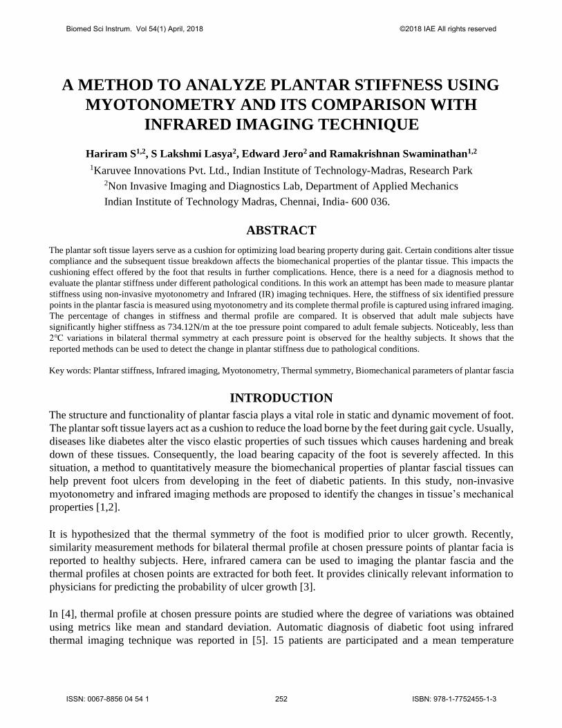
A METHOD TO ANALYZE PLANTAR STIFFNESS USING
MYOTONOMETRY AND ITS COMPARISON WITH
INFRARED IMAGING TECHNIQUE
Hariram S1,2, S Lakshmi Lasya2, Edward Jero2 and Ramakrishnan Swaminathan1,2
1Karuvee Innovations Pvt. Ltd., Indian Institute of Technology-Madras, Research Park2Non Invasive Imaging and Diagnostics Lab, Department of Applied Mechanics
Indian Institute of Technology Madras, Chennai, India- 600 036.
ABSTRACT
The plantar soft tissue layers serve as a cushion for optimizing load bearing property during gait. Certain conditions alter tissue
compliance and the subsequent tissue breakdown affects the biomechanical properties of the plantar tissue. This impacts the
cushioning effect offered by the foot that results in further complications. Hence, there is a need for a diagnosis method to
evaluate the plantar stiffness under different pathological conditions. In this work an attempt has been made to measure plantar
stiffness using non-invasive myotonometry and Infrared (IR) imaging techniques. Here, the stiffness of six identified pressure
points in the plantar fascia is measured using myotonometry and its complete thermal profile is captured using infrared imaging.
The percentage of changes in stiffness and thermal profile are compared. It is observed that adult male subjects have
significantly higher stiffness as 734.12N/m at the toe pressure point compared to adult female subjects. Noticeably, less than
2℃ variations in bilateral thermal symmetry at each pressure point is observed for the healthy subjects. It shows that the
reported methods can be used to detect the change in plantar stiffness due to pathological conditions.
Key words: Plantar stiffness, Infrared imaging, Myotonometry, Thermal symmetry, Biomechanical parameters of plantar fascia
INTRODUCTION
The structure and functionality of plantar fascia plays a vital role in static and dynamic movement of foot.
The plantar soft tissue layers act as a cushion to reduce the load borne by the feet during gait cycle. Usually,
diseases like diabetes alter the visco elastic properties of such tissues which causes hardening and break
down of these tissues. Consequently, the load bearing capacity of the foot is severely affected. In this
situation, a method to quantitatively measure the biomechanical properties of plantar fascial tissues can
help prevent foot ulcers from developing in the feet of diabetic patients. In this study, non-invasive
myotonometry and infrared imaging methods are proposed to identify the changes in tissue’s mechanical
properties [1,2].
It is hypothesized that the thermal symmetry of the foot is modified prior to ulcer growth. Recently,
similarity measurement methods for bilateral thermal profile at chosen pressure points of plantar facia is
reported to healthy subjects. Here, infrared camera can be used to imaging the plantar fascia and the
thermal profiles at chosen points are extracted for both feet. It provides clinically relevant information to
physicians for predicting the probability of ulcer growth [3].
In [4], thermal profile at chosen pressure points are studied where the degree of variations was obtained
using metrics like mean and standard deviation. Automatic diagnosis of diabetic foot using infrared
thermal imaging technique was reported in [5]. 15 patients are participated and a mean temperature
Biomed Sci Instrum. Vol 54(1) April, 2018 ©2018 IAE All rights reserved
ISSN: 0067-8856 04 54 1 252 ISBN: 978-1-7752455-1-3
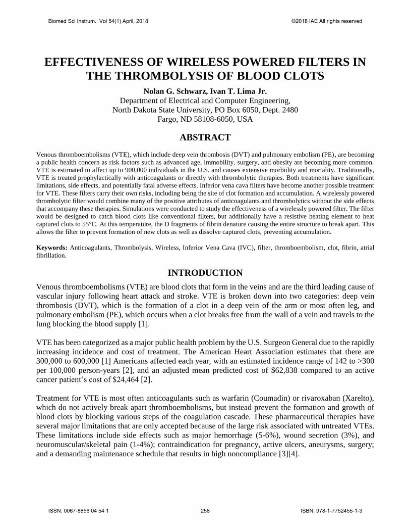
EFFECTIVENESS OF WIRELESS POWERED FILTERS IN
THE THROMBOLYSIS OF BLOOD CLOTS
Nolan G. Schwarz, Ivan T. Lima Jr.
Department of Electrical and Computer Engineering,
North Dakota State University, PO Box 6050, Dept. 2480
Fargo, ND 58108-6050, USA
ABSTRACT
Venous thromboembolisms (VTE), which include deep vein thrombosis (DVT) and pulmonary embolism (PE), are becoming
a public health concern as risk factors such as advanced age, immobility, surgery, and obesity are becoming more common.
VTE is estimated to affect up to 900,000 individuals in the U.S. and causes extensive morbidity and mortality. Traditionally,
VTE is treated prophylactically with anticoagulants or directly with thrombolytic therapies. Both treatments have significant
limitations, side effects, and potentially fatal adverse effects. Inferior vena cava filters have become another possible treatment
for VTE. These filters carry their own risks, including being the site of clot formation and accumulation. A wirelessly powered
thrombolytic filter would combine many of the positive attributes of anticoagulants and thrombolytics without the side effects
that accompany these therapies. Simulations were conducted to study the effectiveness of a wirelessly powered filter. The filter
would be designed to catch blood clots like conventional filters, but additionally have a resistive heating element to heat
captured clots to 55°C. At this temperature, the D fragments of fibrin denature causing the entire structure to break apart. This
allows the filter to prevent formation of new clots as well as dissolve captured clots, preventing accumulation.
Keywords: Anticoagulants, Thrombolysis, Wireless, Inferior Vena Cava (IVC), filter, thromboembolism, clot, fibrin, atrial
fibrillation.
INTRODUCTION
Venous thromboembolisms (VTE) are blood clots that form in the veins and are the third leading cause of
vascular injury following heart attack and stroke. VTE is broken down into two categories: deep vein
thrombosis (DVT), which is the formation of a clot in a deep vein of the arm or most often leg, and
pulmonary embolism (PE), which occurs when a clot breaks free from the wall of a vein and travels to the
lung blocking the blood supply [1].
VTE has been categorized as a major public health problem by the U.S. Surgeon General due to the rapidly
increasing incidence and cost of treatment. The American Heart Association estimates that there are
300,000 to 600,000 [1] Americans affected each year, with an estimated incidence range of 142 to >300
per 100,000 person-years [2], and an adjusted mean predicted cost of $62,838 compared to an active
cancer patient’s cost of $24,464 [2].
Treatment for VTE is most often anticoagulants such as warfarin (Coumadin) or rivaroxaban (Xarelto),
which do not actively break apart thromboembolisms, but instead prevent the formation and growth of
blood clots by blocking various steps of the coagulation cascade. These pharmaceutical therapies have
several major limitations that are only accepted because of the large risk associated with untreated VTEs.
These limitations include side effects such as major hemorrhage (5-6%), wound secretion (3%), and
neuromuscular/skeletal pain (1-4%); contraindication for pregnancy, active ulcers, aneurysms, surgery;
and a demanding maintenance schedule that results in high noncompliance [3][4].
Biomed Sci Instrum. Vol 54(1) April, 2018 ©2018 IAE All rights reserved
ISSN: 0067-8856 04 54 1 258 ISBN: 978-1-7752455-1-3
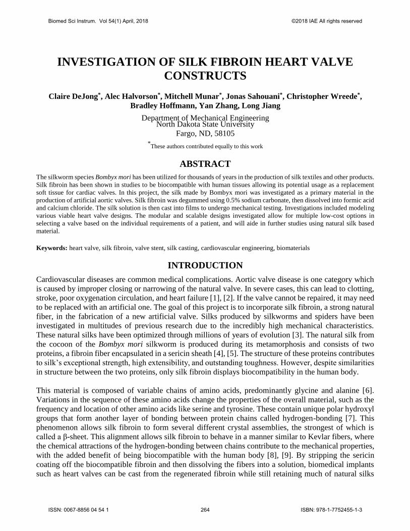
INVESTIGATION OF SILK FIBROIN HEART VALVE
CONSTRUCTS
Claire DeJong*, Alec Halvorson*, Mitchell Munar*, Jonas Sahouani*, Christopher Wreede*,
Bradley Hoffmann, Yan Zhang, Long Jiang
Department of Mechanical Engineering North Dakota State University
Fargo, ND, 58105
*These authors contributed equally to this work
ABSTRACT
The silkworm species Bombyx mori has been utilized for thousands of years in the production of silk textiles and other products.
Silk fibroin has been shown in studies to be biocompatible with human tissues allowing its potential usage as a replacement
soft tissue for cardiac valves. In this project, the silk made by Bombyx mori was investigated as a primary material in the
production of artificial aortic valves. Silk fibroin was degummed using 0.5% sodium carbonate, then dissolved into formic acid
and calcium chloride. The silk solution is then cast into films to undergo mechanical testing. Investigations included modeling
various viable heart valve designs. The modular and scalable designs investigated allow for multiple low-cost options in
selecting a valve based on the individual requirements of a patient, and will aide in further studies using natural silk based
material.
Keywords: heart valve, silk fibroin, valve stent, silk casting, cardiovascular engineering, biomaterials
INTRODUCTION
Cardiovascular diseases are common medical complications. Aortic valve disease is one category which
is caused by improper closing or narrowing of the natural valve. In severe cases, this can lead to clotting,
stroke, poor oxygenation circulation, and heart failure [1], [2]. If the valve cannot be repaired, it may need
to be replaced with an artificial one. The goal of this project is to incorporate silk fibroin, a strong natural
fiber, in the fabrication of a new artificial valve. Silks produced by silkworms and spiders have been
investigated in multitudes of previous research due to the incredibly high mechanical characteristics.
These natural silks have been optimized through millions of years of evolution [3]. The natural silk from
the cocoon of the Bombyx mori silkworm is produced during its metamorphosis and consists of two
proteins, a fibroin fiber encapsulated in a sericin sheath [4], [5]. The structure of these proteins contributes
to silk’s exceptional strength, high extensibility, and outstanding toughness. However, despite similarities
in structure between the two proteins, only silk fibroin displays biocompatibility in the human body.
This material is composed of variable chains of amino acids, predominantly glycine and alanine [6].
Variations in the sequence of these amino acids change the properties of the overall material, such as the
frequency and location of other amino acids like serine and tyrosine. These contain unique polar hydroxyl
groups that form another layer of bonding between protein chains called hydrogen-bonding [7]. This
phenomenon allows silk fibroin to form several different crystal assemblies, the strongest of which is
called a β-sheet. This alignment allows silk fibroin to behave in a manner similar to Kevlar fibers, where
the chemical attractions of the hydrogen-bonding between chains contribute to the mechanical properties,
with the added benefit of being biocompatible with the human body [8], [9]. By stripping the sericin
coating off the biocompatible fibroin and then dissolving the fibers into a solution, biomedical implants
such as heart valves can be cast from the regenerated fibroin while still retaining much of natural silks
Biomed Sci Instrum. Vol 54(1) April, 2018 ©2018 IAE All rights reserved
ISSN: 0067-8856 04 54 1 264 ISBN: 978-1-7752455-1-3
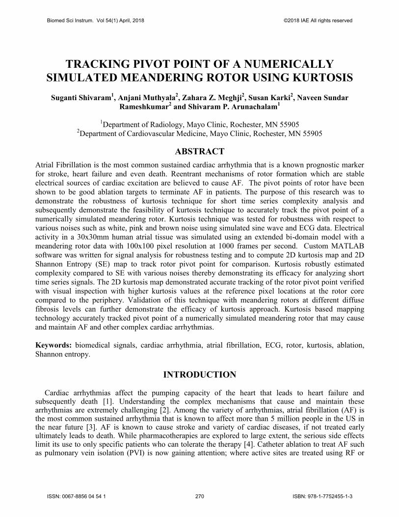
TRACKING PIVOT POINT OF A NUMERICALLY
SIMULATED MEANDERING ROTOR USING KURTOSIS
Suganti Shivaram1, Anjani Muthyala
2, Zahara Z. Meghji
2, Susan Karki
2, Naveen Sundar
Rameshkumar2 and Shivaram P. Arunachalam
1
1Department of Radiology, Mayo Clinic, Rochester, MN 55905
2Department of Cardiovascular Medicine, Mayo Clinic, Rochester, MN 55905
ABSTRACT
Atrial Fibrillation is the most common sustained cardiac arrhythmia that is a known prognostic marker
for stroke, heart failure and even death. Reentrant mechanisms of rotor formation which are stable
electrical sources of cardiac excitation are believed to cause AF. The pivot points of rotor have been
shown to be good ablation targets to terminate AF in patients. The purpose of this research was to
demonstrate the robustness of kurtosis technique for short time series complexity analysis and
subsequently demonstrate the feasibility of kurtosis technique to accurately track the pivot point of a
numerically simulated meandering rotor. Kurtosis technique was tested for robustness with respect to
various noises such as white, pink and brown noise using simulated sine wave and ECG data. Electrical
activity in a 30x30mm human atrial tissue was simulated using an extended bi-domain model with a
meandering rotor data with 100x100 pixel resolution at 1000 frames per second. Custom MATLAB
software was written for signal analysis for robustness testing and to compute 2D kurtosis map and 2D
Shannon Entropy (SE) map to track rotor pivot point for comparison. Kurtosis robustly estimated
complexity compared to SE with various noises thereby demonstrating its efficacy for analyzing short
time series signals. The 2D kurtosis map demonstrated accurate tracking of the rotor pivot point verified
with visual inspection with higher kurtosis values at the reference pixel locations at the rotor core
compared to the periphery. Validation of this technique with meandering rotors at different diffuse
fibrosis levels can further demonstrate the efficacy of kurtosis approach. Kurtosis based mapping
technology accurately tracked pivot point of a numerically simulated meandering rotor that may cause
and maintain AF and other complex cardiac arrhythmias.
Keywords: biomedical signals, cardiac arrhythmia, atrial fibrillation, ECG, rotor, kurtosis, ablation,
Shannon entropy.
INTRODUCTION
Cardiac arrhythmias affect the pumping capacity of the heart that leads to heart failure and subsequently death [1]. Understanding the complex mechanisms that cause and maintain these arrhythmias are extremely challenging [2]. Among the variety of arrhythmias, atrial fibrillation (AF) is the most common sustained arrhythmia that is known to affect more than 5 million people in the US in the near future [3]. AF is known to cause stroke and variety of cardiac diseases, if not treated early ultimately leads to death. While pharmacotherapies are explored to large extent, the serious side effects limit its use to only specific patients who can tolerate the therapy [4]. Catheter ablation to treat AF such as pulmonary vein isolation (PVI) is now gaining attention; where active sites are treated using RF or
Biomed Sci Instrum. Vol 54(1) April, 2018 ©2018 IAE All rights reserved
ISSN: 0067-8856 04 54 1 270 ISBN: 978-1-7752455-1-3
AN EXPERIMENTAL STUDY OF PULSATILE FLOW PAST
COMPLIANT AORTIC VALVE USING PARTICLE IMAGE
VELOCIMETRY
Ruihang Zhang and Yan Zhang
Department of Mechanical Engineering, North Dakota State University, Fargo, ND
ABSTRACT
The fluid-structural interaction inside an aortic valve is complicated due to the highly pulsatile flow and the compliant structure
of the aortic root. A quantitative understanding of the flow characteristics and the corresponding wall deformation is of great
importance for improvements of prosthetic aortic valve design and patient-specific pre-surgical assessments. An in vitro
experiment was conducted to investigate the turbulent flow features and boundary deformation of an intact silicone aortic root
model in this study. Based on a pulsatile cardiovascular flow simulator, particle image velocimetry (PIV) measurements were
performed to measure the flow field characteristics as well as the structural responses. Results suggest the jet flow velocity and
vortex structures vary significantly in different phases of a cardiac cycle. Significant shear stress was observed after the peak
systole phase when flow started to decelerate. Deformations of the aortic root were observed, the area strain was greater at
aortic annulus and least at sinus, the magnitude of which was found comparable to results from previous clinical studies.
Cardiac output plays an important role in determining the strength of hemodynamics and structural responses of the aortic valve
root. Turbulence level was greater for larger cardiac outputs. The deformation of aortic root and leaflet opening, represented
by area strain (AS) and effective orifice area (EOA), were proportional to cardiac outputs due to the increase of peak systolic
pressure. When cardiac output increased from 1 L/min, 2 L/min to 4 L/min, AS at aortic annulus increased from 1.8 %, 3.5 %
to 5.3 %, and EOA increased from 0.7, 1.0 to 1.6 cm2. This study reveals fundamental fluid-structural interaction phenomena
which could provide benchmark validations for future patient-specific computational modeling.
Keywords: aortic valve; polymeric; hemodynamics; pulsatile flow; shear stress; deformation; particle image velocimetry;
INTRODUCTION
Valvular heart disease is a leading cause of death in the US. About five million patients are diagnosed
with valvular diseases each year [1]. Valvular diseases that occur in the aortic root, including aortic
stenosis, calcification, and regurgitation, are the most common cases among all [2]. Valve replacement
surgery is often required to treat more than 50% of patients with severe aortic stenosis [3]. For procedures
like trans-catheter aortic valve replacement (TAVR), an accurate and fast evaluation of patient-specific
aortic hemodynamics is of paramount importance. Bioengineering study of aortic root hemodynamics
would significantly improve our understandings regarding the fundamental nature of the local flow and
provide insights for future clinical practices and computational modeling.
Bioengineering research has also contributed to the improvements of prosthetic heart valve designs. In
past decades, the design of prosthetics has experienced a transition from mechanical to bioprosthetic
valves [4]. Mechanical valves are the most widely used valve type due to its low cost and mechanical
durability [5]. However, because of the unrealistic leaflet design, some mechanical heart valves were
reported to cause separated blood flow and high shear stresses, which significantly increase the risk of
thromboembolism [6]. Patients are burdened with life-long uses of anticoagulation medications [7]. Bio-
prosthetic aortic valves, in contrast, provide better hemodynamic performances. Due to larger orifice
opening than mechanical valves, they can better reduce turbulence and pressure drop, and provide better
material compliance and hemodynamic performance [8,9]. However, bioprosthetic valves are often more
expensive and less durable than mechanical valves [9,10]. The tissue-engineering materials usually fails
Biomed Sci Instrum. Vol 54(1) April, 2018 ©2018 IAE All rights reserved
ISSN: 0067-8856 04 54 1 278 ISBN: 978-1-7752455-1-3
INFUSING SYNTHETICALLY SPUN SPIDER SILK WITH RIFAMPICIN
Nasim Soufizadeh-Balaneji1, Alec Staiger2, Nathan Johnson1, Adam Forness3, Greg Fondong3, Oluwaseyi Ogundolani3, Pranothi Mulinti3, Bradley Hoffmann2, and Amanda E. Brooks3
1Department of Electrical and Computer Engineering 2Department of Mechanical Engineering 3Department of Pharmaceutical Sciences
North Dakota State University Fargo, ND
ABSTRACT Antibacterial resistance is growing exponentially, teetering on a crisis that can only be solved with “out-of-the-box”
solutions. The silk-based delivery systems seems to be a promising approach to combat this problem. This system includes particles and surfaces with grafted antibiotics; however, acquisition of raw silk protein for such applications poses a significant obstacle to clinical translation. Although natural silk is readily used in several biomedical applications, artificial production of fibers may be cost effective for customized applications. Hence, to produce fibers with tailored mechanical, electrical, and biochemical characteristics, the development of silk spinning systems that can mimic the complexity of in vivo silk spinning glands is necessary. To assess the feasibility of infusing an antibiotic directly into a silk fiber, natural and recombinant spider silk as well as natural silkworm silk were dissolved in an appropriate solvent. Both microfluidic and wet spinning techniques have been used to artificially respun the silk solutions. A modification of a standard Kirby Bauer zone of inhibition (i.e. disk diffusion) assay against S. aureus has been used to evaluate the efficacy of this method. Both silk fibers as well as rifampicin incorporated fibers were placed on S. aureus spread LB agar plates. While no zones of inhibition (ZOI) were seen for silk fibers without any drug infused, the ZOI for fibers containing the rifampicin ranged from 5 mm to 27 mm; depending on the length, diameter, and material of silk fibers. Our observations showed that the drug could successfully be integrated into fibers and could potentially be used as suture materials as a novel way to address and prevent antibiotic resistant infections.
Keywords: silkworm silk, spider silk, recombinant spider silk, Rifampicin, wet spinning, microfluidic spinning, drug delivery
INTRODUCTION
Fast-growing antibiotic-resistant bacteria have become a widespread dilemma that has compromised the effectiveness of antibacterial medication for many years [1], [2]. Even though the mechanisms of resistance are relatively well understood, inappropriate prescribing and poor patient compliance remain the two main causes of the epidemic. In fact, based on studies, treatment indication, choice of agent, or duration of therapy are inaccurately prescribed 30-50 % of the time [3], [4]. Thus, discovering and developing either new antibiotics or improving the use of existing antibiotics, although a lengthy and non-lucrative pursuit, is no longer a luxury [5], [6]. Without creative strategies to successfully act against increasingly-resistant bacteria, debilitating and lethal diseases will continue to increase in frequency and scope [7].
Before the 1950s, the only method for drug delivery was an orally dissolving pill [11]. Since then, new methods have been developed containing features such as sustained release, time release, extended release, etc. The use of controlled drug release systems may offer a promising alternative to preserve the efficacy of our antibiotic arsenal and slow or prevent the evolution of antibiotic resistance. By regulating the release of a drug into an environment, the pharmacokinetics of drug release will be more accurate,
Biomed Sci Instrum. Vol 54(1) April, 2018 ©2018 IAE All rights reserved
ISSN: 0067-8856 04 54 1 286 ISBN: 978-1-7752455-1-3
HYPOXIA RESPONSIVE LIPID INCORPORATION
INTO BOVINE MILK EXOSOMES
Jessica Pullan1, Pooja Chemeiti3, Matthew Confeld1, Li Feng1, James Froberg2, YongkiChio2, Sanku Mallik1
1Department of Pharmaceutical Sciences, North Dakota State University 2Department of Physics, North Dakota State University
3Sheyenne High School, Fargo, ND
ABSTRACT
Pancreatic cancer is one of the deadliest cancers, killing 80% of all patients diagnosed within five
years of diagnosis. Patients not only suffer from a high mortality rate but also a high
morbidity rate due to the toxic nature of chemotherapeutic treatments. Targeted localized
delivery of these drugs could improve patient outcomes. Solid malignant tumors, often found in
pancreatic cancer, have a hypoxic microenvironment due to the lack of vascularization.
Hypoxia-responsive lipids incorporated into exosomes could deliver the necessary controlled
chemotherapeutic dose to pancreatic tumors, thereby improving the safety and efficacy of
toxic anticancer medications. Here, manufactured hypoxia-responsive bovine exosomes
(HRB) were tested in a hypoxic environment created with microsomes, NADPH, and nitrogen
gas. Fragmented pieces of the HRBs were observed in atomic force microscopy and transmission
electron microscopy. Future studies will look at the ability to encapsulate an anticancer drug into
the HRBs and reduce cell viability of pancreatic cancer cells.
Keywords: Exosomes, Hypoxia Responsive Lipid, Controlled Drug Delivery, Hypoxic Conditions
INTRODUCTION
More than 80% of people diagnosed with pancreatic cancer greater than stage 1A will not
survive five years. [1] The solid, malignant, pancreatic tumors often show significantly reduced
oxygen levels in the interior. [3-4] The hypoxia created by the lack of vascularization
complicate the effective delivery of anticancer drugs to cancerous cells. In addition, the slow
cell turnover rate and high drug resistance also contribute to the complications. [4] Although
these conditions make drug delivery difficult, the biochemical abnormalities can be utilized to
trigger the release of anticancer drugs from nanoparticles. By creating a nanoparticle
that is specific to the microenvironment of the tumor, cytotoxicity can be reduced
significantly.
Nanoparticles can be created from a variety of materials. Exosomes, extracellular vesicles
secreted naturally in different organs and found in fluids such as blood and milk, may be the
most promising nanoparticle for intracellular drug delivery. [5] The size of these vesicles ranges
between 30 to 120 nm. The exosomes mediate cell-to-cell signaling, transportation of
bioactive molecules, and immune response. [3–6] Inherent stability, biocompatibility, biological
barrier permeability, low toxicity, and low immunogenicity are critical for the exosome’s natural
functions. [5] As a result, exosomes have the potential to be anticancer drug carriers. Due to
the movement of exosomes, their concentration in some fluids is higher than others. The
concentration of exosomes per liter of human blood serum is only about 2 mg/L. [9] Raw bovine
Biomed Sci Instrum. Vol 54(1) April, 2018 ©2018 IAE All rights reserved
ISSN: 0067-8856 04 54 1 293 ISBN: 978-1-7752455-1-3
ASSESSMENT OF ANIMAL MODELS AS SURROGATES
FOR HUMAN TUMORS FROM THREE DIFFERENT
ORGANS Ibrahim O. Farah, Zikri Arslan, Michelle Tucci, Hamed Benghuzzi, and Joseph A. Cameron
Jackson State University and the University of Mississippi Medical Center
ABSTRACT The compositional balance and distribution of trace metals/elements in various body tissues are essential key players in tissue
and cellular homeostasis. Low Zn levels as well as overexpression of metalothioneins were implicated in the development and
progression of various cancers including the prostate. Nonetheless, wider elemental profiles that relate cancer and normal
phenotypes with regards to metal homeostasis were not well elucidated in the literature. Moreover, laboratory animals are
currently used as accepted models for studying cancer but the level of their representation of actual cancer tissues was not clear.
This study is attempting to assess the relevance of animal models currently in use, as surrogates for cancer and establish their
relationship to actual normal and cancer tissues from humans. The major focus of this study was to investigate the differential
relationship of metal concentrations and profiles in cancer and normal tissues from cadavers of humans and their comparison
to established animal models representing organ cancers. The working hypothesis was that elemental/metal concentrations and
profiles seen in post mortem will show significant differences between normal and cancer-derived tissues as well as between
various tissue types in humans, rats, and dogs. This study also establishes critical elemental/metal profiles that may be relevant
in providing correlations with the development of three major cancers. Normal human and tumor tissues of cadaverous lung,
breast, and liver used in this study were obtained from US Biomax Company and relevant animal models (Sprague-dawley and
Brown Norwegian rats as well as dogs; were obtained from Jackson Laboratories and the Mississippi State Veterinary
Laboratory in Pearl, MS), to analyze for elements and test the hypothesis. Tissue samples were prepared using standardized
digestion procedures necessary for use with the Inductively Coupled Plasma-Atomic Emission mass Spectrometry (ICP-MS)
to determine the concentrations and profiles of 21 elements including Ag, Al, As, Ba, Ca, Cd, Co, Cr, Cu, Fe, Mg, Mn, Na, Ni,
Pb, Sb, Se, Sr, Tl, V, and Zn. Our data supports the notion that metal/elemental homeostasis is essential for normal tissue
function and that elemental variations in content, distributions, and ranking are tissue specific as well as carcinoma and species-
specific. Analysis of data showed significant variations in elemental content and distribution profiles/ranking between animal
models and actual human tissues consistent with the hypothesis. It is concluded that elemental homeostasis is essential for
normal tissue function and that shifts in their distribution and content are essential in determining the use of animal models as
surrogates for studying cancer. These results are promising and warrant further studies to confirm the relevance of animal
models in relation to their use as pre-clinical tools for examining targeted cancer therapeutics.
Keywords: essential, non-essential metal load, metal profile, tumors and cancer tissues, animal models,
breast, lung, liver, ICP analysis, homeostasis.
INTRODUCTION Lung, liver, and breast cancers are highly prevalent in the United States [1-2]. Many studies have shown
that homeostasis of metals/trace elements are very essential to the development of the cancer phenotype in many
cancers. Essential trace elements are engaged in four major functions as stabilizers, elements of structure, essential
elements for hormone function, and cofactors in enzymes. Studies have also indicated a role for Ba through its
toxicity and involvement in chronic disease with no established evidence in human cancers [3-6]. Calcium is
recognized as a messenger in cell signaling and studies have established a role for Ca2+ as regulators and Ca channels
were found to be different in their distribution and expression in cancer and normal human tissues [7-10]. Elevated
Fe causes free radicals that are linked to cancer, heart disease, aging, as well as liver and pancreatic damage [11,
12]. It has also been found that Ni, Cu, and Fe concentrations in cancerous human stomach were significantly higher
than those in non-cancerous stomach tissue samples, as well as that high Zn concentrations were found in both
paired stomach samples.
Another evidence for the importance of metals and elemental discrepancy is provided through the up-
regulation of metallothioneins in many cancers [13, 37]. Sodium regulates blood pressure as well as having an
established role in cellular homeostasis; however, there was no established role for Na and Vanadium (V) in human
cancer [14-18]; in contrast, a role for Zn was established in prostate cancer [19-21]. Generally, literature review
Biomed Sci Instrum. Vol 54(1) April, 2018 ©2018 IAE All rights reserved
ISSN: 0067-8856 04 54 1 301 ISBN: 978-1-7752455-1-3

ACETIC ACID REMEDIATION OF ANTHROPOGENIC
CONTAMINATION OF WATER AT THE GBNERR IN
MISSISSIPPI Ibrahim O. Farah, Willis O. Lyons, Zikri Arslan, Michelle Tucci*, and Paul B. Tchounwou
Department of Biology, Jackson State University, Box 18540, Jackson, MS 39217, USA
*Department of Anesthesiology, University of Mississippi Medical Center, Jackson, MS 39216
ABSTRACT Grand Bay National Estuarine Research Reserve (GBNERR) is an important ecosystem in the Mississippi Gulf Coast. The
GBNERR may be a potential source for contamination with anthropogenic bacterial pathogens that may play a significant role
in the causation of waterborne human diseases. The objective of this study was to evaluate the interaction of physicochemical
and microbiological water quality parameters at the GBNERR to determine quantitative levels and establish the potential for
remediation of post-contamination of water and seafood by human fecal pollution from anthropogenic sources at the reserve.
Water samples were collected aseptically from Bayous Heron, Cumbest, Point Aux Chenes Bay and Bangs Lake (Pine-O-Pine).
Physicochemical parameters were determined using standard protocols. Eight bacterial species including Campylobacter were
concentrated from water samples by membrane filtration. Water samples were tested for the presence of traditional indicator
microorganisms including: heterotrophic (HPC), total coliforms (TC), fecal coliforms (FC), and enterococcus (ENT) in CFU/ml
concentrations. Mean values of temperature, specific conductivity, dissolved oxygen, and pH were within acceptable levels in
comparison to MDEQ, USEPA, and the USGS standards during the time of investigation. However, the values of turbidity in
Grand Bay water exceeded USEPA recommended levels in several occasions during the investigation. Data from this study
indicates significant variability (p < 0.0001) in mean bacteria concentrations between sites. The data also indicates significant
impact of acetic acid treatment in the remediation of post contamination and survival of pathogens from the GBNERR Bayous
Heron, Cumbest, and Pine-O-Pine when compared with control findings. The interaction of physicochemical and
microbiological parameters of water through external chemical manipulation by acetic acid may provide utility in the
remediation of post-contamination with anthropogenic pathogens such as E. coli, Enterococci, Campylobacter, Vibrio, Giardia,
and Cryptosporidium. Presence of high numbers of indicator bacteria suggests public health concerns for oyster and shellfish
consumers as well as other water contact activities. Hence, control strategies should be developed and implemented to prevent
or remediate any future contamination of the GBNERR waters citing the economic impact of such contamination on shellfish
fishing activities on the reserve.
Keywords: Acetic acid, anthropogenic, water and foodborne disease, natural remediation, shellfish
INTRODUCTION Water quality monitoring is the foundation for first-hand data on environmental water management. A
reasonable water quality monitoring network should not only meet the needs for long-term data
accumulation, water quality assessment, and trend analysis but also reflect in a timely manner, the dynamic
status of the water environment, and water pollution. Water quality monitoring provides scientific
guidance for water resource management and water environment protection [1]. There has been great
emphasis on the importance of assessing the microbiological quality of water in Grand Bay National
Estuarine Research Reserve (GBNERR) in Mississippi due to the increased impact of pathogenic bacteria
from anthropogenic sources. Improving access to safe water sources can result in significant benefits to
both health and quality of life [2, 3].
An estimated 1.1 billion people worldwide rely on water supplies that are at high risk of fecal
contamination [4]. The deterioration of aesthetic aspects of drinking water, such as taste, odor, and color
represents up to 80% of consumer complaints to water utilities [5]. Diarrheal disease is a major cause of
death and disease, especially among young children in low-income countries where the most common
causes are fecal-contaminated water and food, or poor hygiene practices. The bacterial, viral, and
Biomed Sci Instrum. Vol 54(1) April, 2018 ©2018 IAE All rights reserved
ISSN: 0067-8856 04 54 1 309 ISBN: 978-1-7752455-1-3
TOMOGRAPHIC PIV OF LARGE INTRACRANIAL ANEURYSM MODELS
Timothy Rossman, M.S.1, Kendall Dennis, M.S.1, Adam Stolt, B.S.1,2, Yu-Ju Chen, B.S.1,3, David Kallmes, M.D.4,5, Jordi Estevadeordal, Ph.D.2, Dan Dragomir-Daescu, Ph.D.6,7
1Division of Engineering, Mayo Clinic, Rochester MN, USA 2North Dakota State University, Fargo ND, USA
3 Department of BioMedical Engineering, National Cheng Kung University, Tainan, Taiwan 4Department of Radiology, Mayo Clinic, Rochester MN, USA
5Department of Neurosurgery, Mayo Clinic, Rochester MN, USA 6Department of Physiology and Biomedical Engineering, Mayo Clinic, Rochester MN, USA
7Mayo Clinic College of Medicine and Science, Rochester MN, USA
ABSTRACT
In vitro measurements of brain aneurysm hemodynamics can aid in the validation of computational fluid dynamics models with the intent of helping clinicians assess risk of rupture in patient specific anatomy. Transient tomographic particle image velocimetry (PIV) measurements were conducted on a glass replica of a realistic brain aneurysm geometry recently published in a computational fluid dynamics (CFD) challenge study. To enable higher spatial resolution of the resolved velocity vectors from PIV, the physical aneurysm geometry was a 2X-scaled model fabricated by scientific glass blowing. Because this fluid volume exceeded the depth of field of our PIV cameras, a translational stage was used to acquire overlapping volumetric data with multiple stage positions, and custom scripts were used to assemble a contiguous volume of velocity vectors for multiple pulsatile cycle time points allowing for a precise identification of flow features within the aneurysm model. In addition, another novelty of the work was assessing measurement error from sampling variation using the coefficient of variation (CoV) of the velocity vector components. When compared at each discretization point, the CoV was successfully used as an objective means to merge cells from multiple temporal image datasets, producing smoother velocity contour isolines when the volumetric datasets were combined. In conclusion, this approach enabled flow field measurements at various points along a cardiac cycle on a relatively large flow volume.
Keywords: Particle image velocimetry, experimental fluid mechanics, intracranial aneurysm, tomographic PIV
INTRODUCTION Tomographic PIV is an image-based particle tracking measurement tool that is well-suited to study the complex fluid dynamics associated with intracranial aneurysms which can aid in validation of CFD models and clinical decision-making [1, 2]. Using patient-based models, tomographic PIV can measure 3D velocity fields that could otherwise be unapparent with conventional 2D or even Stereo PIV. The two later technologies have been leveraged for validating computational fluid dynamics (CFD) simulations of intra-aneurysmal flow [2-4]. This is especially important as CFD simulations have been shown to vary greatly among different users [5]. PIV is also used to study different treatment options such as stents and flow diverters where CFD becomes even more computationally challenging with the complexities of anatomical geometry and small feature size of treatment devices [6-8].
Not all of these studies utilized tomographic PIV, though there clearly would have been great benefit from gathering larger volumes of 3D vectors to further reveal the flow complexities. Should seeding
Biomed Sci Instrum. Vol 54(1) April, 2018 ©2018 IAE All rights reserved
ISSN: 0067-8856 04 54 1 317 ISBN: 978-1-7752455-1-3
EFFECT OF MR CONTRAST AGENTS ON MYOCARDIAL
TISSUE ELASTICITY IMAGING: A PILOT STUDY
Suganti Shivaram1, Susan Karki
2, Anjani Muthyala
2, Zahara Z. Meghji
2, Naveen Sundar
Rameshkumar2 and Shivaram P. Arunachalam
1
1Department of Radiology, Mayo Clinic, Rochester, MN 55905
2Department of Cardiovascular Medicine, Mayo Clinic, Rochester, MN 55905
ABSTRACT
Tissue elasticity imaging namely ultrasound and magnetic resonance (MR) elastography techniques can
assess in vivo tissue stiffness that can serve as novel biomarkers for variety of diseases. Cardiac MR
Elastography (CMRE) can provide in vivo myocardial tissue stiffness estimates which are shown to
differentiate normal healthy volunteers and patients with cardiac conditions such as heart failure with
preserved ejection fraction (HFpEF), cardiac amyloidosis, hypertrophic cardiomyopathy (HCM) etc.
The purpose of this pilot study is to evaluate the effect of MR contrast agent in CMRE application, to
provide important insights into clinical protocol development. CMRE imaging using GE 1.5 T scanner
was performed on two HCM patients as a pilot study before and after Gadolinium injection at 15 min
and 30 min respectively. MRE stiffness was calculated pre and post-contrast and % change was
calculated. MRE stiffness increased 28.32% with CMRE imaging after 15 min post-contrast in HCM
patient 1 and 12.38% 30 min post-contrast imaging on HCM patient 2. The results suggest that MR
contrast agents can alter the stiffness estimates based on the timing of the contrast injection and suggest
the importance of pre-contrast CMRE imaging. MR contrast agents such as Gadolinium increases the
stiffness estimates using CMRE technique.
Keywords: ultrasound elastography, MRI, magnetic resonance elastography, stiffness, tissue elasticity
imaging, hypertrophic cardiomyopathy, contrast agents, myocardial stiffness.
INTRODUCTION
In the past, physicians attempted to estimate tissue elasticity using palpation for diagnosing various
diseases [1]. However, with the practical difficulties to palpate internal organs and tissues and for a more
accurate disease diagnosis, tissue elasticity imaging modality emerged that merges several fields such as
tissue elastic constants, tissue contrast, tissue motion, imaging systems and the study of vibrating targets
using coherent radiation [2]. Therefore, tissue elasticity imaging techniques are an integrated approach
that combines the concept of tissue elasticity and modern imaging systems to assess the elastic
properties of tissues such as Young's modulus, Poisson's ratio, and viscosity [2]. Among those,
ultrasound elastography [3] and magnetic resonance elastography [4] are gaining high popularity with
several applications for prognosis and diagnosis of variety of diseases.
There are several ultrasound based techniques such as strain elastography [5], acoustic radiation force
impulse (ARFI) strain imaging [6], shear wave imaging techniques such as transient elastography [7],
point shear wave elastography [8] and two-dimensional (2D) Shear wave elastography [9]. All these
techniques have proven success in variety of applications. However, they each face several technical
Biomed Sci Instrum. Vol 54(1) April, 2018 ©2018 IAE All rights reserved
ISSN: 0067-8856 04 54 1 325 ISBN: 978-1-7752455-1-3

THE EFFECT OF PLATELET-RICH PLASMA ON
COMPLETE TEARS OF THE MEDIAL COLLATERAL
LIGAMENT: A CRITICALLY APPRAISED TOPIC
Bradley J. Conant and Shannon L. David PhD, ATC
Department of Health, Nutrition, and Exercise Sciences
North Dakota State University, Fargo, ND 58102
ABSTRACT
Medial collateral ligament (MCL) tears are relatively common in athletic populations, and conservative treatment is often the
first approach. Platelet-rich plasma has been used to treat many soft-tissue injuries, but evidence regarding its effectiveness
with MCL injuries is lacking. The purpose of the study is to identify whether current research supports the addition of platelet-
rich plasma to conservative treatment to improve healing and reduce recovery time in individuals with complete MCL tears. A
literature search was performed to locate all studies that investigated the healing process or outcomes of complete MCL tears
treated with platelet-rich plasma. The available evidence is inconclusive regarding the effect of platelet-rich plasma injections
on the healing of isolated MCL tears. The low risk associated with platelet-rich plasma injection and lack of indication toward
negative outcomes may make it an appropriate option for some patients. However, any decision to include platelet-rich plasma
in a conservative treatment plan should be made on an individual basis guided by the athlete’s goals. No clinical
recommendation can be made until stronger evidence is available. Consistent results must be obtained from higher-level studies
before an evidence-based recommendation can be made for the treatment of MCL tears with platelet-rich plasma.
Keywords: platelet-rich plasma, medial collateral ligament, conservative treatment, wound healing
INTRODUCTION
Among athletic populations, the medial collateral ligament (MCL) is one of the most commonly injured
structures in the body [1], [2]. Even in the case of a complete, full-thickness tear of the MCL, conservative
treatment is often sufficient when there is no concurrent knee pathology. Conservative treatment of an
isolated MCL tear typically involves immobilization and rehabilitation, and frequently requires at least
four to eight weeks for recovery [1], [2]. Athletes in season often cannot afford a recovery time of this
length. An intervention that could shorten recovery time would minimize the amount of training and
competition an athlete misses because of the injury. The use of platelet-rich plasma to attempt to enhance
tissue healing has gained popularity throughout orthopedics and sports medicine, and it may be an option
to supplement traditional conservative treatment and shorten time of recovery from an MCL tear.
Research has consistently found evidence of the benefits platelet-rich plasma can provide in tissue
healing and regeneration [3]-[11]. Platelet-rich plasma contains a variety of growth factors, glycoproteins,
and adhesion molecules believed to contribute to an improved healing response when activated. Several
proteins have been the focus of research and are believed to be associated with the augmented healing
process. They include growth factors such as epidermal growth factor (EGF), fibroblast growth factor
(FGF), hepatocyte growth factor, insulin-like growth factor 1 (IGF-1), platelet derived growth factors
(PDGF), transforming growth factor-β (TGF-β), and vascular endothelial growth factor (VEGF), as well
as three adhesive proteins, fibrin, fibronectin, and vitronectin. Collectively, these proteins, as well as their
mechanisms within the healing processes of various tissues, have been studied and reviewed extensively
[3], [8]-[11]. Despite this promising preclinical evidence, clinical research has been widely varied
regarding the benefits of platelet-rich plasma. Given the prevalence of MCL tears in athletic populations,
and that conservative treatment is generally used, it is valuable to investigate whether an evidence-based
Biomed Sci Instrum. Vol 54(1) April, 2018 ©2018 IAE All rights reserved
ISSN: 0067-8856 04 54 1 331 ISBN: 978-1-7752455-1-3
ASSESSING THE ABILITY OF WOMEN’S LACROSSE
HELMETS TO REDUCE RISK OF HEAD INJURY
Emily E. Kieffer, Megan L. Bland, Steven Rowson
Biomedical Engineering and Mechanics, Virginia Tech, Blacksburg, VA 24061
ABSTRACT
Often underrepresented in concussion research, females sustain higher rates of concussions in sports than their male
counterparts. Women’s lacrosse is characterized as non-contact, with the required equipment consisting of goggles and a
mouthguard. However, concussions make up 9.8% of injuries in women’s collegiate lacrosse. Recent public awareness has
pushed forward the development of women’s lacrosse helmets. The objective of this study was to evaluate the ability of
women’s helmets to reduce risk of head injury. This was done through a test series with a pendulum impactor. A headform
developed by National Operating Committee on Standards for Athletic Equipment (NOCSAE) for helmet testing was
instrumented with three linear accelerometers and three angular rate sensors to determine peak linear and rotational
accelerations. A total of 72 tests were completed at the front boss and side locations over a range of 2.0, 3.0, and 4.0 m/s. Each
impact configuration was trialed four times. The performance of the Hummingbird and Cascade LX helmets were analyzed
based on resultant accelerations and compared to that of a bare headform. Results show significant reductions in linear and
rotational acceleration of the headform with both Hummingbird and Cascade LX helmets at all impact velocities. This study
demonstrates potential benefits of women lacrosse players wearing helmets in the event of incidental head impact.
Keywords: Concussion, head impact, biomechanics, lacrosse, female
INTRODUCTION
The US Centers for Disease Control and Prevention (CDC) estimates that up to 3.8 million concussions
occur in sports and recreational activities each year [1]. Although sports-related concussions were once
thought to only result in transient symptoms and temporary neurocognitive impairment, recent research
has shown the possibility of long-term neurodegenerative processes as a result of repetitive concussions.
Recent findings have shown a gender-specific difference in occurrence and outcome of mild traumatic
brain injury (mTBI) [2]. When studying male and female high school and collegiate sports, the majority
of studies found that females overall have a higher rate of concussion and they account for a higher
percentage of total injuries in female athletes [3]. While female specific concussion tolerance is largely
unknown, there are several biomechanical differences between males and females that could be a factor
leading to females’ higher concussion rates. Females tend to have smaller head-neck segments, which
would result in a higher acceleration for a given impact force [4]. Additionally, they have been found to
have similar sized head when compared to males, but weaker necks to support it, specifically in the C3-
C5 neck vertebrae (which stabilize the neck during whiplash) [5]. This could contribute to the higher
prevalence of concussion in females as their smaller necks would not be able to resist deceleration as well
as a male’s larger neck. Findings like these have increased awareness of head injury and pushed for safety
in sports by modifying practice techniques, changing game rules, and improving protective headgear.
Women’s lacrosse varies significantly from men’s lacrosse, as it is considered non-contact. As of 2002,
mouthguards and goggles are mandatory to play, but the use of protective headgear for field players is not
required. There is a higher rate of concussion in men’s lacrosse, which can be attributed to full contact in
the game [6]. However, women still remain at high risk for head and face injury, due to contact with the
ball and stick. In fact, in a 10 year analysis of the National Electronic Injury Surveillance System (NEISS)
database, females sustained more of these types of injuries than males [7]. The increased concern of long
Biomed Sci Instrum. Vol 54(1) April, 2018 ©2018 IAE All rights reserved
ISSN: 0067-8856 04 54 1 340 ISBN: 978-1-7752455-1-3
EFFECT OF ANVIL ANGLE ON IMPACT KINEMATICS IN LABORATORY EVALUATION OF BICYCLE HELMETS
Megan L. Bland, Craig McNally, Steven Rowson Biomedical Engineering and Mechanics Virginia Tech, Blacksburg, VA 24061
ABSTRACT Cycling is the leading cause of sport-related head injuries in the US. Bicycle helmets are subject to standards limiting peak linear acceleration (PLA) in normal impacts. However, real-world cycling accidents occur at a variety of impact angles and involve both normal and tangential incident velocities. Real-world head impacts also induce rotational acceleration, a major contributor to brain injury. The objective of this study was to assess performance differences of bicycle helmets across impact angles under real-world cyclist impact conditions. Ten helmet models were impacted on a custom drop tower using biofidelic headform and neck surrogates. Impacts were against a 0° or 30° anvil and at two impact locations and velocities common in cyclist accidents. PLA was not significantly different across anvil (p>0.40) while peak rotational acceleration (PRA) was higher at 0° (p<0.01), reflecting differences in resultant force proximity to the assembly center of gravity. Kinematic results produced a considerable range in concussion risk, with risks differing up to 50% across anvil angle in matched impact configurations. These data suggest that evaluating helmet performance under a variety of impact angles may aid in the development of bicycle helmet technologies equipped to mitigate injury risk under a wider variety of loading scenarios.
Keywords: cycling, head injury risk, concussion, acceleration, biomechanics
INTRODUCTION Cycling is a popular recreational activity, sport, and mode of transportation in the U.S. and throughout the world. Despite its popularity, it is not a risk-free activity; cycling caused the most head injuries treated in U.S. emergency rooms in 2015 out of any sport or recreational activity, accounting for over 81,000 cases [1]. The societal and economic burden from these injuries is considerable, with associated annual healthcare costs estimated to exceed $2 billion [2]. Fortunately, risk of sustaining a head injury has been shown to be significantly reduced by wearing a bicycle helmet [3, 4].
Bicycle helmets are presently subject to standards limiting peak linear acceleration (PLA) in specified impact tests [5]. While standards ensure that helmets lower head injury risk, the prescribed impact testing has limited representation of real-world cyclist accidents. The metal half-headform and the rigid neck joint are not biofidelic and preclude assessment of relative head-neck motion during impact. Further, all impacts are normal to the impact surface, while real-world cyclist head impacts frequently involve both normal and tangential incident velocities (termed “oblique”) [6]. Real-world head impacts also induce rotational acceleration, a major contributor to diffuse brain injury such as concussion [7].
Given the limitations of standards impact testing, several research groups have developed oblique impact rigs with more biofidelic test setups [4, 8-11]. These rigs differ in boundary conditions. For instance, many groups drop helmets onto an angled anvil to simulate oblique impacts, but the choice of anvil angle is not consistent [8, 10, 11]. This is attributable to the range of head impact angles relative to the ground that a cyclist may experience, which spans from close to normal to more commonly between 30° and 60° [12, 13]. Linear and rotational impact accelerations likely vary as a function of angle; however, to-date this possible range is relatively unexplored. The several studies that have subjected helmets to both normal and oblique impacts have used different test rigs for the two impact types, have evaluated different
Biomed Sci Instrum. Vol 54(1) April, 2018 ©2018 IAE All rights reserved
ISSN: 0067-8856 04 54 1 347 ISBN: 978-1-7752455-1-3
EFFECT OF FACEMASK WEIGHT ON HELMET
PERFORMANCE Abigail M. Tyson, Emily E. Kieffer, Steven Rowson
Biomedical Engineering and Mechanics Virginia Tech, Blacksburg, VA 24061
ABSTRACT
Current football helmet standards and ratings evaluate helmets without facemasks; however, all football players wear
facemasks during play. With the existence of 10-20 facemask variations per helmet, it is often impractical to test each helmet-
facemask combination when evaluating helmets. The most differentiating characteristic of facemasks is weight; therefore, the
purpose of this study was to determine the effect of facemask weight on helmet performance in order to inform future
helmet testing methods. The heaviest and lightest facemasks were chosen for five helmet models. Dummy head impact tests
were conducted using a custom pendulum impactor with a rigid impactor face measuring 8-in. in diameter with a 5-in.
radius of curvature representative of a helmet shell. Tests were performed to a front impact location on the helmet shell above
the facemask at 3.0, 4.6, and 6.1 m/s. Two trials were conducted for two samples of each helmet-facemask combination
totaling 120 tests. One-way ANOVAs were performed for each helmet and impact velocity condition. No significant
differences were observed between the heavy and light facemasks for peak linear acceleration. Significant differences did
exist in angular acceleration between facemask types for one helmet model. For most helmets tested, facemask weight has
a negligible effect when impacting a helmet shell; however, this effect varies by helmet model.
Keywords: football, facemask, impact testing, pendulum, concussion
INTRODUCTION
Football helmet standards established by the National Operating Committee on Standards for
Athletic Equipment (NOCSAE) [1] and other ratings [2] evaluate football helmets in the laboratory.
These tests use dummy heads representative of 50th percentile males and impact energies and locations
representative of those experienced on the field during play [3]. However, tests are performed without
facemasks even though facemasks are required on the field. A previous study evaluated differences in
linear and angular head kinematics and severity index in helmets tested with and without facemasks [4].
Two hockey helmet models and two football helmet models were impacted, with and without a
facemask. One facemask sample was used per helmet model. Tests were performed with a linear
impactor at 6 and 9 m/s to the front, side and rear boss of each helmet. Significant differences were
found in tests with and without facemasks, although most differences were small and varied by helmet
type, impact location, and speed. On average, the inclusion of a facemask resulted in lower linear and
angular accelerations with differences of approximately ±5%. The effect of facemask was greater in
football compared to hockey helmets which the authors predicted was due to increased stiffness in
football helmets from the nature of the facemask attachment mechanisms.
In adapting football helmet standards and ratings to include facemasks, the problem arises of
which facemask to choose. Many helmet models can be paired with upwards of 20 facemask models,
making the testing of all possible helmet-facemask combinations impractical. Current facemask
models vary in material, weight, and design, and are recommended to be used for different playing
positions. The most common facemask material is carbon steel, although other materials include
stainless steel and titanium. Titanium is the most light-weight, yet is often more than twice the price of
carbon or stainless steel. In choosing facemasks to be used in football helmet testing, it is common to
choose the most popular styles [4, 5], typically made of carbon steel. However, carbon steel models still
significantly vary in weight.
Biomed Sci Instrum. Vol 54(1) April, 2018 ©2018 IAE All rights reserved
ISSN: 0067-8856 04 54 1 355 ISBN: 978-1-7752455-1-3
ESTIMATING THE BRAIN STRAIN RATES DURING
TRAUMATIC BRAIN INJURY
Mohammad Hosseini Farid, Mohammadreza Ramzanpour, Mariusz Ziejewski, and Ghodrat
Karami
Department of Mechanical Engineering,
North Dakota State University, Fargo, ND 58108-6050, USA
ABSTRACT
Traumatic brain injuries (TBIs) happen when human brain tissue experiences a high rate of strain. Accurate material
modeling of the brain is needed to examine the biomechanics of brain deformation during highly dynamic loads such as blunt
impact or blast related injuries. Recent experimental studies have indicated that the strain rate is the most influential
biomechanics parameter to be measured for any TBI diagnosis. Also, accurate measurement of strain rates is highly
dependent on proper constitutive modeling of the brain. In this study, a validated Finite Element Model (FEM) of a human
head has been developed for the biomechanical analysis of the brain under various conditions of impact and blast related TBI.
Different cases of frontal and lateral blunt impact as well as helmeted and un-helmeted head under blast wave loading were
simulated and the ranges of brain strain rates were measured. It is shown that the strain rates for impacts cases are in the
range of 101±17.5 s-1 (mean ± SD), and for the blast incidents would be 240 and 202 s-1 for un-helmeted and helmeted
situations, respectively. The study will further provide new insights into the selection of strain rates for the constitutive
material properties of the brain.
Keywords: Traumatic brain injury, FEM, brain tissue, material characterization, strain rate.
INTRODUCTION
Human head and particularly the brain tissue has been identified as the most sensitive organ involved in
life-threatening injuries due to blunt impact and blast waves [1]. Due to complex mechanical and
physical reactions of the head and brain under blunt impact and explosion waves, the mechanism of TBI
is not well understood. Numerical models are powerful tools of determining biomechanical analysis of
the head when exposed to those high rated loads [2]. To study in vivo intracranial behavior under TBI
condition, FE models have been introduced to predict the brain deformation for different applied loads
[3-9]. The biofidelity of such computer simulations is closely related on the validity of the material
properties used to model these tissues.
Mechanical properties of brain tissue are a fundamental subject of biomechanics and have
been extensively studied in the last few decades, both experimentally and theoretically [10]. It was
shown that the mechanical behavior of the brain is strongly dominated by the load rating and varies
nonlinearly with any change in strain, as well as with its rate [11]. Typically, soft biological materials
such as brain tissue present complex mechanical responses characterized by large strains, rate
sensitivity and load history [12, 13]. Since brain tissue is a rate-dependent material, great care should
be taken for selecting proper material properties from associated rates in different scenarios. It was
shown [11] that besides selecting an appropriate constitutive model, using the material constants
derived from a mismatched strain rate may considerably affect the validity of the results.
Knowing what ranges of strain rate the brain will experience through TBI simulations, could help in
determining the material properties at that rate. In this paper, an estimation of the strain rate ranges for
TBI caused by blunt impact and blast wave is provided. A FE human head model is developed,
then some impact and blast simulations are conducted to predict the strain rates. Since both the
Biomed Sci Instrum. Vol 54(1) April, 2018 ©2018 IAE All rights reserved
ISSN: 0067-8856 04 54 1 361 ISBN: 978-1-7752455-1-3
COMPUTATIONAL SIMULATION OF BRAIN INJURY BY
GOLF BALL IMPACTS IN ADULT AND CHILDREN
Mohammad Hosseini Farid, Mohammadreza Ramzanpour, Ashkan Eslaminejad, Mariusz
Ziejewski, and Ghodrat Karami
Department of Mechanical EngineeringNorth Dakota State University, Fargo, ND 58108-6050
ABSTRACT
The injury caused by sports-related impacts is one of the most leading causes of head injury in the United States. Tragic
impacts have been observed by flying golf balls or club’s head in golf. Although, the head impact by the ball may not cause a
concussion in an adult, but for the child, it will be more damaging. In this study, using finite-element method (FEM), an adult
and an 8-year old child head responses are examined under the impact by a ball in sagittal and lateral directions. The head
acceleration, as well as brain intracranial pressure (ICP) and shear strains under different impact speeds of 10 and 15 m/s, are
provided. The results indicate that while the child head size is approximately 23% smaller than the adult head, the outcome
acceleration peak is at least two times greater than the adult one. The findings show that the condition of mild head impact
for the adult could be considered as a severe head impact for youngsters which possibly could lead to brain injury.
Keywords: Finite-Element Method, Biomechanics of Head Impact, Traumatic Brain Injury, Adult and Children, Golf Ball
INTRODUCTION
Golf has become one of the most well-known sports in the world, and just in the United States there are
an estimated 23 million golfers [1]. Each year, numerous individuals are admitted to emergency rooms
after being injured at play, most by errant golf balls and flying club heads [2]. The possibility of being
hit by an occasional golf ball exists for both the players and the people who are watching the golf play
for adults or children. Only a few studies have reported the risks of golf balls traveling at relatively high
speeds and causing head injury [3, 4]. However, they claim a strong concern, since they can lead
to permanent neurological sequelae and death [5, 6]. Studying the biomechanics of golf ball
traumatic brain injury (TBI) will help to ameliorate the awareness of the risk of significant cerebral
injuries and prevent the complications from golf ball trauma.
Several research studies have been conducted to report the ball related head injury. Although, Batt
[7] and Theriault and Lachance [8] provided an extensive overview of golf and golfing injuries, they particularly concentrated on non-head related injuries. McGuffie et al. [9] analyzed the number and pattern of golf-related head injuries in children over a three month period.
In some studies, several morbidities of golf related injuries in children are documented and described in
order to report specific injury patterns [2, 3, 6, 10, 11]. Most of these clinical studies are performed as
post-mortem and statistical studies. None of them could present information to predict the TBI
mechanism. Indeed, the available clinical data alone, could not describe the intracranial brain
deformation which is needed for understanding the TBI mechanism [12]. To properly filled this gap and
determine the brain response, finite element (FE) simulations could be utilized to model the human head
at any scenario of golf related-injury. Developing a verified mechanical head model using FE method,
would be a powerful tool to estimate the injury procedure, location, severity and distortion on the human
brain [13-16].
Heow Pueh Lee et al. [17] has done a numerical simulation based on the FE method to investigate the
head injury in children due to golf ball impact. They considered several ball falling angles and simulated
Biomed Sci Instrum. Vol 54(1) April, 2018 ©2018 IAE All rights reserved
ISSN: 0067-8856 04 54 1 369 ISBN: 978-1-7752455-1-3
Author Index
Adah Felix 129, 102
Adah Osasu 102, 129
Akiyama Michelle 83
Anandh KR
Arslan Zikri
Arslan Zikri
Arunachalam Shivaram P.
Ayers Reed
Azarmia Fardad
Bagh Niraj
Bajwa Dilpreet
Baldini Todd
Benghuzzi Hamed
Benghuzzi Hamed A
Bland Megan L
Brooks Amanda
Brooks Amanda E
Brooks A
Brooks Benjmin D
Cameron Joseph A.
Campolettano Eamon T
Chemeiti Pooja
Chen Yu-Ju
Chio Yongki
Conant Bradley
Confeld Matthew
David Shannon L
DeJong Claire
Dennis Kendall
Diptasree Maitraghosh
Duma Stefan M
Eslaminejad Ashkan
Estevadeordal Jordi
Farah Ibrahim O
Farah Ibrahim
Feng Li
Flood William C
Fondong Greg
Forness Adam
159
301, 309
153, 270, 325
83
116
145
90
83
138, 102,129, 301
97, 109
9, 340, 347
116, 189, 196
123, 217, 286
184
217
301
1, 24, 39
293
317
293
331
293
331
264317
244
9
69, 76, 379
317
301, 309
138
293
61
286
286
Fouladi Farnaz 196
Froberg James 293
Garner Angelia D 97, 109
Gatlin JC 167
Gellner Ryan A 24, 32, 39, 46
Gordy David 102
Gunaseelan K. 202
Halvorson Alec 265
Hariram S 252
Hasan Raquib 189
Henning Christian 123
Hoffman Bradley 123, 265, 286
Hosseini Farid Mohammed 69, 76, 361, 369
Huang Min 102
Jahani Babak 116
Jero Edward 252
Jiang Long 265
Johnson Nathan 286
Kallmes David 317
Karami Ghodrat 69, 76, 361, 369
Karki Susan 153, 270, 325
Kauk Jon 189
Kavitha I 210
Kelley Mireille E 54, 61
Kieffer Emily E 340, 355
Kleck Christopher 83
Lasya S Lakshmi 252
Leaf Benjamin 123
Leclerc Estelle 176
Lima, Jr. Ivan T 258
Lyons Willis O 309
Masoudi S 167
McNally Craig 347
McNamara Liam P 54
Meghji Zahara Z. 153, 325
Meghji Zahara 270
Melbye Julie 217
Miller Logan E 54
Mohmaed Adel 138
Morris Tyler P 32, 46
Munar Mitchell 265
Muthyala Anjani 153, 270, 325
Navaneethakrishna Makaram 222
Oakey 167
Ogundolani 286
Pullan 293
Punitha 228
Punitha 210
Rahnavard 167
Ramakrishnan 210, 222, 236, 244, 252
Ramakrishnan 228
Ramesh 159
Rameshkumar 153, 270, 325
Ramzanpour 69, 76
Ramzanpour 361, 369
Reddy 145
Rohini 236
Rossman 317
Rowson 9
Rowson 1, 9, 24, 32, 39, 46, 340, 347, 355
Sahouani 265
Schaper 196
Schleske 196
Schroeder 189
Schwarz 258
Shivaram 153, 270, 325
Shojaeiarani 90
Shreffler 189, 196
Singh 184
Singh 217 Soufizadeh-Balaneji 286
Staiger 286
Stark 184
Stitzel 16, 54, 61
Stolt 317
Storey 189
Sujatha 159
Sundar 236
Tamilnidhi 202
Tchounwou 309
Thiyagarajan 176
Tomblin 16
Tucci 97, 109
Tucci
JS
Oluwaseyi
Jessica
Niraj
N
N
Swaminathan
S
Michelle
Naveen Sundar
Ramzanpour
Mohammadreza
M. Ramasubba Palanisamy Timothy Bethany
Jonas
Codi
Hunter
Meredith
Nolan G
Suganti
Jamileh
Jacob
J
John-Luke
Nasim
Alex
K.
Joel D
Adam
Zachariah
CM
S
M.
Paul B
Swetha
Brian
Micehlle A
Michelle 102, 129, 138, 301, 309
Steven

Tyson Abigail M 355
Umaña Daniela Chavarría 123
Urban Jillian E 16, 54, 61
Vetter Stefan 176
Wang B 184
Wang Yechun 217
Wilson Gerri 129
Wreede Christopher 265
Wright CHG 167
Yon K 184
Zhang Ruihang 278
Zhang Y 184
Zhang Yan 217, 278
Ziejewski Mariusz 69,76, 361, 369
Zimmerman Barret 61