Embed Size (px)
Citation preview

FACTA UNIVERSITATIS Series: Physical Education and Sport Vol. 6, No 1, 2008, pp. 51 - 66
Review paper
BIOMECHANICAL ANALYSIS OF SHOTS AND BALL MOTION IN TENNIS AND THE ANALOGY WITH HANDBALL THROWS
UDC 796.012.342
Tijana Ivančević1,2, Bojan Jovanović3, Milorad Đukić3, Saša Marković4, Natalia Đukić1
1University of Adelaide, 2University of South Australia, Australia
E-mail: [email protected] 3University of Novi Sad
4University of Niš, Serbia
Abstract. The purpose of this paper is to present modern tennis science to a wider scientific audience. A biomechanical analysis of tennis shots and the corresponding racket/ball dynamics is presented together with its analogy with handball throws. The main difference between these two sport games is in the tennis racket (as an additional body segment with its inertial and elastic characteristics). This presents anatomical, physiological and biomechanical analysis of the tennis serve, forehand and backhand, as well as a 3D Newton-Euler dynamical analysis of the tennis racket motion during these shots. In the future, numerical simulations will necessarily support similar analysis, together with the racket stress-strain elasticity analysis, as well as experimental measurement based on inertial sensors (accelerometers and gyroscopes, embedded in the frame of a racket) for tracking velocities and accelerations of a player's movements in the tennis serve, forehand and backhand.
Key words: tennis shots, handball throws, biomechanical analysis, Newton-Euler dynamics, vestibular and artificial sensors
1. INTRODUCTION
Tennis science started as a science of manufacturing official tennis balls for big tennis tournaments. The International Tennis Federation (ITF) rules of tennis specify that a ten-nis ball dropped from a height of 2.54 m must rebound to a height of 1.346 m ≤ y ≤ 1.473 m, i.e. the coefficient of restitution is 0.73 ≤ e ≤ 0.76 (ITF, 2003). Brody (1979) demon-strated that the e of a tennis ball decreases with an increase in impact velocity. Work by Cross (1999a, 1999b) was carried out in an attempt to characterize the bounce character- Received March 17, 2008
52 T. IVANČEVIĆ, B. JOVANOVIĆ, M. ĐUKIĆ, S. MARKOVIĆ, N. ĐUKIĆ
istics of tennis balls. Subsequently, Cross (2000) showed that, for impacts between bod-ies, e depends on the stiffness of each body. As a result, during impacts against a "rigid" surface, almost all of the energy dissipation occurs in the tennis ball.
On the other hand, the main difference between tennis and other ball games is not in the ball itself, but rather in the tennis racket. The active racket surface has different points (Figure 1), most notably the "Sweet spot" (Brody, 1981), which are effectively used by experienced players to obtain the optimal ball trajectory.
Fig. 1. Important points on a tennis racket. The common "sweet spot" is technically
called the center of percussion (the point along the racquet's length where an impact produces no impulse reaction at the axis of rotation). Just below it is the vibration node. Note that the center of mass is below the racket-head.
Ducher et al. (2005) studied bone geometry in response to long-term tennis playing and its relationship with muscle volume. Their quantitative magnetic resonance imaging study showed that the greater bone mineral content (BMC) induced by long-term tennis playing at the dominant radius was associated to a marked increase in bone size and a slight improvement in volumetric BMD, thereby improving bone strength. In addition to the muscle contractions, other mechanical stimuli seemed to exert a direct effect on bone
tissue, contributing to the specific bone response to tennis playing. Wright and Jackson (2007) used fMRI techniques to study brain regions concerned
with perceptual skills in tennis. They found that the task of judgment of serve direction produced two different patterns of response: activations in the MT/MST and STS areas in the posterior part of the temporal lobe concerned with primarily the analysis of motion and body actions, and activations in the parietal and frontal cortex associated specifically with the task of identification of direction of serve.
Recently, Girard et al. (2008) studied neuromuscular fatigue in tennis. The time course of alteration in neuromuscular function of the knee extensor muscles was charac-terized during a prolonged intermittent exercise. Central activation failure and alterations in excitation–contraction coupling were identified as probable mechanisms contributing to the moderate impairment of the neuromuscular function during prolonged tennis playing.
Recently, Bazzucchi et al. (2008) have found that tennis players, with a constant practice in controlling forces around the elbow joint, learn how to reduce co-activation of muscles involved in the control of this joint. This has been shown by the lower antagonist muscular activity of triceps brachii muscle during isokinetic elbow flexion found in ten-nis players with respect to non-players.

Biomechanical Analysis of Shots and Ball Motion in Tennis and the Analogy with Handball Throws 53
There have been several physiological studies of tennis performance; see, e.g., (Bergeron et al., 1991). Recently, Reid and Schneiker (2008) reviewed current research and practice of strength and conditioning in tennis. They have pointed out that virtually all professional tennis players are in continuous pursuit of enhanced performance. With the modern game becoming increasingly dynamic and tournament schedules no less de-manding, the importance of physical fitness is well accepted. Indeed, most professional tennis players resource strength and conditioning specialists on a full- or part-time basis. As tennis play is characterized by intricate bio-energetics, planning specific strength and conditioning interventions represents a significant challenge for the specialist.
In the present paper we will present anatomy, physiology and biomechanics of tennis shots, accompanied by the Newton—Euler dynamics of the tennis racket.
Fig. 2. Three phases of tennis serve: preparation (with the 'toss'), jump and shot
2. TENNIS BIOMECHANICS
2.1. Descriptive Mechanics of Tennis Shots
In tennis, we transfer the energy from our body to the ball via a tennis racket to gen-erate speed and spin of the ball. Energy can be either potential (stored energy) or kinetic energy (energy of movement). A specific type of potential energy is elastic energy (that is, the energy which causes, or is released by, the elastic distortion of a solid or a fluid). An example of elastic energy is the energy stored in a spring under tension. The human equivalent would be energy stored in muscles and their tendons under tension. On the other hand, kinetic energy specifically refers to the work required to accelerate the ball from a resting position to a desired velocity.
Let's examine how the body transfers the necessary energy to the ball in a tennis stroke. Here, we think of the body as a series, or a chain, of linkages connected to one another and affecting each other in a specific sequence. For example, the foot is a link, which is connected to the leg by the ankle joint, which is in turn connected to the thigh by the knee joint and so on. During the initiation of a forehand ground-stroke (Figure 3) the feet are oriented for either an open stance or close stance position. The shoulder and torso are turned approximately 45 degrees, which in turn causes a "coiling" of the abdomen and pelvis, which in turn produce a slight knee bend. With the current forehand the racket is held fairly high at about head level. In this position there exists a great deal of potential

54 T. IVANČEVIĆ, B. JOVANOVIĆ, M. ĐUKIĆ, S. MARKOVIĆ, N. ĐUKIĆ
energy, both in the form of gravity with the racket head up high and the form of elastic stored energy in the tensed muscles that are stretched in the coiled position (both internal and external abdominal obliques muscles, pectorals major, forearm muscles, hip girdle musculature, quadriceps femoris). This energy is released and sequence and there is an overlap in the sequence of linkages. As the racket starts to drop and begin an oval path (loop) the hips start to uncoil. The hips and knees begin to straighten. In sequence with the uncoiling of the hips the next event is the uncoiling of the torso and then the shoul-ders as the racket is brought forward to contact the ball. At the same time, the back leg is fully extended to powerfully drive the body up and forward. In fact, many professional players actually leave the ground during this point. At ball contact only medium grip pressure is required to guide and stabilize the racket. This is because the forward mo-mentum will carry the racquet through the ball without much effort. After contact the shoulder and torso and hips naturally rotate towards the non-dominant side following the path of the racquet resulting in a stretch of the opposite side musculature which deceler-ates the racquet.
Fig. 3. Two phases of a tennis forehand: preparation and shot
Completely analogous is the biomechanics of the 'two-handed' backhand (Figure 4).
Fig. 4. Two phases of a tennis backhand: preparation and shot

Biomechanical Analysis of Shots and Ball Motion in Tennis and the Analogy with Handball Throws 55
Naturally, all of this occurs in one fluid motion with precise timing so that maximum energy (and momentum) transfer occurs from loading to releasing. And, for maximum racket-head speed, some body segments may be slowed down to increase the speed of the racket, as in cracking a whip. Thus, we see that not only we do need some basic biome-chanics at all levels of our tennis maturity, but as we advance in tennis, we even need a special biomechanics of whip-like movements, which is crucial to make every serve, forehand and backhand maximally efficient.
In particular, a topspin shot (Stepanek, 1988; Elliott et al., 1989a, 1989b) is hit by sliding the racquet up and over the ball as it is struck. By dragging the racquet over the ball, the friction between the racquet's strings and the ball is used to make the ball spin forward, towards the opponent. The shot dips down after impact and also bounces at an angle lower to the ground than a shot hit with no topspin. As a ball travels towards a player after bouncing, it has natural topspin that is caused by the friction of the tennis court. When hitting a topspin shot, the player is reversing the spin of the ball, which re-quires more energy.
On the other hand, a backspin shot is hit in the opposite manner, by sliding the rac-quet underneath the ball as it is struck. This causes the ball to spin towards the player who just hit it as it travels away. Generating slice, or backspin, requires only about half the racket head speed compared to hitting topspin, because the player is not required to change the direction in which the ball is spinning. The oncoming ball bounces off the court with topspin, spinning from top to bottom as it comes toward the player. When a player returns the ball with a slice shot the direction in which the ball spins around the axis of rotation is maintained. The direction of the shot changes, but the ball continues to spin from top to bottom, from the player's perspective as it moves away from the player.
2.2. Anatomical Description of Tennis Shots
2.2.1. The Serve
Instead of the fastest serve in the world, Andy Roddick's serve (which we will address later), we have chosen to analyze the standard serve (see Elliott et al., 1995), what happens to be Roger Federer's serve, which is also similar to Novak Đoković's serve. The following information explains the steps and muscles used to create this serve. Federer's serve has three stages: (i) the ball toss, (ii) the jump, and (iii) the finishing smash (see Figure 2).
The ball toss
In Federer's case, the ball toss is thrown with the left arm. The feet are apart, and the ball toss is performed with the contractions of the left deltoideus, the biceps and the palmar flexors muscles. This movement is done simultaneously with two other preparatory actions.
The first one of these preparatory actions is raising the right arm, "loading", part 1. The muscles used to carry this out are the right deltoideus, supraspinatus (a muscle going over the shoulder blade) and the biceps. The second action is bending the knees, and thus preparing for the second stage of the serve (the jump). There are no muscles used to bend the knees, not even the hamstring or knee-flexor muscles, for the bending of knees is accomplished by gravity alone.

56 T. IVANČEVIĆ, B. JOVANOVIĆ, M. ĐUKIĆ, S. MARKOVIĆ, N. ĐUKIĆ
The jump
Federer's serve jump is performed high and forward. It is achieved by instantaneous actions of all the leg extensor muscles; left and right soleus, quadriceps and gluteus maximus muscles. Jumping is the second part of "loading" in Federer's serve. At the same time as he lifts off, the racquet is placed behind the body, in a "back-scratching" position, and the right shoulder's rotation towards the ball begins. This movement involves the right biceps and wrist extensor muscles. While in the air, the feet naturally join together (with Federer, the feet join in the air, not on the ground).
The finishing smash
The finishing smash takes place in the air, before Federer returns to the ground. To end the serve, the shoulders are rotated and the ball is hit simultaneously. By then, the shoulders should have been fully rotated and the feet prepared for landing. The internal and external obliques abdominal muscles complete shoulder rotation. Hitting the ball is performed by the latissimus, then pectoralis major and finally triceps muscles. To add a bit of spin or slice to the serve, the wrist is flicked slightly at the end, using the palmar flexors.
2.2.2. The Forehand
We will analyse the standard forehand (see Elliott et al., 1997), what happens to be the forehand of Roger Federer, which is also similar to Novak Đoković's forehand. It basically has two phases: (i) preparation, or "loading", and (ii) hitting the ball.
Preparation, or "loading"
Preparation for the forehand includes two simultaneous actions. One is stepping into the right position, with the left leg forward (this applies for right-handers). The other is the first half of the "Sydney harbour" movement, lifting the racquet above the shoulders in a curved c-shaped movement. (This does not need to be too far back, like in Lleyton Hewitt's forehand, but can be more to the side.) This is accomplished by the right del-toideus and biceps muscles.
Hitting the ball
Hitting the ball includes four main movements. The first of these four is a right hip rotation towards the ball, while the feet are still on the ground. The right gluteus maximus and medius muscles carry out this action.
Secondly, a leap into the air is necessary to be able to hit the ball from a higher body position, so as not to hit the net. This is performed by all the leg extensor muscles; left and right soleus, quadriceps and gluteus maximus, working at the same time.
Thirdly, an arm swing of the racquet, the second part of the "Sydney harbour" action. The right pectoralis major, deltoideus and biceps muscles complete this.
Lastly, to create topspin; a slight twist of the wrist to just brush over the ball. This is done by the right palmar flexors.

Biomechanical Analysis of Shots and Ball Motion in Tennis and the Analogy with Handball Throws 57
2.2.3. The Backhand
We will next analyse one of the best double-handed backhands of tennis, Novak Đok-ović's. Like the forehand, it basically has two stages: (i) preparation, or "loading", and (ii) hitting the ball.
Preparation, or "loading"
Preparation for the double-handed backhand includes two phases. One is stepping into the right position, with the right leg forward (this applies for right-handers). The other is lifting the racquet in a movement similar to the "Sydney harbour" of the forehand, only this time the racquet tends not to go further than about shoulder-level. This is accom-plished by both the left and right deltoideus and biceps muscles.
Hitting the ball
Hitting the ball includes four main movements. The first of these four is a left hip ro-tation towards the ball. The left gluteus maximus and medius muscles carry out this ac-tion, helped by the left knee extension, which is performed by the quadriceps femoris.
Secondly, a rotation of the left shoulder towards the ball, is achieved by both the in-ternal and external obliques abdominal muscles.
Thirdly, the arm swing of the racquet is performed by pulling of the right arm and pushing of the left arm. The pulling action of the right arm is accomplished by the right latissimus, pectoralis major and triceps muscles. The pushing action of the left arm is accomplished by the left deltoideus, pectoralis major and biceps muscles.
Lastly, to create topspin, a slight twist of both wrists is needed. This is done by the left palmar flexors and right dorsal flexors.
2.2.4. The Mystery of Roddick's Serve
A former world number one, and currently one of the top 10 ATP-ranked players, American Andy Roddick, holds the record for the world's fastest tennis serve: 153 m/h (or 246 km/h) fired at Queen's Club, UK, in 2004 (note that at the last Australian Open, Roddick fired the strongest serve of 237 km/h – and still lost the match). When he first met Patrick McEnroe, his Davis Cup coach, he said: "Whatever you do, don't say any-thing to me about my serve. If I think about it, I'm in trouble." Why? Because it is all subconsci-ous, reflex movement, more precisely the stretch-reflex-based movement (see Houk, 1967, 1979; Ivančevic, 2005, 2006). If you think about something that is per-formed reflexively, you simply mess it up. Therefore, it is crucial that the elite player develops a fully reflex-based technique that will generate the highest possible racket-head speed and thus maximize the athlete's performance / efficiency (assuming that they are already capable of consistently getting the serve into the square, keep their serve deep, able to serve to the opponent's backhand, body and/or forehand at will, and effectively use slice and/or topspin kick).
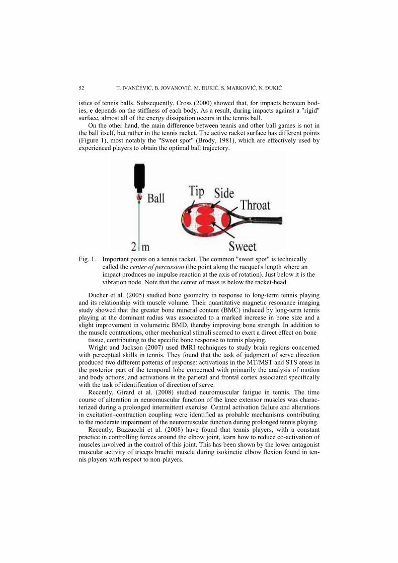
However, coaches and sports scientists should analyze the most efficient movements to be able to teach the model techniques. For example, Dr. Bruce Elliott from the Univer-sity of Western Australia, has extrapolated the contributions of the body segments to racket-head speed (see Figure 5) using 3D video-and computer analysis. "These contri-
58 T. IVANČEVIĆ, B. JOVANOVIĆ, M. ĐUKIĆ, S. MARKOVIĆ, N. ĐUKIĆ
butions vary from person to person," Elliott says, "but the data shows the clear impor-tance of the trunk, shoulder internal rotation and wrist flexion in the swing to impact."
Fig. 5. A snapshot of Andy Roddick's record serve (246 km/h), showing the contribution
(in percentages) of the involved body segments and partial body movements.
2.3. Main Degrees-of-Freedom in Human Joints
All human movements take place in synovial rotational joints (Figure 6). Some joints are only slightly movable, formed by two bones held together by cartilage, without joint cavity (e.g., an intervertebral joint in the spine consists of two vertebras and an intervertebral disc between them). On the other hand, major joints involved in human movement, like shoulder, hip, elbow and knee, are composed of several bones separated by a joint cavity, lubricated by synovial fluid and enclosed in a fibrous joint capsule (Marieb, 1998). Different joints have

Biomechanical Analysis of Shots and Ball Motion in Tennis and the Analogy with Handball Throws 59
different degrees-of-freedom (DOF) of movement: hinge joints have 1 DOF, gliding and saddle joints have 2 DOF, while ball-and-socket joints have 3 DOF.1
Fig. 6. Main Degrees-of-Freedom in Human Joints
2.4. The Basic Biomechanical Unit
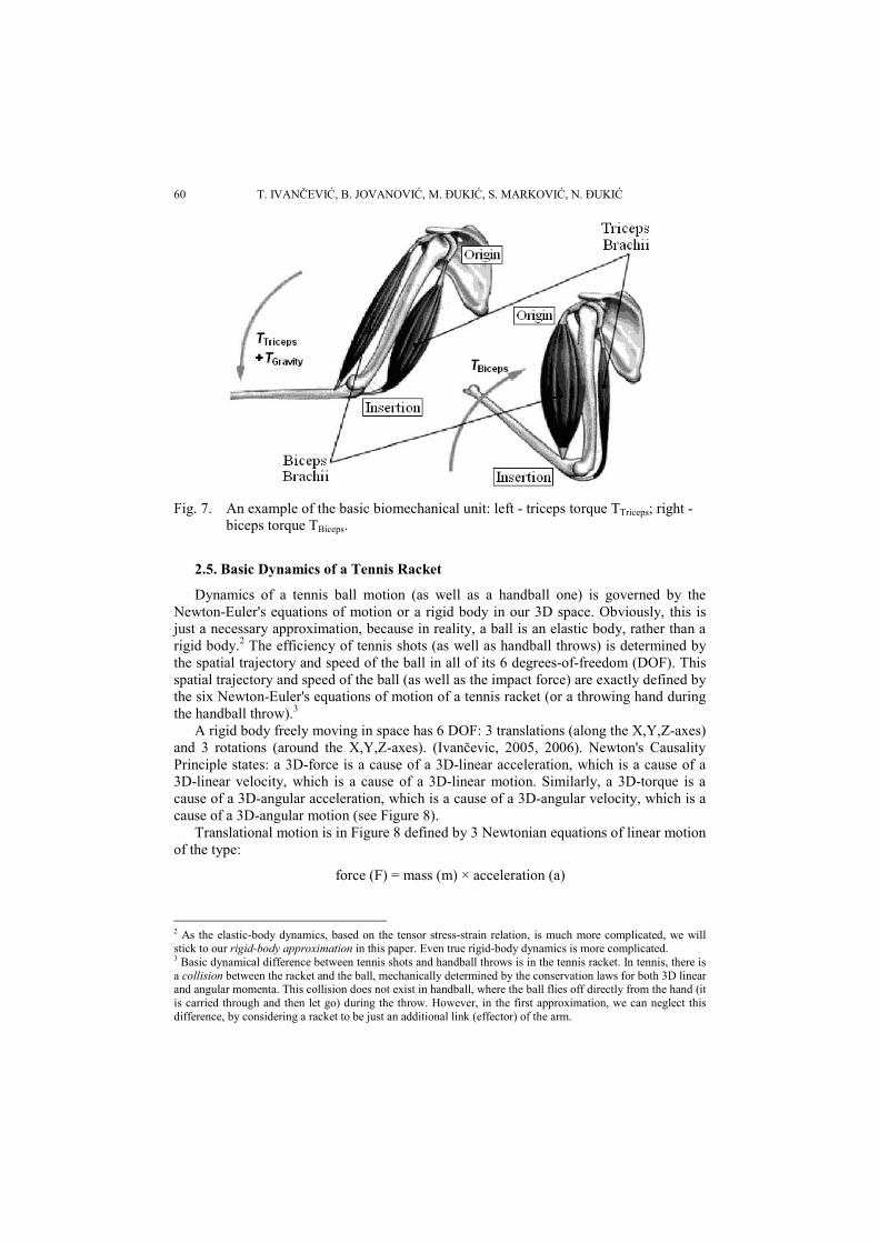
Every human movement is driven by synergistic action of the basic biomechanical units. The basic biomechanical unit consists of a pair of mutually antagonistic muscles producing a common muscular torque, TMus, in the same joint, around the same axes. The most obvious example is the biceps-triceps pair (see Figure 7). Note that in the normal vertical position, the triceps downward action is supported by gravity, that is the torque due to the weight of the forearm and the hand (with the possible load in it).
An overview of main muscular groups used in tennis is given in Appendix.
1 Note that this is a simplified picture that is more valid for robot joints than for human joints. In reality, e.g., shoulder has 6 DOF, as well as hip and knee (the knee-cap travels about 7 cm from maximal flexion to maxima extension). If we take into account micro-translations that always follow the corresponding rotational movements in synovial joints, then the number of DOF is human joints is much higher, so an arm with a tennis racket represents a highly-redundant kinetic system.
60 T. IVANČEVIĆ, B. JOVANOVIĆ, M. ĐUKIĆ, S. MARKOVIĆ, N. ĐUKIĆ
Fig. 7. An example of the basic biomechanical unit: left - triceps torque TTriceps; right -
biceps torque TBiceps.
2.5. Basic Dynamics of a Tennis Racket
Dynamics of a tennis ball motion (as well as a handball one) is governed by the Newton-Euler's equations of motion or a rigid body in our 3D space. Obviously, this is just a necessary approximation, because in reality, a ball is an elastic body, rather than a rigid body.2 The efficiency of tennis shots (as well as handball throws) is determined by the spatial trajectory and speed of the ball in all of its 6 degrees-of-freedom (DOF). This spatial trajectory and speed of the ball (as well as the impact force) are exactly defined by the six Newton-Euler's equations of motion of a tennis racket (or a throwing hand during the handball throw).3
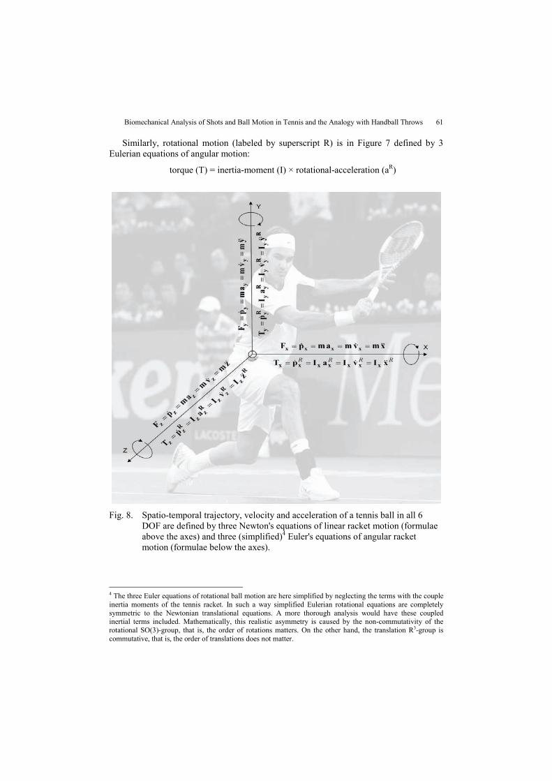
A rigid body freely moving in space has 6 DOF: 3 translations (along the X,Y,Z-axes) and 3 rotations (around the X,Y,Z-axes). (Ivančevic, 2005, 2006). Newton's Causality Principle states: a 3D-force is a cause of a 3D-linear acceleration, which is a cause of a 3D-linear velocity, which is a cause of a 3D-linear motion. Similarly, a 3D-torque is a cause of a 3D-angular acceleration, which is a cause of a 3D-angular velocity, which is a cause of a 3D-angular motion (see Figure 8).
Translational motion is in Figure 8 defined by 3 Newtonian equations of linear motion of the type:
force (F) = mass (m) × acceleration (a)
2 As the elastic-body dynamics, based on the tensor stress-strain relation, is much more complicated, we will stick to our rigid-body approximation in this paper. Even true rigid-body dynamics is more complicated. 3 Basic dynamical difference between tennis shots and handball throws is in the tennis racket. In tennis, there is a collision between the racket and the ball, mechanically determined by the conservation laws for both 3D linear and angular momenta. This collision does not exist in handball, where the ball flies off directly from the hand (it is carried through and then let go) during the throw. However, in the first approximation, we can neglect this difference, by considering a racket to be just an additional link (effector) of the arm.
Biomechanical Analysis of Shots and Ball Motion in Tennis and the Analogy with Handball Throws 61
Similarly, rotational motion (labeled by superscript R) is in Figure 7 defined by 3 Eulerian equations of angular motion:
torque (T) = inertia-moment (I) × rotational-acceleration (aR)
Fig. 8. Spatio-temporal trajectory, velocity and acceleration of a tennis ball in all 6
DOF are defined by three Newton's equations of linear racket motion (formulae above the axes) and three (simplified)4 Euler's equations of angular racket motion (formulae below the axes).
4 The three Euler equations of rotational ball motion are here simplified by neglecting the terms with the couple inertia moments of the tennis racket. In such a way simplified Eulerian rotational equations are completely symmetric to the Newtonian translational equations. A more thorough analysis would have these coupled inertial terms included. Mathematically, this realistic asymmetry is caused by the non-commutativity of the rotational SO(3)-group, that is, the order of rotations matters. On the other hand, the translation R3-group is commutative, that is, the order of translations does not matter.

62 T. IVANČEVIĆ, B. JOVANOVIĆ, M. ĐUKIĆ, S. MARKOVIĆ, N. ĐUKIĆ
The symbols in Figure 7 have the following specific meaning: The dot over a variable denotes the derivative (rate of change) with respect to time; m
is the racket mass; Fx, Fy, Fz – three components of the total translational force acting on the racket (in-
cluding muscular force, gravity and air resistance) – along the (X,Y,Z)-coordinate axes; ax, ay, az – three components of the total translational acceleration of the racket –
along the coordinate axes; px, py, pz – three components of the total translational momentum of the racket –
along the coordinate axes; vx, vy, vz – three components of the total translational velocity of the racket – along
the coordinate axes; Ix, Iy, Iz – three inertia moments of the racket – around the (X,Y,Z)-coordinate axes; Tx, Ty, Tz – three components of the total torque (moment) acting on the racket (in-
cluding muscular, gravity and air resistance torques) – around the (X,Y,Z)-coordinate axes;
aRx, aR
y, aRz – three components of the total rotational (angular) acceleration of the
racket – around the coordinate axes; pR
x, pRy, pR
z – three components of the total rotational (angular) momentum of the racket – around the coordinate axes;
vRx, vR
y, vRz – three components of the total rotational (angular) velocity of the racket
– around the coordinate axes. In biomechanics, the only active force is the muscular force that generates any human
motion by coordinated action (synergy) of a number of basic biomechanical units (Figure 6). On the other hand, from the perspective of muscular-training physiology, the most important quality is power, which incorporates both strength and speed, mechanically defined as:
power (P) = force (F) × velocity (v)
Physiologically, it corresponds to the area under the force-velocity curve (see Hill, 1938; Ivančevic, 2005, 2006). Muscular power is the key element of all power-sports, including tennis and handball.
The future research along the proposed lines will have to include the following com-ponents:
− Estimation/calculation of all included inertial, elasticity and biomechanical parameters;
− Computer simulation based on numerical solution of the above Newton-Euler equations of the tennis racket motion, for various values of parameters, initial conditions and inputs (muscular forces), with the objective to determine the 3D translational and rotational trajectories, velocities and accelerations of the tennis (respectively, handball) ball; and
− Embedding the micro-electro-mechanical (MEMS) inertial sensors (a three-axial accelerometer and a three-axial gyroscope) into the frame of a tennis racket – this technology is still in the development phase.

Biomechanical Analysis of Shots and Ball Motion in Tennis and the Analogy with Handball Throws 63
2.6. Vestibular system: 3D-sensor for the general Newton-Euler dynamics of human motion
The general human motion also obeys the general Newton-Euler dynamics. This gen-eral Newton-Euler dynamics of human motion is continuously being tracked by neuro-dynamical sensors residing within the human head. The vestibular organ in the inner ear helps maintaining the head equilibrium by sending the brain information about the head motion, both linear/translational and angular/rotational.
The vestibular organs consist of three membranous semi-circular canals (SCC), and two large sacs, the utricle and saccule. All the vestibular organs share a common type of receptor cell, the hair cell. The SCC within the vestibular organ of each ear contains fluid and hair receptor cells encased inside a fragile membrane called the cupula. The cupula is located in a widened area of each canal called the ampulla. When you move your head, the fluid in the ampulla lags behind, pushing the cupula a very tiny bit that causes the hairs to also bend a very tiny bit. The bending hairs stimulate the hair cells, which in turn trigger sensory impulses in the vestibular nerve going to the brain to "report" the move-ment. Hair cells are amazingly sensitive. For example, a cupula movement of even a thousandth of an inch is detected by the brain as a big stimulus (see Molavi, 1997; Marieb,1998; Enoka, 2001; Ivancevic, 2006).
The SCC are positioned roughly at right angles to one another in the three planes of space. Thus, the canals react separately and in combination to detect different types of angular head movement. They detect when we nod in an up and down motion (pitch), when we tilt our head to the side towards our shoulder (roll), and when we shake our head ``no" in a side-to-side motion (yaw). The SCC are responsible for detecting any kind of rotational motion in the head, thus effectively sensing the Eulerian dynamics.
Two other vestibular organs are located in membranous sacs called the utricle and the saccule. On the inside walls of both the utricle and the saccule is a bed (a macula) of sev-eral thousand hair cells covered by small flat piles of calcium carbonate crystals which look like sand, imbedded in a gel-like substance. The crystals are called otoliths, a word which literally means "ear stones". In fact, the utricle and the saccule are often called the otolith organs.
When a person's head is in the normal erect position, the hair cells in the utricle lie approximately in a horizontal plane. When the head is tilted to one side, the stones want to slide "downhill". This moves the gel just enough to bend the sensory hairs. The bend-ing hairs stimulate the hair cells, which in turn send a signal to the brain about the amount of head tilt. The stones also move if the person is accelerated forward and back, or side-to-side. Similarly, the hair cells in the saccule are oriented in somewhat of a vertical po-sition when the head is erect. When a person tilts their head, or is accelerated up and down (as in an elevator), or moved forward and back, the otoliths move and a signal is sent to the brain. The signals from the otoliths in the saccule and the utricle complement each other and give us an integrated signal about our movement. The otolith organs are primarily responsible for detecting any degree of linear motion of the head, thus effec-tively sensing the Newtonian dynamics.

64 T. IVANČEVIĆ, B. JOVANOVIĆ, M. ĐUKIĆ, S. MARKOVIĆ, N. ĐUKIĆ
3. CONCLUSION
In this paper the basic biomechanics of tennis shots has been presented together with their analogy with handball throws. The main difference between these two ball games is in the tennis racket (with its own inertial and elastic characteristics). The importance of the present study is in presenting, for the first time in Serbian tennis literature, the basic tennis biomechanics and its analogy with the handball throws.
REFERENCES 1. Bazzucchi, I., Riccio, M.E., & Felici, F. (2008). Tennis players show a lower coactivation of the elbow
antagonist muscles during isokinetic exercises. (in press) 2. Bergeron, M., Maresh, C., & Kraemer, W. (1991). Tennis - A physiological profile during match play.
Int. J. Sports Med. 12, 474-479, 3. Brody, H. (1981). Physics of the tennis racket II: The "Sweet spot". Am J Physics, 49, 816-819. 4. Brody, H. (1979). Physics of the tennis racket. Am J Physics, 47, 482-487. 5. Cross, R. (1999). Dynamic properties of tennis balls. Sports Eng, 2, 23-33. 6. Cross, R. (1999). The bounce of a ball. Am J Physics, 67, 222-227. 7. Cross, R. (2000). The coefficient of restitution for collisions of happy balls, unhappy balls, and tennis
balls. Am J Physics, 68, 1025-1031. 8. Ducher, G., Courteix, D., Meme, S., Magni, C., Viala, J.F., & Benhamou, C.L. (2005). Bone geometry in
response to long-term tennis playing and its relationship with muscle volume: A quantitative magnetic resonance imaging study in tennis players. Bone, 37 457-466.
9. Elliott, B., & Marsh, T. (1989a). Overheu, P., A biomechanical comparison of the multisegment and sin-gle unit topspin forehand drives in tennis. Int J Sport Biomech, 5, 350-364.
10. Elliott, B., Marsh, T., & Overheu, P. (1989b). The topspin backhand drive in tennis. J Hum Mov Studies, 16, 1-16.
11. Elliott, B., Takahashi, K, Noffal, G. (1997). The influence of grip position on upper limb contributions to racket head velocity in a tennis forehand. J Appl Biomech, 13, 182-196.
12. Elliott, B.C., Marshall, R.N., & Noffal, G. (1995). Contributions of upper limb segment rotations during the power serve in tennis. J Appl Biomech, 11, 433-442.
13. Enoka, R.M. (2001). Neuromechanics of Human Movement. Champaign (3rd ed): Human Kinetics. 14. Girard, O., Lattier, G., Maffiuletti, N.A., Micallef, J.-P., & Millet, G.P. (2008). Neuromuscular fatigue
during a prolonged intermittent exercise: Application to tennis. J Electromyogr Kinesiol, (in press). 15. Houk, J.C. (1967). Feedback control of skeletal muscles. Brain Res, 5, 433-451. 16. Houk, J.C. (1979). Regulation of stiffness by skeletomotor reflexes. Ann Rev Physiol 41, 99-114. 17. International Tennis Federation (ITF) (2003). The rules of tennis 2003. London: ITF, 2. 18. Ivancevic, V., & Ivancevic, T. (2005). Human-Like Biomechanics: A Unified Mathematical Approach to
Human Biomechanics and Humanoid Robotics, Springer, Berlin. 19. Ivancevic, V., & Ivancevic, T. (2006). Natural Biodynamics, World Scientific, Singapore. 20. Marieb, E.N. (1998). Human Anatomy and Physiology (4th ed.) Benjamin/Cummings, Menlo Park, CA. 21. Molavi, D.W. (1997). Neuroscience Tutorial. School of Medicine, Washington Univ. 22. Reid, M., & Schneiker, K. (2008). Strength and conditioning in tennis: Current research and practice. J
Sci Med Sport (in press). 23. Stepanek, A. (1988). The aerodynamics of tennis balls - The topspin lob. Am J Physics, 56, 138-142. 24. Wright, M.W., & Jackson, R.C. (2007). Brain regions concerned with perceptual skills in tennis: An
fMRI study. Int J Psychophysiology, 63, 214-220.

Biomechanical Analysis of Shots and Ball Motion in Tennis and the Analogy with Handball Throws 65 5.
App
endi
x –
Min
dMap
of t
he b
asic
mus
cle
grou
ps
66 T. IVANČEVIĆ, B. JOVANOVIĆ, M. ĐUKIĆ, S. MARKOVIĆ, N. ĐUKIĆ
BIOMEHANIČKA ANALIZA UDARACA I KRETANJA LOPTE U TENISU I NJIHOVA ANALOGIJA SA IZBAČAJIMA U
RUKOMETU
Tijana Ivančević, Bojan Jovanović, Milorad Đukić, Saša Marković, Natalia Đukić
Cilj ovog rada je prikaz savremene teniske nauke široj naučnoj javnosti. Prezentirana je biomehanička analiza udaraca u tenisu i odgovarajuća dinamika reketa i lopte, zajedno sa analog-nim izbačajima u rukometu. Osnovna razlika između ove dve sportske igre je u teniskom reketu (kao dodatnom segmentu tela sa njegovim inercijalnim i elastičnim karakteristikama). Ovaj rad prezentuje anatomsku, fiziološku i biomehaničku analizu teniskog servisa, forhenda i bekhenda, kao i 3D Njutn-Ojlerovu dinamičku analizu kretanja tensikog reketa u toku ovih udaraca. U buduć-nosti, ovakva analiza ce morati da bude podržana numeričkim simulacijama, kao eksperimentalnim merenjem baziranim na inercijalnim senzorima (akcelerometrima i žiroskopima umetnutim u okvir teniskog reketa) za merenje brzina i ubrzanja i pokreta igrača u toku servisa, forhenda i bekhenda. Ključne reči: udarci u tenisu, izbačaji u rukometu, biomehanička analiza, Njutn-Ojlerova
dinamika, vestibularni i veštački senzori