Embed Size (px)
Citation preview

BIOMATERIALSENT 311/4
Lecture 11 Orthopaedic Implant: Internal
FixationPrepared by: Nur Farahiyah Binti Mohammad
Date: 15th September 2008
Email : [email protected]

2
Teaching Plan
COURSE CONTENT
Define various types of internal fixation. Identify failure modes of internal fixation.Describe and compare three types of fixation methodsDescribe and discuss types of joint replacements.Describe and recommend biomaterial use to make components of joint replacement.
DELIVERYMODE
LectureSupplement reading
LEVEL OF COMPLEXITY
KnowledgeRepetitionAnalysis Evaluation
COURSE OUTCOMECOVERED
Ability to select biomaterials that can be used for different medical applications and explain the criteria that will lead to a successful implants

3
Purpose: To stabilize fractured bone until natural healing processes have restored sufficient strength so that implant can be removed.
Material requirement: Biocompatible Sufficient strength Corrosion resistance
1.0 Internal Fixation

4
1.0 Internal Fixation
Material used: Stainless steel Co-Cr alloys Titanium alloys Biodegradable polymer
To treat minimally loaded fractures Eliminate the need for second surgery

5
1.0 Internal Fixation
Types:1. Wires2. Pins3. Screw4. Plates5. Intramedullary nails

6
1.0 Internal Fixation
1. WIRES Used to reattach large fragments of bone Useful especially for spiral breaks and
reattaching greater trochanter of hip Wires suffer from twisting and knotting
The deformed region of the wire are more prone to corrosion

7
1.0 Internal Fixation
2. PINS Used to hold fragments of bone together
temporarily or permanently and to guide large screw insertion.
Have different tip design Trocar tip – most efficient in cutting
- often used for cortical bone Diamond tip

8
1.0 Internal Fixation
3. SCREWS Two types of bone screws:
1. Cortical (Compact) bone screw-small treads
2. Cancellous screws –large tread to get more thread-to bone contact

9
1.0 Internal Fixation
It can be used alone or along with other devices.
The general principle is that bone heals better if the fracture fragments are aligned and pressed closely together.
The idea is to stabilize the fracture and keep the bone in anatomic alignment.

10
1.0 Internal Fixation

11
1.0 Internal Fixation
Principle application of bone screw: As interfragmentary fixation devices to lag
or fasten bone fragment together. To attach a metallic plate to bone

12
1.0 Internal Fixation
4. PLATES Plates come in several types, and are
named for their function.
In general, there are: Dynamic Compression plates Neutralization plates Buttress (support) plates.

13
1.0 Internal Fixation
The dynamic compression plate is one of the most common types of plates, and can be recognized by its special oval screw holes. These holes have a special beveled floor
to them with an inclined surface. If desired, this inclined surface can be
used to pull the ends of the bone together as the screws are tightened.

14
1.0 Internal Fixation
Compression plates are used for fractures that are stable in compression. They may be used in combination with lag
screws, and they may provide dynamic compression when used on the tension side of bone.

15
1.0 Internal Fixation
Neutralization plates are designed to protect fracture surfaces from normal bending, rotation and axial loading forces. They are often used in combination with
lag screws.

16
1.0 Internal Fixation
Resorbable bone plate
As the strength of the fracture site increase due to normal healing processes, the resorption of the implant begins to take place.

17
1.0 Internal Fixation
Buttress plates are used to support bone that is unstable in compression or axial loading. These plates are often used in the distal
radius and tibial plateau to hold impacted and depressed fragments in position once they have been elevated.

18
Buttress plate bridging a humeral neck fracture -

20
1.0 Internal Fixation
5. INTRAMEDULLARY NAILS

21
1.0 Internal Fixation
Used as internal struts to stabilize long bone fracture.
Inserted into medullary cavity Should have some spring to provide
elastic force and prevent rotation. IM nails are better than plates at
resisting multi-directional bending However, torsional resistance is less
than plate

22
1.0 Internal Fixation
When designing IM nails: A high polar moment of inertia is desirable
to improve torsional rigidity and strength Problems:
Destroy intramedullary blood supply.

23
1.0 Internal Fixation
Biomaterial applications in Internal FixationMaterial Properties Applications
Stainless steel Low cost, easy fabrication Surgical wire, Pin, plate, screw, IM nails
Titanium alloy High costLow density and modulusExcellent bony contact
Surgical wirePlate, screw, IM nails
Co-Cr alloys High costHigh density and modulusDifficult fabrication
Surgical wireIM nails
Polylactic acid Resorbable Pin, screw

24
Failure modes of internal fixation devices
Failure mode Failure location Reason for Failure
Overload Bone fracture siteImplant screw holeScrew thread
Small size implantUnstable reduction
Fatigue Bone fracture siteImplant screw holeScrew thread
Small size implantUnstable reductionFracture
Corrosion Screw head-plate holeBent area
Different alloy implantOver tightening screwMisalignment of screwOver bent
Loosening Screw MotionWrong choice of screw type

BIOMATERIALSENT 311/4
Lecture 11 (cont)Orthopaedic Implant: Joint
replacementPrepared by: Nur Farahiyah Binti Mohammad
Date: 18th September 2008
Email : [email protected]

26
1.0 Introduction
Total joint replacement An arthritic or damaged joint is removed
and replaced with an artificial joint called prosthesis.
Goal - to relieve the pain in the joint caused by the damage done to the cartilage.

27
1.0 Introduction

28
Joint degradation
Also called Osteoarthritis. Is the end stage of a process of destruction
of the articular cartilage. Results in:
Severe pain Loss of motion An angular deformity of the extremity
Cartilage unable to regenerate So, when exposed to a severe mechanical,
chemical or metabolic injury, the damage is permanent.

29
Joint degradation

30
Types of Total Joint Replacement (TJR)
Joint Type
Hip Ball and socket
Knee Hinged, semiconstrained, surface replacement, unicompartment or bicompartment
Shoulder Ball and socket
Ankle Surface replacement
Elbow Hinged, semiconstrained, surface replacement
Wrist Ball and socket, space filler
Finger Hinged, space filler

31
Implant fixation method
Three type of methods of fixation:1. Mechanical interlock
This fixation achieved by press-fitting the implant by using PMMA bone cement
Bone cement Bone cement is a substance commonly used to hold
implants in bone and filling the space between the skeleton and the total joint device.
Often cement is used for hip replacement and knee replacement surgery.
Cement implies that the material sticks the implant into the bone

32
Implant fixation method
In reality, bone cement should really be called bone grout.
The reason is that this material actually acts as a space-filler, to create a tight space for the implants to be held against the bone.
Bone cement does not stick substances together, rather it fills the void between the implant and the surrounding bone.

33
Implant fixation method
On the X-ray pictures one sees the bone cement as a white layer around the shadows of the total hip device.

34
Implant fixation method
The microscopic structure of bone cement is made by two substances glued together.
One substance are the small particles of pre-polymerized PMMA (PolyMethylMetaAcrylate), so called "pearls.
These pearls are supplied as a white powder. The other substance is a liquid monomer of
MMA(MethylMetacrylate). Both substances are mixed together at the operation table with added catalyst that starts the polymerization of the monomer fluid.

35
Honeycomb structure gives the bone cement the ability to absorb downward (compression) loads.
An important characteristics in an otherwise brittle material.
the bone cement act mechanically as an shock absorber
(1) The unloaded phase: the bone cement net is regular, not deformed.
( 2) Load applied by body weight impact on the total hip (its femoral component): the bone cement net within marrow cavity deforms elastically, but does not brake and stays in contact with both total hip and skeleton.
( 3 ) When the load ceases the bone cement's structure returns to its original regular form.

36
Implant fixation method
2. Biological fixation Which is achieved by using textured or
porous surface, that allow bone to grow into the interstices.
Porous in growth fixation Pore size range should be 100 to 350μm Pores should be interconnected with each other
with similar size of opening.

37
Implant fixation method
3. Direct chemical bonding between implant and bone for example by coating the implant with
calcium hydroxyapatite, which has mineral composition similar to bone.
Material used as coating such as Bioglass and glass ceramic.

38
Biomaterials for TJRMaterial Application Properties
Co-Cr alloy Stem, head (ball), Cup, porous coating, metal backing
Heavy, hard, stiffHigh wear resistance
Titanium alloy Stem, porous coatingMetal backing
Low stiffnessLow wear resistanceLight
Pure titanium Porous coating Excellent osseointegration
Tantalum Porous structure Excellent osseointegrationGood mechanical strength
Alumina Ball, cup Hard, Brittle, High wear resistance
Zirconia Ball Heavy and high toughnessHigh wear resistance
UHMWPE Cup Low friction, wear debris, low creep resistance
PMMA Bone cement fixation Brittle, weak in tensionLow fatigue strength

39
Comparison between Specific Orthopedic Implant Prosthetic Materials

40
Comparison between Specific Orthopedic Implant Prosthetic Materials

41
Total hip replacement
Hip joint is a ball and socket joint Can be divided into 2 types:
1. Monolithic: Consist of one part, less expensive, less prone to corrosion
2. Modular: Consist of 2 or more parts , allow customizing of the implant during future revision surgery.

42
Total hip replacement
Design aspect Prosthetic hip component are optimized to
provide wide range of motion to prevent dislocation.
Must enable implants to support loads Proper femoral neck length will decrease
bending stress

43
Total hip replacement
If femoral stem is designed with sharp corner: bone in contact with sharp corner may necrose and resorb.
Replaceability: possible to remove one part without removing the other.

44
Hip joint replacement
Component or Total Hip joint:
1. Femoral component Stem –Neck Ball/head
2. Acetabular component Cup
Backing Insert
Femoral component
Acetabular component
stem
neck
45
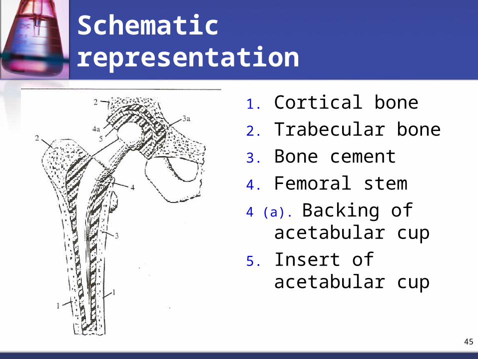
Schematic representation
1. Cortical bone2. Trabecular bone3. Bone cement4. Femoral stem4 (a). Backing of
acetabular cup5. Insert of acetabular
cup

46
Materials used for each component of THR
Femoral component Acetabular componentStem Ball/head Cup BackingCo-Cr alloy
Titanium alloy
Co-Cr alloy
Alumina
Zirconia
Co-Cr alloy
UHMWPE
Alumina
Metal

47
Possible combination of THR
Femoral component Acetabular component
Fixation Stem Head Cup Backing Fixation
PMMA
Bone ingrowth
Press fitting
Co-Cr alloy
Titanium alloy
Co-Cr alloy
Alumina
Zirconia
Co-Cr alloy
UHMWPE
Alumina
Metal
None
PMMA
Screw or press fitting
Bone ingrowth

48
Total hip replacement
1. STEM Titanium alloy Advantages:
Excellent corrosion resistance Highly reactive material Lowest rate dissolution
Disadvantages: Wear Generation of fine wear particles:
inflammation and implant loosening

49
Total hip replacement
2. HEAD Alumina: elicit minimal response from
host tissue Advantages:
High Wear resistance Reasonable fracture toughness Extremely stable (undergo little
physical/chemical deformation)

50
Total hip replacement
Disadvantages: Degradation in-vivo:WEAR
Weakens the material Change shape that may effect fuction Produce biology active particles Low creep resistance: can influence the
behaviour of joint

51
Total hip replacement
3. CUP UHMWPE Advantages:
Tough inert Disadvantages:
Wear debris cause inflammatory reaction

52
Most frequent problems
1. Infection2. Wear3. Migration and failure of implants4. Loosening

53
WEAR
Degradation in vivo Wear debris produced by load bearing
and motion of the prosthesis 150 000 particles generated each step Cells from the immune system of the
host, such as macrophages, will identify the wear particles as foreign matter and initiate a complex inflammatory response

54
WEAR
Problem that will be occur as a result of wear are: Rapid focal bone lose (osteolysis) Bone resorption Loosening Fracture of bone

55
Shoulder Joint Replacement
Humerus fractures -caused by a direct blow or by a fall or osteoporosis (thinning of the bones).
the ball is removed from the top of the humerus and replaced with a cobalt chrome or titanium implant. This is shaped like a half-moon and attached to a stem inserted down the center of the arm bone.
The socket portion of the joint is shaved clean and replaced with a plastic socket that is cemented into the scapula.

56
Elbow Joint replacement
Humeral component replaces the lower end of the humerus in the
upper arm. has a long stem that anchors it into the
hollow center of the humerus.
Ulnar component replaces the upper end of the ulna in the
lower arm. has a shorter metal stem that anchors it into
the hollow center of the ulna.
The hinge between the two components is made of metal and plastic.
The plastic part of the hinge is tough and slick. allows the two pieces of the new joint to glide
easily against each other as you move your elbow.

57
Knee Joint Replacement
Femoral component upper part of a knee system made of a strong polished metal. covers the end of the femur
Patellar component replaces the kneecap in the center
of the knee.
Tibial component covers the top end of tibia covered with a metal tray topped with a disk-shaped
polyethylene insert (sits on a highly polished surface and rotates around a conical post)

58
Knee Joint Replacement

59
Ankle Joint Replacement
Tibial component replaces the socket portion of
the ankle (the top section) metal tray is attached directly
to the tibia bone plastic cup fits onto the metal
piece, forming a socket for the artificial ankle joint.
Talus component replaces the top of the talus. made of Titanium fits into the socket of the
tibial component