Embed Size (px)
Citation preview

Biology of .Bats
Volume I
Edited by William A. Wimsatt Division of Biological Sciences Cornell Ullicersity Ithaca, New York
1970
Academic Press New York and L()ndon

Chapter 4
The Muscular System Terry A. Vaughan
L Introduction 140 [I. Muscles UnicpH::' to Bats 141
IlL Muscles of the Pectoral Cirdle and Limb 145 A. Trapezius Crollp 14,5 B. Costo-spino-scapular G1'0l1p 148 C. Latissimus-Subscapularis Grollp 1.53 D. Deltoid Group 155 E. Suprascapular Group 1.57 F. Triceps Group 158 G. Extensor Group of Forearm 159 H. Pectoralis Group 164 1. Flexor Group of Arm 166 J. Flexor Group of Forearm 167
K. Extensor Croup of ~'fanus 170 L. Flexor Group of I'vfaIlI is 170
IV. Muscles Attaching to the Pelvic Girdle and Limb 173 A. Iliacus Group 177 B. Gluteal Group 178 C. Quadriceps Femoris Croup 179 D. Tibial Extensor Group 179 E. Peroneal Group 180 F. Adductor Group 181 G. Ischiotrochanteric Group 183 H. I Iarnstring Grollp 183 1. Flexor Group of Leg 185
J. Flexor Group of Foot 18(, V. Chiropteran Muscle Specializations for Flight 188
A. Weights of Major Flight Muscles m Bats 188 B. Reduction of the Weight of the Wing 190 C. Muscular Control of the "'ling-beat Cycle 192 Heferences 193
139

140 TEHHY /\_ VAUCIfA);
I. Introduction
The HlHscular systcm of bats cliffers markecllv from that of terrestrial lllalnll1ak Not onlv arc difIerenccs in relative sizes of lllllscles involved . _ , but origins, insertions, and functions of homologous muscles frequcntlv differ between these groups of mammals_ Each group uses a different type of locomotion, and with each type is associated a dominance of certain types of muscles_ In terrestrial mammals the limbs are held beneath the body, more or less vertical to the substrate, and locomotion is due primarily to anteroposterior movements of the limbs_ The main propulsion stroke of the limbs is toward the rear of the animals; consequently, flexors and extensors of the limbs are the most important muscles_ The situation is entirelv different in bats, in which the limbs are , held more or less out to the sides of the body_ The main movements associated with aerial locomotion in bats involve adduction (the downstroke) and abduction (the upstroke) of the forelimbs_ The propulsion stroke is the downstroke of the wings, and the largest muscles arc those causing this action_ The flight membranes are controlled by both the forelimbs and hiwj limbs, and the appendages arc also used for some sort of quadrupedal locomotion ill most bats_ Although the appendages of bats are primarily adapted for flight, the evolution of the limbs has apparently been within the limits imposed by their importance in both aerial and terrestrial locomotion_
The following descriptions cover muscles that attaeh to the pectoral and pelvic girdles and are therefore primarily important in locomotion. TIlese muscles arc of particular interest becallSe of their specializations associated with flight and with types of terrestrial locolnotion unique to bats_ The descriptions of muscles serve as a background for later considerations of the muscular control of flight and of the major muscular specializations for flight
The descriptions are based on the following four bats of the suborder Microchiroptera: Eumops perotis (Molossidae), M yotis veliter (Vespertilionidac), M acrotus waterhousii (Phyllostomatidae), and Hipposideros armiger (Rhinolophidae) _ Eumops perotis is a North American insectivorous bat with long, narrow wings and unusually fast flight It roosts in crevices in rocks and in manmade structures, and can run rapidly and easily_ Mljotis veliter, another North American species, eats insects and has moderatelv slow, maneuverable flight It roosts in caves, crevices,
~ , and buildings, and can crawl easily_ Macrotus waterhousii occurs in the southwestern United States, Mexico, and the West Indies_ This species eats insects and some fruit and its flight is highly maneuverable_

4. TilE MUSCULAH SYSTEM 141
It roosts by hanging pendant from tlw ceilings of caves, mines, or buildings, and is unable to crawl. llipJlosideros armiger lives in southern Asia and is insectivorous. Its flight is slow and maneuverable and it roosts by hanging ill caves, hollow trees, and buildings. Ilipposideros can crawl slowly. These four bats arc used in the following descriptions to illustrate somc of the morphological diversity that occurs bctween eli llerent taxa of bats.
Discussions of functions of muscles arc bascd on careful considcrations of the anatomical relationships involved. In my judgement this method leads to an accurate general understanding of functional relationships. Only careful expcrimental work, however, can clarify complex divisions of labor between muscles (Basmajian, 1962).
Descriptive terms arc applied as they are to terrestrial mammals; that is to say, the limbs are assumed to extend ventrad, beneath the animals. It should be remembered, however, that this is not the normal posture for the limbs in bats. Indeed, I know of no bat that can bring its hind limbs into this position.
Descriptions of muscles arc hased on El1mops. Additional descriptions arc given for those muscles in the other genera that differ from the corresponding muscles in El1mops.
H. Muscles Unique to Bats (Figs. 1-3)
J\,f. OCCIPITO-POLLlCALlS
Origin. From lambodoidal crest adjacent to midline. I nsertion. On distal half of anterior surface of second metacarpal. Remarks. This muscle is fleshy from the lambdoidal crest to the an-
terior surface of the shoulder. Between the shoulder and the carpus the muscle is represented first by clastic fibers and then by a tendon. In El1mops this muscle is bound to the shoulder by a tendon from a slip from the distal part of the M. c1avodeltoideus and by connective tissue anchored to the adjacent part of the M. pectoralis. In Hipposideros the attachment is by two tendons from slips of the M. c1avodcltoideus and one tendon from a slip of the M. pectoralis. In eaeh of the other two genera a short tendon anchors the muscle. The inscrtion in II ip)losieleros is on the medial base of the thumb.
Action. This muscle pulls the propatagium craniad and ventrad, thereby increasing the area of the proximal segment of the wing and improving its airfoil.
142 TEHHY ii, V,\UCIIAN
serratus antenor -
peet. obdom.--"
,1 i) i 1
1)11
I ~!/ Ii j ,I I If ~ 1 r " occipito'pollicalis --~ r pectoralis
coraco·cutaneus
7 -~ tensor plagiopatagii
- - calcar
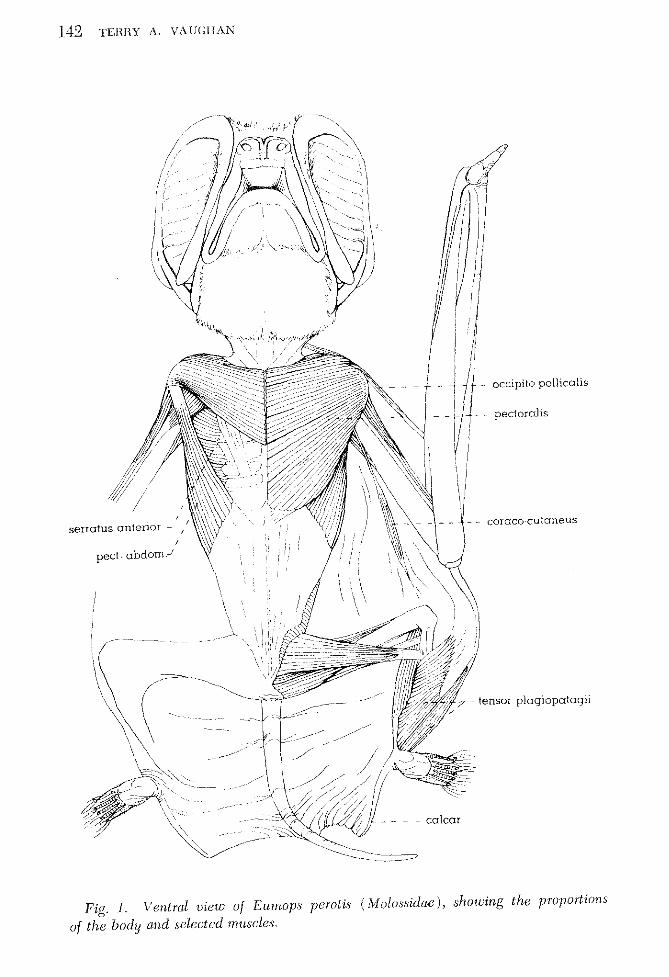
Fig,/. Ventral view of Eumops perotis (iy[o[ossidae), showing the proportions
of the body and selected muscles,

\. I. ~/ , ,
, I
4. THE IvruSCULAH SYSTE~l 143
~_clavotrapezius
toccipi to-pollicalis
I-acromiotrapczius
J rhomboideus
-spinod eltoideus
teres major
'- - serratus anterior
-latissimus dorsi
- -spmotrapezius
- - - -gluteus maximus
- -quadriceps femoris
- - -semitendinosus
Fig. 2. Dorsal view of llipposidcros arrniger (Rhinolophidae), showing selected muscles. Some of the superficial muscles have heen removed from the left side of the shoulder area.
M. COHACO-CUTANEUS
Origin. From distal part of medial ridge of humerus. I nsertion. Into axillary part of plagiopatagium. Remarks. This muscle is relatively largest in Eumops. In Myotis the
origin is on the tip of the coracoid process, ill 1I1acrotus, on the dorsal surface of the coracoid head of thc biceps, and in lIipposideros, on fascia in the axilla.
Action. This muscle attaches distally to networks of c:Iastic fibers and

144 TEHHY A. VAm;lIACi
hy anchoring these to the axilla braces and helps maintain the tautness of the proximal part of the plagiopablgiurn during Hight.
M. IlUlvfEHOl'ATA(;IALlS
Origin. From fascia on medial surface and medial epicondyle of llll
merus ,mel horn posteromedial surface of base of ulna. Remarks. The distal part of this muscle gives way to a diverging
series of elastic fibers that merge with the network of fibers in the distal part of the plagiopatagium. In the bats under discussion, this muscle
___ proximal segment --
coraco-cutaneus , - propatagium I r _ occipito-pollkalis ,
humeropatagialis
+- ~ - - - -distal segment - - - - - - ~ .. - - - -- .,
-tensor plagiopataqii ~ __ plagiopataglUID
Fig. 3. Ventral lAew of the left wing of Eutnops perotis (Alulossidae), shotving the muscles and part at the s,!stem at elastic fibers that brace the uAllg membranes.
is present only in Ewnops. In Macrotus and Hipposideros bundles of clastic fibers take the place of this muscle.
Action. This muscle tightens and braces the distal part of the
plagiopatagium.
M. DEPRESSOH OSSlS STYLIFOHMIS
Origin. From dorsolateral surfaces of calcaneus and adjacent base
of fifth metatarsal. Insertion. Along proximal two-thirds of anterior surface of calcar. Remarks. This muscle is similar in the four genera but is relatively
largest in Eumops. Action. This muscle helps sprcad the uropatagium by swinging the
calcar laterad, away from the shank.

4, TIlE :\1USCULAH SYST/<:i\[ 145
M, TENSOH PLACIOPATAGH
Origin, In two parts: the first, from distal three-quarters of medial and anterior surfaces of tibia amI from ventral surface of tarsus; the second, from medial surface of internal cuneiform and hilSC of first metatarsaL
Insertion, First part: into part of the plagiopatagium adjacent to anterior surface of slmr1k, Second part: along trailing edge of plagiopatagium, giving way distally to clastic fibers,
Remarks, Among the bats under consideration, this muscle occurs only in EU1110PS, but is present also in all other molossid bats,
Action, This muscle anchors and braces the trailing edge of the plagiopatagium anel the part of this membrauc that attaches to the shank
III. Muscles of the Pectoral Girdle and Limb (Figs. I-to)
A. 'Trapezius Group
M, ACHOMIOTIlAPEZIUS AND CLAVOTHAPEZlUS
Origin, On middorsal line from last cervical to fifth thoracic vertebra, I nsertion, On medial surfaces of distal end of clavicle and entire
acromion process, and on rim of scapular spine, Remarks, In Macrotus the insertion is on the distal third of the clavicle
and the acromion process. The two parts of this muscle arc separate in HipT!Osideros, The 1\1, c1avotrapezius originates by a broad tendon on the neural ridge on the last cervical vertebra and inserts on the distal part of the clavie!c. The origin of the M, aeromiotrapezius is on the neural ridge on the fused last cervical and first two thoracic vertebrae, TI1C insertion is on the acromion process,
Action, This muscle pulls the clavicle and scapula mediad and tips the vertebral border of the scapula ventrad. The large size of this muscle and its partial insertion on the acromion process suggest that it helps in the upstroke of the wing by bracing the scapula against the pull of the deltoideus, supraspinatus, and infraspinatus muscles.
M. SPINOTRAPEZIUS
Origin, Along middorsal line from thoracic vertebrae eight to thirteen, I nsertion, On middle third of vertebral border of scapula. Remarks. In Myotis the origin is from thoracic vertebrae seven to

146 TEnny A. VAUGIlA~
/' ~ _ - - - - - aCTomlOtrapezlUs
~~~07=·!€~ ocromlOdeltoideus
- - - ~ - - spinodeltoideus
- spinotrapezius
- - teres major
/ _ - - - triceps longus
'\~, I __ -_ - - - rhomboideus \\ - - - - - - triceps lateralis
- - - - - - brachiahs
\ ______ serratus anterior " _ ~ __ - biceps brachii
, ______ latissimus dorsi
A
- - omocervicohs
- pectoralis
clavodeltOldeus -- - subclavius
- - - - - serratus anterior ____ - - supraspinatus
_ - - -' - - teres minor ___ - - -levator scapulae
- infraspinatus
-, - subscapularis
_ - - - teres major
__ - - - - triceps lateraEs
- - triceps longus
- - - _. - serratus anterior
B
Fig. 4. (A) Dorsal view of the shollidcr region of Eltmops perotis (Molossidac). ( B) Dorsal view of the shoulder region with some superficial rnuscles removed.

I Ilatissimus dorsi J I' L triceps longus
I
L - triceps lateral is
triceps medialis
4. THE "lUSCULAH SYSTE:\l 147
___ - pectoral ridge
- biceps brachii (glenoid head)
biceps brachii (coracoid head)
- subclavius
- serratus anterior
- -subscapularis
- - serratus anterior
A
- - -biceps brachii (glenoid head)
- - - - lesser tuberosity
coracoid process
- - flange of scapula
-. --subscapularis
B
~00~GI~ ~ ~ ~ -serratus anterior I'
Fig. $. (A) Ventral view of the shoulder region of E1Imaps perotis (M alossidae). (ll) Ventral view of the proximal part of the forelimb.
i I

148 TEnHY A. VAUGHAN
tcn, ill Macl'Otus, from thoracics tcn to thirteen, and in Hipposicleros, from thoracic tcn to the second lurnbar vertebra. The insertion is similar in the four genera, but is relatively least extensive in II ipposideros. in which it extcnds along the middle fifth of the vertebral border of thc scapula.
Action. This muscle pulls the scapula caudad and mediad and tips the vertebral border ventrad. Its function in the wing-beat eycle is probably similar to that of the M. acromiotrapezius.
" occipito-pollicalis
;- - propatagium
,- -forearm r ." plagiopatagium
B
A 1----C - - --- - - - - - - - - - - - - ~ -=-~ -~ -----------E-------
Fig. 6. Semidiagrammatic cross section through the proximal segment of the wing of Myotis Gvoiis (Vespeltilionidae). A is the plane of motion. B is the chord line of the airfoil. C is the angle of attack.
B. Costo-spino-sca,!ular Group (Figs. 4 and 5)
M. LEVATOR SCAPULAE
Origin. From eervical vertebrae four to seven, by four large slips. I nsertion. On vertebral border of scapula from the posterior end of
the £lange to junction of spine and vertebral border. Remarks. The attachments of this muscle are similar in all four genera. Action. This muscle draws the antcromedial border of the scapula
ventromcdiad and slightly craniad and probably acts with the trapezius and rhomboideus muscles to brace the scapula during the upstrokc of the wing.
M. SERRATUS ANTERIOR (ANTERIOR DIVISION)
Origin. By four slips, from a broad band along the middle sections of ribs one to four.
Insertion. On rim and anteromedial surface of flange of scapula. Remarks. In Myotis the origin is from ribs one to five, in Macrotus,

L __ -lateral epicondyle
ulna
I
L _ spinous process
~- -
4. TIlE MUSCULAH SYSTE"-l 149
- triceps brachii
brachialis
- biceps brachil (glenoid head)
biceps brachii (coracoid head)
extensor carpi radialis longus
extensor carpi radialis brevis
supinator
-' ~ -- - - extensor digitorum communis
- - extensor carpi ulnaris
A
B
abductor polliCls longus
extensor carpi radialis longus
pronator teres
palmaris longus
flexor carpi ulnaris
- - extensor carpi ulnaris
r flexor carpi radialis
I r ext· carpi radialis brevis
flex. digit. profund.
Fig. 7. Lateral view (A) and medial view (B) of the forearm of Eumops
perotis (Molossidae).

150 TERHY A. VAUGHAN
from the first rib and first costal cartilage, and in II ipposideros, from the anterodorsal part of thc pectoral ring.
Action. This muscle is ideally situatcd to pull the anteromedial border
of the scapula vcntrad.
M. SERRATUS ANTERIOR (POSTEIUOI\ DIVISION)
Origin. From a band that widens from 2 mm anteriorly to 10 mm posteriorly along distal surfaces of ribs one to eight.
r- .~ extensor pollicis brevis
/ ,- -- adductor pollicis
, - extensor indicus ;' .f-'; K2:':=::~ ~ - - extensor carpi radialis longus
-extensor carpi radialis brevis
- - - cuneiform
__ _ extensor carpi ulnaris
\' ~.
ext digitorurn communis
.- interosseus dorsale
Fig. 8. Lateral view of the carpus of Eumops perotis (Molossidae).
Insertion. On posterior two-thirds of lateral bordcr of scapula, adjacent lateral 3 mm of M. subscapularis, lateral edge of M. infraspinatus, and on posterior, cartilaginous extcnsion of scapula.
Remarks. In Myotis the origin is from the first costal cartilage and on ribs two to ninc. The origin extends from ribs two to ten in Macrotus. In Hipposideros the arca of origin is unusually extensive, including the middle of the posterovcntral surface of the pectoral ring and roughly the distal halves of ribs three to eleven (all of the unfused ribs).
Action. This muscle and the M. subscapularis are roughly equal in weight and are, next to the M. pectoralis, the largest muscles in bats

4. TilE l\WSCULAH SYSTK\! 1.51
(Table I). The posterior division of the serratus anterior, the pectoralis, and the subscapularis arc by far the most important muscles powering the downstroke of the wings. Uni(jue attachments of the postcrior diviSlOn of the serratus anterior enable this muscle to help in this action.
r- scaphoid / - - adductor pollicis
/' /' ,..,.- ..-- - oCcipito.pollicalis ·-adductor digiti secundi I f pisifonn
, , I
r abd.pollicus interosseus dorsale I ,flex. poll.brev. I I I , '-
,
, ,I ~, .
..... , "'- - Interossei
"-- abductor digiti quinti
"-, " ..... ""'-- -adductor digiti quinti , ,
, ..... ,'~ flex. digit· profund
-- apponens dig. quinti
L - flexor carpi ulnaris I L _ extensor carpi ulnaris ,
L - flexor digitorurn profundus
~ -- palmaris longus
I ~ - flexor carpi radialis
L - abductor pollicis longus
L - extensor carpi radialis longus
Fig. 9. Medial view of the carpus of E umops perotis (M olossidae ).
In terrestrial mammals, in whieh the long axis of the scapula lies more or less at right angles to the vertebral column, the serratus anterior inserts on the vertebral border of the scapula and connects the scapula to the rib cage. During terrestrial locomotion, therefore, the body is cradled between the scapulae by this muscle. In bats, howcver, in which the long axis of the scapula lies roughly parallel to the vertebral column,

152 TEnnY A. VAUGHAN
this lIluscle inserts on the lateral border (axillary border) of the scapula and pulls this part of thc scapula ventrad. Because the anterior end of the scapula is braced against the distal end of the clavicle, to which it is attached by ligaments, the pOint of contact between these bones partially establishes the pivotal axis of the scapula. The scapula probably
-:n ........ Qcromion process
coracoid process
~ .. biceps brachii (coracoid head)
~ .. biceps brachil ! (glenoid head)
. . . . : ~
Fig. 10. Front view of the shoulder ioint shOWing the relative sizes of the coracoid and glenoid heads of the biceps brachii muscles in Antrozous pallidus ( top), a vespertilionid, and E umops perotis (bottom), a molossid.
TABLE I RELATIVE WEIGHTS OF THE THBEE MAJOR FLIGHT MUSCLES IN SEVERAL BATS"
Serratus anterior Species Pectoralis (post. division) Subscapularis
7'adarida brasiliensis (4) 67.5 15.3 17.2 Eumops peTotis (4) 67.2 16.7 16.1 M yotis yumanensis (6) 63.4 18.1 18.5 M. velifeT (7) 64.0 18.6 17.4 Antrozous pallidus (3) 59.5 21. 1 19.4
a Muscles were dried to a COIt::3tunt weight III a drying oven and weighed OIl an analytieal balance. }'igurcs in parentheses indicate the number of specimens weighed.
responds to ventral pull exerted on either its medial or lateral border primarily by rocking on this axis. Consequently, when the serratus an-

4. TilE MUSCULAH SYSTEM 153
terior (posterior division) pulls the lateral border of the scapula ventrad the scapula rocks on its long axis causing the medial border to swing dorsad; the reciprocal movements are caused by the contraction of the trapezius and rhomboideus muscles.
Owing to a series of modifications of the scapulohumeral articulation in bats, the greater tuberosity of the humerus locks against the scapula at the top of the upstroke of the wing. This locking transfers the work of stopping the upstroke largely to the serratus anterior (posterior division). By anchoring the lateral edge of the scapula this muscle stops the upstroke, and by pulling this edge of the scapula ventrad the muscle moves the locked humerus downward and begins the downstroke of the wing.
M. RHOMBOIDEUS
Origin. On thoracic vertebrae one to seven. I nseltion. Along postspinous section of medial border of scapula. Remarks. This muscle originates on thoracic vertebrae one to five
in My 0 tis, and in Macrotus and Ilipposideros, from thoracics one to SIX.
Action. This muscle pulls the scapula mediad and tips its medial border ventrad. Aeting with the trapezius muscles and the anterior division of the serratus anterior, the rhomboideus helps control the upstroke of the wing.
M. OMOCERVICALIS
Origin. From base of transverse process of atlas. Insertion. On end of acromion process. Remarks. In Macrotus this muscle is cspccially large and inserts on
the middle of the clavicle. The omocervicalis is absent in Ilipposideros. Action. Because the seapula of bats is strongly braced by some of
the largest muscles associated with flight, the omocervicalis probably functions mainly to move thc neck caudad or laterad.
C. Latissimus-Subscapularis Group (Figs. 2, 4, and 5)
M. LATISSIMUS DORSI
Origin. Along middorsal line from thoracic vertebrae ten to thirteen and from lumbodorsal fascia to level of fourth lumbar vertebra.
Insertion. On distal end of medial ridge of humerus by tendon shared with M. teres major.

154 TElillY A. VAUGHAN
Remarks. In Myotis the origin is from the c1eventh thoracic to the second lumbar vertebra. The origin in Macrotus is from the tenth thoracic to the fourth lumbar vertebra, and in Hipposidcros, from the seventh thoracic to the second lumbar vertebra. In all but Eumops the insertion is by a tendon separate from that of the teres major.
Action. The muscle flexes and rotates (pronates) the humerus. Because all joints in the forclimb distal to the scapulohumeral articulation allow movement in but one plane, the entire limb is rotated by the action of this muscle. During the upstroke of the wing the rotational stability of the wing may be rriaintained in part by the latissimus dorsi and . teres major acting as counter rotators against the deltoideus and infraspinatus muscles. The latissimus dorsi is also advantageously situated to help in the propulsion part of the stride of the forelimb in terrestrial locomotion.
M. TEllES MAJOll
Origin. From lateral part of dorsal surface of posterolateral facet of scapula.
Insertion. On distal end of medial ridge of humerus by tendon shared with latissimus dorsi.
Remarks. In Myotis this muscle origin[ltes from the entire posterolater[li facet of the scapula. Some fibers take origin from the surface of the infraspil1[ltus in Macrotus. In both Macrotus and Hipposideros this muscle is larger, rclative to the latissimus dorsi, than in the other genera. In all but Eumops the tendinous insertion is on the medi[ll ridge of the humerus just distal to the insertion of the latissimus dorsi.
Action. This muscle flexes and rotates the humerus. The sharing of a common tendon of insertion by this muscle and the btissimus dorsi in Eumops suggests that in this bat these muscles frequently act together.
M. SUIlSCAPULARlS
Origin. From entire ventral surface of seapub including inner surface of flange and ventml surface of posterior, cartilaginous extension.
I nsertion. On lesser tuberosity of humerus. Remarks. Ne[lrly 100 years ago M[lcalister stated (1872, p. 143) that
"prolmbly the largest subscapulars in the [lnimal kingdom arc possessed by bats." In bats the M. subseapubris is surpassed in size only by the M. pectoralis and is roughly equal to the posterior division of the serratus anterior (Table I). In addition to being remarkably large, the subscapularis is highly specialized in being composed of two bipinnatc parts,

4. TilE MUSCULAH SYSTEI\[ ISS
with the fibcrs of each part inserting on a ccntral, tendinous sheet. This complex arrangement occurs in muscles with attachments that allow a short contraction to produce thc neccssary action. Such a musclc contains morc fibers that docs an undivided muscle of cqual mass, and can act morc powcrfully. In Eumops and Myotis the large anteromedial Rangc of the scapula, the complex faccting, and the posterior, cartilaginous cxtension sccm to be modifications serving most importantly to increase thc area of origin of the subscapularis.
Action. This muscle, together with the M. pcctoralis and posterior division of the serratus anterior, adducts and extends the humerus, the two most important actions producing the downstroke of the wings. During terrestrial locomotion this muscle helps support the weight of the anterior part of the body by adducting the humeri.
D. Deltoid Group (Figs. 2 and .5)
M. CLAVODELTOIDEUS
Origin. Along distal quarter of ventral surface of clavicle. Insertion. On proximal half of pectoral ridge of humerus. Remarks. In Myotis and Macrotus the origin is from the distal third
of the clavicle. In II ipposideros this muscle has an extensive area of origin including the entire posteroventral surface of the clavicle and the ventral part and transverse ridge of the manubrium. This muscle is imperfectly separated from the pectoralis in Eumops and Myotis; in the other two genera the c1avodeltoideus and the pectoralis arc clearly separated.
Action. This muscle extends and adducts the humerus, movements causing the downstroke of the wing. Also, extension of the humerus at the start of the stride of the forelimb during terrestrial locomotion is probably mostly under the control of this muscle.
M. ACROMIODELTOIDEUS
Origin. From distal three-quarters of spine of scapula and lateral surface of acromion process.
Insertion. On latcral surface of pectoral ridge of humerus. Remarks. The origin in Myotis is from the lateral surface of thc acro
mion; the inscrtion is on the distal part of the latcral surface of the pectoral ridgc and thc adjacent lateral surface of the humcrus. The muscle in M acrotus is cOlnposed of two parts. One part originates on

156 TEEHY A. VAUGHAN
the anterior cnd of the acromion process ane! inserts on the lateral surface of the pectoral ridge. The other part takes origin from all but the end of the acromion and inserts on the lateral surface of the humerus just beyond the pectoral ridge. In IIipposideros the origin is from the distal "knob" of the acromion; insertion is along the lateral surface of the pectoral ridge.
Action. This rnuscle elevates (abducts), rotates, and flexes the humerus. Its action is similar to that of the spinodcltoideus and infraspinatus muscles. Together with these muscles and the large muscles binding the medial border of the scapula to the axial skeleton (the trapeziUS and rhomboideus muscles) the acromiotrapezius controls the upstroke of the wing.
The result of the common action of the deltoideus and infraspinatus muscles is not only to raise the humerus but to rotate it such that the leading edge of the wing is tipped upward. It should be remembered that the humcrobraehial and carpal joints allow movement only in the anteroposterior plane; rotation of the humerus, therefore, causes rotation of the entire wing and changes its angle of attack. (The angle of attack is the angle the chord line of the airfoil makes with the plane of motion. Sec Fig. 6.) High-speed photographs of bats in flight show that during the upstroke the wing is raised and partially flexed and maintains a fairly high angle of attack. Because of this angle, the airstream helps with the upstroke and the wing probably produces some lift during this phase of the wing-beat cycle. The control of the angle of attack and of rotational stability of the wing during the upstroke is of basic aerodynamic importance. This control is seemingly affected by the deltoideus and infraspinatus muscles serving as counter rotators against the latissimus dorsi and teres major muscles.
M. SPINODELTOIDEUS
Origin. From postspinous part of vertebral border of scapula and medial quarter of spine.
Insertion. On posterior edge of aeromiodeltoideus and on dorsal surface of pectoral ridge of humerus.
Remarks. 11Je origin in Myotis includes the medial three-quarters of the spine of the scapula as well as the postspinous part of the medial border; insertion is on the lateral surface of the humerus opposite the middle of the pectoral ridge. This muscle is in two parts in Macrotus: the first part has attachments similar to those in Myotis; the second part originates on the posterior half of the acromion and the anterior

4. TIlE MUSCUI.AH SYSTEM 157
half of the spine of the scapula and inserts on the lateral surface of the humel'lls at the distal base of the greater tuberosity. In IIipposideros the origin is from the proximal part of the "knob" of the acromion, the liginnent betwccn the ~tcromion and the medial border of the scapula, and the posterior two-thirds of the medial border. The insertion is along 3 mill of the lateral surface of the hUlllerus opposite the distal end of the pectoral ridge.
Action. This muscle raises, flexes, and rotates tbc humerus. Because the insertion of the spinodcltoideus is farthest from the long axis of the humerus in Eumops, this muscle probably acts as a rotator most effectively in this genus. Action of this muscle is discussed under M. acromiodeltoideus.
M. TICHES MI~OH
Origin. Frorn lateral edge of scapula just posterior to glenoid fossa. I nsertion. On greater tuberosity of humerus. Remarks. This Illuscle is similar in all four genera but is especially
narrow and delicate in Myotis. Action. This muscle Hexes and rotates the humerus.
E. Supr(13capular GroU]! (Fig. 4)
M. SUPHASPINATUS
Origin. From supraspinous fossa, medial surface of scapular spine, and ligament that spans supraspinous fossa from anteromedial Hange to tip of acromion and base of coracoid process.
Insertion. On lateral surface of greater tuberosity of humerus. Hemal'ks. This muscle is similar in all genera and is relatively largest
ill H ipposideros. Action. This muscle raises (abducts), extends, and rotates the hu
merus. The supraspinatus is ideally situated to bring the greater tuberosity into position to lock against the scapula at the elld of the upstroke.
M. INFHASPINATUS
Origin. Lateral surface of scapular spine and all of infraspinous fossa but lateral third of posterolateral facet.
Insertion. On distal part of greater tuberosity of humerus. B.emarks. This muscle is bipinnatc in all but If ipposideros, in which
158 TEHHY A. VAUGHAN
it is pinnate. Relative to the supraspinatus, the infraspinatus is considerably smaller in II ipposideros than in the other three genera.
Action. This muscle raises (abducts). flexes, and rotates the humerus. Togethcr with the deltoideus muscles, the infraspinatus helps power the upstroke of the wing. This muscle also partly controls the rotational stability of the wing during the upstroke.
F. Triceps Group (Figs. 4 and 5)
M. TRICEPS BRACIIII( LATERAL HEAn)
Origin. Along posterolateral surface of humcrus from distal base of greater tuberosity to distal end of pectoral ridge, and from posterior surface of medial ridge and adjacent posterior surface of humerus.
I nsertion. On proximal end of olecranon. Remarks. Attachments of this muscle are similar in all four genera.
M. TlUCEPS BRACHII (MEDIAL HEAD)
Origin. Along distal three-quarters of posterior surface of humerus. I nsertion. On proximal end of 01 eeranon deep to other tendons of
triceps group. Remarks. In Myotis the origin is from the distal third of the humerus;
the distal tendon enlarges into a thiek cartilaginous pad that rcsts against thc proximal surface of the trochlea of the humerus. A small sesamoid bone in the distal part of the pad is connected to the oleeranon by a short tendon. This muscle originates in M aCl'Otus on the distal end of the mcdial ridge of the humerus. The tendons of the three parts of the triceps in Macrotus merge and form a single, thick, padlike tendon that inserts on the olecranon. The origin of the medial head in Hipposicicl'OS is from the distal sixth of the posterior surface of the humerus and the insertion is on a large sesamoid bone that lies against the posterior surface of the trochlea and is bound by ligaments to the proximal part of the olecranon.
M. TRICEPS BRACHII (LONG HEAD)
Origin. Along one-fifth of lateral border of scapula just posterior to glenOid fossa.
Insertion. On proximal end of olecranon. Remarks on the triceps group. In all four genera the long head is
the largest division of the triceps and is composed of two indistinctly

4. TIlE lvIUSCULAH SYSTK\I 159
divided portions that each give nse distally to a tendon. The medial head is the smallest division and is especially reduced in II ip)!osideros. Several types of specializations allow the tendons of the long and lateral heads of the triceps group to pass smoothly over the posterior surface of the trochlea. In Eumops and Hipposideros these tendons ride in a groove in the posterior surface of a large sesamoid bone that rests against the posterior surface of the trochlea. In Myotis these tendons pass over a cartilaginous pad. All of the triceps tendons in M acrotus form a single, enlarged, padlike tendon that inserts on the olecranon.
Action. These muscles extend the forearm. To develop maximum thrust and lift from the wing-beat cycle, the wing must be extended rapidly at the start of the downstroke and kept fully extended throughout its duration. This action is largely under the control of the lateral and long heads of the triceps. The antagonistic actions of the biceps and triceps muscles arc important in maintaining the rigidity of the distal part of the wing during the downstroke.
The long head of the trieeps is seemingly of greatest importance in maintaining extension of the wing after the humerus is extended. Because of the position on the scapula of the origin of the long head of the triceps, the distance from origin to insertion is increased when the humerus is extended. This partly compensates for the proximal movement of the point of insertion (the olecranon) when the forearm is extended and allows the long head to function most effectivcly when the forearm is extended. The central tendons in the belly of the muscle and the fibrous sheath that partially invests it in many bats serve to reduce the elasticity of the muscle. These modifications probably allow the long head of the triceps to affect some degree of extension of the forearm without muscular contraction when the humerus is extended by other muscles.
The small medial head of the trieeps is a weak extensor of the forearm and probably serves most importantly in Eumops and Hipposideros to braee the sesamoid bone on which it inserts.
G. Extensor Group of Forearm (Figs. 7-9)
M. BRACIIIOHADIALIS
Origin. From 2 mm of the anterolateral surface of the distal part of the humerus.
Insertion. Along 10 mm of anteromedial surface of proximal third of humerus.

lGO TEnny A. VA UGIIAK
Hemarks. This muscle occurs only in H ipTiosirieros. Action. Flcxion of the forearm.
M. EXTENSOR CAnp] l\A])]ALIS LONGUS
Origin. From short ridge extending from lateral epicondyle of humerus to distal end of shaft.
Insertion. On lateral base of first metacarpal and anterolateral base of second mctacarpal.
Remarks. In all but Eumops the ongm is from the pr9ximal part of the lateral epicondyle of the humerus. In Hipposideros the insertion is on the anterior surface of the second metacarpal just distal to its base. In all four genera this large musele is partly enveloped by a glistening fibrous sbeath.
Action. This musele extends the second digit and indirectly extends all digits but the first. The action of this musele is discussed in the account of the extensor carpi radialis brevis.
M. EXTENSOR CARP] nAllIALIS BHEVIS
Origin. On ridge on lateral epicondyle of humerus. I nsertion. On anterolateral surface of third metacarpal just distal to
base. Remarks. In Eumops this musele contains a sesamoid bone just distal
to its origin. The inscrtion in l-Iipposirieros is on the lateral surfaces of the base of metacarpals one, two, and three. In all but Ilipposideros the tendon of this muscle has a sesamoid bone just short of the insertion.
Action. Thc extensor carpi radialis longus and brevis are doubtless the most irnportant flight Illuscles in the forearm. These muscles extend digits one, two, and three. Because digits two and three are attached by membranes to digits four and five, this action extends all of the digits and spreads the distal segment of the wing (chiropatagium). This part of the wing develops the thrust necessary for propulsion during flight; to develop maximum thrust the ehiropatagium must be kept fully spread during the downstroke. Due to specializations of their structure and attachments, the slender extensor carpi radialis muscles are able to perform this demanding function effectively.
In all four genera the origins of these muscles arc not at the center of rotation of the humerobraehial joint but are at the proximal base of the lateral epicondyle of the hUlllcrus (Fig. 7). Therefore, with extension of the forearm, the distance between origin and insertion of these muscles is increased considerably. This increase amounts to roughly

4. TilE MUSCULAR SYSTEM 161
3 mm in EwnoTJs. If nonelastic, then, these muscles would automatically spread the ehiropatagiulll with extension of the forearm. Indeed, the elasticity of these muscles is greatly reduced by their heavy tendons that extend to within the proximal fourth of the muscles and by fibrous sheaths that invest most of the bellies of the muscles. With but little muscular effort these muscles can probably function as nonelastic cords. Scemingly, the most important effect of these rnodifications is to transfer much of the burden of extcnding the distal part of the wing from thc small extensor carpi radialis muscles to the larger, proximally located, triceps muscles.
Extcnsion of the thumb by the extensor earpi radialis longus helps spread and maintain the tautness of the propatagium.
M. SUPINATOR
Origin. From ccnter of lateral epicondyle of humerus. Insertion. Along proximal eighth of anterolateral surface of radius. Remarks. The tendinous origin of this muscle contains a sesamoid
bone in all four genera. Action. This muscle probably serves primarily to brace the humero
brachial articulation.
M. EXTENSOR POLLICUS BHEVIS
Origin. Along lateral surface of proximal part of ulna and anterolateral surface of slender shaft of ulna, and from fascia on M. abductor pollicus longus and M. extensor inclicus.
Insertion. On metacarpophalangeal joint and distal end of second phalanx of first digit.
Remarks. In Hipposideros the origin is from the proximal part of the ulna. The distal tendon becomes a broad sheet that divides into three parts over the carpus. The insertions arc as follows: (l) on the lateral surface of the first metaearpophalangeal jOint; (2) on the lateral part of the base of the second metacarpal; (3) on the base of the third metacarpal.
Action. This muscle extends the first three digits.
M. ABDUCTOH POLLICUS LONGUS
Origin. From all but proximal 10 mm of posterior surface of radius. Insertion. On scaphoid bone of carpus. Remarks. This muscle is similar in all four genera.

162 TEnny A. VAUGHAN
Action. This muscle helps brace the ventral part of the fifth carpometacarpal articulation. The insertion and function of this muscle arc unique in bats, for in most mammals this muscle inserts on the thumb and abducts this digit.
The plagiopatagiurn probably creates most of the lift devcloped during the wing-beat cycle; the amount of lift depends in part on the angle of attaek of this segment of the wing. The fifth digit forms the distal edge of the plagiopatagium and, together with the hind limbs, determines its angle of attack. Probably due to the great aerodynamic importance of maintaining the plagiopatagium at a fairly constant angle of attaek during flight, a series of morphological specializations in bats serve to brace the fifth carpometacarpal joint against any other movement than flexion and extension. At this joint there is almost no "give" of the sort that would allow changes in the angle of attaek. The pisiform, which in most bats spans the ventral surface of the fifth carpometacarpal jOint, is attached almost immovably by ligaments to the medial base of the fifth metacarpal. The proximal end of the pisiform is bound to the medial surface of the trapezium and the scaphoid but may movc slightly against the carpus. By pulling the scaphoid craniad the M. abductor pollieus longus anchors the pisiform and braces the fifth digit against "give" that would change the angle of attack of the plagiopatagium.
M. EXTENson DIGITOnUM COMMUNIS
Origin. On lateral epicondyle of humerus. Insertion. On lateral surfaces of second phalanges of digits three to
five. Remarks. The insertion of this muscle on the second digit is lost 111
Eumops and is seemingly in various stages of reduction in the other genera. Only in M yotis does this muscle retain a connection with the first phalanx of the second digit. A tiny, vestigial tendon from this muscle inserts on the second metacarpal in Macrotus. In Hipposideros, whereas the tendons to the other digits insert on the jOints between the first and second phalanges, the tendon to the second digit inserts on the proximal third of the metacarpal.
Action. By extending the phalanges of digits three to five this muscle spreads the distal part of the wing. Although this is an important function, this muscle is not especially large, suggesting that the force of the airstream helps keep this part of thc wing membrane spread during flight.

4. TilE J\fUSCULAH SYSTEM 163
M. EXTENSOH INDICUS
Origin. Opposite middle half of forearm from posterior surface of radius and anterolateral surface of ulna.
I nsertion. By aponeurosis, on anterolateral surface of carpus. Remarks. In Eurnops the thickest part of the distal aponeurosis inserts
on the lateral base of the second metacarpal. In the other genera insertion is entirely on the second rnetacarpal. In 11 ipposideros this muscle is unusually large. Its origin extends along most of the ulna and nearly all but the distal quarter of the radius. .
Action. By' extending the second digit this muscle spreads the distal part of the wing. Its action is thus similar to that of the radial extensors. The unusually large size of the extensor indieus in 11ipposideros may be a reflection of the fact that the origins of the radial extensors in this genus arc not as advantageously located as in the other bats in terms of allowing these muscles to operate effectively as automatic extensors of the hand (see account of M. extensor carpi radialis brevis). In Ilipposideros these muscles may need considerable help from the extensor indieus.
M. EXTENSOH DIGITI QUIKTf PHOPlUUS
Remarks. Apparently this muscle is not present in Eumops or Hipposideros. In Macratus and Myotis the insertion is on the lateral base of the fifth metacarpal. In 1'.1 yotis the origin is from the lateral base of the ulna and the adjacent surface of the extensor digitorum communis; the origin in A1 acrotus is on the lateral epicondyle of the humerus. The insertion in both bats is on the second phalanx of digit five.
Action. In 1'.1 yotis and 1'.1 acrotus, extension of the fifth digit.
M. EXTENSOH CAHPI UI"NAIUS
Origin. From proximal part of ulna and posterolateral surfaee of distal half of radius.
Insertion. On anterolateral base of third metaearpaL Remarks. In Myotis the insertion is the same as that in Eumops, but
in 1'.1 acrotus and If ipposideros the insertion is on the lateral base of the fifth metacarpal.
Action. In Eumops and Myotis this muscle flexes the third digit and thus closes the distal part of the wing. This muscle is probably important in keeping this part of the wing closed during terrestrial locomotion,

164 TElmY A. VAUCHA:--i
when it Cotllltcracts the automatic aetion of the radial extensors when the forearm is extended. In Macrotus and I1ipposideros, neither of whieh llses its forelimbs in terrestrial locomotion to any extent, the extensor carpi uluaris retains its primitive insertion on thc fifth metacarpal and flexcs the fifth digit.
II. Pectoralis Grollp (Figs. 1 and 4)
M. SUBCLAVIUS
Origin. On ventral surface of first costal cartilage. I nsertion. Along proximal three-quarters of posterodorsal surface of
clavicle. Remarks. In 'Myotis the origin is from the distal end of the first rib.
The origin in II ipposideros is on the Haring lateral tubcrosity of thc pcetoral rin g.
Action. This muscle moves the clavicle caudad and ventrad and probably acts primarily to steady the clavicle against the pull of the dorsal musculature.
M. PEcrOHALIS
In the bats under discussion this large sheet of muscle is more or less clearly divided into two parts. One part originates OIl the clavicle and the other, on the sternum. Because of uncertainty as to the terminology of the divisions of the pectoralis muscle in bats, I refer to the parts as anterior and posterior divisions.
M. PECTOHALIS (ANTElUOH DIVISION)
Origin. From proxirnal three-quarters of ventral surface of clavicle. 1 nscrtioll. Along dorsal edge of pectoral ridge of humerus. ll.emarks. This muscle is similar in all genera, but in H ipposideros
this muscle is larger and its origin is vastly more extensive than in the other genera. The origin in Hipposicieros is from the entire ventral surface of the clavicle and from all of the ventral part of the pectoral ring medial to the lateral tuberOSity.
Action. This muscle pulls the humerus downward and forward and rotates it by tilting the pectoral ridge ventrad. Together with the posterior division of the pectoralis, this is the most important muscle eOIltrolling the downstroke of the wings.

4. TIlE MUSCULAH SYSTE1\[ 165
M. PECTORALIS (POSTEHlOH DIVISION)
Origin. Along entire sternum and mid ventral raphe from ventral arm of manubrium to anterior part of xiphoid process, and from a band extending along the medial 5 mm of the seventh costal c;artilage.
Imertion. On anterior surface of pectoral ridge. Remarks. This muscle has similar attachments in all four genera, but
in Hipposideros it is smaller, relative to the anterior division of the pectoralis, than in the other bats. Considering both divisions of the pectoralis together, this is easily the largest muscle in bats (Table I).
Action. The posterior division of the pectoralis' helps produce the downstroke of the wing by adducting the hnmerus and pulling its leading edge downward.
Because of the very extensive origin of the pectoralis, extending from the distal part of the clavicle to the xiphoid process of the sternum, this muscle alone can control the downstroke of the wing through a considerable range of planes. During the normal wing-beat cycle, both divisions of this muscle doubtless work together. Their common aetion not only adducts the hUlllerus but pulls it well forward, spreading the wing.
The insertion of the pectoralis muscle is anterior to the long axis of the humerus. COllsequently this muscle aets as a rotator of the humerus. Such action controls the angle of attack and the rotational stability of the wing by operating against the counter rotational action of the biceps branehii. Because of the variety of directions from which the fibers of the pectoralis pull on the humerus and the extensiveness of their insertion, however, the action of this muscle alone imparts considerable rotational stability to the humerus.
M. PECTOHALIS ABDOMINALIS
Origin. From abdominal fascia opposite xiphisteruum (Fig. 1). Insertion. On middle of ventral edge of pectoral ridge. Remarks. The insertion in Macrotus is between the pectoral ridge
and the greater tuberosity of the humerus. In Hipposideros the insertion is on the posterior border of the anterior division of the pectoralis.
Action. This muscle is primarily a flexor of the humerus. Owing to its length and its attachments, this muscle can pull the humerus through a wide arc and is probably important primarily in producing the backward component (propulsion stroke) of the stride during terrestrial locomotion.
166 TEnEY A. VAUGIIAK
l. Flexor GroUJ! of Ann (Figs. 7 and 9)
M. COl\ACOBHACHIALlS
Remarks. This muscle is absent ill Eumops but occurs in the other thwe genera. In thcse bats the origin is from the tip of the coracoid process of the scapula. The insertion in Myotis is on the medial surface of the humerus opposite the distal end of the pectoral ridge. The insertion is on roughly the middle of the medial surface of the humerus in Macrotus. This muscle is relatively larger in Hipposidems than in the other genera and inserts along the medial surface of the humerus from opposite the middle of the pectoral ridge to ncar the middle of the humerus.
Action. This muscle is a weak adductor and extensor of the humerus. The loss of this muscle in Eumops is probably associated wi th the specializations of the coracoid head of the biceps in this bat.
M. BlCEPS BRACHII
Origin. Coracoid head (short head): from all but medial surface of distal half of coracoid process. Glenoid head (long head): on lateral hase of coracoid process.
I nscrtion. Into flexor fossa of radius (Chapter 3, Fig. 16). Remarks. The coracoid head is much the larger of the two divisions
of this muscle in Eumops, but in 1\1 yotis the heads are of roughly equal size (Table II). In Macmtus and Hipposidems the coracoid head is the smaller.
Action. Both heads of thc biceps Hex the forearm, extpud the humerus, and rotate the forelimb. In addition, at least in Eumops, the coracoid head adducts the humerus.
During the downstroke of the wing-beat cycle the wing must remain fully extended against the force of the airstream in order to produce the maximum thrust and lift. Photographs of bats in flight show that the biceps tendons are taut and arc drawn away from the leading edge of the humerus during the downstroke. Throughout this phase the wing is apparently steadied in a fully extended position partly by the antagonistic actions of the biceps and triceps. The flexing of the wing during the upstroke may be largely passive. The position of the insertion of the biceps tendons gives these muscles much greater mechanical advantage than that of the triceps muscles. Consequently, the forearm probably Hexes under the control of the tonus of the biceps.
Because the insertion of the biceps muscles is medial to the long

4. TIlE ~fUSClJLAH SYSTE,! 167
axis of the radius, these muscles tend to rotate the wing such as to raise its leading edge. Although an analysis of such complex functional relationships is most difficult, on the basis of anatomical evidence it seems probable that the biceps muscles aid in the maintenance of rotational stability during the wing-beat cycle.
Whereas the coracoid process turns more or less laterad in the other genera, it eurves mediad in Eumops, allowing the coracoid head of the biceps to serve as an adductor of the humerus during the lower part of the downstroke in this bat (Fig. 10). The unusually large coracoid head of the biceps in this genus is-probahly correlated with this partial change in function.
TABLE II RELATIVE WEIGHTS OF THE COHACOID AND GLENOID HEADS OF THE BICEPS
BHACHII MUSCLES IN SEVEHAL SPECIES OF BATS"
Weight. of Weight. of glenoid head cora,eoid head Cora.coid head
Species (gm) (gm) Glenoid head
l'adarida brasiliensis (7) o . nO:1!) 0.0081 211.1 Eumops perotis (4) O.OH):; 0.0:139 173.8 M yotis yvrnanensis (7) 0.0022 0.0020 91 0 M. velifer (9) 0.00:]7 0.0032 86 ;)
Antrozous pallid11" (:1) O.OlO7 O.OOG!) 64.8
a TvIusclcs were dried to It constant \veight, 11l a drying oven find weighed on an analytical balance. Figures in parentheses indieate the number of specimens measured.
M. BRACHIALIS
Origin. On roughly middle half of anterior surface of humerus. Insertion. Into flexor fossa of radius (sec Chapter .3, Fig. 16). Remarks. In all hut Eumops this muscle originates along the third
quarter of the humerus. Action. Flexion of the forearm.
f. Flexor Group of Forearm (Figs. 7 and 9)
M. FLEXOH CARPI m~NARIS
Origin. From tip of spinous process of medial epicondyle of humerus. Insertion. On proximal part of pisiform.

16K TEHHY A. VAUGHAN
Remarks. In Eumops heavy fibrous shects surrounding most of the belly of this muscle reduce its clasticit\', and in Myotis clasticity is reduced b\' a tendinous core within the hell\' of the muscle. The flesh\'
.J /'./
origin of the rllnsclc in iv[acrotus is on the mcdial surface of thc ulna and most of the distal part of thc posterior border of the radius. In llipposideros, in contrast to the other genera, this is the smallcst flexor musclc in the forearm and originates along the proximal half of thc medial surface of the ulna.
Action. This muscle flexes the fifth digit by acting on the proximal end of the pisiform, to which the fifth metacarpal is bound by ligaments. The degree of flexion or extension of the fifth digit helps govern the tautness of the plagiopatagium and the amount of extension of the ehiropatagium.
In Eumops and Myotis this muscle controls the degree of flexion or extension of the fifth digit automatically, s(~emingly with but little mus~ eular eflort. (This digit, it should be remembered, can extend no further than to roughly right angles with the radius.) In both of these bats the origin of this muscle is on tbe tip of the elongate spinous process of the medial epicondyle. The tip of this process is well away from the axis of rotation of the humerobrachial joint (sec Chapter 3, Fig. 1,5) and during flexion and extension of the forearm the tip moves through a wide arc. The origin of tbe flexor carpi ulnaris is thus moved proximad with flexion of the forearm, amI distad with extension. Because this muscle is fairly inelastic, rnovement of its origin is transmittcd to the pisiform, and the fifth digit is Hexed or extended with corresponding movements of the forearm. This arrangement transfers the work of regulating the extension of the fifth digit to the proximally located flexors and extensors of the forearm (the biceps and triceps brachii, respee~ tivdy) and allows the size of the flexor carpi 1I1naris to be kept to a minirnurn.
In Maerotus and Jlipposideros the origin of this musele is on the forearm and the muscle does not act as an automatic flexor of the fifth digit.
M. PALMARIS LONGUS
Origin. From base of spinous process of medial epicondyle of humerus. Insertion. On anteromedial surface of carpus, on thumb pad, and on
surface of abductor digitorum quinti. Remarks. This muscle is absent in MyOfis. The origin in MacrotHs
is from the medial epicondyle and the surface of the flexor digitorum

4. THE "'lUSCULAH SYSTEM 169
profundus; the insertion is on the anterior surface of the distal end of the first metacarpal and 011 the proximal part of the third metacarpal. III H ipposideros the insertion is Oil the proximal part of the medial surface of each metacarpal.
Action. Flexion of the digits of the hand.
M. FLEXOH CARPI HADIALIS
Origin. On distal part of pronator teres. I nsertion. In fascia at medial base of first metacarpal. Remarks. In Eumops and Myotis this mnscle is vestigial and is prob
ably functionless. Tllis muscle is robust in Macrotus and originates along the middle third of the pronator teres. The insertion is on the medial base of the third metacarpal. This is the largest flexor muscle of the forearm in Hipposidems and originates on the medial epicondyle, the proximal part of the medial aspect of the radius, and on the surface of the pronator teres. The insertion is on the medial base of the first metacarpaL
Action. In Macl'Otus this muscle flexes the third, and in Ilipposideros,
the first digit.
M. PHONATOR TERES
Origin. From base of spinous process of medial epicondyle. Insertion. Along proximal eighth of medial surface of radius. Remarks. This muscle is considerably smaller in Eumops than in the
other genera. Action. This muscle probably functions prirnarily to brace the elbow
joint.
M. FLEXOR DICITORUM PROFUNDUS
Origin. From proximal three-fifths of posterior border of radius and adjacent border of ulna.
Insertion. On medial base of second phalanx of thumb and medial base of fifth metacarpal.
Remarks. This is a large muscle in all four genera and is the largest flexor muscle of the forearm in Eumops. In Myotis the origin is from the spinous process of the medial epicondyle and the insertion is on the second phalanges of digits one and three and the third phalanx of digit four. The insertion in Macrotus is on the second phalanx of the thumb and the third phalanx of the third digit. In IIipposideros

170 TEHI\Y A. VAUGHAN
tbe insertion is OIl tbe second phalanges of digits one and three and on the first phalanx of digit four.
Action. Flexion of the digits of the haud.
K. Extensor Group of Manus (Fig. 8)
M. INTEHOSSEUS DOHSALE
Origin. From posterolateral base of sccond metacarpal. Insertion. On antcTomcdial base of first phalanx of third digit. Remarks. This muscle occurs only in Eumops. Action. Of the bats considered here, only in Eumops is flexion and
extension of the first phalanx of the third and fourth digits ill the anteroposterior plane. (This same pattern of flexion, however, occurs in all molossid bats.) This muscle extends the first phalanx of the third digit and thus spreads part of the end of the wing.
L. Flexor Group of IlIa nus (Fig. 9)
I have had difficulty identifying certain muscles of the hand in bats. The names M. abductor digiti quinti and M. opponens digiti quinti arc tentatively applied; these muscles may not be homologous with those bearing the same names in other mammals.
M. ABDUCTOR POLLICUS BREVIS
Origin. From trapezium and ligament between this bone and medial base of second metacarpal.
Insertion. Into pad on medial surface of first metacarpophalangeal
joint. Remarks. This musele is similar in the four genera but is unusually
small in M acrotus. Action. Abduction and flexion of first digit.
M. FLEXOR POLLICUS BHEVIS
Origin. From medial base of second metacarpal, ligament from trapezium to second metacarpal, and tendon of flexor digitorum
profundus. Insertion. On posteromedial part of thumb pad. Remarks. This musele is relatively largest in Eumops and is very small

4. THE l\fUSCULAH SYSTE~I 171
m M acrolus. The tendon of insertion extends to the base of the second phalanx of the thumb in llipposicie1'Os.
Action. This muscle flexes the first metacarpal in all but Hipposideros, in whieh the entire thumb is flexed.
M. ADDUcrOH POLLICUS
Origin. Along proximal 3 mm of posteromedial surface of second metacarpal.
Insertion. On lateral base of second phalanx of thumb and on tendon of M. extensor pollicus brevis.
Remarks. This muscle is absent in Mac1'OtuS' and Ilipposideros. Action. Adduction and rotation of the thnmb.
M. ADDUCTOR DIGITI SECUNDI
Origin. On posteromedial surface of trapezium. Insertion. Along proximal 9 rnm of medial surface of second
metacarpal. Remarks. This muscle is relatively largest in EumopS' and is absent
in MacrotuS'. One part of this muscle originates on the tendon of the flexor digiti profundus in Hipposicie1'Os.
Action. This muscle flexes the second digit posteriorly, thus folding the chiropatagium.
M. ABDUCTOR DIGITI QUINTI
Origin. On posterior edge of scaphoid. I nscrtion. On medial surface of fifth metacarpophalangeal joint. Remarks. The origin of the muscle in Eumops is by a thiek tendon
that gives way distally to a fascial sheet that invests the belly of the muscle. In Mljotis the origin is from the pisiform. This muscle in Macrotus is represented by a tendon that extends from the pisiform and the adjacent sesamoid to the fifth metacarpophalangeal joint. In Hipposideros the origin is from the scaphoid and the insertion is on the medial base of the second phalanx of the fifth digit.
Action. The primary function of this muscle is probably to brace the fifth carpometacarpal joint and to help maintain the camber of the fifth metacarpal. This muscle also flexes the first phalanx 'of the fifth digit in Eumops and Mljotis and the second, terminal phalanx of this digit in Hipposideros.
The fifth digit has the important aerodynamic function of help-

172 TERRY A, VAUGHAN
ing to control the angle of attack and cam her of the wing during flight. The abductor digiti quinti is one means by which the fifth digit is reinforced against the force of the airstream during the downstroke, The effectiveness of thc mllsclc~ is increased in several ways, It is fairly inelastic in both Eumops and Macrotus and in these genera acts like a bowstring to resist forces tending to straighten the laterally bowed fifth metacarpaL In addition, the structure of the carpus is such that the abductor digiti quinti is stretched when the digits are fully extended, Further, contraction of thc abductor pollicus longus pnlls the scaphoid craniad and in Eumops and Hipposideros slightly stretches the abductor digiti quinti,
M, ADDUCTOR DIGITI QUINTI
Origin, On proximal 4 mm of posteromedial surface of second metacarpaL
I nsertion, Along proximal fifth of anteromcdial surface of fifth metacarpal.
Remarks, This muscle is present only in Eumops, in which it is one of the largest muscles in the hand,
Action, This muscle pulls the second and Hfth metacarpals together and helps fold the chiropatagium, This action is important mainly during terrestrial locomotion or when the bat is roosting in confined quarters, The retentioll of this muscle in Eumops is apparently associated with the importance of terrestrial locomotion in this baL
M, OPPONENS DIGIT! QUINTI
Origin, From distal part of pisiform. I n~ertion, On medial surface of fifth metacarpophalangeal joint Remarks, This muscle, conSiderably smaller than the abductor digiti
quinti, is similar in the four genera, Action. This muscle braces the fifth metacarpal and is similar in func
tion to the abductor digiti quinti.
MM, INTEROSSEI
In Eumops and Ilipposideros there are four of tllCse muscles; their attachments are as follows: ( 1) from posteromedial base of second metacarpal and distal part of tendon of adductor digiti secundi to posterior surface of third metacarpophalangeal joint; (2) from proximal end of pisiform to posterior surface of proximal end of first phalanx

4. THE MUSCULAH SYSTE~l 173
of third digit; (3) from posteromedial base of third metacarpal and anteromedial base of fourth metacarpal to anteromedial surface of fourth metacarpophalangeal jOint; (4) from sesamoid bone that lies Oil posteromedial base of fourth metacarpal to posteromedial surface of fourth metacarpophalangeal jOint.
Remarks. The posterior interosseus muscle of the third digit is not present in Myotis or Macrotus. The interosseus muscles are otherwise roughly alike in the four genera.
Action. These muscles brace the third and fourth carpometacarpal articulations and flex the phalanges of these digits. In Eumops the anterior interosseus muscle of the fourth digit extends the first phalanx because in this genus this bone flexes posteriorly rather than medially.
psoas minor - -
psoas major "
Fig. 11. Lateral view of the pelvic region in Eumops perot is (Molossidae).
IV. Muscles Attaching to the Pelvic Girdle and Limb (Figs. 11-14)
M. PSOAS MINOR
Origin. From strip 1 mm wide along ventrolateral surfaces of lumbar vertebrae two to four.
Insertion. On tip of pubic spine. Remarks. The origins of this muscle m'e slightly different in eaoh
genus: in Myotis the origin is on lumbar vertebrae one to three, in Macrotus, on the last thoracic and first three lumbars, and in Ilipposideros, on lumbars three and four. At least one-third of the length of the muscle is tendinous in Eumops and Myotis, whereas in the other two genera it is almost entirely fleshy. This muscle is remarkably large in I-iipposideros and inserts on a prominent knob adjacent to the antcroventral rim of the iliac fenestra.

174 TEnny A. VAUGHAN
semitendinosus
gastrocnernlUS - - - ...... "-
popliteus - - -- "
plantaris -~ "-
adductor longus
- - - gracilis
semimembranosus
~~~~~-- quadriceps femoris
"- - biceps femoris
--semimemb.
flexor digitorum libularis - - - - -..,
tibialis posterior
II ,
A
extensor hallucis longus
adductor longus
- - - - semimembranosus
B
gemellus
adductor magnus
obturator extemus
adductor brevis .'"
psoas major - - -'
pectineus - - - - - - - - - - -
c
Fig. 12. (A) Medial view of the hind limb of Eumops perotis (Molossidae).
(B) and (C) Progre,,;vely deeper muscles of the thigh.

A
gracilis - -
B
4. TIlE ~fUSCULAn SYSTEl\r 17.5
~ - - tensor fasciae latae
~ - ~gluteus maximus
- - - semitendinosus
- - - - - quadriceps femoris
.- - - - - biceps femoris
~ - - - - - - semimembranosus
- - - - .- - - gracilis
- - - - - - flexor digitorum fibularis r .- .- - - peroneus brevis
I r" - - - - .- peroneus longus , - - - - extensor digltorum longus , .
,- - extensor hallucls longus ,
- - caudofemoralis
. - gluteus medius
- - - semitendinosus
. quadriceps femoris
.. biceps femoris
- - - - semimembranosus
- - - - - gracilis
Fig. 13. (A) Lateral view of the hind limb of Eumops perot is (Molossidae). (B) Deep muscles of the thigh.

17G TEHHY A. VAUCIIAN
extensores breves
- - - extensor hallucis longus
- - extensor digitorum longus
-. peroneus brevis
- peroneus longus
. -flex. dig. fib- lumbricales - --
abel. hall. brev c -. - gastroc.
A \.- -- - depressor ossis styliformis med. tor5.- - - '"
flexor digitorum brevis plantaris
tibialis posterior -
flexor dlgltoTum hbulans - -
II;
B
D
Fig. 14. (A) Dorsal view of left foot of EllnlOl's perotis (Molossidoe). (I3), (C), and (D) Progressively deeper muscles of the ventral surface of the left foot.
Action. This muscle Hexes the posterior part of the vertebral column, tending to pull the pelvis downward anc! forward. This action doubtless comes into play when a bat curls up and "pouches" its uropatagium when catching insects as Mljotis and Lasiurus have been shown to do (Webster and Griffin, 19(2). The unusually large size of this musele in Macrotus and H ipposidcros suggests that this muscle is important in bracing the dorsally arched vertebral column and the pelvis against the shoek transmitted to them in the hind limhs when these bats alight
on ceilings of eaves or buildings.

4. THE l\fUSCULAH SYSTnI 177
A. ILiacus Group (Figs. 11 anrl12)
M. ILIACUS
Origin. From lumbar vertebrae two to five, lateral half of the expanded anterior end of the ilium, and most of lateral rim of ilium.
[mertion. On distal end of lesser tuberosity of femur. Remarks. The vertebral part of the origin of this muscle in Myotis
is from the iliac crest; the insertion is on the middle of the medial ridge of the femur. This muscle in lIipposirleros originates on the descending ramus of the ilium and inserts along the medial ridge of the femur.
Action. This muscle Hcxes and rotates the femur in such a wav as , to swing the shank downward and forward. This muscle is an especially strong rotator in Macrotus andl1ipposirleros.
In Eumops and Myotis this muscle helps produce the forward component of the stride used in terrestrial locomotion and during Hight serves to change the angle of attack of the uropatagium by swinging the shank downward and forward. In all four genera this muscle can help maintain rotational stability of the hind limbs during Hight be acting against counter rotators and extensors.
M. PSOAS MAJOR
Origin. From ventrolateral surfaces of lumbar vertebrae three to six, medial half of ventral surface of iliac crest, and from pubis just posterior to spine.
Imertion. On anterior surface of lesser trochanter of femur. Remarks. cDlC origin in Myotis is OIl lumbars three to five, in Macrotus,
on lumbars three to six and from the ilium and pubis as in Eurnops. In Hipposideros the origin is on lumbars five to seven; the muscle passes deep to the desccnding rarnus of the ilium and through the iliac fenestra to the insertioIl on the lesser trochanter. Some fibers originate from the anterior and dorsal rim of the iliac fenestra.
Action. Flexion and rotation of the femur. The function of this muscle is probably similar to that of the iliacus.
M. PECI1NEUS
Origin. On latcral surface and base of pubie spine. lnsel"tion. Along roughly proximal half of anterovcntral surface of
femur starting 3 mm distal to lesser trochanter.

178 TEHHY A. VAUGHAN
Remarks. In lvlacrotus and lfip)1osirieros the insertion is on the medial ridge of the humerus; III the latter the origin is from the anteriormost part of the pubis.
Action. This muscle is mostly an adductor of the fcnmr in Eumops and Myotis. In these genera this muscle hclps support the weight of the body during terrestrial locomotion and works with the other adductors in maintaining dorsovcntral stability of the femur during Hight. In M acrotus and especially in H ipposideros the origin is displaced craniad and the insertion is on the medial ridge well medial to the axis of rotation of the femur. Due to these differences, the' muscle is mostly a flexor and rotator of the femur in these genera.
B. Gluteal Group (Fig. 13)
During Hight the force exerted by the airstream against the Hight membranes is transrnitted to the hinel limbs, which anchor the proximal part of the trailing edge of the wing and the anterior and lateral edges of the uropatagiurn. Movements of the hinel limbs during Hight affect the angle of attack, camber, and tautness of the membranes. Accordingly, it is of considerable aerodynamie importanee that the hind limbs be braeed against thc pull exerted from a variety of angles due to the changes in positions of the wings during the wing-beat cycle. The glnteal group of muscles in Eumops and Myotis help maintain the dorsoventral stability of the hind limbs in Hight by resisting the ventral pull of the wing membranes during the lower part of the downstroke. In Macrotus and Hipposidcros, because of the spiderlike postures of the hind limbs, these muscles resist lateral pull by the wing membranes.
IvL TENSOR FASCIAE LATAE
Origin. Along posterodorsal edge of iliac crest and neural ridge of first sacral vertebra.
I nsertion. On anterior edge of M. gluteus maxirnns and middle of lateral surface of femur.
Remarks. This muscle is roughly similar in all four genera. Action. Abduction and flexion of the femur.
M. GLUTEUS MAXIMUS
Origin. On neural spines and middorsal fascia of last three sacral and first caudal vertebrae.
Insertion. Along middle 3 mm of lateral surface of femur. Remarks. In Myotis the insertion is along the second fifth of the femur,

4. THE MUSCULAH SYSTK\f 179
and in Macrotus and Ilipposideros the insertion is within the proximal
third of the femur. Action. Abduction and f1cxion of the femur.
M. GLUTEUS MEDIUS
Origin. Along posterodorsal rim of iliac crest and from entire iliac
fossa. I nsertion. On greater trochanter of fcmur. Remarks. This muscle is similar in all four genera. Action. Flcxion, abchlction, and rotation of the femur.
C. Quadriceps Femoris Group (Fig. 13)
M. QUADRICEPS FEMOlUS
In the hats nnder discussion, this muscle is composed of two parts. One part, probably composed of the fused vastus latcralis, vastus medialis, and vastus intermedins, originates on the femur. The origin of the second part, the rectus femoris, is on the ilium. It is not clear with what muscles in other mammals the first part is homologous. For the sake of convenience, this part is here called the vastus lateralis.
Origin. M. rectus femoris: from ilium immediately anterior to acctabuhun. M. vastus latcralis: from greater trochanter, proximal two-thirds of anterolateral surface, and entire anterior surface of fcmur.
Insertion. On patclla. Remarks. I n all but M acrotus the vastus lateralis is larger than the rec
tus femoris: in M acrotus the rectus femoris is the larger and the two divisions have separate tendons of insertion. The vastus latera lis originates in Myotis along the proximal third of the femnr, and in Macrotus and Hipposideros, on all but the distal third of the femur.
Aetion. This muscle extends the shank. Acting against the powerful flexors of the shank (the gracilis, semimembranosus and semitendinosus), the quadriceps femoris probably is important in steadying the shank during Hight. This muscle also causes extension of the shank at the end of the rearward part of the stride during terrestrialloeomotion.
D. Tibial Extensor Group (Figs. 13 and 14)
M. EXTENSOR DIGITORUM LONGUS
Origin. From lateral condyle of femur and anterior surface of M. ex
tensor digitorum longus.
180 TEl\nY A. VAU(;IlA"
Insertion. Oil dorsal bascs of distal phalanges of digits two to five. Remarks. The insertion in Afljotis and Maemtn., is on digits one to
five. Action. Extension of the digits.
M. EXTENSOH llALLUGIS LO:<!I;US
Origin. From lateral condyle of femur and anterior surface of M. extensor digitormn longus.
I nsertion. On medial surface of base of first metiltarsaL Hemarks. In Mljotis the origin is on all but the most proximal 3 mm
of the posterior surface of the tibia and from the lateral surface of the distal third of the fibula. The insertion is on the lateral part of the base of the first phalanx of the first digit. This is a delicate muscle in Macrotus and originates on the lateral surface of the distal half of the tibia. The origin in Ilipposideros is along the proximal half of the tibia.
Action. This muscle rotates the foot eraniad and dorsad. The large size of the muscle in Eumops is probably clue to the importance of crawling in this bat, and the slllall size of the muscle in Macrotus may be associated with this animal's inability to crawl.
MM. EXTENSOHES BHEVES
Origin. This musclc is composed of seven slips. The medial slip originates on the anterodorsal surface of the distal end of the fibula, and the lateral slip. from the dorsal surface of the proximal part of the calcaneus. The other five slips originate on the dorsal projection on the distal part of the calcaneus.
Tnsertion. Medial slip; on dorsornedial surface of base of first phalanx of first digit. Lateral slip: on dorsolateral surface of base of first phalanx of fifth digit. The other slips insert on the distal phalanges of digits one to five.
Remarks. This muscle is similar in the four genera. Action. Extension of the digits.
E. Peroneal Group (Fig. 13)
M. PEHONEUS LONGUS
Origin. From lateral condyle of tibia and proximal third of anterolateral surface of fibnla.

4. THE ~IUSCULAfl SYSTE~1 181
I nsertiOIl. On ventrolateral base of fourth metatarsal. Remarks. In Myotis this muscle originates on the head and lateral
surface of the proximal two-thirds of the fibula. The insertion is on the ventral base of the third mclatarsaL The origin in Macrotus is from the base and the distal vestige of the fibula, and the insertion is on the base of the second metatarsal.
Action. This muscle rotates the foot dorsad and laterad. This action aids the M. gastrocnemius in giving a final push at the end of the stride used in terrestrial locomotion in Eumops and Myotis.
M. PERONEUS BHEVIS
Origin. From middle half of anterolateral surface of fibula. Insertion On dorsolateral base of fifth digit Remarks. In Myotis the insertion is on the dorsal surface of the shaft
of the fifth metatarsal. Action. The action is similar to that of the M. peroneus longus.
F. Adductor Group (Fig. 12)
M. GRAClLlS
Origin. Along ventrolateral edge of pelvis from postcrovcntral angle to tip of pubic spine and along posterior 3 mm of insertional tendon of M. psoas minor.
I nsertioll. On posterornedial surface of tibia one-third of way along shank, by a common tendon with the semitendinosus.
Remarks. This muscle is roughly equal to the semimembranosus in size in EU1llops, and these arc the largest nmseles in the hind limb. The gracilis muscle is also large in the other genera. In M yotis the insertion is approximately one-sixth of the way along the shank. The insertion in Macrotus is by separate tendon one-tenth of the way along the shank. This is the largest muscle of the pclvic girdle in Jl ipposideros. The origin is unusually extensive, extending along the vC'l1trolatcral border of the pelViS from the junction of the pubis and ilium to the posteriormost part of the ischium. The insertion is at the end of the proximal fifth of the tibia.
Action. This muscle flexes the shank and ad ducts the femur. The common tendon of insertion of the gracilis and the semitendinosus in the three genera suggests that these muscles perform their most important functions together. Together they form the strongest fUllctional unit of the musculature of the pclvie girdle.

lR2 TElmV A. VAl]CIIAN
In EIiIlIOI)S and Myo/is a major function of this muscle lllay he to maintain partial flexion of the shank against the force of the lateral pull exerted by the plagiopatagiml1 during flight. This flexion is important is fully spreading the uropatagiulll and anchoring the proximal part of the plagiopatagiUlIl. In these genera, when the claws of the feet have purchase OIl the substrate, the COlllmon action of the gracilis and the semitendinosus extends and ad duets the femur. These movements tend to support the weight of the body and move the hind limb through part of the propulsion stroke used in terrestrial loeornotion. In Macrotus and Ilipposideros, because of the posture of the hind limbs, the gracilis helps maintain dorsoventral stability of these limbs during the wing-beat cycle. By Hexing the shank the graeilis may also help in movements made while the hats arc hanging.
M. AD])UCrOH LONGUS
Origin. Along lateral surface of ventral half of ascending ramus and posterovcntral angle of ischium and from pubiS posterior to spine.
lnsertion. On roughly proximal third of posteromedial surface of femur.
Remarks. In Myotis the origin is on the postcroveutral angle of the ischilllll; the insertion is along the middle third of the femur. This Jlluscle is large and is in two parts in Maerotus and llipposideros. Thc first part takes origin roughly as in Ewnops; the second part originates along the ascending ralllUS of the ishiulll mostly dorsal to the origin of the first part. Considering both divisions, the insertion extends along the posterior surface of the medial ridge and extends wcll beyond its distal enel.
Action. Adduction and extension of the femur.
M. ADDUGfOR BREVIS
Origin. Along lateral surface of postcrovcntral nm of obturator fenestra.
Insertion Along 4 mm of proximal part of posterolateral surface of femur.
Remarks. This muscle originates on the ascending ramus of the ischium in Myotis, but otherwise differs but little between the four genera.
Action. In Myotis this muscle extends, and in the other genera it aclduets, the femur.

,1. TIlE c\IUSCCLAH SYSTE,\! un
M. AIJIJUCTOH MAGNUS
Origin. Along 4 nun of ventral border of obturator fenestra. I nsertion. On posterior part of greater trochanter of femur. Remarks. In tvlyotis and Mac)'otus the origin is from the ischium pos
terior to the acetabulum. Action. In Eumops and llipposicieros this muscle adduets the femur
and rotates it such that the shank swings downward and forward. Because of the different placement of the origin in Myotis and MacJ'Otlls, this muscle in these genera acts primarily as an extensor of the femur.
M. OBTUHATon EXTEHNUS
Origin. Along posterovcntral rim of obturator fenestra. I mertion. On posteromedial surface of greater trochanter of femur. Remarks. This muscle is absent in Ilipposicieros, but is present and
difIers little in the other genera. Action. Adduction and extension of the femur.
C. lsciLiotrociwnteric Group (Fig. 12)
M. GEMELLUS
Origin. On dorsal ramus of ischium from near posterior rim of acetabulum to dorsal tuberosity.
I nsertion. On posterior part of greater trochanter of femU!'. Remarks. This muscle is absent in Myotis and lS very small in
M acrotus. It is similar in size and attachments in EUn!ops and Hipposideros.
Action. Extension and rotation of the femur.
11. 11 amstring Croup (Fig. 13) ,
The muscles of this group serve mainly to extend the femur and to Hex the shank. In Eumops and M yotis this action is important in controlling the propulsion part of the stride in terrestrial locomotion. The "pouching" of the uropatagium when Myotis catches insects seemingly involves rapid flexion of the shank and some degree of extension of the femur, actions caused in part by muscles of the hamstring group. The dilferent posture of the limbs in Macrotus and lIipposideros allows these muscles in these genera to aid in maintaining dorsoventral stability

lil4 TEHHY A. VAU(;lJA"
of the hind lilllbs dmillg flight. i'vfovPll1cnts made by these bats while hanging frccpll'ntiy involve flexion of the shank, an action primarily nnder the control of the hamstring group.
M. CAUllOFEMORALlS
Origin. On last sacral and first caudal vertebrae. I nsertion. Along proximal half of posterior surface of femur. Hemarks. The origin in Myotis is from the last sacral vertebra; the
insertion is along the proximal third of the femur. In Ifipposideros the origin is from the dorsal ramus of the ischium, and in this genus and in j\[ acrotlls the insertioll is along the proximal quarter of the femur.
Action. Extension of the femur.
M. SEMITENDINOSUS
Origin. From dorsal ischial tuberosity. Insertion. On posterior border of tibia, by common tendon with the
M. gracilis. Remarks. III all but Macrotus insertion is by a common tendon with
the M. gracilis. III Myotis the insertion is one~sixth of the way along the tibia. This muscle originates by two heads in Macrotus: (1) from the dorsal ischial tuberosity; (2) from the middle of the dorsal ramus of the ischium. The insertion is at the distal end of the proximal eighth of the tibia. The origin extends from the posterior angle of the ischium to just posterior to the acetabulum in II ipposideros.
Action. Extension of the femur and flexion of the shank. This muscle is ideally situated to help power the rearward component of the stride in Eurno]ls and M1jotis.
M. SEMIMEMBHANOSUS
Origin. From dorsal tuberosity and ascending ramus of ischium. Insertion. On posteromedial surface of tibi,l at end of proximal quarter
of shank. Rerw:lrks. The insertion in M yotis is one~sixth, in M acrotus, one~eighth,
and in Hipposideros, onc~tenth of the way along the shank. Action. Extension of the femur and flexion of the shank.
M. BICEPS FEMORIS
Origin. From tip of dorsal ischial tuberosity. I n~ertion. On lateral condyle of tibia.

4. TIlE MUSCULAH SYSTEM 185
Remarks. This musclc is absent in Myotis and 1-lipposideros and is vestigial in M acrotus.
Action. In Eumops this musclc extends thc femur and flexes the shank.
I. Flexor Group of Leg (Fig. 12)
M. GASTHOCNEMIUS
.~ Origin. Medial head: from posteromedial surface of femur just proximal to medial condyle. Lateral head: on posterior surface of head of fibula and lateral condyle of femur.
Insertion. On proximal end of calcaneus. Remarks. TIlis muscle differs little between the four genera. Action. Flexion of the foot.
M. PLANTAHIS
Origin. On proximal two-thirds of posterior surface of tibia and adjacent surface of proximal half of fibula.
Insertion. On ventral surfaces of distal phalanges of all five digits. Remarks. In all four genera the large tcndon of this muscle broadens
into a plantar aponeurosis joined by the tendon of the flexor digitorum fibularis (Fig. 14). The origin of the plantaris in Myotis is from the proximal two-thirds, and in Macrotus and 1-lipposideros, from the proximal half of the tibia.
Seemingly the primitive pattern in terrestrial mammals is for the plantaris tendon to divide in the plantar region and to give rise to a deep and a superficial aponeurosis. From the deep part originates the flexor digitorum brevis. The aponeurosis of the flexor digitorum fibularis lies deep to the two plantar aponeuroses and gives origin to the Mm. lumbricales. From this plan the bats considered here differ as follows: the superficial plantar aponeurosis is absent; the flexor digitorum brevis does not originate on the plantaris; the decp plantar aponeurosis is fused distally to that of the flexor digitorum fibularis; the lumbricales originate on the ventral surface of the plantar aponeurosis.
Action. Flexion of the digits of the foot. The fusion of the tendons of the plantaris and the flexor digitorum fibularis suggest that these muscles usually work together. These muscles form the strongest museular unit in the shank and probably act primarily when the claws grip the substrate.

186 TEHHY A. VAUGHAN
M. POPLITEUS
Origin. From medial part of head of fibula. Insertion. On proximal half of posterolateral surface of tibia. Remarks. This muscle is vestigial in M !l0tis and inserts on the tibia
just distal to the head. In Macrotus this mnscle fills most of the concavity in the posterior surface of the tibia adjacent to the head and insert~ along the proximal eighth of the tibia. The popliteus is absent in II ipposideros.
Action. This muscle binds thdibula to the tibia.
M. TIBIALIS POSTERIOR
Origin. From distal two-thirds of medial surface of fibula and anterior surface of M. flexor digitorum fibularis.
I nsertion. On ventral surface of tarsus. Remarks. In Myotis this muscle originates along the distal third, in
Macrotus, along the distal three-quarters, and in Hipposideros, along the distal half of the shank.
Action. Extension of the foot.
M. FLEXOR DIGITORUM FIBULARIS
Origin. From nearly entire length of posteromedial surface of fibula. Insertion. On ventral surfaces of distal phalanges of all five digits. Remarks. The aponeurosis of this muscle and that of the plantaris
are fused at their distal ends in all but Eumops, in which the fusion involves the entire aponeurosis.
Action. Flexion of the digits of the foot.
]. Flexor Group of Foot (Fig. 14)
M. FLEXOH DIGITOHUM BREVIS
Origin. On proximal end of calcaneus. Insertion. On lateral surfaces of second phalanges of digits two to four. Remarks. The identification of this muscle is tentative. Each tendon
divides at the proximal end of the first phalanx and sends a tendon on either side of the common tendon of the plantaris and flexor digitorum fibularis. In Macrotus an additional tendon inserts on the fifth digit.
Action. Flexion of the digits served.
I

4. THE MUSCULAR SYSTEM 187
M. ABDUCrOR HALLUCIS BHEVIS
Origin. On distal part of medial tarsal bone. r nsertion. On medial sesamoid of first metatarsophalangeal joint. Remarks. This muscle is similar in the four genera. Action. Abduction of thc first digit.
M. ABDUcrOR OSSIS METATAHSI QUINTI
Origin. From ventrolateral part of calcaneus. Insertion. On lateral border of plantar aponeurosis. Remarks. In Hipposideros this muscle inserts on the expanded lateral
part of the base of the fifth metatarsal. Action. This muscle pulls tbe plantar aponeurosis laterad and prox
imad in all but Hipposideros; in this genus the action is abduction of the fifth digit.
M. ADBUcrOR DIGITI QUINTI
Origin. On ventral surface of distal end of calcaneus and lateral part of base of fifth metatarsal.
I nsertion. On lateral sesamoid of fifth metatarsophalangeal joint. Remarks. This muscle is divided in Hipposideros and one tendon
inserts on each side of the fifth metatarsophalangeal joint. Action. Abduction of the fifth digit.
M. ADDUcrOR HALLUCIS
Origin. From ventral bases of second and third metatarsals and raphe between this muscle and the M. adductor digiti quinti.
I nsertion. On lateral base of first phalanx of first digit and lateral sesamoid of first metatarsophalangeal joint.
Remarks. This muscle is vestigial in Hipposideros. Action. Adduction of the first digit.
M. ADDUcrOR DIGITI QUINTI
Origin. From ventral bases of metatarsals two and three and raphe between tbis muscle and adductor hallucis.
I nsertion. On medial base of first phalanx of fiftb digit and medial sesamoid of fifth metatarsophalangeal joint.
Remarks. This muscle is absent in Hipposideros. Action. Adduction of fifth digit.

188 TEHHY A. VAUGHAN
MM. LUMBRICALES
Origin. From ventral surface of plantar aponeurosis. Insertion. Along dorsal surfaces of second phalanges of digits one
to five. Remarks. There are nine lumbrieales. 11H:y send a tendon spiraling
around eaeh side of digits two to five, and one tendon around the medial surfaee of the first digit.
Action. Flexion of the digits.
M. INTEROSSEI
Origin. By 10 slips, one from the lateral and one from the media! base of each metatarsal.
Insertion. On lateral and medial base of first phalanx of eaeh digit and the sesamoid bones of eaeh metatarsophalangeal joint.
Action. Flexion of the digits.
V. Chiropteran Muscule Specializations for Flight
In the evolution of bats the muscles that enabled terrestrial mammals to crawl, run, or climb were modified into a system effective primarily in controlling wings during flight. Many of the typically chiropteran muscular specializations are clearly associated with specifie demands of flight, sueh as the necessity for lightness of the appendages, particularly the wings. Many fairly minor morphologieal changes were of major importance in perfeeting the bat as a flying animaL
A. Weights of Maior Flight Muscles in Bats
The peetoralis muscle, the largest and most important flight muscle in bats, comprises less than 10% of the total weight of these animals (Table III). Hclative to the peetoralis of birds (Table IV), that of bats is small and is less variable in weight. Among the bats I have studied, the variation in pectoral size is from 5.4% of the body weight in Natalus stramineus (Natalidae) to 8.5% in Pteropus sp. (Pteropodidae) and Artibeus lituratus (Phyllostornatidae). Hartman (1963) found that within a single family of birds (the flyeatehers, Tyrannidae) the pectoralis varies from 12 to 22% of the body weight, and that whereas the peetoralis is 5.7% of the body weight in a grebe (Podilymbus sp.), this muscle is 33% of the body weight in a dove (Leptotila). The above

4. THE MUSCULAR SYSTEM 189
data support the conclusion that there is a greater similarity between styles of flight in bats than in birds.
TABLE III PEHCENTAGE OF THE TOTAL WEIGHT THAT THE PECTORALIS
MUSCLE COMPIUSES IN SELECTED BATS"
Body weight Pectoralis weight Pectoralis weight
Species (grn) (gm) Body weight
Pteropus sp. (1) 836.4 70.84 8.5 Pteronotus davyi (3) 9.9 0.61 8.4 Sturnira /ilium (22) 14.8 1. 02 6.8 Artibeus hirsutus (21) 31. 2 2.19 6.9 A. jamaicensis (6) 30.9 2.15 6.9 A. lituratus (18) 46.8 4.06 8. ,) N atalus stramineus (3) 6.0 0.32 5.4 Lasiurus borealis (5) 10.2 0.86 8.4 Molossus ater (19) 28.7 2.37 8.3
a Weights were taken from preserved animals. Numbers in parentheses indicate the number of specimens weighed and averaged.
TABLE IV PERCENTAGES OF THE TOTAL WEIGHTS THAT THE PECTOHALIS
MUSCLE COMPRISES IN SELECTED BIRDS"
Species
Pintail duck (Anas aeuta) Black vulture (Coragyps atratus) Duck hawk (Falco peregrinus) Bobwhite quail (Colinus virginianus) Killdeer (Charadrius voei/erans) Laughing gull (Larus atricilla) Rock dove (Columbia livia) Lesser nighthawk (Chordeiles acutipennis) Chestnut-sided warbler (Dendroiea pennsylvanica) Field sparrow (Spizella pusilla) Coot (Fulica americana)
a Hartman, 1963.
Weight of pectoralis muscle expressed as percentage
of total weight
21.3 14.3 18.2 21.6 21. 5 12.6 20.3 19.6 13.6 18.3 8.2
The three largest muscles that power the downstroke in bats are the pectoralis, the posterior division of the serratus anterior, and the subscapularis (Vaughan, 1959, p. 117). The relative weights of these muscles are fairly uniform even between distantly related bats (Table I). The pectoralis is approximately 65% of the total weight of these muscles;

190 TERRY A" VAUGHAN
the other two muscles roughly equal each other in weight. The slight differences between the relative weights of the three major flight muscles in different bats are probably associated both with differences in the structure of the bones of the pectoral girdle and with differences in styles of flight Because most bats fly at night and depend on echolocation for the perception of obstacles, maneuverability is of great importance. The consistency of the relative weights of the major flight muscles suggests that an optimal division of muscular labor for controlling maneuverable flight has evolved in bats. Wide departures from the usual pattern would perhaps impair maneuverability.
In the three vespertilionid bats listed in Table II, the coracoid head of the biceps is appreciably smaller than the glenoid head, whereas in the molossids measured the coracoid head is roughly twice the weight of the glenoid head. This striking difference in relative weights of muscles lends support to the supposition, considered in the description of the biceps brachii muscles, that the coracoid head of this muscle is better adapted to help power the downstroke in molossids than it is in other bats (see Vaughan, 1966).
B. Reduction of the Weight of the Wing
Concentration of weight near the center of gravity and reduction of the weight of the wings arc adaptations for flight common to all flying animals. These specializations occurred in the ptcrosaurs, the large flying reptiles of the Mesozoic Era, and are apparent today in insects, birds and bats. Many of the most important adaptive features of the chiropteran wing are associated with a reduction or redistribution of the weight of the wing.
Propulsion is obtained in all flying animals by movements of the wings, and the kinetic energy produced by such movements depends upon the speed of the wing and its weight. The amplitude of a stroke and its speed are progressively greater toward the wing tip. Consequently, reduction of the weight of the distal parts of the wing results in reducing the kinetic energy developed during a wing stroke. A considerable advantage in metabolic economy is thus gained, for the less kinetic energy developed during each stroke the less energy necessary to control the wings. In addition, light wings can be controlled with speed and precision during the extremely rapid maneuvers used when bats chase flying insects.
The most important modifications serving to lighten the wings of bats involve both skeleton and musculature and serve to transfer the burden

4. THE MUSCULAR SYSTEM 191
of controlling the wings from small, distally situatcd musclcs, to large, proximally situatcd muscles. In general, thc mechanical arrangements of the muscles of the forclimb are such that extension of the humerus by the large muscles that power the downstroke causes automatic extension d the forearm and spreading of the distal part of the wing (extension of the digits).
Several muscles of the forearm are especially important. The extensor carpi radialis longus and brevis tend to extend the metacarpals automatically when the forearm is extended, an action facilitated by the placements of the attachmcnts of these muscles and by adaptations that reduce their clasticity (sec accounts of these muscles). The flexor carpi ulnaris regulates the degree of flexion or extension of the fifth digit automatically during corresponding movements of the forearm. These thin and largely tendinous muscles act mostly to transfer force exerted on them indirectly by more proximally situated muscles. Along the upper arm (brachium) lic the biceps and triceps muscles; these cause flexion and extension of the forearm, respectively. The bellies of these muscles are proximally located and arc made relatively inelastic by investing sheaths of connective tissue. The triceps muscles arc stretched when the humerus is extended and thus in part use force exerted on them by more proximally placed muscles to extend the forearm. The biceps muscles are aided in their action of flexion of the forcarm by being stretched when more proximally placed muscles flex the humerus. It is apparent, then, that some of the most important flight muscles of the forearm and upper arm, thc muscles that control extension and flexion of the hand and forearm, serve partly to transfer distally the force exerted on them by the large muscles originating near the center of gravity on the scapula or axial skelcton. Because of this functional arrangement, these light, slender muscles can effectively perform their demanding functions.
There is considerablc variation in the size of the musculature of the forearm in bats. For example, the musculature of the forearm is larger and heavier, relative to the total weight of the animal, in the primitive Hipposideros armiger than it is in the more advanced genus Molossus ater. The lightness in the advanced species results both from greater perfection of automatic devices and 'from the simplification of the musculature by the loss of certain muscles.
Thc hind limbs in most bats are lightly built and the largest muscles are situated near the center of gravity. In contrast to the forelimbs, however, there are no automatic devices in the hind limbs that contribute to lightness of the distal musculature.
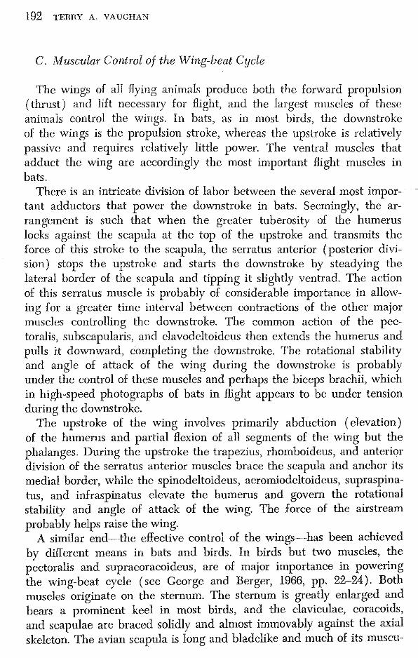
192 TERny A. VAUGHAN
C. Muscular Control of the Wing-beat Cycle
The wings of all flying animals produce both the forward propulsion (thrust) and lift necessary for flight, and the largest muscles of these animals control the wings. In bats, as in most birds, the downstroke of the wings is the propulsion stroke, whereas the upstroke is rclatively passive and requires relatively little power. The ventral muscles that adduct the wing are accordingly the most important flight muscles in bats.
There is an intricate division of labor between the several most important adductors that power the downstroke in bats. Seemingly, the arrangement is such that when the greater tuberosity of the humerus locks against the scapula at the top of the upstroke and transmits the force of this stroke to the scapula, the serratus anterior (posterior division) stops the upstroke and starts the downstroke by steadying the lateral border of the scapula and tipping it slightly ventrad. The action of this serratus muscle is probably of considerable importance in allowing for a greater time interval between contractions of the other major muscles controlling the downstroke. The common action of the pectoralis, subscapularis, and clavodeltoideus then extends the humerus and pulls it downward, completing the downstroke. The rotational stability and angle of attack of the wing during the downstroke is probably under the control of these muscles and perhaps the biceps brachii, which in high-speed photographs of bats in flight appears to be under tension during the downstroke.
The upstroke of the wing involves primarily abduction (elevation) of the humerus and partial flexion of all segments of the wing but the phalanges. During the upstroke the trapezius, rhomboideus, and anterior division of the serratus anterior muscles brace the scapula and anchor its medial border, while the spinodcltoideus, acromiodeltoideus, supraspinatus, and infraspinatus elevate the humerus and govern the rotational stability and angle of attack of the wing. The force of the airstream probably helps raise the wing.
A similar end-·the effective control of the wings .. --has been achieved by different means in bats and birds. In birds but two muscles, the pectoralis and supracoracoideus, are of major importance in powering the Wing-beat cycle (sec George and Berger, 1966, pp. 22-24). Both muscles originate on the sternum. The sternum is greatly enlarged and bears a prominent keel in most birds, and the claviculae, eoraeoids, and scapulae are braced solidly and almost immovably against the axial skeleton. The avian scapula is long and bladelike and much of its muscu-

4. THE MUSCULAH SYSTEM 193
lature serves most importantly to bind this bone to the rib cage and vertebral column. This morphological plan makes the bird's body deeper than it is wide, and keeps the center of gravity low, an advantageous feature in terms of aerodynamic stability.
In bats a very different arrangement occurs. No less than four muscles (pectoralis, subscapularis, posterior division of the serratus anterior, clavodeltoidcus) are important in controlling the downstroke, and at least an equal number serve directly to raise the wing during the upstroke. Only one of these muscles originates on the sternum. In further contrast, the sternum is not greatly enlarged nor does it bear a large keel in most bats, and the scapula is large, complexly faceted, and has considerable frcedom of movement. Indeed, the unique chiropteran arrangement for muscular control of the downstroke is based on the ability of the scapula to rock slightly on its long axis. Rcviewing briefly, then, in birds most of the power for the wing-beat cycle is provided by but two muscles, both originating on the sternum, and the scapula is firmly bound to the axial skeleton. In bats, in contrast, a rclatively large numbcr of muscles control the wing-beat cycle, only one of these muscles originates on the sternum, and mobility of thc scapula is essential to the control of the cycle.
Why are the adaptations for the control of the wings so different in bats and birds? The differences may have resulted from a predeliction of bats, during the period of their early evolution, to rcst and escape danger by day in narrow cracks and crevices. Any modifications causing a considerable increase in dorsoventral thickness of the body, such as occurred in birds, would have made it difficult for bats to use such confining retreats. The assemblage of muscular and osteological specializations typical of bats avoids great enlargement of the pectoralis muscle and a resultant increase in dorsoventral thickness. The reptilian posture of the hind limbs typical of most bats may also have developed in response to crevice dwelling. Flight and movement within a crevice demand the same general type of limb posture, and it is possible that the course of the early evolution of bats was "guided" by both of these types of locomotion (Vaughan, 1959, p. 134).
REFERENCES
Basmajian, J. V. (1962). "Muscles Alive, Their Functions Revealed by Electromyography." Williams & Wilkins, Baltimore, Maryland.
George, J. C., and Berger, A. J. (1966). "Avian Myology." Academic Press, New York.

194 TERRY A. VAUGHAN
Hartman, F. A. (1963). Some /light mechanisms in bats. Ohio J. Sci. 63, 59-65. Macalister, A. (1872). The myology of the cheiroptera. Phil. Trans. Roy. Soc.
London 162, 125-171. Vaughan, T. A. (1959). Functional morphology of three bats: Eumops, Myotis,
Macrotus. Publ. Mus. Natur. Hist. Univ. Kan. 12, 1-153. Vaughan, T. A. (1966). Morphology and flight characteristics of molossid bats.
]. Mammal. 47, 249-260. Webster, F. A., and Griffin, D. R. (1962). The role of the flight membranes in
insect capture by bats. Anim. Behav. 10, 332-340.

StercoradiogNq)/t of fossil hat, a/lout 50 rnillioH W:al'S old, dcscrihed in Chapter 7. Two tirnes natural size. Cf, fourth caudal vertebra; lil, lcft humerus, pmtly opaque tu x~raljs; Tir, right tibia, distal end; \V'r, right wrist.