Embed Size (px)
Citation preview

The Digestive System “Alimentary Canal”

Alimentary: Concerning food, nourishment, and the organs of
digestion. From the Latin alimentum meaning nourishment.

The Digestive System I) Introduction Introduction your digestive system is responsible for four major
processes: ingestion
taking in of food digestion
breaking down of food, can be: physical (mastication) chemical

The Digestive System I) Introduction
ingestion taking in of food
digestion breaking down of food, can be:
physical (mastication) chemical
absorption absorbing nutrients in food and making them available
to your circulatory system for transport elimination
getting rid of indigestible food.

The Digestive System I) Introduction

The Digestive System I) Introduction
Ingestion Digestion Absorption Elimination
Undigested material
Chemical digestion (enzymatic hydrolysis)
Nutrient molecules enter body cells
Small molecules
Mechanical digestion
Food
Pieces of food
1 2 3 4

The Digestive System I) Introduction
absorption absorbing nutrients in food and making them available to your
circulatory system for transport elimination
getting rid of indigestible food.
the goal of the digestive system is to ingest food, to digest into small molecules that can cross plasma membranes, to absorb nutrients and to eliminate non-digestible wastes
in humans digestion of food is an extracellular process.
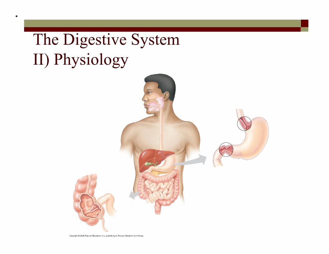
The Digestive System II) Physiology

Cecum
Anus
Ascending portion of large intestine
Gall- bladder
Small intestine
Large intestine
Small intestine
Rectum
Pancreas
Liver
Salivary glands
Tongue Oral cavity
Pharynx Esophagus
Sphincter
Stomach
Sphincter
Duodenum of small intestine
Appendix


Anus
Liver
Pancreas
Small intestine
Large intestine
Rectum
Stomach Gall- bladder
A schematic diagram of the human digestive system
Esophagus
Salivary glands
Mouth

The Digestive System II) Physiology A) Mouth/Oral Cavity (Bouche)
food is chewed in the mouth (mastication) and mixed with saliva (chemical digestion)
i) Teeth among mammals, dentition differs according to the
mode of nutrition humans are omnivores therefore our dentition is
nonspecific to both vegetables and meat diets.

The Digestive System II) Physiology
among mammals, dentition differs according to the mode of nutrition humans are omnivores therefore our dentition is nonspecific to both
vegetables and meat diets.
adult humans have: 32 teeth one-half of each jaw has teeth of four
different types: ~ 2 chisel shaped incisor for biting ~ 1 pointed canine for tearing ~ 2 fairly flat premolars for grinding ~ 3 molars, well flattened for crushing

The Digestive System II) Physiology
adult humans have: 32 teeth one-half of each jaw has teeth of four different types:
~ 2 chisel shaped incisor for biting ~ 1 pointed canine for tearing ~ 2 fairly flat premolars for grinding ~ 3 molars, well flattened for crushing
The Digestive System II) Physiology
~ 2 chisel shaped incisor for biting ~ 1 pointed canine for tearing ~ 2 fairly flat premolars for grinding ~ 3 molars, well flattened for crushing
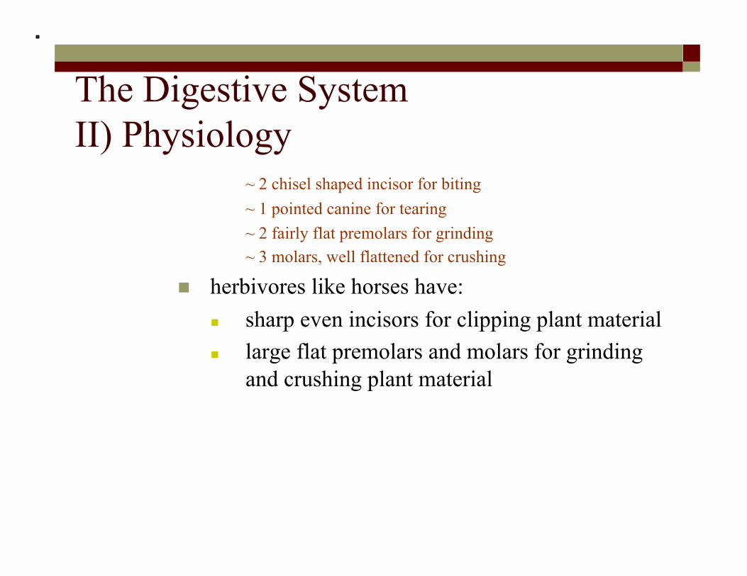
herbivores like horses have: sharp even incisors for clipping plant material large flat premolars and molars for grinding
and crushing plant material

The Digestive System II) Physiology
herbivores like horses have: sharp even incisors for clipping plant material large flat premolars and molars for grinding and crushing
plant material

The Digestive System II) Physiology
herbivores like horses have: sharp even incisors for clipping plant material large flat premolars and molars for grinding and crushing
plant material
carnivores have: pointed incisors and enlarged canine teeth to
tear off pieces of meat that is small enough to be swallowed.

The Digestive System II) Physiology
carnivores have: pointed incisors and enlarged canine teeth to tear off pieces of
meat that is small enough to be swallowed.

The Digestive System II) Physiology
ii) Salivary Glands there are three pairs of salivary glands
parotid sublingual submandibular
all three secrete saliva through the salivary ducts lubricates the food contains salivary amylase

The Digestive System II) Physiology
parotid sublingual submandibular
all three secrete saliva through the salivary ducts lubricates the food contains salivary amylase

The Digestive System II) Physiology
submandibular they secrete saliva through the salivary ducts
lubricates the food contains salivary amylase
this is the first site of chemical digestion starch is broken down into maltose
starch + H2O maltose (disaccharide)
salivary
amylase

The Digestive System II) Physiology
iii) Tongue food is manipulated by the tongue
tongue has: pressure receptors chemical receptors called taste buds.
the tongue pushes the chewed and saliva mixed food (the bolus) to the back of the pharynx and initiates the swallowing reflex.

The Digestive System II) Physiology
pressure receptors chemical receptors called taste buds.
the tongue pushes the chewed and saliva mixed food (the bolus) to the back of the pharynx and initiates the swallowing reflex.
B) Esophagus the esophagus conducts food the digestive and respiratory passages interact at pharynx.
a small flap called the epiglottis covers the trachea. the bolus passes through the pharynx entering the esophagus if food enters the trachea (wind pipe) we choke.

The Digestive System II) Physiology B) Esophagus the esophagus conducts food the digestive and respiratory passages interact at pharynx.
a small flap called the epiglottis covers the trachea. the bolus passes through the pharynx entering the esophagus if food enters the trachea (wind pipe) we choke.
when food enters the esophagus a rhythmic contraction of muscles occurs (peristalsis) pushing the bolus down the esophagus to the stomach.

Larynx Trachea
Epiglottis up
Pharynx
Tongue
Glottis
Esophagus
Esophageal sphincter contracted
Food
To stomach
To lungs

Larynx Trachea
Epiglottis up
Pharynx
Tongue
Glottis
Esophagus
Esophageal sphincter contracted
Food
To stomach
To lungs
Epiglottis down
Esophageal sphincter relaxed
Glottis up and closed

Larynx Trachea
Epiglottis up
Pharynx
Tongue
Glottis
Esophagus
Esophageal sphincter contracted
Food
To stomach
To lungs
Epiglottis down
Esophageal sphincter relaxed
Glottis up and closed
Epiglottis up
Esophageal sphincter contracted
Sphincter relaxed
Relaxed muscles
Contracted muscles
Relaxed muscles
Stomach
Glottis down and open
http://www.hopkins-gi.org/multimedia/database/intro_250_Swallow.swf

Digestion Check In Anatomy Nutrient Mechanical
Digestion Chemical Digestion Enyzmes Acidic or
Basic
Mouth
Carbohydrates Yes Yes Salivary Amylase
(polysaccharide to disaccarides) Acidic Proteins Yes No N/A
Fats Yes No N/A


Esophagus
Sphincter
Stomach
Sphincter
Duodenum of small intestine Small
intestine
Stomach
Esophagus

The Digestive System II) Physiology C) Stomach site of:
food storage initial protein digestion
contains three layers of muscle that run in different directions.
this allows for muscle contraction to churn the food rather than just squish it.
movement of food to and from the stomach is regulated by circular muscles called sphincters.

The Digestive System II) Physiology
this allows for muscle contraction to churn the food rather than just squish it.
movement of food to and from the stomach is regulated by circular muscles called sphincters.
sphincters act like draw strings on a bag. contraction of the lower esophageal sphincter
(previously known as the cardiac sphincter) , the LES, closes the opening to the stomach. the LES prevents food and acid from being
regurgitated up into the esophagus.

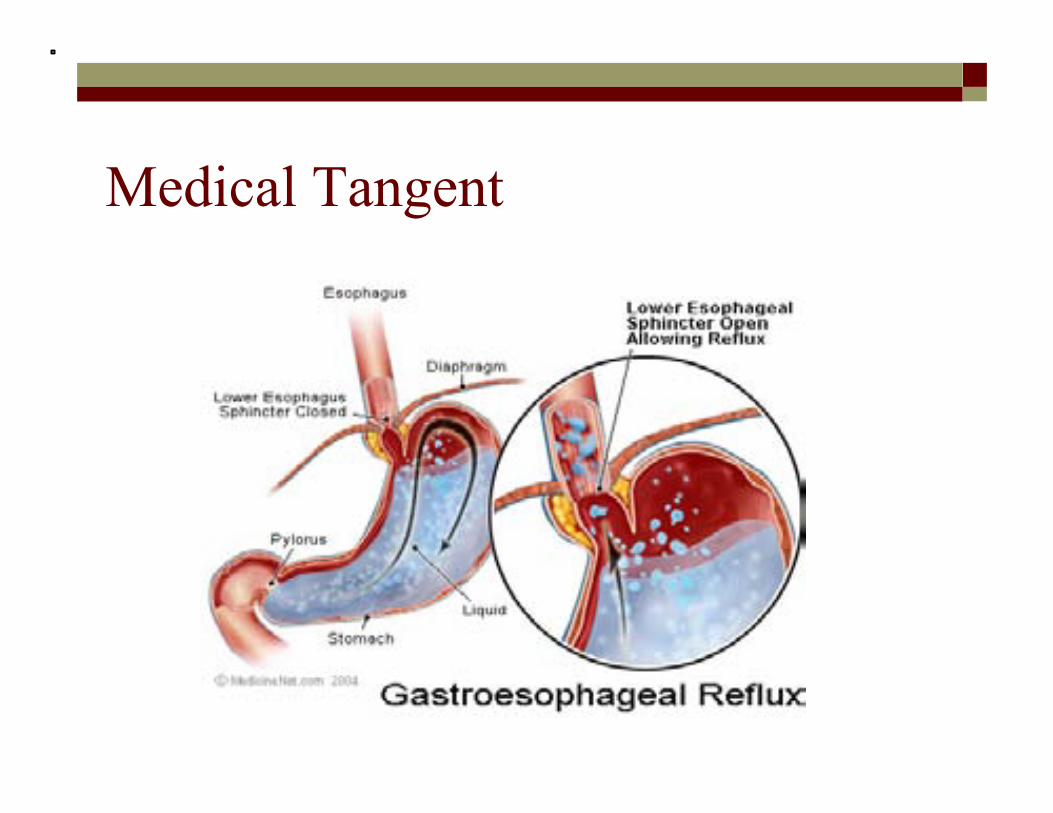
Medical Tangent Acid Reflux/Heartburn Gastroesophageal reflux disease
commonly referred to as GERD or acid reflux the liquid content of the stomach regurgitates (backs up or
refluxes) into the esophagus. can inflame and damage the lining (cause esophagitis)
of the esophagus
Medical Tangent

Medical Tangent the liquid content of the stomach regurgitates (backs up or refluxes) into the
esophagus. can inflame and damage the lining (cause esophagitis) of the esophagus
regurgitated liquid usually contains: acid and pepsin that are produced by the stomach. may contain bile that has backed-up into the stomach
from the duodenum. Acid is believed to be the most injurious component
of the refluxed liquid GERD is a chronic condition
Once it begins, it usually is life-long.

Medical Tangent the body has mechanisms to protect itself from the harmful
effects of reflux most reflux occurs during the day when individuals are
upright. in the upright position, the refluxed liquid is more
likely to flow back down due to the effect of gravity. while individuals are awake, they repeatedly swallow,
whether or not there is reflux. the salivary glands in the mouth produce saliva, which
contains bicarbonate. the bicarbonate neutralizes the small amount of
acid

The Digestive System II) Physiology
the LES prevents food and acid from being regurgitated up into the esophagus.
the pyloric sphincter regulates the movement of food and stomach acids into the small intestine. Lower esophageal
sphincter (LES)
Pyloric sphincter
Diaphragm
Esophagus
Stomach
Duodenum
Muscle Layers To Small Intestine

The Digestive System II) Physiology the L-shaped stomach has numerous ridges called rugae
allow for expansion millions of cells line the inner wall of the stomach.
these cells secrete gastric fluids or juices that aid digestion.
the main components of gastric juice are: mucus hydrochloric acid pepsinogens

The Digestive System II) Physiology
the main components of gastric juice are: mucus hydrochloric acid pepsinogens
parietal cells of the stomach secretes hydrochloric acid (HCl(aq))
lowers the pH of the stomach to around 2 kills many harmful substances that are indigested with
food. deactivates salivary amylase
converts pepsinogen into its active form pepsin.

The Digestive System II) Physiology
kills many harmful substances that are indigested with food. deactivates salivary amylase
converts pepsinogen into its active form pepsin. peptic/Chief cells of the stomach
secretes pepsinogen pepsinogen is an inactive enzyme.
it stays inactive until it reaches the lumen of the stomach and is activated by HCl(aq) (it is now called pepsin)

The Digestive System II) Physiology
Pepsinogen Pepsin
HCl
Long Chain Amino Acids
(Proteins)
Shorter Chain Amino Acids (Peptones)
peptic/ Chief cells of the stomach secretes pepsinogen
pepsinogen is an inactive enzyme. it stays inactive until it reaches the lumen of the stomach and
is activated by HCl (it is now called pepsin)

The Digestive System II) Physiology
mucus cells of the stomach secretes mucus
mucus provides a protective coating to prevent self digestion of the stomach lining
another chemical secreted by the cells of the stomach is renin. renin works to slow down the movement of milk through
the digestive system


Interior surface of stomach
Esophagus
Chief cells
Small intestine
Epithelium
Stomach
Sphincter
Parietal cell
Pepsinogen and HCl are secreted.
HCl converts pepsinogen to pepsin.
Pepsin activates more pepsinogen.
Chief cell
Folds of epithelial tissue
Pepsin
Sphincter
Pepsinogen
HCl
H+ Cl–
Parietal cells
Mucus cells
Gastric gland
1
2
2
3
3
1
5 µm

The Digestive System II) Physiology
another chemical secreted by the cells of the stomach is renin. renin works to slow down the movement of milk through the digestive system
after the action of gastric juice (chemical digestion) and stomach contractions (physical digestion) the contents of the stomach have a thick, soupy consistency. this is called chyme.
the base of the stomach has a sphincter called the pyloric sphincter.

Medical Tangent Peptic Ulcer when the protective mucus lining of the stomach breaks down
the cell membrane is exposed to HCl and pepsin the breakdown of the cell membrane of the stomach is a
peptic ulcer. must ulcers are the result of an infection by the bacterium
Helicobacter pylori. peptic ulcers can be treated with antibiotics if found early
enough

The Digestive System II) Physiology
the base of the stomach has a sphincter called the pyloric sphincter.
when the sphincter relaxes a small amount of chyme passes into the duodenum (the first part of the small intestine) chyme entering the duodenum sets off a reflex that
causes the sphincter to contract and close the opening temporarily. this adaptation allows for a slower more thorough
digestion.

Digestion Check In Anatomy Nutrient Mechanical
Digestion Chemical Digestion Enyzmes Acidic or
Basic
Mouth
Carbohydrates Yes Yes Salivary Amylase
(polysaccharide to disaccarides) Acidic
Proteins Yes No N/A
Fats Yes No N/A
Stomach
Carbohydrates Yes No N/A
Acidic Proteins Yes Yes Pepsinogen/Pepsin
(proteins to peptones)
Fats Yes No N/A

Gall- bladder
Small intestine
Large intestine
Small intestine
Pancreas
Liver
Stomach
Sphincter
Duodenum of small intestine
Liver
Pancreas
Small intestine
Stomach Gall- bladder

The Digestive System II) Physiology D) Small Intestine site of majority of digestion and absorption is approximately 7 metres in length it is called the small intestine because it is narrower than the
large instestine. is made up of three regions
duodenum jejunum ileum
the mucus membrane layer of the small intestine has ridges and furrows that give it corrugated appearance.

The Digestive System II) Physiology the mucus membrane layer of the small intestine has ridges and furrows that give
it corrugated appearance. on the surface of these ridges and furrows are small fingerlike
projections called villi. cells on the surface of the villi have minute projections called microvilli.
the villi and microvilli greatly increase the effective surface area of the small intestine.
if the small intestine was a smooth tube, it would have to be 500 to 600 meters long to have a comparable surface area.

Muscle layers
Vein carrying blood to hepatic portal vein
Villi
Intestinal wall
Key
Nutrient absorption
Large circular folds

Fig. 41-15b Microvilli (brush border) at apical (lumenal) surface
Key
Nutrient absorption
Blood capillaries
Epithelial cells
Villi
Lymph vessel
Basal surface
Lacteal Epithelial cells
Lumen

The Digestive System II) Physiology
the villi and microvilli greatly increase the effective surface area of the small intestine.
if the small intestine was a smooth tube, it would have to be 500 to 600 meters long to have a comparable surface area.
when chyme enters the duodenum: proteins and carbohydrates are partly digested fat digestion still needs to be carried out.
more digestion is still required before nutrients can be absorbed through the intestinal wall. two accessory glands, the liver and the pancreas send
secretions to the duodenum to complete digestion.

The Digestive System II) Physiology more digestion is still required before nutrients can be
absorbed through the intestinal wall. two accessory glands, the liver and the pancreas send
secretions to the duodenum to complete digestion. i) liver ii) pancreas

The Digestive System II) Physiology i) liver
the liver produces bile bile is
stored in the gall bladder is sent to the duodenum via the bile duct contains bile salts which are emulsifiying agents
(Emulsifying agents break up fat into fat droplets so they can mix with water)

The Digestive System II) Physiology
contains bile salts which are emulsifiying agents (Emulsifying agents break up fat into fat droplets so they can mix with water)
emulsified fats are more easily digested by enzymes (more surface area)
fat fat droplets bile salts

The Digestive System II) Physiology
emulsified fats are more easily digested by enzymes (more surface area)
when the stomach is empty bile is stored and concentrated in the gall bladder.
when there is fats in the small intestine the hormone cholecystokinin (CCK) is released. CCK is carried in the blood stream to the gall
bladder. triggers the release of bile salts
fat fat droplets bile salts

The Digestive System II) Physiology
CCK is carried in the blood stream to the gall bladder. triggers the release of bile salts once inside the duodenum the bile salts
emulsify fat into fat droplets (this is physical digestion because chemical bonds are not broken)


Secretin and CCK
Stomach
Gallbladder Liver
+
Duodenum of small intestine
Bile
Gastrin
Secretin
Pancreas CCK
CCK Key
Stimulation Inhibition
+
–
+
+ + –

The Digestive System II) Physiology
once inside the duodenum the bile salts emulsify fat into fat droplets
(this is physical digestion because chemical bonds are not broken)
bile also contains pigments the liver breaks down hemoglobin (Hb) from red
blood cells (RBC) and stores the products in the gall bladder for removal the characteristics brown colour of feces
results from Hb breakdown

The Digestive System II) Physiology
the characteristics brown colour of feces results from Hb breakdown
other liver functions: synthesis produces bile salts manufactures blood proteins breakdown/conversion removes toxic nitrogen group from amino acids
forming urea converts the toxic components of hemoglobin
(excreted by bile salts)

The Digestive System II) Physiology
breakdown/conversion removes toxic nitrogen group from amino acids forming urea converts the toxic components of hemoglobin (excreted by bile salts)
converts glucose into glycogen and vice versa to maintain a constant blood sugar level.
storage stores glycogen stores vitamins A, B12 and D detoxification converts harmful compounds (ie. alcohol) to less
harmful products.

The Digestive System II) Physiology
detoxification converts harmful compounds (ie. alohol) to less harmful products.
ii) Pancreas when acid enters the small intestine, a chemical called
prosecretin is converted into secretin secretin is absorbed into the bloodstream and carried to the
pancreas it signals the pancreas to release a solution containing
bicarbonate ions (HClO3-)
bicarbonate travels through the pancreatic duct to the small intestine.
in the small intestine bicarbonate raises the pH from about 2.5 (acidic) to 9.0 (basic)

The Digestive System II) Physiology
in the small intestine bicarbonate raises the pH from about 2.5 (acidic) to 9.0 (basic)
the basic pH inactivates pepsin. the small intestine is protected from the stomach
acids and protein digesting enzymes. pancreatic secretions contain enzymes that digest all
three major nutrients: ~ protiens ~ carbohydrates ~ lipids

The Digestive System II) Physiology
pancreatic secretions contain enzymes that digest all three major nutrients: ~ protiens ~ carbohydrates ~ lipids
pancreatic secretions or “juice” contains: sodium bicarbonate (for neutralization) pancreatic amylase trypsinogen/trypsin lipase erepsin

The Digestive System II) Physiology
pancreatic secretions or “juice” contains: sodium bicarbonate (for neutralization) pancreatic amylase trypsinogen/tryspin lipase erepsin
pancreatic amylase digest any remaining starches to maltose
(polysaccharides disaccharides)

The Digestive System II) Physiology
pancreatic amylase digest any remaining starches to maltose
(polysaccharides disaccharides)
trypsinogen an inactive enzyme is activated by another enzyme called enterokinase in the
lumen of the duodenum

The Digestive System II) Physiology
tyrpsinogen an inactive enzyme is activated by another enzyme called enterokinase in the lumen of the
duodenum
trypsinogen (inactive) enterokinase
trypsin (active)
proteins &
peptones
smaller amino chains (peptides)
erepsins
amino acids

The Digestive System II) Physiology
tyrpsinogen an inactive enzyme is activated by another enzyme called enterokinase in the lumen of
the duodenum
erepsins complete protein digestion by breaking the bonds
between short-chain peptides, releasing amino acids. released from the pancreas and the small intestine
lipase enzymes released by the pancreas break down lipids two types

The Digestive System II) Physiology
lipase enzymes released by the pancreas break down lipids two types
pancreatic lipase ~ most common ~ breaks down fats into fatty acids and glycerol
phospholipase ~ breaks down phospholipids

The Digestive System II) Physiology
pancreatic lipase ~ most common ~ breaks down fats into fatty acids and glycerol
phospholipase ~ breaks down phospholipids
back to the small intestine at this point:
fats are fully digested carbohydrates are partially digested proteins are fully and partially digested.
epithelial cells of the villi produce intestinal enzymes (intestinal juice)
this completes the digestion of peptides and sugars

The Digestive System II) Physiology
fats are full digested carbohydrates are partially digested proteins are fully and partially digested.
epithelial cells of the villi produce intestinal enzymes (intestinal juice) this completes the digestion of peptides and sugars
peptides amino acids
disaccharides maltase
lactase monosaccharides
peptidase

Digestion Check In Anatomy Nutrient Mechanical
Digestion Chemical Digestion Enyzmes Acidic
or Basic
Small Intestine
Carbohydrates X
Pancreatic Amylase (glycogen, starch → maltose)
Carbohydrases sucrase (sucrose → glucose + fructose)
maltase (maltose → 2 glucose) lactase (lactose → glucose + galactose)
Basic (pH = 8)
Lipids X Lipase
(Lipids → fatty acids + glycerol)
Proteins X
Proteases Trypsin (Peptides → smaller peptides)
Chymotrypsin (Peptides → smaller peptides) Peptidases
(peptides → smaller peptides and amino acids)

The Digestive System II) Physiology absorption of nutrients
each villus contains a network of blood vessels and a small lymphatic vessel called a lacteal after glycerol and fatty acids are absorbed by
epithelial cells, they are recombined into fats within these cells
these fats are mixed with cholesterol and coated with protein, forming molecules called chylomicrons, which are transported into lacteals


Lumen of small intestine
Lacteal
Chylomicron
Phospholipids, cholesterol, and proteins
Triglycerides
Monoglycerides
Triglycerides
Fatty acids
Epithelial cell

The Digestive System II) Physiology these fats are mixed with cholesterol and coated with protein,
forming molecules called chylomicrons, which are transported into lacteals
amino acids and sugars pass through the epithelium of the small intestine and enter the bloodstream
capillaries and veins from the lacteals converge in the hepatic portal vein and deliver blood to the liver and then on to the heart


Bloodstream
Veins to heart
Lymphatic system
Small intestine
Esophagus Stomach
Lipids Mouth
Hepatic portal vein
Absorbed food (except lipids)
Absorbed water
Secretions from the gastric glands of the stomach
Secretions from the pancreas and the liver
Liver
Rectum
Anus
Large intestine


The Digestive System II) Physiology E) Large Intestine
chemical digestion is complete the cecum
the first area where food from the small intestine arrives. has a small blind pouch attached to it called the
appendix. the colon:
the largest part of the large intestine. stores wastes long enough to reabsorb water inorganic salts, minerals and vitamins are reabsorbed
with the water. home to Escherichia coli (E. coli) bacteria


The Digestive System II) Physiology
inorganic salts, minerals and vitamins are reabosrbed with the water. home to Escherichia coli (E. coli) bacteria
uses waste materials to synthesize vitamins B and K. as wastes build up receptors in the cell wall send a signal
to the central nervous system promoting a bowel movement. bowel movements ensure removal of potentially toxic
wastes from the body. people who do not eat enough fibre (cellulose) have
fewer bowel movements.

bowel movements ensure removal of potentially toxic wastes from the body.
people who do not eat enough fibre (cellulose) have fewer bowel movements.
wastes of the digestive tract, the feces, become more solid as they move through the colon
feces pass through the rectum and exits via the anus feces is stored in the rectum until they can be eliminated two sphincters between the rectum and anus control
bowel movements

The Digestive System II) Physiology

The Digestive System II) Physiology interesting adaption
herbivores generally have longer alimentary canals than carnivores, reflecting the longer time needed to digest vegetation


Cecum
Small intestine
Herbivore Carnivore
Colon (large intestine)
Stomach Small intestine

Carbohydrate digestion
Polysaccharides
Smaller polysaccharides, maltose
Polysaccharides
Maltose and other disaccharides
Disaccharides
Protein digestion Nucleic acid digestion Fat digestion
Proteins
Small polypeptides
Monosaccharides
Small peptides
Amino acids
Amino acids
Polypeptides
Smaller polypeptides
DNA, RNA Fat globules
Nucleotides Fat droplets
Nucleosides
Nitrogenous bases, sugars, phosphates
Glycerol, fatty acids, monoglycerides
(starch, glycogen) (sucrose, lactose)

Oral cavity, pharynx, esophagus
Stomach
Lumen of small intes- tine
Epithelium of small intestine (brush border)
Carbohydrate digestion
Polysaccharides
Smaller polysaccharides, maltose
Polysaccharides
Maltose and other disaccharides
Disaccharides
Protein digestion Nucleic acid digestion Fat digestion
Proteins
Small polypeptides
Pepsin
Pancreatic amylases
Salivary amylase
Disaccharidases
Monosaccharides
Small peptides
Amino acids
Amino acids
Polypeptides
Smaller polypeptides
Pancreatic trypsin and chymotrypsin
Pancreatic carboxypeptidase
Dipeptidases, carboxypeptidase, and aminopeptidase
DNA, RNA
Pancreatic nucleases
Fat globules
Nucleotides Fat droplets
Nucleosides
Nitrogenous bases, sugars, phosphates
Nucleotidases
Nucleosidases and phosphatases
Glycerol, fatty acids, monoglycerides
Bile salts
Pancreatic lipase
(starch, glycogen) (sucrose, lactose)

The Digestive System III) Control of Digestion control of digestion is exerted by the nervous and endocrine
(hormonal system) seeing, smelling and tasting food will produce gastric
secretions before there is any food in the stomach. swallowing stimulates gastric secretions even if the
esophagus is empty. hormones play a large role in control

hormones play a large role in control
secretin is released when acid from the stomach moves into
the small intestine. travels to the pancreas via the blood stream where it
stimulates the release of bicarbonate to raise the pH. gastrin
produced when the wall of the stomach are distended by the presence of food and by the presence of partially digested protein.
travels in the blood and stimulates the parietal cells of the stomach to release HCl.

gastrin produced when the wall of the stomach are distended by the presence of
food and by the presence of partially digested protein. travels in the blood and stimulates the parietal cells of the stomach to
release HCl.
the size of the meal a larger meal will trigger more receptors in your stomach
causing more forcefull contractions and faster emptying. a fatty meal will cause the small intestine to secrete
enterogastrone which will slow peristaltic movement allowing more time for fat digestion and absorption.