Embed Size (px)
Citation preview

Biofiles
Centrifugation
Volume 6, Number 5
Centrifugation Basics
Density Gradient Media
Cell Viability and Proliferation
Organelle Isolation
Biofilescontents
Introduction 3
Centrifugation Basics 4
Centrifugation Separations 6
Histopaque® Troubleshooting Guide 14
Cell Viability and Proliferation 17
Organelle Isolation 22
ECACC® Cell Lines 26
Centrifugation Equipment 27
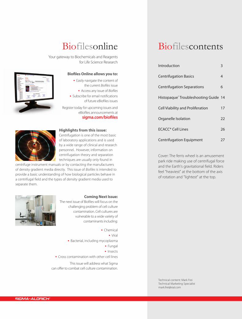
Cover: The ferris wheel is an amusement park ride making use of centrifugal force and the Earth's gravitational field. Riders feel "heaviest" at the bottom of the axis of rotation and "lightest" at the top.
BiofilesonlineYour gateway to Biochemicals and Reagents
for Life Science Research
Biofi les Online allows you to:
• Easily navigate the content ofthe current Biofi les issue
• Access any issue of Biofi les
• Subscribe for email notifi cationsof future eBiofi les issues
Register today for upcoming issues andeBiofi les announcements at
sigma.com/biofi les
Highlights from this issue:Centrifugation is one of the most basic of laboratory applications and is used by a wide range of clinical and research personnel. However, information on centrifugation theory and separation techniques are usually only found in
centrifuge instrument manuals or by contacting the manufacturers of density gradient media directly. This issue of Biofiles is intended to provide a basic understanding of how biological particles behave in a centrifugal field and the types of density gradient media used to separate them.
Coming Next Issue: The next issue of Biofi les will focus on the
challenging problem of cell culture contamination. Cell cultures are
vulnerable to a wide variety of contaminants including:
• Chemical
• Viral
• Bacterial, including mycoplasma
• Fungal
• Insects
• Cross contamination with other cell lines
This issue will address what Sigmacan off er to combat cell culture contamination.
Technical content: Mark FreiTechnical Marketing [email protected]

Order sigma.com/order Technical service sigma.com/techinfo sigma.com/lifescience 3
Separation of particles by sedimentation is one of the most powerful tools in biology. Even though sedimentation using centrifugation is not a new technology, it is essential for cutting edge genomic and proteomic research by providing purified particles of interest. In a survey at the US National Institutes of Health, over 65% of research workers replied that using centrifugation to purify cells, subcellular organelles, viruses, proteins, and nucleic acids is an integral part of their work.1
Density gradient centrifugation is a technique that allows the separation of particles on the basis of their size, shape, and density. A density gradient is typically created by layering media of increasing density in a centrifuge tube. When a sample is layered on top of a density gradient and centrifuged, the various particles move through the gradient at different rates. The particles appear as bands or zones in the gradient with the more dense and larger particles migrating furthest.
A number of different compounds have been investigated as density gradient media. One of the first density gradient centrifugation techniques was developed in the 1950s and used a buffered sucrose solution for the purification of cell organelles. Sucrose quickly became the density medium of choice for separating homogenized mammalian tissues. Later, cesium chloride gradients were used to separate DNA of different densities. Meselson and Stahl in 1958 used cesium chloride density gradient centrifugation in an elegant experiment to support the semi-conservative model of DNA replication. Colloidal-silica suspensions were first manufactured by DuPont and sold under the name of LUDOX®.2
In 1977, the stabilized silica colloid coated with polyvinylpyrrolidone (PVP) called Percoll® became available for separating cells and subcellular particles. In 1968, Boyum described methods for the isolation of mononuclear cells from circulating blood and bone marrow using mixtures of polysaccharide and a radiopaque contrast medium. This led to the development of the first nonionic iodinated density gradient medium, metrizamide, in the 1970s.3 Now, a large selection of commercial iodinated density gradient media are available.
This issue of Biofiles examines the theory and practice of biological separation. The beginning section provides a primer on the basic concepts of centrifugation. Three basic types of centrifugal separations are highlighted; differential centrifugation, rate-zonal centrifugation, and isopycnic centrifugation. A concise description of each is given along with the separation principles involved. Cell viability kits, technical support information, and centrifugation equipment are also included.
We hope you find the reference information and products relevant. For a comprehensive list of our centrifugation media products, please visit sigma.com/handh
References:1. Biological Centrifugation, J. Graham, p. 12. Centrifugation, A Practical Approach (2nd Edition), D.
Rickwood, p.353. Iodinated Density Gradient Media, A Practical Approach, D.
Rickwood, p.1
IntroductionMark FreiTechnical Marketing [email protected]

4
The earth's gravitational force is sufficient to separate many types of particles over time. A tube of anticoagulated whole blood left standing on a bench top will eventually separate into plasma, red blood cell and white blood cell fractions. However, the length of time required precludes this manner of separation for most applications. In practice, centrifugal force is necessary to separate most particles. In addition, the potential degradation of biological compounds during prolonged storage means faster separation techniques are needed.
The rate of separation in a suspension of particles by way of gravitational force mainly depends on the particle size and density. Particles of higher density or larger size typically travel at a faster rate and at some point will be separated from particles less dense or smaller. This sedimentation of particles, including cells, can be explained by the Stokes equation, which describes the movement of a sphere in a gravitational field.1 The equation calculates the velocity of sedimentation utilizing five parameters (see Figure 1).
v = d2 (p–L) 3 g
18 n
v = sedimentation rate or velocity of the sphered = diameter of the spherep = particle densityL = medium densityn = viscosity of mediumg = gravitational force
Figure 1. The Stokes equation.
From the Stokes equation five important behaviors of particles can be explained:
1. The rate of particle sedimentation is proportional to the particle size.
2. The sedimentation rate is proportional to the difference in density between the particle and the medium.
3. The sedimentation rate is zero when the particle density is the same as the medium density.
4. The sedimentation rate decreases as the medium viscosity increases.
5. The sedimentation rate increases as the gravitational force increases.
Most particles are so small that gravitational force is insufficient to overcome the random molecular forces of particles to influence separation. Centrifugation, the name given to separation applications which involve spinning around an axis to produce a centrifugal force, is a way to increase the magnitude of the gravitational field. The particles in suspension experience a radial centrifugal force moving them away from the axis of rotation.2 The radial force generated by the spinning rotor is expressed relative to the earth's gravitational force and therefore is known as the relative centrifugal force (RCF) or the "g force." The g force acting on particles is exponential to the speed of rotation (defined as revolutions per minute; rpm). Doubling the speed of rotation increases the centrifugal force by a factor of four. The centrifugal force also increases with the distance from the axis of rotation. These two parameters are of considerable significance when selecting the appropriate centrifuge. Table 1 summarizes the applications that can be classified by the relative centrifugal force.3
Centrifugation Basics
Centrifugation Basics
Parameters Low speed High speed UltracentrifugeSpeed ranges (r.p.m. x 103) 2–6 18–22 35–120
Maximum RCF (x 103) 8 60 700
Pelleting Applications
Bacteria - Yes Yes*
Animal and plant cells Yes Yes Yes*
Nuclei Yes Yes Yes*
Precipitates Some Most Yes*
Membrane organelles Some Yes Yes
Membrane fractions Some Some Yes
Ribosomes/polysomes - - Yes
Macromolecules - - Yes
Viruses - Most Yes* Can be done but not usually used for this purpose
Table 1. Classes of centrifuges and their applications

Order sigma.com/order Technical service sigma.com/techinfo sigma.com/lifescience 5
RCF is dependent on the speed of rotation in rpm and the distance of the particles from the center of rotation. When the speed of rotation is given in rpm (Q) and the distance (r) is expressed in centimeters, RCF can be calculated by using the formula in Figure 2.
RCF = 11.18 3 r 3 Q1000
2
Figure 2. Formula for relative centrifugal force (RCF)
A nomogram can also be used to obtain the speed of a centrifuge rotor necessary for a desired RCF (see Figure 3). This quick estimate is useful for low speed centrifugation applications. However, it is more accurate to use the RCF calculation for speeds in excess of 10,000 rpm.
Radius(cm) g Rpm
A B C
30
25
201918171615141312
1110
9
8
7
6
5
4
3
7000600050004000
3000
2000
1500
1000800700600500400
300
200
150
1008070605040
30
2015
10
5000
4500
4000
3500
3000
2500
2000
150014001300120011001000
900
800
700
600
500
Figure 3. Nomogram for estimation of centrifuge rpm setting.
Nomogram instructions:
1. Measure the radius (cm) from the center of the centrifuge rotor to the end of test tube carrier.
2. Obtain the relative centrifugal force necessary for the application.
3. A straight line connecting the value of the radius with the relative centrifugal force (g) value will enable the speed of the rotor (rpm) to be read off of the right column.
References:1. Laboratory Techniques in Biochemistry and Molecular
Biology, P.T. Sharpe, p.182. Biological Centrifugation, J. Graham, p. 33. Centrifugation, Essential Data, D. Rickwood, p.12
biomolecules
Bioguarantee.Sigma® Life Science offers a collection of more than 50,000 antibodies, all 100% Bioguaranteed.*
©2011 Sigma-Aldrich Co. LLC. All rights reserved. SIGMA and SIGMA-ALDRICH are trademarks of Sigma-Aldrich Co. LLC, registered in the US and other countries. Where bio begins is a trademark of Sigma-Aldrich Co. LLC.
Find the antibody you need:sigma.com/antibodyexplorer
*Experimental results must be submitted via the Antibody Bioguarantee Form within 12 months of the date of purchase. All required fi elds of the Antibody Bioguarantee Form must be completed. Refunds and replacements contingent to claim review by technical service team. Credit covers the cost of antibody. Product replacements depend on product availability.

6
There are two types of centrifugal techniques for separating particles, differential centrifugation and density gradient centrifugation. Density gradient centrifugation can further be divided into rate-zonal and isopycnic centrifugation.
Differential CentrifugationThe simplest form of separation by centrifugation is differential centrifugation, sometimes called differential pelleting (see Figure 1). Particles of different densities or sizes in a suspension will sediment at different rates, with the larger and denser particles sedimenting faster. These sedimentation rates can be increased by using centrifugal force. A suspension of cells subjected to a series of increasing centrifugal force cycles will yield a series of pellets containing cells of decreasing sedimentation rate.
Centrifugation Time
Cent
rifug
al F
orce
A. B. C. D.
Figure 1. Diff erential Centrifugation
Particles of diff erent densities or size will sediment at diff erent rates with the largest and most dense particles sedimenting the fastest followed by less dense and smaller particles.
Differential pelleting is commonly used for harvesting cells or producing crude subcellular fractions from tissue homogenate. For example, a rat liver homogenate containing nuclei, mitochondria, lysosomes, and membrane vesicles that is centrifuged at low speed for a short time will pellet mainly the larger and more dense nuclei. Subsequent centrifugation at a higher centrifugal force will pellet particles of the next lower order of size (e.g., mitochondria) and so on. It is unusual to use more than four differential centrifugation cycles for a normal tissue homogenate.
Due to the heterogeneity in biological particles, differential centrifugation suffers from contamination and poor recoveries. Contamination by different particle types can be addressed by resuspension and repeating the centrifugation steps (i.e., washing the pellet).1
Rate-Zonal CentrifugationIn rate-zonal centrifugation the problem of cross-contamination of particles of different sedimentation rates may be avoided by layering the sample as a narrow zone on top of a density gradient (see Figure 2). In this way the faster sedimenting particles are not contaminated by the slower particles as occurs in differential centrifugation. However, the narrow load zone limits the volume of sample (typically 10%) that can be accommodated on the density gradient. The gradient stabilizes the bands and provides a medium of increasing density and viscosity.
Sample zone
Density gradient
Cent
rifug
al F
orce
A. B. C.
Figure 2. Rate-Zonal Centrifugation
Sample is layered as a narrow zone on the top of a density gradient (2B). Under centrifugal force, particles move at diff erent rates depending on their mass (2C).
The speed at which particles sediment depends primarily on their size and mass instead of density. As the particles in the band move down through the density medium, zones containing particles of similar size form as the faster sedimenting particles move ahead of the slower ones. Because the density of the particles is greater than the density of the gradient, all the particles will eventually form a pellet if centrifuged long enough.2
Isopycnic CentrifugationIn isopycnic separation, also called buoyant or equilibrium separation, particles are separated solely on the basis of their density. Particle size only affects the rate at which particles move until their density is the same as the surrounding gradient medium. The density of the gradient medium must be greater than the density of the particles to be separated. By this method, the particles will never sediment to the bottom of the tube, no matter how long the centrifugation time (see Figure 3).
Centrifugation Separations
Centrifugation Separations

Order sigma.com/order Technical service sigma.com/techinfo sigma.com/lifescience 7
Cent
rifug
al F
orce
A. B.
Figure 3. Isopycnic Centrifugation
Starting with a uniform mixture of sample and density gradient (3A) under centrifugal force, particles move until their density is the same as the surrounding medium (3B).
Upon centrifugation, particles of a specific density sediment until they reach the point where their density is the same as the gradient media (i.e., the equilibrium position). The gradient is then said to be isopycnic and the particles are separated according to their buoyancy. Since the density of biological particles is sensitive to the osmotic pressure of the gradient, isopycnic separation may vary significantly depending on the gradient medium used. Although a continuous gradient may be more suited for analytical purposes, preparative techniques commonly use a discontinuous gradient in which the particles band at the interface between the density gradient layers. This makes harvesting certain biological particles (e.g., lymphocytes) easier.
No single compound can satisfy all of the above criteria. Therefore a wide range of gradient media are used for the different types of samples (see Table 1). Most media are capable of producing the range of densities required and being easily removed from the particles of interest.
The effect of osmolality on biological particles requires special consideration. The osmolality of most mammalian fluids is 290-300 mOsm.4 This is the osmolality of balanced salt solutions (e.g., 0.85-0.9% NaCl) and most common media. High osmolality solutions not only remove water from the interior of membrane-bound particles, they also remove water bound to macromolecules like DNA. Loss of water from cells will reduce their size and increase their density, thereby affecting their buoyancy and rate of sedimentation. The osmotic effect on cells and macromolecules may be reversible, though it is a possible source of error that should be avoided.
Over the years, a variety of different compounds have been developed as density gradient media in order to enhance the separation process and to overcome osmolality and viscosity problems. There are five main classes of density gradient medium:
• Polyhydric (sugar) alcohols• Polysaccharides• Inorganic salts• Iodinated compounds • Colloidal silica
Suitable Density Gradient Medium SelectionThe primary function of density gradient centrifugation is to separate particles, either on the basis of their buoyancy density or their rate of sedimentation. For rate-zonal separations, the function of the gradient is to provide a gradient of viscosity which improves particle resolution while stabilizing the column from convection currents. For isopycnic separations, the important feature is that the maximum density of the gradient media is higher than that of the particles. An ideal density gradient media has the following properties:3
• Suffi cient solublilty to produce the range of densities required
• Does not form solutions of high viscosity in the desired density range
• Is not hyperosmotic or hypoosmotic when the particles to be separated are osmotically sensitive
• Solutions of the gradient should be adjustable to the pH and the ionic strengths that are compatible with the particles being separated
• Does not aff ect the biological activity of the sample
• Nontoxic and not metabolized by cells• Does not interfere with assay procedures or
react with the centrifuge tubes• Exhibits a property that can be used as a
measure of concentration• Easily removed from the purifi ed product• Autoclavable• Reasonable cost
Gradient medium type Principle usesPolyhydric alcoholsSucrose Organelles, membrane vesicles, viruses, proteins, ribosomes, polysomesGlycerol Mammalian cells (infrequent), proteins
Sorbitol Nonmammalian subcellular particlesPolysaccharidesFicoll®, polysucrose and dextrans Mammalian cells (sometimes in combination with iodinated density gradient media), mammalian subcellular particles
(infrequent)Inorganic saltsCsCl DNA, viruses, proteinsCs2SO4 DNA, RNAKBr Plasma lipoproteinsIodinated gradient mediaDiatrizoate Mainly as a component of commercial lymphocyte isolation mediaNycodenz®, Histodenz™ Mammalian cells, organelles, membrane vesicles, virusesIodixanol Mammalian cells, organelles, membrane vesicles, viruses, plasma lipoproteins, proteins, DNAColloidal silica mediaPercoll® Mammalian cells, organelles, membrane vesicles (infrequent)
Table 1. Density gradient media types and their principle uses.

8
Polyhydric alcoholsPolyhydric alcohols are considered nonionic gradient media. Some of the first centrifugation techniques developed in the 1950s used sucrose in the purification of cell organelles. Sucrose gradients are widely used for the rate-zonal separation of macromolecules and for the isopycnic separation of viruses and cell organelles.The advantages are its stable nature, inertness, and low cost. The disadvantages lie in the fact that concentrated solutions are viscous and hypertonic. Reagent-grade sucrose may be contaminated with RNAses or heavy metals and therefore are unsuitable for DNA and RNA purification. Glycerol solutions are less dense than corresponding sucrose solutions. However, glycerol solutions of the same density of sucrose solutions are much more viscous. Glycerol helps to preserve the activity of certain enzymes and it can be removed through vacuum.
Sucroseα-D-Gluco pyran osyl β-D-fructo furan o side; α-D-Glc-(1→2)-β-D-Fru; D(+)-Saccharose; Sugar; β-D-Fructo-furan osyl-α-D-gluco pyrano side[57-50-1] C12H22O11 FW 342.30
� BioXtra, ≥99.5% (GC)
Insoluble matter.............................. passes filter testign. residue............. ≤0.01% Cu..................... ≤0.0005%(as SO4) Fe.............................. ≤0.0005%pH........5.5-7.5, 1 M H2O (20 °C) K.............................. ≤0.005%chloride (Cl-)..................≤0.005% Li.............................. ≤0.0005%sulfate (SO4
2-)............≤0.005% Mg..............................≤0.0005%Al.............................. ≤0.0005% Mn............................. ≤0.0005%As.............................. ≤0.0001% Mo............................. ≤0.0005%Ba.............................. ≤0.0005% Na.............................. ≤0.005%Bi.............................. ≤0.0005% Ni.............................. ≤0.0005%Ca.............................. ≤0.001% Pb.............................. ≤0.0005%Cd.............................. ≤0.0005% Sr.............................. ≤0.0005%Co.............................. ≤0.0005% Zn.............................. ≤0.0005%Cr.............................. ≤0.0005%
S7903-250G 250 gS7903-1KG 1 kgS7903-5KG 5 kg
� ≥99.5% (GC)RNase..............................none detected
S9378-10MG 10 mgS9378-500G 500 gS9378-1KG 1 kgS9378-5KG 5 kgS9378-10KG 10 kg
� for molecular biology, ≥99.5% (GC)DNase, RNase, protease..............................none detectedFree glucose..............................<0.1%heavy metals (as Pb).............................. <5 ppm
S0389-500G 500 gS0389-1KG 1 kgS0389-5KG 5 kg
� BioReagent, suitable for cell culture, suitable for insect cell culture, ≥99.5% (GC)
Use to create sucrose gradients for purification of viruses and proteins.
S1888-500G 500 gS1888-1KG 1 kgS1888-5KG 5 kg
Gly cerol1,2,3-Propane triol; Gly cer in[56-81-5] HOCH2CH(OH)CH2OH FW 92.09density.............................................................................................1.25 g/mL
� for molecular biology, ≥99%reagentDNase, RNase, NICKase, and protease...............none detected
Fe.............................. ≤5 ppm Mg.............................. ≤5 ppm
heavy metals (as Pb)................................................................. <5 ppm
G5516-100ML 100 mLG5516-500ML 500 mLG5516-1L 1 L
� BioXtra, ≥99% (GC)Phosphorus (P)...........................................................................≤0.0005%
ign. residue.................... ≤0.1% K.................................... ≤0.005%chloride (Cl-)............. ≤0.001% Mg.............................. ≤0.0005%sulfate (SO4
2-)........... ≤0.002% Na.................................. ≤0.005%Al................................ ≤0.0005% NH4
+................................ ≤0.05%Ca................................ ≤0.0005% Pb................................... ≤0.001%Cu................................ ≤0.0005% Zn............................... ≤0.0005%Fe................................ ≤0.0005%
G6279-500ML 500 mLG6279-1L 1 LG6279-4X4L 4 × 4 L
� ≥99% (GC)heavy metals (as Pb).............................. ≤5 ppm
G9012-100ML 100 mLG9012-500ML 500 mLG9012-1L 1 LG9012-2L 2 LG9012-1GA 1 gal
� BioUltra, for molecular biology, anhydrous, ≥99.5% (GC)
Component of loading buffer in agarose gel electrophoresis of nucleic acids previously denatured with glyoxal1; Preparation of phage and plasmid DNA, for the storage of pure cultures2
DNases.................................................................................. none detectedinsoluble matter........................................................... passes filter test
phosphatases................................................................... none detectedproteases............................................................................. none detectedRNases.................................................................................. none detected
ign. residue.................... ≤0.1% Fe............................... ≤1 mg/kgpH.......5.5-8, 5 M H2O (25 °C) K.............................. ≤20 mg/kgchloride (Cl-)......... ≤1 mg/kg Li................................ ≤1 mg/kgsulfate (SO4
2-)..... ≤10 mg/kg Mg............................ ≤1 mg/kgAg................................ ≤5 mg/kg Mn............................ ≤1 mg/kgAl................................. ≤1 mg/kg Mo............................ ≤1 mg/kgAs............................. ≤0.1 mg/kg Na........................... ≤20 mg/kgBa................................ ≤1 mg/kg NH4
+......................... ≤5 mg/kgBi.................................. ≤1 mg/kg Ni............................... ≤1 mg/kgCa................................ ≤5 mg/kg Pb.............................. ≤1 mg/kgCd................................ ≤1 mg/kg Sr................................ ≤1 mg/kgCo................................ ≤1 mg/kg Tl................................ ≤5 mg/kgCr................................. ≤1 mg/kg Zn............................. ≤1 mg/kgCu............................... ≤1 mg/kg
Lit cited: 1. R.C. Ogden, D.A. Adam, Meth. Enzymol. 152, 79 (1987); 2. H. Miller, Meth. Enzymol. 152, 145 (1987);
49767-100ML 100 mL49767-250ML 250 mL49767-1L 1 L
PolysaccharidesPolysaccharides circumvent the high osmotic strength issues that arise with using sucrose solutions. The most common polysaccharide medium used is Ficoll®. Ficoll is produced by the polymerization of sucrose molecules with epichlorohydrin to give a polysaccharide with the average molecular weight of 400,000. Ficoll solutions below 20% (w/v) have a density of 1.07 g/cm3 and are considered osmotically inert. The main disadvantage is Ficoll solutions are more viscous than comparable sucrose solutions.
Dextran solution from Leuconostocmesenteroides[9004-54-0]Use of dextrans as long and hydrophilic spacer arms to improve the performance of immobilized proteins acting on macromolecules.
� 20 % (w/w) (Autoclaved)average mol wt ~500,000store at:: 2–8 °C
D8802-25ML 25 mLD8802-50ML 50 mL
Order sigma.com/order Technical service sigma.com/techinfo sigma.com/lifescience 9
Colloidal Silica MediaColloidal silica media are not true solutions, but are colloidal suspensions of silica particles coated with polyvinylpyrrolidone (PVP) with a diameter of 15–30 nm. The most widely known colloidal silica medium is Percoll®. The polyvinylpyrrolidone minimizes the particle interactions with biological material and stabilizes the colloid. Being a colloid, the osmotic strength of Percoll is extremely low and changes little with density. The osmolality of Percoll gradients can be adjusted by adding appropriate amounts of sucrose or buffer solution. Percoll gradients self-form when centrifuged in fix-angle rotors (swing out rotors are not satisfactory for self-forming gradients).5 Percoll can be removed from suspensions by differential pelleting. Percoll gradients are mainly used in isopycnic separations of cells, organelles, membrane vesicles, and even some viruses. The main limitation is that the sample particle size must be larger than the colloidal silica particles, otherwise the particles of silica pellet before the sample bands.
Percoll®
Percoll® consists of colloidal silica particles of 15–30 nm diameter (23% w/w in water) which have been coated with polyvinylpyrrolidone (PVP). It is used in cell separation and organelle isolation.
aseptically filled
� pH 8.5-9.5 (25 °C)store at: 2–8 °C
P1644-25ML 25 mLP1644-100ML 100 mLP1644-500ML 500 mLP1644-1L 1 L
� pH 8.5-9.5 (25 °C), cell culture testeddensity..................................................1.13 g/mL±0.005 g/mL, 25 °Cstore at: 2–8 °C
P4937-25ML 25 mLP4937-100ML 100 mLP4937-500ML 500 mL
Dextran sulfate sodium salt fromLeuconostoc spp.[9011-18-1]
� mol wt 6,500-10,000
D4911-1G 1 gD4911-10G 10 gD4911-50G 50 gD4911-100G 100 g
� average mol wt 9,000-20,000
D6924-1G 1 gD6924-10G 10 gD6924-50G 50 g
� average mol wt >500,000 (dextran starting material), contains 0.5-2.0% phosphate buffer, pH 6-8
store at:: 2–8 °C
D6001-1G 1 gD6001-10G 10 gD6001-50G 50 gD6001-100G 100 gD6001-500G 500 g
Precipitating agent in the quantitation of HDL cholesterol
D8787-1G 1 gD8787-5G 5 g
Ficoll® solution[26873-85-8]A nonionic synthetic polymer of sucrose.
Used in electrophoresis and as a hapten carrier. Most commonly used to prepare density gradients.
� Type 400, 20% in H2O0.2 μm filteredstore at: 2–8 °C
F5415-25ML 25 mLF5415-50ML 50 mL
▼ Ficoll® 400Poly sucrose 400 [26873-85-8]A nonionic synthetic polymer of sucrose.
Used for cell separation and organelle isolation.
Ficoll® 400� BioXtra, Type 400-DL, lyophilized powderInsoluble matter................................................................................≤0.1%Phosphorus (P)............................................................................≤0.0005%
ign. residue.................... <0.1% K................................... ≤0.005%chloride (Cl-)............... ≤0.05% Mg............................... ≤0.001%sulfate (SO4
2-)............. ≤0.05% Na................................... ≤0.01%Al................................. ≤0.0005% NH4
+.............................. ≤0.05%Ca................................... ≤0.001% Pb................................ ≤0.001%Cu................................ ≤0.0005% Zn............................. ≤0.0005%Fe................................. ≤0.0005%
F1418-25G 25 gF1418-100G 100 g
Ficoll® 400� lyophilized powder, γ-irradiated, BioXtra, suitable
for cell cultureDialyzed
F8636-25G 25 g
Ficoll® 400� lyophilized powder, Type 400-DL, BioReagent,
suitable for cell cultureDialyzed
F8016-5G 5 gF8016-100G 100 gF8016-500G 500 g
Ficoll® 400� Type 400-DL, lyophilized powderDialyzed
F9378-5G 5 gF9378-10G 10 gF9378-25G 25 gF9378-100G 100 gF9378-500G 500 g
Ficoll® 400 ▲
Ficoll® PM 70Poly(sucrose-co-epi chlor hydrin) [72146-89-5]A nonionic synthetic polymer of sucrose.
� Type 70mol wt ~70,000
F2878-100G 100 gF2878-500G 500 g
Ficoll® PM 400Poly sucrose 400 [26873-85-8]A nonionic synthetic polymer of sucrose.
Used for cell separation and organelle isolation.
� Type 400powder
spray-dried
F4375-10G 10 gF4375-25G 25 gF4375-100G 100 gF4375-500G 500 g
Poly sucrose 400[26873-85-8]A nonionic synthetic polymer of sucrose.
Used for cell separation and organelle isolation.
� powdermol wt 300,000-550,000
Similar to Ficoll® 400, but from a different supplier.
P7798-100G 100 gP7798-500G 500 g

10
Ionic Metal SaltsIonic gradient media, comprised of concentrated heavy metal salts, are almost exclusively used for isopycnic separations of nucleic acids.6 Cesium chloride and cesium sulfate are the most widely used heavy metal salts with gradient densities of up to 1.91 g/cm3. Other useful salts include sodium iodide, sodium bromide and the rubidium salts. The steepness and shape of the ionic density gradient formed depends on the centrifugal force and type of rotor respectively. Ionic gradient media are highly ionic and non-viscous with high osmolarities. It should be kept in mind that the density of the sample is highly dependent on the sample's hydration, which in turn depends on the dehydration power of the ionic gradient media.
Cesium chloride[7647-17-8] CsCl FW 168.36Used for the preparation of electrically conducting glasses.1,2 Used to make solutions for the separation of RNA from DNA by density gradient centrifugation.3
Lit cited: 1. Tver’yanovich, Y.S. et al., Glass Phys. Chem. 24, 446 (1998); 2. J. Am. Ceram. Soc. 90, 1822 (2007); 3. Molecular Cloning: A Laboratory Manual , Cold Spring Harbor Laboratory Press (Cold Spring Harbor, : 1989),7.19-7.22;
� Grade I, ≥99.0%Solutions may contain a slight haze.
C3011-25G 25 gC3011-50G 50 gC3011-100G 100 gC3011-250G 250 gC3011-500G 500 gC3011-1KG 1 kg
� BioXtra, ≥99.5% (titration)Insoluble matter.............................. passes filter test
pH...5.0-7.5, 3 M H2O (20 °C) Fe................................. ≤0.0005%sulfate (SO4
2-).......... ≤0.002% K...................................... ≤0.005%Al................................. ≤0.0005% Li................................... ≤0.0005%As.............................. ≤0.00001% Mg.............................. ≤0.0005%Ba................................ ≤0.0005% Mn.............................. ≤0.0005%Bi.................................. ≤0.0005% Mo.............................. ≤0.0005%Ca................................... ≤0.005% Na..................................... ≤0.02%Cd................................ ≤0.0005% Ni................................. ≤0.0005%Co................................ ≤0.0005% Pb................................ ≤0.0005%Cr................................. ≤0.0005% Sr.................................. ≤0.0005%Cu................................ ≤0.0005% Zn................................. ≤0.0005%
C3309-10G 10 gC3309-50G 50 gC3309-250G 250 g
� Grade II, ≥98%Solutions may contain slight haze.
C6914-500G 500 gC6914-1KG 1 kg
Nonionic Iodinated Density Gradient MediaThe iodinated aromatic compounds, originally devised for X-ray contrast applications, solve the more serious deficiencies of the other classes of gradient media. Iodinated gradient media have much lower osmolarities and viscosities than sucrose at any concentration. Polysaccharides such as Ficoll® are even more viscous than sucrose at all densities. Ionic gradient media, such as cesium chloride, have higher densities and lower viscosities than other density gradient media. However, their use is restricted due to the high osmolarities and ionic strength which affect the hydration of osmotically sensitive particles and can disrupt or otherwise modify the integrity of biological particles.
Because of their positive properties, iodinated gradient media are used in a wide range of applications. The structures of most iodinated compounds used as gradient media are based on tri-iodobenzoic acid with hydrophilic groups attached to increase their solubility. The first of these nonionic density gradients, iohexol (e.g., Nycodenz® and Histodenz™), became available in the 1970s. Iohexol solutions are more dense at any given concentration than the other gradient media types. This means that a lower media concentration is needed for any particular concentration which minimizes the possibility of biological particles becoming dehydrated. Iohexol is nontoxic and not metabolized by mammalian cells.7
Histopaque®
Sigma Life Science offers a complete line of products for the separation or extraction of leukocytes, viruses, DNA, RNA, organelles as well as many other applications. Featured in the product line is ACCUSPIN™, a sterile, 2-chamber tube separated by a porous frit.
Whole blood can be added directly to the ACCUSPIN™ tube without the risk of mixing with the Histopaque®-1077 contained in the lower chamber.
Histopaque®-1077� sterile-filtered, density: 1.077 g/mLA solution containing polysucrose and sodium diatrizoate, adjusted to a density of 1.077 g/mL. This medium facilitates the recovery of large numbers of viable mononuclear cells.
endotoxin.............................................................................................. tested
Histopaque® 10771-100MLstore at: 2–8 °C
10771-100ML 100 mL10771-6X100ML 6 × 100 mL10771-500ML 500 mL
Histopaque®-1077 Hybri-Max™� liquid, sterile-filtered, BioReagent, suitable for
hybridomaUsed to create a density medium for the purification of lymphocytes and other mononuclear cells.endotoxin.............................................................................................. testeddensity............................................................................1.077 g/mL, 25 °Cstore at: 2–8 °C
H8889-100ML 100 mLH8889-500ML 500 mL
Histopaque®-1083� sterile-filtered, density: 1.083 g/mLA solution containing polysucrose and sodium diatrizoate, adjusted to a density of 1.083 g/mL. Facilitates the recovery of viable mononuclear cells from rats, mice, and other small mammals.endotoxin ............................................................................................. testedstore at: 2–8 °C
10831-100ML 100 mL10831-6X100ML 6 × 100 mL

Order sigma.com/order Technical service sigma.com/techinfo sigma.com/lifescience 11
Histopaque®-1119
� sterile-filtered, density: 1.119 g/mLA solution containing polysucrose and sodium diatrizoate, adjusted to a density of 1.119 g/mL. Combined with Histopaque®-1077, it permits the separation of mononuclear cells and granulocytes.endotoxin.............................................................................................. testedstore at: 2–8 °C
11191-100ML 100 mL11191-6X100ML 6 × 100 mL
ACCUSPIN™ System-Histopaque®-1077store at: 2–8 °C
Gamma irradiated 12 mL polypropylene tubes fitted with a HDPE porous barrier and sterile-filled with 3 mL of Histopaque® 1077. Each tube will separate 3-6 mL of anticoagulated blood.
A6929-40X3ML 40 × 3 mL
Gamma irradiated 50 mL polypropylene tubes fitted with a HDPE porous barrier and sterile-filled with 15 mL of Histopaque® 1077. Each tube will separate 15–30 mL of anticoagulated blood.
A7054-12X15ML 12 × 15 mL
Gamma irradiated 50 mL polypropylene tubes fitted with a HDPE porous barrier and sterile-filled with 15 mL of Histopaque® 1077. Each tube will separate 15–30 mL of anticoagulated blood.A0561-100X15ML 100 × 15 mL
ACCUSPIN™ Tubes Sterile, 50 mL CapacityPolypropylene radiation sterilized tube fitted with a high density polyethylene barrier. Each tube will accept 15 mL of density gradient.
A2055-10EA 10 ea
ACCUSPIN™ Tubes Sterile, 12 mL Capacity
Polypropylene radiation sterilized tube fitted with a high density polyethylene barrier. Each tube will accept 3 mL of density gradient.
A1805-20EA 20 ea
Centrifuge
30 Minutes
Plasma
Red Cells
Histopaque 1077
Mononuclear cells
Granulocytes
Histopaque 1119
Blood
Histopaque 1077
Histopaque 1119
700 x g
400 x g
Blood
Histopaque 1077
Histopaque 1077
Centrifuge Plasma
Mononuclear Cells
Red Cells
30 Minutes
Density Gradient Centrifugation using Histopaque®-1077An aliquot of Histopaque®-1077 medium is carefully layered with blood. The tube is then centrifuged (400 × g) for30 minutes at room temperature. A visible layer of mononuclear cells forms at the Histopaque®-1077/plasma interface. The solution above the mononuclear layer is carefully aspirated and discarded. The mononuclear layer is recovered, washed, and is ready for studies.
Centrifuge
10 Minutes
Plasma
Mononuclear Cells
Histopaque 1077
Blood
Frit
1000 x g
Frit
Red Cells
Density Gradient Centrifugation using Histopaque®-1119This medium is used in conjunction with Histopaque®-1077 according to a double gradient technique. In this way a layer of cells of the granulocytic series is separated from a zone containing lymphocytes, other mononuclear cells, and platelets. Histopaque®-1077 is layered on top of Histopaque®-1119 followed by a layer of blood. After centrifuging (700 × g) for 30 minutes at room temperature, two distinct layers of cells become visibly evident. These separate fractions are recovered by aspiration, washed, and are ready for granulocyte and mononuclear downstream applications.
ACCUSPIN™ System-Histopaque®-1077 employs centrifuge tubes specially designed with two chambers separated by a porous high-density polyethylene barrier ("frit"). The lower chamber contains Histopaque®-1077 which allows the addition of anticoagulated whole blood without risk of mixing with the separation medium. On centrifugation, the whole blood descends through the frit to contact with the Histopaque®-1077 below the frit, giving a clear separation of the blood components. The erythrocytes aggregate and the granulocytes become slightly hypertonic, increasing their sedimentation rate, resulting in pelleting at the bottom of the ACCUSPIN™ tube. Lymphocytes and other mononuclear cells, i.e., monocytes, remain at the plasma-Histopaque®-1077 interface.

12
Iodinated Compounds▼ Diatrizoic acid
NH
CH3NH
H3C
OHO
I I
I
OO
Diatrizoic acidAmido trizoic Acid[117-96-4] C11H9I3N2O4 FW 613.91
D9268-50G 50 g
Diatrizoic acid dihydrate[50978-11-5] C11H9I3N2O4 · 2H2O FW 649.94
� meets USP testing specifications
D9809-10G 10 g
Diatrizoic acid ▲
Histodenz™
5-(N-2,3-Dihydroxy pro pyl acet amido)-2,4,6-tri iodo-N,N'-bis(2,3-dihydroxy pro pyl)iso phthal amide[66108-95-0] C19H26I3N3O9 FW 821.14
I
I I
NH
O
NH
O
N CH3
O
OHOH
OHHO
OHHO
Useful as a nonionic density gradient medium.store at: room temp
D2158-100G 100 g
Meglumine diatrizoateN-Methyl-D-gluc amine diatrizoate[131-49-7] C7H17NO5 · C11H9I3N2O4 FW 809.13
M5266-10G 10 gM5266-25G 25 gM5266-100G 100 g
OptiPrep™ Density Gradient Medium
HO NH
N NNH
OHOH OH
O OII OH
O CH3 O CH3
ONH
NH
OHO OHOH OH
I I I I
Iodixanol solution
60% (w/v) solution of iodixanol in water (sterile).
Density gradient suitable for the isolation of cells and cell organelles.
D1556-250ML 250 mL
Sodium diatrizoate hydrate
NH
NH
ONaO
I I
ICH3
O
H3C
O
• xH2O
3,5-Di acet amido-2,4,6-tri iodo benzoic acid sodium salt; Diatrizoic acid sodium salt hydrate; 3,5-Bis(acetyl-amino)-2,4,6-tri iodo benzoic acid, sodium salt
C11H8I3N2NaO4 · xH2O FW 635.90 (Anh)
� ≥99%
S4506-10G 10 gS4506-50G 50 gS4506-100G 100 gS4506-250G 250 gS4506-500G 500 g
References:1. Biological Centrifugation, J. Graham, p. 52. Laboratory Techniques in Biochemistry and Molecular Biol-
ogy, P.T. Sharpe, p.243. ibid., p.264. Biological Centrifugation, J. Graham, p. 245. Centrifugation, A Practical Approach (2nd Edition),
D. Rickwood, p.376. ibid., p.387. Laboratory Techniques in Biochemistry and Molecular Biol-
ogy, P.T. Sharpe, p.33

Order sigma.com/order Technical service sigma.com/techinfo sigma.com/lifescience 13
Name Applications Cells Organelles Viruses Macromolecules
ACCUSPIN™ Suitable for separation of mononuclear cells from human peripheral blood or bone marrow.
•
Cesium chloride Used to make solutions for the separation of RNA from DNA by density gradient centrifugation. May be used for the separation of ribosomal subunits, proteins, glycoproteins, and viruses.
• •
Dextran Used to prepare leukocyte rich plasma. •Dextran sulfate Used to precipitate lipoproteins. •Diatrizoic acid Used with polysucrose 400 to produce density gradients
capable of purifying living cells and cell fragments. •
Ficoll® A nonionic synthetic polymer of sucrose. Used for cell separation and organelle isolation. See also Polysucrose 400.
• •
Histodenz® Useful as a nonionic density gradient medium. Autoclavable, universal centrifugation medium recommended for isolation of macromolecules, nucleoproteins, cell organelles, and a variety of cell types and viruses.
• • • •
Histopaque® 1077 Suitable for separation of mononuclear cells from human peripheral blood or bone marrow. In cell culture, used to separate live from dead cells.
•
Histopaque® 1083 Suitable for separation of mononuclear cells from rat, mouse, or other mammalian peripheral blood or bone marrow.
•
Histopaque® 1119 Used in conjunction with Histopaque 1077 for the separation of granulocyes and mononuclear cells from human peripheral blood or bone marrow.
•
Meglumine diatrizoate
Used in conjunction with sodium diatrizoate to separate a variety of cell types and bacterial spores.
•
OptiPrep™ Density Gradient Medium
60% (w/v) solution of iodixanol in water (sterile).Density gradient suitable for the isolation of cells and cell organelles, as well as viruses and proteins.
• • • •
Percoll® Possesses nearly ideal physical characteristics for the separation of cells, viruses, organelles and other subcellular particles. Percoll is isosmotic throughout the gradient, non-toxic and is easily removed from the purified materials. It is supplied as a sterile colloidal suspension comprised of silica particles,15-30 nm in diameter, coated with polyvinylpyrrolidone (PVP).
• • • •
Polysucrose 400 Nonionic synthetic polymer of sucrose. Used for cell separation and organelle isolation. May be used to increase the sedimentation rate of red blood cells. Acts as a stabilizing agent in protein solutions. Useful in preparing density gradients and high density solutions.
• •
Sodium diatrizoate Used with polysucrose 400 to produce density gradients capable of purifying living cells and cell fragments. May also be used to obtain complete leukocyte suspensions and to isolate and prepare pure thrombocytes and reticulocytes.
•
Sucrose Used to prepare gradients for various rate-zonal and isopycnic procedures. May be used in the purification of proteins, nucleic acids, ribosomes and polysomes. May be used for cell separation when viability is not essential.
•
Selection Table
14
Histopaque® Troubleshooting Guide
Histopaque® Troubleshooting Guide
The following recommendations for troubleshooting the use of Histopaque and ACCUSPIN™ in cell separation techniques have been compiled by Sigma-Aldrich® Technical Services. These are based on observations and experience from resolving customer problems. This guide addresses the most common sources of error observed
when using Histopaque but is not meant to be a comprehensive list.
Typical problems observed when using Histopaque/ACCUSPIN • Diffi culty in separating mononuclear or
polymorphonuclear cells from human blood
Cause Solution
Blood drawn >24 hours prior to separation
Use blood drawn from 2–6 hours prior to separation. Blood drawn >24 hours will be more difficult to separate, and percent recovery and viability will be lower.
Blood has coagulated due to absence of anticoagulant
Use of vacuum tubes with premeasured amounts of anticoagulant is suggested. Use of a syringe (without anticoagulant) is not recommended.
Blood has coagulated due to incomplete mixing
Ensure vacuum tubes with anticoagulant are mixed well after blood draw.
Anticoagulant used is not suitable for use in Histopaque separation application
The following anticoagulants are suitable for use with Histopaque:
• 15–30 units heparin per mL whole blood
• 1.25–1.75 mg EDTA per mL whole blood
ACA-A (Acid Citrate Dextrose formula A) may be used as an anticoagulant. After separation, the mononuclear cell band produced will be wider than for other anticoagulants. This is normal and the cells are acceptable for use.
Blood sample is too warm and separation attempted too soon after blood draw
Red blood cell contamination may be a result of blood not being at room temperature.
After drawing, the blood should be allowed to cool at room temperature for minimum 30-45 minutes. If used immediately after being drawn, the number of mononuclear cells collected will be low. Separation procedures are optimal when both blood and Histopaque are at 18–20 °C, with an acceptable temperature range of 18–26 °C.
Blood sample diluted with high salt concentration PBS
The historic cell separation technique used with Histopaque includes dilution of the blood with PBS (1:1). Subsequently, we have found not all PBS formulations are suitable for use with Histopaque.
Solutions containing 150 mM phosphate buffer and 150 mM sodium chloride are PBS solutions with salt concentrations too high for use with Histopaque. PBS molarities closer to 10 mM phosphate are more suitable for use with Histopaque. Cell viability will remain higher when high salt concentration PBS is replaced by a suitable cell culture media.
Differences in donor blood physiology (when single sample has poorer recovery)
High lipid levels, rheumatoid factor, anemia, and drug treatment are all possible causes for poor separation of a specificdonor’s blood.
If the plasma is not clear, this is an indication of high lipid levels. Contamination of small animal sample with other fluids or solids
Blood obtained from a non-needle bleed, e.g., through tail sampling, is often unacceptable for use due to the presence of hairs and other body fluids in the collected blood. This often results in the coagulation cascade being initiated, producing clotting.
Recommend the animal be sacrificed and the blood obtained through either the aorta or vena cava or other large blood vessel. Alternatively, a pediatric vacuum tube may be used for drawing blood.
Blood or Cell Samples Troubleshooting Tips
• Diffi culty in separating mononuclear or polymorphonuclear cells from rat or mouse blood
• Low recovery
• Red blood cell, platelet, or neutrophil contamination in separated cells

Order sigma.com/order Technical service sigma.com/techinfo sigma.com/lifescience 15
Blood or Cell Samples Troubleshooting Tips—Cont'd
Histopaque/ACCUSPIN Troubleshooting Tips
Cause Solution
Occlusion of white blood cells in samples with high levels of white cells
Aggregates may form when red cells come in contact with the polysucrose, resulting in occlusion of white blood cells.Once trapped in these aggregates, the white blood cells will travel to the bottom of the centrifuge tube.
For routine blood specimens, percent recovery will often be higher for undiluted blood. However, bone marrow, umbilical cord blood, and samples from donors with abnormally high white cell counts should be diluted to reduce the size of the red cell aggregates and improve recovery of white cells.
Improper dilution of blood or dilution with contaminated PBS or media
When blood is diluted prior to loading on the Histopaque®, there is a chance either the buffer or media formulation is incompatible with Histopaque, or that the PBS or cell culture media is contaminated.
If the blood has been diluted and yields are poor, repeat experiment without sample dilution. Dilution using cell culture media containing FBS or other carrier protein
Cell culture media without FBS can be substituted for PBS when diluting samples for use with Histopaque. If the blood is diluted with cell culture media containing FBS, the number of cells recovered will be lower.
FBS may be added to the culture media for subsequent washing or storage after the cells have been separated and washed at least once to remove the Histopaque. After the first wash, adding protein such as FBS to the wash media will further protect the cells from damage.
Isolating cells from buffy coats using Histopaque
A buffy coat is prepared by collecting blood in a suitable anticoagulant and centrifuging. Red cells will pellet to the bottom of the tube. Directly on top of the red cell layer will be a gray layer. This gray layer is the buffy coat and represents all the white blood cells in the blood. Above the gray layer will be the plasma.
Buffy coats prepared from fresh blood are suitable for separation using Histopaque. If the cells are several days old, e.g., if the buffy coats are obtained from donated units of blood, cells may not provide acceptable yields.
Buffy coats must be diluted a minimum of 1:2 or 1:4 before being loaded onto the Histopaque gradient.Isolating cells from buffy coats using ACCUSPIN™ tubes
Buffy coat preparations cannot be used in ACCUSPIN tubes. The ACCUSPIN tubes require a certain quantity of red blood cells to work properly, and buffy coats lack sufficient red blood cells for use in ACCUSPIN tubes
Cause Solution
Bacterial contamination of Histopaque
Histopaque is manufactured as a sterile solution. There are no preservatives present to reduce chance of contamination. If contamination is suspected, use fresh bottle of Histopaque.
Histopaque temperature is too cold If Histopaque or filled ACCUSPIN tubes are used cold, yields are typically very low and clumping of the cells may be observed. Red blood cell contamination may also be a result of Histopaque not being at room temperature.
Temperature is extremely important when performing the procedure. A 100 mL or 500 mL bottle of Histopaque stored at 2–8 °C may take several hours to reach 18–20 °C. When planning to use Histopaque, we recommend removing the Histopaque from the refrigerator the previous day and let the bottles stand on the bench overnight. This ensures the solution is at room temperature and ready for use.
Another option is to transfer the proper amount of Histopaque to each centrifuge or ACCUSPIN tube and allow the smaller volumes to acclimate to room temperatures.
After the cell band has been collected and the first wash performed, it is acceptable to perform the remaining wash steps at 4 °C.

16
Technique Troubleshooting TipsCause Solution
Delay in processing samples after layering blood on Histopaque
When layering the blood onto the gradients intended for mononuclear cells, precise layering is not required. If a little mixing occurs between the blood and the Histopaque, the cell band will still form properly.
Once the blood has been layered, it is important to immediately place the centrifuge tubes into the centrifuge. If too much time elapses, the entire layer of Histopaque will be tinged red from the red blood cells. Depending upon the amount of “droplet formation,” this may lower the percent recovery of mononuclear cells. The number of tubes processed at one time should be limited to reduce this cell dispersal.
Improper layering of Histopaque® 1077 and Histopaque 1119 when isolating neutrophils
When using both Histopaque 1077 and Histopaque 1119 to isolate neutrophils, careful technique must be used to prevent mixing at the solution interface.
Check the integrity of the interface using Schlieren optics. Any swirling or mixing at the interface between the layers should be evident when holding the tube up against the light. If the interface was properly prepared, there should be a sharply demarcated line.
If the sharply defined line is not present or if swirling is present at the interface, discard the tube and start over.
It is also important to use the gradient as soon as it is formed. There are no chemical differences between 1077 and 1119; the two solutions have the same components and will start diffusing together over time. When this happens, recoveries will be poor or nonexistent.
Contamination from powder used in powdered gloves
Powdered gloves should not be used. The glove powder causes the cells to aggregate. Use powder free gloves only.
Incorrect centrifugation force used The centrifuge should be checked to ensure proper calibration. Excessive force (higher centrifugation speed) will lower yield.
Histopaque was autoclaved to sterilize prior to use
Histopaque cannot be sterilized by autoclaving; autoclaving will caramelize the polysucrose in the solution, turning it brown. Sterile filtering Histopaque through a 0.22 micron filter is acceptable.
Platelet or neutrophil contamination in cells after separation
Platelet contamination or neutrophil contamination is generally the result of collecting too much plasma or Histopaque 1077 or 1083 when the cell band is being collected. Reduce the amount of plasma or Histopaque collected.
Too large a volume used to wash cell pellet
Washing the cell pellet typically removes platelets. If a large amount of wash solution is left over the cell pellet, this results in more platelets in the cell suspension.
Remove as much wash media as possible without disturbing the cell pellet. Platelets normally do not pellet below a centrifugal force of 1000 × g for 10 minutes. At the lower centrifugation speeds recommended for the wash steps, the platelets should stay suspended in the wash media or supernatant.
Cell pellet does not form single cell suspension
When resuspending the cell pellet, only a small amount of wash solution should be used. We recommend no more than 0.5 mL wash media when performing the procedure in a 15 mL or 50 mL centrifuge tube. This wash media is gently rinsed over the pellet.
It may be necessary to draw the cell suspension into the pipet and dispense several times. Once the cells are in a single cell suspension, the remaining wash solution can be added and the solution centrifuged.
Loss of cells during washing due to adhesion to centrifuge tube wall
Occasionally cells stick to the walls of the centrifuge tube. If the cells do adhere to the polystyrene tube, the tube will appear to become cloudy.
Change to another lot of polystyrene centrifuge tubes or switch to another type of plastic such as polyethylene. Polystyrene tubes are most commonly observed to cause this problem, but other types of plastic have also been reported to have cell adhesion.

Order sigma.com/order Technical service sigma.com/techinfo sigma.com/lifescience 17
Cell Viability and Proliferation
Cell Viability and Proliferation
Assays to measure proliferation, viability, and cytotoxicity are commonly used to monitor the response and health of cells in culture after treatment with various stimuli. The proper choice of an assay method depends on the number and type of cells used as well as the expected outcome. Assays for cell proliferation may monitor the number of cells over time, the number of cellular divisions, metabolic activity, or DNA synthesis. Cell counting using viability dyes such as trypan blue or calcein-AM can provide both the rate of proliferation as well as the percentage of viable cells.
5(6)-Carboxyfluorescein diacetate N-succinimidyl ester (CFSE) is a popular choice for measuring the number of cellular divisions a population has undergone. Upon entering the cell, CFSE is cleaved by intracellular esterases to form the fluorescent compound and the succinimidyl ester group covalently reacts with primary amines on intracellular proteins. Upon division, the fluorescence intensity of each daughter cell is halved which allows for the simple detection of the number of cell divisions by flow cytometry.
Assays that measure metabolic activity are suitable for analyzing proliferation, viability, and cytotoxicity. The reduction of tetrazolium salts such as MTT and XTT to colored formazan compounds or the bioreduction of resazurin only occurs in metabolically active cells. Actively proliferating cells increase their metabolic activity while cells exposed to toxins will have decreased activity.
Cell Viability KitsCell Counting Kit
� sufficient for 500 testsThe Cell Counting Kit (CCK) is used for the fluorometric detection of living cell numbers. The amount of a fluorescent dye, calcein, produced from calcein-AM by esterases in cells is directly proportional to the number of viable cells in a culture media.
ComponentsCalcein-AM reagent DPBS Protocol store at: −20 °C
03285-1KT-F 1 kit
Cell Counting Kit - 8
Cell Counting Kit-8 (CCK-8) allows convenient assays using WST-8 (2-(2-methoxy-4-nitrophenyl)-3-(4-nitrophenyl)-5-(2,4-disulfophenyl)-2H-tetrazolium, monosodium salt), which produces a water-soluble formazan dye upon bioreduction in the presence of an electron carrier, 1-Methoxy PMS. CCK-8 solution is added directly to the cells, no pre-mixing of components is required. WST-8 is bioreduced by cellular dehydrogenases to an orange formazan product that is soluble in tissue culture medium. The amount of formazan produced is directly proportional to the number of living cells. Since the CCK-8 solution is very stable and it has little cytotoxicity, a longer incubation, such as 24 to 48 hours, is possible.
Cell Counting Kit-8 allows sensitive colorimetric assays for the determination of the number of viable cells in the proliferation and cytotoxicity assays. The detection sensitivity is higher than any other tetrazolium salts such as MTT, XTT or MTS.store at: −20 °C
96992-500TESTS-F 500 test96992-3000TESTS-F 3000 test
In Vitro Toxicology Assay Kit, LacticDehydrogenase based
LDH reduces NAD+, which then converts a tetrazolium dye to a soluble, colored formazan derivative.
For spectrophotometric measurement of viable cells. Absorbance of converted dye is measured at a wavelength of 490 nm.
1 kit sufficient for 500 testsstore at: −20 °C
TOX7-1KT 1 kit
In Vitro Toxicology Assay Kit, MTT based
Conversion of MTT to a water-insoluble colored formazan derivative which is then solubilized in acidic isopropanol.
For spectrophotometric measurement of cell viability by mitochodrial dehydrogenase. Absorbance of converted dye is measured at a wavelength of 570 nm.
1 kit sufficient for 1,000 testsstore at: 2–8 °C
TOX1-1KT 1 kit
In Vitro Toxicology Assay Kit, Neutral Redbased
Neutral red is taken up by viable cells and stored in the lysosomes. The dye is extracted and the uptake is quantitated by spectroscopy.
For spectrophotometric measurement of viable cells. Absorbance of converted dye is measured at a wavelength of 540 nm.
1 kit sufficient for 1,000 testsstore at: 2–8 °C
TOX4-1KT 1 kit
In Vitro Toxicology Assay Kit, Resazurinbased
Bioreduction of the dye reduces the amount of its oxidized form (blue) and concomitantly increases the fluorescent intermediate (red).
For spectrophotometric measurement of metabolic activity of living cells. Absorbance of converted dye is measured at a wavelength of 600 nm.
1 kit sufficient for 2,000 testsstore at: 2–8 °C
TOX8-1KT 1 kit

18
In Vitro Toxicology Assay Kit,Sulforhodamine B based
Dye binds to cellular protein and is then solubilized in base.
For spectrophotometric measurement of biomass (viable and non-viable cells) by total protein. Absorbance of dye is measured at a wavelength of 565 nm.
1 kit sufficient for 1,000 tests
TOX6-1KT 1 kit
In Vitro Toxicology Assay Kit, XTT based
Conversion of XTT to a water-soluble colored formazan derivative.
For spectrophotometric measurement of cell viability by mitochodrial dehydrogenase. Absorbance of converted dye is measured at a wavelength of 450 nm.
1 kit sufficient for 1,000 testsstore at: −20 °C
TOX2-1KT 1 kit
Live/Dead Cell Double Staining Kit
Staining kit for live/dead cells The Live/Dead Cell Double Staining Kit contains calcein-AM and propidium iodide (PI) solutions for the simultaneous fluorescent detection of viable and dead cells in cell cultures.
passes test for fluorescence
ComponentsSolution A (Calcein AM solution) 4 × 50 μlSolution B (propidium iodide solution) 300 μlstore at: −20 °C
04511-1KT-F 1 kit
Senescence Cells Histochemical StainingKit� sufficient for 100 testsReplicative senescence is a growth-arrest state associated with loss of division potential, changes in cell morphology, shape and physical appearance, and the pattern of gene expression in cells.
Histochemical staining of β-galactosidase activity is performed at pH 6.0. Under these conditions, β-galactosidase is a biomarker specific for senescent cells, but is not found in quiescent, immortal, or tumor cells.
Manufactured under license to US Patent Nos. 5,491,069 and 5,795,728.
A. Human Foreskin Fibroblasts passage 28 (senescent cells)
B. Human Foreskin Fibroblasts passage 5 (control)
Detection of senescent cells. Primary Human Foreskin Fibroblasts (HFF) at early and late passages (5 and 28 passages, respectively) were stained using the Senescent Cell Staining Kit (Prod. No. CS0030). The HFF cells at passage 28 show a blue staining indicating that these cells are senescent, (A) whereas at passage 5, senescent cells are absent. (B)
ComponentsX-gal solution 4 mLStaining Solution, 10 x 15 mLFixation Buffer 10 x 15 mLReagent B 1.5 mLReagent C 1.5 mLDulbecco's Phosphate Buffered Saline (PBS) 10x 60 mLstore at: −20 °C
CS0030-1KT 1 kit
MTTCell Viability Applications
MTT (3-[4,5-dimethylthiazol-2-yl]-2,5-diphenyltetrazolium bromide; thiazolyl blue) is a water soluble tetrazolium salt yielding a yellowish solution when prepared in media or salt solutions lacking phenol red. Dissolved MTT is converted to an insoluble purple formazan by cleavage of the tetrazolium ring by dehydrogenase enzymes.1 This water insoluble formazan can be solubilized using isopropanol or other solvents and the dissolved material is measured
spectrophotometrically yielding absorbance as a function of concentration of converted dye.
The cleavage and conversion of the soluble yellow dye to the insoluble purple formazan has been used to develop an assay system alternative to the conventional 3H-thymidine uptake and other assays for measurement of cell proliferation. Active mitochondrial dehydrogenases of living cells will cause this conversion. Dead cells do not cause this change. This has been applied in measurement of interleukin-2 activity in a multiwell assay.2 Modification has improved the sensitivity.3 Other uses such as measurement of cytotoxicity4 and cell number have also been developed.
In our testing we dissolve MTT, (Cat. No. M5655), 5 mg/ml in RPMI-1640 without phenol red. This medium is available as a powder (Cat. No. R8755) or liquid (Cat. No. R7509). The solution is filtered through a 0.2 μm filter and stored at 2–8 °C for frequent use or frozen for extended periods.
Routinely, MTT stock solution (5 mg/ml) is added to each culture being assayed to equal one-tenth the original culture volume and incubated for 3 to 4 hr. At the end of the incubation period the medium can be removed if working with attached cells and the converted dye may be solubilized with acidic isopropanol (0.04-0.1 N HCl in absolute isopropanol). When working with suspension cells the dye is added directly and dissolution is accomplished by trituration. Absorbance of converted dye is measured at a wavelength of 570 nm with background subtraction at 630–690 nm.
MTT may also be used to score hybridoma development or clonal development. Clones will convert the dye and become readily visible without magnification. It should be noted that MTT is a mutagen and the resultant cells may be affected. The concentration used may be reduced by dilution of the stock solution (5 mg/ml) to 0.1 mg/ml and adding a tenth volume to each well. Incubation should be monitored by observing for stained clones. Cells can be recovered by gently washing the cells and adding growth medium.

Order sigma.com/order Technical service sigma.com/techinfo sigma.com/lifescience 19
Technique Summary
MTT stock solution: 5 mg/mLTypical use: Add 1/10th of culture volumeSolvent: 0.04-0.1 N HCl in isopropanolCan also use DMSO:isopropanol (1:1)Spectrophometric reading: 570 nmBackground wavelength: 630-690 nm
References1. Slater, T.F., et al., Biochim. Biophys. Acta, 77, 383 (1963).2. Mossman, T., J. Immunol. Methods, 65, 55 (1983).3. Denizot, F. and Lang, R., J. Immunol. Methods, 89, 271
(1986).4. Carmichael, J., et al., Cancer Research, 47, 936 (1987).
Thia zolyl Blue Tetra zolium Bro mideMTT; 3-(4,5-Dimethyl-2-thia-zolyl)-2,5-diphenyl-2H-tetra-zolium bro mide; Methyl thia-zolyl diphenyl-tetra zolium bro mide [298-93-1] C18H16BrN5S FW 414.32
NN
NN
N
S
CH3
CH3Br
� 98%store at: 2–8 °C
M2128-100MG 100 mgM2128-250MG 250 mgM2128-500MG 500 mgM2128-1G 1 gM2128-5G 5 gM2128-10G 10 g
� powder, BioReagent, suitable for cell culture, suitable for insect cell culture, ≥97.5% (HPLC)
store at: 2–8 °C
M5655-5X1G M5655-100MG 100 mgM5655-500MG 500 mgM5655-1G 1 g
RPMI-1640 Medium
� Modified, with sodium bicarbonate, without L-glutamine and phenol red, liquid, sterile-filtered, suitable for cell culture
endotoxin.............................................................................................. testedstore at: 2–8 °C
R7509-500ML 500 mLR7509-6X500ML 6 × 500 mLR7509-24X500ML 24 × 500 mLR7509-1L 1 LR7509-6X1L 6 × 1 L
� Modified, with L-glutamine, without phenol red and sodium bicarbonate, powder, suitable for cell culture
Formulated to contain 10.4 grams of powder per liter of medium.
Supplement with 2.0 g/L sodium bicarbonate.store at: 2–8 °C
R8755-10X1L 10 × 1 LR8755-10L 10 L
Trypan BlueTrypan Blue is one of several stains recommended for use in dye exclusion procedures for viable cell counting. This method is based on the principle that live (viable) cells do not take up certain dyes, whereas dead (non-viable) cells do. Staining facilitates the visualization of cell morphology.
NOTE: Trypan Blue has a greater affinity for serum proteins than for cellular protein. If the background is too dark, cells should be pelleted and resuspended in protein-free medium or salt solution prior to counting.
Protocol for Viable Cell Counting using Trypan Blue
1. Prepare a cell suspension in a balanced salt solution (e.g., Hanks' Balanced Salts [HBSS], Cat. No. H9269).
2. Transfer 0.5 ml of 0.4% Trypan Blue solution (w/v) to a test tube. Add 0.3 ml of HBSS and 0.2 ml of the cell suspension (dilution factor = 5) and mix thoroughly. Allow to stand for 5 to 15 minutes.
Note: If cells are exposed to Trypan Blue for extended periods of time, viable cells, as well as non-viable cells, may begin to take up dye.
3. With the cover-slip in place, use a Pasteur pipette or other suitable device to transfer a small amount of Trypan Blue-cell suspension mixture to both chambers of the hemacytometer. Carefully touch the edge of the cover-slip with the pipette tip and allow each chamber to fill by capillary action. Do not overfill or underfill the chambers.
4. Starting with chamber 1 of the hemacytometer, count all the cells in the 1 mm center square and four 1 mm corner squares (see Diagram I). Non-viable cells will stain blue. Keep a separate count of viable and non-viable cells.
Note: Count cells on top and left touching middle line of the perimeter of each square. Do not count cells touching the middle line at bottom and right sides (see Diagram II).
5. Repeat this procedure for chamber 2.
Note: If greater than 10% of the cells appear clustered, repeat entire procedure making sure the cells are dispersed by vigorous pipetting in the original cell suspension as well as the Trypan Blue-cell suspension mixture. If less than 200 or greater than 500 cells (i.e., 20–50 cells/square) are observed in the 10 squares, repeat the procedure adjusting to an appropriate dilution factor.
6. Withdraw a second sample and repeat count procedure to ensure accuracy.
Calculations
Cell Counts - Each square of the hemacytometer, with cover-slip in place, represents a total volume of 0.1 mm3 or 10-4 cm3. Since 1 cm3 is equivalent to approximately 1 ml, the subsequent cell concentration per ml (and the total number of cells) will be determined using the following calculations:
Cells Per mL = the average count per square × dilution factor × 104 (count 10 squares)
Example: If the average count per square is 45 cells × 5 × 104 = 2.25 × 10⁶ cells/ml.
Total Cells = cells per ml × the original volume of fluid from which cell sample was removed.
Example: 2.25 × 10⁶ (cells/ml) × 10 ml (original volume) = 2.25 × 10⁷ total cells.
Cell Viability (%) = total viable cells (unstained) ÷ total cells (stained and unstained) × 100.
Example: If the average count per square of unstained (viable) cells is 37.5, the total viable cells = [37.5 × 5 × 104] viable cells/ml × 10 ml (original volume) = 1.875 × 10⁷ viable cells. Cell viability (%) = 1.875 × 10⁷ (viable cells) ÷ 2.25 × 10⁷ (total cells) × 100 = 83% viability.

20
Counting Cells Using Hemocytometer
DIAGRAM ISTANDARD HEMOCYTOMETER CHAMBER
The circle indicates the approximate area covered at 100× microscope magnification (10× ocular and 10× objective). Include cells on top and left touching middle line (O). Do not count cells touching middle line at bottom and right (Φ). Count 4 corner squares and middle square in both chambers (one chamber represented here).
DIAGRAM IICORNER SQUARE (ENLARGEMENT)
▼ Trypan Blue
N NN NOHNH2 OH NH2
SO
OONaSNaO
O
OS ONaO
OSNaOO
O
H3C CH3
[72-57-1] C34H24N6Na4O14S4 FW 960.81
Use to detect dead and dying cells in cytotoxicity assays and for routine assessment of cell viability.
Trypan BlueDirect blue 14 C34H24N6O14S4Na4
� powder, BioReagent, suitable for cell culture
composition Dye content ~40%
T6146-5G 5 gT6146-25G 25 gT6146-100G 100 g
Trypan Blue solution� 0.4%, liquid, sterile-filtered, suitable for cell culturePrepared in 0.81% sodium chloride and 0.06% potassium phosphate, dibasic.
T8154-20ML 20 mLT8154-100ML 100 mL
Trypan Blue ▲
Stains, Dyes, and IndicatorsGiemsa stainAzure mixture sicc. Giemsa stain[51811-82-6] C14H14ClN3S FW 291.80
� powder, BioReagent, suitable for cell cultureUsed as a chromosome stain and to differentiate nuclear morphology of platelets, RBCs, WBCs, and other cell types.
G9641-5G 5 gG9641-25G 25 g
� certified by the Biological Stain CommissionCertified for Giemsa stain for blood films and for the Wolbach's Giemsa method for paraffin sections.
G5637-5G 5 gG5637-25G 25 g
Neutral Red
3-Amino-7-dimethyl-amino-2-methyl-phenazine hydrochloride; Toluylene red; Basic Red 5 [553-24-2] C15H17ClN4
FW 288.78
NH
NH3C
H2N NCH3
CH3
Cl
Useful as an indicator for preparing neutral red paper, and as a biological stain.
� powder, BioReagent, suitable for cell culture
composition Dye content ≥90%
Purifiedstore at: 2–8 °C
N4638-1G 1 gN4638-5G 5 g
� certified by the Biological Stain CommissionCertified for use in the method of Doan & Ralph for the supravital staining of living blood cells.
composition
Dye content 50-60%
861251-25G 25 g
Neutral Red Solu tion (0.33%)
Neutral Red solution; Toluylene red; 3-Amino-7-dimethyl amino-2-methyl-phenazine hydro chloride
[553-24-2] C15H17ClN4
FW 288.78
NH
NH3C
H2N NCH3
CH3
Cl
� 3.3 g/L in DPBS, sterile-filtered, BioReagent, suitable for cell culture
Can be used as a vital stain, to stain living cells.endotoxin.............................. testedstore at: 2–8 °C
N2889-20ML 20 mLN2889-100ML 100 mL
NigrosinAcid black 2; Nigrosin water soluble [8005-03-6]
� certified by the Biological Stain CommissionCertified for use in Dorner's spore stain. Used as a negative stain in place of India ink for spirochetes, bacteria, protozoa and fungi.
198285-25G 25 g198285-100G 100 g
Nigrosin water solubleAcid black 2 [8005-03-6]
� powder, BioReagent, suitable for cell cultureUsed as a viable cell staining agent especially in stem cell biology.
N4763-25G 25 g
▼ Phenol RedSuitable for use as a pH indicator.
Phenol RedPhenol sulfo n phthalein [143-74-8] C19H14O5S FW 354.38
SO
O O
HO
OH
� powder, BioReagent, suitable for cell culture
P3532-5G 5 gP3532-25G 25 g
Phenol Red sodium saltPhenol sulfo ne phthalein sodium salt [34487-61-1] C19H13NaO5S FW 376.36
HO O
SO
OONa
� powder, BioReagent, suitable for cell culture, suitable for insect cell culture
P5530-5G 5 gP5530-25G 25 gP5530-50G 50 g
Phenol Red ▲

Order sigma.com/order Technical service sigma.com/techinfo sigma.com/lifescience 21
Hemacytometer
Bright-Line™ Hemacytometer
H-shaped moat forms two cell-counting areas. The surface features enhanced Neubauer rulings. Replacement cover slips sold separately.
Supplied with two cover slips.
Z359629-1EA 1 ea
Bright-Line™ Hemacytometer replacement cover slip
Z375357-1EA 1 ea
Resazurin sodium salt
7-Hydroxy-3H-pheno xazin-3-one-10-oxide sodium salt [62758-13-8] C12H6NNaO4 FW 251.17
N
OO ONa
O
For the measurement of metabolic activity and proliferation of living cells.1,2 The bioreduction of the dye reduces the amount of the oxidized form (blue) and concomitantly increases the fluorescent intermediate (red).Lit cited: 1. Ahmed, S.A., et al., A new rapid and simple non-radioactive assay to monitor and determine the proliferation of lymphocytes: an alternative to [3H]thymidine incorporation assay. J. Immunol. Methods 170, 211-24 (1994); 2. DeFries, R., and Mitsuhashi, M., Quantification of mitogen induced human lymphocyte proliferation: comparison of alamar blue assay to 3H-thymidine incorporation assay. J. Clin. Lab Anal. 9, 89-95 (1995);
� powder, BioReagent, suitable for cell culturecomposition
Dye content ~80%
R7017-1G 1 gR7017-5G 5 g
� certified by the Biological Stain Commissioncomposition
Dye content 75%
199303-1G 1 g199303-5G 5 g199303-25G 25 g
Sulfo rhod amine B sodium salt
[3520-42-1] C27H29N2NaO7S2
FW 580.65
O
S OO
O
SONa
O O
NNH3C
H3C
CH3
CH3
� powder, BioReagent, suitable for cell cultureUse as a fluorescent dye to quantify cellular proteins in cultured cells.
composition
Dye content ~75%
S1402-1G 1 gS1402-5G 5 gS1402-25G 25 g
bioediting
Bioaccess.Permanently knockout any human, mouse or rat gene with CompoZr® Knockout Zinc Finger Nucleases.
©2011 Sigma-Aldrich Co. LLC. All rights reserved. SIGMA and SIGMA-ALDRICH are trademarks of Sigma-Aldrich Co. LLC, registered in the US and other countries. CompoZr is a registered trademark of Sigma-Aldrich Co. LLC. Where bio begins is a trademark of Sigma-Aldrich Co. LLC.
CompoZr Knockout ZFNs are more accessible than everwith new lower pricing and the addition of every gene in the mouse and rat genomes. A proven technology with validated Knockout ZFNs for nearly 60,000 genes.
Create knockout cell lines or animal models for research with confi dence.
sigma.com/knockout
22
Organelle Isolation
Organelle Isolation
The isolation of subcellular fractions by centrifugation is a commonly used technique and is widely applicable across multiple cell and tissue types. Because organelles differ in their size, shape, and density, centrifugation can be easily employed to separate and purify organelle fractions from gently homogenized samples. To be suitable for further analysis, isolated organelles must be relatively pure, and structurally and functionally intact. It is also important to verify the organelle integrity with robust assays.
Sigma Life Science offers several organelle isolation and enrichment kits, companion organelle functionality assays, and antibodies that are suitable for evaluation of samples from plant, animal, and cell culture sources.
Density Gradient KitsEndoplasmic Reticulum Isolation Kit
� isolation of intact ER from mammalian soft tissues and cultured cellsThe Endoplasmic Reticulum Isolation Kit is used for the efficient enrichment of functional endoplasmic reticulum (ER) from mammalian tissue or cell culture. The isolated ER are useful for protein synthesis, degradation and metabolic pathway analysis. Metabolic abnormalities of ER include carnitine acetyltransferase deficiency in children, cystic fibrosis, and many more ER protein retention disorders. Suitable for up to 50 g of tissue or ~20 mL of packed tissue.
• Specially formulated extraction reagents for research scale applications—save time and minimize waste
• Includes a calcium chloride solution—for quick and simple precipitation of rough ER without need for ultracentrifugation
• Produces functional, intact organelles—resulting ER are suitable for functional studies, lipid metabolism, and protein profiling
• Compatible with products for structure confirmation—easily confirm intactness with companion kit test for cytochrome C reductase (CY0100)
• Can be used to isolate ER from soft animal tissues and cultured cells
ComponentsIsotonic Extraction Buffer 5X 100 mLHypotonic Extraction Buffer 10 mLCalcium chloride, 2.5 M solution 5 mLOptiPrep™ Density Gradient Medium (Sigma D1556) 100 mLBlunt Nosed Needle 1 eastore at: 2–8 °C
ER0100-1KT 1 kit
Golgi Isolation Kit� sufficient for 50 g (tissue)The Golgi Isolation Kit provides a method for isolating Golgi membranes from mammalian soft tissues by discontinuous density gradient.
The Golgi Isolation kit was optimized using rat liver and tested on rat kidney, spleen, and heart.
ComponentsIsolation Buffer 5X 120 mL2.3 M Sucrose Solution 120 mLProtease Inhibitor Cocktail (Sigma P8340) 5 mLstore at: 2–8 °C
GL0010-1KT 1 kit
Lysosome Isolation Kit
� sufficient for 25 g (tissue), sufficient for 20 mL (packed cells), enrichment of lysosomes from tissues and packed cells
The Lysosome Isolation Kit provides a method for isolating lysosomes from animal tissues and from cultured cells by differential centrifugation followed by density gradient centrifugation and/or calcium precipitation. The presence of lysosomes can be measured by assaying the activity of the lysosomal marker Acid Phosphatase, with the Acid Phosphatase Assay Kit (Cat. No. CS0740). Separation from other organelles can be measured using the appropriate marker detection kits available from Sigma.
Lysosomes are organelles ubiquitous in most prokaryotic and eukaryotic cells. They contain many acid hydrolases that take part in protein degradation in the cell. Lysosomes also contain lipases, nucleases, and polysaccharidases, and deficiencies in some of these enzymes lead to specific lysosomal storage diseases such as Tay Sachs, Gaucher, Hunter disease and others. They also contribute to maintaining cellular homeostasis. Malfunctions in this organelle directly impact cell behavior and fate. Lysosomes may also be involved in other cellular processes such as Albinism and aging.
• Generates functional organelles for metabolism and disease research
• Includes protease inhibitor to manage degradation
• Measures integrity with included Neutral Red dye
• Choose from multiple options to obtain enriched fractions
• Enriched fractions for intact and functional lysosomes from tissues and cells
• Suitable for functional studies including protein degradation
ComponentsCalcium chloride, 2.5 M solution 1 mL2.3M Sucrose 25 mLExtraction Buffer 5x OptiPrep™ Dilution Buffer 20 mLProtease Inhibitor Cocktail (Sigma P8340) 5 mLNeutral Red Reagent 1 mLstore at: 2–8 °C
LYSISO1-1KT 1 kit
Nuclei Isolation Kit: Nuclei PURE Prep
� sufficient for 15 nuclei preparations (~1-10×107 cells or 1g of tissue per preparation)
Centrifugation through a dense sucrose cushion protects nuclei and strips away cytoplasmic contaminants. High yield has been obtained from common cell lines (Jurkat, HFN7.1, COS7, HEK293 and MDCK) and tissues (spleen and liver).
For preparation of pure nuclei and fragile nuclei from cell lines and solid tissues.nuclease and protease........................................................................free

Order sigma.com/order Technical service sigma.com/techinfo sigma.com/lifescience 23
ComponentsNuclei PURE Lysis Buffer 180 mLNuclei PURE Storage Buffer 90 mLNuclei PURE Sucrose Cushion Buffer 120 mLNuclei PURE 2 M Sucrose Cushion Solution 475 mL10% Triton® X-100 Solution 1.7 mLstore at: 2–8 °C
NUC201-1KT 1 kit
Peroxisome Isolation Kit
� isolate peroxisomes from tissues and cellsThe Perixosome Isolation Kit provides all the necessary reagents and a detailed protocol for the isolation of highly purified peroxisomes from animal tissues and cells, by differential density gradient centrifugation using iodixanol [OptiPrep™]. This kit has been used for preparation of peroxisomes from rat liver, rat kidney and rabbit liver as well as HEK293 and HepG2 cells.
Isolated peroxisomes are used for studying lipid β-oxidation,1 amino acid metabolism2 and biosynthesis of ether-linked glycerolipids3 and bile acids.4,5
• Specially formulated extraction reagents for research scale applications - save time and minimize waste
• Produces functional intact organelles - resulting peroxisomes are suitable for functional studies, metabolic assays, protein profiling, and disease state analysis
• Compatible with products for structure confirmation - easily confirm intactness with companion test kit, Cytochrome C Reductase
Assay Kit (Cat. No. CY0100)
ComponentsPeroxisome Extraction Buffer 5X 100 mLOptiPrep™ Dilution Buffer 10 mLOptiPrep™ Density Gradient Medium (Sigma D1556) 100 mLProtease Inhibitor Cocktail (Sigma P8340) 5 mLLit cited: 1. Lazarow, P.B., and De Duve, C., A fatty acyl-CoA oxidizing system in rat liver peroxisomes; enhancement by clofibrate a hypolipidemic drug. Proc. Natl. Acad. Sci. U. S. A. USA 73, 2043-2046 (1976); 2. Mannaerts, G.P., and Van Veldhoven, P.P., Metabolic pathways in mammalian peroxisomes. Biochimie 75, 147-158 (1993); 3. Burdett, K., et al., Peroxisomal localization of Acyl-coenzyme A reductase (long chain alcohol forming) in guinea pig intestine mucosal cells. J. Biol. Chem. 266, 12201-12206 (1991); 4. Rodrigues, C.M.P., et al., Formation of delta22-bile acids in rats is not gender specific and occurs in the peroxisomes. J. Lipid Res. 37, 540-550 (1996); 5. Une, M., et al., Comparison of side chain oxidation of potential C27-bile acid intermediates between mitochondria and peroxisomes of the rat liver: presence of β-oxidation activity for bile acid biosynthesis in mitochondria. J. Lipid Res. 37, 2550-2556 (1996); store at: 2–8 °C
PEROX1-1KT 1 kit
Centrifugation-based KitsCelLytic™ NuCLEAR™ Extraction Kit� For mammalian tissue or cultured cellsWithin this kit is a complete system for preparing nuclear and cytoplasmic protein extracts from mammalian tissue or cultured cells. All reagents necessary for extraction are included.
A number of different procedures in the detailed technical bulletin enable the selection that best fits a particular application. For example, choose between detergent and non-detergent extraction of nuclear protein or between the standard hypotonic lysis buffer for most cell types and isotonic lysis buffer for fragile cells. In addition, the kit provides a procedure for salt reduction from the nuclear extract with dilution buffer. CelLytic™ NuCLEAR™ offers the flexiblity you need for optimal protein extraction. Extracts can be prepared in less than 2 hours and are highly pure since there is little or no cross-contamination between nuclear and cytoplasmic extracts.
1 kit sufficient for 10 extractions (1 ml packed cell volume)
1 kit sufficient for 100 extractions (100 μl packed cell volume)
Free Probe
Oct-1
CellFraction
HeLA CHO COS PC-12C NC N C NC N
Highly Purified Nuclear Fractions
Figure 1. Purity of Cytoplasmic and Nuclear Proteins.
The double stranded 32P-labeled Octamer motif oligonu-cleotide was incubated with either cytoplasmic fraction (C) or nuclear extract (N) prepared from HeLa, CHO, COS and PC-12 cells using the CelLytic™-NuCLEAR™ extraction kit.
Free Probe
Oct-1
N C N C Fraction
1 2 3 4
Flexibility of Protocol
Probe: Octamer MotifProbe: Oct-1 MotifPreparation Method: IGEPAL® Syringe
Figure 2. Gel Shift Assay of Oct-1. Double stranded 32P-labeled Octamer motif oligonucle-otide was incubated with either cytoplasmic (C) or nuclear extracts (N) of HeLa cells prepared using the CelLytic™ Nu-CLEAR™ Extraction kit. Two diff erent methods, described in the protocol were utilized: Method 1 used NP-40 detergent; Method 2 relied on passages through a syringe instead of a detergent to extract the nuclei.
Components10× Lysis Buffer, Hypotonic 7 mL3× Dilution and Equilibration Buffer 90 mL1 M DTT 0.4 mLExtraction Buffer 10 mLIGEPAL® CA-630 10% Solution 4 mL5× Lysis Buffer, Isotonic 14 mLProtease Inhibitor Cocktail 1 mLstore at: −20 °C
NXTRACT-1KT 1 kit

24
Mitochondria Isolation Kit
The Mitochondria Isolation Kit provides a fast and easy isolation of an enriched mitochondrial fraction from animal tissues as well as the testing of the electrochemical proton gradient (ΔΨ) of the inner mitochondrial membrane. Useful for mitochondria mediated apoptosis studies.
Liver mitochondria2.5 mgP/ml
Heart mitochondria2.5 mgP/ml
Controlcytochrome c
50 µg/ml
94
48.6
36.4
29.8
20.6
6.5
Integrity of Isolated MitchondriaAnother indication for the integrity of mitochondria is the presence of cytochrome c in the intermembrane space (between the inner and outer membranes). The presence of free cytochrome c in whole mitochondria can be dem-onstrated by a Western blot of the preparations produced using the MITO-ISO kit. Mitochondria from heart show more cytochrome c (at the same total protein concentra-tion) than mitochondria from liver.
Samples were analyzed by 20% SDS-PAGE and then blotted. Antibody specifi c to cytochrome c was then used in Western blot analysis. Protein concentrations of the samples used are indicated at the bottom.
These results show that functional, intact mitochondria are prepared using the Mitochondria Isolation Kit. In addition, the use of this kit together with the Cytochrome c Oxidase Assay Kit (CYTOC-OX1) facilitates a quick and easy determination of the functionality of both the inner and outer mitochondrial membranes.
� sufficient for 10–20 g (animal tissue), sufficient for 50 assays (2 mL), isolation of enriched mitochondrial fraction from animal tissues
store at: −20 °C
MITOISO1-1KT 1 kit
� sufficient for 50 applications (2-5 x 10⁷ cells), isolation of enriched mitochondrial fractionfrom cells
The Mitochondria Isolation Kit provides a fast and easy method for the isolation of an enriched mitochondrial fraction from cells.store at: −20 °C
MITOISO2-1KT 1 kit
Nuclei Isolation Kit: Nuclei EZ Prep� sufficient for 25 nuclei preparations (~1-10×107 cells/preparation)Provides a high yield of nuclei from commonly used mammalian cells, including both adherent (e.g., HEK293 and COS7) and non-adherent (e.g., Jurkat and HFN7.1) cell lines and peripheral blood mononuclear cells (PBMCs).
For rapid isolation of nuclei from mammalian cells.nuclease and protease........................................................................free
ComponentsNuclei EZ Lysis Buffer 200 mLNuclei EZ Storage Buffer 5 mLstore at: 2–8 °C
NUC101-1KT 1 kit
Assay Kits for Verifi cationThe following kits and reagents are recommended for determination of organelle presence or integrity after isolation. Antibodies are used to confirm the presence of organelle in the lysate. Enzyme activity kits are used to evaluate the integrity of the organelles.
β-N-Acetyl gluco saminidase Assay Kit� sufficient for 50 reactions (1 mL), sufficient for
500 reactions (100 μL)N-acetyl-D-glucosaminidase (NAG) in mammals is a lysosomal enzyme, which takes part in the intracellular degradation of glycolipids and glycoproteins. High activities of this enzyme have been detected in human kidney, lung and liver lysosomes. Elevated levels of serum NAG are associated with certain forms of cancer.
The kit provides the reagents required for a fast and convenient detection of β-N-Acetylglucosaminidase activity in cell lysates, tissue homogenates or purified enzyme. It is a useful tool for the detection of lysosomes in fractionated cell/tissue samples.
The kit assay is based on the hydrolysis of NAG substrate (NP-GLcNAc) by the enzyme. The enzymatic hydrolysis of the substrate liberates p-nitrophenylate ion. The reaction product is detected colorimetrically at 405 nm.
ComponentsDilution Buffer 8 mL4-Nitrophenyl-N-acetyl-β-D-glucosaminide 50 mgCitrate Buffer Solution, 0.09 M 100 mLp-Nitrophenol Standard Solution, 10 mM 1 mLβ-N-Acetylglucosaminidase from Jack beans 1 vialSodium carbonate 5 gstore at: 2–8 °C
CS0780-1KT 1 kit
Acid phosphatase Assay Kit� 1 kit sufficient for 1,000 assays (multiwell plates),
1 kit sufficient for 100 assays (tubes)Acid phosphatase resides in lysosomes and is a marker for the identification of lysosomes in sub-cellular fractionations.
Kit detects acid phosphatase activity in tissues, whole cell extracts, column fractions, and purified enzyme.
Componentsp-Nitrophenyl phosphate tablets 5 mg 20 tabletsCitrate buffer solution, 0.09 M pH 4.8 100 mLp-Nitrophenol Standard solution 10 mM 1 mLAcid phosphatase control enzyme 0.2 mLstore at: −20 °C
CS0740-1KT 1 kit
Catalase Assay Kit� sufficient for ≥100 tests, enzymatic, determination
of catalase activity in tissues and cellsCatalase is a ubiquitous antioxidant enzyme which catalyses the decomposition of hydrogen peroxide (H2O2) to water and oxygen. Hydrogen peroxide is formed in the eukaryotic cell as a by-product of various oxidases and superoxide dismutases. Hydrogen peroxide accumulation in cells causes oxidation of cellular targets such as DNA, proteins, and lipids leading to mutagenesis and cell death. Removal of the H2O2 from the cell by catalase provides protection against oxidative damage to living cells and its role in oxidative stress related diseases has been widely studied.
The Catalase Assay Kit is a simple colorimetric assay for studying catalase activity in various tissues and subcellular organelles.store at: 2–8 °C
CAT100-1KT 1 kit
Cytochrome c Oxidase Assay Kit
� sufficient for 100 tests, soluble and membrane bound mitochondria
Cytochrome c oxidase [EC 1.9.3.1] is located on the inner mitochondrial membrane dividing the mitochondrial matrix from the intermembrane space, and has traditionally been used as a marker for this membrane. It is also located in the cytoplasmic membrane of bacteria. Cytochrome c oxidase provides energy for the cell by coupling electron transport through the cytochrome chain with the process of oxidative phosphorylation.
The Cytochrome c Oxidase Assay Kit uses an optimized colorimetric assay based on observation of the decrease in absorbance of ferrocytochrome c measured at 550 nm, which is caused by its oxidation to ferricytochrome c by cytochrome c oxidase. This kit is suitable for the detection of mitochondrial outer membrane integrity and for the detection of mitochondria in subcellular fractions.

Order sigma.com/order Technical service sigma.com/techinfo sigma.com/lifescience 25
Antibodies
Product Name Host Clone No. Form Gene SymbolSpeciesReactivity Application Cat. No.
Anti-GM130 (C-terminal) rabbit - affinity isolated antibody Golga2, ratGolga2, mouseGOLGA2, human
humanmouserat
IF (i)IPWB CL
G7295-200UL
Anti-GRP78/BiP (ET-21) rabbit - IgG fraction of antiserum Hspa5, mouseHSPA5, humanHspa5, rat
Xenopuschickenhamsterhumanmouserat
ICCWB
G9043-200UL
Anti-Nuclear Pore Complex Proteins
mouse 414 purified immunoglobulin - Xenopushumanmouseratyeast
EMICCIPWB
N8786-200UL
Anti-PMP70 rabbit - affinity isolated antibody Abcd3, mouseABCD3, humanAbcd3, rat
humanmouserat
IF (i)IPWB CL
P0497-200UL
ComponentsAssay Buffer 5× 25 mLEnzyme Dilution Buffer 2× 20 mLCytochrome c (Sigma C2506) 50 mgCytochrome c positive control 1 vialn-Dodecyl β-D-maltoside (Sigma D4641) 10 mgDL-Dithiothreitol solution, 1 M .4 mLstore at: −20 °C
CYTOCOX1-1KT 1 kit
Cytochrome c Reductase (NADPH)Assay Kit� 1 kit sufficient for 100 tests, determining
cytochrome c reductase activityEukaroytic NADPH-cytochrome c reductase (NADPH-cytochrome P450 reductase, EC1.6.2.4) is a flavoprotein localized to the endoplasmic reticulum. It transfers electrons from NADPH to
several oxygenases, the most important of which are the cytochrome P450 family of enzymes, responsible for xenobiotic detoxification. NADPH-cytochrome c reductase is widely used as an endoplasmic reticulum marker and as one of the biomarkers for ecological pollution and dietary lipid uptake.
The Cytochrome C Reductase Assay Kit contains all the reagents required for a fast and convenient determination of NADPH-Cytochrome C Reductase activity in cell and tissue extracts and in purified microsomes. It has been tested on samples prepared from various species of mammalian tissues such as liver, kidney, brain, spleen, and heart muscle, as well as on lysates from cell lines such as HeLa, HepG2, and Jurkat. In addition, it has been tested on samples prepared from the yeast strains P. pastoris and S. cerevisiae.
ComponentsAssay Buffer 100 mLEnzyme Dilution Buffer 25 mLCytochrome c 50 mgβ-Nicotinamide adenine dinucleotide 2'-phosphate reduced (Sigma N6505) 25 mgCytochrome c Reductase (NADPH) Positive Control (Sigma C9363) 50 μLCytochrome c Oxidase Inhibitor Solution 1 mLstore at: −20 °C
CY0100-1KT 1 kit
Application Abbreviation TableApplication Abbreviation
ANA-indirect immunofl uorescence IF (ANA)
Capture ELISA ELISA (c)
Direct ELISA ELISA (d)
Direct immunofl uorescence IF (d)
Dot blot DB
Dot immunobinding DIBA
Electron microscopy EM
Enzyme immunoassay EIA
Flow cytometry FACS
Immunoblotting WB
Immunoblotting (chemiluminescent) WB CL
Immunocytochemistry ICC
Immunoelectrophoresis IEP
Application Abbreviation
Immunohistochemistry IHC
Immunohistochemistry (formalin-fi xed, paraffi n-embedded sections)
IHC (p)
Immunohistochemistry (frozen sections) IHC (f )
Immunoprecipitation IP
Indirect ELISA ELISA (i)
Indirect immunofl uorescence IF (i)
Microarray ARR
Neutralization Neutral
Ouchterlony double diff usion ODD
Particle immunofl uorescence PIFA
Quantitative precipitin assay QPA
Radioimmunoassay RIA

26
ECACC® Cell Lines
ECACC® Cell Lines
Sigma Life Science has over 1500 ECACC® cell lines available in Europe and the United States. Many hard-to-get cell lines are included so that the time and effort required for procurement will be greatly
reduced. All cell lines are authenticated and mycoplasma tested so you can use them in your applications with confidence. The cell lines can be used in a variety of applications, including cancer research, neurobiology,
and toxicology. For the comprehensive and up-to-date list of ECACC® cell lines Sigma provides, visit sigma.com/ecacc.
Name Description Biological Source Cat. No.
A2780 Cell Line ovarian carcinoma human 93112519-1VL
A2780cis Cell Line ovarian carcinoma human 93112517-1VL
A549 Cell Line human lung carcinoma human 86012804-1VL
B9 Cell Line Mouse B cell hybridoma from mouse 96080128-1VL
C2C12 Cell Line C3H muscle myoblast from mouse 91031101-1VL
CACO-2 Cell Line Caucasian colon adenocarcinoma human 86010202-1VL
1301 Cell Line T-cell leukemia human 01051619-1VL
1321N1 Cell Line Glial cells from brain astrocytoma human 86030402-1VL
293 Cell Line embryonic kidney human 85120602-1VL
CHO-K1 Cell Line ovary from hamster 85051005-1VL
HeLa Cell Line epitheloid cervix carcinoma human 93021013-1VL
Hep G2 Cell Line hepatocyte carcinoma human 85011430-1VL
Jurkat E6.1 Cell Line leukemic T cell lymphoblast human 88042803-1VL
L929 Cell Line mouse C3H/An connective tisue from mouse 85011425-1VL
MCF7 Cell Line breast adenocarcinoma human 86012803-1VL
MDA-MB-231 Cell Line human breast adenocarcinoma human 92020424-1VL
MRC-5 pd13 - - 05011802-1VL
MRC-5 PD 19 Cell Line Human fetal lung human 05072101-1VL
Nb2-11 Cell Line Thymus/lymph node, lymphoblast morphology from rat 97041101-1VL
NIH 3T3 Cell Line Embryonic fibroblast murine 93061524-1VL
OE33 Cell Line Caucasian esophageal carcinoma human 96070808-1VL
U-2 OS Cell Line osteosarcoma human 92022711-1VL
RAW 264.7 Cell Line Macrophage from blood murine 91062702-1VL
SH-SY5Y Cell Line Neuroblast from neural tissue. human 94030304-1VL
SP2/O-Ag14 Cell Line Non-producing hybridoma murine 85072401-1VL
THP 1 Cell Line Leukemic monocyte human 88081201-1VL
tsa201 Cell Line embryonal kidney, SV40 transformed human 96121229-1VL
U937 Cell Line Lymphoblast from lung human 85011440-1VL

Order sigma.com/order Technical service sigma.com/techinfo sigma.com/lifescience 27
Centrifugation Equipment
Centrifugation Equipment
Corning® Microcentrifuges
Corning® LSE™ high speed micro centrifuge
Easy-to-use, digital control interface and high-speed performance for quick nucleic acid and protein separations. Has two control knobs to precisely set rotor speed and run time, and a “Speed/G-Force” knob to toggle the display between rpm and calculated g-force. Incorporates a brushless drive and unique air cooling system to both reduce noise and minimize sample heating even during prolonged runs at maximum speed.
• Fast acceleration to a maximum speed 13,300 rpm/16,300 × g
• Complete with easy access, 24 × 1.5/2.0 mL rotor
CE compliant.............................. Yes
Mfr Designation AC Cat. No.
Corning® No.6766-HS 230 V (Schuko plug) CLS6766HS-1EA
Corning® No.6767-HS 230 V (UK plug) CLS6767HS-1EA
Corning® No.6765HS 120 V CLS6765HS-1EA
Corning® LSE™ mini micro centrifuge
Designed for quick spin downs of micro-samples, the operation is simple and convenient. After loading sample tubes, close the lid and the rotor accelerates quickly to 6000 rpm (2000 × g). This speed range is ideal for bringing small droplets to the bottom of the tubes, for micro-filtrations, or basic separations. Includes an 8-place rotor that will accept standard 1.5 mL to 2.0 mL microcentrifuge tubes, and adapters are included for 0.5/0.4 mL and 0.2 mL tubes. Also included is a 4-place rotor for PCR strip tubes.CE compliant.............................. Yes
Mfr Designation AC Cat. No.
Corning® No.6766 230 V (Schuko plug) CLS6766-1EA
Corning® No.6767 230 V (UK plug) CLS6767-1EA
Corning® No.6765 120 V CLS6765-1EA

28
Hettich® CentrifugesHettich® EBA 20 Centrifuge
Practical and handy, the EBA 20 is ideal for a small quantity of samples. This small, microprocessor-controlled centrifuge is supplied with an 8-place fixed angle rotor. It accepts a variety of tubes up to a volume of 15 mL and generates an RCF of 3,461. This centrifuge is supplied with an 8-place fixed angle rotor.
• Small, economy benchtop centrifuge
• Non-refrigerated
• Capacity: 8 × 15 mL
• Max RPM/RCF: 6,000/3,421(Fixed Angle)
• Dimensions (H×W×D): 216 mm (8.5 in.) × 231 mm (9.0 in.) × 292 mm (11.5 in.)
• One multi-functional 8-place angled rotor included supporting: blood tubes up to 10 mL, round bottom glass & plastic tubes up to 15 mL; conical tubes up to 15mL
• Weight: 4 kg (8.75 lbs)
• CE Compliant
Product Mfg. No. AC Input Cooling Cat. No.
EBA 20 Hettich® 2002-01 115 V No Z601039-1EA
Hettich® EBA 21 Centrifuge
Maximum performance in microliter centrifugation coupled with reliability for routine laboratory tasks. The EBA 21 is equipped with a variable, maintenance-free frequency drive that generates a max RCF of 23,907. With its' wide range of rotors (rotor not included), the EBA 21 can easily meet all the demands placed on a small, high-performance centrifuge.
• Basic economy benchtop centrifuge
• Non-refrigerated
• High performance
• Capacity: 6 × 50 mL
• Max RPM/RCF: 18,000/23,907 (Fixed Angle)
• Dimensions (H×W×D): 247 mm (9.75 in.) × 275 mm (10.75 in.) × 330 mm (13.0 in.)
• Multiple rotors available supporting: microliter tubes (biocontained), round bottom tubes up to 50 mL, conical tubes up to 50 mL; blood tubes up to 10 mL
• Compact with a small footprint
• Weight: 11 kg (24.25 lbs)
• CE Compliant
Rotors and other centrifuge accessories are sold separately
Product Mfg. No. AC Input Cooling Cat. No.
EBA 21 Hettich® 1004-01 115 V No Z601136-1EA

Order sigma.com/order Technical service sigma.com/techinfo sigma.com/lifescience 29
MIKRO 200/200R Centrifuges
The MIKRO 200 microliter centrifuges are not only fast, but the MIKRO 200R refrigerates quickly: down to + 4 °C in 10–15 minutes with the Fast Cool function. Highly reliable refrigeration ensures that even thermosensitive samples are centrifuged gently. Rotors sold separately.
• Standard micro centrifuge• Available in non-refrigerated (200) and refrigerated (200R) versions• Capacity: 24 × 1.5/2.0 mL• Max RPM/RCF: 14,000/18,626 (Fixed Angle)• Choice of 24 or 30 place rotors, PCR strip rotor, or Cryo tube rotor• Bio-containment, autoclavable; phenol resistant lids available• Fast cool function: down to +4 °C in 10-15 minutes
• CE CompliantRotors and other centrifuge accessories are sold separately.
Product Mfg. No. AC Input Cooling Cat. No.
MIKRO 200 Hettich® 2400-01 115 V No Z652105-1EA
MIKRO 200 Hettich® 2400 240 V No Z652091-1EA
MIKRO 200R Hettich® 2405-01 115 V Yes Z652121-1EA
ROTANTA 460/460R Centrifuges
Enhanced performance and an extended range of accessories guarantee fast, trouble-free operation in daily laboratory use. With a maximum capacity of 3 liters per run, the ROTANTA 460 centrifuges are proving themselves in clinics and laboratory facilities, as well as in biotechnology and life science departments. Rotors sold separately.
• Large capacity universal benchtop centrifuge• Available in non-refrigerated (460) and refrigerated (460R) versions• Temperature Range (460R): –20 °C to + 4 °C• Capacity: 4 × 750 mL (Swing-out)• Max RPM/RCF: 15,000/24,400 (Fixed Angle)• 89 user defi ned programs• High speed plate rotor (6,446 g)• Special braking ramp for spinning blood bags• Bi-Directional serial interface (optional)• Control Panel lock with key (optional)• CE Compliant
Speciality applications available:
• Blood bags• Cytology
• Chrome bath and Schlenk tubes
Rotors and other centrifuge accessories are sold separately.
Product Mfg. No. AC Input Cooling Cat. No.
ROTANTA 460 Hettich® No.5600-01 115 V No Z623520-1EA
ROTANTA 460 Hettich® No. 5600 240 V No Z623512-1EA
ROTANTA 460R Hettich® No.5605-01 115 V Yes Z623547-1EA
ROTANTA 460R Hettich® No.5605 240 V Yes Z623539-1EA

30
ROTINA 420/420R Centrifuges
The ROTINA 420 is a compact benchtop centrifuge that is well equipped for all centrifuge applications. This centrifuge has been conceived for high sample throughput as well as for high sample volumes. With a maximum capacity of 4 × 600 mL, 140 blood collection tubes or 16 microtiter plates they are ideal centrifuges for applications in clinical chemistry as well as biotechnology and life science research facilities. Rotors sold separately.
• Available in non-refrigerated (420) and refrigerated (420R) versions • Maximum capacity of: 4 × 600 mL, 52 × 15 mL conical tubes, 84 blood/urine tubes or
16 microtiter plates• Noise Dampening Technology• Self diagnostic testing
• 98 user defi ned programs
Rotors and other centrifuge accessories are sold separately.
Product Mfg. No. AC Input Cooling Cat. No.
ROTINA 420 Hettich® No.4701-01 115 V No Z723428-1EA
ROTINA 420 Hettich® No.4701 240 V No Z723525-1EA
ROTINA 420R Hettich® No.4706-01 115 V Yes Z723630-1EA
ROTINA 420R Hettich® No.4706 240 V Yes Z723754-1EA
ROTOFIX Centrifuge
The ROTOFIX 32A is ideally equipped for tasks in clinical chemistry and cytology. However, this versatile centrifuge can handle a lot more than just medical applications. With its' wide variety of accessories, the ROTOFIX 32A can also be used for the preparation of samples in industrial and research laboratories. Rotors sold separately.
• Economy universal benchtop centrifuge• Non-refrigerated• Capacity: 4 × 100 mL• Max RPM/RCF: 6,000/4,186 (Swing-out)• Dimensions (H×W×D): 257 mm (10.0 in.) × 366 mm (14.5 in.) × 430 mm (17.0 in.)• Rugged, versatile and indispensable for routine laboratory tasks• Wide variety of accessories• Exchangeable rotors available support: round bottom tubes up to 100 mL, conical tubes up
to 50 mL; cytology up to 8 slides carriers• Weight: 23 kg (50.5 lbs)
• CE Compliant
Rotors and other centrifuge accessories are sold separately.
Product Mfg. No. AC Input Cooling Cat. No.
ROTOFIX 32A Hettich® 1206-01 115 V No Z601446-1EA

BiointegritySigma® Life Science has been a trusted
manufacturer of reliable, high-qualitymedia, sera and reagents for over 25 years.
Cultivating healthy cell growth means depending on a quality mix of media, sera and reagents. That’s why we offer an unparalleled line of cell culture solutions. From classical and specialty media, to fetal bovine serum, supplements and reagents, our offering is extensive and comprehensive — exactly what you’d expect from a leader in research and technology.
For more information on how our cell culture offering can speed your research, visit sigma.com/cellculture

Order/Customer Service (800) 325-3010 • Fax (800) 325-5052 Technical Service (800) 325-5832 • sigma-aldrich.com/techserviceDevelopment/Custom Manufacturing Inquiries (800) 244-1173Safety-related Information sigma-aldrich.com/safetycenter
Sigma-Aldrich® Worldwide Offices
NUU76742-511360
1091
©2011 Sigma-Aldrich Co. LLC. All rights reserved. SIGMA, SAFC, SIGMA-ALDRICH, ALDRICH, and SUPELCO are trademarks of Sigma-Aldrich Co. LLC, registered in the US and other countries. FLUKA is a trademark of Sigma-Aldrich GmbH, registered in the US and other countries. Sigma brand products are sold through Sigma-Aldrich, Inc. Purchaser must determine the suitability of the product(s) for their particular use. Additional terms and conditions may apply. Please see product information on the Sigma-Aldrich website at www.sigmaaldrich.com and/or on the reverse side of the invoice or packing slip. Hettich is a registered trademark of Andreas Hettich GmbH & Co. KG, Nycodenz is a registered trademark of Axis-Shield PoC AS, OptiPrep is a trademark of Axis-Shield PoC AS, Bright-Line is a trademark of Cambridge Instruments, Inc., Corning is a registered trademark of Corning, Inc., LSE is a trademark of Corning, Inc., Ficoll and Percoll are registered trademarks of GE Healthcare Bio-Sciences AB, IGEPAL is a registered trademark of Rhondia Operations, Triton is a registered trademark of Union Carbide Corporation, ECACC is a registered trademark of Health Protection Agency, Where Bio Begins, ACCUSPIN, CelLytic, Histodenz, Hybri-Max and NuCLEAR are trademarks of Sigma-Aldrich Co. LLC., Histopaque is a registered trademark of Sigma-Aldrich Co. LLC., LUDOX is a registered trademark of W.R. Grace &Co.-Conn.
World Headquarters3050 Spruce St.
St. Louis, MO 63103(314) 771-5765
sigma-aldrich.com
Enabling Science to Improve the Quality of Life
ArgentinaFree Tel: 0810 888 7446
Tel: (+54) 11 4556 1472
Fax: (+54) 11 4552 1698
AustraliaFree Tel: 1800 800 097
Free Fax: 1800 800 096
Tel: (+61) 2 9841 0555
Fax: (+61) 2 9841 0500
AustriaTel: (+43) 1 605 81 10
Fax: (+43) 1 605 81 20
BelgiumTel: (+32) 3 899 13 01
Fax: (+32) 3 899 13 11
BrazilFree Tel: 0800 701 7425
Tel: (+55) 11 3732 3100
Fax: (+55) 11 5522 9895
CanadaFree Tel: 1800 565 1400
Free Fax: 1800 265 3858
Tel: (+1) 905 829 9500
Fax: (+1) 905 829 9292
ChileTel: (+56) 2 495 7395
Fax: (+56) 2 495 7396
People’s Republic of ChinaFree Tel: 800 819 3336
Tel: (+86) 21 6141 5566
Fax: (+86) 21 6141 5567
Czech RepublicTel: (+420) 246 003 200
Fax: (+420) 246 003 291
DenmarkTel: (+45) 43 56 59 00
Fax: (+45) 43 56 59 05
FinlandTel: (+358) 9 350 9250
Fax: (+358) 9 350 92555
FranceFree Tel: 0800 211 408
Free Fax: 0800 031 052
Tel: (+33) 474 82 28 88
Fax: (+33) 474 95 68 08
GermanyFree Tel: 0800 51 55 000
Free Fax: 0800 64 90 000
Tel: (+49) 89 6513 0
Fax: (+49) 89 6513 1169
HungaryTel: (+36) 1 235 9055
Fax: (+36) 1 235 9068
IndiaTelephoneBangalore: (+91) 80 6621 9400
New Delhi: (+91) 11 4358 8000
Mumbai: (+91) 22 4087 2364
Pune: (+91) 20 4146 4700
Hyderabad: (+91) 40 3067 7450
Kolkata: (+91) 33 4013 8000
FaxBangalore: (+91) 80 6621 9550
New Delhi: (+91) 11 4358 8001
Mumbai: (+91) 22 2579 7589
Pune: (+91) 20 4146 4777
Hyderabad: (+91) 40 3067 7451
Kolkata: (+91) 33 4013 8016
IrelandFree Tel: 1800 200 888
Free Fax: 1800 600 222
Tel: +353 (0) 402 20370
Fax: + 353 (0) 402 20375
IsraelFree Tel: 1 800 70 2222
Tel: (+972) 8 948 4222
Fax: (+972) 8 948 4200
ItalyFree Tel: 800 827 018
Tel: (+39) 02 3341 7310
Fax: (+39) 02 3801 0737
JapanTel: (+81) 3 5796 7300
Fax: (+81) 3 5796 7315
KoreaFree Tel: (+82) 80 023 7111
Free Fax: (+82) 80 023 8111
Tel: (+82) 31 329 9000
Fax: (+82) 31 329 9090
LuxembourgTel: (+32) 3 899 1301
Fax: (+32) 3 899 1311
MalaysiaTel: (+60) 3 5635 3321
Fax: (+60) 3 5635 4116
MexicoFree Tel: 01 800 007 5300
Free Fax: 01 800 712 9920
Tel: (+52) 722 276 1600
Fax: (+52) 722 276 1601
The NetherlandsTel: (+31) 78 620 5411
Fax: (+31) 78 620 5421
New ZealandFree Tel: 0800 936 666
Free Fax: 0800 937 777
Tel: (+61) 2 9841 0555
Fax: (+61) 2 9841 0500
NorwayTel: (+47) 23 17 60 00
Fax: (+47) 23 17 60 10
PolandTel: (+48) 61 829 01 00
Fax: (+48) 61 829 01 20
PortugalFree Tel: 800 202 180
Free Fax: 800 202 178
Tel: (+351) 21 924 2555
Fax: (+351) 21 924 2610
RussiaTel: (+7) 495 621 5828
Fax: (+7) 495 621 6037
SingaporeTel: (+65) 6779 1200
Fax: (+65) 6779 1822
SlovakiaTel: (+421) 255 571 562
Fax: (+421) 255 571 564
South AfricaFree Tel: 0800 1100 75
Free Fax: 0800 1100 79
Tel: (+27) 11 979 1188
Fax: (+27) 11 979 1119
SpainFree Tel: 900 101 376
Free Fax: 900 102 028
Tel: (+34) 91 661 99 77
Fax: (+34) 91 661 96 42
SwedenTel: (+46) 8 742 4200
Fax: (+46) 8 742 4243
SwitzerlandFree Tel: 0800 80 00 80
Free Fax: 0800 80 00 81
Tel: (+41) 81 755 2511
Fax: (+41) 81 756 5449
ThailandTel: (+66) 2 126 8141
Fax: (+66) 2 126 8080
United KingdomFree Tel: 0800 717 181
Free Fax: 0800 378 785
Tel: (+44) 1747 833 000
Fax: (+44) 1747 833 313
United StatesToll-Free: 800 325 3010
Toll-Free Fax: 800 325 5052
Tel: (+1) 314 771 5765
Fax: (+1) 314 771 5757
VietnamTel: (+84) 8 3516 2810
Fax: (+84) 8 6258 4238
Internetsigma-aldrich.com