Embed Size (px)
Citation preview
© 2013 Barrefelt et al. This work is published by Dove Medical Press Ltd, and licensed under Creative Commons Attribution – Non Commercial (unported, v3.0) License. The full terms of the License are available at http://creativecommons.org/licenses/by-nc/3.0/. Non-commercial uses of the work are permitted without any further
permission from Dove Medical Press Ltd, provided the work is properly attributed. Permissions beyond the scope of the License are administered by Dove Medical Press Ltd. Information on how to request permission may be found at: http://www.dovepress.com/permissions.php
International Journal of Nanomedicine 2013:8 3241–3254
International Journal of Nanomedicine Dovepress
submit your manuscript | www.dovepress.com
Dovepress 3241
O r I g I N a l r e s e a r c h
open access to scientific and medical research
Open access Full Text article
http://dx.doi.org/10.2147/IJN.S49948
Biodistribution, kinetics, and biological fate of sPION microbubbles in the rat
Åsa Barrefelt1,2,*Maryam saghafian2,*raoul Kuiper3
Fei Ye4
gabriella egri5
Moritz Klickermann5
Torkel B Brismar1
Peter aspelin1
Mamoun Muhammed4
lars Dähne5
Moustapha hassan2,6
1Department of clinical science, Intervention and Technology, Division of Medical Imaging and Technology, Karolinska Institutet, and Department of radiology, Karolinska University hospital-huddinge, stockholm, sweden; 2experimental cancer Medicine, Department of laboratory Medicine, Karolinska Institutet, stockholm, sweden; 3Karolinska Institute core Facility for Morphologic Phenotype analysis, clinical research center, Karolinska University hospital-huddinge, stockholm, sweden; 4Division of Functional Materials, Department of Materials and Nano Physics, royal Institute of Technology, stockholm, sweden; 5surflay Nanotec gmbh, Berlin, germany; 6clinical research center, Karolinska University hospital-huddinge, stockholm, sweden
*These authors contributed equally to this work
correspondence: Moustapha hassan experimental cancer Medicine, clinical research center, Karolinska University hospital huddinge, se-141 86 stockholm, sweden Tel +468 5858 3862 Fax +468 5858 3800 email [email protected]
Background: In the present investigation, we studied the kinetics and biodistribution of a contrast
agent consisting of poly(vinyl alcohol) (PVA) microbubbles containing superparamagnetic iron
oxide (SPION) trapped between the PVA layers (SPION microbubbles).
Methods: The biological fate of SPION microbubbles was determined in Sprague-Dawley
rats after intravenous administration. Biodistribution and elimination of the microbubbles
were studied in rats using magnetic resonance imaging for a period of 6 weeks. The rats were
sacrificed and perfusion-fixated at different time points. The magnetic resonance imaging results
obtained were compared with histopathologic findings in different organs.
Results: SPION microbubbles could be detected in the liver using magnetic resonance imaging
as early as 10 minutes post injection. The maximum signal was detected between 24 hours and
one week post injection. Histopathology showed the presence of clustered SPION microbubbles
predominantly in the lungs from the first time point investigated (10 minutes). The frequency
of microbubbles declined in the pulmonary vasculature and increased in pulmonary, hepatic,
and splenic macrophages over time, resulting in a relative shift from the lungs to the spleen
and liver. Meanwhile, macrophages showed increasing signs of cytoplasmic iron accumulation,
initially in the lungs, then followed by other organs.
Conclusion: The present investigation highlights the biological behavior of SPION microbub-
bles, including organ distribution over time and indications for biodegradation. The present
results are essential for developing SPION microbubbles as a potential contrast agent and/or a
drug delivery vehicle for specific organs. Such a vehicle will facilitate the use of multimodality
imaging techniques, including ultrasound, magnetic resonance imaging, and single positron
emission computed tomography, and hence improve diagnostics, therapy, and the ability to
monitor the efficacy of treatment.
Keywords: biodistribution, microbubbles, superparamagnetic iron oxide, pharmacokinetics,
magnetic resonance imaging, histopathology
IntroductionThe increasing number of newly developed nanodevices, nanoparticles, and multi-
functional carriers for biomedical application highlights the importance of thorough
preclinical investigation of their toxicity, biodistribution, pharmacokinetics, and
clearance, as well as their interaction with host tissues. Several investigations utilizing
in vivo imaging and histopathologic surveys are presently being conducted to determine
the biological properties, biodistribution, and toxicity of different drug carriers or
nanodevices.1–3 Potentially suitable nanocarriers are widely variable, ranging from
nanoscale biologically derived or artificial virus-like particles to engineered (up to
micron-sized) particles; consequently, they have a highly variable biological impact
In
tern
atio
nal J
ourn
al o
f Nan
omed
icin
e do
wnl
oade
d fr
om h
ttps:
//ww
w.d
ovep
ress
.com
/ by
95.2
16.9
9.24
on
11-A
pr-2
019
For
per
sona
l use
onl
y.
Powered by TCPDF (www.tcpdf.org)
1 / 1
International Journal of Nanomedicine 2013:8submit your manuscript | www.dovepress.com
Dovepress
Dovepress
3242
Barrefelt et al
on the target species. For instance, Singh et al2 studied a
plant virus, ie, cowpea mosaic, that is used for the ability
of its coating proteins to attach to a variety of molecules by
means of genetic modifications of the virus. These authors
showed that cowpea mosaic is relatively safe and nontoxic,
but also that it could cause leukopenia at high doses. Silica
nanoparticles are an example of a nonbiological vehicle used
for drug delivery and hybridization. Using 125I-radiolabeled
silica nanoparticles and histologic survey, Xie et al3 found
accumulation in the lungs, liver, and spleen of mice due to
uptake by macrophages. The authors suggest that uptake of
silica nanoparticles by macrophages and subsequent activa-
tion might increase the risk of liver injury in mice. Preclinical
studies are of great importance in evaluating the potential
toxicity and adverse effects of nanoparticles prior to clinical
investigations.
Superparamagnetic iron oxide nanoparticles (SPIONs)
have previously been reported in the literature for several
nanomedicine applications. One example is the use of hydro-
tropic magnetic micelles for combined magnetic resonance
(MR) imaging and cancer treatment,4–6 chemotherapy,7 and
targeting of hepatocytes using SPION-loaded chitosan-linoleic
acid.8 They can also be used for delivery of drugs such as doxo-
rubicin, platinum compounds, anti-inflammatory drugs, and
prodrugs.9–12 SPIONs are interesting nanoparticles with clinical
potential both in the imaging field and for drug delivery.
Small injectable particles have also been shown to be of
use as a diagnostic aid to improve the visibility of internal
body structures, enhance the contrast between normal and
diseased tissue, visualize abnormalities in tissue, and follow
the progress of disease.13–20 For instance, SPION nanoclusters
have been used as a negative contrast agent in liver and heart
imaging,21 and SPION-folate-poly(ethylene glycol) has been
used for lung cancer imaging.22 The microbubbles used in
this investigation represent a novel microdevice consisting
of poly(vinyl alcohol) (PVA) containing SPION in between
the shells. SPION microbubbles are designed to function as
a contrast-enhancing device for ultrasound imaging and also
as a contrast-enhancing agent for MR. The PVA shell has the
capacity to be functionalized by several ligands for further
attachment of radioactive tracers such as 99 mTc or antibodies.
Thus, the microbubbles are of interest as a multifunctional
microdevice for targeted drug delivery and multimodal
imaging. At present, information on tissue distribution and
toxicity and/or adverse effects of the particles exceeding
nanoscale is limited.
In a previous study using multimodal imaging,
single photon emission computed tomography (SPECT)/
computed tomography/MR imaging, we showed that
SPION microbubbles are mainly distributed to the lungs
and liver in the rat and are slowly eliminated.23 In the
current study, we further investigated the biological
fate and kinetics of SPION SPECT in different tissues.
We utilized a histopathologic survey to compare, correlate,
and confirm the results obtained from MR examination.
MR imaging is a noninvasive approach for measuring
and following small changes in the same animals over
time. This technique may help in reducing the number of
animals, decreasing intraindividual variation, and refining
the experiments, which may enhance the statistical power
of the experiment.24–26
Materials and methodssynthesis of microbubbles containing sPIONThe SPION microbubbles used in the current study were
synthesized and produced as described previously. Briefly,
PVA was dissolved in water (20 g/L). Next, 7.6 g of sodium
periodate was added and the mixture was incubated at 85°C
for one hour, cooled to room temperature, then suspended
for 3 hours using an Ultra Turrax (IKA-2000, Staufen,
Germany) at 8,000 rpm followed by washing three times
in a cream centrifugal separator (RZ OPS, 3,000 rpm,
Merryfarm, Kiev, Ukraine). The microbubbles were then
modified with aminoguanidine to enable further coupling
with different layers according to the following proce-
dure: the microbubbles (100 mL, approximately 7.1 × 10¹⁰
microbubbles) were washed in 0.1 M HEPES buffer at pH
8 (200 mL), aminoguanidine (5 mg/mL) was added, and
the suspension was put on a shaker for 3 days followed by
washing and centrifugation.
grounding using the layer-by-layer methodFollowing aminoguanidine grounding, the microbubbles
were coated using the layer-by-layer technique 27 and
functionalized using different layers of polystyrene sul-
fonate 70,000/polyethyleneimine 25,000/polystyrene
sulfonate 70,000/polyethyleneimine 25,000/polystyrene
sulfonate 70,000. The coating solutions were 2 g/L in 50 mM
acetate buffer, pH 5.6, and 0.2 M NaCl. After each layer was
added, the microbubbles were washed four times, the upper
phase was separated by centrifugation (30 × g, 40 minutes),
and the subnatant was removed. This was followed by addi-
tion of the next layer.27
In
tern
atio
nal J
ourn
al o
f Nan
omed
icin
e do
wnl
oade
d fr
om h
ttps:
//ww
w.d
ovep
ress
.com
/ by
95.2
16.9
9.24
on
11-A
pr-2
019
For
per
sona
l use
onl
y.
Powered by TCPDF (www.tcpdf.org)
1 / 1
International Journal of Nanomedicine 2013:8 submit your manuscript | www.dovepress.com
Dovepress
Dovepress
3243
Biological fate of sPION microbubbles in rats
coating with magnetite (iron oxide)A poly(allyl) amine hydrochloride (PAH, 15,000) layer was
attached to the microbubbles to undercoat the magnetite.
The coating solution contains 1 g PAH/L in a 50 mM acetate
buffer (pH 5.6) plus 0.1 M NaCl. The aqueous magnetite
coating solution was added to the microbubbles (12 mL/109
microbubbles) plus 1 M acetate buffer (pH 5.6) to give an
acetate concentration of 50 mM. NaCl (1 M) was added
dropwise until the NaCl concentration reached 0.1 M. The
sample was put on a shaker at room temperature overnight,
the upper phase was separated by centrifugation (30 × g,
40 minutes), and the subnatant was removed. When the
first magnetic layer had been attached to the microbubbles,
they were washed and separated in a magnetic field; thus,
no further centrifugation was necessary. This procedure
was repeated for each layer. The microbubbles (with the
layers: magnetic layer (Mag1)/PAH1/Mag2/PAH2/Mag3/
PAH3) were coated by (2 × 80 mL polystyrene sulfonate)
per 5 × 10¹⁰ microbubbles as described above. PAH was
labeled with fluorescein isothiocyanate at pH 8 overnight.
The product was purif ied by dialysis. The negatively
charged microbubbles with polystyrene sulfonate as the
outmost layer were then coated with the fluorescein-labeled
positively charged PAH. Thereafter, the microbubbles were
again coated with polystyrene sulfonate to provide a nega-
tively charged surface.
characterization of sPION microbubblesThe microbubbles were analyzed by bright field and fluo-
rescent microscopy (image size 550 µm × 550 µm, Leica
TCS SPE, Wetzlar, Germany). The morphology of the
SPION microbubbles was characterized using field emis-
sion transmission electron microscopy (TEM, JEM-2100F,
JEOL, Tokyo, Japan). The TEM sample was prepared by
depositing an aqueous suspension of SPION microbubbles
onto a copper grid covered with a carbon film. The grid
was dried, loaded in the sample holder, and examined
under TEM operating at an accelerating voltage of 200
kV. The hydrodynamic size of the SPION microbubbles
was measured using a flow cytometer (Partec CyFlow,
Partec GmbH, Münster, Germany). Forward scatter as
well as sideward scatter was measured to determine the
size and surface area of the microbubbles. Moreover,
three fluorescent lasers of different wave lengths (FL1
[536 ± 40 nm], FL2 [590 ± 50 nm], and FL3 [675 nm])
were applied to the microbubble solution to give informa-
tion as to whether the dye was homogenously distributed
among the microbubbles.
animal studiesThe study was approved by the Stockholm Southern
Ethical Committee on Animal Research and was performed
in accordance with Swedish Animal Welfare law. Male
Sprague-Dawley rats weighing 250 ± 50 g were purchased
from Charles River (Charles River Laboratories, Sulzfeld,
Germany). Upon arrival, the animals were allowed to accli-
matize for at least one week before the start of the experiment.
The animals had access to food and water ad libitum, and
were kept at a facility with a 12-hour light/dark cycle, con-
trolled humidity (55% ± 5%), and controlled temperature
(21°C ± 2°C).
The rats were anesthetized using an intraperitoneal injec-
tion of 60 mg/kg sodium pentobarbital (APL, Kungens Kurva,
Sweden). The rats were injected intravenously with SPION
microbubbles (0.6 mL of 0.5 × 10⁹ microbubbles per mL)
via the tail vein.
Mr imaging and pharmacokinetic modelingThree rats were imaged using a commercially available
3 Tesla MR scanner (Siemens Trio, Siemens, Erlangen,
Germany). The rats were anesthetized, placed head first in
the prone position in an extremity coil, and imaged in the
coronal plane at 3 Tesla. The rats were then injected with
SPION microbubbles and imaged for 2 hours. Repeat imag-
ing was performed at 3, 6, 12, 24, and 48 hours and at one,
2, 3, 4, 5, and 6 weeks. A gradient-echo T2* sequence with a
repetition time of 2,000 msec, field of view of 148 × 250 mm,
slice thickness of 3 mm, and 12 stepwise increasing echo
times of 2–22.9 msec was used to scan the rats using one or
two signal averages. The duration of the one signal average
T2* sequence was 3.5 minutes, and the two signal average
sequence had a duration of 7.4 minutes. The signal intensity
was measured manually by placing 10–40 mm2 circular
regions of interest on the images with different echo times.
In the liver, full transaxial relaxation occurred very quickly;
therefore, the calculations were based on fewer echo times
(2–11.5 msec) in order to prevent underestimation.23 T2* was
calculated as the slope of a semilog plot of the signal intensi-
ties versus echo times. The signal intensity was used as an
indicator for SPION concentrations in the liver tissue. The
elimination half-life (t½) was calculated from pharmacoki-
netic modeling based on R2* (ms-1) versus time (hours).
The modeling was performed and the curve was fitted to a
one-compartment open model using WinNonLin version 5.0
(Pharsight, Mountain View, CA, USA). The slope of the ter-
minal elimination phase was used to calculate the half-life.
In
tern
atio
nal J
ourn
al o
f Nan
omed
icin
e do
wnl
oade
d fr
om h
ttps:
//ww
w.d
ovep
ress
.com
/ by
95.2
16.9
9.24
on
11-A
pr-2
019
For
per
sona
l use
onl
y.
Powered by TCPDF (www.tcpdf.org)
1 / 1
International Journal of Nanomedicine 2013:8submit your manuscript | www.dovepress.com
Dovepress
Dovepress
3244
Barrefelt et al
To exclude the effects of the magnetic field from the
MR scanner on SPION microbubbles, four additional rats
were injected with SPION microbubbles. Two animals were
scanned repeatedly by 3 Tesla MR imaging over one week
and two animals served as controls (without scanning). All
four rats were euthanized, perfused, and investigated histo-
logically one week (168 hours) post injection.
Necropsy and histologyTo verify findings on MR imaging, rats were injected and anes-
thetized as described above. At 10 minutes, one, 3, 6, 12, 24,
and 48 hours, as well as one, 2, 3, 4, and 6 weeks post injection,
three rats per time point were anesthetized by intra peritoneal
injection of 60 mg/kg sodium pentobarbital and fixed onto a
surgical pad in the supine position. A surgical incision along
the chest midline was made, and the skin, the anterior part
of the peritoneum, and the diaphragm were cut. The right
atrium was incised to release intravascular pressure, after
which 40 mL of heparinized phosphate-buffered saline and
60 mL of ice-cold fixation solution (paraformaldehyde 4%,
Solveco, Rosersberg, Sweden) were injected slowly through
a needle inserted into the left ventricle. The perfusion of the
corporal circulation was monitored by observing the liver
turning pale. Tissues were then dissected and transferred to
paraformaldehyde 4%. After 48 hours, the paraformaldehyde
was replaced by 70% ethanol before further dehydration and
paraffinization of selected tissue samples in a vacuum infiltra-
tion processor. Lungs, liver, spleen, adrenal glands, kidneys,
brain, eyes and Harderian glands, inguinal and mesenteric
lymph nodes, as well as the sternum were embedded in paraf-
fin according to RENI trimming guidelines.28 Sections (4 µm)
were mounted on superfrost glass slides and stained with
hematoxylin and eosin and Perl’s Prussian blue staining for
iron. Unstained sections were used to verify remaining fluo-
rescence of fluorescein isothiocyanate-labeled microbubbles
in tissue sections. In addition, immunohistochemistry was
performed with a primary antibody against CD68 macrophage
marker (MCA 341, AbD Serotec, Kidlington, UK) in order
to confirm phagocytosis of microbubbles by macrophages
in the lungs, liver, and spleen. The secondary antibody was
alkaline phosphatase-conjugated rabbit anti-mouse (A4312,
Sigma, St Louis, MO, USA), and Vector red substrate
(SK5100, Vector Laboratories Inc, Burlingame, CA, USA)
was used to produce a red signal for bright field as well as
red fluorescent microscopy according to the manufacturer’s
protocol. Slides were coverslipped utilizing 4′,6-diamidino-
2-phenylindole (DAPI) containing mounting medium (Vector
Vectashield® H1200, Vector Laboratories Inc) to produce a
nuclear counterstain. The slides were evaluated using an A1R
confocal microscope (Nikon Corporation, Tokyo, Japan) with
appropriate fluorescence filters for fluorescein isothiocyanate,
Cy5, and DAPI.
For histologic quantification of microbubbles in tissues,
Perl’s Prussian blue stained slides were digitally scanned
using a Pannoramic slide scanner (3DHistech, Budapest,
Hungary). Repeated measurement of a minimum area of
1 mm2 was selected for lungs, liver, and spleen (n = 5) from
each animal for digital counting of microbubbles based
on their iron content, using Elements BR software (Nikon
Instruments Inc, Melville, NY, USA).
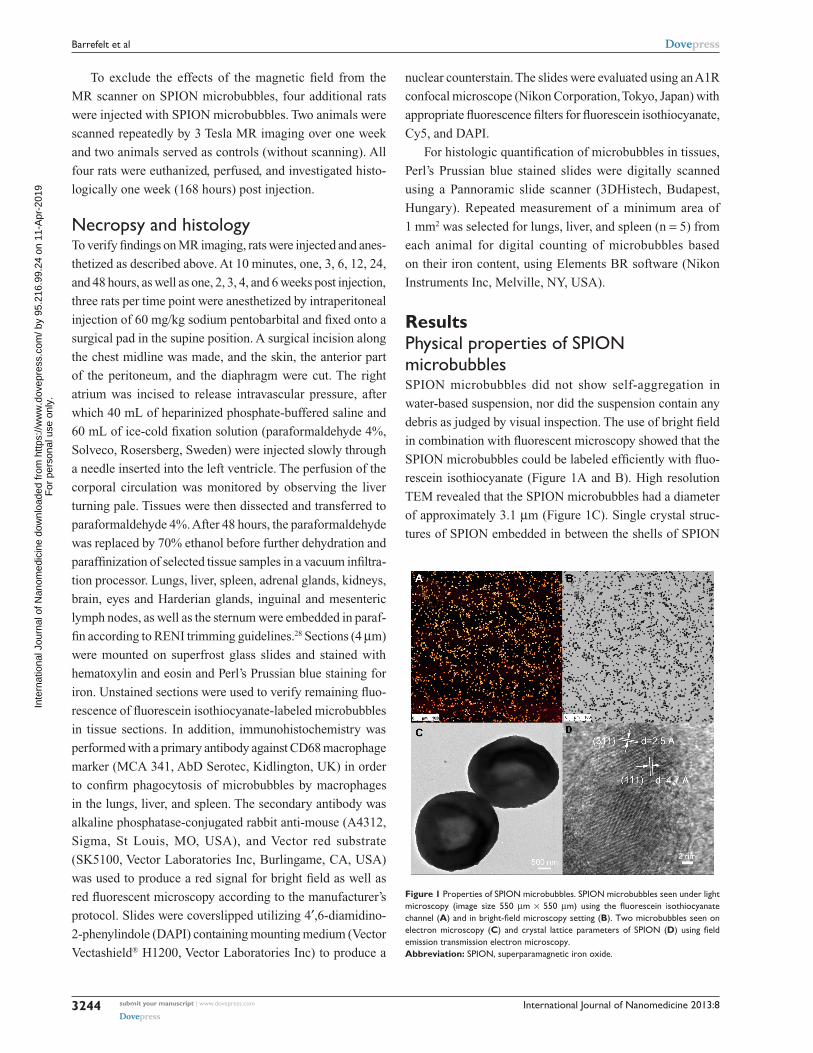
ResultsPhysical properties of sPION microbubblesSPION microbubbles did not show self-aggregation in
water-based suspension, nor did the suspension contain any
debris as judged by visual inspection. The use of bright field
in combination with fluorescent microscopy showed that the
SPION microbubbles could be labeled efficiently with fluo-
rescein isothiocyanate (Figure 1A and B). High resolution
TEM revealed that the SPION microbubbles had a diameter
of approximately 3.1 µm (Figure 1C). Single crystal struc-
tures of SPION embedded in between the shells of SPION
Figure 1 Properties of sPION microbubbles. sPION microbubbles seen under light microscopy (image size 550 µm × 550 µm) using the fluorescein isothiocyanate channel (A) and in bright-field microscopy setting (B). Two microbubbles seen on electron microscopy (C) and crystal lattice parameters of sPION (D) using field emission transmission electron microscopy. Abbreviation: sPION, superparamagnetic iron oxide.
In
tern
atio
nal J
ourn
al o
f Nan
omed
icin
e do
wnl
oade
d fr
om h
ttps:
//ww
w.d
ovep
ress
.com
/ by
95.2
16.9
9.24
on
11-A
pr-2
019
For
per
sona
l use
onl
y.
Powered by TCPDF (www.tcpdf.org)
1 / 1
International Journal of Nanomedicine 2013:8 submit your manuscript | www.dovepress.com
Dovepress
Dovepress
3245
Biological fate of sPION microbubbles in rats
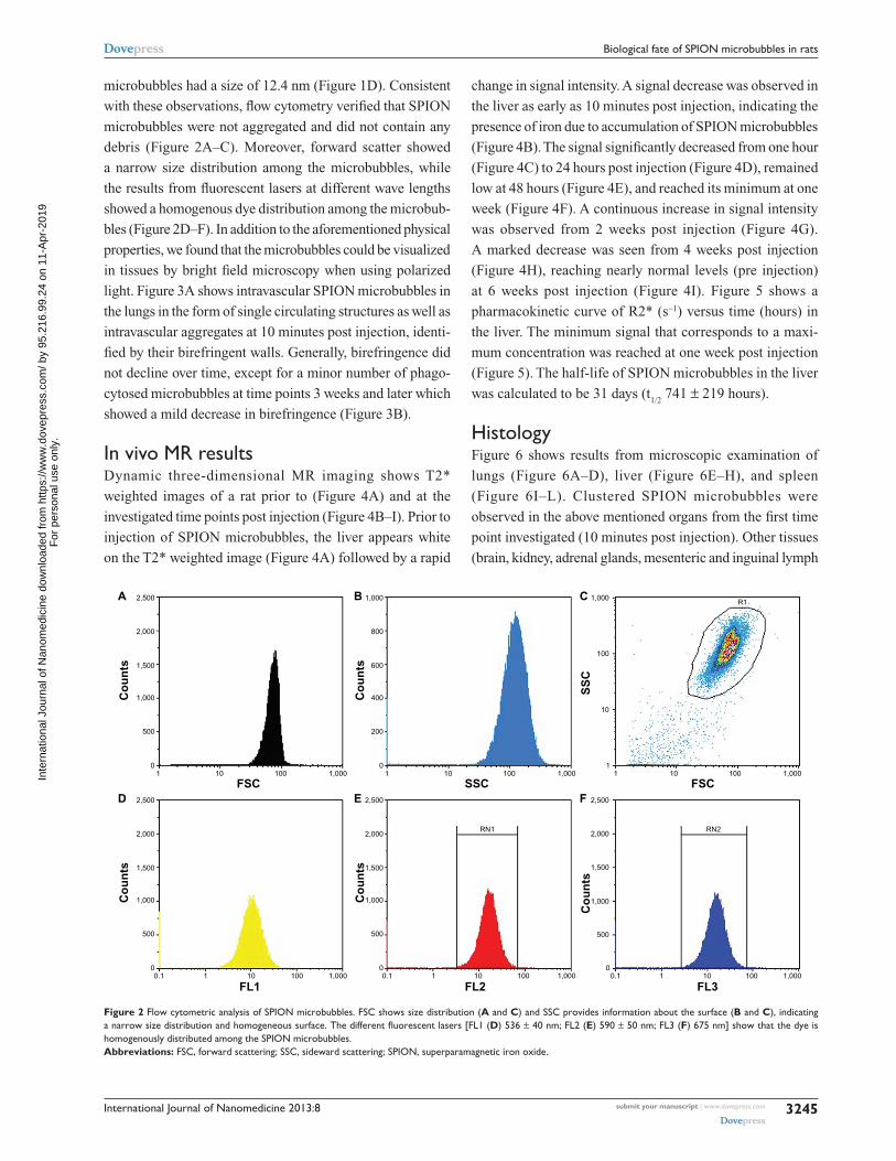
microbubbles had a size of 12.4 nm (Figure 1D). Consistent
with these observations, flow cytometry verified that SPION
microbubbles were not aggregated and did not contain any
debris (Figure 2A–C). Moreover, forward scatter showed
a narrow size distribution among the microbubbles, while
the results from fluorescent lasers at different wave lengths
showed a homogenous dye distribution among the microbub-
bles (Figure 2D–F). In addition to the aforementioned physical
properties, we found that the microbubbles could be visualized
in tissues by bright field microscopy when using polarized
light. Figure 3A shows intravascular SPION microbubbles in
the lungs in the form of single circulating structures as well as
intravascular aggregates at 10 minutes post injection, identi-
fied by their birefringent walls. Generally, birefringence did
not decline over time, except for a minor number of phago-
cytosed microbubbles at time points 3 weeks and later which
showed a mild decrease in birefringence (Figure 3B).
In vivo Mr resultsDynamic three-dimensional MR imaging shows T2*
weighted images of a rat prior to (Figure 4A) and at the
investigated time points post injection (Figure 4B–I). Prior to
injection of SPION microbubbles, the liver appears white
on the T2* weighted image (Figure 4A) followed by a rapid
change in signal intensity. A signal decrease was observed in
the liver as early as 10 minutes post injection, indicating the
presence of iron due to accumulation of SPION microbubbles
(Figure 4B). The signal significantly decreased from one hour
(Figure 4C) to 24 hours post injection (Figure 4D), remained
low at 48 hours (Figure 4E), and reached its minimum at one
week (Figure 4F). A continuous increase in signal intensity
was observed from 2 weeks post injection (Figure 4G).
A marked decrease was seen from 4 weeks post injection
(Figure 4H), reaching nearly normal levels (pre injection)
at 6 weeks post injection (Figure 4I). Figure 5 shows a
pharmacokinetic curve of R2* (s-1) versus time (hours) in
the liver. The minimum signal that corresponds to a maxi-
mum concentration was reached at one week post injection
(Figure 5). The half-life of SPION microbubbles in the liver
was calculated to be 31 days (t1/2
741 ± 219 hours).
histologyFigure 6 shows results from microscopic examination of
lungs (Figure 6A–D), liver (Figure 6E–H), and spleen
(Figure 6I–L). Clustered SPION microbubbles were
observed in the above mentioned organs from the first time
point investigated (10 minutes post injection). Other tissues
(brain, kidney, adrenal glands, mesenteric and inguinal lymph
2,500
2,000
1,500
1,000
500
01 10 100 1,000
A
Co
un
ts
FSC
1,000
800
600
400
200
01 10 100 1,000
B
Co
un
ts
SSC
1,000
100
10
11 10 100 1,000
R1C
SS
C
FSC2,500
2,000
1,500
1,000
500
010.1 10 100 1,000
D
Co
un
ts
FL1
2,500
2,000
1,500
RN1 RN2
1,000
500
00.1 1 10 100 1,000
E
Co
un
ts
FL2
2,500
2,000
1,000
500
1,500
00.1 1 10 1,000100
F
Co
un
ts
FL3
Figure 2 Flow cytometric analysis of sPION microbubbles. Fsc shows size distribution (A and C) and ssc provides information about the surface (B and C), indicating a narrow size distribution and homogeneous surface. The different fluorescent lasers [FL1 (D) 536 ± 40 nm; Fl2 (E) 590 ± 50 nm; Fl3 (F) 675 nm] show that the dye is homogenously distributed among the sPION microbubbles. Abbreviations: Fsc, forward scattering; ssc, sideward scattering; sPION, superparamagnetic iron oxide.
In
tern
atio
nal J
ourn
al o
f Nan
omed
icin
e do
wnl
oade
d fr
om h
ttps:
//ww
w.d
ovep
ress
.com
/ by
95.2
16.9
9.24
on
11-A
pr-2
019
For
per
sona
l use
onl
y.
Powered by TCPDF (www.tcpdf.org)
1 / 1

International Journal of Nanomedicine 2013:8submit your manuscript | www.dovepress.com
Dovepress
Dovepress
3246
Barrefelt et al
Figure 3 Polarizing properties of sPION microbubbles. single and clustered sPION microbubbles in pulmonary vasculature and macrophages (10 minutes post injection) show birefringent walls when viewed using polarized light (A). At 3 weeks post injection, a decrease in birefringence is shown in phagocytosed SPION microbubbles [arrow, (B)]. The size bar represents 10 µm. Abbreviation: sPION, superparamagnetic iron oxide.
Figure 4 Dynamic magnetic resonance imaging of a rat after injection of sPION microbubbles. Magnetic resonance signal intensity changed mostly in the liver over time from 10 minutes to 6 weeks compared with pre-injection. (A) Pre-injection and at (B) 10 minutes, (C) one hour, (D) 24 hours, (E) 48 hours, (F) one week, (G) 2 weeks, (H) 4 weeks, and (I) 6 weeks post injection. Abbreviation: sPION, superparamagnetic iron oxide.
In
tern
atio
nal J
ourn
al o
f Nan
omed
icin
e do
wnl
oade
d fr
om h
ttps:
//ww
w.d
ovep
ress
.com
/ by
95.2
16.9
9.24
on
11-A
pr-2
019
For
per
sona
l use
onl
y.
Powered by TCPDF (www.tcpdf.org)
1 / 1
International Journal of Nanomedicine 2013:8 submit your manuscript | www.dovepress.com
Dovepress
Dovepress
3247
Biological fate of sPION microbubbles in rats
nodes, sternal bone marrow, and eyes) showed rare solitary
microbubbles apparently free in the circulatory system from
10 minutes post injection onwards.
Clusters or aggregates of SPION microbubbles were
largest and most frequent in the lungs after intravenous
injection. At 10 minutes post injection (Figure 6A),
numerous microbubbles were seen in the capillaries of
the alveolar septa and less frequently in the occasion-
ally distended small arterioles along the bronchial tree.
Intravascular clusters are often associated with intravas-
cular, finely fibrillar, and eosinophilic material (consistent
with fibrin). Marked infiltration of inflammatory cells
was absent in all organs investigated except for the lungs
where, from 3 hours after injection, increased numbers of
intravascular and rarely alveolar neutrophils were noted,
as well as a minimal increase in macrophages associ-
ated with the clustered SPION microbubbles. Pulmonary
macrophages became more apparent after 6 hours and
neutrophil numbers further increased to a maximum at
12 hours post injection, after which neutrophil numbers
decreased while macrophages remained and increased in
size. Numbers of freely circulating microbubbles decreased
from 24–48 hours post injection (Figure 6B). Neutrophil
numbers further declined one week after injection and
reached background levels at 2 weeks following injection
(Figure 6C). From 48 hours after injection and later, the
remaining clusters of SPION microbubbles associated
with macrophages decreased in size. A decreased number
of phagocytosed microbubbles were observed at 2 weeks
(Figure 6C), while at 4 weeks following injection (Figure
6D) the now abundant macrophage cytoplasm started to
0.5
0.4
0.3
0.2
0.1
0.00 500 1,000
Time (hours)
R2*
(s−1
)
Figure 5 Pharmacokinetic model of sPION microbubbles. relaxation times (r2*; s-1) of sPION microbubbles in the liver (as indicator for concentrations) versus time (hours) were fitted to a one-compartment open model. The elimination half-life was calculated from the slope. Abbreviation: sPION, superparamagnetic iron oxide.
Figure 6 Histology of the lungs, liver, and spleen of a rat post injection of SPION microbubbles. Abundant intravascular, fibrin-covered microbubbles in lung at 10 minutes (A) and 24 hours (B). a decreased number of phagocytosed microbubbles were observed at 2 weeks (C). Iron pigment becomes apparent in macrophage cytoplasm after 4 weeks (D). In the liver, rare microbubbles are found in the vicinity of Kupffer cells at 10 minutes post injection (E), and increasingly phagocytosed at 24 hours (F). Phagocytosed microbubbles were abundant at 2 weeks post injection (G) and decreased 4 weeks after injection (H). In the spleen, 10 minutes after injection, microbubbles are found in red pulp and marginal zone (I). From 24 hours after injection and onwards, the microbubbles are associated with marginal zone macrophages and rare in red pulp (J). (K) at 2 weeks and (L) at 4 weeks. The size bar represents 10 µm. The arrows show the microbubbles in the different tissues. Abbreviation: sPION, superparamagnetic iron oxide.
In
tern
atio
nal J
ourn
al o
f Nan
omed
icin
e do
wnl
oade
d fr
om h
ttps:
//ww
w.d
ovep
ress
.com
/ by
95.2
16.9
9.24
on
11-A
pr-2
019
For
per
sona
l use
onl
y.
Powered by TCPDF (www.tcpdf.org)
1 / 1
International Journal of Nanomedicine 2013:8submit your manuscript | www.dovepress.com
Dovepress
Dovepress
3248
Barrefelt et al
show a brown color, indicative of accumulation of iron
released from SPION microbubbles.
In the liver, small clusters of microbubbles (1–3) were
found in the sinusoids as early as 10 minutes post injec-
tion, mostly associated with macrophages (Figure 6E).
Microbubble clusters were more frequent in the periportal
areas. Over the following hours, cluster frequency and size
(6–8 microbubbles per Kupffer cell) increased (Figure 6F).
Microbubble clusters became clearly less frequent at
2 weeks post injection (Figure 6G), whereas cluster size
decreased more slowly but recognizably from 3 weeks post
injection (Figure 6H). In the spleen, frequent microbubbles
were observed from 10 minutes post injection, and seen
predominantly as solitary microbubbles and small clusters
(2–3 per cell) associated with macrophages in the marginal
zone (Figure 6I). Fewer solitary bubbles were observed in
the red pulp (Figure 6I). The frequency of microbubbles
in the marginal zone peaked at 12 and 24 hours post injection
(Figure 6J), whereas a mild decrease was observed in the red
pulp as early as 24 hours post injection (Figure 6J). A negli-
gible number of solitary bubbles was occasionally observed in
the periarteriolar lymphoid sheaths from 3 weeks post injec-
tion. The marginal zone was not markedly expanded at any
time point following injection. In the spleen, macrophages
showed signs of cytoplasmic iron accumulation from 2 weeks
post injection (Figure 6K). In the liver, these signs were not
clearly visible on hematoxylin and eosin or Perl’s staining at
any time point investigated. Macrophages in the red pulp of
the spleen showed massive accumulation of brown pigment
(Figure 6L), consistent with iron accumulation, at 6 weeks
post injection (Figure 7I).
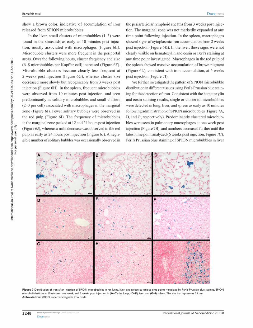
We further investigated the pattern of SPION microbubble
distribution in different tissues using Perl’s Prussian blue stain-
ing for the detection of iron. Consistent with the hematoxylin
and eosin staining results, single or clustered microbubbles
were detected in lung, liver, and spleen as early as 10 minutes
following administration of SPION microbubbles (Figure 7A,
D, and G, respectively). Predominantly clustered microbub-
bles were seen in pulmonary macrophages at one week post
injection (Figure 7B), and numbers decreased further until the
latest time point analyzed (6 weeks post injection, Figure 7C).
Perl’s Prussian blue staining of SPION microbubbles in liver
Figure 7 Distribution of iron after injection of sPION microbubbles in rat lungs, liver, and spleen at various time points visualized by Perl’s Prussian blue staining. sPION microbubbles/iron at 10 minutes, one week, and 6 weeks post injection in (A–C) the lungs, (D–F) liver, and (G–I) spleen. The size bar represents 25 µm. Abbreviation: sPION, superparamagnetic iron oxide.
In
tern
atio
nal J
ourn
al o
f Nan
omed
icin
e do
wnl
oade
d fr
om h
ttps:
//ww
w.d
ovep
ress
.com
/ by
95.2
16.9
9.24
on
11-A
pr-2
019
For
per
sona
l use
onl
y.
Powered by TCPDF (www.tcpdf.org)
1 / 1
International Journal of Nanomedicine 2013:8 submit your manuscript | www.dovepress.com
Dovepress
Dovepress
3249
Biological fate of sPION microbubbles in rats
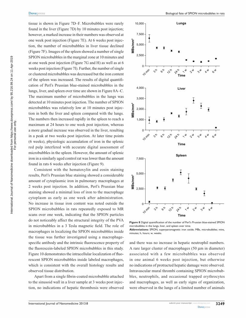
tissue is shown in Figure 7D–F. Microbubbles were rarely
found in the liver (Figure 7D) by 10 minutes post injection;
however, a marked increase in their numbers was observed at
one week post injection (Figure 7E). At 6 weeks post injec-
tion, the number of microbubbles in liver tissue declined
(Figure 7F). Images of the spleen showed a number of single
SPION microbubbles in the marginal zone at 10 minutes and
at one week post injection (Figure 7G and H) as well as at 6
weeks post injection (Figure 7I). Further, the number of single
or clustered microbubbles was decreased but the iron content
of the spleen was increased. The results of digital quantifi-
cation of Perl’s Prussian blue-stained microbubbles in the
lungs, liver, and spleen over time are shown in Figure 8A–C.
The maximum number of microbubbles in the lungs was
detected at 10 minutes post injection. The number of SPION
microbubbles was relatively low at 10 minutes post injec-
tion in both the liver and spleen compared with the lungs.
The numbers then increased rapidly in the spleen to reach a
maximum at 24 hours to one week post injection, whereas
a more gradual increase was observed in the liver, resulting
in a peak at two weeks post injection. At later time points
(6 weeks), physiologic accumulation of iron in the splenic
red pulp interfered with accurate digital assessment of
microbubbles in the spleen. However, the amount of splenic
iron in a similarly aged control rat was lower than the amount
found in rats 6 weeks after injection (Figure 9).
Consistent with the hematoxylin and eosin staining
results, Perl’s Prussian blue staining showed a considerable
amount of cytoplasmic iron in pulmonary macrophages at
2 weeks post injection. In addition, Perl’s Prussian blue
staining showed a minimal loss of iron to the macrophage
cytoplasm as early as one week after administration.
No increase in tissue iron content was noted outside the
SPION microbubbles in rats repeatedly exposed to MR
scans over one week, indicating that the SPION particles
do not noticeably affect the structural integrity of the PVA
in microbubbles in a 3 Tesla magnetic field. The role of
macrophages in localizing the SPION microbubbles inside
the tissue was further investigated using a macrophage-
specific antibody and the intrinsic fluorescence property of
the fluorescein-labeled SPION microbubbles in this study.
Figure 10 demonstrates the intracellular localization of fluo-
rescent SPION microbubbles inside labeled macrophages,
which is consistent with the overall histology results and
observed tissue distribution.
Apart from a single fibrin-coated microbubble attached
to the sinusoid wall in a liver sample at 3 weeks post injec-
tion, no indications of hepatic thrombosis were observed
and there was no increase in hepatic neutrophil numbers.
A rare larger cluster of macrophages (50 µm in diameter)
associated with a few microbubbles was observed
in one animal 6 weeks post injection, but otherwise
no indications of protracted hepatic damage were observed.
Intravascular mural thrombi containing SPION microbub-
bles, neutrophils, and occasional trapped erythrocytes
and macrophages, as well as early signs of organization,
were observed in the lungs of a limited number of animals
6 w
4 w
3 w
2 w
1 w
24 h
12 h6
h3
h
10 m
in
10,000
7,500
5,000
2,500
0
Lungs
Spleen
Time
MB
s/m
m2
4 w
3 w
2 w
1 w
24 h
12 h6
h3
h
10 m
in
7,500
5,000
2,500
0
Time
MB
s/m
m2
Liver
6 w
4 w
3 w
2 w
1 w
24 h
12 h6
h3
h
10 m
in
4,000
3,000
2,000
1,000
0
Time
MB
s/m
m2
Figure 8 Digital quantification of the number of Perl’s Prussian blue-stained SPION microbubbles in the lungs, liver, and spleen over time. Abbreviations: sPION, superparamagnetic iron oxide; MBs, microbubbles; mins, minutes; h, hours; w, weeks.
In
tern
atio
nal J
ourn
al o
f Nan
omed
icin
e do
wnl
oade
d fr
om h
ttps:
//ww
w.d
ovep
ress
.com
/ by
95.2
16.9
9.24
on
11-A
pr-2
019
For
per
sona
l use
onl
y.
Powered by TCPDF (www.tcpdf.org)
1 / 1

International Journal of Nanomedicine 2013:8submit your manuscript | www.dovepress.com
Dovepress
Dovepress
3250
Barrefelt et al
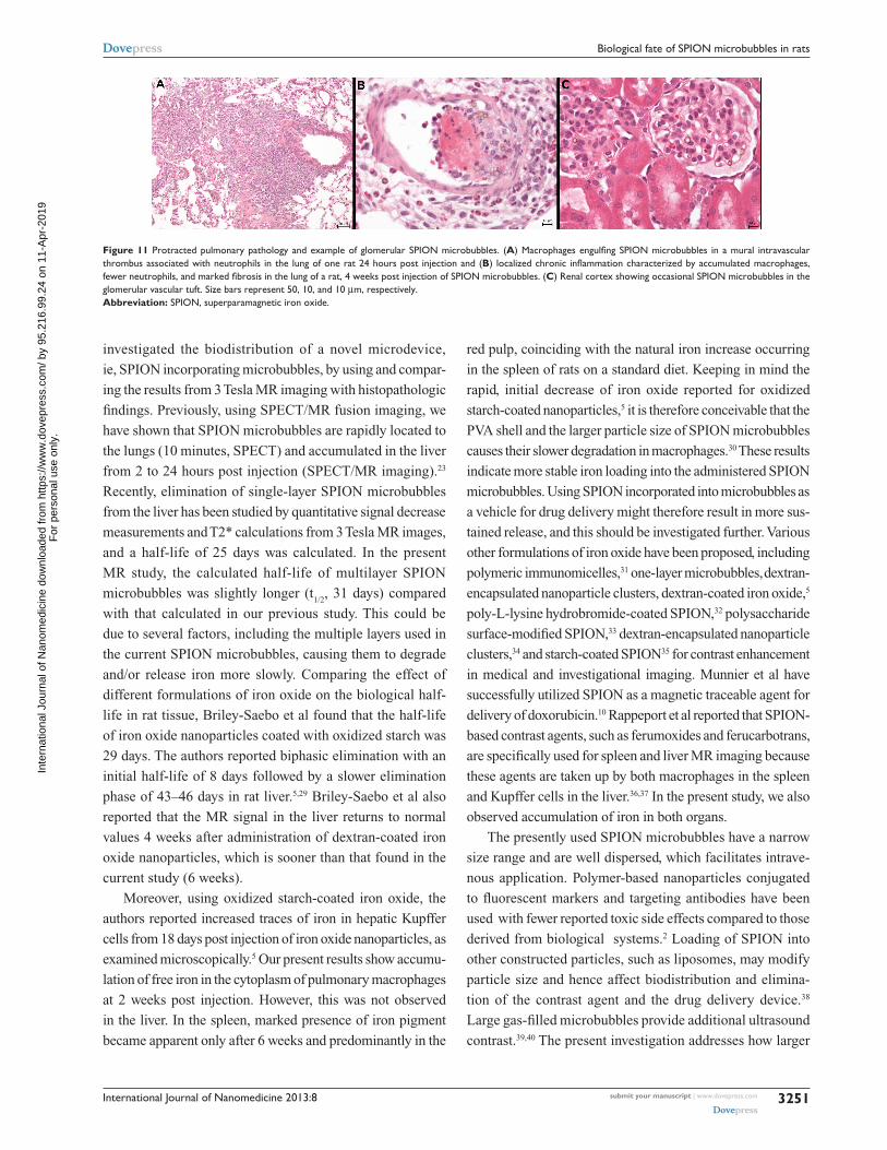
(Figure 11A) at 6 and 12 hours. Thrombi were no longer
observed after 3 weeks and onwards; however, one animal
sacrificed at 3 weeks and another at 6 weeks showed larger
clusters of macrophages with occasional neutrophils and
proliferation of bronchus-associated lymphoid tissue
(Figure 11B), indicating granulomatous pneumonia and a
possible risk of mild, longer persisting pulmonary damage
at the currently used dose.
Marked presence of SPION microbubbles was
not observed in any of the other organs investigated
microscopically. The kidneys showed occasional solitary
microbubbles in the glomerular capillaries (Figure 11C),
increasing up to 6 hours post injection and thereafter
gradually decreasing until becoming absent from 4 weeks
post injection. Microbubbles were not observed in the
mesangium. Rare microbubbles were observed in the corti-
cal interstitial blood vessels until one week post injection.
Occasional mild tubular protein was observed, but not in
relation to glomerular microbubble frequency. However,
this observation was slightly more frequent around 6 and
12 hours post injection, coinciding with the influx of pul-
monary neutrophils, which might indicate a mild cytokine-
related proteinuria at these time points.
Bone marrow showed infrequent solitary microbubbles,
sometimes phagocytosed but also free in the sinuses. The
bone marrow showed no other abnormalities, but gradually
increasing nuclear hypertrophy was recognized in endosteal
cells from 3 weeks onwards. At 6 weeks post injection, some
deposition of osteoid was observed along bone trabeculae,
with a slight brown discoloration of recently deposited
subendosteal lamellar bone (data not shown).
Brain and eye tissue showed rare, mostly free, intravas-
cular bubbles until 48 hours post injection, not related to
brain regions and located in the choroid capillaries of the
eye. Apart from negligible single microbubbles in the cortex
of mesenteric lymph nodes in three animals at different time
points across the study, no microbubbles were observed in
the lymph nodes collected.
DiscussionRecent advances in nanoscience and the development of
nanoparticles have revolutionized biomedicine during the
last decade. The characteristics of engineered nanoparticles
can be easily manipulated for use in drug delivery and vari-
ous routes for tracing, administration, and controlled release.
Microbubbles with a PVA matrix can also be tailored as
an imaging agent or drug carrier. In the present study, we
Figure 9 The amount of iron in the spleen of 12-week-old and 25-week-old control rats compared with a 25-week-old rat injected with sPION microbubbles as visualized using Perl’s Prussian blue staining. (A) The spleen of a 12-week-old control rat (untreated) shows a limited amount of iron, while (B) in the 25-week-old control rat, the amount of iron was increased. (C) shows the amount of iron found in the spleen of the 25-week-old rat injected 6 weeks previously with sPION microbubbles, that was considerably higher compared with the untreated control. Abbreviation: sPION, superparamagnetic iron oxide.
Figure 10 Intracellular localization of fluorescent SPION microbubbles inside labeled macrophages. A primary antibody against CD68 (red) confirms phagocytosis of the microbubbles (green) by macrophages in the lungs. The nuclei were stained blue using DaPI. The size bar represents 25 µm. The upper left part of the image is higher magnification with the size bar of 10 µm. Abbreviations: DaPI, 4′,6-diamidino-2-phenylindole; sPION, superparamagnetic iron oxide.
In
tern
atio
nal J
ourn
al o
f Nan
omed
icin
e do
wnl
oade
d fr
om h
ttps:
//ww
w.d
ovep
ress
.com
/ by
95.2
16.9
9.24
on
11-A
pr-2
019
For
per
sona
l use
onl
y.
Powered by TCPDF (www.tcpdf.org)
1 / 1
International Journal of Nanomedicine 2013:8 submit your manuscript | www.dovepress.com
Dovepress
Dovepress
3251
Biological fate of sPION microbubbles in rats
investigated the biodistribution of a novel microdevice,
ie, SPION incorporating microbubbles, by using and compar-
ing the results from 3 Tesla MR imaging with histopathologic
findings. Previously, using SPECT/MR fusion imaging, we
have shown that SPION microbubbles are rapidly located to
the lungs (10 minutes, SPECT) and accumulated in the liver
from 2 to 24 hours post injection (SPECT/MR imaging).23
Recently, elimination of single-layer SPION microbubbles
from the liver has been studied by quantitative signal decrease
measurements and T2* calculations from 3 Tesla MR images,
and a half-life of 25 days was calculated. In the present
MR study, the calculated half-life of multilayer SPION
microbubbles was slightly longer (t1/2
, 31 days) compared
with that calculated in our previous study. This could be
due to several factors, including the multiple layers used in
the current SPION microbubbles, causing them to degrade
and/or release iron more slowly. Comparing the effect of
different formulations of iron oxide on the biological half-
life in rat tissue, Briley-Saebo et al found that the half-life
of iron oxide nanoparticles coated with oxidized starch was
29 days. The authors reported biphasic elimination with an
initial half-life of 8 days followed by a slower elimination
phase of 43–46 days in rat liver.5,29 Briley-Saebo et al also
reported that the MR signal in the liver returns to normal
values 4 weeks after administration of dextran-coated iron
oxide nanoparticles, which is sooner than that found in the
current study (6 weeks).
Moreover, using oxidized starch-coated iron oxide, the
authors reported increased traces of iron in hepatic Kupffer
cells from 18 days post injection of iron oxide nanoparticles, as
examined microscopically.5 Our present results show accumu-
lation of free iron in the cytoplasm of pulmonary macrophages
at 2 weeks post injection. However, this was not observed
in the liver. In the spleen, marked presence of iron pigment
became apparent only after 6 weeks and predominantly in the
red pulp, coinciding with the natural iron increase occurring
in the spleen of rats on a standard diet. Keeping in mind the
rapid, initial decrease of iron oxide reported for oxidized
starch-coated nanoparticles,5 it is therefore conceivable that the
PVA shell and the larger particle size of SPION microbubbles
causes their slower degradation in macrophages.30 These results
indicate more stable iron loading into the administered SPION
microbubbles. Using SPION incorporated into microbubbles as
a vehicle for drug delivery might therefore result in more sus-
tained release, and this should be investigated further. Various
other formulations of iron oxide have been proposed, including
polymeric immunomicelles,31 one-layer microbubbles, dextran-
encapsulated nanoparticle clusters, dextran-coated iron oxide,5
poly-L-lysine hydrobromide-coated SPION,32 polysaccharide
surface-modified SPION,33 dextran- encapsulated nanoparticle
clusters,34 and starch-coated SPION35 for contrast enhancement
in medical and investigational imaging. Munnier et al have
successfully utilized SPION as a magnetic traceable agent for
delivery of doxorubicin.10 Rappeport et al reported that SPION-
based contrast agents, such as ferumoxides and ferucarbotrans,
are specifically used for spleen and liver MR imaging because
these agents are taken up by both macrophages in the spleen
and Kupffer cells in the liver.36,37 In the present study, we also
observed accumulation of iron in both organs.
The presently used SPION microbubbles have a narrow
size range and are well dispersed, which facilitates intrave-
nous application. Polymer-based nanoparticles conjugated
to fluorescent markers and targeting antibodies have been
used with fewer reported toxic side effects compared to those
derived from biological systems.2 Loading of SPION into
other constructed particles, such as liposomes, may modify
particle size and hence affect biodistribution and elimina-
tion of the contrast agent and the drug delivery device.38
Large gas-filled microbubbles provide additional ultrasound
contrast.39,40 The present investigation addresses how larger
Figure 11 Protracted pulmonary pathology and example of glomerular sPION microbubbles. (A) Macrophages engulfing SPION microbubbles in a mural intravascular thrombus associated with neutrophils in the lung of one rat 24 hours post injection and (B) localized chronic inflammation characterized by accumulated macrophages, fewer neutrophils, and marked fibrosis in the lung of a rat, 4 weeks post injection of SPION microbubbles. (C) renal cortex showing occasional sPION microbubbles in the glomerular vascular tuft. size bars represent 50, 10, and 10 µm, respectively. Abbreviation: sPION, superparamagnetic iron oxide.
In
tern
atio
nal J
ourn
al o
f Nan
omed
icin
e do
wnl
oade
d fr
om h
ttps:
//ww
w.d
ovep
ress
.com
/ by
95.2
16.9
9.24
on
11-A
pr-2
019
For
per
sona
l use
onl
y.
Powered by TCPDF (www.tcpdf.org)
1 / 1
International Journal of Nanomedicine 2013:8submit your manuscript | www.dovepress.com
Dovepress
Dovepress
3252
Barrefelt et al
vehicles would affect the distribution and half-life of the iron
oxide signal using MR imaging. Microscopic tissue sample
analysis confirmed rapid uptake of SPION microbubbles
in the lungs and their redistribution to the liver and spleen
over time. As we have previously hypothesized, and in line
with findings of various authors regarding smaller intrave-
nously administered nanoparticles,3,41–43 histology showed
the prominent role of macrophage uptake along the pulmo-
nary capillary bed and (predominantly periportal) hepatic
sinusoids. Further, these results were confirmed utilizing
immunohistochemistry. Despite distribution to the liver and
the relatively long half-life of SPION microbubbles, we did
not observe signs of necrosis, inflammation, or fibrosis in the
liver and spleen. These findings are in agreement with those
of Bacon et al,45 who studied several biochemical liver param-
eters for redox toxicity (including hepatic, mitochondrial,
and microsomal lipid peroxidation) in rats after intravenous
ferrite injections, without finding evidence of iron-induced
hepatotoxicity or organelle dysfunction. Therefore, it is
unlikely that an increased iron load after injection of SPION
microbubbles will give rise to adverse effects. The loss of
iron from SPION microbubbles in the present study occurred
gradually, and had already appeared while the morphologi-
cal integrity was still preserved. A marked increase in iron
load in the spleen was finally observed when frequent signs
of integrity loss were seen at 6 weeks post administration.
Additional evaluation of how repeated application of a 3 Tesla
magnetic field might affect integrity of the microbubble wall
and accelerate iron loss to the tissues did not show any his-
tologic indication for increased tissue iron load. The initially
widespread intravascular clustering of SPION microbubbles
in the lungs largely resolved after 3 weeks; however, an indi-
cation of possible adverse effects resulting from intravenous
administration of large numbers of SPION microbubbles was
found in the lungs of two rats at later time points, where we
observed remains of organized vascular thrombi and focal
granulomas. In these rats, localized granulomas had formed
in association with unresolved aggregates of microbubbles.
Further studies addressing the mode, speed of administration,
and biological fate of SPION microbubbles should clarify
if these potentially negative effects can be attenuated and
whether slower degradation of these particles can serve in
a complementary role to existing nanoscale targeted drug
delivery systems.
ConclusionHistologic f indings are generally correlated with MR
imaging. The methods used in the current study appropriately
characterize the properties of SPION microbubbles in terms
of size, size distribution, the effect on rat tissue in various
organs in vivo, cellular distribution, biological fate, and the
relaxivity of SPION microbubbles using MR imaging. Further
investigations should address the dynamics of drug release
from the microbubbles, modifications for organ targeting, size
effect on biodistribution and kinetics, tendency to cluster in
vivo, and delivery dynamics during administration to attenuate
possible adverse effects on the pulmonary vasculature.
AcknowledgmentThis work was supported by the European Commission
project 3MICRON within the framework of the Seventh
Framework Program, the Swedish Cancer Foundation
(CancerFonden), and the Swedish Children’s Cancer Society
(BarnCancerFonden).
DisclosureThe authors declare that they have no conflict of interest in
this work.
References 1. Semete B, Booysen L, Lemmer Y, et al. In vivo evaluation of the
biodistribution and safety of PLGA nanoparticles as drug delivery systems. Nanomedicine. 2010;6(5):662–671.
2. Singh P, Prasuhn D, Yeh RM, et al. Bio-distribution, toxicity and pathology of cowpea mosaic virus nanoparticles in vivo. J Control Release. 2007;120(1–2):41–50.
3. Xie G, Sun J, Zhong G, Shi L, Zhang D. Biodistribution and toxicity of intravenously administered silica nanoparticles in mice. Arch Toxicol. 2010;84(3):183–190.
4. Yoon HY, Saravanakumar G, Heo R, et al. Hydrotropic magnetic micelles for combined magnetic resonance imaging and cancer therapy. J Control Release. 2012;160(3):692–698.
5. Briley-Saebo K, Hustvedt SO, Haldorsen A, Bjornerud A. Long-term imaging effects in rat liver after a single injection of an iron oxide nanoparticle based MR contrast agent. J Magn Reson Imaging. 2004;20(4):622–631.
6. Inoh K, Muramatsu H, Torii S, et al. Doxorubicin-conjugated anti-midkine monoclonal antibody as a potential anti-tumor drug. Jpn J Clin Oncol. 2006;36(4):207–211.
7. Mahmoudi M, Sant S, Wang B, Laurent S, Sen T. Superparamagnetic iron oxide nanoparticles (SPIONs): development, surface modification and applications in chemotherapy. Adv Drug Deliv Rev. 2011;63(1–2): 24–46.
8. Kim do K, Chang JH, Kang YJ. Efficient internalization of peptide-conjugated SPIONs in dendritic cells for tumor targeting. J Nanosci Nanotechnol. 2012;12(7):5191–5198.
9. Vingerhoeds MH, Haisma HJ, van Muijen M, van de Rijt RB, Crommelin DJ, Storm G. A new application for liposomes in cancer therapy. Immunoliposomes bearing enzymes (immuno-enzymosomes) for site-specific activation of prodrugs. FEBS Lett. 1993;336(3): 485–490.
10. Munnier E, Cohen-Jonathan S, Linassier C, et al. Novel method of doxorubicin-SPION reversible association for magnetic drug targeting. Int J Pharm. 2008;363(1–2):170–176.
11. Laroui H, Sitaraman SV, Merlin D. Gastrointestinal delivery of anti-inflammatory nanoparticles. Methods Enzymol. 2012;509:101–125.
In
tern
atio
nal J
ourn
al o
f Nan
omed
icin
e do
wnl
oade
d fr
om h
ttps:
//ww
w.d
ovep
ress
.com
/ by
95.2
16.9
9.24
on
11-A
pr-2
019
For
per
sona
l use
onl
y.
Powered by TCPDF (www.tcpdf.org)
1 / 1
International Journal of Nanomedicine 2013:8 submit your manuscript | www.dovepress.com
Dovepress
Dovepress
3253
Biological fate of sPION microbubbles in rats
12. Wang J, Wang X, Song Y, Zhu C, Wang K, Guo Z. Detecting and delivering platinum anticancer drugs using fluorescent maghemite nanoparticles. Chem Commun (Camb). 2013;49(27):2786–2788.
13. Carney CE, Tran AD, Wang J, Schabel MC, Sherry AD, Woods M. Towards the rational design of MRI contrast agents: delta-substitution of lanthanide(III) NB-DOTA-tetraamide chelates influences but does not control coordination geometry. Chemistry. 2011;17(37): 10372–10378.
14. Martinez GV, Zhang X, Garcia-Martin ML, et al. Imaging the extracellular pH of tumors by MRI after injection of a single cock-tail of T1 and T2 contrast agents. NMR Biomed. 2011;24(10): 1380–1391.
15. Wu Y, Carney CE, Denton M, et al. Polymeric PARACEST MRI contrast agents as potential reporters for gene therapy. Org Biomol Chem. 2010;8(23):5333–5338.
16. Wu Y, Zhou Y, Ouari O, et al. Polymeric PARACEST agents for enhancing MRI contrast sensitivity. J Am Chem Soc. 2008;130(42): 13854–13855.
17. Borel A, Bean JF, Clarkson RB, et al. Towards the rational design of MRI contrast agents: electron spin relaxation is largely unaffected by the coordination geometry of gadolinium(III)-DOTA-type complexes. Chemistry. 2008;14(9):2658–2667.
18. Woods M, Botta M, Avedano S, Wang J, Sherry AD. Towards the rational design of MRI contrast agents: a practical approach to the synthesis of gadolinium complexes that exhibit optimal water exchange. Dalton Trans. 2005;24:3829–3837.
19. Lin SP, Brown JJ. MR contrast agents: physical and pharmacologic basics. J Magn Reson Imaging. 2007;25(5):884–899.
20. Frullano L, Catana C, Benner T, Sherry AD, Caravan P. Bimodal MR-PET agent for quantitative pH imaging. Angew Chem Int Ed Engl. 2010;49(13):2382–2384.
21. Zhang B, Li Q, Yin P, et al. Ultrasound-triggered BSA/SPION hybrid nanoclusters for liver-specific magnetic resonance imaging. ACS Appl Mater Interfaces. 2012;4(12):6479–6486.
22. Yoo MK, Park IK, Lim HT, et al. Folate-PEG-superparamagnetic iron oxide nanoparticles for lung cancer imaging. Acta Biomater. 2012;8(8):3005–3013.
23. Barrefelt AA, Brismar TB, Egri G, et al. Multimodality imaging using SPECT/CT and MRI and ligand functionalized 99mTc-labeled magnetic microbubbles. EJNMMI Res. 2013;3(1):12.
24. Wells DJ. Animal welfare and the 3Rs in European biomedical research. Ann N Y Acad Sci. 2011;1245:14–16.
25. Madden JC, Hewitt M, Przybylak K, Vandebriel RJ, Piersma AH, Cronin MT. Strategies for the optimisation of in vivo experiments in accordance with the 3Rs philosophy. Regul Toxicol Pharmacol. 2012;63(1):140–154.
26. Schiffelers MJ, Blaauboer BJ, Hendriksen CF, Bakker WE. Regulatory acceptance and use of 3R models: a multilevel perspective. ALTEX. 2012;29(3):287–300.
27. Peyratout CS, Dahne L. Tailor-made polyelectrolyte microcapsules: from multilayers to smart containers. Angew Chem Int Ed Engl. 2004;43(29):3762–3783.
28. Morawietz G, Ruehl-Fehlert C, Kittel B, et al. Revised guides for organ sampling and trimming in rats and mice. Part 3. A joint publication of the RITA and NACAD groups. Exp Toxicol Pathol. 2004;55(6): 433–449.
29. Briley-Saebo KC, Johansson LO, Hustvedt SO, et al. Clearance of iron oxide particles in rat liver: effect of hydrated particle size and coating material on liver metabolism. Invest Radiol. 2006;41(7): 560–571.
30. Shi R, Zhu AC, Chen DF, et al. In vitro degradation of starch/PVA films and biocompatibility evaluation. J Appl Polym Sci. 2010;115(1): 346–357.
31. Sawant RM, Sawant RR, Gultepe E, et al. Nanosized cancer cell-targeted polymeric immunomicelles loaded with superparamagnetic iron oxide nanoparticles. J Nanopart Res. 2009:1777–1785.
32. Albukhaty S, Naderi-Manesh H, Tiraihi T. In vitro labeling of neural stem cells with poly-L-lysine coated super paramagnetic nanoparticles for green fluorescent protein transfection. Iran Biomed J. 2013;17(2): 71–76.
33. Zhu A, Yuan L, Jin W, et al. Polysaccharide surface modified Fe3O
4
nanoparticles for camptothecin loading and release. Acta Biomater. 2009;5(5):1489–1498.
34. Mondalek FG, Zhang YY, Kropp B, et al. The permeability of SPION over an artificial three-layer membrane is enhanced by external magnetic field. J Nanobiotechnology. 2006;4:4.
35. Kim DK, Mikhaylova M, Wang FH, et al. Starch-coated superparamag-netic nanoparticles as MR contrast agents. Chem Mater. 2003;15(23): 4343–4351.
36. Rappeport ED, Loft A. Liver metastases from colorectal cancer: imaging with superparamagnetic iron oxide (SPIO)-enhanced MR imaging, computed tomography and positron emission tomography. Abdom Imaging. 2007;32(5):624–634.
37. Rappeport ED, Loft A, Berthelsen AK, et al. Contrast-enhanced FDG-PET/CT vs SPIO-enhanced MRI vs FDG-PET vs CT in patients with liver metastases from colorectal cancer: a prospective study with intraoperative confirmation. Acta Radiol. 2007;48(4):369–378.
38. Floris A, Ardu A, Musinu A, et al. SPION@liposomes hybrid nano-architectures with high density SPION association. Soft Matter. 2011;7(13):6239–6247.
39. Grishenkov D, Pecorari C, Brismar TB, Paradossi G. Characterization of acoustic properties of PVA-shelled ultrasound contrast agents: ultrasound-induced fracture (part II). Ultrasound Med Biol. 2009;35(7): 1139–1147.
40. Brismar TB, Grishenkov D, Gustafsson B, et al. Magnetite nanoparticles can be coupled to microbubbles to support multimodal imaging. Biomacromolecules. 2012;13(5):1390–1399.
41. Zahr AS, Davis CA, Pishko MV. Macrophage uptake of core-shell nanoparticles surface modified with poly(ethylene glycol). Langmuir. 2006;22(19):8178–8185.
42. Beduneau A, Ma Z, Grotepas CB, et al. Facilitated monocyte- macrophage uptake and tissue distribution of superparmagnetic iron-oxide nanoparticles. PloS One. 2009;4(2):e4343.
43. Nicolete R, dos Santos DF, Faccioli LH. The uptake of PLGA micro or nanoparticles by macrophages provokes distinct in vitro inflammatory response. Int Immunopharmacol. 2011;11(10):1557–1563.
44. Bacon BR, Stark DD, Park CH, et al. Ferrite particles: a new magnetic resonance imaging contrast agent. Lack of acute or chronic hepatotoxicity after intravenous administration. J Lab Clin Med. 1987; 110(2):164–171.
In
tern
atio
nal J
ourn
al o
f Nan
omed
icin
e do
wnl
oade
d fr
om h
ttps:
//ww
w.d
ovep
ress
.com
/ by
95.2
16.9
9.24
on
11-A
pr-2
019
For
per
sona
l use
onl
y.
Powered by TCPDF (www.tcpdf.org)
1 / 1
International Journal of Nanomedicine
Publish your work in this journal
Submit your manuscript here: http://www.dovepress.com/international-journal-of-nanomedicine-journal
The International Journal of Nanomedicine is an international, peer-reviewed journal focusing on the application of nanotechnology in diagnostics, therapeutics, and drug delivery systems throughout the biomedical field. This journal is indexed on PubMed Central, MedLine, CAS, SciSearch®, Current Contents®/Clinical Medicine,
Journal Citation Reports/Science Edition, EMBase, Scopus and the Elsevier Bibliographic databases. The manuscript management system is completely online and includes a very quick and fair peer-review system, which is all easy to use. Visit http://www.dovepress.com/ testimonials.php to read real quotes from published authors.
International Journal of Nanomedicine 2013:8submit your manuscript | www.dovepress.com
Dovepress
Dovepress
Dovepress
3254
Barrefelt et al
Inte
rnat
iona
l Jou
rnal
of N
anom
edic
ine
dow
nloa
ded
from
http
s://w
ww
.dov
epre
ss.c
om/ b
y 95
.216
.99.
24 o
n 11
-Apr
-201
9F
or p
erso
nal u
se o
nly.
Powered by TCPDF (www.tcpdf.org)
1 / 1