Embed Size (px)
Citation preview

Louisiana State UniversityLSU Digital Commons
LSU Historical Dissertations and Theses Graduate School
1978
Biochemical Studies on Growth and Sporulation inBacillus Cereus Strain T.Donald P. BoudreauxLouisiana State University and Agricultural & Mechanical College
Follow this and additional works at: https://digitalcommons.lsu.edu/gradschool_disstheses
This Dissertation is brought to you for free and open access by the Graduate School at LSU Digital Commons. It has been accepted for inclusion inLSU Historical Dissertations and Theses by an authorized administrator of LSU Digital Commons. For more information, please [email protected].
Recommended CitationBoudreaux, Donald P., "Biochemical Studies on Growth and Sporulation in Bacillus Cereus Strain T." (1978). LSU HistoricalDissertations and Theses. 3223.https://digitalcommons.lsu.edu/gradschool_disstheses/3223

INFORMATION TO USERS
This material was produced from a microfilm copy of the original document. While the most advanced technological means to photograph and reproduce this document have been used, the quality is heavily dependent upon the quality of die original submitted.
The following explanation of techniques is provided to help you understand markings or patterns which may appear on this reproduction.
1.The sign or "target" for pages apparently lacking from die document photographed is "Missing Page(s)". If it was possible to obtain the missing page(s) or section, they are spliced into the film along with adjacent pages. This may have necessitated cutting thru an image and duplicating adjacent pages to insure you complete continuity.
2. When an image on the film is obliterated with a large round black mark, it is an indication that the photographer suspected that the copy may have moved during exposure and thus cause a blurred image. You will find a good image of the page in the adjacent frame.
3. When a map, drawing or chart, etc., was part of the material being photographed the photographer followed a definite method in "sectioning" the material. It is customary to begin photoing at the upper left hand corner of a large sheet and to continue photoing from left to right in equal sections with a small overlap. If necessary, sectioning is continued again — beginning below the first row and continuing on until complete.
4. The majority of users indicate that the textual content is of greatest value, however, a somewhat higher quality reproduction could be made from "photographs" if essential to the understanding of the dissertation. Silver prints of "photographs" may be ordered at additional charge by writing the Order Department, giving the catalog number, title, author and specific pages you wish reproduced.
6. PLEASE NOTE: Some peges may have indistinct print. Filmed as received.
University Microfilms International300 North Zeab RoadAnn Arbor, Michigan 48106 USASt. John’s Road, Tyior's GreenHigh Wycombe. Bucks, England HP10 8HR

7 9 0 3 1 IB
BOUDREAUX, DONALD P .BIOCHEMICAL STUDIES ON GROWTH AND SPORULATION IN BACILLUS CEREUS STRAIN T .
THE LOUISIANA STATE UNIVERSITY AND AGRICULTURAL AND MECHANICAL C O L .* P H . D . * 1978
University , MicrofilmsInternational soo n . z e e b r o a d , a n n a r b o r , m i 4 , io 6

BIOCHEMICAL STUDIES ON GROWTH AND SPORULATION IN BACILLUS CEREUS STRAIN T
A Dissertation
Submitted to the Graduate Faculty of the Louisiana State University and
Agricultural and Mechanical College in partial fu lfillm ent of the
requirements for the degree of Doctor of Philosophy
in
The Department of Microbiology
by
Donald P. Boudreaux B.S., Louisiana State University, 1971
August 1978

ACKNOWLEDGEMENT
I would like to thank the faculty and s ta ff of the
Department of Microbilogy for their assistance throughout the course
of niy graduate study. I would like to extend a word of special
appreciation to a few individuals who have aided me in formulating niy
ideas and goals:
Dr. R. M. Jamison of the Louisiana State Univeristy Medical
Center in Shreveport for the chance to learn what science was a ll
about; and the encouragement and counselling he offered during and
after my tenure in his laboratory.
Dr . H. D. Braymer, whose door was always open for the
discussion of ideas relating to a ll phases of learning.
Dr. V. R. Srinivasan for his confidence, guidance and
ideas. I hope memories of his enthusiasm for science and his at
times unorthodox approaches to solving problems w ill guide my
thinking in a ll future endeavors.
And especially to my wife, Susan, without whose commitment
and sacrifice none of this would have been possible.
i i

TABLE OF CONTENTS
Pafle
ACKNOWLEDGEMENT ..................................................................................... 11
TABLE OF CONTENTS............................................................................ i i i
LIST OF FIGURES ....................... V
LIST OF TABLES................................................................................ v ll
ABSTRACT............................................................................................... v i i i
INTRODUCTION ......................................................................................... xi
LITERATURE REVIEW .................................................................................. 1
Morphological-Biochemical Changes DuringSporulation ......................................................... 1
In itia tion of Sporulation .......................................... 8
Transcriptional Control of Sporulation ................ 10Episomal Control of Sporulation ............................... 15
A CONTINUOUS CULTURE STUDY OF GROWTH OF BACILLUS CEREUSSTRAIN T ................................................................................................ 21
Introduction ................................................................. 22Materials and Methods .................................................. 24Results............................................................................ 27Discussion..................................................................... 47
BACTERIOPHAGE MEDIATED CONTROL OF SPORULATION IN BACILLUS CEREUS STRAIN T ..................................................................................... 50
Introduction ................................................................. 51Materials and Methods .................................................. 53Resul ............................................................................ 56Discussion..................................................................... 77
BIBLIOGRAPHY ......................................................................................... 84
i i i

Page
APPENDICES........................................................................................ 94
Comparison of the Chemical Composition of Cell Walls Isolated from the Sporogenic and Oligosporogenic Strains of Bacillus cereus strain T ....................................................................... 95
Growth of High Cell Density Cultures of Vegetative Cells of B acilli by the "Gradient Feed" Technique .......................................... 100
V IT A ....................................................................................................... 109
iv

211
2
3
4
5
6
7
8
9
10
11
12
LIST OF FIGURES
Macromolecular composition of cells grown 1n a continuous culture at different growth rates .
Carbon utilization by cells grown in a chemostat at different growth rates ......................................
Nitrogen utilization by cells grown in a chemostat at different growth rates ...................
Specific activities of two transaminases in cells grown in a chemostat at different growth rates .................................................................
Specific activ ities of malic dehydrogenase and fumarase in cells grown in a chemostat at different growth rates ..............................................
Effect of varied zinc concentration on growth and macromolecular content of cells grown in a chemostat at a constant growth rate ...............
Effect of varied zinc concentration on the utilization of substrate by cells grown in a chemostat at a constant growth rate ...................
Effect of varied manganese concentration on the u tilization of substrate by cells grown in a chemostat at a constant growth rate ...............
Photograph showing the two types of colonies isolated from a chemostat culture of Bacillus cereus strain T .........................................................
Rate of appearance of the oligosporogenic strain from the sporogenic strain in continuous culture
Growth rate and sporulation frequency of the sporogenic and oligosporogenic strains in batch culture ........................................................................
Electron micrograph of the bacteriophage isolated from the oligosporogenic strain . . . .
v

Figure Page
13 Cesium chloride-ethidlum bromide gradient ofDNA of the oligosporogenic s t r a i n ....................... 69
14 Cesium chloride-ethidium bromide gradient ofDNA of the sporogenic strain .................................. 7]
15 Cesium chloride gradient of DNA peaks isolatedfrom cesium chloride-ethidium bromide analysisof the oligosorogenic strain .................................. 73
16 Cesium chloride gradient of DNA of theoligosporogenic strain .............................................. 75
17 Proposed model for the involvment of thebacteriophage on the control of sporulation . . 81
18 Growth of temperature sensitive mutant ofBacillus cereus, strain Ts-17 in a gradientfeed cutture ..................................................................... 105
vi

LIST OF TABLES
Table Page
1 Enhancement of Sporulation Frequency due toAddition of the Induced Phage ............................... 67
2 Total Amino Acid and Hexosamine Levels of the Sporogenic and Oligosporogen Variants ofBacillus cereus strain T ...................................... 96
3 Comparison of the Major Components of the CellWalls of the Sporogenic and Oligosporogenic Strains of Bacillus cereus strain T ................... 98
4 Growth of "High Cell Density" Cultures ofVegetative Cells of Bacilli in a "Gradient Feed" C u ltu re ............................................................. 104
v i i
ABSTRACT
Growth and sporulation of Bacillus cereus T were studied
using the techniques of continuous culture. In a chemostat culture
most members of the population are presumed to be in the same
physiological state. The age of such a culture can be varied by
altering the dilution rate. During growth in the chemostat an
oligosporogenic variant of the parental strain was isolated. The
variant and parental strains exhibited similar growth rates and
phage sensitivities in batch cultures. The two strains did, however,
exhibit a marked difference in their lysozyme sensitivities. The
oligosporogenic strain was much more lysozyme sensitive than the
sporogenic parent. Comparison of amino acid analysis of cell walls
isolated from the two strains was carried out. The oligosporogenic
strain contained less N-acetylglucosamine-N acetylmuramic acid
polymer than the parent. The oligosporogenic strain also contained
lysine rather than diamlnopimelic acid as found in the parent.
A medium for the growth of the oligosporogenic strain was
developed. Growth of the organism was investigated in this medium
under conditions of glucose lim itation in continuous culture. The
medium allowed glucose limited growth to a maximum rate of 0.467 h“l .
DNA, RNA and protein assays were carried out at various dilution rates.
RNA content was found to Increase linearly with increased growth rate.
DNA levels were constant; the protein content of the cells decreased
v i i i

at high growth rates. Enzymes Involved In glutamate u tilization and
energy production increased with increased growth rate. For enzymes
assayed the maximum activities were observed at a growth rate of 0.3
t f l . At growth rates greater than 0.3 h“ ̂ no further increase 1n
activity was observed. This fact suggests that some metabolic shift
occurs at this growth rate.
The effect of the trace metal content of the medium on
growth was also examined. Changes 1n the concentration of zinc or
manganese of as l i t t l e as 10" 7 M from the optimum concentration needed
for growth caused a significant reduction in the steady state popula
tion. Zinc appeared to effect the efficiency of conversion of sub
strate to biomass. Conversely, manganese seemed to be very toxic at
concentrations slightly above the optimum concentration for growth and
thus inhibited substrate u tilization .
A phage was isolated by induction of the oligosporogenic
strain. Similar induction of the sporogenic parent failed to yield
phage. The phage increased the frequency of sporulation of the
oligosporogenic strain by 30 fold on addition to cells grown 1n the
chemostat at a growth rate of 0.25 h_1. Cesium chloride-ethidium
bromide analysis of cleared lysates of the oligosporogenic strain
suggest that the phage is carried in the cytoplasmic state. The
mechanism by which the phage acts to enhance sporulation 1 n the
oligosporogenic strain 1 s proposed.
The "gradient feed" technique was applied for the growth of
four species of b a c illi. The growth period was 10 - 12 hr. 1n either
ix
a 7 1 or 14 1 New Brunswick fermenter equipped with pH control.
During the growth cycle no spores were observed except in the case of
Bacillus subtil is . Yields in the range of 7.5 - 19 g/1 dry weight
were obtained. This technique promises to be a simple and economical
method of the production for large quantities of cells in short
period of time.
x

INTRODUCTION
Bacterial sporulation has been studied as a model of
cellu lar differentiation for many years. Most of these studies have
been conducted in batch culture. Few investigations have been con
ducted under the more constant and rigorous conditions imposed by
continuous culture techniques. None of the continuous culture
studies which appear in the literature have investigated the bio
chemical changes associated with growth and sporulation.
This dissertation presents the results of a continuous
culture study of growth and sporulation in Bacillus cereus strain T.
Growth kinetics and selected biochemical aspects of metabolism of
Bacillus cereus strain T are reported. Some of the data presented
would not be predicted based on similar investigations performed in
batch culture.
Early in the course of these continuous culture studies,
routine platings suggested the presence of a bacteriophage. The
bacteriophage was demonstrated by induction of the carrier strain.
The involvement of the isolated phage in control of sporulation was
established. A model for phage mediated control of sporulation is
presented.
Ancillary studies on the composition of the cell wall of
the strains under investigation and the applicability of the
"gradient feed" technique for the production of high cell yields of
b ac illi are presented as an appendix to this dissertation.
xi

LITERATURE REVIEW
Bacterial sporulation has been studied as a model of
cellu lar differentiation for many years. This bacterial cycle is an
attractive system for the study of differentiation for several
reasons. The entire growth and differentation cycle can be com
pleted in a short time period. Nutritional (auxotrophic) and
developmental (cacogenic) mutants have been isolated (Balassa, 1969).
This has allowed extensive biochemical studies of the developmental
process to be performed. [The term cacogenic was proposed by Freese
(1972) to describe organisms which are prototrophs with respect to
vegetative growth but carry lesions in the genes directing differen
t ia tio n .] The genetic map of Bacillus subtil is (Hoch, 1977) has been
established. This aids in understanding the complex morphological
and physiological changes associated with the developmental cycle.
Morphological - Biochemical Changes During Sporulation
The relationship of morphological and biochemical changes
observed during sporulation has long been examined in an effort to
correlate form and function. These studies (Young and Fitz-James,
1959; Ryta, 1965; Ohye and Murell, 1962; E llar and Lundren, 1966;
Bayen, et a l . , 1967) have demonstrated that some of the biochemical
changes observed during differentiation cannot be readily correlated
with the process of spore formation. This d ifficu lty can in part be
1
2
traced to the fact that in itia tion of sporulation and loss of catabo-
U te repression both occur at the end of vegetative growth. Lalshley
and Bernlohr (1966) showed that not a ll functions released from
repression at this time were Involved with sporulation. Indeed, the
problem of distinguishing sporulation specific changes from dere
pressed vegetative functions 1s complex. For simplicity sake, we
w ill f ir s t consider the biochemical changes which are widely accepted
to be needed for completion of the developmental cycle in conjunction
with the observed morphological changes. Those physiological changes
whose relationship to the sporulation process are in doubt w ill be
discussed later.
Ityter (1966) has divided the differentiation cycle of spore
formers into several discrete stages. In itia tion of the process, to
be discussed la te r, is due to a s t i l l undefined stimulus. Thus,
when growth is no longer possible, presumably due to a nutritional
imbalance, the cell begins its developmental process. Therefore, the
end of vegetative growth 1 s the reference point for timing the sub
sequent changes. The time frame of these changes is described as the
number of hours after the end of vegetative growth (Stage 0). Thus,
stage 0 is termed t 0 and the la te r stages ( I -V II ) are defined as t n
where n is the number of hours after t Q.
Stage I - This stage requires about 1.5 h to complete
( t 0 - t i ^ ) . I t is marked by the loss of the discrete staining
properties of the chromosomal material as seen during vegetative
growth. The chromosomal material forms an axial filament. Several
3
enzymatic changes occur during this stage, but the only one generally
accepted to be vita l to sporulation is appearance of protease. The
involvement of protease in sporulation is implied by the high
incidence of concomitant loss of protease activity and the ab ility to
sporulate (Mandelstam and Waites, 1968). Also, i t has been shown that
protein turnover is very high during sporulation (Young and Fitz-
James, 1959b). Mandelstam and Waites (1968) reported this turnover to
be as high as 95% 1n Bacillus subtil is . Investigators feel the
protease and high protein turnover are required because the sporula
tion process is occurring in an nutritionally depleted medium where
de novo synthesis of large amounts of amino acids would be d iff ic u lt.
Membrane alternations have also been observed during stage I ( Ito , et
a l . , 1971; Guespin-Micheal, 1971; I to, 1973). These membrane function
mutants were isolated by selection with membrane active antibiotics
(Polynyxin B) for antibiotic resistance or with phage for phage
resistance. I t was noted that some of the membrane mutants had also
lost the a b ility to sporulate and did not produce some of the extra
cellu lar products associated with sporulation (RNAse, protease,
alkaline phosphatase and antibiotics). Partial revertants with
respect to membrane function also regained some of the spore markers.
Ito (1973) suggested that the altered membrane caused loss of the
spore associated products due to the inability of these extracellular
enzymes to be released. Thus, Ito (1973) was suggesting that membrane
function was v ita lly involved with the early stages of sporulation.

4
Stage I I - Morphological changes associated with this stage
involve the partitioning of the cellu lar components by an asymmetrical
prespore septum. This transverse septum generates unequal compart
ments by a mechanism similar to that seen in the normal cell division
cycle. No peptidoglycan material is deposited in association with
this prepsore septum. The small compartment is the prespore. The
larger one, the mother c e ll, is destined to lyse. Completion of
stage I I requires about one hour ( ^ 5 - tg , 5 )- Alanine dehydrogenase
appears during this stage. This enzyme is thought to be involved with
spore germination (Warren, 1968). Near the end of this stage and
early in the next (stage I I I ) the enzymes of the c itr ic acid cycle
are induced (Szulmajster and Hanson, 1965; Murrell, 1967). The need
for this enzyme system is believed for generation of the large amounts
of energy needed for completion of the sporulation sequence in a
depleted medium.
Stage I I I - Stage I I I is characterized by the engulfment of
the prespore by the mother cell generated in stage I I . Following
completion of this engulfment the forespore is surrounded by two
complete membranes. The membranes are arranged with opposite
polarities. Before completion of this engulfment process the
differentiation cycle can be aborted and vegetative growth resumed by
transfer)ng the cell to a rich medium. The completion of the fore
spore membrane ( t ^ ) marks the point of irreversible commitment to
spore formation. Several biochemical changes are associated with

5
stage I I I . Apart from the c itr ic acid cycle enzymes mentioned above,
none appear to be indispensable for continuation and completion of the
developmental process. These changes w ill be discussed later.
Stage IV - Cortex deposition is f irs t observed during this
stage. In addition to a mucopeptide, similar to that found in vegeta
tive cells, the cortex also contains large amounts of dipicolinic acid
(DPA) and calcium. I t appears that the heat-resistance property of
spores is due to a calcium-dipicolinic complex. This assumption is
based on the observations tha DPA deficient mutants produce refractile ,
heat-sensitive spores (Wise, Swanson and Halvorson, 1967). Also,
Grelet (1951) showed that in the absence of calcium, heat-sensitive,
re fractile spores are produced. Completion of stage IV requires
about 1.5 h ( t^ g - tg).
Stage V - Stage V is marked by the development of the
proteinaceous spore coat. The coat proteins contain high levels of
cysteine (Vinter, 1959; Fitz-James, 1965). Coat formation requires
one hour (tg - t 7 ). Development of octanol resistance coincides
with the completion of stage V.
Stage VI - Spore maturation, an ill-defined process, occurs
during the one hour (ty - tg) required to complete stage VI. Com
pletion of maturation is marked by the development of heat resistance.
Stage VII - The sporangium disintegrates and the mature
spore is released.
Other biochemical changes have been shown to be associated
with the sporulation process. The involvement of these changes in

6
the developmental process are not well defined. Part of the d iffic u l
ty in determining the relationship of these changes to sporulation is
the a b ility of these markers to vary with respect to the developmental
stage at which they are expressed. For example, the stage at which
a change occurs can vary depending on the experimental conditions.
Also, mutants can be isolated which are unable to produce a substance
in question but retain the ab ility to sporulate. These facts lend
doubt to their involvement in the development process. The fact
that the loss of catabolite repression coincides with in itia tion of
sporulation adds to the confusion. Some of these physiological
changes which occur during the sporulation process, whose relationship
to the cycle is in doubt, are described below.
Laishly and Bernlohr (1966) showed that an arginine
catabolic pathway was induced during stage 0. Further investigation
demonstrated that this pathway could be induced,in the absence of
sporulation by removing glucose from the medium. Similarly, some
c itr ic acid cycle enzymes can be induced in the absence of sporula
tion by adjusting the nutrient environment (Freese, 1976; Chapter
I I ) .
An\ylase has been shown by Lampen (1965) to be produced
during stage I of sporulation. Aniylase negative mutants can be iso
lated which have no altered sporulation properties (Schaeffer, 1968).
Antibiotic and ribonuclease production are also associated with stage
I of the sporulation cycle (tampon, 1965; Bernlohr and NovelH, 1964).
To date, firm evidence does not exist which demonstrates that these

7
products are necessary for continuation of the sporulation process.
Alkaline phosphatase 1s an enzyme which appears during
stage I I I of spore formation. This enzyme cannot be distinguished
from the vegetative enzyme on the basis of amino acid content or
specificity (Tono and Kornberg, 1967). During vegetative growth
synthesis of the enzyme 1s repressed by Inorganic phosphate. Con
versely, during sporulation inorganic phosphate levels have no effect
on its production (Mandelstom and S te rlin i, 1969). Adding to the
confusion is the fact that the stage of appearance of alkaline
phosphatase fluctuates depending on the experimental conditions from
stage I (Warren, 1968) to stage V (M ille t, 1963).
R1bos1dase 1s an enzyme not found 1n vegetative cells but
produced at stage IV of the differentiation cycle (Powell and Strange,
1956). No ribosidase requirement for spore development has been
demonstrated.
Alanine racemase and a heat-resistant catalase are examples
of products which seem to a lter their properties during d ifferentia
tion. Both enzymes become particulate (Lawrence and Halvorson,
1954; Stewart and Halvorson, 1954). Attempts to demonstrate that the
vegetative and sporulation forms of the enzymes are homologous have
not been successful. Inability to solubllze the enzymes has pre
vented this question from being answered.
Waites (1968) showed that antibody prepared against spore
extracts yielded a single band In gel diffusion assays i f the antibody
8
was f irs t extensively adsorbed with extracts of vegetative cultures.
I f unadsorbed anti sera was reacted with spore extracts seven bands
were distinguishable which would not be seen in anti-vegetative c e ll-
vegetative extract preparations. This suggests the existence of only
one antigen unique to the sporulation cycle. The bands which were
removed by adsorption of spore antibody with vegetative extracts are
probably low level components of vegetative cells.
In itia tion of Sporulation
One area of major interest in the study of differentiation
in a ll systems is what in itia tes the process. This stimulus, which
in itiates or induces the development cycle in spore formers, is as
yet unidentified. I t has been demonstrated that in itia tion of sporu
lation is related to the level and type of carbon or nitrogen source
available in the medium (Schaeffer, et a l . , 1965). This type of data
has led to the concept that some type of carbon-nitrogen containing
compound is the actual Inducer (Srinivasan, 1965; Mitani, et a l . ,
1977). The conversion from the vegetative cycle to the developmental
cycle is termed transition. During the transition period the d iffe r
entiation cycle can be aborted by the addition of nutrients to the
medium (Foster, 1956; Freese, et a l . , 1975). Observations of the
types noted above have given rise to debate as to the actual mechanism
of induction. Two basic models have been proposed to describe induc
tion: the "trigger" model and the sequential induction model.

9
As described by Freese (1976), the trigger model 1s based
on a sudden, Irreversible commitment to spore formation. This would
be analogous to the sudden, irreversible commitment to lysis caused
by Infection by a ly tic phage (Hayes, 1968). The demonstration of
altered t-RNAs (Steinberg, 1974; Bold, 1972) and RNA polymerase
(Loslck, 1970; KHer, 1973) during sporulation led to the postulation
that one or both of these molecules was the trigqer. Further Investi
gation demonstrated that the altered t-RNAs ware also found in some
non-sporulating organisms (Void, 1975), and that the RNA polymerase
alterations were artifactual (Rexer, et a l . , 1975; M ille t, 1972).
Also, the irrevers ib ility dictated by the trigger concept is not
compatible with the observed reversib ility seen during the transition
phase (Freese, et a l . , 1975).
Thus, the currently "in vogue" model of induction is that
of sequential induction. The sequential induction theory dictates
that each step in the differention process causes the in itia tion of
one or more subsequent steps. Thus, after some critica l number of
these steps have been initiated the organism is unable to reverse the
process and resume vegetative growth. The cell is therefore commit
ted to sporulate. Such a model would explain the reversible nature
of the in it ia l stages of development as described by Freese, et al.
(1975). This model does not propose what the in ita l trigger might
be although various samll carbon-nitrogen containing molecules have
been suggested (Srinlvasan, 1965; M1tan1, et a l . , 1977; Rhaese,
1975).
10
Rhaese (1975) has recently suggested that the regulatory
molecule might be a highly phosphorylated nucleotide similar to the
"magic spots" observed 1n Escherichia c o li. Subsequent investigation
(Rhaese, 1976) has shown that two of these molecules have a regulatory
function in Bacillus subtil is . The identities of the two active
molecules are Adenosine 3 '(2 ‘ ) triphosphate 51triphosphate, pppAppp,
and a nucleotide, hot yet fu lly identified , of the structure ppZpUp
where Z 1s an unidentified sugar. Rhaese has noted that early in the
differentiation cycle these compounds appear. Coincident with their
appearance m-RNA synthesis drops. Later in the developmental
process the spots disappear and m-RNA synthesis rises.
Transcriptional Control of Sporulation
The amount of information available on the genetics and
genetic control of sporulation has expanded greatly in the last 2 0
years. This growth is due in part to the isolation of well defined
mutants and the development of sensitive techniques for probing gene
expression (Hoch, 1977). Such genetic studies have allowed the
construction of the genetic map of the Bacillus subtil is chromosome
(Hoch, 1977). This map contains a minimum of 33 sporulation operons
and possibly as many as 40 - 60 spore specific operons (Piggot, 1973;
Piggot, 1976; Hranueli, et a l . , 1974). [For the purpose of studying
sporulation, Piggot (1973) has defined operons as: A) separable by
an auxotrophic marker, B) unlinked in transformation, and C) cause
11
developmental blocks at d ifferent morphological stages.] None of
the primary products of these operons have been isolated.
Prior to discussing some of the theories of genetic control
of sporulation, a brief discussion of terms involved 1 n spore
genetics seems to be in order. Sporulation genes are designated as
spo. The point of lesion in the sporulation sequence is designated
by the morphological stage at which the block occurs. Thus, spo 0
and spo IV would indicate strains unable to d ifferentiate past
stage 0 and stage IV, respectively. A number of operons occurring at
different chromosomal loci have been demonstrated for each develop
mental stage. Thus the different operons within a given stage are
designated A, B, C, etc. Piggot (1973) has identified nine stage 0,
seven stage I I , five stage I I I , seven stage IV and five stage V
operons. Morphological changes associated with stage I are obscure
thus no operons are designated for this stage. The spo+ strain
notation denotes reacquisition of the a b ility to sporulate.
The concept of transcriptional control of sporulation has
been considered for more than 20 years (Halvorson, 1965). Observa
tions that transcription must continue throughout the differentiation
cycle (Leighton, 1971), many spore specific m-RNA's have short half-
lives (Leighton, 1974), both vegetative and sporulation specific
m-RNA's are transcribed during development (DiCioccio and Strauss,
1973), and the preferential transcription of phage DNA due to host
physiology (Yehle, 1967) have a ll reinforced the concept of trans
criptional control.

12
The mechanism by which sequential Induction of sporulation
genes takes place has given rise to several hypotheses. The most
direct 1s that the core RNA polymerase (RPase) 1s altered during
differentiation. Specifically, 1t was shown that the B-| subunit was
altered (Loslck, 1970; Klein, 1973). This concept 1s no longer
seriously considered for the reason cited earlier (see Induction
section). Other concepts to be explored are:
1. Altered recognition of RPase for DNA In itia tio n
sites due to binding of some Inhibitor-modulator
type molecule to the vegetative holoenzyme.
2. Altered recognition of RPase for DNA In itia tio n
sites due to substitution of a different recognition
molecule 1 n place of the sigma factor of the
vegetative enzyme.
3. D ifferential transcription in the forespore and
mother ce ll.
The concept of altered specificity of RPase due to Inhibitor
has been extensively studied. Loslck and his group has demonstrated
the presence of polypeptides of molecular weights 85,000 and
27,000 daltons which appear during sporulation (L1nn, T ., et a l . ,
1975). These pepltides co-pur1f1ed with RPase by isolation
procedures used for purification of the vegetative enzyme to homo
geneity (Linn, et a l . , 1975). The RPase binding proteins cannot be
removed from the core enzyme on phosphocellulose chromatography, which
removes sigma. The altered enzyme, which f ir s t appears at about

13
stage I I I , exhibits transcriptional specificity different from the
vegetative enzyme (Segall, et a l , , 1974). Similar reports of a
sporulation specific binding protein have been reported by Nishimoto
and Takahashi (1974).
Fukuda and Doi (1977) have demonstrated that during
differentiation of Bacillus subtil is three species of RPase can be
distinguished. In addition to the expected vegetative enzyme
) , two other enzymes could be observed. Both of these
unique molecules carry the vegetative core enzyme (otl(^ ^ ') but a
variant peptide replaced sigma to form the holoenzyme. The peptides
(S1 andS^) appear in stoichiometric quantities with respect to core
enzyme (Fukuda and Doi, 1977). The S1 enzyme appears at stage I I I
while the ̂ enzyme is f ir s t observed during stage IV. During the
late stages of sporulation (stages IV - VI) a ll three species of
enzyme are present, as would be expected from the observation that
both vegetative and spore specific m-RNA's are present (Dicioccio,
1973; Suminda-Yasamota, 1974). The£-containing enzymes are not
synthesized i f sporulation is blocked by antibiotics or some condi
tional lethal mutations. Unlike the(f-containing enzyme the£-subunit
cannot be removed from the core enzyme by phosphocellulose chromatog
raphy. This is a feature which theS-factor has in common with the
low molecular weight peptide reported by Linn, et a l. (1975). No
large polypeptides of the type reported by L1nn, et a l. (1975) or
Nashimoto and Takahashi (1974) have been associated in stoichiometric
quantities with either of the £ -containing enzymes observed (Linn, et
a l . , 1975).

14
Plggot (1977) has suggested that the sequential Induction
of transcription partitions unequally between mother cell and
forespore. He suggests that certain spore specific genes must be
transcribed 1n the mother cell while others must be transcribed 1n
the forespore. He has developed a transformation system to test this
hypothesis. His system 1s based on the fact that l i t t l e DNA synthe
sis occurs after stage 0. I t has been shown by Sumlda-Yasamoto and
Co1 (1974) that a single strand of transforming DNA 1s Integrated 1n
Bacillus subtHis. Thus 1f one round of DNA replication occurs or 1s
completed a fter Integration of the donor DNA the chromosomes generated
w ill be unequal. One genome w ill enter the forespore and eventually
the spore, while the second chromosome w ill enter the mother cell an
be lost when the sporangium lysis (stage V II) . I f transcription 1s
equally expressed 1n mother cell and forespore, and 1f Integration
and segregation of the chromosome 1s at random, then the transformed
marker should have an equal chance of entering the forespore or
mother c e ll. I f , however, transcription 1s unequally shared between
mother cell and forespore then the frequency with which spores w ill
have obtained the spo+ genotype w ill be dependent on which compart
ment, forespore or mother c e ll, the transforming DNA ultimately resides.
Plggot (1977) showed experimentally that the transcription
of some loci (spo I I IA , spo I I IE , spo VA) 1s required 1n the fore
spore, while another locus (spo IVC) must be transcribed 1n the mother
c e ll. Some loci (spo VB) appeared to allow sporulation i f they are

15
transcribed 1n mother cell or forespore. Examination of these five
loci was possible because they map near the terminus of DNA replica
tion thus diminishing the chance of generating chromosomes which were
homogenous. Additional studies of this type obviously need to be done
with the remaining spore loci.
As should be evident from this brief discussion on trans
criptional control of sporulation, the final chapter is yet to be
written.
Episomal Control of Sporulation
Another concept of control of sporulation is the involvement
of an episomal element in the In itia tio n of sporulation. The term
episome was f ir s t proposed 1n 1959 by Jacob and Wollman. They
proposed at that time that episomes were exemplified by temperate
phage, F plasmids and colicinogenic plasmids. I t was suggested that
cell d ifferentiation and neoplastic transformations in higher organ
isms might also be due to episomal characters. Since 1959 interest
in extrachromosoma1 deoxyribonucleic acid has cascaded. Such extra-
chromosomal elements are now know to be responsible for biochemical
factors such as drug resistance, nitrogen fixation, u tiliza tion of
certain carbon sources, protease synthesis and enterotoxin produc
tion. Jacob and Wollman (1959) proposed the following c rite ria as
prooertles of characters which might be thought to be controlled by
episomes:

16
1. An episomal t r a it is not a necessary component of
the c e ll, therefore they should be expected to
control no essential cellu lar functions but rather
those which can be superimposed on normal cellu lar
metabolism.
2. An episomal t r a it is subject to irreversible loss.
3* An episomally controlled t ra it should be alterna
tive ly rather than permanently expressed.
A cellu lar function which f its these c rite ria and for which
considerable indirect evidence exists is the control of sporulation.
The f ir s t such indirect evidence was reported by Schaeffer, Ionesco,
and Jacob (1959). They reported that Bacillus subtilis was found to
spontaneously generate asporogenlc (Sp-) strains at a frequency of
10“4. Someof these Sp" strains had irreversibly lost the a b ility to
sporulate (Sp-j“) while others were able to revert to the parental
type (Spr “). The Sp" strains were able to produce spores at a rate
of 10"5 that of the Sp+ strain. Exposure of ind"Sp" cells to DNA
from the wild type cells (1nd+Sp+) generated Sp+ organisms. No spon
taneous reversion of Sp-j — ►Sp+ was observed either in the absence of
wild type DNA or due to wild type DNA treated with DNase. The major
ity of cells transformed for the sporulation marker remained indole
negative. Thus the two markers (ind Sp) are not linked. Frequency
of transformation of the ind marker was much greater than that of the
sporulation marker. [This paper presented very l i t t l e quantitation
of results. Statements were very general.] In a follow-up report,

17
Schaeffer and Ionesco (1960) showed that DNA from one Sp" strain was
not capable of transforming a second Sp- strain. This In a b ility of
two Sp" strains to exhibit complementation or recombination suggests
the loss rather than mutation of the Information required for normal
sporulation. This agrees with the results reported by Rogolsky and
Splzizen (1967). They showed that the genetic defects in acriflav in -
induced Sp" mutants are located at the same chromosomal site linked
to the his A marker.
Rogolsky and Slepecky (1964) reported that the frequency
with which asporogenlc strains of Bacillus subtil is could be isolated
was increased due to addition of acriflav ln . Maximum recovery of Sp"
cells was generated by acrlflavin addition during the early lag phase.
These sporulation mutants were prototrophic with respect to amino
acid requirements. Sp+ and Sp" showed the same acrlflavin resistance.
Hirota (1960) had previously shown that acriflavins effect extra-
chromosomal DNA, but not episomes integrated into the host chromosome.
For example, E.. coll cultures which carry a non-integrated F-plasmid
can be cured of this plasmid by acriflavin treatment in complex media.
No curing 1s observed due to acriflavln in Hfr cultures or of F+
cultures treated 1n minimal media.
Asporogenlc variants have also been obtained by other
methods shown to be effective In curing cells of episomes. Among
these are UV lig h t and heat. UV treatment is a common method for
prophage Induction. Schaeffer, Ionesco and Jacob (1959) showed that
the rate of appearance of Sp" strains could be increased by exposure
18
to UV lig h t. At levels lethal to 99.9% of the population, the rate
of Sp" isolation increased 30 fold. Sp+ and Sp" cells have the same
UV sensitivity. Efstathiou and McKay (1976) showed that heat could
be used to cure Streptococcus lactis of plasmid DNA responsible for
lactose metabolism and protease ac tiv ity . Sim ilarly, Northrop and
Slepecky (1967) showed that treatment of a Bacillus subtil is spore
pellet at 90 - 100°C under vacuum for 9 - 12 hr gave up to 15% Sp'
cells. Balassa (1969) also showed that cultivation of Bacillus
subtil is at 37°C increased the rate of Sp" isolation significantly.
None of the above mentioned reports showed significant levels of
auxotrophy due to the treatments.
Rolfe (1965) observed a s a te llite DNA band in alkaline CsCl
gradients of Bacillus subtil is DNA. No follow-up has been reported
dealing with either the nature or function of this s a te llite DNA.
In 1966 Douthit and Halvorson reported s a te llite DNA in Bacillus
cereus strain T. This band was of increased density in alkaline
CsCl and was observed in freshly germinated cells . The band
intensity decreased and could no longer be observed 75 min. after
germination. In neither the case of Rolfe (1963) nor Douthit (1966)
have follow-up reports appeared in the litera ture .
Based on the above mentioned circumstantial evidence a
model was proposed by Rogolsky and Slepecky (1968). This model
proposed that the episome carries a regulatory gene(s). Eradication
of the regulator genes creates an asporogenic strain. These asporo-
genic variants carry a ll the structural genes for sporogenesis, but

19
the deletion of the regulatory mechanism causes Impaired expression of
these genes. Jacob, Schaeffer and Wollman (1959) had previously
postulated a model for sporulation involving episomes. Their model
suggested that Induction of the episome was due to environmental
stress. The Induced episome in itia tes sporulation. The cytoplasmic
episome is again integrated into the host chromosome upon spore
germination.
Carlton and Helinski (1969) demonstrated exlrachromosomal
circular DNA in Bacillus megateriurn. This ONA Mounted to as much as
30% of the total DNA recovered. Sedimentation values showed this
material to be very heterogeneous in size while being very homogeneous
to the host in density and sequence. I t was subsequently accepted as
being host DNA of random size and composition (Henneberry and
Carlton, 1969).
Lovett (1973) f i r s t demonstrated covalently closed circular
DNA 1n the genus Bacillus. Bacillus pumilis NRS 576, an asporogenic
strain was found to carry a plasmid. This plasmid, pPL 576, had a
molecular weight of 28 x 10® daltons. Treatment of this DNA with
EcoRl endonuclease yielded three fragments of constant size. This
non-random base sequence of the plasmid suggests that the DNA is not
cyclized random host DNA (Lovett and Framucci, 1975). Variants of
strain NRS 576, which formed spores rapidly, had lost the plasmid.
Subsequent studies of other strains of Bacillus pumilis and Bacillus
subtil is have demonstrated other plasmids of distinct size and
density (Lovett and Bramucci, 1975).
20
The evidence which has appeared since 1959 seems to support
the involvement of extrachromosomal DNA in sporogenesis. The chief
technical problem of quantitation and c larification of the phenomenon
is the fact that sporogenesis is not an all-or-none phenomenon.
Asporogenlc strains are in fact able to form spores at a low frequency.
They are thus better termed oligosporogenic. Old cultures (72 - 96
hr) of these oligosporogenic strains exhibit considerable lysis.
The presence of spores suggests that regulator like genes carried by
these oligosporogenic strains are transcribed at a low frequency.
Therefore, these gene products are unable to reach an effective level.
Thus, i f oligosporogenic strains carry a ll the necessary structural
genes, as suggested by Rogolsky and Slepecky (1968), i t is conceiv
able that due to lysis of a portion of the population the remainder
of that population is induced to sporulate by increasing the number
of regulatory molecules per cell to an effective level. Considerably
more investigation is obviously necessary before documentation of
the true involvement of extrachromosomal DNA is sporogenesis can be
assessed.
A CONTINUOUS CULTURE STUDY OF
GROWTH OF BACILLUS CEREUS STRAIN T
21

INTRODUCTION
Bacterial sporulation has been studied as a model for
cellu lar differentiation by several investigators. Throughout these
studies the mechanism by which the vegetative cell is able to repress
the expression of sporulation characters has remained obscure. There
is evidence to suggest that due to the depletion of some carbon and/
or nitrogen containing molecule the repression of spore genes is
relieved (Schaeffer, et a l . , 1965; Dawes and Mandelstam, 1969). The
identity of such a molecule has not been determined.
The vast majority of these attempts in understanding the
controls of spore formation have been performed in batch culture.
These studies used media which were designed to give e ffic ien t
sporulation; they were poorly optimized with respect to the
requirements necessary for vegetative growth. Investigations
conducted under constant conditions which are optimum for either
growth or sporulation may give a better insight into the process of
sporulation.
One and possibly the best method by which these parameters
can be achieved is by continuous culture. Dawes and Thornley (1970)
demonstrated that differentiation could be studied under the con
ditions imposed by continuous culture. The main shortcoming of most
continuous culture studies of d ifferentiating organisms has been the
in ab ility to grow the organism at rates approaching the maximum
22

23
specific growth rate under conditions of carbon limited growth.
We have developed a medium for the growth of a variant
strain of Bacillus cereus strain T. This medium allows growth under
conditions of glucose lim itation to a maximum specific growth rate of
0.467 h "l. During the development of this medium we observed great
sensitivities to the concentration of various trace metals. The
sensitivities not only altered yields but also altered the metabolic
character of the culture. Under optimum growth conditions, enzyme
activ ities could not be predicted based on data obtained from batch
culture studies (Srinivasan, 1965). In this report we present these
findings and emphasize the potentiality of chemostat cultures for
investigations on the metabolism of differentiating micro-organisms.
MATERIALS AND METHODS
Organism-Medium
A variant strain of Bacillus cereus strain T was isolated
by a modification of the method of Holliday (1956). This strain was
designated BCT-Sp+ or Sp+. An oligosporogenic mutant of this strain
was isolated from a chemostat culture of the sporogenic strain and
designated BCT-OSp or OSp. Cultures were maintained on plates of
either 0.8% nutrient agar supplemented with 0.2% yeast extract or
glucose-glutamate (GG) agar. The defined medium was designed from
experiments conducted in continuous culture. The medium contained
1.1 x 10"2m glucose, 1.5 x 10"2m glutamate, 4.5 x 10"4M (NH4) 2S04,
1 x 10“3M K2HP04, 4 x 10“4M NaH2P04, 4.92 x 10“4M MgCl2*6H20, 2.04 x
10_5M CaCl2-2H20, 4 x 10'10M CuS04*5H20, 1.69 x 10“8m CoC12*6H20,
5.10 x 10“7M MnCl9*4H20, 3.3 x 10"7M ZnS04 *7H20 and 1.8 x 10“6M
FeS04*7H20. This medium allowed cultivation of the OSp strain under
conditions of glucose lim itation to a c ritica l dilution rate of
0.467 h-1 .
Unless otherwise noted in the text, dilutions were prepared
in a buffer consisting of 0.15M NaCl, 0.05M Tris and 5 x 10“ M̂ EDTA
at pH 7.5 (STE).
Cultivation
Batch cultures were prepared by inoculating appropriate
volumes of liquid medium with either heat shocked spores (80°C, 30 min)24

25
or vegetative cells obtained from 24 - 36 hr plates. Following 16 hr
of Incubation at 23°C the cultures were Incubated at 34°C until the
desired density was obtained. Fresh medium was Inoculated with a
volume equal to 10% of the final volume of culture and again incu
bated at 34°C until the desired density was obtained.
The chemostat consisted of a one l i t e r flask with a 500 ml
working volume. The temperature was maintained at 34°C by a constant
temperature bath. The culture was agitated with a magnetic s tirre r
and aerated by sparging with 1.5vvm of filte re d a ir . The pH was
maintained at 7.0 by automatic addition of 0.5 N NaOH.
Care was taken to avoid precipitation during the preparation
of the medium. The medium was prepared two fold concentrated in two
40 1 pyrex carboys of s te rile water containing sufficient concentra
ted HC1 to give a final pH of 2.0. To one carboy was added s te rile
carbon, nitrogen, and phosphorous sources. The other carboy con
tained the complement of trace metals. The medium was delivered from
the two carboys to the growth vessel in equal volumes by a peris ta ltic
pump (Harvard Apparatus, Mi 11 is , Mass.).
After any change in the system, either due to sampling or
change 1n medium composition, the culture was allowed a sufficient
number of residence times to re-establish a steady state.
Sampling and assay procedures
Samples of approximately 100 ml were taken as a single
aliquot. All analytical determinations were performed from this

26
sample. A minimum of 10 residence times was allowed between samples.
Analysis of residual nutrients in the medium were performed on
supernatant fluids a fter harvesting the cells by centrifugation at
8000 x g for 10 min (Sorvall RC2B). Glucose, phosphate and ammonia
levels were determined by the methods of M iller (1959), F1sk (1925),
and Weatherburn (1967), respectively. Residual glutamate was
determined by a modification of the dinitrophenylation reaction of
Ghuysen (1966). The DNA and RNA content of the cells were determined
by the diphenylamine and orcinol reactions, respectively as described
by Schneider (1956). The protein content was determined either by
the method of Lowry (1951) or Kalb and Bernlohr (1977).
Enzyme activ ities were monitored on cleared homogenates.
Cells were broken by exposure to sonic oscillation for 3 min. Cell
debris was removed by centriguation. Fumarase ac tiv ity was
determined by the method of Kanarek and H ill (1964). Malic dehydro
genase and alkaline phosphatase were determined as described in
Worthington Enzymes (1972). Glutamic-oxalacetic transaminase (GOT)
and glutamic-pyruvic transaminase (GPT) ac tiv ities were assayed as
described in Sigma technical bulletin number 55-UV.
Dry weight determinations were done in tr ip lic a te by
removing cells from 10 ml of culture. The pellet was washed two
times with d is tilled water. Samples were dried to constant weight at
70°C.

RESULTS
The culture was grown under conditions of glucose lim itation
in the specified medium over a wide range of dilution rates. As seen
1n Figure 1, the dry weight produced at a ll dilution rates was
re latively constant up to a maximum growth rate of 0.467 h " l. At
growth rates s lightly above this value a slow wash out began which
gave complete loss of the culture a fte r 24 residence times. The
maximum growth rate obtained in batch culture with the same medium
was 0.47 h- ^.
The RNA content of the culture increased linearly with
growth rate. The RNA content can thus be related directly to growth
rate. DNA content was constant over a ll growth rates examined.
Protein content decreased as a function of growth rate.
As expected from the constant nature of the dry weight
values, the amount of caron u tilized over the dilution rates tested
was constant. Figure 2 demonstrates carbon u tiliza tion expressed
as mg/ml of glucose and glutamate carbon removed from the medium.
Glucose was at a lim iting concentration at a ll dilution rates
examined up to the c r itic a l dilution rate. Likewise, glutamate up
take was constant at most dilution rates, and decreased only s lightly
as the c r itic a l dilution rate was approached.
Nitrogen u tiliza tion by the culture was also monitored.
The total nitrogen removed from the medium increased linearly with
27

Figure 1. Macromolecular composition of cells grown in a continuous culture at d ifferent growth rates
• Dry Weight A DNA a RNA O Protein

OryWMght •((■!
t.n • i*n uoumiaS' r ' f r
01
e «■
Hoc 4
- »
of
C L
6Z
Figure 2. Carbon u tiliza tion by cells grown in a chemostat at different growth rates
• Dry Weight o Total Carbon A Glutamate Carbon A Glucose Carbon

31
m u if'
oir
*«i|i
it
Min
i
32
growth rate (Figure 3). This linear increase could occur either due
to variable use of ammonium sulfate nitrogen or the a b ility of the
organism to trap the nitrogen derived from glutamate metabolism. The
culture was unable to achieve glucose lim itation at a growth rate
of 0.1 h"̂ i f ammonium sulfate was omitted from the medium (data not
shown). To investigate the mode of glutamate u tiliza tio n , two
transaminases involved in glutamate u tiliza tion were monitored at
various growth rates. Both glutamic-oxalacetic transaminase (GOT)
and glutamic-pyruvic transaminase (GPT) activ ities increased with
increasing growth rate as seen in Figure 4. The specific activ ity
of both enzymes increased with increasing growth rate. The maximum
activ ity was observed near a growth rate of 0.3 h“^. This informa
tion together with the nitrogen u tiliza tion data presented earlier
suggests that the nitrogen derived from glutamate metabolism at
slow growth rates is not e ffic ie n tly scavenged. This nitrogen is
apparently lost by the cell as ammonium ions.
As aluded to e a rlie r, batch studies have suggested that
various c itr ic acid cycle enzymes are not active in vegetative cells
and are induced only a fte r depletion of readily fermentable
carbohydrates and/or the induction of sporulation. I f this assumption
were correct, one would expect a decrease in the specific activ ities
of the c itr ic acid cycle enzymes as the growth rate is increased. We
followed the activ ities of fumarase and malic dehydrogenase. Both are
c itr ic acid cycle enzymes shown in batch studies (Charba and Nakata,

33
Figure 3. Nitrogen u tiliza tion by cells grown in a chemostat at different growth rates
• Dry Weight O Total Nitrogen A 61utamate Ni trogen a Ammonium Sulfate Nitrogen

Dry Wa
Igllt mg/ml
,«l • la v uoimno *' f VT---------- 1------------T
— *
c -
n

Figure 4 Specific ac tiv ities of two transaminases of cells grown in a chemostat a t d ifferent growth rates
▲ Glutamic-oxalacetic Transaminase ■ Glutamic-pyruvic Transaminase

36
450 -
«■
<
tt•300
130
.5,3Dliutfcfl R u t h~%

37
1977} to be at higher activ ities during sporulation than during
vegetative growth. Alkaline phosphatase was monitored as a marker of
sporulation. No alkaline phosphatase activ ity was observed over the
dilution rates examined. The activ ities of both fumarase and malic de
hydrogenase increased with increased growth rate to a maximum value
at a growth rate of 0.3 h""* as seen 1n Figure 5.
Early during these studies we observed that a significant
fluctuation 1n the population was caused by a slight alteration in
the trace metal composition of the medium. In order to examine the
role of trace metals in the growth of Bacillus, we operated the
chemostat at a constant dilution rate of 0.4 h“^. The concentration
of the trace metal under study was varied and the fluctuation in the
dry weight of cells at steady state monitored. Such experiments were
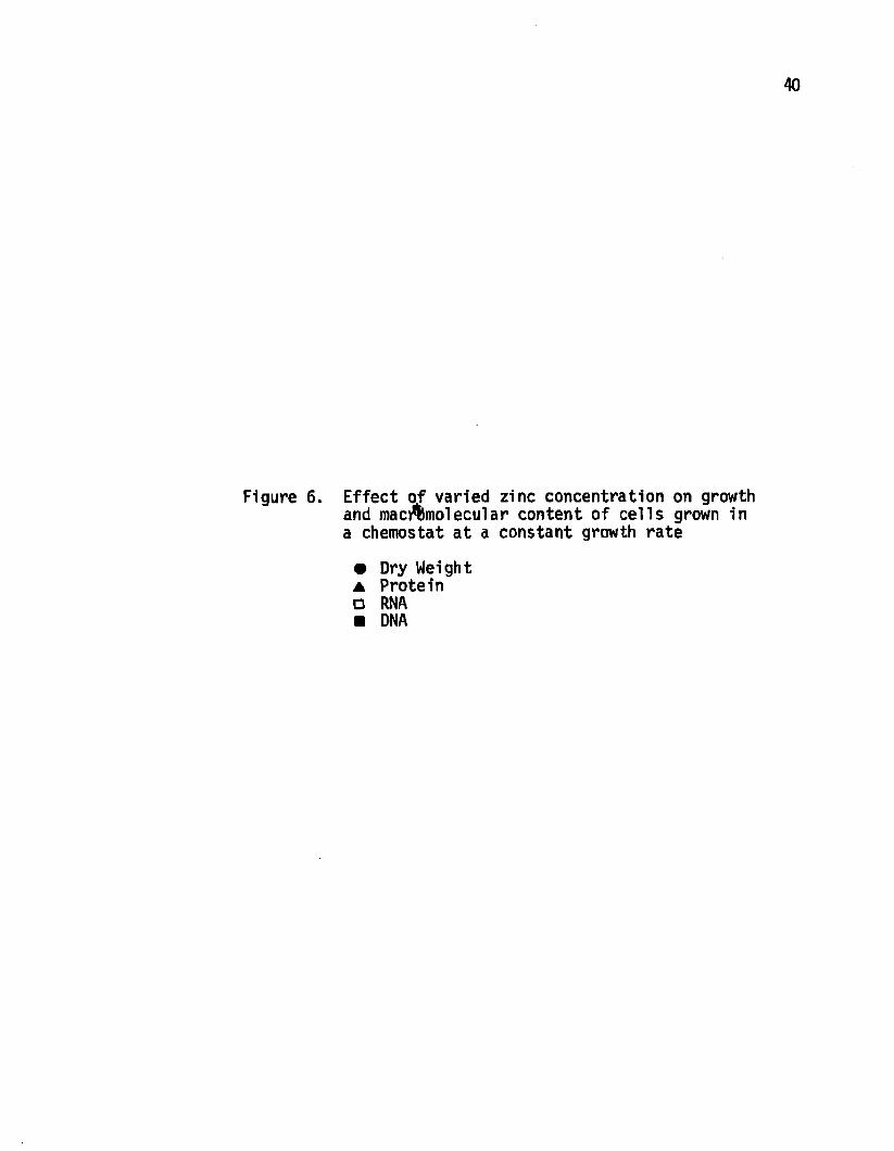
performed with zinc and manganese. As seen in Figure 6 the dry
weight of cells 1s dependent on zinc concentration. The RNA content
increased linearly with zinc concentration. Cellular DNA content
was constant while the protein level varied with zinc concentration.
The reason that the dry weight increased at zinc concentrations 10
fold lower than those necessary for optimum growth rate could be due
either to increased substrate u tiliza tion at these zinc concentra
tions or a more e ffic ie n t conversion of substrate to biomass. As
seen in Figure 7 the amount of substrate used over the zinc concen
trations studied did not fluctuate to a degree which would account
for the increased dry weight. Thus, a reasonable explanation for
the variable yield appears to be increased efficiency of metabolism

38
Figure 5 Specific activ ities of malic dehydrogenase and fumarase in cells grown in a chemostat at d ifferent growth rates
▲ Malic Dehydrogenase ■ Fumarase
J
ActMtf ■
• ̂ • c l f l c A e l l v l l f A
40
Figure 6. Effect of varied zinc concentration on growth and macmnolecular content of cells grown in a chemostat a t a constant growth rate
• Dry Weight ▲ Protein □ RNA ■ DNA

41
l« l m /aw m « t * « «ia
**• *
mi
•Illtiiu
aata
tail

42
Figure 7. Effect of varied zinc concentration on the u tiliza tio n of substrate by cells grown in a chemostat at a constant growth rate
• Dry Weight A Nitrogen O Carbon □ Phosphate

V*»4 2 - «t *
s>k& *■
0
1axi 14-
2 * f
1-----«— L . , ,..xri r[Zinc] M*

44
rather than increased substrate u tiliza tio n .
The examination of manganese content on growth was con
ducted with the zinc concentration which gave the maximum dry weight.
Under these conditions, alterations in the manganese concentration
produced a significant variation in dry weight due to slight altera
tions in metal concentration. In this case, however, substrate
u tiliza tion closely followed the dry weight (Figure 8). Thus for
manganese i t appears that at values below the optimum concentration
for growth the culture is limited by manganese and at values
slightly above the optimum manganese concentration manganese toxicity
inhibits growth. As one might predict from this data, the RNA
content showed a linear decrease with increased manganese concentra
tion (data not shown). DNA and protein values were constant at a ll
manganese concentrations tested.

45
Figure 8. Effect of varied manganese concentration on the u tiliza tion of substrate by cells grown on a chemostat at a constant growth rate
• Dry Weight o: Nitrogen □ Carbon a Phosphorous

Substrate Used mg/ml □ Dry Weight mg/mlwK>
asaP90.»P<*09ft
s
Substrate Used ug/inl xio^ (Oa )
9 fr

DISCUSSION
The data presented in this report demonstrates two obser
vations which one might not predict based on information obtained from
batch studies. These are: a) certain c itr ic acid cycle enzymes are
active during vegetative growth, and b) slight alternations in the
trace metal content significantly alters primary metabolism.
Charba and Nakata (1977) showed in batch cultures 1n "G"
medium that fumarase and malic dehydrogenase were at very low levels
at the end of vegetative growth and only during differentation did
the activ ities of the enzymes rise. Other investigators (Hanson,
1963 and Freese, 1976) working with batch cultures have reported
similar trends. We have shown that, in continuous culture under
conditions of glucose lim itation in a defined medium, these enzymes
are active even during vegetative growth. The level of enzyme
was related to ce llu lar energy requirements for the specific growth
rate rather than d ifferentiation.
Weinberg (1970) has suggested that trace metal concentra
tions required for secondary metabolism may be three orders of
magnitude higher than needed for growth. He feels that these
elevated levels of trace metals do not in terfer with vegetative
growth. This concept 1s in contradiction to our findings. Slight
(10“7 M) fluctuations of either zinc or manganese content greatly
47

48
alters the vegetative metabolism of the c e ll. The trace metal effect
can be observed at growth rates lower than those presented however
the effect is not as striking. We have shown that by altering the
trace metal content of the medium the efficiency of substrate u tiliz a
tion can be altered. This conversion of carbon source to biomass is
very e ffic ien t under conditions of optimum growth. Indeed, i t is
close to the predicted values of 0.5 for glucose and 0.4 for
glutamate.
No doubt there are certain problems involved with continuous
culture of differentiating organisms. Probably the most often cited
one is selection of asporogenic mutants. This problem can be circum
vented by not operating the chemostat for extended periods of time.
The oligosporogenic strain used in this study was isolated
from a chemostat culture. The spore mutant was indistinguishable
from the parent strain on the basis of any nutritional markers. The
sporogenic and oligosporogenic strain had equivalent growth rates in
batch culture. Further studies on the sporogenic parent are
obviously necessary. To circumvent the problem of mutant selection
the chemostat can be run for short time periods as suggested by
others. The medium used 1n this study is able to support a 0.47 h"1
growth rate in batch culture, thus no further optimization of the
medium should be necessary.
The potential of the chemostat in the study of sporulation
is great. Techniques similar to those of Lopez, e t a l . (1975), which
showed a marked dependence of competency on growth rate, can be applied
49
to other Investigations. For example, the a b ility to In it ia te spore
formation following a sh ift 1n some parameter such as growth rate,
carbon source, nitrogen source, e tc ., could be monitored. A chemostat
culture of a sporulatlon mutant grown at the rate of maximum
competency could be used to enhance the frequency of transformation
or conjugation thus enhancing the a b ility to do genetic mapping.
Thanks to the enhanced yields obtained in continuous culture, larger
amounts of ce llu lar mass or some extracellular enzyme could be
produced, either by a single stage fermenter at an appropriate growth
rate or a two stage fermenter where the f irs t stage is the cell
generator (with maximum specific growth rate) and the second stage is
used for aging the culture under appropriate conditions to obtain
maximum yield of the desired product.
BACTERIOPHAGE MEDIATED CONTROL OF
SPORULATION IN BACILLUS CEREUS STRAIN T
50

INTRODUCTION
The formation of bacterial spores has long been studied as
a model for in tracellu lar d ifferentiation. These studies of spore-
formation have led to varied hypotheses as to the mechanism of control
of transition of vegetative cells to sporulation. Most of the pro
posed mechanisms can be viewed as operating under either a negative
or positive control system. Limitations of carbon and/or nitrogen
are accepted as examples of negative control of spore in itia tio n .
Alternatively, positive controls might take the form of the production
or accumulation of some substance which in itia tes the differentiation
process. In either case the final result of d ifferentiation , forma
tion of mature spores, 1s due to the sequential induction of subse
quent gene function.
A positive control system governed by cytoplasmically
carried genes was postulated by Jacob (1960). Other investigators
have also suggested extrachromosomal control of sporulation (Rogolsky
and Slepecky, 1969; Schaeffer, et a l . , 1959; Schaeffer and Ionesco,
1960). Rogolsky and Slepecky (1964) reported that the treatment of
sporogenic strains of Bacillus subtil is with acriflavin increased the
frequency of generation of asporogenic variants. The asporogenic
strains showed no altered response to acriflav in , nor could they be
scored as auxotrophs for any character except their sporulation
efficiency. Treatment of bacterial cells with acriflavin had been
52
shown to be effective in curing F-plasmids located cytoplasmically
but had no significant effect on those in the integrated state
(Hirota, 1960). Similar reports of generating olIgosporogenic mutants
with lesions presumed to be located only at the site of regulation
of sporogenesis have been obtained using heat (Norton and Slepecky,
1967) and u ltrav io let ligh t (Schaeffer, et a l . , 1959).
In this report we present data which suggest the involve
ment of a lysogenic phage in regulating the in itia tio n of sporulation.
Introduction of the isolated phage into an oligosporogenic host
increased the efficiency with which that host formed heat resistant
spores. The experiments indicate that the phage may be carried in
the cytoplasm in a form other than a covalently closed circ le .

MATERIALS AND METHODS
Organism-Medium
A variant strain of Bacillus cereus strain T was isolated
by a modification of the method of Holliday (1956). This strain was
designated BCT-Sp+. An oligosporogenic mutant of this strain was
isolated from a chemostat culture of the sporogenic strain and
designated BCT-OSp or OSp. Cultures were maintained on plates of
either 0.8% nutrient agar supplemented with 0.2% yeast extract or
glucose-glutamate (GG)agar. The defined medium was designed from
experiments conducted in continuous culture. The medium contained
1.1 x 10"2 M glucose, 1.5 x 10⁻² M glutamate, 4.5 x 10⁻⁴ M (NH4)2S04,
1 x lO"3 M K2HP04, 4 x 10"4 M NaH2P04, 4.92 x 10“4 M MgCl2*6H20,
2.04 x TO"5 M CaCl2-2H20, 4 x 10"10 M CuS04*5H20, 1.69 x 10⁻⁸ M
CoC12*6H20, 5.10 x 10'7 M MnCl2*4H20, 3.3 x 10“6 M ZnS04*7H20 and 1.8
x 10-8 M FeS04*7H20. This medium allowed cultivation of the OSp
strain under conditions of glucose lim itation to a c ritic a l dilution
rate of 0.467"!.
Unless otherwise noted in the text, dilutions were prepared
in a buffer consisting of 0.15 M NaCl, 0.05 M Tris and 5 x 10"8 M
EDTA at pH 7.5 (STE).
Cultivation
Batch cultures were prepared by inoculating appropriate
53

54
volumes of liquid medium with either heat shocked spores (80°C,
30 min) or vegetative cells obtained from 24 - 36 hr plates.
Following 16 hr of incubation at 23°C the cultures were Incubated at
34°C until the desired density was obtained. Fresh medium was
inoculated with a volume equal to 10% of the final volume of culture
and again incubated at 34°C until the desired density was obtained.
Phage Induction-Purification
Cultures were induced with either u ltravio let light or
mitomycin C. U ltravio let induction consisted of irradiation of cul
tures with a GE germicidal lamp at a distance of 10 cm for 30 sec.
The cultures were then incubated at 30°C for 3 hr. Mitomycin C was
used to induce cultures by addition of the drug to mid-log cultures at
1 ug/ml final concentration and incubated for 3 hr at 30°C. In
either case the cells were removed by centrifugation at 5000 rpm for
30 min. EDTA was added to a final concentration of 10“2 M. Phage
particles were pelleted by centrifigation under conditions sufficient
to pellet particles of 50s. The 50s pellet was resuspended in STE and
layered on a performed gradient of 5 - 20% sucrose in STE. Gradients
were centrifuged in a Beckman SW65 rotor for 90 min at 25,000 rpm.
Fractions were collected from the bottom of the tube. The presence
of phage was determined either by electron microscopy, radioactivity
or absorbance at 260 nm.

55
Isop.vcirtc Centrifugation
Isopycnic centrifugation with or without ethldium bromide
was carried out by a modification of the procedure of Lovett (1966).
Cells from the mid-log phase of growth were pelleted and washed
twice with STE. These cells were resuspended in 2 ml STE containing
lysozyme (200 ug/ml) and RNase (50 ug/ml). The EDTA concentration
was increased to 0.1 M. Samples were incubated for 40 min at 37°C.
Sarkosyl (0.8%) and predigested pronase (500 ug/ml) were added and
incubation continued at 37°C for 30 min to effect lysis. STE was
added to give a final volume of 5 ml. Seven grams of CsCl was added
and 7 ml of the CsCl solution was combined with 3 ml of either
ethidium bromide (4 mg/ml in 0.1M NaH2P0̂ pH 7.0) or STE. Five
m illil ite rs were delivered to each tube of a Ti-50 rotor and
topped with mineral o il . Equilibrium was obtained by centrifugation
at 36,000 rpm for 42 hr at 15°C. Fractions were collected from a
hole pierced in the bottom of the tube. Fractions were spotted on
Whatham f i l t e r paper circles, dried and counted by liquid s c in tilla
tion.
RESULTS
A strain of Bacillus cereus strain T was isolated which
was able to grow and sporulate on a medium containing glucose,
glutamate and salts. Continuous culture techniques were employed to
optimize this medium. Early during these experiments, routine plating
on glucose-glutamate plates (G6) demonstrated two colony types (Fig.
9). The parental strain produced small, convex, mucoid colonies
which contained greater than 95% free spores after 48 hr at 34°C.
The second colony type was larger, non-mucoid and essentially free of
spores a fte r similar incubation for 96 hr. Occasional sector colonies
could also be observed.
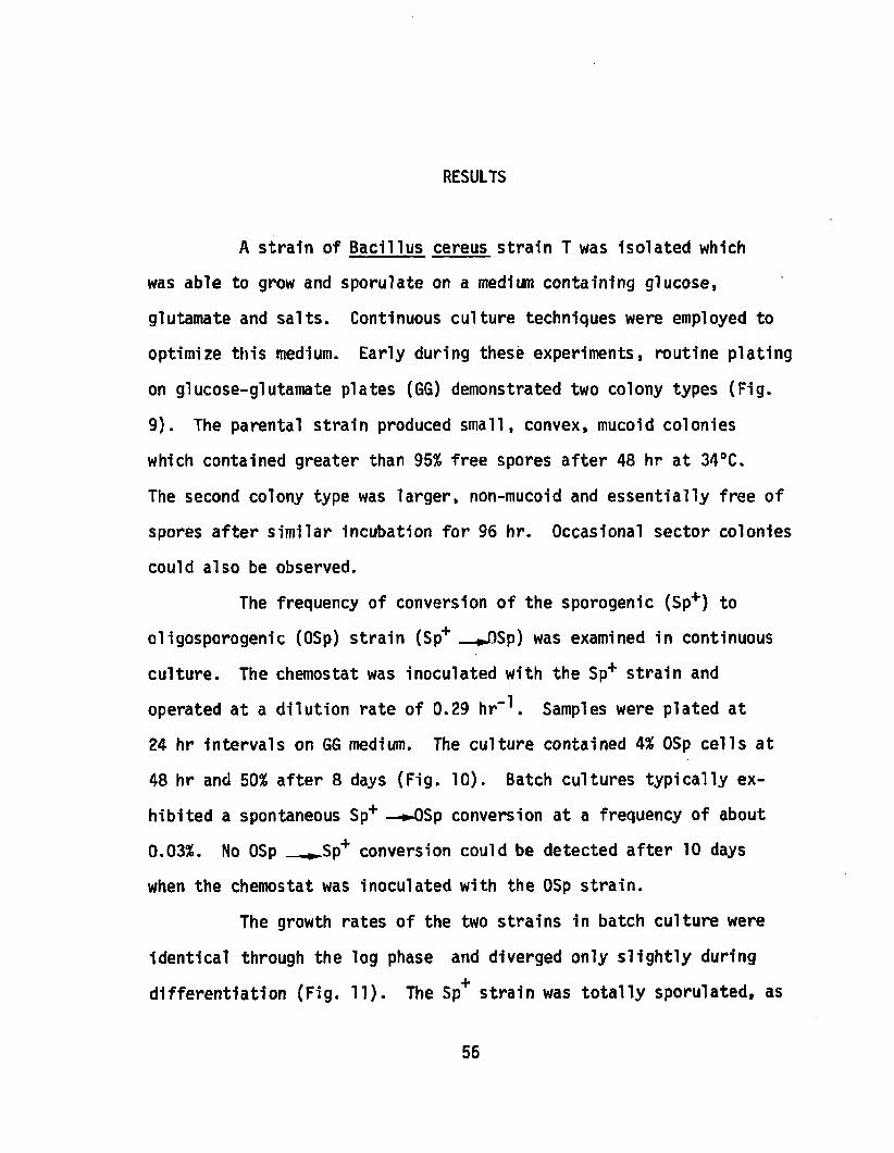
The frequency of conversion of the sporogenic (Sp+) to
oligosporogenic (OSp) strain (Sp+ *J3Sp) was examined in continuous
culture. The chemostat was inoculated with the Sp+ strain and
operated at a dilution rate of 0.29 hr-1 . Samples were plated at
24 hr intervals on GG medium. The culture contained 4% OSp cells at
48 hr and 50% after 8 days (Fig. 10). Batch cultures typically ex
hibited a spontaneous Sp+ —»-OSp conversion at a frequency of about
0.03%. No OSp ►Sp+ conversion could be detected a fter 10 days
when the chemostat was inoculated with the OSp strain.
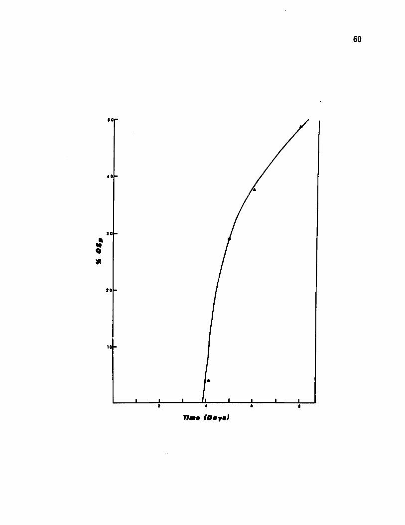
The growth rates of the two strains in batch culture were
identical through the log phase and diverged only s lightly during
differentiation (Fig. 11). The Sp+ strain was to ta lly sporulated, as
56

Figure 9 Photograph showing the two colony types isolated from a chemostat culture of Bacillus cereus


59
Figure 10. Rate of appearance of the oligosporogenic from the sporogenic strain in continuous culture
60
40
40
10
• fO*
10
61
Figure 11. Growth rate and sporulation frequency of the sporogenic and oligosporogenic strain in batch culture
O Turbidity Sp+• Turbidity OSp ▲ % Spores Sp a % Spores OSp

62
t*

63
determined by heat resistance, a fter 24 hr. The OSp strain produced
only 1% spores a fte r 24 hr and a maximum of 6% a fte r 36 hr. Both
strains were sensitive to the phage 0CL, a phage ly tic for EL cereus
strain T.
The high rate of generation of OSp colonies from the Sp+
strain , the observation of sector colonies and the pitted nature of
old colonies led to experiments to determine the possible involvement
of a phage in the conversion of the sporogenic to oligosporogenic
phenotype. Induction of the OSp strain with UV-light or mitomycin C
caused liberation of a phage at low levels (Fig. 12). Similar
attempts to induce the Sp+ parent did not liberate any phage-like
particles as determined by electron microscopic examination of
concentrated culture supernatant liquid.
The inducible phage could also be detected at extremely
low levels in lysates produced due to infection of either the OSp or
Sp+ strain with the ly tic phage (J)C1. The isolated phage had a head
diameter of 45 nm, a rig id ta il 145 nm long and a whip-like appendage
similar to that of Bacillus subtil is phage PBS 1. Isopycnic centri
fugation in CsCl showed the phage density to be 1.595. All attempts to
obtain either a phage sensitive host or a conditional lethal mutant
of the phage have so fa r been unsuccessful. Thus, i t has been
d iff ic u lt to obtain large quantities of the phage.
Concentrated culture supernatants of induced OSp cultures
were banded on 5 - 20% sucrose. Fractions containing intact phage

Figure 12. Electron micrograph of the bacteriophage isolated from the oligosporogenic strain

65
VC'UM*r'ii'./ ;
j l f i r Vr f».

66
were pooled, and dialyzed exhaustively against buffer (0.1 M KgHOP̂ ,
pH 7 .0 ). The phage preparation was stored at 4°C over chloroform.
Addition of this phage preparation to batch cultures of the OSp
strain increased the level of sporulatlon (Table 1). Additions of
dialysis buffer sim ilarly stored in the presence of chloroform gave
no increase 1n sporulation. Increased phage dosage resulted in
increased sporulatlon. Maximum increase with batch cultures was
10-fold. Further increase of phage dose caused a loss of the
sporulation enhancement. Similar experiments using cells obtained
form a chemostat culture at a dilution rate of 0.23 h""* increased
the rate of sporulation 30 fold. With chemostat grown cells , as
with batch grown ce lls , a decrease in ac tiv ity was observed at high
phage doses. A few spores generated following infection of OSp
cultures with phage produced colonies of the Sp+ phenotype upon
germination.
Cesium chloride-ethidium bromide analysis was carried out
to detect the presence of plasmid DNA. Cultures of both the Sp+ and
OSp strains were prepared 1n GG medium supplemented with 0.05% yeast
extract. These cultures were labeled with 10 uCi/ml ^H-d-adenosine
(Sp. Act. 5 C/mM) for 12 hr at room temperature followed by an
additional 4 hr a t 34°C. The density of the culture was approximately
150 Klett units. The cells were harvested by centrifugation and washed
twice with STE (pH 7 .5 ). The cells were resuspended in 2 ml STE and
prepared for isopycnic centrifugation as described in Materials and

67
Table 1. Enhancement of Sporulation Frequency
Due to Addition of the Induced Phage
ml Tot viable spores phage xl0"8 xlO-6 %Sp Increase
Cells fromBatch 0 1.18 .25 .217Culture .1 1.00 .847 3.99
.2 2.60 2.203 10.39
.4 0.30 .254 1.20
ills fromContinuous 0 1.50 .11 .073Culture .15 5.5 2.267 30.90
.30 3.3 .1467 20.00
68
Methods. Gradients were centrifuged at 36,000 rpm for 42 hr at 15°C
in a T1 SO rotor. One-tenth (0.1) m i l l i l i te r fractions were collected
Ten m icroliter samples were spotted on Whatman f i l t e r paper circles,
dried and counted by liquid sc in tilla tio n . This experiment was
repeated several times. On each occasion a s a te llite peak was ob
served at a position less dense than the primary peak (Fig. 13). The
s a te llite contained about 2.5% of the counts found in the main peak.
No s a te llite DNA was seen in lysates of the Sp+ strain (Fig. 14).
Fractions containing the main peak were pooled as were those con
taining the s a te llite . The ethidium bromide was removed by dialysis
and the two preparations were again subjected to isopycnic centrifu
gation. The main peak had a density of approximately 1.690 and the
s a te llite peak banded at a density of 1.63 (Fig. 15). When labeled
lysates were subjected to equilibrium centrifugation in the absence
of ethidium bromide, the banding pattern of the OSp strain was the
same as that seen in the presence of ethidium bromide (Fig. 16). No
s a te llite band was found in the lysates of the Sp+ strain.
Figure 13. Cesium chloride-ethidium bromide gradient of DNA of the oligosporogenic strain
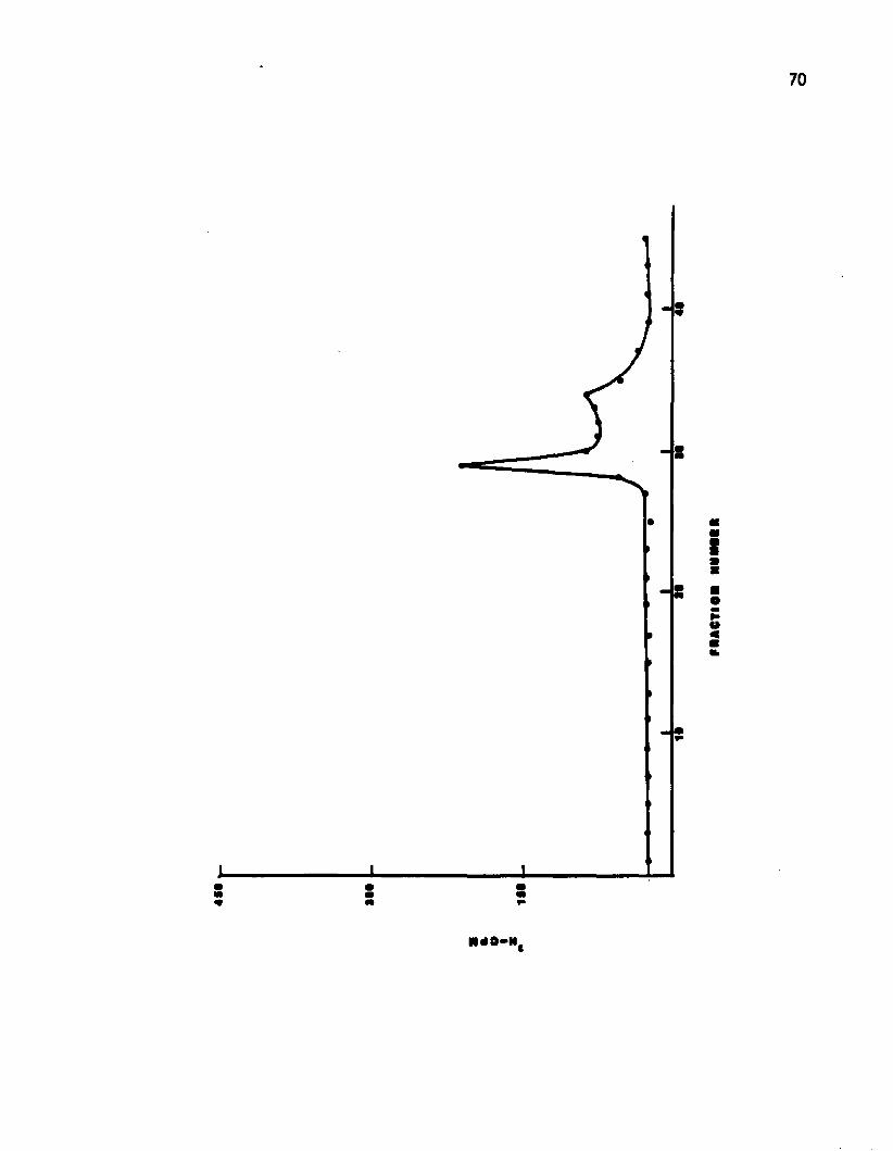

71
Figure 14. Cesium chloride-ethidium bromide gradient of DNA of the sporogenic strain

CPU
«*• r-
I I I
"i r
«•
p h a c t io n n u m m

73
Figure 15. Cesium chloride gradient of DNA peaks isolated from cesium chloride-ethidium bromide analysis of the oligosporogenic strain
O Fractions 1-5 • Fractions 6-10

74
430
300
150
• 4
30

Figure 16. Cesium chloride gradient of DNA of the oligosporogenic strain

s
4
i
1
301 0
ON OCT (ON NUM M N
O*

DISCUSSION
The selective advantage of OSp mutants in a chemostat has
been reported previously (Dawes and Mandelstam, 1970; Dawes and
Thornley, 1970). This characteristic of spore formers in continuous
culture was exploited to isolate an OSp strain which carried an
inducible phage in the cytoplasm. Steady state continuous cultures
operate on the assumption that most cells in the population are
dividing at the same rate. Any cells which are undergoing d iffe r
entiation are selected against because during differentiation they do
not divide. Spore mutants are likewise unable to divide a fter the
cell undergoes transition ( t 0). I f the chemostat is operated at a
dilution rate which approaches the maximum growth rate (umax)of the
c u ltu m a ll organisms which undergo transition w ill be washed out of
the culture. At slower growth rates (u-m-|x.) d ifferentiating wild
type cells w ill s t i l l be unable to contribute to maintaining the
population. Spore mutants with lesions before commitment have been
shown capable of resuming, division under appropriate conditions.
These facts infer that OSp mutants may only be enriched for in con
tinuous culture i f either the mutant exhibits an accelerated or expand
ed vegetative growth cycle or the mutant is unable to undergo commit
ment to sporulation (tg) (Mandelstam, 1969). The growth rate of the
OSp mutant which we have isolated does not d iffe r from that of the Sp+
strain in batch cultures (Fig. 11). Thus, the enrichment observed
77
78
must be due to a lesion a fter transition but before commitment.
Microscopic examination by phase contrast confirms that the OSp
strain has an early block 1n the sporulatlon sequence.
The phage can be Isolated from Induced cultures of the OSp
strain. CsCl examination of lysates suggests that the genome is
carried in the extrachromosomal state. This phage increases the
sporulation frequency when reintroduced into the OSp strain.
Organisms from continuous culture showed a greater response to the
phage. This is not surprising since most cells in the continuous
culture are dividing at the same rate. Conversely, in batch culture
a more heterogeneous population 1s found. These facts suggest that
the dilution rate employed resulted in more of the culture being in
a phage sensitive phase. The OSp ►Sp+ conversion of a significant
proportion of the spores generated in such an experiment shows that
the phage genome has undergone some change during packaging or
reinfection. Such a change could be the generation of complementary
ends at the time of sizing of the genome. Such a molecule might be
expected to cycllze on infection. The circular genome could pair
with complementary sites on the host genome and allow integration
according to Cambell's model (Cambell, 1962).
Attempts to label the phage with 3H-thymidine were not
successful, however 3H-d-adenosine was Incorporated by the phage.
Our inab ility to label the phage at high levels might be due to either
ineffic ien t induction or replication of phage genome. Although the

79
number of counts which appeared in the s a te llite peak was small» the
results were reproducible.
The buoyant density of the phage genome and the s a te llite
peak in CsCl were both approximately 1.63. These data suggest that
the sa te llite peak and phage genome are identical. More complete
analysis of the phage and s a te llite DNA must await isolation of
mutants which permit purification of greater quantities of these
components.
We feel that the data presented supports the hypothesis
that the isolated phage carries a gene(s) involved in the control
of in itia tio n of sporulation. The origin of this gene can only be
speculated on at present. I t might be of phage origin with the
function of turning o ff host metabolism during times of stress.
Alternately, i t may be a host gene which lies near the site of
phage integration. I t could then be picked up by the phage at the
time of excision similar to the incorporation of the host
galactose operon into the phage A (Morse, et a l . , 1956). The a b ility
of such a phage to exist cytoplasmically has been previously
reported (Lieb, 1970; Pemberton, 1977). The a b ility of these
genomes to replicate independent of the host genome varies from
one system to another (Lieb, 1970).
The low level of sporulation which we observed in our OSp
strain can be explained in either of two ways. Either a low level
of transcription of the cytoplasmically carried phage is occurring

80
or faulty termination of transcription of host operons allows read
through into spore specific regions. In either case a threshold
level of messenger could be produced which would be capable of
in itia tio n of subsequent events in the sporulatlon sequence.
In Figure 17 we present a model by which the isolated
phage may be involved in sporulation. The phage, carrying regulatory
gene(s), is excised generating a linear molecule. The observed
frequency in batch culture is 10"4 (Schaeffer, et a l . , 1959). The
excised genome is carried in the linear form in the cytoplasm.
Some members of the population might lose the phage by abortive
transduction. The cytoplasmically carried phage may be induced,
either spontaneously or a r t if ic ia l ly , giving rise to intact phage.
These phage particles carry a genome capable of forming a circular
molecule upon infection of either the phage deficient or phage
carrying OSp strain. The circular form of the phage is more
e ffic ie n tly copied and is more like ly to integrate into the host
genome. In either case i t should increase the frequency of
sporulation.
This model for phage involvement in sporulation takes
several of its elements from the model for sporulation proposed by
Rogolsky(1968). The model of Rogolsky suggested episomal regulation
of sporulation. The possibility of the episome existing in the
integrated state was not considered. This episomal control model
has been critic ized on the basis that several sporulation markers,

Figure 17. Proposed model for the involvement of the bacteriophage on the control of sporulation

9 Jo ) /o

83
not located near each other on the host chromosome, are lost as a
block when the culture is treated to cure i t of episomes (Mandelstam,
1969). This is not a valid criticism because loss of the regulatory
mechanism could block expression of genes controlled by that
regulator. This would appear phenotypically as a loss of the
structural genes for these spore associated markers.
Bramucci and Lovett (1977) reported the transfer of
sporogenesis by a phage. This system, however, relied on the chance
isolation of phage from nature. These phage preparations were then
introduced into a variety of auxotrophic mutants. While such an
experiment does show the a b ility to transfer the a b ility to sporulate
by using a phage, i t does not exclude the possibility of either
transduction or complementation of an essential spore marker not
involved with spore in itia tio n .

BIBLIOGRAPHY
Bauer, S.
Balassa, i
Bayen, H.
Beck, B. !
Bernlohr,
Bramucci,
Cambell, i Carlton, I
Caslavska
Charba, 0
and E. Ziv. 1976. Dense growth of aerobic bacteria 1n a bench scale fermenter. Biotech. Bloeng. 18: 81-94.
5. 1969. Biochemical genetics of sporulation: I . Unidirectional pleiotrophic interactions among genes controlling sporulation in Bacillus subtilis . Mol. Gen. Genet. 104: 73-103.
, C. Frehel, A. Ryter, and M. Sebold. 1967. Etude cytologique de la sporulation chez Clostridium hlstolyticum: sou che sporogene et mutants de sporulation.Ann. Inst. Pasteur. 113: 163-173.
). and J. J. Park. 1976. Activity of three murein hydrolases during the cell division cycle of Escherichia coli K12 as measured in toluene-treated cells. J.B acterid . 126: 1250-1260.
R. W. and G. D. Novell 1. 1964. Bacitracin biosynthesisand spore formation: The physiological role of anantib io tic . Arch. Biochem. Biophys. 103: 94-104.
M. G., K. M. Keggins and P. S. Lovett. 1977. Bacteriophage conversion of spore-negative mutants to spore- positive in Bacillus pumilis. J. Bacteriol. 22: 194-202.
\. 1962. Advances in Genetics. 11: 101-146.
i. C. and D. R. Helinski, 1969. Heterogenous circular DNA elements in vegetative cultures of Bacillus megaterium. Proc. Natl. Acad. S c i., U.S.A., 64: 592-599.
, J . , J. Pazlarova-Kodesova and I . Horakova. 1973. The effect of continuous cultivation 1n a chemostat on the formation and reversion of basic ultrastructural changes of bacteria. Folia Microbiol. 18: 266-268.
. F. and H. M. Nakata. 1977. Role of glutamate in the soorogenisis of Bacilluscereus. J. Bacteriol. 130 : 242- 248.
84

85
Dawes, I . W. and J. Mandelstam. 1969. Biochemistry of sporulation of Bacillus subtil is 168: continuous culture studies.Proceedings 4th In t. Symp. Continuous Culture of Microorganisms (Prague).
Dawes, I . W., and J. Mandelstam. 1970. Sporulation of Bacillus subtil is in continuous culture. J. Bacteriol. 103: 529- 3̂5.
Dawes, I . W. and J. H. M. Thornley. 1970. Sporulatlon in Bacillussu b tilis . Theoretical and experimental studies in continuous culture systems. J. Gen. Microbiol. 62: 49-66.
DiCiocci, R. A. and N. Strauss. 1973. Patterns of transcription in Bacillus subtilis during sporulation. J. Mol. Biol. 77: 35S32B7-----------
Douthit, H. A. and H. 0. Halvorson. 1966. S a te llite deoxyribonucleicacid from Bacillus cereus strain T. Nature. 153: 182-183.
Efstathios, J. D. and L. L. McKay. 1976. Plasmids in Streptococcus la c tis : Evidence that the lactose metabolism and proteaseactiv ity are plasmid linked. Applied and Environ. Micro.32: 38-44.
E llar, D. J. and D. G. Lundgren. 1966. Fine structure of sporulation in Bacillus cereus grown in chemically defined medium. J. Bacteriol. 92: 1748-1764.
Ellwood, D. C. 1970. The wall content and composition of Bacillus subtilis var niqer qrown in a chemostat. Biochem. J. 118: 367-3737
Ellwood, D. C. and D. W. Tempest. 1969. Control of teichoic acid and teichuronic-acid biosynthesis in chemostat cultures of Bacillus subtilis var. niger. Biochem. J. I l l : 1-5.
Fisk, C. H. and Y. Subbarow. 1925. The colorimeter determination of phosphorous. J. Biol. Chem. 66: 375-400.
Fitz-James, P. C. 1965. Spore formation in wild and mutant strains of EL cereus and some effect of inhibitors, p. 524-544.In: M. J-C Senez (ed .), Collog In t. Centre Natl. Res. S c i., Marseille.
Foster, J. W. 1956. Morphogenisis 1n bacteria: some aspects of spore formation. Q. Rev. Biol. 31: 102-118.

86
Freese, E., W. Klofat and E. Galliers. 1970. Commitment to sporulatlon and induction of glucose-phosphoenolpyruvate tran- ferases. Biochem. Biophys. Acta. 22: 265-289.
Freese, E. 1972. Sporulation of b a c il l i , a model of cellu lar differentiation , p. 85-124. In: A. A. Muscona and A.Monroy (eds.), Current Topics in Development Biology.Vol. 7. Academic Press, New York.
Freese, E. B., P. Cooney and E. Freese. 1975. Conditions controlling commitment of d ifferentiation in Bacillus megaterium.Proc. Natl. Acad. Sci. , U. S. A. 72: 4037-4o4T.
Freese, E. and Y. Fujita. 1976. Control of enzyme synthesis during growth and sporulation. p. 164-184. In: D. Schelssinger(ed .), Microbiology. 1976. American Society for Microbiology, Washington, D. C.
Ghuysen, J. M., D. T. Tipper and J. L. Strominger. 1966. p. 685- 699. In: E. F. Neufeld and V. Ginsburg (eds.), Methodsin Enzymology. V I I I . Academic Press, New York.
Grelet, N. 1957. Growth lim itation and sporulation. J. Appl. Bact. 20: 315-324.
Guespin-Michel, J. F. 1971. Pleiotropic reversion in some early blocked sporulation mutants of Bacillus subtilis 168: Isolation and phenotype identification of partial revertants. J. Bacteriol. 108: 241-247.
Halvorson, H. 0. 1965. Sequential expression of biochemical eventsduring in tracellu lar d ifferentiation, p. 343-368. In:M. R. Pollock and M. H. Richmond (eds.), Symp. Soc. Gen. Microb. 15. Cambridge Univ. Press, N. Y.
Hanson, R. S ., V. R. Srinlvasan and H. 0. Halvorson. 1963. Biochemistry of sporulation. I I . Enzymatic change during sporulation of Bacillus cereus. J. Bacteriol. 86: 45-50.
Harden, W. and J. G. Kuenen. 1977. A review: Microbial selectionin continuous culture. J. Applied Bacteriol. 43: 1-24.
Hayes, W. 1968. The genetics of bacteric and their viruses.Second Edition, p. 61. John Wiley and Sons, Inc ., New York.
Hemphill, H. E. and H. R. Whiteley. 1975. Bacteriophage of Bacillus subtilis . Bacteriol. Rev. 39: 257-315.

87
Henneberry, R. C. and B. C. Carlton. 1973. Characterization of Polydispense closed circular deoryribonucleic acid molecules of Bacillus megaterium. J. Bact. 114: 625-631.
Hirota, Y. 1960. The effect of acridine dyes on mating type factors in Escherichia co li. Proc. Natl. Acad. S c i., U.S.A. 46:57-W.
Hoch, J. 1977. Developmental genetics at the beginning of a new era, p. 119-121. In: G. Chambliss and J. C. Vary (eds.), Spores V II. American Society for Microbiology, Washington, D. C.
Holliday, R. 1956. A new method for the identification of biochemical mutants of microorganisms. Nature. 178: 987.
Hranueli, D., P. J. Piggot and J. Mandelstom. 1974. Statistical estimate of the total number of operons specific for Bacillus subtilis sporulation. J. Bacteriol. 119: 684-690.
Ito , J ., G. Mildna and J. Spizizen. 1971. Early blocked asporogenous mutants of Bacillus subtilis 168. I . Isolation and characterization of mutants resistant to antibiotics produced by sporulating Bacillus subtilis 168. Mol. Gen.Genet. 112: 104-109.
Jacob, F ., P. Schaeffer and E. L. Wollman. 1960. Episomic elements in bacteria. Symp. Soc. Gen. Microbiol. 10: 67-91.
Kalb, V. F. and R. W. Bernlohr. 1977. A new spectrophotometric assay for protein in cell extracts. Anal. Biochem. 82: 362-371.
Kanarek, L. and R. H il l . 1964. The preparation and characterization of fumarase from swine heart muscles. J. Biol. Chem.239: 4202-4208.
K lier, A. F ., M. M. Lecadet and R. Dedonder. 1973. Sequential modifications of DNA-dependent RNA polymerase during sporulation in Bacillus thuringiensis. Eur. J. Bioch. 36: 317-327.
Lalshley, E. J. and R. W. Bernlohr. 1966. Catabolite repression of "three sporulatlon enzymes" during growth of Bacillus licheniformis. Biochem. Biophys. Res. Comm. 24: 85-50.
Lampen, J. 0. 1965. Secretion of enzymes by microorganisms. Symp.Soc. Gen. Microbiol. 15: 115-133.

88
Lawrence*
Leighton,
Leighton,
Lieb, M.
Linn, T .,
Lopez, R.
Losick, R
Lovett, P
Lovett, P
Lovett, P
Lovett, P
Lowry, 0.
N. and H. 0. Halvorson. 1954. Studies on the spores of aerobic bacteria. IV. A heat-resistant catalase from spores of Bacillus terminalis. J. Bacteriol. 68: 334-337.
T. J ., and R. H. Doi. 1971. The s ta b ility of messenger ribonucleic acid durinq sporulation on Bacillus subtills .J. Biol. Chem. 246: 3189-3195.
T. 1964. Further studies on the s ta b ility of sporulation m-RNA in II. subtil is . J. Biol. Chem. 249: 7808-7812.
1970. A mutants which persist as plasmids. J. V iro l. 6: 218-225.
A. L. Greenwood and R. Losick. 1975. RNA polymerase from Bacillus subtil is : Purification and properties of a modified form of enzyme containing two sporulation polypeptides. J. Biol. Chem. 250: 9256-9261.
, A. Tapia and A. Portoles. 1975. The influence of glutamic acid and arginine on the competence of Bacillus subtil is growing in a chemostat. fiolec. Gen. Genet. T36: S/-94.
. , R. G. Shorenstein and A. L. Sonenshein. 1970. Structural alteration of RNA polymerase during sporulation. Nature (London). 227: 910-913.
. S. 1973. Plasmid in Bacillus pumilis and the enhanced sporulation of plasmid-negative variants. J. Bacteriol.115: 291-298.
. S. and M. G. Bramucci. 1975. Evidence for a non-random base sequence in a Bacillus pumilis plasmid: EcoRlendonuclease digestion of pPL576. J. Bacteriol. 123: 377-379.
. S. and M. G. Bramucci. 1975. Plasmid deoxyribonucleic acid in Bacillus subtilis and Bacillus pumilus. J. BacterioTi 124: 484-490.
. S. 1976. Bacillus pumilus plasmid pPHO: properties andinsertion into Bacillus subtilis 168 by transformation. J. Bacteriol. 127: 817-828.
H., N. J. Rosenbrough, A. L. Farr and R. J. Randall. 1951. Protein measurement with the fo lin phenol reagent. J. Biol. Chem. 193: 265-275.
Mandelstam, J. and W. M. Waites. 1968. Sporulation in Bacillussubtilis . The role of exo-protease. Biochem. J. Tti5: 793- 801.

89
Mandelstum, J. 1969. Regulation of bacterial spore formation.Symp. Soc. Gen. Microbiol. 19: 377-401.
Mandelstum, J. and J. M. S terlln . 1969. Commitment to sporulation1n Bacillus su b tilis , p. 180-185. In: L. L. Campbell (ed.), Spores TV. American Society for Microbiology, Bethesda, Md.
M ille r, G. L. 1959. Use of d1n1trosal1cyl1c acid reagent for determination of reducing sugar. Analytical Chem. 31: 426-428.
M ille t, J. 1963. La phosphatase alcaline de Bacillus megaterium. Comptes Rendus. 257: 784-787.
M ille t, J ., P. Kerjan, J. P. Aubert and J. Szulmajster. 1977.Porteolytic convesion In vitro of B.. subtilis vegetative RNA polymerase into the homologous spore enzyme. FEBS Lett. 23: 47-50.
Mitanl, T ., J. E. Helnze and E. Freese. 1977. Induction of sporulation 1n Bacillus subtilis by decoyinlne or hadacidin. Biochem. Biophy. Res. Comm. 77: 1118-1125.
Morse, M. L ., E. M. Lederberg and J. Lederberg. 1956. Transduction in Escherichia coll K 12. Genetics. 41: 142-156.
Murrel, W. G. 1967. The biochemistry of the bacterial endospore.Adv. Microbiol. Physiol. 1: 133-251.
Nishimoto, H. and I . Takahashi. 1974. Template specificity and subunits of RNA polyermase from asporogenous mutants of Bacillus su b tilis . Can. J. Biochem. 52: 966-973.
Norton, J. and R. A. Slepecky. 1967. Sporulation mutations induced by heat in Bacillus su b tilis . Nature (London). 155: 838- 839.
N y iri, L. K. Strategies and problems related to computer controlled fermentation systems, p. 120-126. In: T. Hasegawa (ed .), 1st Intersectional Congress IAMS. Vol. 5. Publ. Science Council of Japan.
OHye, D. F. and W. G. Murrell. 1962. Formation and structure of the spore of Bacillus coagulans. J. Cell Biol. 14: 111-123.
Pemberton, J. M. and W. T. Tucker. 1977. Naturally occurring viral R plasmid with a circular supercolled genome in the extracellu lar state. Nature (London). 266: 50-51.
90
Plggot, P
Plggot, P
Piggot, P
Powel1, J
Rexer, B.
Rhaese, H
Rhaese, H
Rogolsky,
Rogolsky,
Rogolsky,
. J. 1973. Mapping of asporogeneous mutations of Bacillus su b tilis : A minimum estimate of the number of sporulation operons. J. Bacteriol. 114: 1241-1253.
. J. and J. G. Coote. 1976. Genetic aspects of bacterial endospore formation. Bacteriol. Rev. 40: 908-962.
. 1977. Organization of spo locus expression duringsporulation of Bacillus su b tilis : Evidence for differentloci being expressed in the mother cell and in the forespore, p. 122-126. In: G. Chamblis and J. C. Vary (eds.)» Spore V II. American Society for Microbiology, Washington,D. C.
. F. and R. E. Strange. 1956. Biochemical changes occurring during sporulation in Bacillus species. Biochem. J. 63: 661-668.
, V. R. Srinivasan and W. Z il l ig . 1975. Regulation of transcription during sporulation of Bacillus cereus T.Eur. J. Bioch. 53: 271-281.
. -J . , H. Dichtelmuller and R. Grade. 1975. Studies on the control of development. Accumulation of guanosine tetraphosphate and pentaphosphate in response to inhibition of protein synthesis in Bacillus s u b tilis . Eur. J. Bloch. 56: 385-392.
. -J . , R. Grade and H. Dichtelmuller. 1976. Studies on the control of development. Correlation of in itia tion of differentiation with synthesis of highly phosphorylated nucleotides in Bacillus su b tilis . Eur. J. Bioch. 64: 205- 213.
M. and R. A. Slepecky. 1964. Elimination of a genetic determinant for sporulation of Bacillus subtilis with acriflavin . Biochem. Biophy. Res. Com. 16: 204-208.
M. and J. Spizizen. 1967. Chromosomal localization of spore genes in Bacillus su b tilis , p. 22. Bacteriol. Proc. 1967.
M. and R. A. Slepecky. 1968. The response of sporogenesis in Bacillus subtilis to acriflav in . Can. J. Microbiol. 14: 61

91
Rolfe, R. 1963. Changes 1n the physical state of DNA during the replication cycle. Proc. Natl. Acad. Sc1., U.S.A. 49: 386-392.
Ryter, A. 1965. Etude morphologlque de la sporulation de Bacillus su b tilis . Annls. Inst. Pasteur, Paris. 108: 40-501
Ryter, A ., P. Schaeffer and H. Ionesco. 1966. Classification cytologlque, par leur stades de biocage, de mutants de sporulation de Bacillus subtilis Marburg. Annls. Inst. Pasteur, Paris. 110: 305-315.
Schaeffer, P ., H. Ionesco and F. Jacob. 1959. Sur le determinlsme genetique de la sporulation bacterienne. Comptes Rendus. 249: 577-578.
Schaeffer, P. and H. Ionesco. 1960. Contribution a letude genetique de la sporogeneses bacterienne. Comptes Rendus. 249: 3125- 3127.
Schaeffer, P., J. M ille t and J.-P. Aubert. 1965. Catabolicrepression of bacterial sporulation. Proc. Natl. Acad.Sc1. , U.S.A. 54: 704-711.
Schneider, U. C. 1956. Determination of nucleic acids in tissues by pentose analysis, p. 680-684. In: S. P. Colowick and N. 0. Kaplan (eds.), Method in Enzymology. I I I . Academic Press, New York.
Segall, J ., R. Tjlan, J. Pero and R. Losick. 1974. Chloramphenicol restores sigma factor activ ity to sporulating Bacillus s u b tilis . Proc. Natl. Acad. S c i., U.S.A. 71: 4860-4863.
Spackman, D. H., W. H. Stein and S. Moore. 1958. Automatic recording apparatus for use in chromatography of amino acids. Anal. Chem. 30: 1190-1206.
Srinivasan, V. R. 1965. In tracellu lar regulation of sporulation of bacteria, p. 64-74. In: L. L. Campbell and H. 0. Halvorson (eds.), Spores I I I . American Society for Microbiology,Ann Arbor, Mich.
Steinberg, W. 1974. Properties and developmental roles of thelysyl- and trlptophenyl-transfer ribonucleic acid synthetases of Bacillus su b tilis : common genetic origin of thecorresponding spore and vegetative enzymes. J. Bacteriol. 118: 70-82.

92
Stewart, B. T. and H. 0. Halvorson, 1954. Studies on the spores of aerobic bacteria. I I . The properties of an extracted heat stable enzyme. Archs. Biochem. Biophys. 49: 168-178.
Strominger, J. L ., J. T. Park and R. E. Thompson. 1959. Composition of the cell wall of Staphylococcus aureus: Its relationto the mechanism of action of pen ic illin . J. Biol. Chem. 234: 3263-3268.
Sumida-Yasumoto, C. and R. H. Doi. 1974. Transcription from the complimentary deoxyribonucleic acid strands of Bacillus subtilis during various stages of sporulation. J.Bacteriol. 117: 775-782.
Szulmajster, J. and R. S. Hanson. 1965. Physiological control of sporulation in Bacillus su b tilis , p. 162-173. In L. L. Campbell and H. 0. Harvorson (eds.), Spores I I I . Am.Soc. for Microbiol., Ann Arbor, Mich.
Tono, H. and A. Kornberg. 1967. Biochemical studies of bacterialsporulation. I I I . Inorganic pyrophosphatase of vegetative cells and spores of Bacillus subtilis . J. Biol. Chem. 242: 2375-2382.
Vinter, V. 1959. Sporulation of b ac ili. V II. The participation of cysteine and cystine in spore formation by Bacillus megateriurn. Folia Microbiol. 4: 216-219.
Void, B. S. and S. Minatogawa. 1972. Characterization of changes intransfer ribonucleic acids during sporulation in Bacillussu b tilis , p. 254-263. In H. 0. Halvorson, R. Hanson and L. L. Campbell (eds.), Spores V. American Society for Microbiology, Washington D. C.
Void, B. S. 1975. l 5 oaccepting species of lysyl-transfer ribonucleic acids in Bacillus subtilis ce lls , p. 282-289. In P. Gerhardt, R. N. Castilow and H. L. Sadoff (eds.), Spores VI. American Society for Microbiology, Washington, D. C.
Waites, W. M. 1968. Sporulation in Bacillus su b tilis . Antigenicchanges during spore formation. Biochem. J. 109: 803-810.
Warren, S. C. 1968. Sporulation in Bacillus su b tilis . Biochemical changes. Biochem. J. 109: 811-818.
Weatherburn, M. W. 1967. Phenol hypochlorite reaction fordetermination of ammonia. Anal. Chem. 39: 971-974.

93
Weinberg,
Wise, J .,
Yehle, C.
Young, E.
Young, E.
Young, F.
E. D. 1970, Biosynthesis of secondary metabolites. Rolesof trace metals. Adv. Microbiol. Phys. 4: 1-44.
Swanson, A. and H. 0. Halvorson. 1967. Dlpicolinic acid-less mutants of Bacillus cereus. J. Bacteriol. 94:2075-2076.
0. and R, H. Doi. 1967. D ifferential expression of bacteriophage genomes in vegetative and sporulating cells of Bacillus su b tilis . Virology. 1: 935-941.
1. and P. C. Fitz-James. 1959a. Chemical and morphological studies of bacterial spore formation. I . Formation of spores in Bacillus cereus. Biophys. Biochem. Cytol. 6: 467-482.
I . and P. C. Fitz-James. 1959b. Chemical and morphological stages of bacterial spore formation. I I . Spore and parasporal protein formation in Bacillus cereus var A lesti. Biophys. Biochem. Cytol. 6: 483-498.
E., J. Spizizen and I . P. Crawford. 1963. Biochemical aspects of competence in the Bacillus subtilis transformation system. J. Biol. Chem. 238: 3119-3125.

APPENDICES
Comparison of the Chemical Composition of Cell Walls Isolated from a
Sporogenic and an Oligosporogenic strain of Bacillus cereus strain T.
The selective pressures Imposed by continuous culture
techniques have been well documented (Harden and Kuenen, 1977). The
enrichment for asporogenic mutants in chemostat cultures of Bacillus
sp. has often been cited as a problem (Daves and Mandelstam, 1970;
Daves and Thornley, 1970). In addition to sporulation mutants,
chemostat cultures have been shown to give rise to strains which have
an altered cell wall (Ellwood, 1970; Ellwood and Tempest, 1970;
Caslauska, et a l . , 1973). We have isolated an oligosporogenic strain
Bacillus cereus strain T from a chemostat culture operated under
conditions of glucose lim itation. This sporulation mutant demonstrat
ed a greater sensitiv ity to lysozyme than the sporogenic parent.
Cell walls were isolated from both the sporogenic parent and
the oligosporogenic variant by d ifferentia l centrifugation as de
scribed by Young, et a l . (1963). Isolated walls were assayed for free
primary amino groups by the dinitrophenylation reaction (Ghuysen, et
a l . , 1966). Hydrolyzed walls (3N HC1, 100°C, 8 hr) were also subjected
dinitrophenylation. The hydrolyzed samples were also assayed for total
hexosamlnes by the Morgan-Elson reaction as described by Ghuysen, et
a l. (1966). The results of these assays are seen 1n Table 2. The
cell wall from the oligosporogenic strain contains a higher level of
free amino groups but less total hexosamines than the sporogenic
strain. The low peptidoglycan level and the elevated level of dinitro-
fluorobenzene positive material 1n the non-hydrolyzed walls of the
95

Table 2. Total Amino Acid and Hexosamine Levels of the Sporogenic Parent and Oligosporogenic Variants of Bacillus cereus strain T
Sp+ OSpuq/mq cell wall
Dinitrofluorobenzene Positive
Prehydrolysis 80 116
Post-hdyrolysis 500 800
Total Hexosamines 233 168

97
oligosporogenic strain suggests that the wall of this organism is not
cross-linked to the extent found in the sporogenic strain. This might
explain the increased lysozyme sensitivity of the oligosporogenic
strain.
In an attempt to further elucidate the differences between
the two strains, hydrolyzed cell walls of the two organisms were
assayed for amino acids and amino sugars on a Beckman 120C amino acid
analyzer by the method of Spackman, et a l . (1958). A comparison of
the two strains with respect to the major wall components is pre
sented in Table 3. These results reinforce the pattern reported
above. The sporogenic strain contained more hexosamine than the
oligosporogenic strain. Of greater interest was the fact that the
oligosporogenic strain seemed to have substituted lysine for much of
the diaminopimelic acid which was found in the parental strain.
Glycine was also found at a much higher level in the oligosporogenic
strain. This sh ift from diaminopimelic acid to lysine and the con
comitant appearance of glycine is noteworthy.
The cell walls of many gram positive organisms other than
the genus Bacillus contain a pentaglycine bridge which cross-links the
i, -amino group of lysine with a nearby free carboxyl group of alanine.
This linkage furnishes added strength to the wall matrix (Strominger,
1959). In ligh t of this fact, the results presented here could be
explained in the following manner:
1. The oligosporogenic strain contains about one-half the
amount of peptidoglycan as found in the lysozyme

98
Table 3. Comparison of the Major Components of the Cell Walls of the Sporogenic and Oligosporogenic Strains of Bacillus cereus strain T
Sp+ OSpum/mg cell wall
Alanine 443.6 401.6
Diaminopimelicacid 216.0 120.0
Lysine 17.9 111.8
Glycine 28.0 176.5
Glucosamine 289.8 140.5
Galactosamine 351.9 300.45
Ammonia 306.5 432.5

99
resistant strain thus the individual polymers must
be more widely spaced in the wall.
2. The elevated glycine level in the walls of the
oligosporogenic strain may be an attempt by the cell
to bridge the widely spaced peptidoglycan polymers
and thus restore rig id ity and lysozyme resistance
to the wall.
3. The appearance of lysine Jn the wall of the
oligosporogenic strain may reflect a regulatory
problem in the lysine biosynthetic pathway. Such
a flaw might allow accumulation of the end product
(lysine) at the expense of a precursor (diaminopimelic
acid).
Obviously a more detailed investigation needs to be con
ducted before the va lid ity of such a hypothesis can be established.

Growth o f High Cell Density Cultures o f Vegetative Cells o f B a c il l i
by the "Gradient Feed11 Technique
The importance of obtaining high cell densities in cultures
of bacteria for research or in industrial fermentations for obtaining
products cannot be overemphasized. Investigations on the isolation
of factors or enzymes present only at very low levels in the organisms
may be fac ilita ted to a very great extent i f a large amount of cells
were readily available. Many laboratories have neither the access
to the large p ilo t plants nor the funds to carry out such studies.
We developed the concept of "gradient feed" as a technqiue for ob
taining cellulomonas in high cell densities at a re lative ly short
time during our studies on the production of single cell proteins.
The foregoing reasons prompted us to investigate the fea s ib ility of
application of the technique for growing other organisms in bench
scale fermenters to obtain high cell densities. Bauer and Ziv (1976)
have reported the growth of several non-spore formers in high cell
densities; however they were unable to grow large quantities of
Bacillus by their technique. The concentration of b ac illi grown by
the usual laboratory technique varies from two to five grams of wet
weight per l i t e r of the culture. Recently Beck and Park (1976) ob
tained 70 grams of cell paste of Bacillus subtilis grown in 30 lite rs
of medium. Our laboratory has been interested in factors Involved in
sporulation of b ac illi for several years. Hence we decided to study
100

101
the growth of b a c illi by the "gradient feed" method. The present
communication reports the technique for obtaining high cell yields,
with general applicab ility , varying from 50 - 100 gms/liter of
vegetative cells of four different species of b a c illi.
Bacillus cereus strain T, a temperature sensitive mutants
Ts-17, Bacillus subtilis c 6001, Bacillus megaterium and Bacillus
thuringiensis were used in these investigations. The medium for the
growth of these organisms was the same and contained the following
nutrients per l i t e r of the medium: Glucose 5 g. Ammonium Sulfate
1.5 g; K2HP04 0.36 g; NaHgPO^HgO 0.12 g; Mg C12*6H20 0.125 g; CaCl2‘
2 H2 O 10 mg; FeS04 '7H20 2.5 mg; ZnS0 4 *7 H2 0 0.8 mg; CaCl2*6H20 0.02 mg;
CuS0 4 *5 H2 0 0.0005 mg; MnS0 4 ' 4 H2 0 0.02 mg; Yeast extract 0.5 g. When
higher concentrations of glucose are used, then the concentration of
a ll the constituents are increased proportionately.
Inoculum was prepared in the following manner: One ml
suspension containing 108 - 109 spores of the organisms was heat
shocked at 65°C for 15 min and added to 10 ml of s te rile "G"
medium containing 0.256 glucose and 0.5% yeast extract. The spore
suspension was incubated for 30 - 60 min until the majority of the
spores germinated and outgrew into vegetative cells . The organisms
were inoculated Into either 4.5 lite rs or 9 lite rs of s te rile medium
in a 7 l i t e r or 14 l i t e r New Brunswick fermenter equipped with a
sterilizab le pH electrode. The medium consisted of 0.01 percent
glucose with proportionate concentrations of a ll other constituents.
The pH of the culture was maintained at 6.8 throughout the fermentation
102
by the addition of 2N NaOH using a Radiometer TTT1 au to -titra tor.
The temperature of the culture was kept constant at 32°C. The cultures
were aerated in it ia l ly at a rate of 0.5 vvm and gradually increased
with the increase of population in the fermenter until the final
aeration was at a rate of 3 vvm. Air was dispersed by mechanical
agitation starting with 200 r.p.m. and increased to 500 r.p.m. at
high cell densities. Concentrated nutrients were pumped Into the
fermenter in such a manner that the concentration of the nutrients
increased linearly with time. A simple gradient mixture usually
used in column chromatographic separation in the laboratory was used
to generate a linear gradient of the concentration of nutrients.
The concentrations of the nutrients were adjusted such that the
composition of the medium increased to a final concentration of 3.5%
glucose in the fermenter. Mixing of the nutrients in such high
concentrations usually resulted in the precipitation of several
constituents. In order to avoid such precipitations, the constituents
of the media were divided into two groups in which the nutrients
remained completely in solution. The carbon source, ammonium sulfate
and phosphates were mixed together in one vessel and the rest of the
nutrients were dissolved into a second one. The s p lit stream of
nutrients were pumped into the vessel with the aid of a perista ltic
pump (Harvard Apparatus Co.). The rate of flow was adjusted so that
the total time of pumping was 10 hours for a ll the b a c illi. The
final increase in volume of the culture after the addition of a ll
the concentrated nutrients was not more than 10 - 15% of the original

103
volume. Growth was monitored by taking samples at intervals and
measuring the turbidity after suitable dilution in a Klett-Summerson
photoelectric colorimeter using a #66 red f i l t e r . Dry weight deter
minations were carried out by taking a 10 ml sample, centrifuging at
10,000xgfor 15 minutes in a Sorvall refrigerated centrifuge, washing
and drying to constant weight at 80°C under vacuum.
Table 4 presents the results of the experiments on the
growth of vegetative cells of four different b a c illi. The total time
of fermentation varied between 10 - 12 hours from the time of inocula
tion. No significant sporulation was noticed 1n the cultures
excepting the Bacillus subtilis where a few cells contained re frac tile
bodies intracellu larly .
Preliminary studies were in itia ted to investigate the
growth and sporulation of a temperature sensitive mutant of Bacillus
cereus strain T. This mutant has the potential of growing and
sporulating normally at 30°C. However, at 40°C the growth appears to
be similar to that of the parent but the organism produces a large
number of "spore ghosts" because of a defect in the integration of
cortex during sporulation. This mutant was also grown by the gradient
feed method and the growth curve is presented in Fig. 18. At the end
of 24 hours more than 90 per cent of the organisms in the culture
sporulated and a major portion of 1t was in the form of free spores.
We have not come across any report 1n the lite ra tu re describing such
high cell densities of either spores or vegetative cells obtained in
Table 4. Growth o f "High Cell Density" o f VegetativeCells o f Bad! 11 in "Gradient Feed" Culture
Organism Absorbence (K lett units) In it ia l Final
wet wt.a/1
dry wtall
Bacillus cereus strain T 5 4000 93 19.1
Bacillus thuringiensis 3 3360 64 13.9
Bacillus megaterlum 2 2800 67 11.3
Bacillus subtilis C 6001 2 2240 56 7.5

Figure 18. Growth of temperature sensitive mutant of
Bacillus cereus strain Ts-16 in a "gradient
feed" culture
TIM
E
(hr)
C E L L C O N C . K L E T T U N I T S
Mm MO
«D
M _

107
bench-scale fermenters.
The concept of "gradient feed" was developed on the basis
of certain simple perhaps naive assumptions. The organisms require
balanced nutrients for growth at maximum potential rate. The
balanced nutrients include not only carbon, nitrogen, and phosphorous
sources but also oxygen, trace minerals and other accessory growth
factors. Excess or insufficiency of any of the constituents of the
nutrient mixture at any time may lead to an inhibition of growth
rate. I t is well known that the addition of a ll carbon source such
as glucose, at the start of the fermentation results in i t ia l ly 1n the
rapid assimilation of glucose with a concomitant increase in the
growth rate. However, the dissimilative processes are induced in the
organism and glucose is preferentailly converted into by-products
leading to a decline in the rate of growth (N y iri, 1975). Thus
i t is clear that in order to maintain the optimum growth rate of the
organism throughout the period of fermentation, i t is necessary to
keep the concentration of the nutrients per organism constant. The
population of bacteria increases exponentially and hence the nutrients
must be brought into the fermenter as an exponential gradient to
maintain the maximum growth rate since the organisms do not divide
synchronously. This may be accomplished with well-instrumented
computerized fermenters. As an approximation, we have devised a
method of increasing the concentration of nutrients in the fermenter
linearly at a rate dependent upon the growth rate of the particular
organism. The minimum concentration of nutrients required for a

108
specific growth rate can easily be determined by studies on continuous
cultivations of that organism. Although our yields of biomass are
far greater than those reported in the lite ra tu re , they do not repre
sent the maximum productivity obtainable in fermentation. However,
the technique provides a simple method of obtaining large quantities
of cells in bench scale fermenters.

109
VITA
Donald Peter Boudreaux was born on April 12, 1949. He
attended Nicholls State University and received his B.S. degree from
Louisiana State University in 1971. Prior to entering the LSU
graduate school 1n 1973, he worked as a Research Technician at the
Veterans Administration Hospital in Shreveport, Louisiana. In 1972
he married Susan Marie Papelbon. They have one daughter, Lauren
Camille, born in 1977. He is a candidate for the Doctor of Philosophy
degree in August, 1978.

E X A M IN A T IO N A N D T H E S IS R E P O R T
Candidate:
Major Field:
T itle of Thesis:
D o n a l d P . B o u d r e a u x
M i c r o b i o l o g y
B i o c h e m i c a l S t u d i e s o n G r o w t h a n d S p o r u l a t i o n i n B a c i l l u s c e r e u s s t r a i n T
Approved:
PvysA. _________M a jo r P ro fessor and C ha irm an
^ V . R . * S r i n i v a s a n
f D ean o f the G radaate School
EXAMINING COMMITTEE:
H . D . B r a y m e r
f t ) _________R . J . S l e b e l i t / g
R . L . A m b o r s k l
S , H . C h a n g
Date of Examination:
June 29 , 1978