Embed Size (px)
Citation preview

International Journal of Health Sciences and Research
DOI: https://doi.org/10.52403/ijhsr.20210558
Vol.11; Issue: 5; May 2021
Website: www.ijhsr.org
Original Research Article ISSN: 2249-9571
International Journal of Health Sciences and Research (www.ijhsr.org) 379
Vol.11; Issue: 5; May 2021
Bioactivity of Kemangi Leaves (Ocimum sanctum)
and Ruku Leaves (Ocimum tenuiflorum)
Fri Rahmawati1, Hertina Silaban
2
1,2
Faculty of Medicine, Universitas Kristen Indonesia, Jakarta
Corresponding Author: Fri Rahmawati
ABSTRACT
Kemangi (Ocimum sanctum) is commonly used as a vegetable (fresh vegetables) and traditional
medicine in Indonesia. Kemangi leaves have been empirically used as an alternative medicine for
wound healing. Apart from kemangi, a morphologically similar plant to kemangi, namely ruku ruku,
is often used by Indonesians as a cooking spice. The leaves of ruku ruku (Ocimum tenuiflorum) have
been empirically used in various alternative medicine, namely to treat fever, cough, gout, nerves,
mouth sores, tinea versicolor, nausea and vomiting. The research was conducted to determine the
antimicrobial activity (antibacterial and antifungal), antioxidants and phytochemical analysis of the
ethanol extract of kemangi leaves and ruku ruku leaves. The extraction method used is the maceration
method using 96% ethanol as a solvent. The antimicrobial test was carried out using the agar diffusion
method. The bacteria used are Salmonella typhi, Staphylococcus aureus, while the fungi used are
Candida albicans. Phytochemical analysis used the Harbone method, and the antioxidant test used the
1,1-Diphenyl-2-picryl Hidrazil (DPPH) method. The results showed that the extracts of kemangi and
ruku ruku leaves contained flavonoid and steroid compounds. The antimicrobial test results showed
that only ruku ruku leaves inhibited the growth of Staphylococcus aureus with an inhibition zone of
9.35 mm, and the two extracts used could not inhibit the growth of the fungus Candida albicans. In
contrast, the results of the antioxidant test showed that the ruku ruku and kemangi leaf extracts had an
antioxidant activity with inhibition concentration 50 (IC50) values of 225.64 ppm and 455.11 ppm.
Keywords: Bioactivity, Kemangi, Phytochemicals, Ruku.
INTRODUCTION
Indonesia is a country rich in various
kinds of plants that can be processed into
traditional medicine. In general, traditional
medicinal ingredients are made from natural
biological sources such as animals and
plants. The use of plants as medicinal plants
is a legacy that has been passed down from
generation to generation. Apart from being
medicinal ingredients, many of the plants
commonly consumed as food include
kemangi and ruku, but the benefits of these
two types of plants for clinical health are not
yet clearly known.
Kemangi (Ocimum sanctum) is a
wild plant found on roadsides and garden
edges. Kemangi is a plant that is commonly
consumed as a vegetable in the form of
fresh vegetables. Apart from being a food
ingredient (vegetable), kemangi can also be
used as traditional medicine. Kemangi
leaves have been used empirically as an
alternative medicine for wound healing. It is
closely related to the content of active
compounds in kemangi. Several studies
have shown that kemangi leaves can be anti-
inflammatory, antimicrobial, antioxidant,
and analgesic. The potential of kemangi
leaves as raw material for herbal medicine is
closely related to the essential oil content
contained in kemangi leaves, including
methyl chavicol and linalool, which are
suspected to be antifungal [1]
. Other classes
of bioactive compounds found in kemangi

Fri Rahmawati et.al. Bioactivity of kemangi leaves (ocimum sanctum) and ruku leaves (ocimum tenuiflorum).
International Journal of Health Sciences and Research (www.ijhsr.org) 380
Vol.11; Issue: 5; May 2021
leaves are flavonoids and phenols. Essential
oil is the main ingredient in kemangi. The
essential oil content in kemangi leaves can
inhibit Staphylococcus aureus bacteria and
the fungus Candida albicans [1]
.
The community has widely used
ruku (Ocimum tenuiflorum). Ruku leaves
can be used to treat fever, cough, gout,
nerves, mouth sores, tinea versicolor, nausea
and vomiting. Ruku seeds can be used to
treat constipation, gonorrhoea, eye diseases,
while ruku root is used to treat skin
diseases. Ruku plants contain flavonoids,
triterpenoids, essential oils, alkaloids,
tannins and saponins [2]
. The ruku ruku plant
has properties as antioxidants,
antimicrobials, anti-mutagens, and allergy
based on its chemical content.
This study aims to determine the
biological activity of kemangi and ruku
leaves in vitro which includes antimicrobial
(antibacterial and antifungal) tests,
antioxidants and to determine the active
compounds from the extracts of the two
plants. The research is expected to provide
information on the active compound class
and bioactivity of the kemangi and ruku
leaves.
LITERATURE REVIEW
1. Kemangi Leave (Ocimum sanctum)
Figure 1. Kemangi Leave (Ocimum sanctum)
(https://lampungkita.id/manfaat-daun-kemangi-bagi-kesehatan/)
Kemangi is a plant that grows a lot
in the tropics and is a herb or plant that has
wet stems and does not have wood, has a
height of 0.3-1.5 m, has many branches, is
often purplish green, and is very fragrant,
and it can be seen in Figure 1.
Macroscopically, kemangi leaves
have oval, elongated, round, round
elongated leaves, pointed tip, leaf base
pointed or blunt to round, pinnate leaf
bones, serrated edges shallow or flat and
wavy, thin leaf flesh, fine-haired surface,
leaf length 2.5 cm to 7.5 cm, width 1 cm to
2.5 cm, round cross-section, smooth hair.
Whereas microscopically, it can be seen
from the cross-section of the leaf across the
leaf bone that the upper epidermis consists
of one layer of small cells, rectangular in
shape, transparent colour, thin walls, thin
and smooth cuticles. Based on several
studies that have been carried out on
kemangi, it is known that kemangi contains
several chemical contents. They are:
analgesic, anti amnesia, nootropic
properties, anthelmintic, antibacterial, anti-
inflammatory, anti-fertility,
antihyperlipidemic, anti-inflammatory, anti-
malarial, anti-lipid peroxidative,
antioxidant, anti-stress, anti-thyroid, anti-
inflammatory, anti-ulcer, chemoprotective,
skin disease, diabetes, immunomodulatory,
radioprotective, hypoglycemic activity,
hypotensive activity, and anticancer.
Chemical content in kemangi includes
tannins, flavonoids, alkaloids, terpenoids,
saponins, glycosides, amino acids, namely
phenylalanine, lysine, and tryptophan [1]
.
2. Ruku Leave (Ocimum tenuiflorum)
Figure 2. Ruku Leave (Ocimum tenuiflorum)
(https://www.kebunpedia.com/media/ruku-ruku.1430/)

Fri Rahmawati et.al. Bioactivity of kemangi leaves (ocimum sanctum) and ruku leaves (ocimum tenuiflorum).
International Journal of Health Sciences and Research (www.ijhsr.org) 381
Vol.11; Issue: 5; May 2021
The ruku ruku plant has many
branches and has a distinctive aromatic
smell, a slightly spicy taste, and green to
brownish-green. Leaf-blade elongated,
pointed tip, spiky/blunt leaf base, pinnate
leaf bone, a shallow serrated edge, thin leaf
flesh, and fine-haired leaf surface [3]
. The
morphology of ruku leaves can be seen in
Figure 2.
The decoction of ruku-ruku leaves is
traditionally believed to treat stomach aches,
toothaches, coughs and washing wounds.
Ruku leaf extract is used as a phlegm
laxative, menstrual laxative, wind release,
nausea prevention, appetite enhancer,
postpartum medication, seizure reliever,
laxative, and externally for rheumatism. At
the same time, the seeds are used as a skin
softener, urine laxative, sweat and reliever
carminative seizures, and antipyretics [2]
.
Chemical content found in ruku-ruku leaves
includes essential oils, tannins, flavonoids,
steroids/triterpenoids. The phytochemical
screening of simplicia powder from ruku-
ruku leaves obtained alkaloids, flavonoids,
glycosides, triterpenoids/steroids, tannins,
and saponins [3; 4]
.
3. Phytochemicals
Phytochemistry is a branch of
science that studies various organic
compounds stored by plants, namely about
the chemical structure, changes,
metabolism, and the natural distribution and
biological function of organic compounds.
Phytochemicals or sometimes called
phytonutrients, in a broad sense, are all
kinds of chemicals or nutrients derived from
plant sources, including vegetables and
fruits [3]
.
Phytochemicals are usually used to
refer to compounds found in plants that are
not required for normal function but have
beneficial effects on health or have a role in
preventing and treating various diseases.
Phytochemical screening aims to determine
which secondary metabolites have
biological activity in plants. Secondary
metabolites are chemical compounds that
are formed in plants. These compounds act
as medicines in plants and can benefit
humans [3; 4]
.
Phytochemical screening is a way to
identify bioactive through a test or rapid
examination to separate natural materials
that contain certain phytochemicals from
natural materials that do not contain certain
phytochemicals. Phytochemical screening is
a preliminary stage in a study aiming to
provide an overview of the class of
compounds in plants [5]
.
Secondary metabolites are
compounds resulting from the biosynthesis
of primary metabolites. Secondary
metabolites are usually produced by higher
plants but are not directly determinants of
survival but result from the organism's self-
defence mechanisms. Several secondary
metabolites have been shown to act as
anticancer, antibacterial, and antioxidants,
including the alkaloids, tannins, polyphenols
and their derivatives. The phytochemical
analysis also aims to determine the
characteristics of active compounds that
cause toxic effects or beneficial effects,
shown by the extract when tested with a
biological system. Phytochemical analysis is
part of the science of pharmacognosy,
which studies methods of analyzing
chemical content in plants or animals in
whole or in part, including how to isolate or
separate chemical substances [4; 6]
.
The phytochemical screening
method is carried out by looking at the
qualitative reaction of colour changes using
a reagent. The important thing that plays an
essential role in phytochemical screening is
the solvent and extraction method.
Phytochemical screening carried out on
samples in simplicia, and wet samples
usually include tests for alkaloids,
flavonoids, terpenoids/steroids, tannins and
saponins according to the procedures
performed by Harbone [3; 4]
.
4. Antibacterial
Antibacterials are agents that kill
microorganisms by suppressing bacterial
multiplication or growth. Antimicrobials are
substances or drugs commonly used to

Fri Rahmawati et.al. Bioactivity of kemangi leaves (ocimum sanctum) and ruku leaves (ocimum tenuiflorum).
International Journal of Health Sciences and Research (www.ijhsr.org) 382
Vol.11; Issue: 5; May 2021
eradicate microbial infections in humans,
including antibiotics, antiseptics,
disinfectants, and preservatives.
Antimicrobial drugs must be selective
toxicity, meaning that the drug or substance
must be toxic to disease-causing
microorganisms but not toxic to the host or
host body [7]
. Antibacterial properties can be
grouped into two, namely bacteriostatic and
bacteriocide. Bacteriostatic are substances
or materials that can inhibit bacterial
growth. Bacteriocides are substances or
materials that can kill bacteria directly [8]
.
Based on an antibacterial
mechanism, it can be divided into five
groups: inhibiting cell metabolism,
inhibiting cell wall synthesis, disrupting cell
integrity, inhibiting cell protein synthesis,
and inhibiting cell nucleic acid synthesis
bacterium. Antibacterial that works by
inhibiting microbial cell metabolism is
usually bacteriostatic. Some of the
antimicrobials that can inhibit the metabolic
process of bacterial cells are sulfonamides,
trimethoprim, p-aminosalicylic acid and
sulfones. Pathogenic bacteria need folic
acid, so the bacteria must synthesize folic
acid from amino benzoate themselves for
survival. If sulfonamides can inhibit the
formation of para-aminobenzoic acid
(PABA), a non-functional folic acid
analogue will be formed so that the
microbial balance will be disturbed [9]
.
Antibacterial can inhibit microbial cell wall
synthesis. In bacteria, there is a cell wall
consisting of polypeptidoglycan. The drugs
in this group are penicillin, cephalosporins,
bacitracin, vancomycin, and cycloserine.
Cycloserine can inhibit the early reaction of
cell wall synthesis. Bacitracin, vancomycin,
and finally, by penicillin and
cephalosporins, because the osmotic
pressure in bacteria is higher, the cell wall
lysis will occur to become the basis for the
bactericidal effect of bacteria [10]
.
Antibacterial can interfere with the integrity
of microbial cell membranes included in this
group are polymyxin, pollen group, and
chemotherapeutic antimicrobial. Polymyxin
damages cell membrane after reacting with
phosphate on microbial cell membrane
phospholipids. Damage to the cell
membrane causes the release of various
components in it, namely nucleic acids,
proteins, nucleotides, and others [6]
.
Antibacterial can inhibit microbial
cell protein synthesis. For the sake of
microbial survival, various protein
syntheses are required. Included in this
group are aminoglycosides, macrolides,
tetracyclines, chloramphenicol, linezolid
and lincomycin. Aminoglycoside drugs will
interfere with the formation of proteins in
the ribosomes by binding to the 3OS or 5OS
ribosomes so that functional 7OS does not
form. Antibacterial can inhibit nucleic acid
synthesis. Works by interfering with the
formation of folic acid so that bacterial life
is disrupted and consequently will be
inhibited. This group includes sulfonamides,
quinolones, rifampin, trimethoprim, azoles,
and sulfones [6]
.
5. Antifungal
Antifungal is a substance that is used
specifically for the treatment of fungal
diseases. A compound is said to be an
antifungal agent if the compound can inhibit
or even stop the growth of fungi. Antifungal
substances work by causing damage to cell
walls, changes in cell permeability, changes
in protein and nucleic acid molecules,
inhibition of enzyme work, or inhibition of
nucleic acid and protein synthesis. Damage
to cell walls, changes in cell permeability,
changes in protein and nucleic acid
molecules, inhibition of enzyme work, or
inhibition of nucleic acid and protein
synthesis can initiate changes that lead to
cell death [11; 12]
.
The antifungals work by destroying
the cell walls of fungi. The cell walls are
protective for cells and also participate in
specific physiological processes. The fungal
cell wall structure can be damaged by
inhibiting its formation or altering it once it
is formed. Antifungal can cause changes in
the permeability of fungal cells. The
cytoplasmic membrane maintains certain
substances in the cell and selectively

Fri Rahmawati et.al. Bioactivity of kemangi leaves (ocimum sanctum) and ruku leaves (ocimum tenuiflorum).
International Journal of Health Sciences and Research (www.ijhsr.org) 383
Vol.11; Issue: 5; May 2021
regulates the flow of substances in and out
between cells and the external environment.
The membrane maintains the integrity of the
cellular components. The membrane is also
the site of several enzyme reactions.
Damage to the membrane results in cell
growth inhibition or causes cell death [11; 12]
.
Antifungal works by changing the
protein molecules and nucleic acids of
fungi; fungal cells depend on the
maintenance of protein molecules and
nucleic acids in the membrane. A condition
or substance that changes the state of
protein molecules and fungal nucleic acids,
namely denaturing proteins and nucleic
acids can damage cells irreversibly. High
temperatures and total concentrations of
several chemicals can lead to coagulation
(denaturation), irreversible (irreversible).
Antifungal inhibits the action of certain
enzymes in fungi. Various enzymes present
in fungal cells are potential targets for the
action of an inhibitor. Many chemicals are
known to interfere with biochemical
reactions. This inhibition can result in
disruption of metabolism or cell death.
Antifungal inhibits the synthesis of nucleic
acids, DNA molecules, RNA and proteins,
which play a significant role in everyday
life processes of cells. It means that any
disturbance in the formation or function of
these substances can result in total damage
to cells [13; 14]
.
6. Anti-fungal Activity Testing Methods
The antifungal compound test is a
test to determine whether a compound can
inhibit fungi' growth by measuring the
response of the growth of the
microorganism (fungal) population to the
antifungal agent [15]
.
Some of the antifungal test methods
include the diffusion method and the
dilution method. The diffusion method can
be done in 3 ways: the cylinder, hole, and
paper disc method. The cylinder method is
to place several cylinders made of glass or
stainless steel on top of the media that has
been inoculated with the fungus. Each
cylinder is placed to stand on the agar
medium, filled with the solution to be tested
and incubated. The hole method (well) is to
make a hole in the solid agar that has been
inoculated with the fungus. The number and
location of the holes are adjusted to the
purpose of the study. The holes are filled
with the solution to be tested. The paper
disc method is to place the disc paper
soaked in the test solution on the solid
media that has been inoculated with the
fungus [16]
.
While the dilution method is carried
out by diluting the test solution to obtain
several concentrations, each concentration
of the test solution is added to the fungal
suspension in the media. Insubstantial
dilution, each concentration of the test
solution is mixed into the agar medium and
solid, then planted with mushrooms. The
dilution method is usually used to determine
the minimum inhibitory level and the
minimum kill rate of an antimicrobial agent.
The principle of the dilution method uses a
series of test tubes filled with a liquid
medium, and a certain number of microbial
cells are tested. Furthermore, each tube was
filled with an antifungal agent that had been
diluted serially. The tube series was
incubated at 37o C for 18-24 hours and
observed the lowest concentration of
antimicrobial turbidity in the tubes, which
was shown by the culture results that began
to appear clear (none fungal growth is the
minimum inhibitory concentration).
Cultures of all clear tubes were grown on a
solid agar medium, incubated for 24 hours
and observed for the presence or absence of
fungal colonies. The lowest concentration of
the antifungal agent in solid medium
culture, indicated by the absence of fungal
growth, is the minimum killing
concentration of the antifungal agent against
the tested fungus [17]
.
7. Antioxidants
Antioxidants are compounds that
have a way of working to delay, slow down
and prevent the process of lipid oxidation.
In a specific sense, antioxidants can delay or
prevent the occurrence of free radical
Fri Rahmawati et.al. Bioactivity of kemangi leaves (ocimum sanctum) and ruku leaves (ocimum tenuiflorum).
International Journal of Health Sciences and Research (www.ijhsr.org) 384
Vol.11; Issue: 5; May 2021
reactions in lipid oxidation. Antioxidants are
electron-giving or reductant compounds.
These compounds have a small molecular
weight but can inactivate the development
of oxidation reactions by preventing the
formation of radicals [18]
. Antioxidant
compounds function as scavengers for free
radicals, form complexes of prooxidant
metals, and reduce compounds.
Antioxidants can scavenge free radicals,
thereby inhibiting oxidative mechanisms
that cause degenerative diseases, namely
heart disease, cancer, cataracts, brain
dysfunction, and arthritis. Based on the
source, antioxidants are divided into two
groups, namely synthetic antioxidants
(antioxidants obtained from the synthesis of
chemical reactions) and natural antioxidants
(antioxidants extracted from natural
materials) [19]
.
Five types of antioxidants are
permitted to be used as food additives, and
their use is widespread throughout the
world, namely butyl hydroxy anisol (BHA),
butylhydroxytoluene (BHT), propyl gallate,
tert-butyl hydroxy quinone (TBHQ), and
tocopherol (vitamin E). ) [20]
. Natural
antioxidants found in food come from: (a)
existing antioxidant compounds from one or
two food components, (b) antioxidant
compounds formed from reactions during
processing, and (c) antioxidant compounds
isolated from sources natural and added to
food as a food additive. The compounds that
are generally contained in natural
antioxidants are phenols, polyphenols, and
the most common are flavonoids (flavonols,
isoflavones, flavones, catechins, flavonoids)
cinnamic acid derivatives, tocopherols, and
polyfunctional organic acids. Now
tocopherols have been produced
synthetically for commercial purposes.
Sources of nutrients that contain
antioxidants include all whole grains, fruits,
vegetables, liver, oysters, poultry, shellfish,
fish, milk, and meat. Natural vitamin E can
be found in wheat germ (wheat), vegetable
oils, green leafy vegetables, egg yolks, and
nuts. Natural vitamin C can be found in
citrus fruits, tomatoes, melons, cabbage,
guava, and strawberries. Beta carotene (pro-
vitamin A), an important antioxidant of
carotenoids, can be found in apricots,
carrots, beets, cassava leaves, spinach
leaves, and red sweet potatoes [21]
.
The mechanism of action of
antioxidants is grouped into two, namely
primary antioxidants and secondary
antioxidants. A compound can be said to be
a primary antioxidant if the compound can
provide hydrogen atoms quickly to lipid
radicals (R*, ROO*) or change it to a more
stable form, while the derivative of
antioxidant radicals (A*) has a more stable
state than lipid radicals. The work of the
secondary antioxidant system is by slowing
down the autoxidation rate by various
mechanisms outside the mechanism of
breaking the autoxidation chain by
converting lipid radicals to a more stable
form. A suitable antioxidant will react with
fatty acid radicals as soon as these
compounds are formed. The mechanism of
antioxidants in inhibiting oxidation or
stopping the chain reaction of free radicals
from oxidized fats can be caused by four
kinds of reaction mechanisms, namely: (1)
the release of hydrogen from antioxidants;
(2) the release of electrons from
antioxidants; (3) addition of fat to the
aromatic ring in antioxidants; and (4) the
formation of a complex compound between
the fat and the aromatic ring of the
antioxidant [22]
.
The method to see antioxidants
using DPPH (2,2-diphenyl-1-
picrylhydrazyl) as free radicals are most
often used to screen the antioxidant activity
of various medicinal plants. The method is
based on reducing a methanol solution in
coloured DPPH free radicals with free
radical inhibition. This method involves
measuring the reduction in DPPH uptake at
its maximum wavelength, proportional to
the concentration of free radical inhibitors
added to the DPPH reagent solution [23]
.
IC50 (inhibition concentration 50) is
a value that shows the ability to inhibit the
oxidation process by 50% a sample
concentration (ppm). The smaller IC50 value

Fri Rahmawati et.al. Bioactivity of kemangi leaves (ocimum sanctum) and ruku leaves (ocimum tenuiflorum).
International Journal of Health Sciences and Research (www.ijhsr.org) 385
Vol.11; Issue: 5; May 2021
indicates the higher the antioxidant activity.
A compound is said to have potent
antioxidant activity if the IC50 value is less
than 50ppm, the potent antioxidant for IC50
is 50-100ppm, moderate antioxidant if the
IC50 value is 100-150 ppm, and weak
antioxidants if the IC50 value is 151-
200ppm.
RESEARCH METHOD
The research design used was a
laboratory experimental research method to
determine the active compound
(phytochemical analysis) and bioactivity
(antibacterial, antifungal, and antioxidant)
groups of kemangi (Ocimum sanctum) and
ruku ruku (Ocimum tenuiflorum) leaves.
The research was conducted at the
Microbiology Laboratory, Parasitology
Laboratory, Faculty of Medicine, Christian
University of Indonesia and the
Biopharmaca Laboratory of IPB Bogor. The
study was conducted from September to
December 2019. Kemangi leaves (Ocimum
sanctum) and ruku ruku (Ocimum
tenuiflorum) were taken from the Kramat
Jati wholesale and Padang herbal grocery
stores, which were extracted using 96%
ethanol at the Microbiology Laboratory of
Faculty of Medicine, Christian University of
Indonesia. The formula used to determine
the sample size is Federer's formula.
(n-1)(k-1) > 15
(n-1)(6-1) > 15
(n-1)5 > 15
5n-5 > 15
n > 4
Information:
n = number of samples (repetitions)
k = number of treatments
Based on the results above, the
sample size used is 4. The tools used in the
study were test tubes, test tube racks, Petri
dishes, filter paper, tweezers, Bunsen,
Erlenmeyer tubes, measuring pipettes,
micropipettes, incubators, ose, scales and
evaporators. The research materials used in
the study included kemangi leaves, ruku
ruku, test bacteria (Staphylococcus aureus
and Salmonella typhi), Candida albicans
fungi, Mueller Hinton Agar (MHA) media,
Saboroud Dextrose Agar (SDA) media,
Mannitol Salt Agar media. (MSA),
Salmonella Shigella agar (SSA) medium,
1,1-diphenyl-2-picrillhydrazil (DPPH)
reagent, 96% ethanol, sterile distilled water,
NH3, CHCl3, H2SO4, Mayer reagent,
Wagner reagent, Dragendorf reagent. The
stages of testing carried out are as follows:
a) Phytochemistry (Harbone Method) [3]
; b)
Antibacterial Test (Kirby Bauer Method) [6;
10]; c) Antifungal Test (Diffusion Method)
[4]; and d) Antioxidant Test (DPPH Method)
[8; 9]. The data was obtained descriptively by
recording the inhibition zone results of
Gram-positive Staphylococcus aureus
bacteria and Salmonella typhi harmful
bacteria after being treated with the extract
of kemangi and ruku leaves, negative
control (sterile distilled water), and also
positive control (chloramphenicol).
Recording inhibition zone results in
Candida albicans fungi after being treated
with kemangi and ruku leaf extracts,
negative control (sterile distilled water) with
the positive control (fluconazole).
Antioxidant comparison data were obtained
using the DPPH method to assess IC50. Data
presented descriptively in tabular form.
RESULT AND DISCUSSION
Table 1. Phytochemical Test Results of Kemangi and Ruku
Leaves Extracts
Treatment Result
Kemangi Ruku
Alkaloids
Wagner - -
Mayer - -
Deggendorf - -
Flavonoids + +
Tannins - -
Saponins - -
Steroids + +
Triterpenoids - -
Note: (-) was not detected to contain the secondary metabolite
compound tested, and (+) was detected to contain a small number of secondary metabolite compounds being tested
Phytochemical Analysis of Kemangi
Leaves and Ruku Leaves Extracts - The
phytochemical analysis included tests of
alkaloids, flavonoids, tannins, saponins,
steroids and triterpenoids and quinones. The
results of the phytochemical test of the
Fri Rahmawati et.al. Bioactivity of kemangi leaves (ocimum sanctum) and ruku leaves (ocimum tenuiflorum).
International Journal of Health Sciences and Research (www.ijhsr.org) 386
Vol.11; Issue: 5; May 2021
extract of kemangi leaves and ruku ruku
leaves can be seen in Table 1.
Based on Table 1, it is known that
the extracts of kemangi leaves and ruku
leaves contain active compounds of the
flavonoid and steroid groups that may occur
due to processing errors such as maceration,
improper simplicia. In a study with the same
method, Kemangi leaves and ruku contained
active alkaloid compounds, saponins,
flavonoids, tannins, and steroids [24; 25]
. In
research with the thin layer chromatography
method of kemangi leaves and ruku, there
are active alkaloids, saponins and tannins.
Flavonoids have antioxidant,
antibacterial, antifungal and antiviral
activity. Steroids have antibacterial and
antifungal activity. Antibacterial activity
can be caused by the presence of chemical
compounds, namely tannins and flavonoids.
Tannins and flavonoids are a group of
phenolic compounds. The phenol group has
antimicrobial activity, which is bactericidal
but not sporicid [26]
. Phenolic compounds
work by denaturing cell proteins and
damaging the cell walls of bacteria so that
bacteria die. They can also actively promote
protein and damage lipids in cell
membranes by reducing the surface tension
of the cell membranes.
Flavonoids function as antibacterial
by damaging the cytoplasmic membrane of
bacteria. The bacterial cytoplasmic
membrane functions to regulate the entry of
food or nutrients, if the cytoplasmic
membrane is damaged, the essential
metabolites in the bacteria will come out,
and food materials to produce energy cannot
enter, resulting in the inability of bacterial
cells to grow and eventually cell death
occurs. Tannins can shrink and damage the
cell walls of bacteria, thereby disrupting the
permeability of the cells themselves; as a
result, the cells cannot carry out life
activities, and their growth is inhibited or
even causes cell death. Tannin astringent
compounds can induce a complex bond to a
protein, enzyme or microbial substrate and
the formation of a tannin complex bond to
metal ions, increasing the toxicity of the
tannin itself [27]
.
Antibacterial Activity Test of
Kemangi Leaf Extract and Ruku Ruku - The
antibacterial activity test used the agar
diffusion method using disc paper (Kirby
Bauer Method). The choice of this method
was based because it was easy, inexpensive,
and did not require special equipment. The
results of the antibacterial test of kemangi
and ruku leaf extracts against the tested
bacteria can be seen in Figure 2.
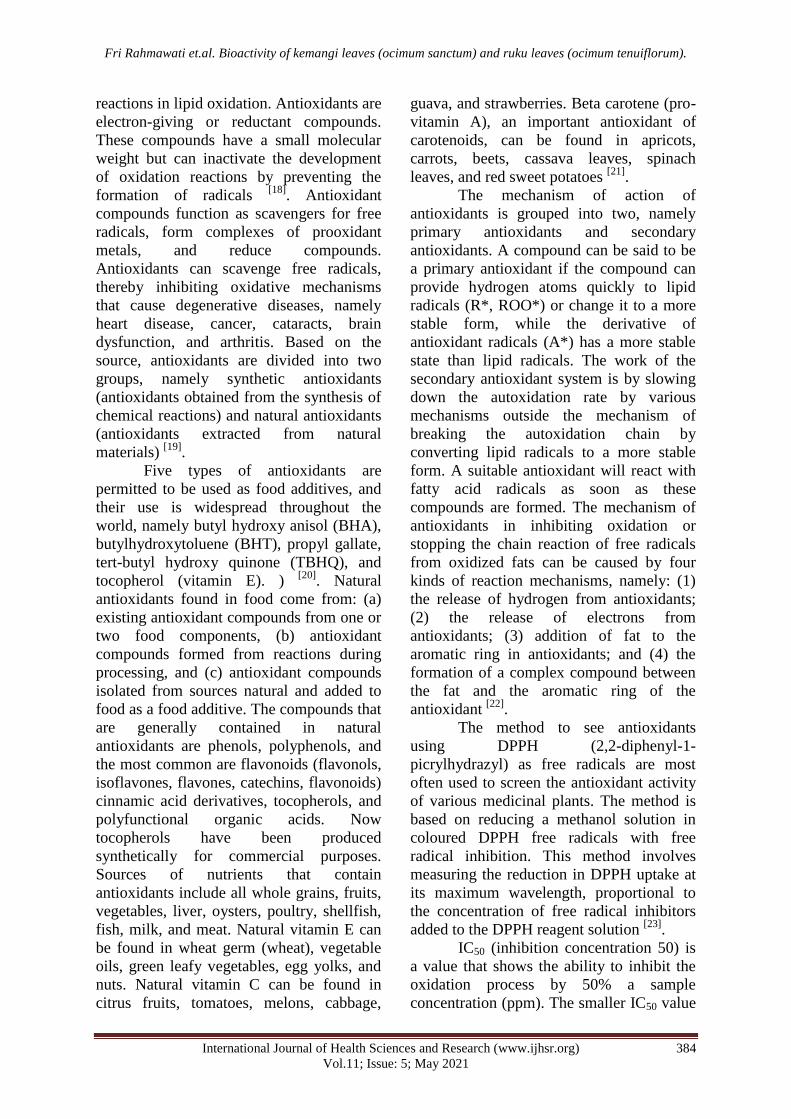
Figure 2. The results of the antibacterial test of kemangi leaf extract (K) and ruku leaf (R) contained bacteria (A) Staphylococcus
aureus and (B) Salmonella typhi
Based on Figure 2, it can be seen
that only ruku ruku leaf extract and positive
control (chloramphenicol) produce a clear
zone (inhibition zone) around the disc
paper. The diameter of the inhibition zone
of the ruku ruku leaf extract and
chloramphenicol can be seen in Figure 3.
B A

Fri Rahmawati et.al. Bioactivity of kemangi leaves (ocimum sanctum) and ruku leaves (ocimum tenuiflorum).
International Journal of Health Sciences and Research (www.ijhsr.org) 387
Vol.11; Issue: 5; May 2021
Figure 3. Diameter of sample inhibition zone, chloramphenicol, distilled water, Staphylococcus aureus and Salmonella typhi bacteria.
Based on the graph in Figure 3, it is
known that the ruku ruku leaves have an
inhibition zone diameter of 9.35 mm. Based
on the classification of inhibition according
to David Stout (Table 2), the ruku ruku leaf
extract is classified as an antibacterial
compound that has moderate inhibition
because it has a diameter in the range of 5-
10 mm. Meanwhile, chloramphenicol
showed potent inhibition in S. aureus and S.
Typhi because it has an inhibition zone
diameter of more than 20 mm.
Table 2. Inhibition Category According to David Stout [28]
Bacterial inhibition Growth Inhibition Response
>20 mm Very strong
10-20 mm Strong
5-10 mm Moderate
<5 mm Weak
The results of the inhibition of ruku
ruku leaf extract, which has a moderate
inhibition zone and kemangi leaf extract that
does not have an inhibition zone may occur
due to errors such as the soil where the
plants are grown, when the plants are
harvested, processing methods such as
maceration, improper simplicia. In the
study, it was known that the concentration
of ethanol extract of kemangi leaves had the
highest inhibition zone in Staphylococcus
aureus, namely the concentration of 100%
without dilution with a result of 21.75 mm.
The concentration of ethanol extract of
kemangi leaves, which has the highest
inhibition zone, is at 80% Salmonella typhi
with a yield of 6.25mm [6]
. In this study, the
ethanol extract of ruku ruku leaves with the
same method had the highest inhibition zone
for Staphylococcus aureus at a
concentration of 50%, and an inhibition
zone of 21.6 mm was obtained [26]
. The
inhibition of ruku ruku extracts against
Salmonella typhi using ethyl acetate extract
and 90% ethanol extract resulted in an
inhibition zone of 6.20 mm and 5.40 mm,
respectively [29]
.
Antifungal Activity Test of Kemangi
Leaf Extract and Ruku Ruku Leaf - The
antifungal test was performed using C.
Albicans as the test fungus and fluconazole
as the standard antibiotic. The sample's
antifungal activity and standard antibiotics
are indicated by forming a clear zone
around the disc paper. The results of the
antifungal test of kemangi and ruku leaf
extracts can be seen in Figure 4.
Figure 4. Antifungal activity test results of kemangi leaf
extract (K) and ruku ruku leaves (R), positive control (+),
negative control (-).

Fri Rahmawati et.al. Bioactivity of kemangi leaves (ocimum sanctum) and ruku leaves (ocimum tenuiflorum).
International Journal of Health Sciences and Research (www.ijhsr.org) 388
Vol.11; Issue: 5; May 2021
Based on Figure 4, it is known that
the clear zone around the disc paper is only
found in fluconazole, while the disc paper
containing extracts of kemangi leaves and
ruku ruku leaves does not produce clear
zones. It means that the two sample extracts
used did not provide inhibition against the
growth of Candida albicans but differed
from standard antibiotics, which produced a
clear zone around the disc paper.
Figure 5. Graph of the antifungal inhibition zone of kemangi and ruku leaves against Candida albicans
Based on the graph in Figure IV.5, it
can be seen that there is no clear zone
around the disc paper containing extracts of
kemangi leaves, ruku ruku, and negative
controls, but the clear zone is produced in
the positive control (fluconazole) with an
inhibition zone diameter of 27.8 mm. shows
relatively strong inhibition power. Based on
these results, it was known that the two
extracts used could not inhibit the fungus
Candida albicans. Kemangi leaf extract
using the same solvent obtained the highest
inhibition zone at a concentration of 100%
with a diameter of 11.9 mm [30]
. Ruku leaf
extract with 70% ethanol solvent and 100%
concentration can inhibit the growth of the
fungus C. Albicans [31]
.
Figure 6. Graph of antioxidant activity of kemangi leaf extract, ruku ruku leaf extract, and vitamin C
Antioxidant Activity Test of
Kemangi Leaf Extract and Ruku Ruku - The
antioxidant test was carried out using the
DPPH method, the choice of this method
was because the DPPH method was simple,
easy, fast, sensitive, and required a small
number of samples. The DPPH method is
easy to apply because the DPPH radical
compound used is relatively stable
compared to other methods [28]
. Antioxidant

Fri Rahmawati et.al. Bioactivity of kemangi leaves (ocimum sanctum) and ruku leaves (ocimum tenuiflorum).
International Journal of Health Sciences and Research (www.ijhsr.org) 389
Vol.11; Issue: 5; May 2021
activity is expressed by the value of
inhibition concentration 50 (IC50). IC50 can
be defined as the substrate concentration
that can cause a 50% reduction in DPPH
activity [8]
. The IC50 value in the extract of
kemangi leaves and ruku leaves can be seen
in Figure 6.
Based on the graph in Figure IV.6, it
is known that the two extracts used in the
study have antioxidant activity with IC50
values of 225.64 ppm and 455.11 ppm,
respectively. Based on the IC50 value
obtained, it can be seen that the ruku ruku
leaf extract has more excellent antioxidant
activity than the Kemangi leaf extract,
indicated by a low IC50 value. A low IC50
value indicates high antioxidant activity.
The antioxidant activity produced by the
extracts of kemangi leaves and ruku ruku
leaves is weak because the IC50 value is
more significant than 200 ppm. Kemangi
leaf extract has a moderate IC50 value of
52.68 ppm [32]
. Ruku leaf extract using the
DPPH method is known to have an IC50
value of 46 ppm [33]
. The smaller IC50 value
indicates the higher the antioxidant activity.
A compound is said to have potent
antioxidant activity if the IC50 value is less
than 50 ppm, the potent antioxidant for IC50
is 50-100 ppm, moderate antioxidant if the
IC50 value is 100-150 ppm, and weak
antioxidants if the IC50 value is 151-200
ppm.
CONCLUSION
The research results show that the
kemangi leaf extract and ruku leaf extract
contain active compounds of the flavonoid
and steroid classes. Kemangi leaf extracts
and ruku ruku leaves could not inhibit the
growth of Salmonella typhi bacteria, but
ruku ruku leaf extract could inhibit
Staphylococcus aureus bacteria with an
inhibition zone of 9.35 mm. Kemangi leaf
extracts and ruku ruku leaves did not inhibit
the growth of the fungus Candida albicans.
However, kemangi leaf extract and ruku
ruku have antioxidant activity. Ruku ruku
leaves have more excellent antioxidant
activity than kemangi leaves extract, with an
IC50 value of 225.64 ppm.
Acknowledgement: None
Conflict of Interest: None
Source of Funding: None
Ethical Approval: Approved
REFERENCES 1. Sentari, M., Harahap, U., Sapiie, T. W. A.,
& Ritarwan, K. (2019). Blood cortisol level
and blood serotonin level in depression mice
with basil leaf essential oil treatment. Open
access Macedonian journal of medical
sciences, 7(16), 2652.
2. Riady, M. H., Rostini, I., Andriani, Y., &
Pratama, R. I. (2019). Effectiveness of the
Ruku-ruku Leaf Solution (Ocimum
sanctum) as a Natural Preservative in Indian
Mackerel (Rastrelliger sp.) during Low-
temperature Storage. Asian Food Science
Journal, 1-13.
3. Parbuntari, H., Etika, S. B., Mulia, M., &
Delvia, E. (2019). A Preliminary Screening
of the Different of Secondary Metabolites
Ruku-Ruku Leaves (Ocimum tenuiflorum
Linnen) in West Sumatera. Eksakta: Berkala
Ilmiah Bidang MIPA (E-ISSN: 2549-
7464), 20(2), 17-24.
4. Pandiyan, A. (2012). Chemical Profiling
and Biological Activity on Leaves of
Ocimum Gratissimum Grown in
Tamilnadu (Doctoral dissertation,
Adhiparasakthi College of Pharmacy,
Melmaruvathur).
5. Gavamukulya, Y., Abou-Elella, F.,
Wamunyokoli, F., & AEl-Shemy, H.
(2014). Phytochemical screening, anti-
oxidant activity and in vitro anticancer
potential of ethanolic and water leaves
extracts of Annona muricata
(Graviola). Asian Pacific journal of tropical
medicine, 7, S355-S363.\
6. Khan, A. M., Qureshi, R. A., Ullah, F.,
Gilani, S. A., Nosheen, A., Sahreen, S., ... &
Murad, W. (2011). Phytochemical analysis
of selected medicinal plants of Margalla
Hills and surroundings. Journal of medicinal
plants research, 5(25), 6055-6060.
7. Wright, P. M., Seiple, I. B., & Myers, A. G.
(2014). The evolving role of chemical

Fri Rahmawati et.al. Bioactivity of kemangi leaves (ocimum sanctum) and ruku leaves (ocimum tenuiflorum).
International Journal of Health Sciences and Research (www.ijhsr.org) 390
Vol.11; Issue: 5; May 2021
synthesis in antibacterial drug
discovery. Angewandte Chemie
International Edition, 53(34), 8840-8869.
8. Birbir, Y., & Birbir, M. (2006). Inactivation
of extremely halophilic hide-damaging
bacteria via low-level direct electric
current. Journal of electrostatics, 64(12),
791-795.
9. Guzzo, M. B. (2017). Molecular
Mechanisms Underlying the Intrinsic
Sulfonamide Resistance in Bacteria. Case
Western Reserve University.
10. Kohanski, M. A., Dwyer, D. J., & Collins, J.
J. (2010). How antibiotics kill bacteria: from
targets to networks. Nature Reviews
Microbiology, 8(6), 423-435.
11. Clemens, M. J., Bushell, M., Jeffrey, I. W.,
Pain, V. M., & Morley, S. J. (2000).
Translation initiation factor modifications
and the regulation of protein synthesis in
apoptotic cells. Cell Death &
Differentiation, 7(7), 603-615.
12. Lü, J. M., Lin, P. H., Yao, Q., & Chen, C.
(2010). Chemical and molecular
mechanisms of antioxidants: experimental
approaches and model systems. Journal of
cellular and molecular medicine, 14(4), 840-
860.
13. Durant, S., & Karran, P. (2003). Vanillins- a
novel family of DNA-PK inhibitors. Nucleic
acids research, 31(19), 5501-5512.
14. Wang, Y., Feng, K., Yang, H., Zhang, Z.,
Yuan, Y., & Yue, T. (2018). Effect of
cinnamaldehyde and citral combination on
transcriptional profile, growth, oxidative
damage and patulin biosynthesis of
Penicillium expansum. Frontiers in
microbiology, 9, 597.
15. Woodhams, D. C., LaBumbard, B. C.,
Barnhart, K. L., Becker, M. H., Bletz, M.
C., Escobar, L. A., ... & Minbiole, K. P.
(2018). Prodigiosin, violacein, and volatile
organic compounds produced by widespread
cutaneous bacteria of amphibians can inhibit
two Batrachochytrium fungal
pathogens. Microbial ecology, 75(4), 1049-
1062.
16. Li, B., Li, Q., Xu, Z., Zhang, N., Shen, Q.,
& Zhang, R. (2014). Responses of
beneficial Bacillus amyloliquefaciens SQR9
to different soilborne fungal pathogens
through the alteration of antifungal
compounds production. Frontiers in
microbiology, 5, 636.
17. Stupar, M., Grbić, M. L., Džamić, A.,
Unković, N., Ristić, M., Jelikić, A., &
Vukojević, J. (2014). Antifungal activity of
selected essential oils and biocide
benzalkonium chloride against the fungi
isolated from cultural heritage
objects. South African Journal of
Botany, 93, 118-124.
18. Li, Y., Jiang, B., Zhang, T., Mu, W., & Liu,
J. (2008). Antioxidant and free radical-
scavenging activities of chickpea protein
hydrolysate (CPH). Food chemistry, 106(2),
444-450.
19. Caleja, C., Barros, L., Antonio, A. L.,
Oliveira, M. B. P., & Ferreira, I. C. (2017).
A comparative study between natural and
synthetic antioxidants: Evaluation of their
performance after incorporation into
biscuits. Food chemistry, 216, 342-346.
20. Tormin, T. F., Almeida, E. S., de Sousa, R.
M. F., & Mathias, E. (2015). Determination
of Butylhydroxytoluene,
Butylhydroxyanisole, and tert-
Butylhydroquinone. Flow Injection Analysis
of Food Additives, 1, 225.
21. Bechoff, A. (2010). Investigating carotenoid
loss after drying and storage of orange-
fleshed sweet potato (Doctoral dissertation,
University of Greenwich).
22. Brewer, M. S. (2011). Natural antioxidants:
sources, compounds, mechanisms of action,
and potential applications. Comprehensive
reviews in food science and food
safety, 10(4), 221-247.
23. Kedare, S. B., & Singh, R. P. (2011).
Genesis and development of DPPH method
of antioxidant assay. Journal of food science
and technology, 48(4), 412-422.
24. Utami, P. S. M., Noorhamdani, N., &
Rahayu, M. (2020). The extract of kemangi
leaves as inhibitor of biofilm from
Staphylococcus aureus in vitro. Journal
AMS, 6(3), 168-173.
25. Parbuntari, H., Etika, S. B., & Delvia, E.
(2021, February). Molecular encapsulation
of bioactive molecules of Ruku-Ruku leaves
(Ocimum tenuiflorum Linnen) as a
preliminary stability study. In Journal of
Physics: Conference Series (Vol. 1788, No.
1, p. 012002). IOP Publishing.
26. Sher, A. (2009). Antimicrobial activity of
natural products from medicinal plants.
27. Chung, K. T., Wong, T. Y., Wei, C. I.,
Huang, Y. W., & Lin, Y. (1998). Tannins
and human health: a review. Critical

Fri Rahmawati et.al. Bioactivity of kemangi leaves (ocimum sanctum) and ruku leaves (ocimum tenuiflorum).
International Journal of Health Sciences and Research (www.ijhsr.org) 391
Vol.11; Issue: 5; May 2021
reviews in food science and nutrition, 38(6),
421-464.
28. Lu’lu’ A’lana, Rafika S, Pratiwi A.
Determination of FICI Value Combination
of Ethanol Extract of Aloe Vera Leaves
(Aloe vera (L.) and Gentamicin Sulphate
Against Escherichia coli Bacteria. Faculty
of Medicine, Tanjungpura University,
Pontianak. 2018.
29. Octaviani, P. (2020, February).
Identification of Chemical Content and
Antihypertensive Activity Test of Ethanol
Extract from Tali Bamboo Shoot
(Gigantochloa apus (Schult. & Schult. F.)).
In 1st International Conference on
Community Health (ICCH 2019) (pp. 237-
241). Atlantis Press.
30. Himawan, H. C., Isa, A. F., & Wiharja, D.
S. (2021, February). Antioxidant Activity
Of 70% Ethanol Extract Combination of
Kemangi Leaf (Ocimum Americanum Linn)
and Binahong Leaf (Anredera cordifolia
(Ten.) Steenis) Using DPPH. In Journal of
Physics: Conference Series (Vol. 1764, No.
1, p. 012009). IOP Publishing.
31. Pandiyan, A. (2012). Chemical Profiling
and Biological Activity on Leaves of
Ocimum Gratissimum Grown in
Tamilnadu (Doctoral dissertation,
Adhiparasakthi College of Pharmacy,
Melmaruvathur).
32. Erviana, L., Malik, A., & Najib, A. (2016).
The free antiradical activity test of the
ethanol extract of basil (Ocimum basilicum
L.) using the dpph method. Jurnal
Fitofarmaka Indonesia, 3(2), 164-168.
33. Parbuntari, H., Etika, S. B., & Mulia, M.
(2020, August). Identification and
Prediction of Potential Drug-like
Compounds from Ruku-Ruku Leaves
(Ocimum tenuiflorum Linnen) in West
Sumatera. In International Conference on
Biology, Sciences and Education
(ICoBioSE 2019) (pp. 248-252). Atlantis
Press.
How to cite this article: Rahmawati F, Silaban
H. Bioactivity of kemangi leaves (ocimum
sanctum) and ruku leaves (ocimum
tenuiflorum). Int J Health Sci Res. 2021; 11(5):
379-391. DOI: https://doi.org/10.52403/ijhsr.
20210558
******