Embed Size (px)
Citation preview

RSC Advances
PAPER
Ope
n A
cces
s A
rtic
le. P
ublis
hed
on 0
5 M
arch
202
0. D
ownl
oade
d on
1/2
2/20
22 1
2:39
:37
PM.
Thi
s ar
ticle
is li
cens
ed u
nder
a C
reat
ive
Com
mon
s A
ttrib
utio
n-N
onC
omm
erci
al 3
.0 U
npor
ted
Lic
ence
.
View Article OnlineView Journal | View Issue
Bio-nanoparticle
aDefence Research Laboratory, DRDO, Post
E-mail: [email protected]; sultan
com; [email protected]; varunbhard
com; [email protected]; p
258534; Tel: +91-3712-258836bSri Sankardeva Nethralaya, Beltola, Gu
[email protected] of Veterinary Science, Assam Agricu
781022, India. E-mail: [email protected]
Cite this: RSC Adv., 2020, 10, 9356
Received 28th November 2019Accepted 11th February 2020
DOI: 10.1039/c9ra09972g
rsc.li/rsc-advances
9356 | RSC Adv., 2020, 10, 9356–93
assembly: a potent on-sitebiolarvicidal agent against mosquito vectors
Nazima Sultana,a Prasanta K. Raul, *a Diganta Goswami,a Dipankar Das,b
Saidul Islam,c Varun Tyagi,a Bodhaditya Das,a Hemanta K. Gogoi,a
Pronobesh Chattopadhyaya and Pakalapati S. Rajua
Background: Vector-borne diseases such as malaria, dengue, yellow fever, encephalitis and filariasis are
considered serious human health concerns in the field of medical entomology. Controlling the
population of mosquito vectors is one of the best strategies for combating such vector-borne diseases.
However, the use of synthetic insecticides for longer periods of time increases mosquito resistance to
the insecticides. Recently, the search for new environmentally friendly and efficient insecticides has
attracted major attention globally. With the evolution of material sciences, researchers have reported the
effective control of such diseases using various sustainable resources. The present investigation
demonstrates a potent on-site biolarvicidal agent against different mosquito vectors such as Aedes
albopictus, Anopheles stephensi and Culex quinquefasciatus. Methods: Stable and photo-induced
colloidal silver nanoparticles were generated via the surface functionalization of the root extract of
Cyprus rotundas. Characterizations of the nanoparticles were performed using assorted techniques, such
as UV-visible spectroscopy, FTIR spectroscopy, DLS and HRTEM. The bioefficacy of the synthesized
nanoparticles was investigated against different species of mosquito larvae through the evaluation of
their life history trait studies, fecundity and hatchability rate of the treated larvae. Histopathological and
polymerase chain reaction-random amplified polymorphic DNA (RAPD) analyses of the treated larvae
were also examined to establish the cellular damage. Results: The synthesized nanoparticles showed
remarkable larvicidal activity against mosquito larvae in a very low concentration range (0.001–
1.00) mg L�1. The histopathological study confirmed that the present nanoparticles could easily enter the
cuticle membrane of mosquito larvae and subsequently obliterate their complete intestinal system.
Furthermore, RAPD analysis of the treated larvae could assess the damage of the DNA banding pattern.
Conclusion: The present work demonstrates a potent biolarvicidal agent using sustainable bioresources
of the aqueous Cyprus rotundas root extract. The results showed that the synthesized nanoparticles
were stable under different physiological conditions such as temperature and photo-induced oxidation.
The effectiveness of these materials against mosquito larvae was quantified at very low dose
concentrations. The present biolarvicidal agent can be considered as an environmentally benign material
to control the mosquito vectors with an immense potential for on-site field applications.
1. Introduction
Vector-borne diseases, for example, malaria, dengue, yellowfever and lariasis are considered to be serious health issues.Mosquito species belonging to the genera Aedes albopictus,
bag no. 2, Tezpur-784001, Assam, India.
[email protected]; digs78@gmail.
[email protected]; gogoi_hk@yahoo.
[email protected]; Fax: +91-3712-
wahati-781028, Assam, India. E-mail:
ltural University, Khanapara, Guwahati –
68
Anopheles stephensi and Culex quinquefasciatus play signicantroles in the transmission of such vector-borne diseases.Anopheles stephensi is the primary malaria vector found in Indiaand other Asian countries and transmits lariasis.1 The Aedesaegypti vector transmits several yellow fevers like dengue, chi-kungunya, viruses and other diseases.2 Culex quinquefasciatus isan important vector of lymphatic lariasis.3 These species arefound abundantly in many tropical areas of the world and breedvery fast in drains and stagnant water reservoirs; their devel-opment involves the holometabolous life cycle, which includesfour life stages: eggs, larvae, pupae and adults. Although the useof synthetic insecticides can interrupt these disease trans-missions, the repeated use of insecticides can easily destroy theecosystem and lead to a resurgence of the mosquito
This journal is © The Royal Society of Chemistry 2020
Paper RSC Advances
Ope
n A
cces
s A
rtic
le. P
ublis
hed
on 0
5 M
arch
202
0. D
ownl
oade
d on
1/2
2/20
22 1
2:39
:37
PM.
Thi
s ar
ticle
is li
cens
ed u
nder
a C
reat
ive
Com
mon
s A
ttrib
utio
n-N
onC
omm
erci
al 3
.0 U
npor
ted
Lic
ence
.View Article Online
population.4,5 In addition, a number of challenging medicaltreatments have been reported to cure an infected person.6
However, the development of control programs for mosquitovectors at their earlier stage is more suitable to prevent suchtransmitted vector-borne diseases.7 Also, the targeting of thelarvae is considered an effective strategy due to their lowermobility in breeding habitats. Since the past few decades,researchers have been searching for eco-friendly, biodegradableand economically viable alternatives to chemical insecticides tosuppress the population of various mosquito disease vectors.8,9
With the evolution of material sciences, nanoparticles arenow considered as a promising candidate to control vector-borne diseases.10 It is considered to be the latest promisingplatform to help reach that goal with a wide range of benets,including increased bioefficacy, durability and controlleddelivery to the specic targets. The use of efficient nanoparticleswith improved physiochemical properties can help to reducethe serious environmental issues of larvicidal agent.10–16 Studieswith silver nanoparticles have been done extensively againstseveral different types of mosquito vectors.10,17,18 Variouschemical and physical methods are used for the synthesis ofnanoparticles. The use of plant sources-based bioactive organicchemicals has gained more attention to synthesize silvernanoparticles.19–23 Phytochemical approaches for the synthesisof silver nanoparticles are considered to be a promising methodfor controlling mosquito vector-borne diseases due to their lowtoxicity and cost effectiveness.24 Various intersections have beenhighlighted between biotechnology and nanotechnology via thedevelopment of environmental benign technologies in the eldof material sciences, whereby such materials can serve asdefence chemicals, like insecticides, anti-feedants, repellents,oviposition deterrents, growth inhibitors, and moultinghormones as well as attractants due to the occurrence ofsecondary metabolites.25 Despite the spectacular activities ofsilver nanoparticles, their large-scale utility is not yet consid-ered due to the requirement of high dose concentrations, whichcan further induce toxicological effects on non-targeted organ-isms present in the environment.26 Therefore, the utilization ofhighly biocompatible and biodegradable polymeric materials tosynthesize the nanoparticles may be a far-reaching goal toovercome such environmental risk factors.
The Cyprus rotundas plant is an important bioresource fornutrition and medicine. It is a carbohydrate-rich leguminousroot species and such polysaccharide-based nutgrass plants arewidely available in South Asia. It is indigenous to India, avail-able mostly in tropical, subtropical and temperate regions. Theextreme black tubers of the Cyprus rotundas plant with a char-acteristic odour have staple carbohydrate-based contents, like a-cyperone, b-selinene, cyperene, patchoulenone, sugeonol,kobusone and isokobusone, which are signicant for medicinaluse, especially as a larvicidal.27 Therefore, in this context weaimed to prepare a stable silver nano-biocomposite materialusing a facile method with Cyprus rotundas root extract. Weobserved an excellent effectiveness of the synthesized biol-arvicidal agent against variousmosquito disease vectors and theobtained results are presented here.
This journal is © The Royal Society of Chemistry 2020
2. Experimental section2.1 Materials and methods
Cyprus rotundas root were collected from the Defence ResearchLaboratory garden in Tezpur, Assam, India. They were washedwith tap water several times to remove dust particles and werethen sun dried. The dried Cyprus rotundas roots were furtherground to make ne powder using a domestic blender.
2.2 Biosynthesis of silver nanoparticles
Cyprus rotundas root powder (2 g) was heated with 100 mL ofwater at 50 �C for 1 h and then ltered using muslin cloth. Next,100 mL of 0.01 (M) AgNO3 solution was added drop-wise with50 mL root extract in a conical ask and with continuous stir-ring at 50 �C for up to 2 h. Aer the termination of the reactiontime, the mixture was allowed to cool down at room tempera-ture and nally a dark brown colloidal solution was obtained.The collected solution was ltered using a muslin cloth andcentrifuged at 960 � g for 10 min to remove any undesired largeparticles. The product was vacuum dried at room temperature.The dried product was further dissolved in water to make1000 mg L�1 stock solution.
2.3 General experimental procedures
A UV-visible spectrophotometer (U2001, Hitachi, Japan) wasused for the detection of the surface plasmon resonance (SPR)peaks. The formation of silver nanoparticles was preliminarilyconrmed with the help of dynamic light scattering (DLS)measurements using a Malvern Nano ZS instrument at 633 nm(a 4 mW He Ne laser was used) and 90� scattering angle. Theprepared colloidal solution was ltered through a 0.22 mmmembrane lter prior to the measurements. The scatteringintensity data were processed using instrumental soware toobtain the average hydrodynamic diameter (dH) of the particlesin the solution. The chemical functionalities of the nano-materials solutions were identied using Fourier transforminfrared spectrometry (FTIR) (Bruker, Model Alpha Eco-ATR)with the accumulation of 15 scans. The spectra were recordedwithin the range of 500–4000 cm�1. The resolution of thespectra was set within the range of 8–2.0 cm�1. Initially, a smallamount of the lyophilized sample was mixed with 1% KBrpowder and ground to reduce the particle size. High-resolutiontransmission electron microscopy (HRTEM) images of thesolution were taken on a JEOL microscope at an operatingvoltage of 200 kV. The sample was prepared for HRTEM bydropping aqueous suspensions of the nanoparticles onto Cugrids coated with a holey carbon lm followed by solventevaporation.
2.4 Mosquito larvae
Larval strains of Aedes albopictus, Aedes aegypti, Culex quinque-fasciatus and Anopheles stephensi were collected from themosquito culture facility of the Defence Research Laboratory,Tezpur, India. The mosquito larvae were maintained at 28 �2 �C and 75–85% relative humidity under 14 : 10 (light : dark).
RSC Adv., 2020, 10, 9356–9368 | 9357

RSC Advances Paper
Ope
n A
cces
s A
rtic
le. P
ublis
hed
on 0
5 M
arch
202
0. D
ownl
oade
d on
1/2
2/20
22 1
2:39
:37
PM.
Thi
s ar
ticle
is li
cens
ed u
nder
a C
reat
ive
Com
mon
s A
ttrib
utio
n-N
onC
omm
erci
al 3
.0 U
npor
ted
Lic
ence
.View Article Online
They were fed with amixture of 1 : 3 (dog biscuits : yeast extract)for their survival.
2.5 Larvicidal activity of the silver nanoparticles
The synthesized silver nanoparticles were subjected to a dose–response bioassay for larvicidal activity against Aedes albopictus,Aedes aegypti, Culex quinquefasciatus and Anopheles stephensispecies of the larvae. For the bioassay test, initially 20 numbersof 3rd and 4th instars larvae were taken in a 100 mL beaker.Different concentrations of silver nanoparticles ranging from0.01–5.0 mg L�1 were prepared to study the larvicidal activityagainst the mosquito larvae. The control was set up withdeionized water and kept for 24 h observation. The numbers ofdead larvae were counted aer 24 h of exposure and thepercentage mortality was reported from the average of threereplicates. Mortality was calculated using Abbott's formula.28
The dose–response data were subjected to probit regressionanalysis using IBM SPSS Statistics soware.29 Lethal doseconcentrations at the upper condence limit (LC90) and at thelower condence limit (LC50) were calculated with 95% con-dence intervals.
2.6 Life history traits studies of the mosquito vectors
Aedes albopictus and Culex quinquefasciatus larvae were treatedwith silver nanoparticles and their complete life history traitswere studied. Adult mosquitoes were maintained in optimumconditions at 28 � 2 �C and 75–85% relative humidity under14 : 10 (light : dark) photoperiod.30 Male and female mosqui-toes were housed together in the same size of cage (30 � 30 �30 cm) for about one week in order to mate. Honey-soakedcotton pads were provided as a sugar source to them prior toa blood meal. Mated females were collected and fed upon rabbitblood in order to undergo egg maturation prior to testing.
2.7 Histological studies of the nanoparticle-treated larvae
For histological tests, both the silver nanoparticles-treated anduntreated third instars larvae of Aedes albopictus were takenfrom the laboratory colony. The LC50 dose concentrations of thetreated larvae were incorporated aer 24 h exposure and keptfor examination. Aerwards, they were xed with 10% formal-dehyde solution for up to 24 h and then dehydrated using anincreasing acetone gradient, i.e. 30–100%. The treated larvaewere embedded and cut with glass knives in a rotary microtome.The sections were stained with haematoxylin–eosin, analyzedand photographed using a photomicroscope.
2.8 DNA extraction of the silver nanoparticles-treated larvae
DNA from the mosquito larvae was extracted using the reportedmethod by Ballinger-Crabtree et al.31,32 Each adult femalemosquito was ground with 200 mL lysis buffer (100 mM Tris–HCl, pH 8.0; 1% sodium dodecylsulphate; 50 mM NaCl; 50 mMEDTA). The mixture was kept immediately on ice for 15 minfollowed by heating at 65 �C for 30 min. Subsequently, thehomogenous mixture was further treated with 30 mL of 5 Mpotassium acetate for 1 h at 10 �C followed by centrifugation at
9358 | RSC Adv., 2020, 10, 9356–9368
�13 000 rpm. The suspension was extracted by chilled ethanoland kept overnight at �20 �C. The suspension was furthercentrifuged at 13 000 rpm at 10 �C and the precipitated DNAwas washed twice with 70% ethanol at room temperature. Thepalleted DNA were kept for air drying and nally dissolved in 50mL TE buffer. Concentrations of DNA extracted from variouslarvae were determined using spectrophotometer.
2.9 Polymerase chain reaction (PCR) and random ampliedpolymorphic DNA (RAPD)
We screened 20 different numbers of 10 mole oligonucleotideRAPD primers. Out of these, only two primers were assignedhere as primer 1 (GTGGCTTGGA) and primer 2 (GTAAACCGCC),which showed good results. These were selected for the rearedAedes albopictus mosquito strains in our laboratory. RAPDamplication was performed with a 10 mL PCR mixture con-taining 10mMPCR buffer at pH 9.0, 50mMMgCl2, 2 mMdNTP,0.2 (M) primer and 0.5 U of Tag DNA polymerase. Steriledeionized water was used for the volume make-up. 10 mg ofextracted DNA was also added in to each PCR tube. The reactionmixture was given a short spin for thoroughly mixing thecocktail of components. The PCR tubes were loaded ontoa thermal cycler (iCycler, Bio-Rad PCR System) for 4 min at94 �C, followed by 35 cycles of 1 min at 94 �C, 1 min at 40 �C and2 min at 72 �C. The nal extension step was performed at 72 �Cfor 10 min.
2.10 Agarose gel electrophoresis
The amplication products were analyzed by the electropho-resis method using 1% agarose gel (w/v).33 Along with the PCR-amplied products, 100 bp DNA ladders were used as thestandard marker and subjected to electrophoresis using 1%agarose gel in 0.5� TAE buffer and then stained using ethidiumbromide solution. The molecular sizes of the markers were1000, 900, 800, 700, 600, 500, 400, 300, 200 and 100 bp. Theamplied pattern was visualized on a UV trans-illuminator andthen photographed. Polymorphism was evidenced both in thepresence or absence of DNA fragments between the samples.The RAPD proles of the treated larvae were evaluated using thesame 1% agarose gel run for h at 100 volts followed by 80 voltsfor about 3 h with 0.5� TAE buffer.
3. Results and discussion3.1 Origin of the silver nanoparticles
Cyprus rotundas root extract was used as a precursor tosynthesize silver nanoparticles in the present work. In general,plant-based materials contain an untapped reservoir of carbo-hydrate sources, like cellulose, pectin, chitin, starch, chitosanand many more that can be used as templates for the synthesisof nanoparticles. Therefore, during the hydrolysis process, anaqueous extract of the material decomposes carbohydratemolecules and forms surface-functionalized circumstances bygenerating different functional groups, such as hydroxyl,carbonyl, carboxyl and epoxy groups, in the solution mixture.10
This process helps to liberate a substantial amount of protons
This journal is © The Royal Society of Chemistry 2020
Paper RSC Advances
Ope
n A
cces
s A
rtic
le. P
ublis
hed
on 0
5 M
arch
202
0. D
ownl
oade
d on
1/2
2/20
22 1
2:39
:37
PM.
Thi
s ar
ticle
is li
cens
ed u
nder
a C
reat
ive
Com
mon
s A
ttrib
utio
n-N
onC
omm
erci
al 3
.0 U
npor
ted
Lic
ence
.View Article Online
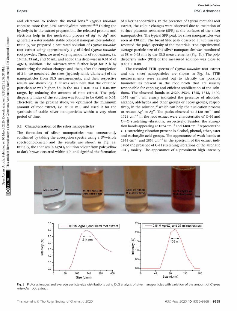
and electrons to reduce the metal ions.34 Cyprus rotundascontains more than 35% carbohydrate content.35,36 During thehydrolysis in the extract preparation, the released protons andelectrons help in the nucleation process of Ag+ to Ag0 andgenerate a water soluble stable colloidal nanoparticles solution.Initially, we prepared a saturated solution of Cyprus rotundasroot extract using approximately 2 g of dried Cyprus rotundasroot powder. Then, we used varying amounts of root extract, i.e.10mL, 35mL, and 50mL, and added this drop-wise in 0.01 M ofAgNO3 solution. The mixtures were further kept for 2 h bymonitoring the colour changes and then, aer the completionof 2 h, we measured the sizes (hydrodynamic diameter) of thenanoparticles from DLS measurements, and their respectiveresults are shown Fig. 1. It was seen here that the obtainedparticle size was higher, i.e. in the 103 � 0.01–214 � 0.04 nmrange, by reducing the amount of root extract. The poly-dispersity index of the solution was found to be 0.662 � 0.02.Therefore, in the present study, we optimized the minimumamount of root extract, i.e. at 50 mL, and used it for thesynthesis of stable silver nanoparticles within a very shortperiod of time.
3.2 Characterization of the silver nanoparticles
The formation of silver nanoparticles was concurrentlyconrmed by taking the absorption spectra using a UV-visiblespectrophotometer and the results are shown in Fig. 2a.Initially, the changes in AgNO3 solution colour from pale yellowto dark brown occurred within 2 h and signied the formation
Fig. 1 Pictorial images and average particle-size distributions using DLSrotundas root extract.
This journal is © The Royal Society of Chemistry 2020
of silver nanoparticles. In the presence of Cyprus rotundas rootextract, the colour changes were observed due to excitation ofsurface plasmon resonance (SPR) at the surfaces of the silvernanoparticles. The typical SPR peak for silver nanoparticles wasseen at 430 nm. The broad SPR peak observed at 430 nm rep-resented the polydispersity of the materials. The experimentalaverage particle size of the silver nanoparticles was monitoredat 58 � 0.05 nm by the DLS measurements (Fig. 2b). The poly-dispersity index (PDI) of the measured solution was close to0.462 � 0.08.
The recorded FTIR spectra of Cyprus rotundas root extractand the silver nanoparticles are shown in Fig. 3a. FTIRmeasurements were carried out to identify the possiblebiomolecules present in the root broth that are usuallyresponsible for capping and efficient stabilization of the solu-tions. The observed bands at 3420, 2934, 1715, 1643, 1400,1074 cm�1, etc. clearly indicated the presence of alcohols,alkanes, aldehydes and ether groups or epoxy groups, respec-tively, in the solution,37 which can help the nucleation processto reduce Ag+ to Ag0. The peaks observed at 3420 cm�1 and1724 cm�1 in the root extract were characteristic of O–H andC]O stretching vibrations, respectively. Besides, the absorp-tion bands appearing at 1074 cm�1 and 1400 cm�1 represent theC–O stretching vibration present in alcohol, phenol, ether, esterand carboxylic acid groups. The appearance of weak bands at2934 cm�1 and 2854 cm�1 in the spectrum of the extract indi-cated the presence of C–H stretching vibrations of the aliphatic–CH2 moiety. The appearance of a prominent high intensity
analysis of silver nanoparticles with variation of the amount of Cyprus
RSC Adv., 2020, 10, 9356–9368 | 9359

Fig. 2 (a) UV-vis spectrum and (b) average particle-size analysis of the silver nanoparticles.
Fig. 3 (a) FTIR spectra of Cyprus rotundas root extract (blue line) and silver nanoparticles (red line); (b) thermogravimetric analysis of the silvernanoparticles.
RSC Advances Paper
Ope
n A
cces
s A
rtic
le. P
ublis
hed
on 0
5 M
arch
202
0. D
ownl
oade
d on
1/2
2/20
22 1
2:39
:37
PM.
Thi
s ar
ticle
is li
cens
ed u
nder
a C
reat
ive
Com
mon
s A
ttrib
utio
n-N
onC
omm
erci
al 3
.0 U
npor
ted
Lic
ence
.View Article Online
peak and decrease in their intensity aer the formation ofnanoparticles manifested the aptitude of the strong bindinginteractions of the capping agent without any agglomeration.The thermogravimetric analysis (TGA) results in Fig. 3b wererecorded on a Shimadzu TG 50 analyzer under a constantnitrogen ow (30 mL min�1) at a heating rate of 10 �C min�1.Fig. 2b represents the weight loss of the solution in the 165–550 �C temperature range and the amount of weight loss wasfound to be 21.16%. This may be due to the desorption of thebio-organic compounds of the root extract. However, thepresent synthesized nanoparticles represented a stable form upto a very high temperature range.
In general, nanoparticles have shown unique propertiesrelated to their particle size. Transmission electron microscopy(TEM) is an excellent technique to analyze the accurate particlesize of the prepared nanoparticles. Therefore, the size andmorphology of the silver nanoparticles were further monitoredusing HRTEM analysis and the results are shown in Fig. 4a.Here, most of the silver nanoparticles were found to be spher-ical in shape and were uniformly dispersed without anyagglomeration. The diameters of the silver nanoparticlesranged between 5–20 nm. The monitored nanoparticles sizefrom using DLS and HRTEM analysis was found to be mis-matched. The HR-TEM analysis images showed a very tinyportion of the sample with the possibility of the presence of
9360 | RSC Adv., 2020, 10, 9356–9368
a few particles being present in the nanoparticles solution.However, DLS measures the relaxation time for the decay of theautocorrelation function of the scattered light, from which thediffusion coefficient is inversely proportional to the averageparticle size of the materials. In Fig. 4b, the selected area elec-tron diffraction (SAED) patterns demonstrated the presence ofbright spots within the concentric diffraction rings corre-sponding to the (200) plane of the face-centred-cubic (fcc) latticeof silver nanoparticles. The crystalline nature of the single silvernanoparticles is further shown in Fig. 4c and d, while theinverse fast Fourier transform (IFFT) images ascertained thelayer spacing of silver nanoparticles was close to 0.21 nm.
3.3 Larvicidal activity
Nowadays, controlling mosquito disease vectors is an importantaspect for the comfort of human health. In concern to envi-ronment issues, conventional synthetic insecticides presentserious threats all over the world. In general, the use of herbalplants is one of the best alternatives to control mosquitovectors.38,39 However, the bioactive compounds present inphytochemical extracts are not resistant to their medicinalproperties due to presence of toxicity and biodegradabilityissues at higher dose concentrations.40 Therefore, with theevolution of material science, the uses of metal nanoparticlesare being developed to overcome these hazardous
This journal is © The Royal Society of Chemistry 2020

Fig. 4 (a) HRTEM images of Cyprus rotundas root extract-stabilized silver nanoparticles; (b) SAED images of randomly selected silver nano-particles; (c) HRTEM images of single silver nanoparticles; (d) IFFT images of silver nanoparticles, also showing the layer spacing.
Paper RSC Advances
Ope
n A
cces
s A
rtic
le. P
ublis
hed
on 0
5 M
arch
202
0. D
ownl
oade
d on
1/2
2/20
22 1
2:39
:37
PM.
Thi
s ar
ticle
is li
cens
ed u
nder
a C
reat
ive
Com
mon
s A
ttrib
utio
n-N
onC
omm
erci
al 3
.0 U
npor
ted
Lic
ence
.View Article Online
environmental issues. However, there are several factors thatcan affect their effectiveness; for example, the dose concentra-tion range, particle-size distribution, bioavailability andstability as well as solvent medium used to prepare the nano-particles.41 In view of these factors, we chose a medicinal andrich bioresource-based material, i.e. Cyprus rotundas root, andprepared an aqueous root extract by using a facile hydrolysismethod to synthesize the silver nanoparticles.
Initially, we monitored the efficacy of the materials againstAedes albopictus mosquito larvae with varying amounts of rootextract, as shown in Fig. 1. It was observed that the amount ofnanoparticles required for the mortality was 100 times higherthan that of the optimized form. The optical images of both thetreated and untreated larvae are shown in Fig. 5. In the case ofthe treated larvae 2, we can see that the nanoparticles haddeeply penetrated into the larvae body due to the smaller size ofthe nanoparticles, whereas such a kind of observation wasfound to be missing for the untreated larvae and the treatedlarvae 1. Therefore, we further carried out our studies using thenanoparticles prepared through the 50 mL of root extract. Weobserved an excellent effect in terms of the bioefficacy of thesematerials against different mosquito disease vectors at very lowdose concentrations and the obtained results are discussed inthe following sections. The larvicidal activities of the nano-particles were tested against the 3rd and 4th instars larvae of the
This journal is © The Royal Society of Chemistry 2020
Aedes albopictus, Aedes aegypti, Culex quinquefasciatus andAnopheles stephensi mosquito vectors. The observed rates ofmortality are further listed in Table 1. It could be seen that themaximum percentage of mortality occurred for all those speciesat 0.68 mg L�1 solution of the synthesized silver nanoparticles.
The average rates of mortality for all three replicates werefurther subjected to probit regression analysis and the lethalconcentrations calculated (LC50 and LC90) in mg L�1. Theresults are shown in Table 2, where it can be seen that the lethaldose concentrations at the lower condence limit (LC50) and atthe upper condence limit (LC90) with 95% ducial limits ofsilver nanoparticles lay within the ranges of 0.05–0.225 mg L�1
and 0.675–2.75 mg L�1, respectively. Compared with the re-ported literature data, the present synthesized silver nano-particles had a higher rate of bioefficacy against mosquitolarvae at very low dose concentrations. For example, Veer-akumar et al. used Sida acuta leaves to synthesize silver nano-particles and reported their larvicidal activity against A.stephensi, A. aegypti, and C. quinquefasciatus mosquito vectors.42
The reported dose concentrations followed LC50 and LC90
values at 21.92 and 41.07 mg L�1; 23.96 and 44.05 mg L�1; 26.13and 47.52 mg L�1, respectively. Raman et al. reported theeffectiveness of silver nanoparticles using Pithecellobium dulceextract against the C. quinquefasciatus mosquito vector, whichwas mainly due to their higher surface-to-volume ratio, with an
RSC Adv., 2020, 10, 9356–9368 | 9361

Fig. 5 Optical images of the untreated and treated Aedes albopictus mosquito larvae using varying amounts of root extract, i.e., 35 mL of rootextract (treated larvae 1) and 50 mL of root extract (treated larvae 2) applied for the preparation of the nanoparticles.
Table 1 Larvicidal activity of the silver nanoparticles against both third and fourth instars larvae of Aedes albopictus, Aedes aegypti, Culexquinquefasciatus and Anopheles stephensi (control: nil mortality)
Dose(mg L�1)
Mosquito species
A. albopictus A. aegypti C. quinquefasciatus A. stephensi
3rd instarslarvae
4th instarslarvae
3rd instarslarvae
4th instarslarvae
3rd instarslarvae
4th instarslarvae
3rd instarslarvae
4th instarslarvae
% Mortality (mean value of three replicates, n ¼ mean � standard error)1.0 mg L�1 100 � 0.0 100 � 0.0 100 � 0.0 100 � 0.0 100 � 0.0 100 � 0.0 100 � 0.0 100 � 0.00.50 mg L�1 81.3 � 1.3 85.6 � 4.3 80.9 � 2.1 81.1 � 2.9 82.9 � 0.13 84.9 � 4.6 80.4 � 5.2 83.7 � 5.20.30 mg L�1 69.1 � 4.3 75.7 � 2.9 72.5 � 3.1 74.9 � 1.3 71.9 � 1.9 75.9 � 2.9 71.8 � 4.1 74.2 � 1.10.10 mg L�1 51.9 � 2.13 59.2 � 2.8 55.1 � 3.1 53.1 � 4.0 58.0 � 3.1 59.1 � 4.1 52.6 � 4.7 56.2 � 4.20.05 mg L�1 31.7 � 3.5 38.0 � 2.9 36.1 � 5.5 33.1 � 1.5 37.1 � 4.7 34.7 � 5.1 34.1 � 1.0 35.1 � 2.90.03 mg L�1 15.1 � 3.3 19.4 � 5.1 16.9 � 1.7 15.9 � 1.0 17.9 � 4.1 14.9 � 0.9 19.8 � 2.9 18.1 � 4.5
RSC Advances Paper
Ope
n A
cces
s A
rtic
le. P
ublis
hed
on 0
5 M
arch
202
0. D
ownl
oade
d on
1/2
2/20
22 1
2:39
:37
PM.
Thi
s ar
ticle
is li
cens
ed u
nder
a C
reat
ive
Com
mon
s A
ttrib
utio
n-N
onC
omm
erci
al 3
.0 U
npor
ted
Lic
ence
.View Article Online
LC50 value of 21.56 mg L�1.43 Rawani et al. reported that silvernanoparticles synthesized from aqueous extracts of dry leaves,fresh leaves and berries of Solanum nigrum showed LC50 valuesof 1.33, 1.59 and 1.56 mg L�1 respectively against A. stephensivectors.44 Whereas the LC90 values were reported to be 3.97, 7.31and 4.76 mg L�1 against the A. stephensi vectors, respectively.They suggested that due to the smaller size of silver nano-particles, they could easily pass through the epithelialmembrane of mosquito vectors and inactivate the enzymaticpathway, then leading to cell death. Kumar et al. studied thelarvicidal activity of synthesized silver nanoparticles usingMorinda tinctoria extract against C. quinquefasciatus species,where their reported LC50 value was 1.44 mg L�1.45 Further-more, synthesized silver nanoparticles using a herbicidal plant
9362 | RSC Adv., 2020, 10, 9356–9368
extracts, like Tinospora cordifolia (LC50 � 6.35 mg L�1) andNelumbo nucifera (LC50 � 0.69 mg L�1), showed potentiallarvicidal activity against A. stephensi and C. quinquefasciatuslarvae, respectively.46,47 Similarly, the reported silver nano-particles synthesized from the bark of A. indica (Neem) were alsotested for larvicidal and pupicidal activities against the malariavector A. stephensi and lariasis vector C. quinquefasciatus,48
where the 1st and 2nd instars larvae of C. quinquefasciatus had100% mortality aer 30 min exposure to the nanoparticles.However, pupicidal activity was observed at an LC50 of 4 mg L�1
for A. stephensi and C. quinquefasciatus vectors, whereas theirLC90 values were recorded at 11 and 13 mg L�1, respectively,aer 3 h exposure. Vimala et al. synthesized silver nanoparticlesusing Couroupita guianensis leaf extract and reported their
This journal is © The Royal Society of Chemistry 2020

Table 2 Lethal concentrations (LC50 (mg L�1) (LCL–UCL) and LC90 (mg L�1) (LCL–UCL)) of the silver nanoparticles against both third and fourthinstars larvae of Aedes albopictus, Aedes aegypti, Culex quinquefasciatus and Anopheles stephensi
Dose (mg L�1)
Mosquito species
A. albopictus A. aegypti C. quinquefasciatus A. stephensi
3rd instarslarvae
4th instarslarvae
3rd instarslarvae
4th instarslarvae
3rd instarslarvae
4th instarslarvae
3rd instarslarvae
4th instarslarvae
LC50 (mg L�1)(LCL–UCL)a
0.078(0.05–0.12)
0.077(0.05–0.11)
0.139(0.09–0.20)
0.210(0.14–0.34)
0.133(0.09–0.18)
0.126(0.09–0.18)
0.223(0.16–0.34)
0.218(0.15–0.31)
LC90 (mg L�1)(LCL–UCL)a
0.674(0.40–1.50)
0.686(0.41–1.55)
1.028(0.62–2.29)
2.730(0.29–10.37)
0.930(0.57–2.05)
0.904(0.55–1.95)
1.507(0.62–2.29)
1.34(0.79–1.57)
a Lethal concentration for silver nanoparticle-treated larvae, LCL – lower concentration limit, UCL – upper concentration limit.
Paper RSC Advances
Ope
n A
cces
s A
rtic
le. P
ublis
hed
on 0
5 M
arch
202
0. D
ownl
oade
d on
1/2
2/20
22 1
2:39
:37
PM.
Thi
s ar
ticle
is li
cens
ed u
nder
a C
reat
ive
Com
mon
s A
ttrib
utio
n-N
onC
omm
erci
al 3
.0 U
npor
ted
Lic
ence
.View Article Online
extensive mortality rate, with LC50 at 2.1 mg L�1 and LC90 at5.59 mg L�1.49
Amongst all those recent studies of silver nanoparticles, herewe report the exclusive effectiveness of our present silvernanoparticles materials, where LC50 and LC90 values wererecorded at very low concentrations, i.e. 0.05 mg L�1 and0.68 mg L�1, respectively. These may be due to the presence ofstable and highly photoinduced-based synthesized nano-particles materials that could be uniformly distributed into thelarval body because of their smaller sizes. Despite the numberof literature reports about the larvicidal activity of silver nano-particles, it is very rare to nd details of the life history traits ofthese materials owing to issues around the toxicity and poorstability of the reported nanoparticles till date. Therefore, weextended our studies to examine the life history traits assay ofmosquito vectors followed by their an investigation into theirmechanism.
3.4 Life history trait assays of nanoparticles-treatedmosquito species
A series of experiments was carried out in the present investi-gation to quantify the complete life cycle traits by assay of
Fig. 6 (A) Plots of the number of Culex quinquefasciatus larvae developethe silver nanoparticle-treated larvae.
This journal is © The Royal Society of Chemistry 2020
mosquito species, including their survival rate and tnessconsequences aer treatment. The overall experiments werecarried out at 28 � 2 �C and 75–85% relative humidity under14 : 10 light and dark photoperiods at variable concentrationsof the nanoparticles. Since the nanoparticles-treated larvae nearthe concentration range of the LC50, i.e. at 0.05–0.225 mg L�1,showed 100% mortality within 48–72 h, we chose the concen-tration range of nanoparticles near the LC30, i.e. 0.02 mg L�1, tounderstand the life history traits by assay of A. albopictus and C.quinquefasciatus larvae. Initially, the survival rate for the treatedlarvae were recorded within a xed period of time andcompared with untreated larvae under identical conditions.Interestingly, only 30% of the treated larvae survived tocomplete their life cycle under the same circumstances(Fig. 6A). However, 70% of them had a delayed life cycle growthand stayed in the larvae stage up to a minimum of 30 days andthen they showed fatality at that stage. While the untreatedlarvae species completed their whole life cycle stages withina very short period of time (1–12 days). A comparative survivalrate study of nanoparticles-treated and untreated larvae isshown in Fig. 6B. Fig. 4b shows the signicant linear tting dataof the nanoparticles-treated larvae, where the calculated
d for both the treated and untreated larvae and (B) linear fitted data of
RSC Adv., 2020, 10, 9356–9368 | 9363

Fig. 7 Survival rates of A. albopictus andC. quinquefasciatusmosquitospecies of the control and silver nanoparticle-treated larvae and pupaeat the range of the LC30 dose concentration.
RSC Advances Paper
Ope
n A
cces
s A
rtic
le. P
ublis
hed
on 0
5 M
arch
202
0. D
ownl
oade
d on
1/2
2/20
22 1
2:39
:37
PM.
Thi
s ar
ticle
is li
cens
ed u
nder
a C
reat
ive
Com
mon
s A
ttrib
utio
n-N
onC
omm
erci
al 3
.0 U
npor
ted
Lic
ence
.View Article Online
probability of the ndings (P) and regression time (R) werefound to be <0.0001 and �0.0998, respectively. The reported Pand R values in Fig. 6B indicate the accuracy of the presentexperimental results.
In addition, to understand the physical tness level of those30% surviving mosquito larvae in both the treated anduntreated stage, we collected them and further transferredthem into two different cages and allowed them to emerge asadults. The number of days required to complete their life cyclestages was recorded.
In Fig. 7, it is shown that in comparison to untreated larvae,the treated larvae took a longer period of time to complete theirlife cycles. For instance, the 3rd instars of untreated larvaegenerally took up to 7–12 days to become pupae, whereas thetreated larvae took up to 18–40 days. Similarly, the treated larvaeat the pupae stage took 15–28 days to become adults. In thiscase, we can say that aer the treatment of 0.02 mg L�1
concentration of nanoparticles materials, those 30% survivinglarvae also had an impaired growth capability to complete theirlife cycles. In contrast to the studies performed by Koella et al.,we can expect that the present nanoparticles materials incurredan immunological stress in the earlier stages of the treatedlarvae at that particular dose concentration, which then caused
Table 3 Effect of the silver nanoparticles against the fecundity and egg h
Dose (mg L�1) Species
Number of eggmasses laid Number o
Mean � SE Mean � SE
0.001 mg L�1 A. albopictus 8.23 � 0.09 317.0 � 0.C. quinquefasciatus 8.36 � 0.17 319.0 � 0.
0.005 mg L�1 A. albopictus 5.19 � 0.21 257.0 � 0.C. quinquefasciatus 4.96 � 0.12 243.0 � 0.
0.010 mg L�1 A. albopictus 2.03 � 0.07 97.0 � 0.C. quinquefasciatus 1.76 � 0.15 89.0 � 0.
Control A. albopictus 10.0 � 0.00 417.0 � 0.C. quinquefasciatus 10.0 � 0.00 423.0 � 0.
9364 | RSC Adv., 2020, 10, 9356–9368
a delay in their life cycle stages.50 In addition, the possibility ofbond formation between the nanoparticles materials and thesulphur- or phosphorous-containing compounds, like DNA,proteins, etc., present in the mosquito species can also inhibittheir enzymatic actions, leading to cell death aer a shortinterval of time.51
3.5 Fecundity and hatchability rate studies of the silvernanoparticles-treated larvae
In continuing the life history traits analysis, the fecundity andhatchability rate of the nanoparticles-treated pupae were alsokept under observation and compared with the same rate forthe untreated mosquito species. Upon emergence as adults,both the surviving treated and untreated pupae of the aboveexperiments were separated out in two different cages andprovided 10% honey solutions for sustenance and a bloodmeal prior to the female mosquito adult producing eggs.Under one week observation, the produced eggs were furthercollected and kept in two different enamel trays for hatchingnew larvae from the treated and untreated species. It wasinteresting to note that, those eggs collected from thenanoparticles-treated A. albopictus and C. quinquefasciatusadult mosquitoes did not hatch to start their new life cycle.However, the control mosquitoes eventually hatched andstarted their life cycle within a very short period of time.Therefore, this could further suggest that due to the probabledisturbance of their enzymatic pathways, those survivingmosquitoes aer treatment with the nanoparticles did noteven start their new life cycle. Furthermore, the studies wereextended to manifest the reduced formation of egg masses anddiminished hatching of eggs by varying the dose concentrationbelow the LC30. Therefore, we observed these activities atdifferent concentrations ranging from 0.001–0.01 mg L�1 andthe results are shown in Table 3. At 0.01 mg L�1 concentrationof nanoparticles, the numbers of egg masses were reduced to2.03 � 0.07 and 1.76 � 0.15 for A. albopictus and C. quinque-fasciatus respectively as compared to 10.00 � 0.00 egg massesin the case of the control. Whereas, at 0.005 and 0.001 mg L�1
concentrations, the egg masses laid were 8.23 � 0.09 and 5.19� 0.21 for A. albopictus; while similar reduced values of 8.36 �0.17 and 4.96 � 0.12 were found for the C. quinquefasciatus
atching rate of A. albopictus and C. quinquefasciatusmosquito species
f eggsTotal number ofegg laid
Number ofeggs hatched
Percentage hatchingof eggs
Mean � SE Mean � SE Mean � SE
98 2162.53 � 3.12 216.60 � 0.57 64.23 � 0.9276 2675.13 � 2.90 199.44 � 0.39 69.68 � 0.4121 1622.44 � 5.22 126.22 � 0.71 48.03 � 0.0834 1475.61 � 4.30 115.56 � 0.88 42.89 � 0.6814 672.52 � 1.16 39.27 � 0.11 17.23 � 0.7120 499.81 � 1.35 35.10 � 0.44 19.55 � 0.9958 3647.22 � 3.12 365.66 � 0.53 89.99 � 1.2336 3915.13 � 3.68 398.22 � 0.41 89.83 � 1.09
This journal is © The Royal Society of Chemistry 2020

Paper RSC Advances
Ope
n A
cces
s A
rtic
le. P
ublis
hed
on 0
5 M
arch
202
0. D
ownl
oade
d on
1/2
2/20
22 1
2:39
:37
PM.
Thi
s ar
ticle
is li
cens
ed u
nder
a C
reat
ive
Com
mon
s A
ttrib
utio
n-N
onC
omm
erci
al 3
.0 U
npor
ted
Lic
ence
.View Article Online
mosquito species. The signicant reduction of eggs in eachegg mass was monitored at all the concentrations of silvernanoparticles. The numbers of eggs laid per egg masswere 97.0 � 0.14 and 89.0 � 0.20 for A. albopictus andC. quinquefasciatus mosquitoes as compared to 417.0 � 0.58and 423.0 � 0.36 for the control, respectively. Similarly, at0.001 and 0.005 mg L�1 concentrations, the eggs laid per eggmass were 317.0 � 0.98 and 257.0 � 0.21 for A. albopictus,whereas the same values were 319.0 � 0.76 and 243.0 � 0.34for C. quinquefasciatus, respectively. The total number ofeggs laid by female mosquitoes also diminished whena nanoparticles-treated diet was provided to them. In the caseof the untreated experiment, 3647.22 � 3.12 and3915.13 � 3.68 eggs for A. albopictus and C. quinquefasciatuswere laid, which were decreased by up to 672.52 � 1.16 and499.81 � 1.35 at 0.01 mg L�1 of nanoparticles mixtures,respectively. Likewise, at 0.001 and 0.005 mg L�1 of nano-particles mixtures, the eggs laid were reduced to2162.53 � 3.12 and 2675.13 � 2.90 for A. albopictus;2675.13 � 2.90 and 1475.61 � 4.30 for C. quinquefasciatus,respectively. The percentage hatching of the eggs was alsoaffected by feeding on a nanoparticles-treated diet, wherebythe maximum egg hatching was approximately 19% at0.01 mg L�1 of nanoparticles mixture, which further increasedwith the decrease in concentration of the solution. Therefore,the overall studies demonstrated that when adult females of A.albopictus and C. quinquefasciatus mosquitoes were fed silvernanoparticles-treated diets, a drastic change in fecundity andthe egg hatching rate occurred. Thus the tremendous anti-ovicidal effectiveness of the synthesized silver nanoparticlesmaterials against various mosquito species may be increaseddue to the presence of Cyprus rotundas root extract, whichcontains various carbohydrate contents, like a-cyperone,
Fig. 8 Longitudinal section of both untreated (A–C) and silver nanopart
This journal is © The Royal Society of Chemistry 2020
b-selinene, cyperene, patchoulenone, sugeonol, kobusoneand isokobusone etc., to enhance their properties.
The deterrent activity was directly proportional to theconcentration of the nanoparticles mixtures. The presentinvestigations are compatible with the previously reportedstudies of plant or oil extract materials and their derivatives. Forexample, Pushpalatha et al. reported that the fecundity rates ofC. quinquefasciatus were decreased by up to 72.4% and 85.4%over the control at 50 ppm concentrations of Croton hirtus andPogostemon quadrifolius leaf extract, respectively.52 Similarly,inorescence oil extract of Piper marginatum did not show anti-oviposition effectiveness towards A. aegypti females at 50 ppm.53
In contrast to these reported data, the present results of ourwork indicate an onsite potency of the nanoparticles mixture atvery low dose concentrations, i.e. 0.001 mg L�1 (0.001 ppm).Therefore, one can speculate that the use of such a sustainablenanoweapon to control the aforesaid vectors has immensepotential against mosquito vectors in the practical eld ofapplications. However, further studies on the mechanisticactions of the nanoparticles mixtures are also required for thedevelopment of such onsite biolarvicidal agents.
3.6 Histological studies of nanoparticles-treated anduntreated larvae
To understand the morphological activity of thenanoparticles-treated mosquito vectors, we further investi-gated the histological studies of Aedes albopictus larvae. InFig. 8A–C, it can be seen that for the untreated larvae, thedark intercellular structure of the Aedes albopictus larvaealong with their plasma membrane and nuclei are in thenormal form; whereas for the nanoparticles-treated larvae,dramatic changes have occurred towards the intercellularlarval body structures, which indicates that due to the
icle-treated (D–F) third instars larvae of Aedes albopictus.
RSC Adv., 2020, 10, 9356–9368 | 9365

Fig. 9 Comparison of the RAPD–PCR profiles of 3rd instars larvae ofuntreated and silver nanoparticle-treated A. albopictus larvae. (a)Primer 1: (GTGGCTTGGA) and (b) primer 2 (GTAAACCGCC).
RSC Advances Paper
Ope
n A
cces
s A
rtic
le. P
ublis
hed
on 0
5 M
arch
202
0. D
ownl
oade
d on
1/2
2/20
22 1
2:39
:37
PM.
Thi
s ar
ticle
is li
cens
ed u
nder
a C
reat
ive
Com
mon
s A
ttrib
utio
n-N
onC
omm
erci
al 3
.0 U
npor
ted
Lic
ence
.View Article Online
penetration of silver nanoparticles into the larval body, theinternal body textures have been gradually damaged. Alsowith the increase in time, the nanoparticles mixtures spreadfurther into the overall body part of the larvae and by thenwith mixing the gut contents with the haemolymph of thelarvae body eventually cause cell death.11,54 In addition, thereis also a possibility of disturbing the enzymatic pathways fordestruction of the larvae cells.54
3.7 RAPD assay for the detection of DNA damage of thetreated larvae
The extracted DNA samples of 3rd instars A. albopictus larvae wereexposed to silver nanoparticles at the LC50 dose and their DNA bandwas evaluated. Initially, 20 numbers of RAPD primers were screenedwith a laboratory-reared strain of A. albopictus mosquito larvae inorder tond a suitable primer that could be used further to generatethe ngerprints of A. albopictusmosquito species. Out of these, onlytwo primers, assigned here as primer 1 and primer 2, generatedclear and discrete banding patterns with the laboratory-reared A.albopictus mosquito DNA. Both the primers had reproducibleresults with their genetic ngerprints obtained with the agarose gel.It was interesting to note that the observed numbers of bands forthe nanoparticles-treated larvae dramatically decreased with thebreakage of their strands as compared to the control larvae (Fig. 9).
Therefore, the genomic alteration analysis of the silvernanoparticles-treated larvae revealed the modications of theRAPD patterns and the changes of the primer binding sites ofthe A. albopictus mosquito. As a result, structural changes tookplace due to the DNA damage of A. albopictus larvae. Besides,the changes observed in the RAPD prole treatment, includingthe loss of various bands, also indicated the formation ofcovalent bonds between the nanoparticles mixtures and the S-or N-atoms present in the DNA of the larvae.55 In consequence,this leads to mutational or cytogenetic changes inside the larvalbody, which can lead to cell death.
9366 | RSC Adv., 2020, 10, 9356–9368
4. Conclusions
The present work demonstrated the efficacy of a sustainablebioresource-based nanoparticles material to control the larvicidalactivity of mosquito vectors. The larvicidal activity of silver nano-particles against various common mosquito species, like Aedesalbopictus, Anopheles stephensi and Culex quinquefasciatus, wasobserved at very low concentrations of nanoparticles. A facile andecofriendly method was established to synthesize the materialsusing aqueous Cyprus rotundas root extract. The synthesizedmaterials were demonstrated to be an efficient onsite biolarvicidalagent at very low dose concentrations, i.e. 0.001–0.02 mg L�1.Cyprus rotundas root contains several organic moieties, such assteroids, alkaloids, avonoids, triterpenoids (terpenoids),saponin, coumarin and quinine, or other compounds, like myr-cene, limonene, xanthatin, xanthinin, that help to generatesurface-functionalized circumstances during preparation of theroot extract and can lead to the formation of stable and highlyphotoinduced silver nanoparticlesmaterials. The details of the lifehistory traits by assay of the nanoparticles materials indicated thepotential use of such a sustainable nanoweapon against theaforementioned mosquito vectors for practical eld applications.The mechanistic actions of the nanoparticles were conrmed byhistopathological and RAPD analyses. It could be seen that thenanomaterials could penetrate and be uniformly distributed inthe larvae body. Thereby, it can affect their intracellular bodyfunctions and cause a respective effect of synergism. As a result,the present studies demonstrate the use of sustainablebioresource-based materials to promote a promising biolarvicidalcandidate in the direction of controlling the serious threat ofmosquito vectors around the world.
Funding
This study was supported by the grant BT/518/NE/TBP/2013dated Dec 12, 2014 (received from DBT, Govt. of India), DRL,Tezpur and DRDO, New Delhi. The funders had no role in studydesign, data collection and analysis, decision to publish, orpreparation of the manuscript.
Authors' contributions
PKR, DG and NS designed the study. NS performed thesynthesis and characterization of materials. DG and BD per-formed mosquito larvicidal experiment. DD and SI performedhistological studies. HKG and PSR were responsible for nan-cial support. VT and PC reviewed the manuscript. All authorsread and approved the nal manuscript.
Abbreviations
UV
Ultraviolet spectroscopy FTIR Fourier transforms infrared spectroscopy DLS Dynamic light scattering TGA Thermo gravimetric analysisThis journal is © The Royal Society of Chemistry 2020

Paper RSC Advances
Ope
n A
cces
s A
rtic
le. P
ublis
hed
on 0
5 M
arch
202
0. D
ownl
oade
d on
1/2
2/20
22 1
2:39
:37
PM.
Thi
s ar
ticle
is li
cens
ed u
nder
a C
reat
ive
Com
mon
s A
ttrib
utio
n-N
onC
omm
erci
al 3
.0 U
npor
ted
Lic
ence
.View Article Online
HRTEM
This journal
High-resolution transmission electron microscopy
RAPD Random amplied polymorphic DNA PCR Polymerase chain reaction DNA Deoxyribonucleic acidConflicts of interest
The authors declare that they have no competing interests.
Acknowledgements
Authors are grateful to DBT, India for nancial assistancethrough the grant BT/518/NE/TBP/2013 dated Dec 12, 2014.DRL, Tezpur and DRDO, New Delhi, are duly acknowledged forproviding research platform and necessary permission. Authorsare also grateful to Mr M. G. Vairale, Scientist-D, Botanist, DRLTezpur for identify the grass used here.
References
1 P. K. Mittal and S. K. Subbarao, ICMR Bull., 2003, 33(4), 1–10.2 Filariasis, in Ciba Foundation Symposium, ed. J. W. Mak, D.Evered and S. Clark, John Wiley & Sons, Chichester, UK,2007, vol. 127.
3 N. Nitatpattana, C. Apiwathnasorn, P. Barbazan,S. Leemingsawat, S. Yoksan and J. Gonzalez, SoutheastAsian J. Trop. Med. Public Health, 2005, 36(4), 875–878.
4 T. Mathivanan, K. Govindarajan, K. Elumalai andA. Ananthan, J. Vector Borne Dis., 2000, 47(3), 178–180.
5 M. Tiwary, S. N. Naik, D. K. Tewary, P. K. Mittal and S. Yadav,J. Vector Borne Dis., 2007, 44(3), 198–204.
6 J. M. Cohen, D. L. Smith, C. Cotter, A. Ward and G. Yamey,Malar. J., 2012, 11(2), 122–138.
7 A. F. Howard, G. Zhou G and F. X. Omlin, BMC Public Health,2007, 7(1), 199–205.
8 R. M. Al-Mehmadi and A. A. Al-Khalaf, J. King Saud Univ.,2010, 22(8), 77–85.
9 C. Kamaraj, A. Abdul Rahman, A. Bagavan, A. Abduz Zahir,G. Elango, P. Kandan, G. Rajakumar, S. Marimuthu andT. Santhoshkumar, Trop. Biomed., 2010, 27(2), 211–219.
10 N. Sultana, P. K. Raul, D. Goswami, B. Das, H. K. Gogoi andP. S. Raju, Environ. Chem. Lett., 2018, 16(3), 1017–1023.
11 S. P. Chandran, M. Chaudhary, R. Pasricha, A. Ahmad andM. Sastry, Biotechnol. Prog., 2006, 22(2), 577–583.
12 D. M. Ledwith, A. M. Whelan and J. M. Kelly, J. Mater. Chem.,2007, 17(2), 2459–2464.
13 S. B. Brichkin, M. G. Spirin, L. M. Nikolenko, D. Y. Nikolenkoand V. Y. Gak, High Energy Chem., 2008, 42(7), 516–521.
14 F. Mafune, J. Kohno, Y. Takeda, T. Kondow and H. Sawabe, J.Phys. Chem. B, 2008, 104(39), 8333–8337.
15 S. Z. Malynych and G. Chumanov, J. Vac. Sci. Technol., A,2003, 21(3), 723–727.
16 C. D. Patil, S. V. Patil, H. P. Borase, B. K. Salunke andR. B. Salunkhe, Parasitol. Res., 2012, 110(5), 1815–1822.
17 K. Velayutham, A. A. Rahuman, G. Rajakumar, S. M. Roopan,G. Elango, C. Kamaraj, S. Marimuthu, T. Santhoshkumar,
is © The Royal Society of Chemistry 2020
M. Iyappan and C. Siva, Asian Pac. J. Trop. Med., 2013, 6(2),95–101.
18 U. Muthukumaran, M. Govindaraja and M. Rajeswary,Parasitol. Res., 2015, 114(5), 989–999.
19 N. K. Mondal, A. Chowdhury, U. Dey, P. Mukhopadhya,S. K. Chatterjee Das and J. K. Datta, Asian Pac. J. Trop. Dis.,2014, 4(1), S204–S210.
20 T. Elijah, O. Adesuji Omolara, O. Oluwaniyi Haleemat,I. Adegoke Roshila, M. Ayomide, H. Labulo Olusola,S. Bodede and O. Charles, J. Nanomater., 2016, 2016,4363751.
21 L. Sintubin, W. Verstraete and N. Boon, Biotechnol. Bioeng.,2012, 109(10), 2422–2436.
22 S. Iravani, Green Chem., 2011, 13, 2638–2650.23 V. Kumar and S. K. Yadav, J. Chem. Technol. Biotechnol., 2009,
84(2), 151–157.24 S. Marimuthu, A. A. Rahuman, G. Rajakumar,
T. Santhoshkumar and A. V. Kirthi, Parasitol. Res., 2011,108(6), 1541–1549.
25 K. Murugan, D. Jeyabalan, N. Senthilkumar, R. Babu andS. Sivaramakrishnan, J. Entomol. Res., 1996, 20, 137–139.
26 E. K. Sohn, S. A. Johari, T. G. Kim, J. K. Kim, E. Kim,G. H. Lee, Y. S. Chung and J. Yu, BioMed Res. Int., 2015,20(5), 1–12.
27 N. Kumar, J. P. Singh, R. Ranjan, S. Devi andV. M. Srinivasan, Adv. Appl. Sci. Res., 2013, 4(4), 299–302.
28 W. S. Abott, J. Econ. Entomol., 1925, 18, 265–266.29 D. J. Finey, Probit analysis: a statistical treatment of the
sigmoid response curve, Cambridge University Press,University of Oxford, Cambridge, Oxford, England, 1947,pp. 256–258.
30 L. Deng, S. Y. Koou, A. B. Png, L. C. Ng and S. G. Lam-Phua,Trop. Biomed., 2012, 29(1), 169–174.
31 M. E. Ballinger-Crabtree, W. C. Black and B. R. Miller, Am. J.Trop. Med. Hyg., 1992, 47(6), 893–901.
32 V. Tyagi, A. K. Sharma, R. Yadav, T. Adak, D. Sukumaran,O. P. Agrawal and V. Veer, European Journal ofBiotechnology and Bioscience, 2015, 3(8), 47–54.
33 J. Sambrook, E. F. Fritsch and T. Maniatis,Molecular Cloning.A Laboratory Manual, Cold Spring Harbor Laboratory Press,Cold Spring Harbor NY, 2nd edn, 1989.
34 M. Ishihara, V. Q. Nguyen, Y. Mori, S. Nakamura andH. Hattori, Int. J. Mol. Sci., 2015, 16(3), 13973–13988.
35 N. Kumar, J. P. Singh, R. Ranjan, C. S. Devi andV. M. Srinivasan, Adv. Appl. Sci. Res., 2013, 4(4), 299–302.
36 G. A. Mohamed, Bull. Fac. Pharm., 2015, 53(1), 5–9.37 B. Ajitha, A. R. Reddy and P. S. Reddy, Mater. Sci. Eng., C,
2015, 49, 373–381.38 S. Tripathu, D. Pradhan andM. Anjan, Int. J. Pharm. Biol. Sci.,
2013, 1(5), 1–7.39 M. Govindarajan, R. Sivaakumar, M. Rajeswary and
K. Yogalakshmi, Asian Pac. J. Trop. Dis., 2012, 2(2), 124–128.40 M. E. Mohammed, Journal of Entomology and Zoology Studies,
2016, 4(2), 483–488.41 S. Agnihotri and S. Mukherji, Nanoscale, 2013, 5, 7328–7340.42 K. Veerakumar, M. Govindarajan and M. Rajeswary,
Parasitol. Res., 2013, 112(12), 4073–4085.
RSC Adv., 2020, 10, 9356–9368 | 9367

RSC Advances Paper
Ope
n A
cces
s A
rtic
le. P
ublis
hed
on 0
5 M
arch
202
0. D
ownl
oade
d on
1/2
2/20
22 1
2:39
:37
PM.
Thi
s ar
ticle
is li
cens
ed u
nder
a C
reat
ive
Com
mon
s A
ttrib
utio
n-N
onC
omm
erci
al 3
.0 U
npor
ted
Lic
ence
.View Article Online
43 N. Raman, S. Sudharsan, V. Veerakumar, N. Pravin andK. Vithiya, Spectrochim. Acta, Part A, 2012, 96, 1031–1037.
44 A. Rawani, A. Ghosh and G. Chandra, Acta Trop., 2013,128(3), 613–622.
45 R. K. Kumar, R. Nattuthurai, J. Gopinath and T. Mariappan,Parasitol. Res., 2014, 14(2), 411–417.
46 T. Santhoshkumar, A. A. Rahuman, G. Rajakumar,A. Marimuthu Bagavan, C. Jayaseelan, A. A. Zahir,G. Elango and C. Kamaraj, Parasitol. Res., 2011, 108(3),693–702.
47 S. Mitra, S. Chandra, P. Patra, P. Pramanik and A. Goswami,J. Mater. Chem., 2011, 21, 17638–17641.
48 N. Soni and S. Prakash, Parasitol. Res., 2014, 113(11), 4015–4022.
9368 | RSC Adv., 2020, 10, 9356–9368
49 R. T. V. Vimala, G. Sathishkumar and S. Sivaramakrishnan,Spectrochim. Acta, Part A, 2015, 135, 110–115.
50 J. C. Koella and C. Boette, Evolution, 2002, 56(5), 1074–1079.51 R. Srinivasan, D. Nataranjan, S. Karthi and
M. S. Shivakumar, International Journal of MosquitoResearch, 2014, 1(4), 66–71.
52 E. Pushpalatha, Adv. Zool. Bot., 2015, 3(3), 38–41.53 E. S. Autran, I. A. Neves, C. S. D. Silva, G. K. Santos,
C. S. D. Camara and D. M. Navarro, Bioresour. Technol.,2003, 100(1), 2284–2288.
54 G. Sutter and E. S. Raun, J. Invertebr. Pathol., 1967, 9, 90–103.55 C. Jones and A. Kortenkamp, Teratog., Carcinog., Mutagen.,
2000, 20(2), 49–63.
This journal is © The Royal Society of Chemistry 2020