Embed Size (px)
Citation preview

Binding of Digitoxin and
Some Related Cardenolides to
Human Plasma Proteins
DANIEL S. LuKAs and AGoNyG. DEMAYIMNO
From the Department of Medicine, Cornell University Medical College,NewYork 10021
A B S T R A C T Tritium-labeled digitoxin, digitoxigenin,digoxin, and digoxigenin of established purity and chem-cal authenticity were used to study the binding of thesecompounds to human plasma proteins. 97% of digitoxinin plasma was nondialyzable. Continuous flow paper elec-trophoresis of plasma containing digitoxin and dialysisexperiments in which human serum albumin competedfor the glycoside with plasma or plasma protein fractionsdemonstrated that digitoxin was almost exclusivelybound by albumin. Equilibrium dialyses revealed thatthe interaction was characterized by a single binding siteon the albumin molecule and an association constant of9.62 X 10' liter/mole at 370C. At 1VC the associationconstant was 4.64 X 10' liter/mole. The interactiontherefore was endothermic; the gain in enthalpy of 3.5kcal/mole and the free energy change of - 7.06 kcal/mole was derived from a large change in entropy of33.8 cal/mole per 'K. The direction of these thermo-dynamic changes suggested the formation of a hydro-phobic bond between digitoxin and albumin. Quenchingof the fluorescence of albumin by digitoxin indicated thatthe conformation of albumin was altered by the bindingprocess.
Digitoxigenin, its mono- and didigitoxosides, digoxin,and digoxigenin competed with digitoxn for its bindingsite on albumin. The affinity of the mono- and didigitoxo-sides for the site was equal to that of digitoxin, but thatof digitoxigenin was only one-third as great. The abilityof the digitoxose residues of the glycosides to enhancebinding to albumin was also observed with digoxin,
Presented in part at the 28th Scientific Sessions of theAmerican Heart Association, Miami Beach, Fla., 16 October1965.
Dr. De Martino was formerly a Postdoctoral ResearchFellow of the National Heart Institute.
Received for publication 15 November 1968 and in revisedform 6 February 1969.
which was more extensively bound by the protein thandigoxigenin.
At concentrations of 2 jg/ml or less in plasma, only23% of digoxin was bound. Albumin, which interactedwith digoxin with an apparent association constant of9 X 102 liter/mole at 370C, was entirely responsible forthe binding. Lowering the temperature from 370 to 1VCdecreased the fraction of digoxin bound to albumin bytwo-thirds.
The marked difference in avidity of digitoxin anddigoxin for serum albumin is reflected by the higherplasma concentrations, lower rate of urinary excretion,and longer half-time of digitoxin as compared to thoseof digoxin when these compounds are administered toman.
INTRODUCTIONInteraction of digitoxin and certain other cardiac gly-cosides with a constituent of serum has been knownsince 1913 when Oppenheimer reported that the toxiceffects of these compounds on the isolated frog heart weremarkedly attenuated when they were dissolved in rabbit,ox, or horse serum rather than in Ringer's solution (1).Subsequent investigators (2-4) demonstrated that theprotective action of serum was related to binding of theglycosides by albumin, and that the globulins did notparticipate in this reaction.
Using equilibrium dialysis methods, Farah (4) stud-ied the ability of serum albumin of various animal spe-cies to bind digitoxin and observed that the extent ofbinding varied directly with the concentration of the pro-tein and the ligand. From his data, it can be calculatedthat in a system at 4VC containing 1.0 g/100 ml of hu-man albumin and 29 pg/100 ml of digitoxin, 85% ofthe glycoside is bound. More recently, Spratt and Okita(5) reported failure of digitoxin-j'C and -'H to migrate
The Journal of Clinical Investigation Volume 48 1969 1041

with albumin during electrophoresis of plasma on starchblock. On the basis of these observations and resultsof dialysis experiments performed with serum proteinsfractionated by ammonium sulfate precipitation, theyquestioned the specificity and avidity of binding of digi-toxin by albumin.
Additional information on the association of digitoxinwith the plasma proteins is needed since, as in the caseof other compounds (6-8), such an association influencesthe distribution, metabolism, and excretion of the glyco-side in man (9). The availability of pure crystalline hu-man serum albumin and of digitoxin-'H with a highspecific activity (9) facilitated reexamination of theproblem. This report concerns the specificity and natureof the interaction between digitoxin and human serumalbumin and the physical constants that govern it. In thecourse of the investigations, data were obtained on bind-ing to albumin of digitoxigenin, the mono- and didigi-toxosides of digitoxigenin, digoxin, and digoxigenin;these data are also presented.
METHODSThe two lots of digitoxin-'H used in these studies were pre-pared by tritium exchange and had specific activities of 581and 991 mc/mmole. The methods employed in preparing andpurifying the compound and establishing its chemical purity-and specific activity have been presented previously (9).Digitoxigenin-'H (528 mc/mmole), digoxin-'H (528 mc/mmole), and digoxigenin-'H (3.0 c/mmole) were prepared,purified, and analyzed by similar methods. The specificactivities of these compounds were determined by convertingthem to acetate derivatives (digitoxigenin acetate, digoxintetraacetate, and digoxigenin diacetate) with acetic-1-14Canhydride of known specific activity and subsequently assay-ing the tritium and IC activities of the radiochemically purederivatives in a liquid scintillation spectrometer. The un-labeled digitoxin' was 99.6% pure. Other cardenolides wereobtained from commercial sources.!
Plasma was obtained from normal individuals and patientswith cardiac disease being treated with digitoxin. Blood wascollected in syringes moistened with heparin solution. Hepa-rin in concentrations of 10 mg/ml was found not to interferewith binding of digitoxin by albumin.
Continuous flow paper (curtain) electrophoresis of 100 mlof fresh normal plasma to which was added 2 pg of digitoxinand 6.0 X 10⁶ dpm of digitoxin-'H was performed in a Spincomodel CP apparatus' at 4°C. The plasma was first dialyzedagainst 2 liters of triethylamine-acetic acid buffer at a pH of9.0, and the dialysate was used in the electrophoretic run(10), which was carried out at 600 v and 95 ma while theplasma was applied to the paper at the rate of 1.5 ml/hour.30 fractions were obtained. An aliquot of each was sub-
1 Kindly supplied by Dr. James A. Dingwall, Squibb Insti-tute for Medical Research, New Brunswick, N. J.
'Digitoxigenin, digitoxigenin monodigitoxoside, and digi-toxigenin didigitoxoside were obtained from BoehringerMannheim Corp., New York, N. Y.; digoxin ("Lanoxin"),from Burroughs Wellcome & Co., Tuckahoe, N. Y.; digox-igenin, from K & K Laboratories Inc., Plainview, N. Y.
'Beckman Instruments, Inc., Spinco Division, Palo Alto,Calif.
mitted to microzone electrophoresis on cellulose acetate, andthe proteins were identified after staining with Ponceau Sred. Fractions with similar electrophoretic patterns werecombined to form 10 fractions of 8-80 ml. Electrophoresison cellulose acetate was performed with an aliquot of eachof the 10 combined fractions, and the protein in the bandswas stained and estimated densitometrically. The digitoxinin 2 ml of each combined fraction was extracted with 10 mlof ethyl acetate. 5 ml of the extract was transferred to acounting vial; the ethylacetate was evaparated, and the tri-tium activity was assayed using a toluene phosphor.' Thismethod of extraction results in complete recovery of digi-toxin, digoxin, and their genins from plasma and solutionsof plasma proteins.
Electrophoreis on starch gel of plasma containing digi-toxin-'H was performed by previously published methods(11) using hydrolyzed starch.' Half the block was stainedto identify the protein bands. The other half was sectionedinto 1 cm segments. Each of the segments was incubated at370C with an amylase preparation,' and the containeddigitoxin was extracted with 10 volumes of purified di-chloromethane (9). The dichloromethane was washed insequence with 1/10 volumes 0.1 N NaOHand 1/10 volumes0.1 Macetic acid before its tritium content was determined.
Equilibrium dialysis was performed with bags 5 cm inlength made from cellulose tubing,7 27/32 inch diameter wheninflated. The bags were washed with 0.05 M Tris-0.1 MNaCl buffer at pH 7.4 and stored in the buffer at 4VC untilused. 3 ml of plasma or a solution of human serum albuminin Tris-NaCl buffer containing 40,000 dpm of digitoxin-'H/ml was pipetted into a bag, which was placed in a 125 mlErlemenyer flask containing 25 ml of Tris-NaCl buffer anddigitoxin. The concentrations of the glycoside ranged from0.01 to 12 ,ug/ml; in a typical run, dialysis with each of sixconcentrations was performed in triplicate. Albumin con-centration was varied, but in experiments designed to obtaindata regarding association constant and binding sites, a con-centration of 6.9 mg/ml was used in order to minimize os-motic effects and to provide' an analytically favorable differ-ence at equilibrium between tritium concentration in thebag and in the dialysate
The flasks were stoppered with silicone rubber stoppers,and were shaken mechanically in a water bath at 370C for20-24 hr. Preliminary experiments with systems containingonly buffer and digitoxin-'H either inside or outside the bagdemonstrated that dialysis attained equilibrium within 14 hr.At 1°C equilibrium was established in 20 hr. Unless statedotherwise all dialyses were performed at 370C and with a0.15 M Tris-NaCl buffer at pH 7.4.
Human serum albumin was obtained from two commercialsources. One of these preparations' was recrystallized fourtimes, and has been shown to contain 1.83-2.50 moles of freefatty acids per mole of albumin (12). The other preparation,'also crystallized, contained 0.06-0.74 moles of fatty acids permole of protein (12). The albumin in each new lot was ana-lyzed by electrophoresis on starch gel to exclude the presenceof protein contaminants before it was used. Immunoelectro-phoresis of each preparation revealed a single, clean-cut
'Liquifluor, Pilot Chemicals, Inc., Watertown, Mass.'Starch-Hydrolyzed, Connaught Medical Research Labs.,
Toronto, Canda." Cotazym, Organon, Inc., West Orange, N. J.7 Dialyzer tubing, Arthur H. Thomas Co., Philadelphia, Pa.'Lot Nos. 7749, 6941, and 9216, Nutritional Biochemicals
Corp., Cleveland, Ohio.'Lot Nos. 9 and 23, Pentex Inc., Kankakee, Ill.
1042 D. S. Lukas and A. G. De Martino

precipitin arc corresponding to albumin and no evidence ofother plasma proteins. By reweighing after storage in avacuum dessicator for 5 wk, moisture content of the albuminwas found to be 2% or less.
At the conclusion of dialysis and after the contents of theflasks reached room temperature, 2 ml of the bag solutionand 2 ml of the dialysate were each extracted with 10 mlof ethyl acetate. 5 ml of extract was pipetted into a countingvial, and after the ethyl acetate was evaporated with theaid of a heating lamp and fan, tritium content was deter-mined. The standard undialyzed protein solution that wasused to prepare the bags was similarly assayed. Ethyl acetateextracts of solutions of albumin and of Tris-NaCl bufferproduced no quenching of tritium activity when added toknown quantities of digitoxin-'H.
In some experiments, digitoxin in ethyl acetate extracts ofbag contents and dialysate was also measured by them-dinitrobenzene color reaction (9). No significant or sys-tematic discrepancies between the isotopic measurements andthe colorimetric data were encountered, and the calculatedvalues for fraction of digitoxin bound agreed within 1%.To check on intactness of the digitoxin, randomly selectedextracts were dried and chromatographed on paper in asolvent system containing benzene, methanol, and water(4:2: 1; v/v) for 6 hr or cyclohexane, dioxane, methanol,and water (4: 4: 2: 1) for 24 hr (9). Radioscans of the chro-matograms showed single peaks corresponding to digitoxin.In some instances, a quantity of digitoxin-23-14C was addedto part of the -extracts before they were dried and chromato-graphed.' The area of the chromatogram containing digi-toxin was eluted and assayed for 'H and 14C. The tritiumactivity, when corrected for losses on the basis of recovered4C, agreed closely with the directly measured tritium content
of the extracts. These experiments further established theabsence of quenching and again indicated that the digi-toxin-'H was not chemically degraded during dialysis. Ran-domly selected dialysates were analyzed for protein withFolin-Ciocalteu phenol reagent (14); these analyses revealedno leakage of protein from the bag except in the rareinstances of obvious loss of bag contents.
Only data derived from dialysis flasks that yielded recoveryof tritium within 95-105% were used. An average recoveryof 97% indicated that adsorption of digitoxin by the bag wasslight if it occurred at all. The fraction of digitoxin withinthe bag that was bound by protein was calculated as follows:fraction bound = (cpm/ml bag - cpm/ml dialysate) /cpm perml bag. From the fraction bound and the concentrationsof tritium activity within the bag and within the dialysate,the molar concentration of unbound digitoxin, A, and themoles of digitoxin bound per mole of albumin, v, were cal-culated. The molecular weight of albumin was regarded as69,000 (15). The association constant, k, for the interactionof digitoxin and a single binding site on the albumin mole-cule and n, the number of binding sites for digitoxin on thealbumin molecule, were determined by solving the equation(16, 17): v/A = kn - kv. This was accomplished graphi-
cally as advocated by Scatchard (16) by plotting experi-mentally derived values for v/A against corresponding valuesfor v and extending the line defined by the points to inter-sect the axes of the graph. At v/A = 0, v = n, and atv=0, v/A=Ikn.
The physical constants governing the interaction weredetermined by standard means (17). The free energy of
' Digitoxin-23-VC was a kind gift of Dr. G. Rabitzsch,Deutsche Akademie der Wissenschaften zu Berlin, Berlin-Buch, Germany.
binding, A F0, was calculated from the relationship: A F =- RT log. k, in which R is the gas constant (1.987 cal/degree per mole); and T is the temperature in 'K. Theenthalpy change, A H0, was calculated from the observedassociation constants, k1 and k2, at two different temperatures,T1 and T2, by the van't Hoff formula: log. (k,/kl = AH-/R)(l/T1 - 1/T2). The entropy change, A So, was derived fromthe equation: AF"=A H"-T A S'.
To study the effects of pH on binding, a 0.15 M phosphatebuffer at pH 6.0 and 0.15 M Tris-NaCl buffer at pH 9.0were substituted for the usual Tris-NaCl buffer. The effectsof possible competing ligands, digitoxose,' 8-angelica lac-tone,' which is structurally identical to the lactone ring ofthe cardenolides, and other cardenolides were assessed byadding the competing substance to the buffer outside the bag.The 8-angelica lactone was distilled at 10 mmHg beforeuse; the fraction boiling at 87"C was collected. Because itwas found that penicillin and streptomycin slightly interferedwith binding of digitoxin by albumin, these agents were notadded to the dialysis flasks in any of the experiments re-ported. The possible influence of the fatty acids bound toserum albumin on the binding of digitoxin was investigatedby using albumin freed of fat by the isooctane-acetic acidextraction method of Goodman (18) and by the charcoalextraction method of Chen (12).
The methods used to study binding of digitoxigenin, di-goxin, and digoxigenin to serum albumin were similar tothose used for digitoxin except for the use of diatol ' as thephosphor in assaying tritium activity of digoxin-H anddigoxigenin-MH. Equilibration times for these compoundswere similar to those for digitoxin.
Optical rotary dispersion was determined with a Carymodel 60 spectropolarimeter using- a cell with a 02 mmlight path. Fluorescence was measured with an Aminco-Bowman spectrophotofluorometer.
RESULTS
Fraction of digitoxin bound in plasma. Dialyses atpH 7.4 and 37°C of multiple samples of fresh normalplasma containing added digitoxin ranging in concen-tration from 0.01 to 12 Fug/ml demonstrated that 97%of the glycoside in the plasma was in bound form. Anidentical value was found with plasma from two pa-tients who had been maintained on the drug and whoseplasma digitoxin concentrations were 1.0-1.9 jg/100ml. The total protein concentration in the plasma sampleswas 7.1-7.3 g/100 ml, and the albumin concentrationwas 4.7-4.9 g/100 ml. Although most of these dialyseswere performed with 0.15 M Tris-NaCl buffer as dialy-sate, the use of a Krebs-Henseleit buffer at pH 7.4 (19)produced no change in the results.
Albumin as the major digitoxin-binding protein inplasma. The results of continuous flow paper electro-phoresis of plasma containing digitoxin-'H are presentedin Fig. 1. The content of digitoxin in the various com-bined fractions closely paralleled the content of albumin.
'Nutritional Biochemicals Corp., Cleveland, Ohio.'K & K Laboratories, Inc., Plainview, N. Y.'A mixture of 500 ml toluene, 500 ml dioxane, 300 ml
methanol, 104 g naphthalene, 6.5 g 2,5-diphenyloxazole, and130 mg p-bis[2-(5-phenyloxazolyl)]benzene.
Binding of Digitoxin to Plssma Proteins 1043

404
30
DIGITOXIN a
%OF TOTAL 20
10
1 2 3 4 5 6 7 8 9 10
30
ALBUMIN020 % OF TOTAL
10
0
FRACTION
FiGURE 1 Results of continuous flow electrophoresis on paper of 100 ml of plasma containing4 ,g of digitoxin-8H demonstrating migration of the glycoside with albumin. The amounts ofdigitoxin (closed circles) and albumin (open circles) in the fractions are expressed as per centof total recovered digitoxin and albumin. Each fraction was a combination of three adjacentsubfractions that exhibited a similar protein pattern on microzone electrophoresis on celluloseacetate. Anode to left.
Fractions 1-5 contained all the albumin and 91% of thedigitoxin. Fractions 6-7 contained a.-, /B'-, and 382-globu-lins and 3% of the digitoxin. Only y-globulins werepresent in fractions 8-10 which contained the remaining6% of the glycoside.
600
H ACTIVITY,cpmcm 400_I
GEL SLICE
200
0
Electrophoresis on starch gel demonstrated a strik-ingly different pattern. The proteins migrated in theusual manner, but the digitoxin remained at the originand fanned out slightly to either side of it (Fig. 2).The albumin band contained almost no digitoxin.
9 10 11 12 13 14
1 GOWl1 ABUMIN111FIGURE 2 Results of electrophoresis on starch gel of 0.4 ml of plasma contain-ing digitoxin-'H at a concentration of 2.7 #g/100 ml. The starch block was cutin two; half was stained for identification of proteins (lower panel). The otherhalf was cut into 1 cm segments, and tritium content of each slice was measured(upper panel). Anode to right.
1044 D. S. Lukas and A. G. De Martino
-4 -3 -2 -1 0 1 2 3 4 5 6 7 8DISTANCE FROMORIGIN, cm
40
- I

%BOUND
40
20
0 1 2 3 4ALBUMIN CONCENTRATION,g/100ml
FIGURE 3 Relationship between the fraction of digitoxin bound to humanserum albumin8 and the concentration of albumin. The dialyses were performedat 370C, pH 7.4, and with digitoxin, 2 ug/ml, in the dialysate.
The possibility that the disparity in the results of thetwo types of electrophoresis was related to an affinityof digitoxin for the starch gel was investigated by allow-ing human serum albumin and starch gel to compete forthe glycoside in a dialysis system. The bag containeddigitoxin-8H and albumin at a concentration of 4.0 g/100 ml, and the dialysate consisted of starch gel thathad been diluted with an equal volume of Tris bufferto prevent setting. After equilibration, the concentrationof digitoxin in the bags was 28% less than in starch-freecontrol systems, and in the dialysate it was almost threetimes greater. With lower concentrations of albumin,displacement of digitoxin from the bags was evengreater.
Additional data on the avidity of albumin for digi-toxin relative to other plasma proteins was obtained bycompetitive dialysis in which the bag contained albuminat a concentration of 4.2 g/100 ml and the dialysate con-tained plasma protein fractions (Cohn) adjusted toconcentrations approximating those in normal plasma(20) and 4 lug of digitoxin/100 ml. In none of the sys-tems containing Cohn fractions was the concentration ofglycoside in the bag lowered by more than 5% of thatin similar systems containing no protein in the dialysate.Relative to the amount bound by albumin, concentrationsof digitoxin bound by the various Cohn fractions wereas follows: fraction II, 0.3%; fraction III, 0.6%; frac-tion III-0, 1.5%; fraction IV-1, 1.8%; fraction IV-4,1.1%. It is of importance in interpreting the data thatall the fractions except fraction II contain significantamounts of plasma albumin.
When normal plasma diluted with Tris-NaCl buffer(dialysate) was dialyzed against a solution of albumin
(bag), the distribution of digitoxin in the systems (ini-tial concentration: 2 ,ug/ml) between bag contents anddialysate was related entirely to the concentration of al-bumin on either side of the membrane.
Quantitative aspects of the interaction between digi-toxin and albumin. The fraction of digitoxin bound byalbumin varied with the concentration of the protein(Fig. 3). At a concentration of 4 g/100 ml, 97% of theglycoside in a milliliter of the protein solution wasbound, and even at an albumin concentration of 1 mg/ml, 50% was bound.
Fig. 4 is a Satchard plot of representative data frommultiple dialyses at 370C and pH 7.4 with the albumin8containing 1.83-2.50 moles of free fatty acid per mole.The regression line derived from the data (21) inter-sected the v axis at 1.13 (SE: ±0.06) and the v/A axisat 9.62 (+0.18) X 10', indicating a single major bind-ing site for digitoxin on the albumin molecule and anassociation constant of 9.62 X 10⁴ liter/mole. Becauseof the limited solubility of the glycoside and consequentinvariable decrease in recovery when its concentrationin the dialysate exceeded 12 Iug/100 ml, it was not pos-sible to obtain values for v greater than 0.67. The pos-sibility that additional points with higher values for ivmight have revealed the existence of additional bindingsites with weaker affinity for digitoxin could, therefore,not be excluded.
Lowering the temperature resulted in progressive de-crease in binding (Table I, Fig. 5). The free energychange, A F0, for the reaction at 370C was - 7.06 kcal/mole. From the changes in k with temperature, changesin enthalpy, A H0, and entropy, A S0, associated with thereaction were calculated. The value of 3.43 kcal/mole for
Binding of Digitoxin to Plasma Proteins 1045

10
8
6P/ALITER/MOLE
O10-4I X 1
4
2
00 0.2 0.4 0.6 0.8 1.0 1.2
v, MOLEDIGITOXIN /MOLE ALBUMIN
FIGURE 4 Scatchard plot of data obtained from equilibriumdialyses of digitoxin and human serum albumin' at pH 7.4and 370C. Each point represents the average value of threeor more dialyses. v denotes the number of moles of digitoxinbound per mole of albumin; A is the concentration of un-bound digitoxin in mole per liter. The intercept of theregression line on the v axis represents the number of bind-ing sites for digitoxin on the albumin molecule; the intercepton the v/A axis is kn, the product of the association constantand the number of binding sites.
A H' calculated with data from the temperature decre-ment 370-1C agreed closely with value of 3.54 kcal/mole calculated with data from the decrement 370-20'C.Since A H0 was positive, the change in free energy wasderived from a large change in entropy; the value forA S0 at 370 was 10.49 kcal/mole or 33.8 cal/mole per 'K.The reaction, therefore, was spontaneous (negativeA F0), endothermic (positive A H0), and was associatedwith a large gain in entropy.
The albumin' preparation with a lower fatty acidcontent manifested significantly greater affinity for digi-toxin; at 370C, k was 11.37 (±0.18) X 104 and the valuefor n, the number of binding sites, was 0.94 ±0.03. Anapproximately similar degree of decreased affinity forthe glycoside with decreasing temperature was ob-served, and values for A H0 and A S were in closeagreement with those determined for the albumin thatcontained more fat (Table I).
Because of the positive enthalpy and large change inentropy associated with binding, evidence of a confor-mational change in the albumin was suspected. The op-tical rotary dispersion spectrum of a solution of albumin,'1 mg/ml in Tris-NaCl buffer at pH 7.4, revealed theanticipated Cotton effect with a peak of dextrorotation at198 niz and maximum levorotation at 233 mub (12, 22).Addition of digitoxin (final concentration: 12 ug/ml) tothe albumin solution produced no changes in the opticalrotation of the protein.
The fluorescence spectrum of albumin has been shown
TABLE I
Physical Constants for Interaction of Digitoxinwith Human Serum Albumin
Albumin preparationTemperature A* BI
ack, liter/mole 5sE X 10-4
n -SE
AFO, kcal/mole
AHO, kcal/mole
ASO, cal/mole per 'K
37 9.62 ±0.1820 6.89 ±0.09
1 4.64 40.1 1
37 1.13 40.0620 1.20 40.09
1 1.02 ±0.10
37 - 7.0620 - 6.49
1 - 5.84
3.43
33.8
11.37 ±0.188.74 ±0.216.20 40.18
0.94 ±0.031.02 ±0.071.08 ±0.11
- 7.16- 6.59- 6.00
2.84
32.3
* See footnote 8.t See footnote 9.
to be sensitive to conformational changes in the pro-tein (12, 22, 23). When excited with light at a wavelength of 285 mju both albumin preparations exhibited apeak of fluorescence extending from 290 to 475 mpwith a maximum at 340-345 mju (Fig. 6). As noted pre-viously by Chen (12), one of the albumin preparations'was definitely more fluorescent. Addition of digitoxin(20 mg/ml) to 2-mg/ml solutions of either albuminpreparation in either distilled water (pH 5.2) or Tris-
10
8
I/ALITER / MOLEx 10-4
4
0
370
U.t U.4 0.0 0.5
i, MOLE DIGITOXIN I MOLEALBUMIN
FIGURE 5 Scatchard plots of data obtained from equilibriumdialyses of digitoxin and human serum albumin8 at 20'Cand at 10C showing decrease in the association constantwith decrease in temperature. The upper regression line isderived from data obtained at 370C. See Fig. 3 for symbolsand interpretation.
1046 D. S. Lukas and A. G. De Martino
0

40
1: ALBUMIN-i2: ALBUMIN-2-WITHOUT DIGITOXIN---WITH DIGITOXIN
RELATIVEFLUORESCENCE t
300 350 400 450WAVELENGTH, mp
FIGURE 6 Fluorescence spectra of human serum albumin with and without digi-toxin. Wavelength of exciting light: 285 m~u. Protein concentration: 2 mg/mlwater. Digitoxin concentration: 20 \g/ml. Albumin-1 was a preparation lowin free fatty acid'; albumin-2 contained 1.8 to 2.3 mole of fatty acid per molealbumin!
NaCl buffer at pH 7.4 resulted in consistent reduction inrelative fluorescence in all solutions that was most ap-parent at wave lengths between 325 and 425 mpt (Fig. 6).At the peak fluorescent wave length of 340-345 mfx, thedegree of quenching approximated 15%.
Effects of pH, electrolytes, and various compounds onbinding of digitoxin. Substitution of a 0.15 Mphosphatebuffer or Krebs-Henseleit buffer at pH 7.4 for theroutine Tris-NaC1 buffer used in the dialyses producedidentical data for k and n. Changes in pH from 6.0 to 9.0also were without effect. Reliable data could not be ob-tained at higher pH because of poor recovery that wasprobably related to alkaline instability of digitoxin (13).
The addition of potassium chloride (10 mmoles/liter)or calcium chloride (2.9 mmoles/liter) to the standard0.15 M Tris-NaCl buffer at pH 7.4 had no effect onbinding.
In attempts to identify the structural features of digi-toxin critical to its binding with albumin, dialyses -wereperformed with various substances added to the dialysate.Digitoxose at a concentration of 4.7 X 10' mole/liter(three times the highest digitoxin concentration) did notaffect binding, nor did 8-angelica lactone at a concentra.tion of 5.5 X 10' mole/liter. Digitoxose, itself, at a con-centration of 25 jug/ml was only 4% bound to albumin.
The didigitoxoside and monodigitoxoside of digitoxi-genin, digitoxigenin, digoxin, and digoxigenin in de-creasing order of activity interfered with binding ofdigitoxin, (Table II, Fig. 7). Because radioisotopicallylabeled digitoxosides of digitoxin were not available,the competitive binding effects of these compounds were
used to determine their association constants (17). Theconcentration of either the di- or monodigitoxoside inthe bag and in the dialysate was determined from thedifference between the concentrations of extractedm-dinitrobenzene-reacting substances (total glycosideconcentration) and digitoxin. The apparent associationconstants were 9.7 X 10' liter/mole for the monodigi-toxoside and 9.7 X 14' liter/mole for the didigitoxoside.
Removal of bound fatty acids by isooctane-acetic acidextraction or charcoal extraction from the albumin prep-aration8 with the higher fat content did not alter itsavidity for digitoxin. The association constant remained
TABLE I IEffects of Related Cardiac Glycosides and Genins on
Associatton Constant (k) of Digitoxin andAlbumin at 370C and pH 7.4
Initial concentrationCompound in dialysate k
jig/ml mpmoles/ liters/moleliter ASE X 10-4
Digitoxin didigitoxoside 20.3 32 4.19 ±0.09Digitoxin monodigitoxoside 16.2 32 4.50 40.16Digitoxigenin 12.0 32 7.34 ±0.14
18.0 48 5.87 ±0.24Digoxin 8.0 10 8.23 40.18Digoxigenin 4.4 10 9.37 ±0.33
40.0 94 6.47 ±0.18
The number of binding sites, n, for digitoxin in these experi-ments ranged from 0.93 to 1.10.
Binding of Digitoxin to Plasma Proteins 1047

i /ALITER /MOLEx10 -4
* DIGITOXIGENINo MONODIGITOXOSIDE* DIDIGITOXOSIDE
4j
2
i. MOLE DIGITOXIN/MOLE ALBUMIN
FIGURE 7 Scatchard plots showing the effects of digitox-igenin, digitoxigenin monodigitoxoside, and digitoxigenindidigitoxoside on the interaction of digitoxin and humanserum albumin. Each competing ligand was added to thedialysate at a concentration of 3.2 X 10-' mole/liter. Dialyseswere performed at 37°C and pH 7.4. The upper line isderived from dialyses with no competing ligand in the systemand is the same as the regression line in Fig. 3.
remarkably similar to that of the parent protein, 9.75(+0.20) X 10' liter/mole and n continued at one (0.90±0.05).
Binding of digitoxigenin. Dialyses with plasma dem-onstrated that at equilibrium concentrations of digitoxi-genin in the plasma of less than 5 ug/ml, 94% of thecompound was bound. In solutions of albumin at 4.0g/100 ml, the extent of binding was identical. The frac-tion bound decreased at lower concentrations of albumin,but even at a concentration of 0.69 g/100 ml, which wasused for determination of the binding constants, 78% ofthe genin was bound. At concentrations of the geningreater than 6.0 ug/ml the degree of binding decreased,and at 170 jpg/ml, only 41% was bound.
On equilibrium dialysis it was possible to obtain datawith values for v as high as 2 moles of digitoxigeninper mole of albumin.8 The Scatchard plot (Fig. 8) wascurvilinear, and was extrapolated to Y kn of 4.06 X 10'and n of 3. Successive approximations (17) led to theconclusion that albumin contains a single primary bind-ing site with k of 3.36 X 10' liter/mole and two sec-ondary binding sites, each with k of 3.5 X 10' liter/mole.The curve defined by these constants fit the experimentaldata closely (Fig. 8).
Binding was enhanced at 1°C. The apparent associa-tion constant increased to 4.6 X 10' liter/mole with nochange in the number of binding sites. The altered affin-ity was related entirely to increase in k of the primarybinding site to 3.9 X 10' with no evident change in thehigher order sites. The enthalpy change associated withbinding at the primary site was - 0.7 kcal/mole, and the
change in entropy was 18.5 cal/mole per 'K. Thus thereaction was spontaneous and exothermic and was as-sociated with a moderate increase in entropy.
The albumin with a low fat content manifested sig-nificantly less affinity for the genin. The associationconstant for the single primary binding site was 1.85 X10' liter/mole. Two higher order binding sites, eachwith k of 3 X 10' liter/mole, were also present. Extrac-tion of fat from the lipid-rich protein lowered its affinityfor digitoxigenin; a Scatchard plot of dialysis data us-ing this protein was superimposible on the data obtainedwith the albumin preparation originally low in boundfatty acid.
The addition of digitoxin at an initial concentration of12 jug/ml to the dialysate interfered with binding of thegenin. Decrease in k of the primary binding site for thegenin to 2.8 X 10' liter/mole with no change in theavidity of the two weaker binding sites indicated thatdigitoxin competed with the genin only for the strongerbinding site. Digoxin (24 ug/ml) and digoxigenin (40jug/ml) depressed binding of digitoxigenin only slightly.
Binding of digoxin. At a concentration of 0.005 jug/mlin plasma, 23 ±2% (mean ±SD) of the digoxin wasbound. With increasing concentrations, the fractionbound decreased (Fig. 9); at 30 ug/ml, only 10 +1%was bound.
Human serum albumin8 at a concentration of 4.0g/100 ml manifested an affinity for digoxin identicalwith that of plasma. Lowering the protein concentration
v, MOLE DIGITOXIGENIN/MLE ALBUMIN
FIGuRE 8 Scatchard plot of data obtained from equilibriumdialyses of digitoxigenin and human serum albumin8 at pH7.4 and 37°C. Each point represents average values of threeor more dialyses. The curve was derived from the equationfi= [n1kiA/(I+kA)] + [n2k2A/(l+k2A)], where ni andn2 represent the number of sites within each of two classes,primary and secondary, and k1 and k2 are the respectiveassociation constants characteristic of a site in each class(17). Other symbols are defined in Fig. 3. The curve that
fitted the experimental data best was derived with the fol-lowing assumed values: ki = 3.36 X 10', ni = 1, k2 = 3.5 X 10w,and n2 =2. The straight lines represent hypothetical plots foreach of the two classes of binding sites.
1048 D. S. Lukas and A. G. De Martino

resulted in decreased binding (Fig. 9); thus, in an albu-min solution of 4.0 g/100 ml, 23 +2% of digoxin at aconcentration of 2 jug/ml was bound, and in an albuminsolution of 1.0 g/100 ml, 13 ±3% was bound. Competi-tive dialysis experiments with albumin at 4 g/100 mlon one side of the membrane and individual plasma pro-tein fractions (Cohn) at normal plasma concentrationson the other side revealed insignificant binding by theglobulin fractions.
Because of the low avidity of digoxin for albumin andlimited solubility of the glycoside, data for a Scatchardplot could not be obtained. The highest value for v was0.03. The points clustered closely to the v/A axis andindicated an apparent association constant of approxi-mately 9 X 10' liter/mole. Reduction in temperaturemarkedly decreased the binding (Fig. 9). At VC, only7 ±2% of digoxin at a concentration of 2 pg/ml wasbound in a 4 g/100 ml solution of albumin. Change inpH between 6.0 and 9.0 had no- effect. Digitoxin, digitoxi-genin, and, to a lesser degree, digoxigenin, when addedto the dialysate, interfered with the binding of digoxin.
Binding of digoxigenin. Digoxigenin was bound to al-bumin ' but to a lesser degree than digoxin. In a 1 jug/mlsolution of digoxigenin at an albumin concentration of4 g/100 ml, only 14±2% of the genin was bound; atlower albumin concentrations the following values forfraction bound were obtained: 2.0 g/100 ml, 10 ±1%;1.0 g/100 ml, 7 ±-2%; 0.5 g/100 ml, 5 ±1%; 0.1 g/100ml, 1.1 +1%.
Effects of digitoxigenin and its glycosides on fluores-cence of albumin. To compare the effects of digitoxi-genin, digitoxin, and the mono- and didigitoxosides of
1 2 3 4 0 10 20ALBUMIN CONCN, gilOOmI DIGOXIN CONCN,/lg/ml
FIGURE 9 Relationship at 37°C and 1°C between the fractionof digoxin bound to human serum albumin' and the albuminconcentration (left panel); the concentration of digoxin was2 ,g/ml. Relationship between digoxin concentration and thefraction of digoxin bound to albumin (concentration: 4 g/100ml) or existing in bound form in human plasma at 37°C(right).
digitoxigenin on the fluorescence spectrum of albumin,each of these compounds was added to a 6.9 mg/ml solu-tion of albumin in water. In each instance, the quantityof compound added was calculated from the binding datato produce a value for v0 of 0.67. Fluorescence was mea-sured in the manner described earlier in the results.Except for digitoxigenin which had no effect, each ofthe compounds reduced the fluorescence of albumin atwavelengths between 325 and 425 mu. The effects on thefluorescence spectrum of both albumin preparations weresimilar to those noted above for digitoxin and albuminin a 2 mg/ml solution (Fig. 6). The degree of quenchingat the wave length, of peak fluorescence varied from 14to 19% with no consistent differences among the com-pounds.
DISCUSSIONThe evidence developed in this investigation indicatesthat in human plasma digitoxin is extensively boundto protein and that albumin is the main carrier of theglycoside. Thus solutions of human serum albumin innormal concentration bound digitoxin as avidly asplasma. In competition for the glycoside, albumin faroutstripped the globulins and on continuous flow paperelectrophoresis of plasma and digitoxin, the glycosidemigrated almost exclusively with albumin. The failureof digitoxin to acompany albumin and, indeed, to movefrom the origin in electrophoresis on starch gel wasattributed to the ability of the gel to bind the glycosideand to strip it off the albumin molecule. Similar be-havior of the compound in plasma during electrophore-sis on a starch bed has been noted previously (5). Dis-tortion of the pattern of distribution of ligands amongthe plasma proteins by electrophoretic supports has beenobserved previously in the case of cortisol and trans-cortin (24), and competition for the ligand betweenthe support material and the binding proteins has proved.to be a problem in the study of thyroxine binding (25).
It is recognized that the possible existence of a plasmaprotein with an avidity for digitoxin greater than al-bumin has not been excluded. The concentration of sucha protein, however, would have to be regarded as smallenough for its effects to escape detection, and therefore,its physiologic significance as compared to albuminwould appear to be minor.
Binding of digitoxin to human serum albumin is fairlystrong relative to the binding of most steroids to thisprotein. The association constants for cortisol, testos-terone, progesterone, and androsterone sulfate rangefrom 3 X 10' to 3.6 X 10' liter/mole (26-31). Thebinding is not as strong as that of thyroxine to albumin(k = 1.3 X 10' liter/mole) (32) or of the long-chainfatty acid anions, stearate, oleate, and palmitate, whichhave k values of 6 X 10⁷-1.1 X 10⁸ liter/mole (33). The
Binding of Digitoxin to Plasma Proteins 1049

avidity exceeds that of many pharmacologic agents(6), such as quinidine which has an association con-stant of 7.7 X 10' liter/mole (34), but it is in the rangeof warfarin sodium, which has a k of 8.7 X 10' liter/mole(35), and phenylbutazone with k of 1.25 X 10' liter/mole (36).
Determination of the number of binding sites fordigitoxin on the human albumin molecule is surroundedby some uncertainty because of inability to extend theScatchard plot beyond values for v of 0.67. At varioustemperatures, with changes in pH, and in competitivebinding experiments, only a single binding site was man-ifest with equilibrium concentrations of digitoxin ashigh as 67 pg/ml. These concentrations were many hun-dred to several thousand times greater than the con-centrations of 1.0-10.0 jug/100 ml found in the plasmaof patients treated with the drug (9, 37). It is apparent,therefore, that a single binding site with a k of 9.62 X10' liter/mole adequately characterizes the interactionof the glycoside and plasma albumin under physiologiccircumstances.
The difference in the values for k obtained with thetwo different preparations of albumin could not be as-cribed to the disparity in the quantity of fatty acid boundby the proteins since extraction of fat from the albuminwith the highest fatty acid content did not alter its affin-ity for digitoxin. In the case of digitoxigenin, however,variation in fat content of albumin appeared to be thecause of the dissimilarity in k values for the primarybinding site. Although the true nature of the interactionbetween albumin and a ligand is best appreciated bystudy of albumin that is free of bound fat and small ions(12, 18), the interaction in vivo undoubtedly occurswith the protein containing a full complement of boundfatty acid anions. The association constants obtainedwith the albumin preparation containing two to threemolecules of free fatty acid per molecule of protein aretherefore of greater physiologic relevance.
The energy changes accompanying formation of thebond provide some insight into the general mechanismof the interaction and the final state of the complexformed. The energy changes, however, may have repre-sented a summation of effects, some of which occurred indivergent directions; it is therefore not possible to becertain of all the mechanisms involved in the bindingprocess, but only of the final net result of possiblechanges in state of the solvent, the protein, and theinterrelationships among them and the ligand. Fromthe design of the dialysis experiments, in which digi-toxin-8H left the protein solution in the bag to enter thedialysate, it is apparent that the interaction is reversible.The free energy change is too small for formation of acovalent bond, but it is within the range of values ob-served for binding of other organic molecules to pro-
tein (26-28, 34, 35, 38-40). The slight effects of pH onthe association constant indicate that ionic effects arenot a significant factor in the interaction. The endo-thermic nature of the reaction and the positive entropychange are characteristic of hydrophobic bonding (40),in which disruption of the water structure about theprotein and the rupture of hydrogen bonds betweenwater and both the protein and ligand consume heat andthe freeing of water molecules produces more disorderand consequent increase in the entropy of the system.
The binding of most substances, including steroids,to protein is facilitated by low temperatures (27, 35, 38,39, 41-43). These interactions are exothermic; part ofthe free energy change that favors formation of theprotein-ligand complex is derived from the negative en-thalpy. In this respect, the reaction between digitoxinand albumin is unusual; the free energy change of thereaction is entirely derived from the associated changein entropy, which is large enough to provide the energyfor the enthalpy change and to drive the reaction.
The entropy change is much larger than the 10-20cal/mole per 'K observed for many binding reactions(27, 35, 38-40). Although the processes involved inhydrophobic bonding account for the gain in entropy ofmany organic substances (39, 40), concomitant altera-tions in the structure of the protein might contribute tothe increase in entropy. Thus the a-helical content ofhuman serum albumin diminishes in the process of bind-ing sodium decyl sulfate (44).
Because of these considerations, it was suspected thatbinding of digitoxin is accompanied by a change in theconformation of albumin. Optical rotary dispersion pro-vided no evidence of decrease in the helicity of the pro-tein, but the changes in fluorescence were similar tothose associated with unfolding of human serum albu-min at pH 4 in the region of its N-F transition (23).The fluorescence spectrum of albumin is derived fromemission by the tyrosine and the single tryptophanresidues in the molecule. The tyrosine residues are di-rectly excited by the incident light and emit light at awavelength of 303 mg whereas the tryptophan residueis excited by energy transfer from the tyrosine residuesand emits light at 345-350 mM. With unfolding of thealbumin molecule, energy transfer from tyrosine totryptophan decreases resulting in diminution of trypto-phan emission and in a relatively greater contributionof tyrosine emission to the fluorescence spectrum (23).The quenching of tryptophan emission from albuminby digitoxin, which in either free or albumin-boundform does not absorb light between 270 and 500 mg,therefore suggests that unfolding of the protein occursin the process of binding and that this conformationalalteration contributes to the gain in entropy. Of in-terest in this regard is the evidence that the binding of
1050 D. S. Lukas and A. G. De Martino
at least one steroid, testosterone, to albumin occurs ina configuration parallel to the ring structure of the tyro-sine side chains of the protein (42).
Although the binding site for digitoxin on the albu-min molecule has not been defined by this study, it isevident from the competitive binding data that the di-and monodigitoxosides of digitoxigenin, digitoxigenin,digoxin, and digoxigenin also attach to it. Both digi-toxin and digoxin have a greater affinity for the bindingsite than their respective genins. In the case of digitoxi-genin, for which association constants could be obtained,the addition of one to three digitoxose residues at the3fl-position of the genin produces a threefold increasein its affinity for albumin and a change in fluorescence ofthe protein. With respect to its exothermic characterand the magnitude of the entropy change, the interactionof digitoxigenin with albumin is similar to that of othersteroids (27) and strikingly different from that of digi-toxin. At equal concentrations of the protein-ligandcomplex, digitoxigenin did not affect the fluorescencespectrum of albumin and therefore did not induce aconformational change in the protein equivalent to thatproduced by the glycosides. The added binding of thedigitoxose residues appears to be responsible for theunfolding of albumin and the consequent augmented en-tropy change and the reversal of enthalpy that accom-pany binding of digitoxin.
The thermodynamic differences between the interac-tions of digitoxigenin with albumin and digitoxin withalbumin are equivalent in direction and magnitude tothose associated with hydrophobic binding of an ali-phatic compound (40). Digitoxose is a polar compoundthat is freely soluble in water, but it is a 2,6-dideoxyhex-ose and, therefore, possesses some aliphatic features.Although it binds weakly, if at all, to albumin, it is pos-sible that when digitoxin binds to albumin the digitoxoseresidues form an additional hydrophobic bond with theprotein and that formation of this bond rather than analteration in albumin is the source of the thermodynamicdifferences between binding of the glycoside and bind-ing of the genin.
Digitoxigenin is the cardioactive moiety of digitoxin;digitoxose is pharmacologically inert. In tests of acutecardiotoxicity, however, digitoxin is 2.6-2.8 times morepotent on a molar basis than the genin (45, 46). Themechanism whereby the digitoxose residues potentiatethe activity of the genin is unknown, although it hasbeen attributed to increased water solubility and cellpenetrability (47) and to protection of the genin frommetabolic attack and inactivation at the 3/3-hydroxylposition (48). The increase in potency conferred on thegenin by the sugars does not appear to vary with thenumber of digitoxose residues in the saccharide sidechain; the monodigitoxoside is as potent as the tridigi-
toxoside, digitoxin (45, 46). Although no conclusionscan be drawn regarding the relative affinity of thesecompounds for their site of action, it is of interest thatthe association constants for the interaction of the mono-digitoxoside, the didigitoxoside, and digitoxin with al-bumin are of similar magnitude and almost three timesgreater than that of the genin. Digoxin is also more po-tent than its genin (45, 46), and it too binds more avidlythan the genin to albumin.
Digoxin differs from digitoxin only in that it con-tains a 12fl-hydroxyl group, but this additional polarsubstituent is responsible for a hundredfold differencein the affinity of the two compounds for human serumalbumin. In this respect, the compounds conform to thegeneral rule that the avidity of steroids for albuminvaries inversely with the number of polar functional-groups they contain (26-28, 49). The marked disparityin the affinity of the two glycosides for human serumalbumin accounts for several of the differences intheir pharmacodynamic behavior.
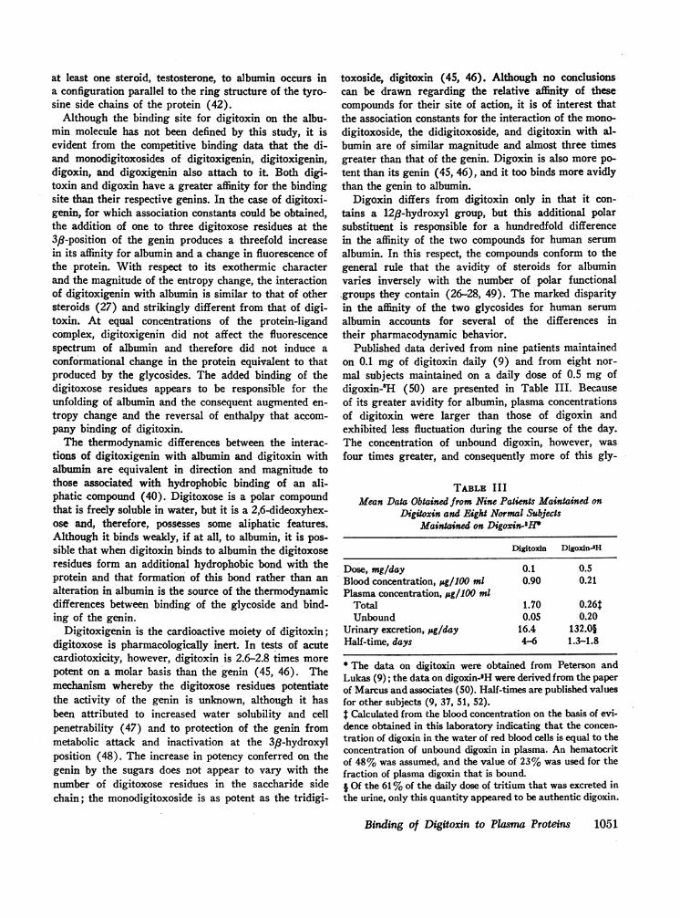
Published data derived from nine patients maintainedon 0.1 mg of digitoxin daily (9) and from eight nor-mal subjects maintained on a daily dose of 0.5 mg ofdigoxin-81H (50) are presented in Table III. Becauseof its greater avidity for albumin, plasma concentrationsof digitoxin were larger than those of digoxin andexhibited less fluctuation during the course of the day.The concentration of unbound digoxin, however, wasfour times greater, and consequently more of this gly-
TABLE IIIMean Data Obtained from Nine Patients Maintained on
Digitoxin and Eight Normal SubjectsMaintained on Digoxin-1P
Digitoxin Digoxin-3H
Dose, mg/day 0.1 0.5Blood concentration, jsg/100 ml 0.90 0.21Plasma concentration, &g/1100 ml
Total 1.70 0.26tUnbound 0.05 0.20
Urinary excretion, ;&g/day 16.4 132.0§Half-time, days 4-6 1.3-1.8
* The data on digitoxin were obtained from Peterson andLukas (9); the data on digoxin-3H were derived from the paperof Marcus and associates (50). Half-times are published valuesfor other subjects (9, 37, 51, 52).t Calculated from the blood concentration on the basis of evi-dence obtained in this laboratory indicating that the concen-tration of digoxin in the water of red blood cells is equal to theconcentration of unbound digoxin in plasma. An hematocritof 48%was assumed, and the value of 23%was used for thefraction of plasma digoxin that is bound.I Of the 61%of the daily dose of tritium that was excreted inthe urine, only this quantity appeared to be authentic digoxin.
Binding of Digitoxin to Plasma Proteins 1051

coside was contained in the water of red blood cells.Also the rate of urinary excretion of digoxin was con-siderably larger, and although the mechanisms of renalexcretion of the two glycosides have yet to be defined,the difference is partly attributable to the higher plasmaconcentration of digoxin in a form susceptible to filtra-tion by the renal glomeruli. The difference in rates ofexcretion of the two compounds are in turn reflected bya difference in their persistence in the body. The half-time of digitoxin in man is 4-6 days (9, 37), whereasthat of digoxin is 1.3-1.8 days (51, 52).
Hypoalbuminemia would not be expected to affect thedistribution of digitoxin in the plasma to a significantextent, since even at a plasma albumin concentration of1 g/100 ml, 92% of the glycoside is bound. The con-centration of free digitoxin in the plasma, however,will be almost tripled. The fraction of unbound digoxinin plasma would be relatively less affected since only23% is bound at an albumin concentration of 4 g/100ml and 13% is bound at a concentration of 1 g/100 ml.
Because the quantity of unbound digitoxin in theplasma is small, hemodialysis is not expected to be anefficient means for depleting the body pool of the glyco-side. For example, after an hour of in vivo dialysis us-ing a dialysis unit with a 1 liter dialysate volume (53),the concentration of digitoxin in the subject's plasmameasured by the double isotope dilution derivativemethod (9) was 1.05 jug/100 ml, whereas in the dialysateit was only 0.05 pg/100 ml. The addition of albumin orplasma to the dialysis fluid should facilitate the re-covery of digitoxin. Even if the concentration of albu-min in a 100 liter dialysate is as low as 0.1 g/100 ml, aconcentration capable of binding 50% of digitoxin insolution, as much as 1 mg of digitoxin theoreticallycould be extracted from a patient with a plasma digi-toxin concentration of 2 ug/100 ml. Although digoxinshould be more readily removed by dialysis, the verylow total plasma concentration of this drug is expectedto limit the efficacy of the procedure.
Since digitoxin and digoxin are bound to the samesite on the albumin molecule, the question may be posedwhether these substances might displace each otherfrom the binding site during the common therapeuticmaneuver of replacing one of the glycosides with theother. The concentrations attained by these compoundsin the plasma during treatment are exceedingly smallfractions of the concentrations needed to demonstratecompetition in the studies reported in this paper. Undertherapeutic circumstances, therefore, it is most unlikelythat binding of either of these glycosides to albumin willbe significantly affected.
ACKNOWLEDGMENTSWe are grateful to Dr. Albert L. Rubin for his assistancein performing the continuous flow electrophoresis and in vivo
dialysis studies, to Dr. Philip E. Schweitzer who as a StudentResearch Fellow contributed extensively to the work ondigoxin, to Mrs. Josefa Cubina for her steadfast and experttechnical assistance, and to Doctors Ralph E. Peterson andEsther M. Breslow for their advice and encouragement.
This investigation was supported by Public Health ServiceResearch Grants HE08950 from the National Heart Instituteand FR47 from the National Institutes of Health, and wasperformed pursuant to Contract No. PH-43-67-1439 with theNational Institutes of Health, U. S. Public Health Service,Department of Health, Education and Welfare.
REFERENCES1. Oppenheimer, E. 1913. Zur Frage der Fixation der Digi-
taliskorper im tierischen Organismus und besondersderen Verhalten zum Blut. Biochem. Z. 55: 134.
2. Lendle, L., and P. Pusch. 1935. tber die Bindung derDigitaliskorper an die Eiweisstoffe des Blutes. Arch.Exp. Pathol. Pharmakol. 177: 550.
3. Fawaz, G., and A. Farah. 1944. A study of the digi-toxin binding power of serum and other soluble tissue-proteins of the rabbit. J. Pharmacol. Exp. Ther. 80: 193.
4. Farah, A. 1945. On the combination of some cardio-active glycosides with serum proteins. J. Pharmacol.Exp. Ther. 83: 143.
5. Spratt, J. L., and G. T. Okita. 1958. Protein binding ofradioactive digitoxin. J. Pharmacol. Exp. Ther. 124: 109.
6. Goldstein, A. 1949. The interactions of drugs andplasma proteins. Pharmacol. Rev. 1: 102.
7. Brodie, B. B., and C. A. M. Hogben. 1957. Somephysico-chemical factors in drug action. J. Pharm.Pharmacol. 9: 345.
8. Tait, J. F., and S. Burstein. 1964. In vivo studies ofsteroid dynamics in man. In The Hormones. Physiology,Chemistry, and Applications. G. Pincus, K. V. Thimann,and E. B. Astwood, editors. Academic Press Inc., NewYork. 5: 441.
9. Lukas, D. S., and R. E. Peterson. 1966. Double isotopedilution derivative asay of digitoxin in plasma, urine, andstool of patients maintained on the drug. J. Clin. Invest.45: 782.
10. Aronson, R. F., G. D. Lubash, and A. L. Rubin. 1962.Separation of plasma protein fractions by continuous-flow paper electrophoresis using a volatile buffer. Anal.Biochem. 4: 306.
11. Pert, J. H., R. E. Engle, Jr., K. R. Woods, and M. H.Sleisenger. 1959. Preliminary studies on quantitativezone electrophoresis in starch gel. J. Lab. Clin. Med. 54:572.
12. Chen, R. F. 1967. Removal of fatty acids from serumalbumin by charcoal treatment. J. Bicl. Chem. 242: 173.
13. Rabitzsch, G. 1963. Digitoxin-(23-'4C). Naturzwissen-schaften. 50: 225.
14. Lowry, 0. H., N. J. Rosenbrough, A. L. Farr, andR. J. Randall. 1951. Protein measurement with the Folinphenol reagent. J. Biol. Chem. 193: 265.
15. Foster, J. F. 1960. Plasma albumin. In The Plasma Pro-teins. Vol. I. Isolation, Characterization, and Function.F. W. Putnam, editor. Academic Press Inc., New York.179.
16. Scatchard, G. 1949. The attraction of proteins for smallmolecules and ions. Ann. N. Y. Acad. Sci. 51: 660.
17. Edsall, J. T., and J. Wyman. 1958. Biophysical Chem-istry. Vol. I. Thermodynamics, Electrostatics, and theBiological Significance of the Properties of Matter.Academic Press Inc., New York. 136, 591.
1052 D. S. Lukas and A. G. De Martino

18. Goodman, D. S. 1957. Preparation of human serum al-bumin free of long-chain fatty acids. Science. 125: 1296.
19. Krebs, H. A., and K. Henseleit. 1932. Untersuchungenuber die Harnstoffbildung im Tierkorper. Z. Phys. Chem.210: 33.
20. Pennel, R. B. 1960. Fractionation and isolation of puri-fied components by precipitation methods. In ThePlasma Proteins. Vol. I. Isolation, Characterization, andFunction. F. W. Putnam, editor. Academic Press Inc.,NewYork. 9.
21. Snedecor, G. W. 1956. Statistical Methods Applied toExperiments in Agriculture and Biology. Iowa StateCollege Press, Ames. 5th edition.
22. Jirgensons, B. 1965. The Cotton effects in the opticalrotatory dispersion of proteins as new criteria of con-formation. J. Biol. Chem. 240: 1064.
23. Chen, R. F. 1966. Fluorescence spectra of human serumalbumin in the pH region of the N-F transition. Bio-chim. Biophys. Acta 120: 169.
24. Daughaday, W. H. 1958. Binding of corticosteroids byplasma proteins. IV. The electrophoretic demonstrationof corticosteroid binding globulin. J. Clin. Invest. 37:519.
25. Robbins, J. Reverse-flow zone electrophoresis. 1956. Amethod for determining the thyroxine-binding capacityof serum protein. Arch. Biochem. Biophys. 63: 461.
26. Sandberg, A. A., W. R. Slaunwhite, Jr., and H. N.Antoniades. 1957. The binding of steroids and steroidconjugates to human plasma proteins. Recent Progr.Hormone Res. 13: 209.
27. Schellman, J. A., R. Lumry, and L. T. Samuels. 1954.The binding of uncharged molecules to proteins. II.Testosterone and bovine serum albumin. J. Amer. Chem.Soc. 76: 2808.
28. Eik-Nes, K., J. A. Schellman, R. Lumry, and L. T.Samuels. 1954. The binding of steroids to protein. I.Solubility determinations. J. Biol. Chem. 206: 411.
29. Pearlman, W. H., and 0. Crepy. 1967. Steroid-proteininteraction with particular reference to testosteronebinding by human serum. J. Biol. Chem. 242: 182.
30. Antoniades, H. N., W. H. Daughaday, and W. R. Slaun-white, Jr. 1960. The binding of steroid hormones andtheir metabolites by plasma proteins. In Hormones inHuman Plasma; Nature and Transport. H. N. Antoni-ades, editor. Little, Brown & Company, Boston. 455.
31. Plager, J. E. 1965. The binding of androsterone sulfate,etiocholanolone sulfate, and dehydroisoandrosterone sul-fate by human plasma protein. J. Clin. Invest. 44: 1234.
32. Steiner, R. F., J. Roth, and J. Robbins. 1966. The bind-ing of thyroxine by serum albumin as measured byfluorescence quenching. J. Biol. Chem. 241: 560.
33. Goodman, DeW. S. 1958. The interaction of human se-rum albumin with long-chain fatty acid anions. J. Amer.Chem. Soc. 80: 3892.
34. Conn, H. L., Jr., and R. J. Luchi. 1961. Some quantita-tive aspects of the binding of quinidine compounds byhuman serum albumin. J. Clin. Invest. 40: 509.
35. O'Reilly, R. A. 1967. Studies on the coumarin anticoag-ulant drugs: interaction of human plasma albumin andwarfarin sodium. J. Clin. Invest. 46: 829.
36. Thorp, J. M. 1964. The influence of plasma proteins onthe action of drugs. In Absorption and Distribution ofDrugs. T. B. Binns, editor. The Williams & WilkinsCompany, Baltimore. 64.
37. Leach, C. N., Jr., R. E. Peterson, and D. S. Lukas. 1966.Metabolism of digitoxin in man. Clin. Res. 14: 254.
38. Karush, F. 1950. Heterogeneity of the binding sites ofbovine serum albumin. J. Amer. Chem. Soc. 72: 2705.
39. Klotz, I. M., and J. M. Urquhart. 1949. The binding oforganic ions by proteins. Effect of temperature. J. Amer.Chem. Soc. 71: 847.
40. Kauzmann, W. 1959. Some factors in interpretation ofprotein denaturation. Advan. Protein Chem. 14: 1.
41. Meyer, C. J., D. S. Layne, J. F. Tait, and G. Pincus.1961. The binding of aldosterone to plasma proteins innormal, pregnant, and steroid-treated women. J. Clin.Invest. 40: 1663.
42. Oyakawa, E. K., and B. H. Levedahl. 1958. Testosteronebinding to bovine and human serum albumin: The role oftyrosine groups. Arch. Biochem. Biophys. 74: 17.
43. Teresi, J. D., and J. M. Luck. 1952. The combinationof organic anions with serum albumin. J. Biol. Chem.194: 823.
44. Markus, G., and F. Karush. 1957. Structural effects ofthe interaction of human serum albumin with sodiumdecyl sulfate. J. Amer. Chem. Soc. 79: 3264.
45. Tamm, C. 1963. The stereochemistry of the glycosidesin relation to biological activity. In New Aspects ofCardiac Glycosides. W. Wilbrandt and P. Lindgren,editors. The Macmillan Company, New York. 11.
46. Chen, K. K. 1963. Possibilities of further developmentsin the glycoside field by modifying the glycoside struc-ture. In New Aspects of Cardiac Glycosides. W. Wil-brandt and P. Lindgren, editors. The Macmillan Com-pany, New York. 27.
47. Moe, G. K., and A. E. Farah. 1965. Digitalis and alliedcardiac glycosides In The Pharmacological Basis ofTherapeutics. L. S. Goodman and A. Gilman, editors.The Macmillan Company, New York. 3rd edition. 665.
48. Repke, K., and L. T. Samuels. 1964. Enzymatic basisfor epimerization of cardiotonic steroids at carbon-3 inrat liver. Biochemistry. 3: 689.
49. Westphal, U., and B. D. Ashley. 1958. Steroid-proteininteractions. IV. Influence of functional groups in A'-3-ketosteroids on interaction with serum albumin andp-lactoglobulin. J. Biol. Chem. 233: 57.
50. Marcus, F. I., L. Burkhalter, C. Cuccia, J. Pavlovich,and G. G. Kapadia. 1966. Administration of tritiateddigoxin with and without a loading dose. A metabolicstudy. Circulation. 34: 865.
51. Doherty, J. E., W. H. Perkins, and G. K. Mitchell. 1961.Tritiated digoxin studies in human subjects. Arch. In-tern. Med. 108: 531.
52. Marcus, F. I., G. J. Kapadia, and G. G. Kapadia. 1964.The metabolism of digoxin in normal subjects. J. Phar-macol. Exp. Ther. 145: 203.
53. Lubash, G. D., K. H. Stenzel, and A. L. Rubin. 1964.Methodology for study of nitrogenous compounds in he-modialysate. Trans. Amer. Soc. Artificial Internal Or-gans. 10: 337.
Binding of Digitoxin to Plasma Proteins 1053