Embed Size (px)
Citation preview

Volume 5 • Issue 2 • 1000153J Bioengineer & Biomedical SciISSN:2155-9538 JBBS an open access journal
Research Article Open Access
Shanthi Priya, J Bioengineer & Biomedical Sci 2015, 5:2 DOI: 10.4172/2155-9538.1000153
*Corresponding author: Shanthi Priya CH, Department of PharmaceuticalSciences, Srikrupa Institute of Pharmaceutical Sciences, Osmania University,Hyderabad, Telangana, India, E-mail: [email protected]
Received: April 21, 2015; Accepted: April 30, 2015; Published: May 07, 2015
Citation: Shanthi Priya CH (2015) Design and Characterization of Mucoadhesive Microspheres for Gastro-Retentive Delivery of Famotidine Hydrochloride. J Bioengineer & Biomedical Sci 5: 153. doi:10.4172/2155- 9538.1000153
Copyright: © 2015 Shanthi Priya CH. This is an open-access article distributed under the terms of the Creative Commons Attribution License, which permits unrestricted use, distribution, and reproduction in any medium, provided the original author and source are credited.
AbstractThis article illustrates the Design and Characterization of Mucoadhesive microspheres with Famotidine Hydrochloride
as drug for Gastro-Retention of drug release. The microspheres were prepared by the Ion Gelation method and Thermal Cross Linking Method. In Ion Gelation method sodium alginate is used as a release controlling biopolymer and Calcium chloride is acts as hygroscopic cross linking agent. In Thermal Cross Linking Method egg albumin is used as binding agent and a Thermal energy is used as cross linking property. The characteristics like shape and structure of prepared microspheres by Optical microscopy and scanning electron microscopy, respectively. In vitro drug release studies were done and drug release was evaluated. Effect on the Mucoadhesion, drug Entrapment Efficiency of microspheres and Drug release were observed. The prepared microspheres exhibited prolonged drug release (17 hrs) the mean particle size increased as the concentration of sodium alginate increased, as the egg albumin concentration increases the Mucoadhesion increased and the drug release rate decreased at higher concentration of sodium alginate. Significant effect of the Encapsulation Efficiency of microspheres was observed. In vitro studies demonstrated the Gastro retentive delivery of drug from the microspheres.
Design and Characterization of Mucoadhesive Microspheres for Gastro-Retentive Delivery of Famotidine HydrochlorideShanthi Priya CH*Department of Pharmaceutical Sciences, Srikrupa Institute of Pharmaceutical Sciences, Osmania University, Hyderabad, Telangana, India
Keywords: Mucoadhesive microspheres; Famotidine; In vitrorelease; Gastric residence time
IntroductionFamotidine is a competitive histamine H2-receptor antagonist.
Their principal pharmacodynamics outcome is the inhibition of gastric secretion. Famotidine is used as for the treatment of peptic ulcer disease (PUD) and gastro esophageal reflux disease (GERD). Famotidine binds competitively to H2-receptors located on the basolateral membrane of the parietal cell, blocking histamine influences. This competitive inhibition outcome in lowered basal and nocturnal gastric acid secretion and reduction in gastric quantity, acidity. The bioavailability of oral doses of famotidine is forty-45%. 1/2-lifestyles are 2.5-3.5 hours. Oral administration is probably the most suitable and desired manner of any drug delivery to the systematic circulation. Medicines which might be effectively absorbed from gastrointestinal tract (GIT) and have quick half of-lives are eliminated rapidly from the systemic circulation. Common dosing of those medications is required to attain suitable therapeutic activity. To avoid this quandary, the progress of oral sustained-controlled liberate formulations is an attempt to release the drug slowly into the gastrointestinal tract (GIT) and preserve an effective drug awareness within the systemic circulation for a very long time. To formulate a orally administered controlled liberate dosage type, it is fascinating to obtain lengthen gastric residence time through the drug delivery. Gastro retentive drug supply is a process to extend gastric time, thereby focusing on site-precise drug unlock in the higher gastrointestinal tract (GIT) for nearby or systemic results [1-10].
Materials and MethodFamotidine was gift sample from SMS Pharmaceuticals Limited
Hyderabad. Andhra Pradesh. Egg albumin, sodium alginate, Liquid paraffin (light), Calcium chloride, Tweens 80 purchased from S.D. Fine Chemicals Limited (Hyderabad) the entire chemical were of analytical grade and double distilled water used throughout the experiment.
Formulation of microspheres
Microspheres with drug (famotidine Hydrochloride) are prepared by the Ion Gelation and Thermal Cross Linking Method. F1 – F4 are
prepared by Thermal Cross Linking Method. F5 – F8 are prepared by Ion Gelation Method.
Thermal cross linking method
Mucoadhesive microspheres of Famotidine Hydrochloride was prepared by thermal cross linking method. 0.4% of Tweens 80 was added to 100mL of light liquid paraffin and it was heated to 70°C till tweens 80 was completely dissolved. It was cooled to room temperature. Prepare 10%W/V solution of egg albumin and drug solution. Add the solution containing egg albumin and drug (Famotidine) to previously cooled Light Liquid Paraffin. Stir it with mechanical stirrer for 10min and heat it to 95oC for 10-15 min. We can see the formation of microspheres. The beads so prepared were collected by decantation, washed with water. Then it was dried over night to become hard microspheres. The process was applied to 4 different formulations by using varying proportions of egg albumin (i.e., F1-F4) [11-15].
Ion gelation method
Accurately weighed about 10% of sodium alginate 20% of egg albumin and kept aside, then it was dispersed in 100 ml of distilled water by using magnetic stirrer at 40°C. Then after complete dispersion, added accurately 100 mg of Famotidine Hydrochloride then the stirring was continued until complete and uniform dispersion was obtained. Then the Calcium chloride solution was prepared by dispersing the 5 gm of Calcium chloride powder in 100 ml of distilled water by heating at 40°C.
Journal of
Bioengineering & Biomedical ScienceJour
nal o
f Bioe
ngineering & Biomedical Science
ISSN: 2155-9538

Citation: Shanthi Priya CH (2015) Design and Characterization of Mucoadhesive Microspheres for Gastro-Retentive Delivery of Famotidine Hydrochloride. J Bioengineer & Biomedical Sci 5: 153. doi:10.4172/2155- 9538.1000153
Page 2 of 6
Volume 5 • Issue 2 • 1000153J Bioengineer & Biomedical SciISSN:2155-9538 JBBS an open access journal
The resulting bubble free dispersion was added manually drop wise with a 5 ml syringe (22 gauze needle) into 100 ml of (5%w/v) calcium chloride solution (CaCl2) and stirred in a 250 ml beaker. The gelation time of 15 min was allowed to complete the curing reaction and produce spherical and rigid microspheres. The beads so prepared were collected by decantation, washed with water and dried in hot air oven at 60°C for 2 hours. The process was applied to 4 different formulations by using varying proportions of egg albumin and sodium alginate (i.e., F5-F8) (Table 1).
Characterization of FormulationProduction yield
The dried microspheres of each batch are weighed separately and percentage yield is calculated by using following equation.
Practical weightPercentage yield 100
Theoretical weight= ×
Estimation of drug content
50 mg of muco adhesive microspheres were weighed and powdered. This was dissolved or extracted in methanol in 100 ml volumetric flask and made up to volume. The solution was shaken occasionally for 1h and filtered. From this 1ml of solution was diluted to 100 ml with pH 1.2 buffer solution in 100 ml volumetric flask. The drug content was analyzed by measuring absorbance in a UV spectrophotometer at 265 nm using pH 1.2 phosphate buffer as blank. The studies were carried out in triplicate [16-18].
Drug entrapment efficiency or incorporation efficiency
To determine the drug entrapment efficiency or incorporation efficiency the microspheres were crushed in glass mortar and powered, then suspended in 10 ml of methanol, after 24 hrs the solution was filtered and filtrate was analyzed for drug content. The drug incorporation efficiency was calculated by the following formula:
100bIncorporationefficiencya
= ×
b = calculated amount of drug present in the formulation,a = theoretical amount of drug present in the formulation
Morphology and particle size determination
The size was measured using an optical microscope, and the mean particle with the help of a calibrated ocular meter.
a) Effect of Stirring Speed on particle size: The effect of stirring speed on the particle size was determined at 500 rpm and 1000 rpm.
b) Surface morphology/Scanning Electron Microscopy (SEM): The external morphology of the microspheres was studied by scanning electron microscopy using apparatus Philip 505.
In vitro wash-off test
The mucoadhesive property of microspheres was evaluated by an in vitro adhesion testing method known as wash-off method. Freshly excised piece of intestinal mucosa (2×2 cm) from albino rat were mounted onto glass slides (3×1 inch) with cyanoacrylate glue. Two glass slides were connected with a suitable support, about 50 microspheres were spread on to each wet rinsed tissue specimen
and immediately thereafter the support was hung onto the arm of a USP tablet disintegrating test machine. When the disintegrating test machine was operated, the tissue specimen was given slowly, regular up and down moment in the test fluid (500 ml pH 1.2 phosphate buffer) maintained at 37ºC. At the end of 30 min, 1h, and hourly intervals up to 8h, the numbers of microspheres adhering to tissue were counted.
Mucoadhesion= (no. of microspheres adhered/no. of microspheres applied) ×100
In-vitro drug release data and profiles
The prepared formulation was evaluated for in-vitro release by USP dissolution apparatus 1 at 50 rpm and at 37°C temperature in order to determine 100% drug release. To evaluate microspheres containing famotidine were exposed to 900 ml of HCl (pH 1.2). The samples were collected in pre-determined time intervals. Famotidine concentrations were determined by UV at 265 nm [19,20].
Result and DiscussionThe mucoadhesive microspheres of sodium alginate and egg
albumin prepared by Ion Gelation and Thermal cross linking method. The polymer sodium alginate was selected to control the release rate and egg albumin as a mucoadhesive polymer. Both are biodegradable and mucoadhesive polymer. The formulation of the present microspheres was based on the solubility behavior of both polymers. Eight Formulations F1-F8 were formulated by varying concentration of sodium alginate and egg albumin (Table 1), to study effect of release of famotidine from the microspheres and effect of polymer concentration on the size, percentage mucoadhesion, drug entrapment efficiency. The particle size and surface morphology was determined with the help of optical microscope and Scanning Electron microscope.
To investigate the effect of release of famotidine from the microspheres eight batches F1-F8 were prepared. The drug release prolonged to 17 hrs in formulation F5.
Product yield:
The results of product yields are shown in Table 2. The percentage yield of formulations was in the range of 86.15 ± 0.3 to 95.56 ± 0.31. The product yield was manageable with little loss of drug during the formulation stage (Table 2).
Estimation of drug Content
The results of drug content are shown in Table 3. The percentage drug content of formulations were in the range of 95.21 ± 0.45 to 98.65 ± 0.31. The low SD and CV value indicates uniform distribution of drug within the various batches of microspheres prepared. The drug content results suggest a negligible loss of drug during the formulation stage (Table 3).
Encapsulation efficiency
Encapsulation efficiency of all the formulations is presented in the Table 4. The percentage encapsulation efficiency of set-1 formulations were in the range of 83.32 ± 0.11 to 98.16 ± 0.45. The results suggest encapsulation efficiency depend upon concentration of sodium alginate used in the formulation. The encapsulation efficiency is increased progressively with increase in the concentration of sodium alginate. This could be attributed due to formation of larger microspheres with increasing concentration of sodium alginate, thus entrapping more

Citation: Shanthi Priya CH (2015) Design and Characterization of Mucoadhesive Microspheres for Gastro-Retentive Delivery of Famotidine Hydrochloride. J Bioengineer & Biomedical Sci 5: 153. doi:10.4172/2155- 9538.1000153
Page 3 of 6
Volume 5 • Issue 2 • 1000153J Bioengineer & Biomedical SciISSN:2155-9538 JBBS an open access journal
amount of drug (Table 4).
Morphology and Particle size DeterminationEffect of stirring speed on particle size
The size analysis of microspheres is carried out by optical microscope. The sizes of microspheres were in the range of 20-90 microns. The size of microspheres is depending upon concentration of sodium alginate used in the formulation. The increase in size of microspheres was observed with increase in concentration of sodium alginate. This could be due to increase in viscosity of the polymeric dispersion, which eventually lead to formation of bigger particle during ionic gelation. The results of the particle size of many of the
formulations were in the limits and comply with the standards (Table 5).
Scanning electron microscopy
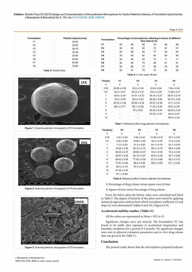
Scanning electron microscopy was used to know surface morphology of microspheres. The SEM photographs of F5 revealed that microspheres were spherical, discrete. The outer surface of microspheres was coarse rough texture, with few pores, mild cracks and completely covered with coat materials (Figures 1-3).
In vitro wash off test
The mucoadhesion is a phenomenon in which two materials, at least one of which is biological are held together by means of interfacial force. The Table 6 shows in vitro mucoadhesion data of mucoadhesive microspheres carried out with everted rat intestinal mucosa in presence of pH 1.2. The percentage of microspheres retained on everted intestinal mucosa after 6 h in set-1 formulations were found in the range of 71-55. The overall results suggest that concentration and type of mucoadhesive polymer doesnot show much more difference in the mucoadhesive property (Table 6).
In-vitro drug release data and profiles
The dissolution conditions used for studying the drug release from the mucoadhesive microspheres of famotidine were:
Apparatus : USP Type 1 (basket)
Agitation speed (rpm) : 50
Medium : 0.1N HCl (pH 1.2), 900 ml
Dissoloution volume : 900 ml
Temperature : 37.0 ± 0.5 C
Time : 0, 0.25, 0.5, 1, 3, 5, 7, 9, 11, 13, 15 and 17 hrs
Wavelength : 265 nm
(i) Release profiles of formulations containing egg albumin: (Table 7)
All the values are represented as Mean ± SD (n=3) (Table 8, Figures 4 and 5).
Kinetic Data AnalysisKinetic data/Model fitting
The formulations having maximum release were selected. The result are exhibited in table and plotted in four modes of data treatments.
1) Percentage of drug release versus time.
2) Log percentage of drug remained versus time.
Ingredients(in %w/v)
Formulation CodeF1 F2 F3 F4 F5 F6 F7 F8
Egg albumin 5 10 15 20 20 20 20 20Sodium alginate - - - - 10 5 3 1Calcium chloride - - - - 5 5 5 5
Light liquid paraffine 100 100 100 100 - - - -Tween 80 0.4 0.4 0.4 0.4 - - - -
Purified water Q.S Q.S Q.S Q.S Q.S Q.S Q.S Q.SDrug(mg) 100 100 100 100 100 100 100 100
Table 1: Shows various formulations.
Formulation Product yield ± SDF1 86.15+0.3F2 88.57 ± 0.21F3 89.25 ± 0.5F4 89.46 ± 0.78F5 95.56 ± 0.31F6 91.9 ± 0.46F7 94.18 ± 0.83F8 93.29 ± 0.87
Table 2: Product yield.
Formulation Theoreticaldrug content(mg)
Practicaldrugcontent(mg)
% Drug content ± SD
F1 40 39.61 98.43 ± 0.46F2 40 39.54 98.16 ± 0.45F3 40 39.53 98.12 ± 0.54F4 40 39.23 96.92 ± 0.25F5 40 39.66 98.65 ± 0.31F6 40 39.4 97.62 ± 0.47F7 40 38.8 95.21 ± 0.45F8 40 39.15 96.62 ± 0.62
Table 3: Drug content.
Formulation Microencapsulation efficiency ± SDF1 83.32 ± 0.11F2 84.11 ± 0.32F3 85.02 ± 0.23F4 85.65 ± 0.44F5 98.16 ± 0.45F6 96.81 ± 0.35F7 96.92 ± 0.25F8 93.23 ± 0.21
Table 4: Encapsulation efficiency.
Citation: Shanthi Priya CH (2015) Design and Characterization of Mucoadhesive Microspheres for Gastro-Retentive Delivery of Famotidine Hydrochloride. J Bioengineer & Biomedical Sci 5: 153. doi:10.4172/2155- 9538.1000153
Page 4 of 6
Volume 5 • Issue 2 • 1000153J Bioengineer & Biomedical SciISSN:2155-9538 JBBS an open access journal
Formulation Percentage of microspheres adhering to tissue at different time interval (h)
F1 50 96 88 79 68 68F2 50 93 82 75 67 67F3 50 96 85 77 65 65F4 50 88 80 64 60 60F5 50 94 83 74 71 71F6 50 89 74 66 61 61F7 50 86 74 58 55 55F8 50 91 78 70 65 62
Table 6: In vitro wash off test.
Time(h) F1 F2 F3 F40 0 0 0 0
0.25 26.28 ± 0.36 22.5 ± 0.36 23.8 ± 0.54 7.94 ± 0.540.5 35.2 ± 0.21 29.23 ± 0.41 28.4 ± 0.29 17.86 ± 0.211 55.6 ± 0.34 47.51 ± 0.72 45.14 ± 0.37 26.47 ± 0.743 72.5 ± 0.36 65.4 ± 0.23 58.38 ± 0.65 35.73 ± 0.365 85.32 ± 0.54 85.92 ± 0.34 65.27 ± 0.36 51.7 ± 0.237 98.2 ± 0.71 89.1 ± 0.56 71.24 ± 0.54 69.2 ± 0.549 - 97 ± 0.23 85.42 ± 0.43 82.05 ± 0.3611 - - 97.02 ± 0.57 94.2 ± 0.4713 - - - 96.6 ± 0.22
Table 7: Release profile of egg albumin microspheres.
Time(hrs) F5 F6 F7 F80 0 0 0 0
0.25 1.02 ± 0.45 3.06 ± 0.43 21.82 ± 0.51 20.7 ± 0.240.5 2.52 ± 0.64 10.06 ± 0.65 32.4 ± 0.35 31.5 ± 0.351 11.2 ± 0.54 21.4 ± 0.56 54.1 ± 0.19 53.1 ± 0.493 19.64 ± 0.45 35.14 ± 0.72 69.4 ± 0.72 68.6 ± 0.595 26.24 ± 0.37 49.62 ± 0.37 74.2 ± 0.45 73.2 ± 0.357 46.67 ± 0.26 64.12 ± 0.57 85.9 ± 0.36 84.7 ± 0.669 66.42 ± 0.54 77.42 ± 0.55 91.3 ± 0.65 90.3 ± 0.7311 77.81 ± 0.65 88.4 ± 0.29 95.2 ± 0.52 97.1 ± 0.4313 82.4 ± 0.19 97.4 ± 0.4515 91.42 ± 0.2417 97.1 ± 0.28
Table 8: Release profile of sodium alginate microspheres.
Formulation Particle size(microns)F1 40-60F2 20-80F3 50-70F4 30-40F5 30-80F6 50-60F7 60-80F8 70-90
Table 5: Particle Size.
Figure 1: Scanning electron micrographs of F5 formulation.
Figure 2: Scanning electron micrographs of F5 formulation.
Figure 3: Scanning electron micrographs of F5 formulation.
3) Percentage of drug release versus square root of time.
4) Square of time versus Percentage of drug release.
From the below plots the kinetic value were calculated and listed in Table 9. The degree of linearity of this plots were tested by applying statistical regression analysis from which correlation coefficient (r) and slope (n) were determined (Tables 9 and 10), (Figures 6-9).
Accelerated stability studies: (Table 11)
All the values are represented as Mean ± SD (n=3)
Significant changes were not noticed. The formulation F5 was found to be stable after exposure to accelerated temperature and humidity conditions for a period of 3 months. No significant changes were seen in physical evaluation parameters and in vitro drug release data was given in the Table 11.
ConclusionThe present study shows that the microspheres prepared polymer

Citation: Shanthi Priya CH (2015) Design and Characterization of Mucoadhesive Microspheres for Gastro-Retentive Delivery of Famotidine Hydrochloride. J Bioengineer & Biomedical Sci 5: 153. doi:10.4172/2155- 9538.1000153
Page 5 of 6
Volume 5 • Issue 2 • 1000153J Bioengineer & Biomedical SciISSN:2155-9538 JBBS an open access journal
0
20
40
60
80
100
120
0 5 10 15 20
Cum
ulat
ive %
dru
g re
leas
e
Time(h)
f5
f6
f7
f8
Figure 5: In vitro drug release of famotidine from sodium alginate (F5-F8).
Time (hrs) LOG T SQRT T % Drug
ReleaseLog % Drug
Release% Drug
RemainedLog % Drug Remained
0 - 0 0 - - 2
1 0 1 11.2 1.049218 88.8 1.948413
3 0.477121 1.732051 19.64 1.293141 80.36 1.90504
5 0.69897 2.236068 26.24 1.418964 73.76 1.867821
7 0.845098 2.645751 46.67 1.669038 53.33 1.726972
9 0.954243 3 66.42 1.822299 33.58 1.526081
13 1.113943 3.605551 82.4 1.915927 17.6 1.245513
17 1.230449 4.123106 97.1 1.987219 2.9 0.462398
Table 9: Ex-vivo drug release of Famotidine from F5 Formulation.
0
20
40
60
80
100
120
0 5 10 15 20
Cum
ulat
ive %
dru
g re
leas
e
Time (h)
f1
f2
f3
f4
Figure 4: In vitro drug release of famotidine from egg albumin (F1-F4).
Figure 6: Zero order release rate of Famotidine from formulation F5.
y = 0.8163x + 0.9733 R² = 0.9559
0
0.5
1
1.5
2
2.5
0 0.2 0.4 0.6 0.8 1 1.2 1.4
LOG
cum
ulat
ive %
dru
g re
leas
e
LOG TFigure 7: Korsemeyer-Peppas kinetics plot of Famotidine from formulation F5.
Figure 8: (Higuchi’sPlot) of famotidine from famotidine F5.
Zero order Korsemeyer -Peppas Model
Higuchi Model First order
Slope m 5.89 0.81 24.90 -0.08
Regression r 0.97 0.96 0.92 0.89
intercept c 3.17 0.97 -13.39 2.15
Table 10: Kinetic Model fitting data.
sod. alginate and egg albumin both have a significant effect on the mucoadhesion, drug entrapment efficiency and drug release. Egg albumin is hydrophilic polymer has good entrapment efficiency and good mucoadhesion but it releases the drug immediately therefore sod. alginate was used to control the release rate as well as the other factors to match the acceptance criteria. After evaluating all the formulation, the formulation F5 which is containing the higher percentage of egg

Citation: Shanthi Priya CH (2015) Design and Characterization of Mucoadhesive Microspheres for Gastro-Retentive Delivery of Famotidine Hydrochloride. J Bioengineer & Biomedical Sci 5: 153. doi:10.4172/2155- 9538.1000153
Page 6 of 6
Volume 5 • Issue 2 • 1000153J Bioengineer & Biomedical SciISSN:2155-9538 JBBS an open access journal
albumin showed the good entrapment efficiency about 98%, in vitro wash off test was found to be about 82% and good drug release profile in 8hrs. Therefore it was selected as the best formulation.
References
1. James EF, Reynolds (1996) The extra pharmacopoeia / Martindale. RoyalPharmaceutical Society London.
2. Singh B, Kim K (2000) Floating drug delivery systems: An approach to oralcontrolled drug delivery via gastric retention. J Control Release 63: 235-259.
3. Tripathi KD (203) Essentials of medical pharmacology. (5thedn), Jay Peebrothers: Medical publishers (P) ltd, New Delhi.
4. Kimura K, Ido K, Saifuku K, Taniguchi Y, Kihira K, et al. (1995) A 1-h topicaltherapy for the treatment of Helicobacter pylori infection. Am J Gastroenterol90: 60-63.
5. Akiyama Y, Nagahara N, Nara E, Kitano M, Iwasa S, et al. (1998) Evaluation of oral mucoadhesive microspheres in man on the basis of pharmacokinetics offurosemide and riboflavin, compounds with limited gastrointestinal absorption sites. J Pharm Pharmacol 50: 159-166.
6. Yellanki SK, Singh J, Syed JA, Bigala R, Goranti S, et al. (2010) Design andCharacterization of Amoxicillin trihydrate Mucoadhesive Microspheres forProlonged Gastric retention. Int J Pharma Sci Drug Res 2: 112-114.
7. Koner P, Saudagar RB, Daharwal SJ (2007) Gastro-retentive drugs: a novelapproach towards floating therapy in drugs: a novel approach towards floating therapy.
8. Arora S, Ali J, Ahuja A (2005) Floating drug delivery systems: a review. AAPSPharm Sci Tech 6: E372-E390.
9. Rao SB, Sharma CP (1997) Use of chitosan as biomaterial: studies on itssafety and hemostatic potential. J Biomed Mater Res 34: 21-28.
10. Akiyama Y, Yoshioka M, Horibe H, Hirai H, Kitamori N, et al. (1993) Noveloral controlled-release microspheres using polyglycerol esters of fatty acids. JControlled Release 26: 1-10.
11. Kiyama Y, Nagahara N, Kashihara T, Hirai S, Toguchi H (1995) In vitro and invivo evaluation of mucoadhesive microspheres prepared for the gastrointestinal tract using poly glycerol esters of fatty acids and poly(acrylic acid). Pharm Res 12: 397-405.
12. Axon AT (1994) The role of acid inhibition in the treatment of Helicobacter pylori infection. Scand J Gastroenterol 29: 16-23.
13. Chiba N, Rao BV, Rademaker JW, Hunt H (1992) Meta-analysis of the efficacy of antimicrobial therapy in eradicating Helicobacter pylori. Am J Gastroenterol87: 1716-1727.
14. Nagahara N, Akiyama Y, Nakao M, Tada M, Kitano M, et al. (1998)Mucoadhesive microspheres containing amoxicillin for clearance of helicobacter pylori. Antimicrobial Agents and Chemotherapy 42: 2492-2494.
15. Gennaro AR (1995) Remington: The science and practice of pharmacy. (19th edn), Mack Publishing Company.
16. Donnell PO, McGinity J (1997) Preparation of Microspheres by the solventevaporation technique. Adv Drug Del Rev 28: 25-42.
17. Rahman Z (2006) Characterization of 5-Fluorouracil Microspheres for ColonicDelivery AAPS Pharm. Sci Tech 7: E-1- E-9.
18. Benita S (1996) Microencapsulation: Methods and Industrial Applications.(2ndedn), Marcel Dekker, New York.
19. Gopferich A, Alonso M, Langer R (1994) Development and characterization ofmicroencapsulated microspheres. Pharm. Research 11: 1568-1574.
20. Jain D, Panda AK, Majumdar DK (2005) Eudragit S-100 Entrapped InsulinMicrospheres for Oral Delivery. AAPS Pharm Sci Tech 6: E100-107.
Citation: Shanthi Priya CH (2015) Design and Characterization of Mucoadhesive Microspheres for Gastro-Retentive Delivery of Famotidine Hydrochloride. J Bioengineer & Biomedical Sci 5: 153. doi:10.4172/2155- 9538.1000153
Figure 9: First order release rate of Famotidine from formulation F5.
Parameters Temperature maintained: 400C;Relative humidity (RH) maintained: 75 ± 5%RH
0 (Initial) 1st month 2nd month 3rd month
Percentage Yield(%) 98.64 ± 0.31 98.63 ± 0.05 98.55 ± 0.04 97.88 ± 0.039
Encapsulation efficiency (%) 98.00 ± 0.45 97.98 ± 0.58 97.45 ± 0.69 97.32 ± 0.61
In vitro wash off test 71 ± 0.06 70 ± 0.01 69 ± 0.19 68 ± 0.14
In vitro drug release(%) 97.1 ± 0.04 97.0 ± 0.058 96.88 ± 0.05 96.70 ± 0.03
Table 11: Accelerated stability studies.