Embed Size (px)
Citation preview

Beth Burgess, MHA, RHIA, CCS
Huff DRG Review Services January 16, 2013

CVA/TIA Respiratory Failure Pneumonia Sepsis CHF AKI

Neurological deficits due to abnormality of the vessels supplying the brain substance and meninges Clinical symptoms (TIA vs. Stroke) Pathology (infarct vs. hemorrhage) Etiology (atherosclerosis; thrombosis; embolic)

TIA Transient episode of
neurological dysfunction caused by focal ischemia to brain, spinal cord, or retina without acute infarction.
Lasts at least 30 seconds and up to several days (3 days average).
Stroke Sudden neurological
deficit related to brain necrosis caused by either infarction (80%) or hemorrhage (20%).
Strokes can be defined by imaging studies or persistent symptoms, typically > 3 days.

Acute/subacute infarction on CT or MRI Hemorrhage on CT or MRI Significant neurological deficits persist at
time of discharge
“Physician must link any abnormal imaging study to the neurological condition”

Primary cause of CVA (ruptured intracerebral vessel)
Conversion of ischemic stroke Complication of thrombolytic therapy Tumor associated Trauma
Distinguish between traumatic vs. nontraumatic (can be difficult even for clinician!)

Obstruction of pre-cerebral arteries (carotid; vertebrobasilar) Pre-cerebral artery occlusion > 70% is clinically significant Physician must link occlusion to signs and symptoms
Obstruction of cerebral arteries Cerebral artery embolism is suggested with neurological
symptoms without suggestion of pre-cerebral artery stenosis and/or cerebral atherosclerosis.

Cerebral Embolus
Cerebral artery embolism is suggested with neurological symptoms without suggestion of pre-cerebral artery stenosis and/or cerebral atherosclerosis. Recurrent TIAs or strokes usually different areas of the brain
(not always with same symptoms)
No significant obstruction of pre-cerebral vessels
No evidence of intracerebral atherosclerosis (small vessel disease; old lacunar infarcts; atherosclerotic dementia;)
Sources of cerebral emboli: Intra-arterial embolization from diseased pre-cerebral vessels
Cardiogenic (from the heart; aorta)

Cardiac Sources of Cerebral Emboli
Atrial fibrillation Ventricular aneurysm Cardiomyopathy Prosthetic heart valves Acute or subacute MI Intimal injury from heart catheterization PFO

CCs Hemiparesis Acute confusion Ischemic encephalopathy
MCCs Brain
compression/herniation Cerebral edema Coma Conversion hemorrhage

Patient with history of afib was admitted for transient aphasia lasting 30 minutes. The patient has a h/o afib but is not on any medical treatment. TIA was diagnosed and patient was treated with Lovenox and Coumadin in the hospital then discharged on Coumadin. The coder assigned TIA as the PDX. Final DRG = 69. Is there a potential for a different PDx? If so, what?

Patient presented with transient right arm weakness & numbness. Head CT showed no acute infarcts. Carotid U/S showed 80% left carotid stenosis. The final diagnosis was TIA. Patient is to f/u for OP left carotid endarterectomy. The coder assigned 433.10 (CAS) as the PDX. Final DRG = 68. Is the PDx correct based on current documentation?

Patient presented with slurred speech and left-sided weakness. The head CT report showed large SDH with midline shift and vasogenic edema. Patient was diagnosed with SDH and given IV Decadron. The coder assigned 434.91 as the PDX with dysphagia and left sided weakness as 2nd DXs. Final DRG 66. Is there a potential for a different DRG? If so, what?

Significant impairment of gas exchange Increased work of breathing Intensive treatment Close monitoring

Normal Lungs Arterial blood PO2 < 60 mmHg.
Or Arterial blood PCO2> 50mmHg
Pre-existing lung disease Arterial pH < 7.35 w/ PCO2
>50mmHg*** Or
Change in baseline PO2 level by 10-15mm ***generally more useful since
baseline PO2 not usually given.

Work of breathing Tachypnea (RR > 20) Accessory muscle use Moderate to severe
respiratory distress SATs < 88%
Treatment High flow oxygen (>4 to 6L
NC or non-rebreather mask)
Controlled oxygen delivery (i.e. Venturi mask) or low flow in persons with CO2 retention
Noninvasive MV (BiPAP) Invasive mechanical
ventilation with forced air delivery via a tube in airway

May be assigned as PDx when it is the condition established after study to be chiefly responsible for occasioning admission (CC 1st Qtr 2005)
Do not code acute respiratory failure as PDx when there is a chapter-specific coding guideline (sepsis, obstetrics, poisoning, HIV, newborn) or an alphabetic index or tabular directive which takes precedence over the general respiratory failure guidelines.
17

Patient with COPD on continuous home O2 at 2 L presented with SOB. SATs on 2L were 75%. The patient was placed on BiPAP, IV Solumedrol was started and patient was admitted to the ICU for acute on chronic respiratory failure due to COPD exacerbation. Can 518.84 be used as the PDx?

Patient with no history of lung disease presented with SOB. The patient was noted to have accessory muscle use and was hypoxic with SATs 87% on room air. ABGs showed PCO2 55. The patient was admitted for acute respiratory failure due to pneumonia. The patient was admitted to telemetry on 4L and treated with IV Azithromycin. What is the appropriate PDx?

Patient was admitted for AECOPD with acute respiratory distress. On admission the patient had RR 20 with SATs 90% on room air. The patient required O2 at 2L and IV steroids. Would you query for acute respiratory failure?

Simple Pneumonia Unspecified Bacterial unspecified Viral/Atypical/Mycoplasma Strep or Hemophilus Community Acquired Healthcare associated Nosocomial Postobstructive Immunocompromised host Nursing home acquired
Complex Pneumonia Specific bacteria** Legionnares Fungus/parasitic Aspiration Gram negative

The treating physician must link any positive cultures, gram stains, and serological tests to the pneumonia before it can be used to specify the pneumonia
Positive cultures not required.

Risk factors GERD PEG tube Bed bound Advanced Parkinson’s Advanced dementia ETOH
abuse/intoxication Seizure Etc.
ABX Coverage Clindamycin Cefepime Piperacillin (Zosyn) Quinilones Flagyl Augmentin
*Check for swallow study although does not r/o aspiration.

Pseudomonas E. Coli Klebsiella Proteus Enterobacter Serratia Actinobacter Haemophilus (DRGs 193-195)

Appropriate antibiotic coverage (≥ 7 days) And
High risk host And
High risk for colonization

ABX Coverage Imipenem (Primaxin) Meropenem (Merem) Doripenem (Doribax) Ertapenem (Invanz) Cefepime (Maxipime) Cetazidime (Fortaz) Ciprofloxacin (Cipro) Levofloxacin (Levaquin) Piperacillin (Zosyn) Ticarcillin (Timentin) Tobramycin Gentamycin Aztreonam (Azactam)
NOT appropriate for gram negative coverage: Penicillin Erythromycin Vancomycin Zithromax Clindamycin Zyvox

High risk host Severe lung disease,
particularly with CF or bronchiectasis
Chronic organ dysfunctions such as CHF, Cirrhosis, CKD
Diabetes Malignancy Immunosuppresive Rx HIV Alcoholics Malnutrition
High risk setting HCAP Postobstructive Bed confinement

Certain types of pneumonia are designated clinically to describe complex respiratory infections but are assigned to pneumonia NOS unless specified as to potential organisms being treated. Hospital-Acquired or Institutional Acquired
HCAP; Nursing Home Acquired Immunocompromised host Gram negative; staphylococcal
Postobstructive (gram negative; anaerobes)
Community Acquired pneumonias (CAPs) are typically Streptococcal, atypical, or Hemophilus influenza or viral. Aspiration pneumonia can be community or hospital acquired.

Patients who develop pneumonia within 90 days of being hospitalized in an acute care hospital for 2 or more days should be approached as if they had hospital-acquired pneumonia, as should residents of long-term care facilities, hemodialysis patients, and patients who received intravenous (IV) antibiotics, wound care, or chemotherapy within 30 days of developing pneumonia.

HCAP Hospital acquired Nosocomial Nursing home acquired Institutional acquired Pneumonia in immuno-compromised host

Organisms: Gram negative bacilli see previous slides for organisms &ABX coverage
Anaerobic bacteria Any of the drugs for gram negative or one of the following: Clindamycin; Flagyl; Augmentin
Staphylococcus aureus (particularly MRSA) Vancomycin; Zyvox; Bactrim; Tetracycline

Pneumonia that occurs as a result of bronchial obstruction. Most commonly seen in patients with bronchogenic carcinoma of lung.
Organisms: Anaerobic bacteria Gram negative bacilli
*Please refer to previous slides for antibiotic coverage.

Patient has ESRD and several admissions over the past 3 months and is now re-admitted for pneumonia. The patient was treated with Zosyn & Cipro for 5 days in the hospital and discharged on both for 4 additional days. What type of pnuemonia does it appear is
being treated?

Patient was admitted for pnuemonia. PMH is notable for HTN, GERD and CHF. The patient was treated with Clindamycin for a total of 7 days. What type of pnuemonia does it appear is
being treated?

Patient was admitted from the nursing home for pnuemonia. The patient has a PMH of hypothyroidism and CKD. The patient was treated with 3 days of Azithromycin and discharged back to the nursing home to continue Azithromycin for 4 additional days. What type of pnuemonia does it appear is
being treated?

Patient recently began chemo for bronchogenic carcinoma of the lung. The patient is now admitted for postobstructive pnuemonia, which was treated with Augmentin for 10 days. What type of pnuemonia does it appear is being
treated?

Systemic Inflammatory Response due to Documented or suspected Infection
Without organ dysfunction
038.xx + 995.91
With organ dysfunction 038.xx + 995.92

3 or more of the following must be present and cannot be explained by other disease process : Fever (≥ 100.4) or hypothermia (≤ 96.8) Leukocytosis (WBC >12), leukopenia (WBC < 4), bandemia (>
10% bands) Tachycardia (HR >90) Tachypnea (RR > 20) Encephalopathy Oliguria (<30cc/hr) Hypotension Increase Anion gap Metabolic acidosis Elevated lactate level CRP elevation Blood sugar >120 in a nondiabetic DIC Shock

Compliance issues Diagnosis of sepsis without clinical support Querying for sepsis without clinical support Querying for sepsis when clinical indicators can
be explained by other disease process Re-sequencing of PDx when complicaiton of
medical care Bacteremia not indicator for SIRS!

Patient presented with a painful leg with erythema. The patient had temp 101, RR 18, HR 95,WBC 12.8, and elevated anion gap. The patient was admitted for cellulitis of the leg and treated with IV ABX. Is it appropriate to query for SIRS/sepsis?

Patient on 6 weeks of IV antibiotic treatment for chronic osteomyelitis was admitted for infected PICC line. The patient had hypotension, T100.8, HR 120, RR 19, WBC 11.5. The patient was admitted for infected PICC line and dehydration. What is the appropriate PDx? Would it be appropriate to query for SIRS/sepsis?

Patient presented with acute confusion. The patient had temp 95.4, HR 75, RR 17, WBC 18.5. Head CT was negative. Urinalysis was positive. The patient was admitted for metabolic encephalopathy due to UTI. Is it appropriate to query for SIRS/sepsis?

Heart failure is a clinical syndrome resulting from a failure of the heart to supply the body’s need for circulation. This may be a structural (valve) or functional (heart muscle weakness) disturbance.

ETIOLOGY: Coronary artery disease; hypertension; valvular; primary cardiomyopathy
ANATOMICAL: Left, right, or biventricular
TYPE (PHYSIOLOGICAL):
Diastolic only (preserved ejection fraction) Systolic heart failure Combined
ACUITY: Acute, chronic, or both

Left Heart Only*** Left ventricular abnormal function Signs: Pulmonary congestion (effusion; edema) Right due to Left (Combined)*** Most common cause of Right HF is Left HF Signs: Pulmonary and peripheral congestion Abnormal left ventricular function with elevated RH pressures Right Heart Failure Only Underlying lung disease or congenital heart disease with significant
shunting Signs of peripheral congestion: swollen liver; ascites; peripheral edema;
pleural effusions; Jugular vein distention Peripheral congestion with pulmonary hypertension and RV pressures with
normal Wedge pressure (Pulmonary venous pressures or PCWP) ***Represents 95% or more of HF

Chronic cor pulmonale (not associated with left heart failure) Intrinsic lung disease such as emphysema, COPD, ILD,
primary PAH Sleep apnea disorders Chronic pulmonary embolus
Acute cor pulmonale : acute pulmonary embolus Pulmonary and tricuspid valve disorders Peripheral AV shunts

Diastolic No systolic dysfunction Preserved Contractility
with EF>45%. Ventricular end diastolic
pressures ELEVATED (unless there is mitral stenosis)
Cardiac output initially normal or elevated
Left ventricular hypertrophy present
Systolic Decreased contractility
with EF<45% Cardiac output
decreased Ventricular end diastolic
pressures are elevated Heart is dilated with
usually minimal or no Left ventricular hypertrophy

Everyone with systolic CHF has diastolic dysfunction but not everyone with diastolic CHF has systolic dysfunction.
MD has to document type of CHF. Cannot
assume!!!

Acute only Chronic (“compensated”) Acute on Chronic (“decompensated”) Unspecified

Symptoms & Signs: SOB, orthopnea, paroxysmal nocturnal (PND), wheezing, and increased signs of peripheral congestion (JVD, hepatomegaly, ascites, and peripheral edema +/-.
Radiographic signs of volume overload - Pulmonary vascular congestion - pleural effusions (usually bilateral) - interstitial edema - pulmonary edema - small pericardial effusions
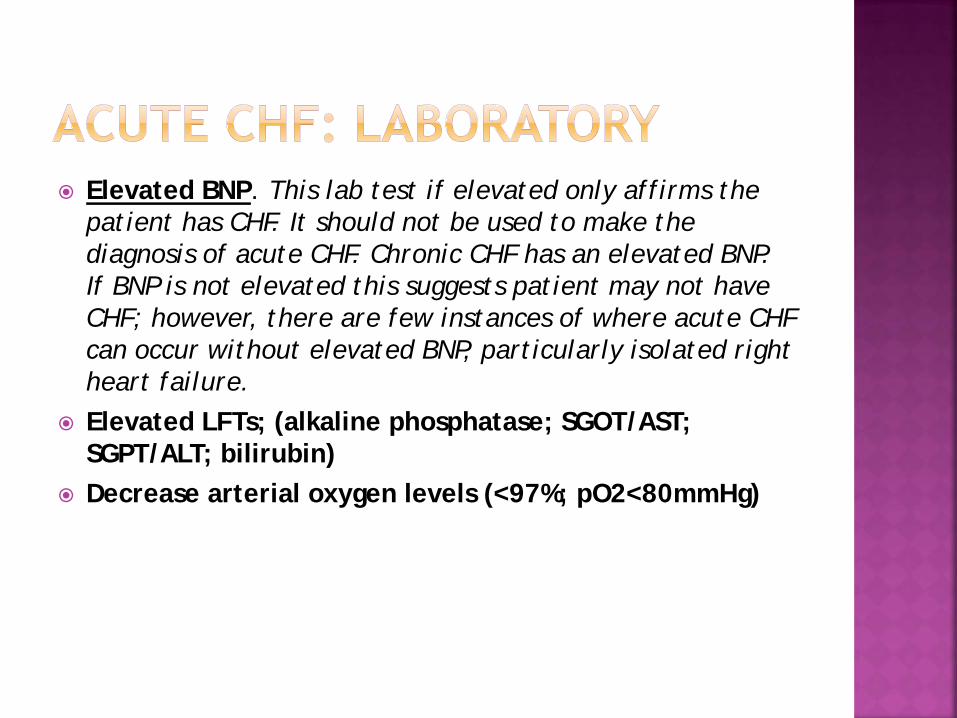
Elevated BNP. This lab test if elevated only affirms the patient has CHF. It should not be used to make the diagnosis of acute CHF. Chronic CHF has an elevated BNP. If BNP is not elevated this suggests patient may not have CHF; however, there are few instances of where acute CHF can occur without elevated BNP, particularly isolated right heart failure.
Elevated LFTs; (alkaline phosphatase; SGOT/AST; SGPT/ALT; bilirubin)
Decrease arterial oxygen levels (<97%; pO2<80mmHg)

Treatment: Intensification of any treatment regimen Intravenous diuretic therapy Intravenous vasodilators Dialysis Emergency cardioversion

Therapy Left HF only Right due to Left HF Right only
ACE drugs/ARBs X X
Beta blockers X X
Diuretics X X X
Lanoxin X X
Vasopressors X X
Vasodilators X X X
Dialysis X X
Biventricular pacing X X
hBNP X X
Calcium channel Blockers X
Viagra and related compounds
X

MCC CC No Impact
CHF NOS or any acuity X
Left HF NOS or any acuity X
Right HF NOS or any acuity X
Biventricular HF (NOS or any acuity) X
Systolic (unspecified or chronic) X
Systolic (Acute or acute on chronic) X
Diastolic (unspecified or chronic) X
Diastolic (Acute or acute on chronic) X
S & D HF (unspecified or chronic) X
S&D (Acute or acute or chronic) X

Ischemic Biventricular
enlargement Ejection fraction (EF)
decreased No LV hypertrophy;
only dilation of LV on ECHO
Hypertensive Long standing HTN Left ventricular
hypertrophy prominent on EKG/Echo
Diastolic dysfunction and/or preserved EF (>45%) on Echo
Enlargement is usually confined to LV unless ischemia and valve disorders present

Acuity Acute Unspecified or chronic Type Noncardiogenic (i.e. ARDS) Cardiogenic - CHF - Volume overload

Cardiogenic Cause by increase
capillary hydrostatic pressures
CHF: due to heart dysfunction (systolic/diastolic) and or valvular heart disease (ECHO abnormal)
Volume overload: no cardiac dysfunction (i.e. ESRD, TACO) ECHO normal
Elevated BNP
Non-cardiogenic Caused by increased
permeability of alveolar membrane
Direct injury caused by infections, fatty emboli, aspirations
Indirect injury due to toxins such as in sepsis, chemicals, etc.
BNP normal

Volume/fluid overload is integral to CHF and not coded separately unless MD says not due to heart failure. CC: 3rd Qtr 1996; 2nd Qtr 2001
If a person is admitted for fluid overload due to missed dialysis and has a h/o CHF, CHF is used as the PDx unless the MD indicates fluid overload was not due to CHF. CC: 2nd Qtr 2001; 3rd Qtr 2007
Treatment of volume overload (pre-load reduction) is the mainstay of treatment for ESRD patients. Dialysis is the main mode of therapy acutely. This is the case regardless of whether the patient has CHF or purely volume overload.
Acute pulmonary edema with heart disease is coded to CHF unless the physician states the pulmonary edema is not related.

If a person has an elevated BNP with LV dysfunction on Echo clarify the nature of the volume overload.
TACO syndrome is a form of volume overload due to transfusion. Acute pulmonary edema is not coded separately.

Do not code unspecified pulmonary edema (514) as a CC if the cause is CHF. Pulmonary edema is integral to CHF. If CHF or acute/decompensated HF is not documented you should try to clarify.

Patient was admitted for acute GI bleed. The patient has a PMH of GERD, HTN and CHF. Home meds including Protonix, Lasix, Coreg & Lisinopril were continued in the hospital. The coder assigned 578.9 as the PDX with 428.0, 401.9 and 530.81 as ODXs. Final DRG was 379. Is the final DRG correct based on current
documentation? Is there potential for a different DRG?

Patient was admitted for AKI from dehydration. PMH includes DM, CHF and CAD. The patient was aggressively hydrated with IVFs. On HD #2 the patient developed SOB. CXR was ordered and showed pulmonary congestion. Echo showed EF 25%. The patient was treated with IV Lasix. The coder assigned 584.9 as the PDX with 428.0, 250.00 & 414.01 as ODXs. Final DRG was 684. Is the final DRG correct based on current
documentation? Is there potential for a different DRG?

Patient with h/o COPD, HTN, CAD and systolic & diastolic CHF presented with SOB & wheezing. The admitting MD noted that CXR showed pulmonary edema. The patient was admitted for COPD and CHF. Treatment included IV Solumedrol and IV Lasix. The coder assigned 491.21 as the PDX with 428.43 as an MCC. Final DRG was 190. Is the final DRG correct based on current
documentation?

Patient with PMH of HTN, CKD IV and diastolic dysfunction was admitted for CHF exacerbation. Echo showed LVH, EF 65% and no valvular disease. The coder assigned 428.33 as the PDX with 403.90 & 585.4 as ODXs. Final DRG was 292. Is the final DRG correct based on current
documentation? Is there potential for a different DRG?

Acute renal insufficiency –593.6- No CC Acute kidney injury/failure- 584.9- CC Acute tubular necrosis – 584.5- MCC Vasomotor nephropathy – 584.5- MCC Toxic nephropathy – 584.5- MCC Acute interstitial nephritis – 580.89-MCC Pre-renal azotemia – 790.6-No CC Pre-renal failure – 788.99-No CC Acute kidney disease –593.9- No CC Renal failure unspecified – 586 – No CC

STAGE Serum Creatinine Urine Output
1
1.5 – 1.9 times baseline within 7 day period
OR > 0.3 mg/dl increase over 48 hrs.
<0.5 ml/kg/hr for 6-12 hrs.
2 2.0 – 2.9 times baseline <0.5 ml/kg/hr for > 12 hrs.
3
3.0 times baseline OR
Increase serum creatinine > 4.0mg/dl OR
Initiation of renal replacement RX
<0.3 ml/kg/h for > 24hrs.

Increase SCr ≥ 0.3 over 48 hrs
Creatinine was 1.8 on admission and over the next 48 hours increased to 2.0. Does this patient have AKI?
Creatinine was 1.4 on admission and over the next 48 hrs increased to 1.7. Does this patient have AKI?

Change in SCr ≥ 1.5 x baseline over 7 days
If CKD but no baseline documented: Lowest SCr x 1.5 = ≥ highest SCr within 7 day period
Ex: If SCr is 1.8 on admission it must decrease to a minimum of 1.2 within 7 days. (1.2 x 1.5 = 1.8)
If no CKD documented then one would expect in a normal person that the CKD would be < 1.0.
If baseline is documented on admission but SCr drops below then the lowest SCr is new baseline.

Change in SCr ≥ 1.5 x baseline over 7 days
Patient admitted with dehydration. Creatinine noted on admission to be 3.6. After rehydration patient was d/c on day 5 with a creatinine of 1.8. Does this patient have AKI?
Patient with CKD was admitted with dehydration. Creatinine noted on admission to be 1.3. After rehydration patient was d/c on day 2 with a creatinine of 1.0. Does this patient have AKI?

70
Pre-renal Intrinsic Post-renal
Hypovolemia Dehydration Hemorrhage Decrease in effective circulating volume Heart failure Septic shock Drugs ACE-inhibitors/ARBs NSAIDs
Acute Tubular Necrosis (ATN) Ischemia prolonged dehydration severe hypotension Toxins rhabdomyolysis drugs (radio-contrast, cisplatin, etc.) Infection pyelonephritis Glomerular nephritis collagen vascular disease malignant hypertension Renal Artery Atherosclerosis renal artery stenosis renal vein thrombosis Interstitial pyelonephritis allergic intestinal nephritis
BPH GU neoplasm Bilateral ureteric stones

AKI/Pre-renal AKI/ATN
U:P Osmolality >1.4:1 1:1
U:P Creatinine >50:1 <20:1
Urine Na (mEq/L) <20 >80
FENa (%) <1 >3
Urine Exam Neg/hyalin casts Granular casts

Acute kidney injury should be sequenced as principal diagnosis instead of dehydration if both are present on admission.
Complication of transplant should be used if acute renal failure occurs in a transplanted kidney.
Sepsis should be used as principal diagnosis if acute renal failure is part of the SIRS or infectious process.

Patient was admitted for PVD. Following angiography the patient had a rise in creatinine from 1.2 to 1.8 over 48 hrs. Contrast induced nephropathy was documented. Could the coder query for a more specific type of kidney injury?

Patient has been on Bactrim for UTI as an OP. The patient is now admitted for CHF. The patient has CKD with a documented baseline creatinine of 1.5. Creatinine on admission was 2.0 and AKI was documented. Could the coder query for a more specific type of kidney injury?

Patient was admitted for CHF. On HD# 2 the patient developed hypotension. Creatinine increased from 1.5 on admission to 3.1. Urinalysis showed granular casts. The physician documented dehydration & AKI from diuresis. Could the coder query for a more specific type of kidney injury?

Please send any additional questions to: [email protected]