Embed Size (px)
Citation preview

Basics of Neuromonitoringand Anesthetic
ConsiderationsShilpa Rao, MDa,*, James Kurfess, MDb,Miriam M. Treggiari, MD, PhD, MPHc
KEYWORDS
� Evoked potentials � Electroencephalography � Burst suppression
KEY POINTS
� Amplitude and latency.
� Intraoperative changes in neuromonitoring signals.
� Effect of anesthesia on neuromonitoring.
� Controversies in neuromonitoring.
INTRODUCTION
Neuromonitoring is a modality involving recording electrical potentials generated byneurons or their axons throughout the nervous system. Commonly used modalitiesinclude electroencephalography (EEG), somatosensory evoked potentials (SSEPs),motor evoked potentials (MEPs), and brainstem auditory evoked potentials (BAEPs).Intraoperative neuromonitoring (IONM) modalities are extensively used in adult and
pediatric intracranial surgeries to facilitate near complete and safe surgical resectionof brain tumors, resection of arteriovenous malformations, aneurysm clipping, andcoiling. They are also widely used in spine surgeries to monitor integrity of the spinalcord during spine fusion/fixation and tumor resections. In addition, neuromonitoringtechniques are commonly used during carotid endarterectomy surgery. Suddenchanges in any signal aids in rapid diagnosis of acute change in clinical conditionand alerts the surgeon and anesthesiologist to intraoperative critical events.In this article, we discuss basics of neuromonitoring, indications, contraindications,
and effect of anesthetic medications on various types of neuromonitoring techniques.We also discuss controversies associated with the use of IONM.
a Division of Neuroanesthesia, Department of Anesthesiology, Yale School of Medicine, YaleUniversity, PO Box 208051, 333 Cedar Street, TMP 3, New Haven, CT 06510, USA; b Departmentof Anesthesiology, Yale University, PO Box 208051, 333 Cedar Street, TMP 3, New Haven, CT06510, USA; c Department of Anesthesiology, Yale School of Medicine, Yale University, POBox 208051, 333 Cedar Street, TMP 3, New Haven, CT 06510, USA* Corresponding author.E-mail address: [email protected]
Anesthesiology Clin 39 (2021) 195–209https://doi.org/10.1016/j.anclin.2020.11.009 anesthesiology.theclinics.com1932-2275/21/ª 2020 Elsevier Inc. All rights reserved.

Rao et al196
ELECTROENCEPHALOGRAPHYIndications and Contraindications
The EEG records electrical potentials generated by the neurons in cerebral cortex.Electrodes made of silver disks with conductive gel are placed on the scalp or steril-ized and placed directly in the surgical field. The most commonly used description forthe location of scalp electrodes is the 10- to 20-lead placement system (Fig. 1), wherethe specific electrodes are placed in relation to specific areas of the cerebral cortex.However, during a craniotomy requiring EEGmonitoring, the complete 10 to 20 sys-
tem cannot be fully used because of surgical incision and exposure, hence the leadsare usually placed as close as possible to the surgical site to facilitate monitoring.Each scalp electrode gives a continuous recording of spontaneous superficial brainactivity covering an area of 2 to 3 cm in diameter (Fig. 2).1
Indications of EEG monitoring include the following procedures:
� Surgery involving eloquent cortex� Carotid endarterectomy to aid in diagnosis of stroke� Aortic arch surgery to monitor cerebral perfusion� Certain seizure surgeries
There are no major contraindications for intraoperative EEGmonitoring. Presence ofscalp infection may preclude the placement of scalp electrodes. Emergency surgeriesoften proceed without any type of neuromonitoring because of clinical urgency.Basic EEG waveforms are described next and shown in Fig. 3A.
� Delta (0.5–4 Hz): Delta rhythm is physiologically seen in deep sleep states and isprominent in the frontocentral head regions. Pathologic delta rhythm presents inawake states in case of generalized encephalopathy and focal cerebral dysfunc-tion. Frontal intermittent rhythmic delta activity is normally present in adults.2
Temporal intermittent rhythmic delta activity is seen in patients with temporallobe epilepsy.3
� Theta (4–7 Hz): This is often seen in the frontocentral regions, and travels poste-riorly, replacing the alpha rhythm during early drowsiness. This waveform isenhanced by heightened emotional states.
Fig. 1. A 10- to 20-EEG lead placement system.

Fig. 2. Photograph showing EEG lead placement in a craniotomy. (Courtesy of Courtney Al-les, CNIM.)
Basics of Neuromonitoring 197
� Alpha (8–12 Hz): The dominant alpha rhythm is typically present in normal awakeEEG recordings in the occipital region. It is best recorded with the eyes closedand during mental relaxation and is attenuated by eye opening.
� Beta (13–30 Hz): This is the most frequently seen rhythm in normal adults andchildren. Most sedatives increase the amplitude and quantity of beta activity.Beta wave attenuation can occur with cortical injury, and any fluid collection inthe brain.
Several factors can affect EEG waveforms including pharmacologic interventions,physiologic factors (mostly sleep and awake status), and disease states.
Pharmacologic interventions
� General anesthesia causes progressive slowing of the raw EEG waveforms andcan potentially cause gradual burst suppression in deeper states.
� Inhaled anesthetics (eg, sevoflurane), intravenous anesthetics (eg, propofol), andbarbiturates produce slowing of the EEG frequency when used in higher
Fig. 3. (A) Normal EEG waveform recording. (B) EEG recording showing burst suppression.(Courtesy of K. Eggan, CNIM, New Haven, CT.)
Rao et al198
concentrations. During deep anesthesia with these agents, a “burst suppression”pattern is noticed (Fig. 3B), which is characterized by high-frequency “bursts”alternating with flat tracings “suppression.”
� Nitrous oxide does not produce burst suppression but produces fast oscillatoryactivity.
� Ketamine increases theta frequencies and decreases alpha oscillations but doesnot produce burst suppression.
� Opiates produce loss of beta waves, slow alpha waves, and an increase in deltawave activity. Commonly used opiates do not cause burst suppression in clinicaldoses.
Pathologic factors
� Hypoglycemia causes EEG changes that are characterized by increased activityin the delta and theta frequencies.
� Any cortical injury that alters the brain homeostasis (eg, trauma, bleeding, orhematoma).
� Diffuse encephalopathy (eg, virus induced, drug induced, or metabolicderangements).
� Hyperventilation-induced hypocapnia (PaCO2 <20 mm Hg), which causes gener-alized slowing of the EEG activity.
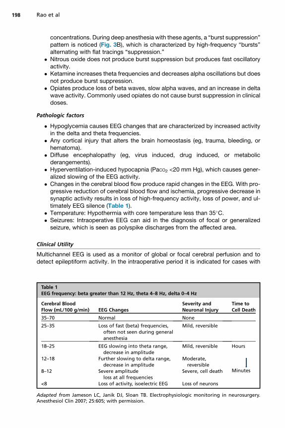
� Changes in the cerebral blood flow produce rapid changes in the EEG. With pro-gressive reduction of cerebral blood flow and ischemia, progressive decrease insynaptic activity results in loss of high-frequency activity, loss of power, and ul-timately EEG silence (Table 1).
� Temperature: Hypothermia with core temperature less than 35�C.� Seizures: Intraoperative EEG can aid in the diagnosis of focal or generalizedseizure, which is seen as polyspike discharges from the affected area.
Clinical Utility
Multichannel EEG is used as a monitor of global or focal cerebral perfusion and todetect epileptiform activity. In the intraoperative period it is indicated for cases with
Table 1EEG frequency: beta greater than 12 Hz, theta 4–8 Hz, delta 0–4 Hz
Cerebral BloodFlow (mL/100 g/min) EEG Changes
Severity andNeuronal Injury
Time toCell Death
35–70 Normal None
25–35 Loss of fast (beta) frequencies,often not seen during generalanesthesia
Mild, reversible
18–25 EEG slowing into theta range,decrease in amplitude
Mild, reversible Hours
Minutes
12–18 Further slowing to delta range,decrease in amplitude
Moderate,reversible
8–12 Severe amplitudeloss at all frequencies
Severe, cell death
<8 Loss of activity, isoelectric EEG Loss of neurons
Adapted from Jameson LC, Janik DJ, Sloan TB. Electrophysiologic monitoring in neurosurgery.Anesthesiol Clin 2007; 25:605; with permission.

Basics of Neuromonitoring 199
a high potential for cortical ischemia, such as carotid endarterectomy, select supra-tentorial surgery, and epilepsy surgery for monitoring epileptic activity. EEG analysiscan also be used to monitor for isoelectricity under hypothermic arrest. Similarly, itis used to avoid isoelectricity by titrating anesthetics to avoid reaching excessivelydeep planes of anesthesia.
Processed Electroencephalography
EEG waveform data is “processed” using power spectral analysis. Sine wavesextracted at different frequencies are plotted over time and then overlaid using Fouriertransformation to produce a single dimensionless value. There are several commer-cially available processed EEG monitors with slight variations in signal processingand information displayed. In general, all processed EEG uses fewer electrodes (usu-ally one to four) compared with the full EEG. These monitoring modalities have simplerelectrode placement in the operating room setting and are more straightforward toanalyze and interpret. Each of the commercially available monitors use propriety algo-rithms to process EEG waveforms (Table 2).The goal of processed EEGs is to monitor the relative density of different waveforms
corresponding to the following states: awake, sedated, surgical anesthesia, and burstsuppression. Data are then used to titrate anesthetic agents to avoid periods ofpossible awareness under anesthesia and unnecessary burst suppression. Bispectralindex is the most widely used with a typical target goal of 40 to 60 for generalanesthesia.
Caveats of Electroencephalography Monitoring
EEG monitoring modalities are expensive, and do not completely guarantee the pres-ence of unconsciousness, lack of awareness, or the absence of cerebral ischemia,especially if there is preexisting neural damage. Furthermore, use of intraoperativeEEG is prone to multiple artifacts, from the use of cautery, skin contact and imped-ance, patient movement, and location of lead placement.
SOMATOSENSORY EVOKED POTENTIALS
Intraoperatively, SSEPs are used in a variety of surgeries to monitor the integrity of theposterior (dorsal) columns of the spinal cord. An electrical stimulus is applied to a pe-ripheral nerve, typically the median or ulnar nerve at the wrist for upper extremitySSEPs and the posterior tibial nerve at the ankle for lower extremity SSEPs, using nee-dle or surface electrodes near the nerve.4 Impulses ascend primarily in the dorsal col-umn fibers of the spinal cord, which then synapse in the lower medulla. These thendecussate at the level of the medulla and travel up the brainstem as the mediallemniscus to synapse in the contralateral thalamus. From there, relay neuron nerve fi-bers form the thalamocortical radiations, which travel through the internal capsule andsynapse in the primary sensory cortex of the parietal lobe. SSEPs are useful in assess-ing the integrity of the sensory system from the peripheral nerves through to the cere-bral cortex (Figs. 4 and 5).Common indications for use of SSEPs include:
� A wide variety of spine surgeries, including scoliosis repair and posterior spinalinstrumentations/fixations
� Carotid endarterectomies� Some intracranial tumors� Cardiovascular surgeries

Table 2Commercially available processed EEG monitors
Index Company Index Range Works with AgentsNot Work with Agents/Disadvantages
1 Bispectrum Index (BSI) Aspect Medical Systems (nowCovidien), United States, 1992
0–100 Propofol, midazolam, andisoflurane
Outperformed all
Nitrous oxide and ketamineProblems with EMG
2 Narcotrend Index NCT MonitorTechnik, Germany, 2000 0–100 Children, sevoflurane propofol/remifentanil
EMG susceptibilityGood artifact removal
Neuromuscular blocking agentsComplex algorithmSlowest response to a change in
sedation
3 Entropy Index Datex-Ohmeda Company,United States, 2003
0–1001–91
Desflurane, sevoflurane propofol,thiopental
Ketamine
4 Patient State Index(PSI) or (PSA)
Physiomatrix (now SED LineSystems), United States, 2001
0–100 Propofol, alfentanil, nitrous oxideEMG susceptibility
—
5 AEP-Monitor (AAI) Danmeter, Denmark, 2001 0–100 or 1–60 Propofol, midazolam, andisoflurane
No effects of nitrous oxide andketamine
6 Snap Index Everest Biomedical Instruments,United States, 2002
Sevoflurane and sevoflurane/nitrous oxide
Sensitive to unintentionalawareness
7 Cerebral State Index (CSI) Danmeter A/S, Denmark, 2004 0–100 Propofol Nitrous oxide
Abbreviation: EMG, electromyographic.Data from Al-Kadi MI, Reaz MB, Ali MA. Evolution of electroencephalogram signal analysis techniques during anesthesia. Sensors 2013;13(5): 6605-35.
Raoetal
200

Fig. 4. Normal SSEP waveform during a spine surgery. (Courtesy of K. Eggan, CNIM, New Ha-ven, CT.)
Basics of Neuromonitoring 201
Factors Affecting the Amplitude and Latency of Somatosensory Evoked PotentialsWaveforms
Similar to the EEGwaveform, several factors including pharmacologic and physiologicand disease states influence SSEP signals.
Pharmacologic agents
� Halogenated inhalational agents cause dose-dependent reduction in amplitudeand increase in latency, with a greater effect on cortex compared with spinal, pe-ripheral, and subcortical tracings.5
� Nitrous oxide works synergistically with inhalational andmost intravenous agentsto decrease amplitude and increase latency of SSEPs.
� Intravenous agents with the notable exceptions of ketamine and etomidatedecrease amplitude and increase latency of SSEP recordings.
� Barbiturates cause dose-dependent decreases in amplitude and increases in la-tency. Barbiturate doses that induce coma are still compatible with SSEP moni-toring6 even when they are dosed to produce burst suppression in the EEG.
� Propofol causes amplitude decrease and increased latency, although less pro-nounced than inhalational agents including nitrous oxide, or midazolam adminis-tered in doses to achieve comparable planes of anesthesia. Propofol is used forSSEP monitoring especially with opioids.7
� Etomidate causes increased amplitude with increased latency on SSEP corticalrecordings.
� Ketamine causes increased amplitude with no change in latency or corticalpotentials.8
� Opioids mildly decrease cortical SSEP amplitude and mildly increase latencywith minimal effect on subcortical and peripheral potentials. Bolus dosing of opi-oids has a greater impact on SSEP changes than continuous infusion.9
� Benzodiazepines alone have little effect on SSEPs but may increase latency withthe concomitant administration of nitrous oxide.

Fig. 5. Loss of greater than 50% amplitude in SSEP waveform during the placement of aspine fixator device, which was immediately removed. (Courtesy of K. Eggan, CNIM, NewHaven, CT.)
Rao et al202

Basics of Neuromonitoring 203
Physiologic factors
� Temperature: Mild hypothermia increases SSEP latency but not amplitude.Although profound hypothermia silences SSEPs completely, mild hyperthermiadecreases latency without affecting amplitude.10
� Tissue perfusion: Similar to EEG changes, cerebral blood flow less than 18 mL/min/100 g of tissue affects SSEPs. Amplitude is initially reduced and corticalSSEPs is lost with worsening hypotension. Regional ischemia caused byvascular injury, surgical traction, clipping, embolic effects, or positioning arecommon causes of altered SSEP monitoring during surgery. If mild, anemiacan actually cause a mild increase in SSEP amplitude and reduced latencycaused by improved viscosity effects, but worsening anemia causes decreasesin amplitude and increased latency.11
� Although early responses to ischemia or hypoxia can manifest as a transient in-crease in SSEP amplitude, severe, progressive hypoxemia is associated with adecrease in SSEP amplitude and an increase in latency, eventually resulting incomplete loss of cortical SSEP waves. Ventilation and PaCO2 levels have little ef-fect on SSEP monitoring.6
� Intracranial hypertension causes decreased SSEP monitoring and eventual lossof responses in conjunction with uncal herniation.
MOTOR EVOKED POTENTIALS
MEPs are measured by exciting the motor cortex and subsequently measuring theelectrical activity in the muscles of the hands or feet. MEP monitoring is a methodof assessing the integrity of the corticospinal tract and the anterior segments of thespinal cord during spinal surgery. It has a high sensitivity and specificity to detect intra-operative neurologic deficits.12
Muscle responses to stimulation of the motor cortex are especially impacted byincreasing concentrations of inhaled anesthetics and use of neuromuscular blockade,because MEPs are extremely sensitive to these medications. Special stimulation tech-niques and certain anesthetic regimens are used to optimize MEPs. Monitoring tech-niques are divided according to the site of stimulation (motor cortex or spinal cord),method of stimulation (electrical potential or magnetic field), and the site of recording(spinal cord or peripheral mixed nerve and muscle).MEPmonitoring is new compared with SSEPs monitoring and has gained popularity
after isolated motor injury without sensory changes was described following idiopathicscoliosis procedures. MEP monitoring is now the standard of care in spinal deformitysurgeries. Importantly, MEP changes were noted in 6% of spine deformity surgeriesand 72% of these changes were reversible.13 The ability to maintain neural integrityand prevent devastating injury has led to MEP monitoring in a growing number of sur-gical cases. When intraoperative use of MEPs is planned, soft bite blocks should beplaced between the upper and lower molars to prevent the patient from biting eitherthe tongue or the endotracheal tube during stimulation.
CRANIAL NERVE ELECTROMYOGRAM MONITORING
Often, individual cranial nerves may be monitored depending on the location of surgi-cal resection. Examples include cranial nerve V and VII during the resection of cerebel-lopontine angle tumors and/or acoustic neuroma resection. Lower cranial nerves (VII,IX, X, XII) are monitored in certain thyroid resections and brainstem tumor resections.The hypoglossal nerve is monitored during open carotid endarterectomy.

Rao et al204
A single nerve electromyography aids in successfully isolating the “at risk” nerveduring establishment of surgical access and while performing the resection, allow-ing preservation of its vital function. The anesthetic considerations focus solely onthe avoidance of neuromuscular blockade during the monitoring period. A special-ized neural integrity monitor electromyographic (EMG) endotracheal tube may berequired in instances that require monitoring cranial nerve X and its laryngealnerves.
BRAINSTEM AUDITORY EVOKED POTENTIALS
BAEPs are recorded using a loud acoustic stimulus in the ear canal with an ear insertdevice. The sound is transduced by ear structures, with information conducted to thebrainstem via the eighth cranial nerve. Recording electrodes are placed at the headnear the mastoid process or ear lobe. Five main short-latency peaks (I to V) are usuallyseen within the first 10 milliseconds after stimulation (Fig. 6).14
BAEPs are commonly used in conjunction with other neuromonitoring modalitiesduring posterior fossa surgeries to assess brainstem function. BAEPs are typicallyresistant to anesthesia as compared with other evoked potentials.
Fig. 6. The circled area captures the intraoperative change in waveform with greater than50% decrease in MEP amplitude, during spine surgery. (Courtesy of K. Eggan, CNIM, NewHaven, CT.)

Basics of Neuromonitoring 205
RECOMMENDATIONS FOR CHOICE OF ANESTHETICS DURING THE USE OF VARIOUSNEUROMONITORING MODALITIES
The effects of medications used for induction of anesthesia typically do not persistlong enough to influence IONM. Conversely, it is important to use an appropriate intra-operative maintenance regime to facilitate monitoring techniques. The authors recom-mend considering the following approaches:
� Use of SSEP monitoring: Propofol infusion at anesthetic doses (titrated to patientage and comorbidities; eg, 80–120 mg/kg/min) along with an opioid infusion (eg,remifentanil at 0.1–0.2 mg/kg/min, fentanyl infusion at 2–3 mg/kg/h, or sufentanilinfusion at 0.15–0.4 mg/kg/h). An alternative to this is the use of a volatile anes-thetic, such as sevoflurane at approximately 0.5 minimum alveolar concentration(MAC) along with propofol infusion and opioid. Neuromuscular blocking agentsare used as necessary if SSEP monitoring is the only monitor that is beingused intraoperatively.
� Use of SSEPs and MEPs monitoring: Propofol infusion at anesthetic doses(titrated to patient age and comorbidities; eg, 80–120 mg/kg/min) along with anopioid infusion (eg, remifentanil at 0.1–0.2 mg/kg/min, fentanyl infusion at 2–3 mg/kg/h, or sufentanil infusion at 0.15–0.4 mg/kg/h). Volatile anesthetics aretypically avoided because of the exquisite sensitivity of MEPs to them. Neuro-muscular blocking agents are avoided during the period of monitoring. A short-acting neuromuscular blocking agent may be used to facilitate intubation.
� Use of SSEPs and cranial nerve EMG: The approach to the anesthetic to whathas already been described for MEP monitoring.
� Use of isolated cranial nerve EMG: No neuromuscular blocking agent during theperiod of monitoring. Up to 1.5 MAC volatile anesthetic and no restriction inopioid administration.
� Use of EEG, SSEPs, and MEPs: Volatile anesthetics are administered at approx-imately 0.5 MAC, and/or a propofol infusion is used in combination with an opioidinfusion (eg, remifentanil at 0.1–0.2 mg/kg/min, fentanyl infusion at 2–4 mg/kg/h, orsufentanil infusion at 0.15–0.4 mg/kg/h). Alternatively, fentanyl boluses may beused to maintain analgesia. Dexmedetomidine is a valuable adjunct when thiscombination is being used and aids in reducing the dose of propofol required.Neuromuscular blocking agents are avoided during the period of monitoring. Ashort- or intermediate-acting neuromuscular blocking agent may be used tofacilitate intubation.
CONTROVERSIES IN NEUROMONITORING
With the increasing use of neuromonitoring modalities in various types of surgeries,several studies have analyzed the benefits and cost effectiveness of their routine use.According to the guidelines for the use of electrophysiologic monitoring for human
spinal cord surgery,15 the use of multimodal IONM including SSEPs and MEPs duringspinal cord/spinal column surgery is a level I recommendation because of its reliabilityand validity in assessing spinal cord integrity. MEP recordings are superior to SSEPrecordings during spinal cord/spinal column surgery as diagnostic adjuncts forassessment of spinal cord integrity and are recommended if used for this purpose.Use of multimodal IONM, including SSEPs andMEP recording, during spinal cord/spi-nal column surgery does not improve gross total tumor resection or improve neuro-logic outcome, when used during intramedullary tumor resection procedures (LevelII evidence).15

Rao et al206
Daniel and colleagues16 performed a literature review and meta-analysis of sixstudies comparing neurologic events with and without IONM. Based on the evidenceprovided in the studies reviewed, they concluded that IONM did not result in fewerneurologic events compared with no monitoring (Level 2 evidence). For surgeriesinvolving intramedullary lesions, there was a trend to fewer neurologic events in pa-tients who underwent surgery with IONM.16
A literature search of Medline database was performed and relevant studies from alllevels were included in a narrative review by Charalampidis and colleagues.17 Nearlyall of these studies investigated the use of IONM in the setting of spine surgery. Over-all, these reports support the use of multimodal IONM in spinal tumor resections. Thecombined use of SSEPs andMEPs seems to provide increased accuracy for detectinginjury to sensory and motor pathways, reaching a high sensitivity, specificity, positivepredictive value, and negative predictive value.17
In 2010, Ayoub and colleagues18 performed a cost-effectiveness analysis on acohort of 210 patients who underwent cervical spine surgery with SSEP monitoring.The total cost of the surgery, hospital stay, neuromonitoring, and medical expendi-tures associated with postoperative neurologic injury was accounted for in the costanalysis. Given an incidence of 0.1% for spinal cord injury, the authors assumedthat without SSEPmonitoring 1 out of 201 patients would have had a permanent spinalcord injury. In their estimation, the total annual cost savings for a single injured patientwould range from $64,074 to $102,192 for their institution, whereas the yearly expen-diture on SSEP amounted to only $31,546.18
Recently, Ney and colleagues19 constructed a simulated cost-effectiveness modelto estimate the value of IONM to avert postoperative neurologic deficits. The modelassumptions included parameters, such as the surgical risk, frequency of casesaverted, and cost per case estimates. The authors concluded that use of IONM in spi-nal procedures was associated with a 49% reduction in relative risk for neurologiccomplications. They further estimated that the cost of monitoring to prevent a singleneurologic injury was $63,387.19
Conversely, Traynelis and colleagues,20 in a single-center study, reported a caseseries of 720 consecutive patients who underwent routine cervical spine procedureswithout the use of IONM. The authors reported a 0.4% rate of postoperative neuro-logic deficits. Furthermore, at 1-year follow-up, all patients had significantly improved,and their neurologic deficits had complete resolution. The authors, therefore, ques-tioned the utility of IONM during routine cervical spine surgery. Additionally, furtheranalysis was performed to explore the economic impact of IONM during cervical spineprocedures. This cost analysis was based on the Current Procedural Terminologyreimbursement codes. They concluded that significant savings could be achievedby not using IONM in simple cervical spine procedures.20
In a large retrospective propensity score matched analysis, using a national data-base, Cole and colleagues21 investigated single-level spinal procedures, with andwithout the use of IONM, with the goal of comparing the occurrence of neurologiccomplications. Trauma, spinal tumors, and revisions were excluded from the anal-ysis. A total of 85,640 patients were included in the analysis with a minority (13%)receiving IONM during the surgery. The authors found no differences in neurologiccomplications between those who did and did not receive IONM. They concludedthat the use of IONM was associated with higher costs ranging from $2859 to$3841.21
In summary, currently there is conflicting evidence regarding the cost effectivenessof use of IONM in routine spine surgeries. Additional expenditures in terms of trainingneuromonitoring personnel, use of specialized equipment for monitoring, and choice

Basics of Neuromonitoring 207
of anesthetic techniques would further complicate the evaluation of these monitoringmodalities.Multimodal IONM is also commonly used in carotid endarterectomies. Hong and
colleagues22 analyzed 668 carotid endarterectomy cases at six surgical centers,and found that a decrease in amplitude of 50% or more in any EEG or SSEP channelshould be used as the criteria to indicate the need for shunting or to initiate a neuro-protective protocol. A reduction of 50% or greater in the beta band of the EEG oramplitude of the SSEP was observed in 150 cases, most of which occurred duringcross-clamping. No patient showed signs of a cerebral infarct after surgery. Selectiveshunting based on EEG and SSEP monitoring can reduce carotid endarterectomyintraoperative stroke rate to a near zero level if trained personnel practice with stan-dardized protocols.In conclusion, the role of multimodal monitoring for the intraoperative detection of
physiologic changes allows the care team to decrease the likelihood of potentialcell or nerve injury. However, with the possible exception of certain spinal proceduresand carotid endarterectomies, the benefits in terms of prevention of permanent neuro-logic complications or cost effectiveness are not well documented and data are gener-ally inconclusive because of the absence of rigorously controlled trials.
DISCLOSURE
No conflicts of interest.
CLINICS CARE POINTS
� With the increasing use of neuromonitoring modalities during intracranial, carotidand spine procedures, it is important to understand the effects of physiologicalchanges and pharmacologic interventions on these monitoring modalities.
� There is currently a level I recommendation for the use of multimodal intraoper-ative neuromonitoring (including SSEPs and MEPs) during procedures involvingthe spine. This recommendation is based on the reliability and validity of thesemonitoring modalities in assessing spinal cord integrity.
� MEPs are superior to SSEP recordings as diagnostic adjuncts for the assess-ment of spinal cord integrity.
� There is insufficient evidence for routine use of neuromonitoring for routine cer-vical spine procedures in neurologically intact patients.
� Controversies continue to exist regarding the cost effectiveness of use of intra-operative multimodal neuromonitoring for procedures other than spine surgery.
REFERENCES
1. Jameson LC, Janik DJ, Sloan TB. Electrophysiologic monitoring in neurosurgery.Anesthesiol Clin 2007;25(3):605–30.
2. Cordeau JP. Monorhythmic frontal delta activity in the human electroencephalo-gram: a study of 100 cases. Electroencephalogr Clin Neurophysiol 1959;11:733–46.
3. Reiher J, Beaudry M, Leduc CP. Temporal intermittent rhythmic delta activity(TIRDA) in the diagnosis of complex partial epilepsy: sensitivity, specificity andpredictive value. Can J Neurol Sci 1989;16(4):398–401.
4. Toleikis JR, American Society of Neurophysiological Monitoring. Intraoperativemonitoring using somatosensory evoked potentials. A position statement by the

Rao et al208
American Society of Neurophysiological Monitoring. J Clin Monit Comput 2005;19(3):241–58.
5. Sloan TB, Heyer EJ. Anesthesia for intraoperative neurophysiologic monitoring ofthe spinal cord. J Clin Neurophysiol 2002;19(5):430–43.
6. Banoub M, Tetzlaff JE, Schubert A. Pharmacologic and physiologic influencesaffecting sensory evoked potentials: implications for perioperative monitoring.Anesthesiology 2003;99(3):716–37.
7. Borrissov B, Langeron O, Lille F, et al. Combination of propofol-sufentanil on so-matosensory evoked potentials in surgery of the spine. Ann Fr Anesth Reanim1995;14:326–30.
8. Schubert A, Licina MG, Lineberry PJ. The effect of ketamine on human somato-sensory evoked potentials and its modification by nitrous oxide [publishedcorrection appears in Anesthesiology 1990;72(6):1104]. Anesthesiology 1990;72(1):33–9.
9. Becker A, Amlong C, Rusy DA. Somatosensory-evoked potentials. In: Koht A,Sloan TB, Toleikis JR, editors. Monitoring the nervous system for anesthesiolo-gists and other health care professionals. Springer; 2017. p. 3–18.
10. Stecker MM, Cheung AT, Pochettino A, et al. Deep hypothermic circulatory arrest:II. Changes in electroencephalogram and evoked potentials during rewarming.Ann Thorac Surg 2001;71(1):22–8.
11. Zanatta P, Bosco E, Comin A, et al. Effect of mild hypothermic cardiopulmonarybypass on the amplitude of somatosensory-evoked potentials. J NeurosurgAnesthesiol 2014;26(2):161–6.
12. Nagao S, Roccaforte P, Moody RA. The effects of isovolemic hemodilution andreinfusion of packed erythrocytes on somatosensory and visual evoked poten-tials. J Surg Res 1978;25(6):530–7.
13. Ushirozao H, Yoshida G, Hasegawa T, et al Characteristics of false-positive alertson transcranial motor evoked potential monitoring during pediatric scoliosis andadult spinal deformity surgery: an “anesthetic fade” phenomenon, J NeurosurgSpine 32(3); 423-431.
14. Holdefer RN, Skinner SA. Motor evoked potential recovery with surgeon interven-tions and neurological outcomes: a meta-analysis and structural causal model forspine deformity surgeries. Clin Neurophysiol 2020;131(7):1556–66.
15. Hadley MN, Shank CD, Rozzelle CJ, et al. Guidelines for the use of electrophys-iological monitoring for surgery of the human spinal column and spinal cord.Neurosurgery 2017;81(Issue 5):713–32.
16. Daniel JW, Ricardo Vieira B, Jeronimo Buzetti M, et al. Intraoperative neurophys-iological monitoring in spine surgery. A systematic review and meta-analysis.Spine 2018;43(Issue 16):1154–60.
17. Charalampidis A, Jiang F, Jamie R, et al. The use of intraoperative neurophysio-logical monitoring in spine surgery. Global Spine J 2020;10(1 Suppl):104S–14S.
18. Ayoub C, Zreik T, Sawaya R, et al. Significance and cost-effectiveness of somato-sensory evoked potential monitoring in cervical spine surgery. Neurol India 2010;58:424–8.
19. Ney JP, van der Goes DN, Watanabe JH. Cost-effectiveness of intraoperativeneurophysiological monitoring for spinal surgeries: beginning steps. Clin Neuro-physiol 2012;123:1705–7.

Basics of Neuromonitoring 209
20. Traynelis VC, Abode-Iyamah KO, Leick KM, et al. Cervical decompression andreconstruction without intraoperative neurophysiological monitoring.J Neurosurg Spine 2012;16:107–13.
21. Cole T, Veeravagu A, Zhang M, et al. Intraoperative neuromonitoring in single-level spinal procedures: a retrospective propensity score-matched analysis in anational longitudinal database. Spine (Phila Pa 1976) 2014;39:1950–9.
22. Hong L, Di Giorgio AM, Williams ES, et al. Protocol for electrophysiological moni-toring of carotid endarterectomies. J Biomed Res 2010;24(6):460–6.