Embed Size (px)
Citation preview

Back to Basics 2015
Dr. Brian WeitzmanDepartment of Emergency Medicine
Ottawa Hospital

Review of 14 Common Emergency Medicine Topics
Today
– Acute Abdominal Pain– Acute Dyspnea– Hypotension/Shock– Syncope– Coma– Cardiac Arrest
Other Emergency Medicine Topics
• Malignant Hypertension• Animal Bites• Burns• Near-drowning• Hypothermia• Poisoning• Urticaria/Anaphylaxis
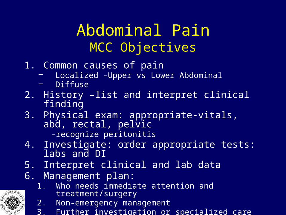
Abdominal PainMCC Objectives
1. Common causes of pain– Localized -Upper vs Lower Abdominal– Diffuse
2. History –list and interpret clinical finding3. Physical exam: appropriate-vitals, abd, rectal, pelvic
-recognize peritonitis4. Investigate: order appropriate tests: labs and DI5. Interpret clinical and lab data6. Management plan:
1. Who needs immediate attention and treatment/surgery2. Non-emergency management 3. Further investigation or specialized care
Case 1:
Sally is an 18 year old woman who presents with a 2 day history of dull periumbilical pain which now localizes to the RLQ.
What disease process is this typical for?
What causes the change in the pain pattern?
What other diseases must you consider?

Neurologic Basis of Abdominal Pain
• Visceral
• Somatic
• Referred

Visceral Abdominal Pain
• Stretch receptors in walls of organs• Stimulated by distention, inflammation • return to spinal cord: bilateral, multiple
levels• Brain cannot localize source
Visceral Abdominal Pain
• Pain felt as crampy, dull, achy, poorly localized
• Associated with autonomic responses of palor, sweating, nausea, vomiting
• Patients often writhing around – Movement doesn’t alter pain
Somatic Abdominal Pain
• parietal peritoneum• Returns to ipsilateral dorsal root ganglion at
1 dermatomal level• Sharp, localized pain• Causes tenderness, rebound, and guarding• Patients lie still, movement increases pain

Referred Pain
• What is it?
• What are some examples?
Referred Pain
• Pain perceived in an area that is distant from the disease process
• Due to overlapping nerve innervations
Examples of Referred Pain
• Shoulder pain with diaphragm stimulation– C 3,4,5 stimulation
• Back pain with biliary colic, pancreatitis, or PID
Differential Diagnosis
• Diffuse vs Localized
Diffuse Abdominal Pain
• Peritonitis• AAA• Ischemic Bowel• Gastroenteritis• Irritable Bowel Syndrome
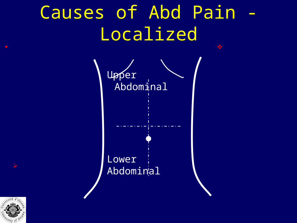
Causes of Abd Pain - Localized
Lower Abdominal
Upper Abdominal

Localized Abdominal Pain
Biliary Colic/Cholecystitis Hepatitis / Hepatic Abscess Pneumonia / Pleurisy
Appendicitis Mesenteric lymphadenitis
Splenic Infarction Splenic Rupture Pneumonia
Incarcerated HerniaBowel obstructionInflammatory bowel diseaseEctopic Ovarian(torsion or cyst)Salpingitis/PID Renal Stones/UTI Testicular torsion
Gastritis,GERD/PUD
Pancreatitis
MI
Diverticulitis
Case 1:
Sally is an 18 year old woman who presents with a 2 day history of dull periumbilical pain which now localizes to the RLQ.

Case 1: Questions
1. What further history do you need from the patient?
2. What would you do in your physical exam?
3. What are you looking for on physical examination?
4. What initial stabilization is required?
5. What is your differential diagnosis?
History* Onset / Duration* Nature / Character / Severity* Radiation* Exacerbating / Relieving Factors* Location* Associated Symptoms
- Nausea / Vomiting- Diarrhea / Constipation / Flatus- Fever- Jaundice / other skin changes- GU (dysuria, freq, urgency, hematuria…)- Gyne (menses, contraception, STDs,,,)
* PMHx- Prior Surgery- Medical Problems
* Medications

SOCRATES• Site - Where is the pain? Or the maximal site of the pain.• Onset - When did the pain start, and was it sudden or
gradual? Include also whether if it is progressive or regressive.
• Character - What is the pain like? An ache? Stabbing?• Radiation - Does the pain radiate anywhere? (See also
Radiation.)• Associations - Any other signs or symptoms associated
with the pain?• Time course - Does the pain follow any pattern?• Exacerbating/Relieving factors - Does anything change the
pain?• Severity - How bad is the pain?

OPQRST
• Onset• Provocation or Palliation• Quality • Region and radiation• Severity. • Time (history) How long, ? Changed, past hx,

High Yield Questions
High Yield Questions
1. Age Advanced age means increased risk.
2. Which came first—pain or vomiting?
1. Pain first is worse (i.e., more likely to be caused by surgical disease).
3. When did it start? Pain for < 48 hrs is worse.
4. Previous abdominal surgery? Consider obstruction.
5. Is the pain constant or intermittent? Constant pain is worse.
6. Previous hx of pain?
7. Pregnant? consider ectopic.

High Yield Questions cont’d8. History of serious illness is suggestive of more serious
disease.
9. HIV? Consider occult infection or drug-related pancreatitis.
10. Alcohol? Consider pancreatitis, hepatitis, or cirrhosis.
11. Antibiotics or steroids? These may mask infection.
12. Did the pain start centrally and migrate to the right lower quadrant? High specificity for appendicitis.
High Yield Questions, cont’d
13. History of vascular or heart disease, hypertension, or atrial fibrillation? Consider mesenteric ischemia and abdominal aneurysm.
Physical Examination

Physical Examination
• Vitals• General appearance: writhing/motionless, diaphoresis,
skin, mental status• Always do brief cardiac and respiratory exam• Abdominal exam: Look, listen, feel • Pelvic, genital and rectal exam in ALL patients with
severe abdominal pain• Assess pulses!
Abdo Exam: Specifics• Always palpate from areas of least pain to areas with
maximal pain• ?Organomegaly, ?ascites• Guarding: voluntary vs. involuntary• Bowel sounds: increased/decreased/absent• Rectal exam: occult/frank blood, ?stool, ?pain, ?masses• Pelvic exam: discharge, pain, masses • Peritonitis:
– suggested by: rigidity with severe tenderness, pain with percussion/deep breath/shaking bed, rebound
Risk Factors for Acute Disease
• Extremes of age• Abnormal vital signs• Severe pain of rapid onset• Signs of dehydration• Skin pallor and sweating

Initial Stabilization
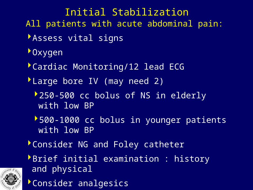
Initial StabilizationAll patients with acute abdominal pain:
Assess vital signs
Oxygen
Cardiac Monitoring/12 lead ECG
Large bore IV (may need 2)
250-500 cc bolus of NS in elderly with low BP
500-1000 cc bolus in younger patients with low BP
Consider NG and Foley catheter
Brief initial examination : history and physical
Consider analgesics
??Do they need immediate surgical consultation?

Pain: ER Management• Is it OK to give a patient pain medications
before you determine their diagnosis?

Abdominal Pain: ER Management
• Anti-inflammatories (NSAIDs):– very effective, esp. for MSK or renal colic pain– Ex. Ketorlac (Toradol) 30 mg IV
• Narcotics– sc/im/iv– very effective, esp. for visceral or undifferentiated pain– Ex. Morphine 2.5-5 mg, hydromorphone 1-2 mg

Nausea/Vomiting: ER Tx

Nausea/Vomiting: ER Tx
• Ondansetron (Zofran) : iv 4-8 mg– very useful in patients with refractory vomiting
• Dimenhydrinate (Gravol): po/pr/im/iv 25-50 mg– beware of anticholinergic side effects– sedating, may cause confusion
• Metoclopramide (Maxeran) 10 mg IV• Prochlorperazine (Stemetil): 10 mg IV
– beware of possible EPS– less sedating; may help with pain control
• Domperidone: po/iv– especially useful with diabetic gastroparesis

Investigations

InvestigationsMost patients with acute abdominal pain require:
- CBC, differential; may need type and cross-match
- electrolytes, BUN, creatinine,
- lactate
- liver function tests
- lipase
- beta-hCG
- urinalysis; stool for OB
They may also need: ECG, cardiac enzymes, ABG,
Investigations
Imaging
ultrasound
CT scan
plain Xrays
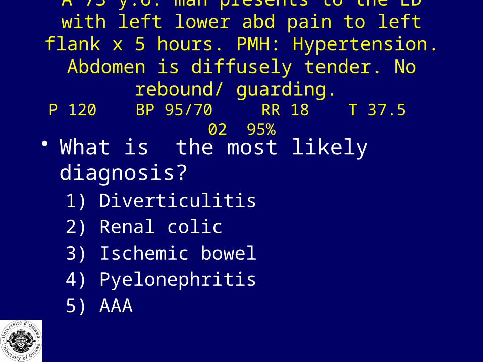
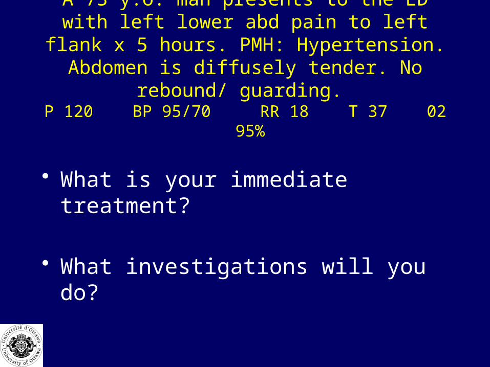
A 73 y.o. man presents to the ED with left lower abd pain to left flank x 5 hours. PMH: Hypertension.
Abdomen is diffusely tender. No rebound/ guarding. P 120 BP 95/70 RR 18 T 37.5 02 95%
• What is the most likely diagnosis?1) Diverticulitis
2) Renal colic
3) Ischemic bowel
4) Pyelonephritis
5) AAA
A 73 y.o. man presents to the ED with left lower abd pain to left flank x 5 hours. PMH: Hypertension.
Abdomen is diffusely tender. No rebound/ guarding. P 120 BP 95/70 RR 18 T 37 02 95%
• What is your immediate treatment?
• What investigations will you do?
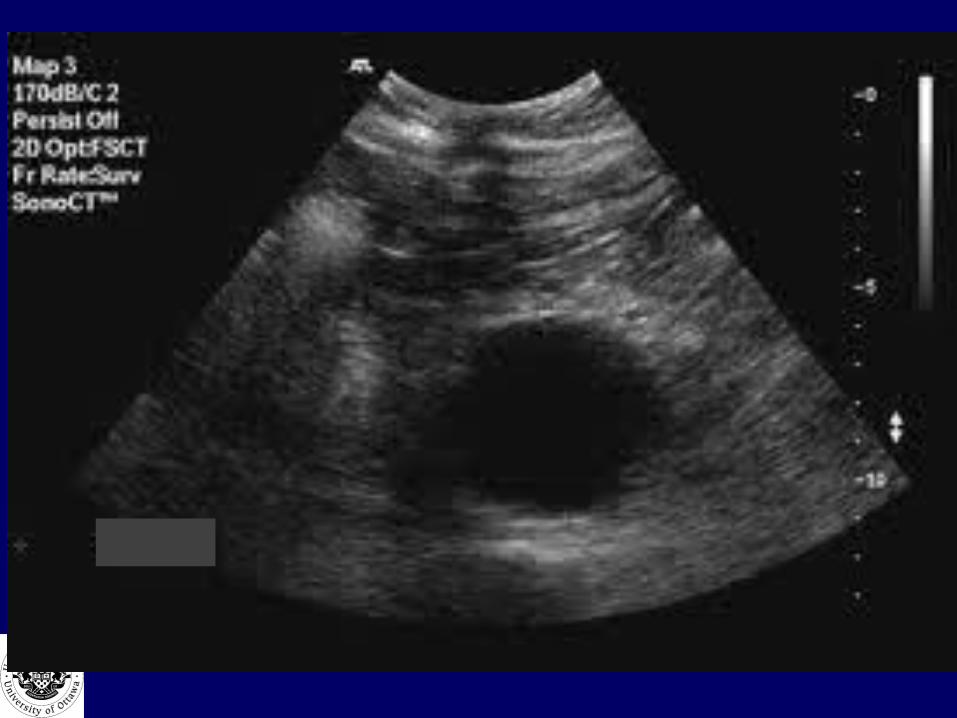
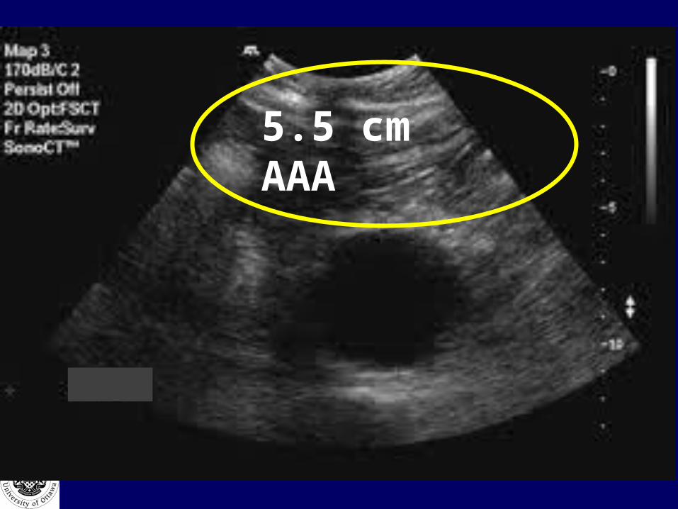
5.5 cm AAA
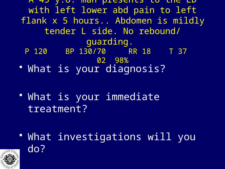
A 45 y.o. man presents to the ED with left lower abd pain to left flank x 5 hours.. Abdomen is mildly
tender L side. No rebound/ guarding. P 120 BP 130/70 RR 18 T 37 02 98%
• What is your diagnosis?
• What is your immediate treatment?
• What investigations will you do?
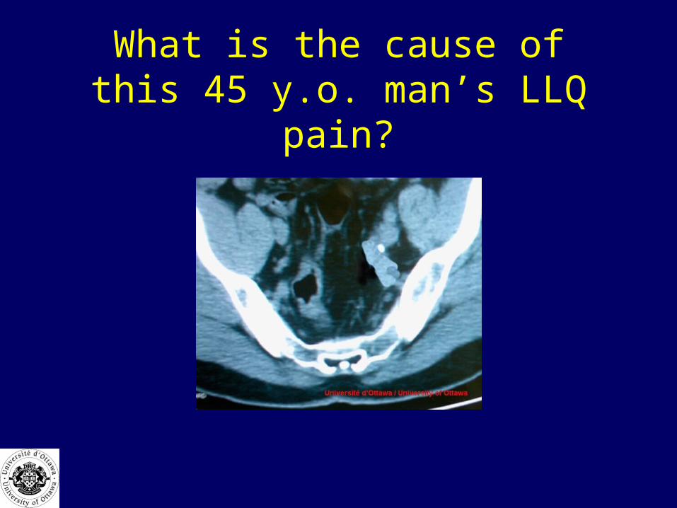
What is the cause of this 45 y.o. man’s LLQ pain?

What is the cause of this 45 y.o. man’s LLQ pain?
• Renal stone
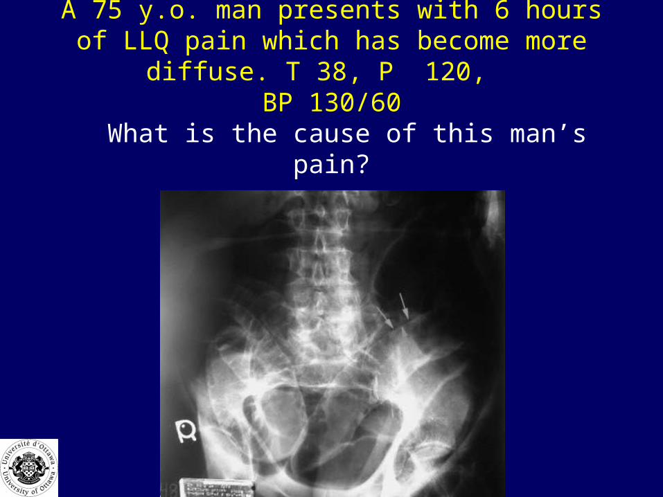
A 75 y.o. man presents with 6 hours of LLQ pain which has become more diffuse. T 38, P 120,
BP 130/60 What is the cause of this man’s pain?
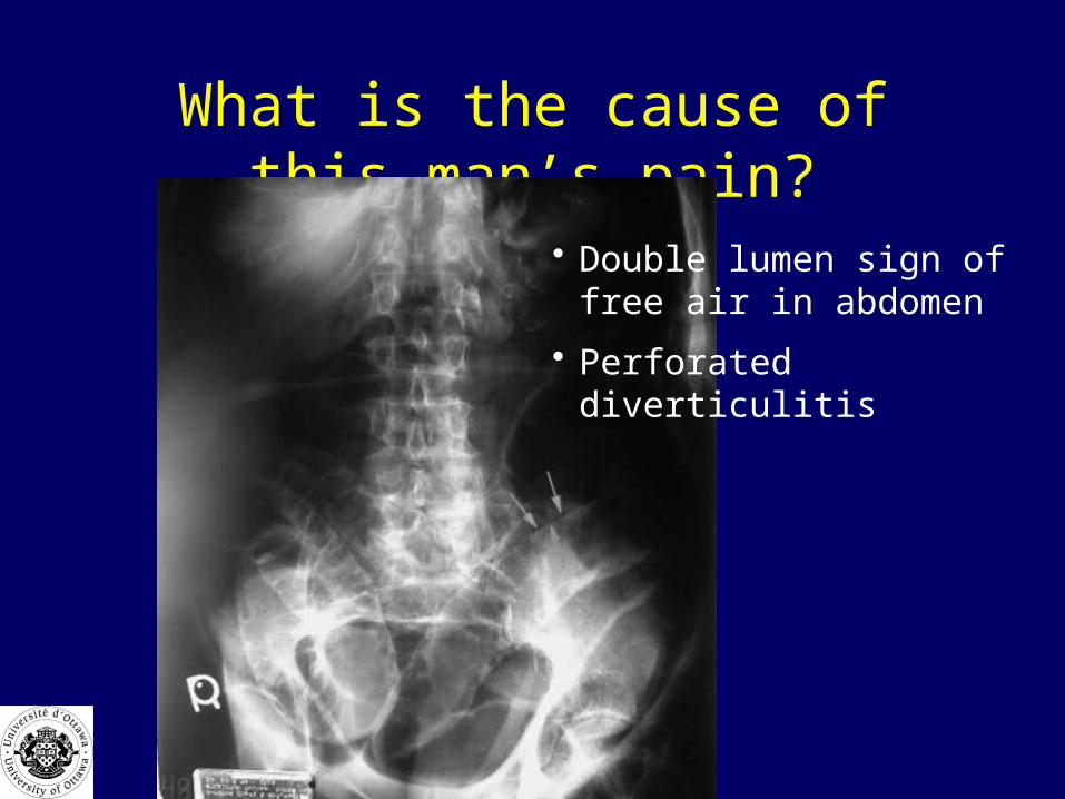
What is the cause of this man’s pain?
• Double lumen sign of free air in abdomen
• Perforated diverticulitis

Why is this woman vomiting?
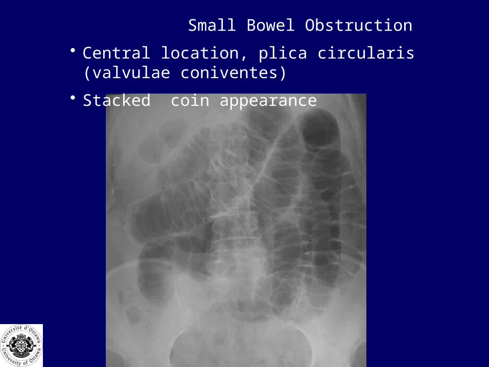
Small Bowel Obstruction
• Central location, plica circularis (valvulae coniventes)
• Stacked coin appearance
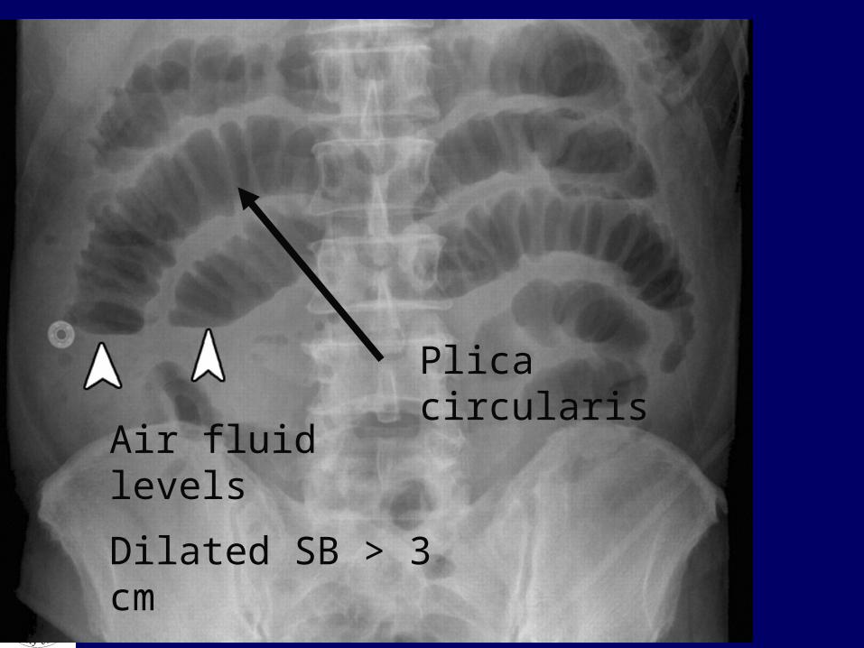
Air fluid levels
Dilated SB > 3 cm
Plica circularis
What are the 3 leading causes of SBO
• 1) adhesions• 2) hernia• 3) neoplasm
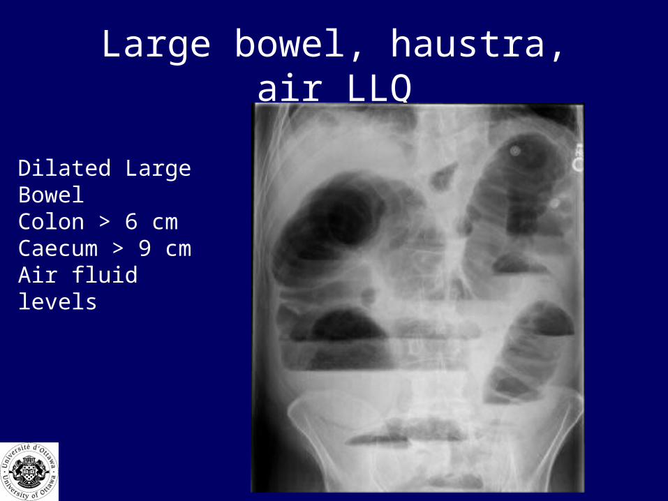
Large bowel, haustra, air LLQ
Dilated Large BowelColon > 6 cmCaecum > 9 cmAir fluid levels
3 Leading causes of LBO
• 1) neoplasm• 2) Diverticulitis• 3) volvulus

Sigmoid Volvulus
34yr female: cerebral palsy, no BM’s, abdo distension
massive bowel
dilation
single loop “bent rubber
tube”

34 y.o. man, alcoholic binge, repeated vomiting. Now abdominal pain, guarding
rebound. What is the cause of this man’s abdominal
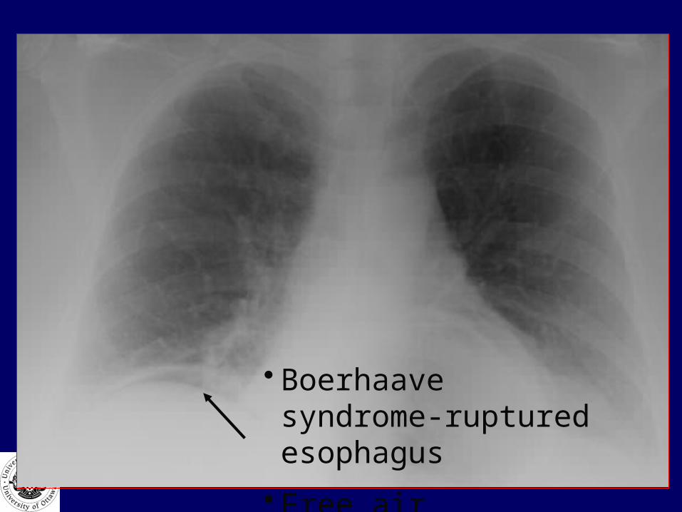
• Boerhaave syndrome-ruptured esophagus
• Free air
Summary: Approach to Abdominal Pain in the ER
• ABC assessment• Stabilize the patient, and refer early if unstable• Careful, detailed history• Focused physical examination• Early, thorough work-up:
– Appropriate laboratory investigation– Diagnostic imaging where indicated
• Continuous reassessment• Consider patient circumstances (age, pmhx, reliability,
home situation)

Summary: Common Causes of Abdominal Pain
MCC Categorization
• Is it diffuse or localized?
• Do they need immediate resuscitation, referral or surgery?

?
Acute Dyspnea (minutes to hours)MCC Objectives
• Differentiate cardiac, pulmonary, central causes
• Assess the A, B, C’s• Diagnose cause, severity and manage acute
dyspnea• Identify life threatening dyspnea• List and interpret clinical and lab data
– ECG, ABG, CXR • Management: acutely, refer prn, plan long-
term Rx if chronic
What drives us to breath?
• Chemoreceptors in medulla, carotid and aortic bodies:– High CO2– High H+ ion– Low 02.
• Stretch and baroreceptors in lungs

Definitions
• Dyspnea:– sensation of shortness of breath

Definitions
• Tachypnea: – rapid, shallowing breathing
• Hyperventilation: – breathing in excess of metabolic needs of body
lowering C02 – Need to rule out organic disease
• A 55 year old woman comes into the ED in obvious respiratory distress. She is very agitated, sitting forward, using her accessory muscles.
What is her problem?

Most Common Causes of Acute Dyspnea (MCC)
• Cardiac: – MI– Valvular heart disease– Pericardial Tamponade– Dysrhythmia– Increased cardiac output (anemia)
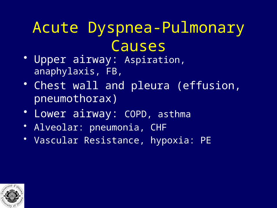
Acute Dyspnea-Pulmonary Causes• Upper airway: Aspiration, anaphylaxis, FB,
• Chest wall and pleura (effusion, pneumothorax)• Lower airway: COPD, asthma • Alveolar: pneumonia, CHF• Vascular Resistance, hypoxia: PE
Acute Dyspnea
–Central causes•Metabolic: acidosis, ASA toxicity

• Our 55 year old woman is still in respiratory distress.
What will you do?

Rapid Assessment
• ABC’s : 5 vitals: P, RR, BP, T, 02 sat, Glucoscan.
• MOVIE– Monitor– O2– Vitals with BS– IV– ECG
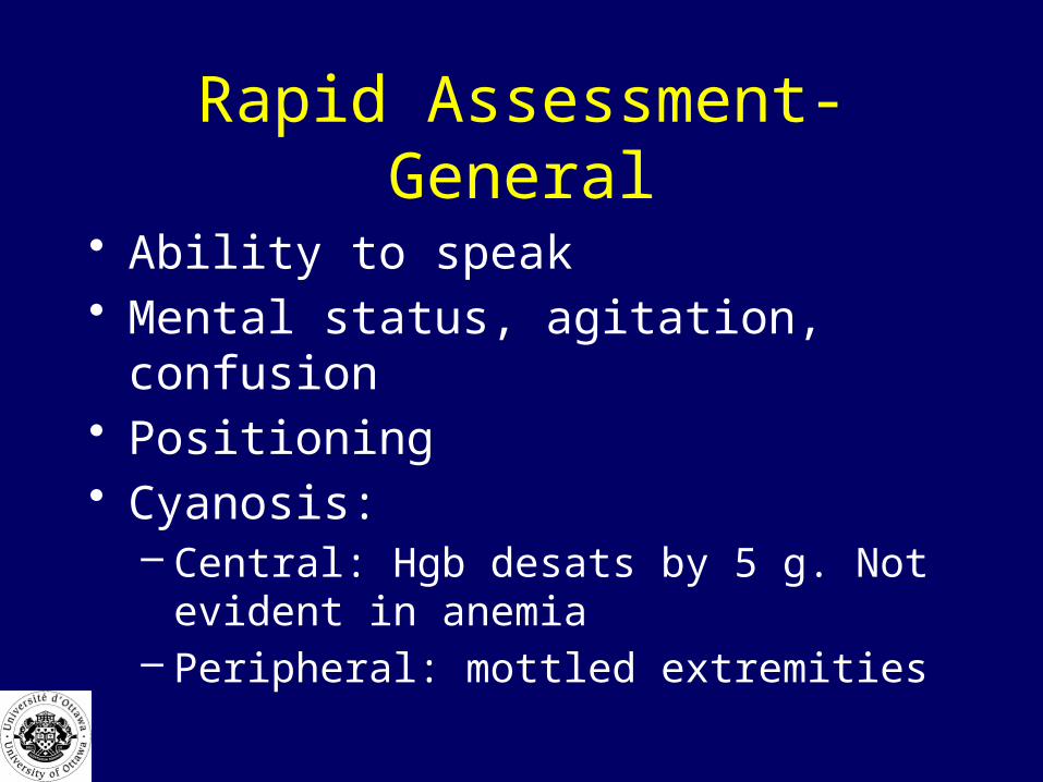
Rapid Assessment-General
• Ability to speak• Mental status, agitation, confusion• Positioning• Cyanosis:
– Central: Hgb desats by 5 g. Not evident in anemia
– Peripheral: mottled extremities
Rapid Assessment
• Airway: – Is the patient protecting it?
• Talking, swallowing, gagging
– Is the patient able to oxygenate and ventilate adequately?
– Is there stridor

Oxygen
• Nasal prongs max. 4-5l/min– Increase FIO2 by 4%/L
• Venturi: up to 50%
• 02 reservoir: 90-95%
5 Reasons to Intubate
• Protection• Creation• Oxygenation• Ventilation• Pulmonary toilet
Breathing
• Look, listen, feel, or IPPA
• Wheezes, rales, rubs, decreased air entry
• Is it adequate? O2 sat?
Circulation
• Pulse, BP, • Heart sounds ? Muffled• JVP• Edema
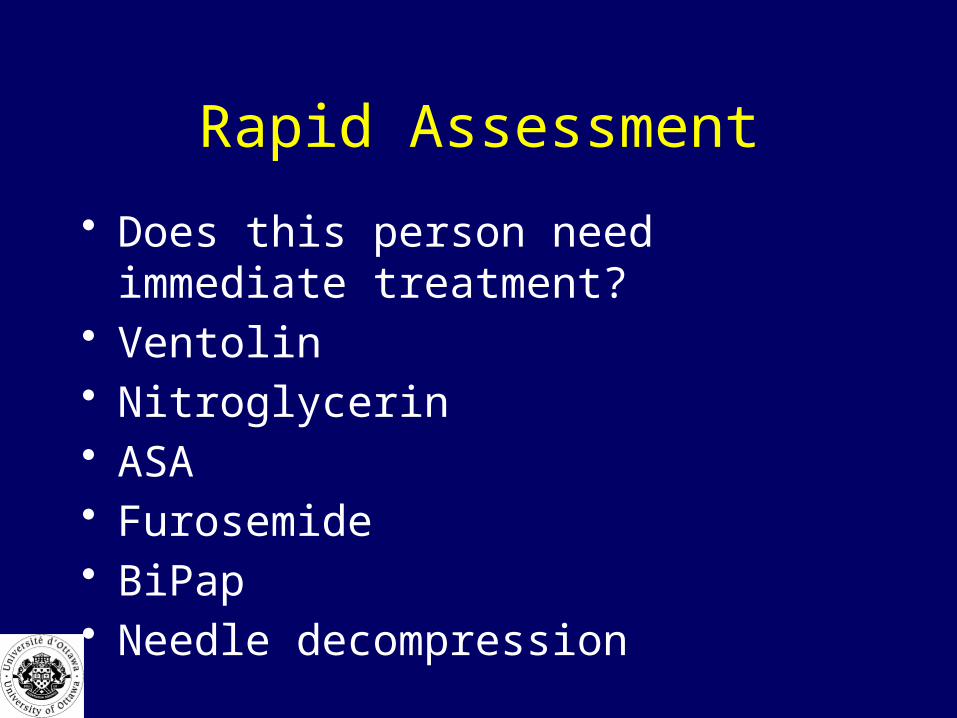
Rapid Assessment
• Does this person need immediate treatment?• Ventolin• Nitroglycerin• ASA• Furosemide• BiPap• Needle decompression

History-What are the key questions?
• Previous hx of similar event• How long SOB• Onset gradual or sudden• What makes it better or worse• Associated symptoms:
– Chest pain, cough, fever, sputum, PND, orthopnea, SOBOE

History-What are the key questions?
• Medications, home 02• Allergies• What has helped in the past• Past medical history:
– Cardiac, pulmonary, recent surgery

Labs/Investigations
• ABG/VBG• CBC, Lytes, Cardiac enzymes• D dimer• ECG• Pulmonary Function Tests
Imaging
• CXR
• Helical CT• Pulmonary angiogram• V/Q –Nuclear ventilation perfusion scan

COPD
72yr female: chronic SOB, worse x few days
vertical heart
increased AP diameter
increased retrosternal
air
flat diaphragm
low set diaphragm
hyperlucent lung fields

Principles of ManagementCOPD
• Oxygen – Titrate with 02 sat:– Monitor pC02, avoid loss of hypoxic drive
• Beta agonists and anticholinergics– Ventolin 1 cc in 2 cc atrovent or MDI
• Steroids ex. Solumedrol 125 mg IV• BiPap • Antibiotics
Status Asthmaticus• 100 % oxygen• continuous ventolin in atrovent• Prednisone P.O. or solumedrol IV• magnesium S04 2 gm over 2 min
• Epinephrine IM or IV has limited role
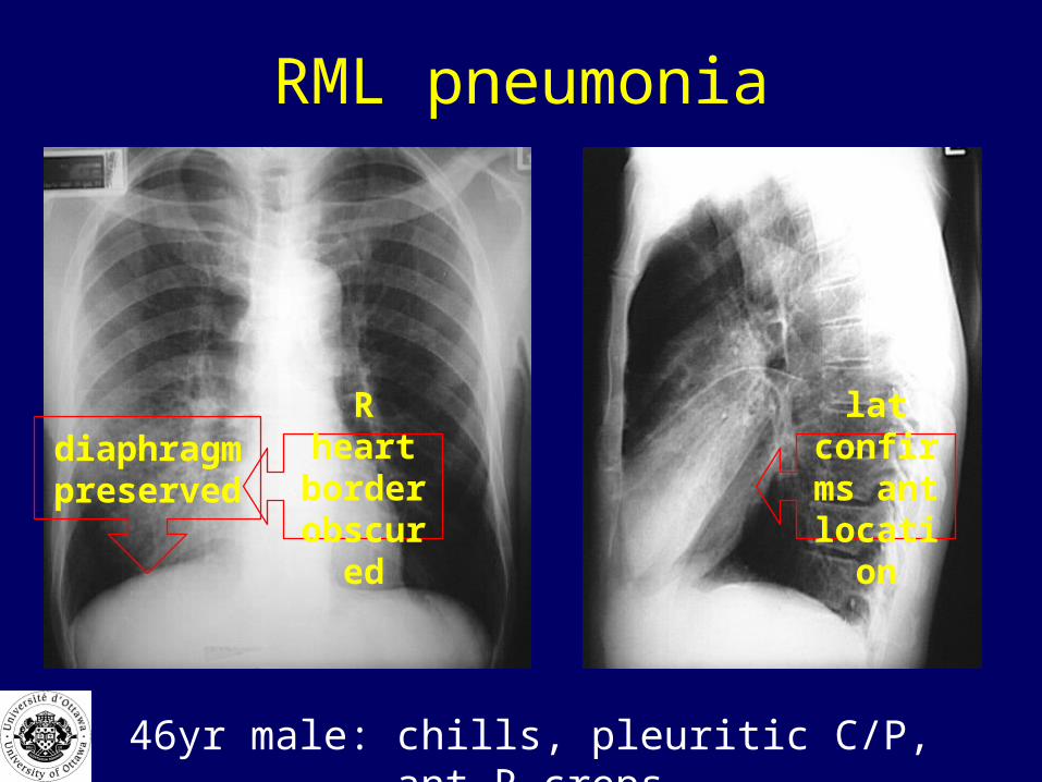
RML pneumonia
R heart border
obscured
lat confirms
ant location
diaphragm preserved
46yr male: chills, pleuritic C/P, ant R creps

LLL pneumonia
58yr female: weakness, cough, SOB
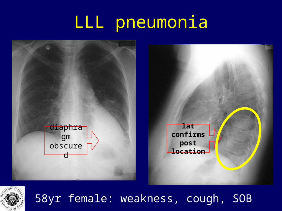
LLL pneumonia
diaphragm obscured
lat confirms post
location
58yr female: weakness, cough, SOB
Principles of ManagementPneumonia
• Oxygen to maintain 02 sat at 92-94%• Antibiotics:
– Macrolides– Fluroquinolones– 2nd or 3rd generation cephalosporin
• Beta agonists and BiPap as required• Considering scoring system for disposition
– CURB-65, CRB-65, Pneumonia Severity Index
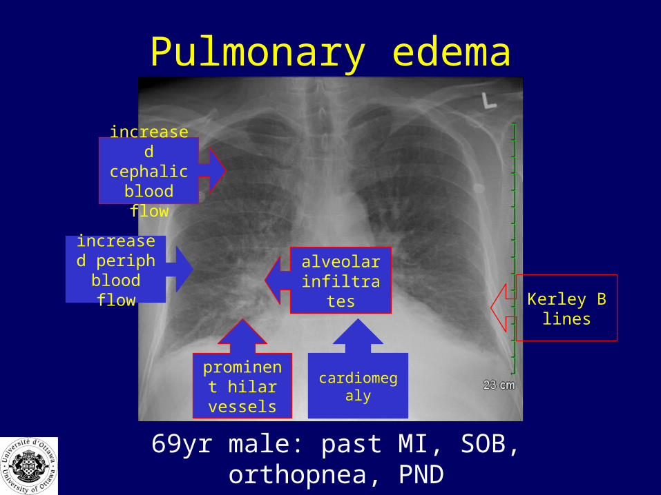
Pulmonary edema
69yr male: past MI, SOB, orthopnea, PND
prominent hilar vessels
cardiomegaly
increased periph blood
flow
increased cephalic
blood flow
alveolar infiltrates Kerley B
lines

Principles of ManagementPulmonary Edema
• LMNOP• Lasix –furosemide 40-160 mg IV• Morphine 2-4 mg IV• Nitroglycerin SL, IV• Oxygen • Position, postive pressure BiPap• ECG-rule out ACS

A 25 year old with dyspnea

Pneumothorax

Principles of ManagementPneumothorax
• Tension: 14 gauge needle 2nd ICS, MCL • 30 Fr chest tube
• Pigtail catheter
• Small spontaneous pneumothorax: @20%– May observe, discharge, repeat CXR 24 hrs

Ruptured Aorta
34yr male: MVC hit tree, unrestrained, c/o chest pain
widened superior
mediastinum
loss of aortic knuckle
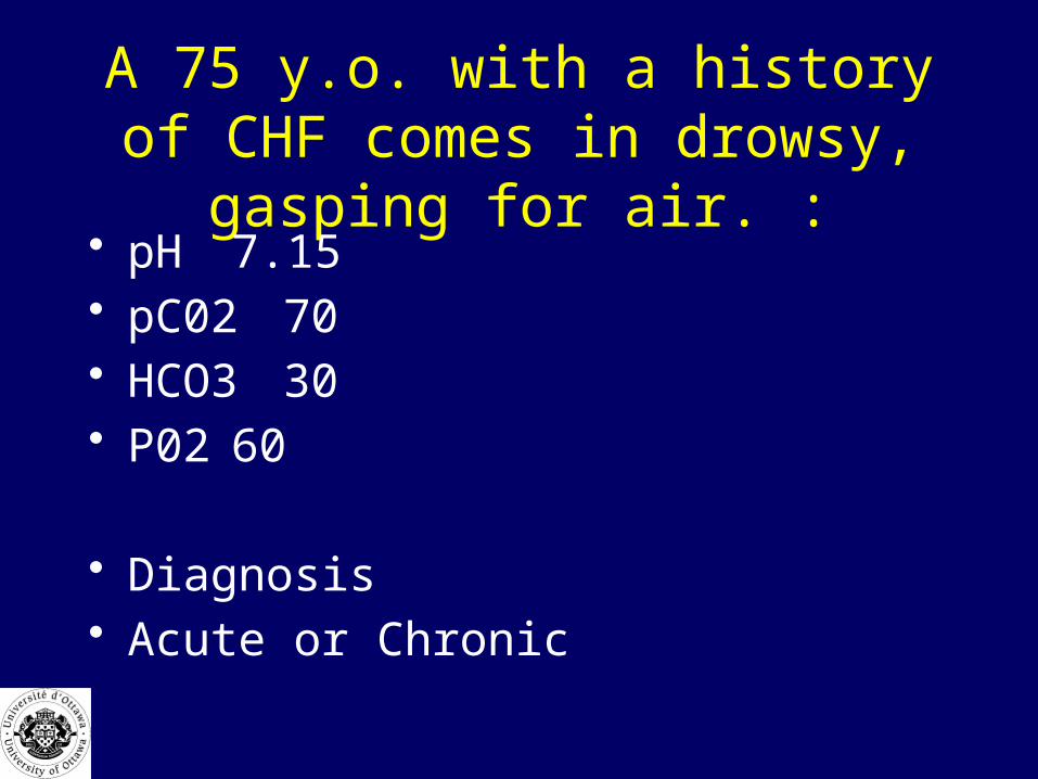
A 75 y.o. with a history of CHF comes in drowsy, gasping for air. :
• pH 7.15• pC0270• HCO3 30• P02 60
• Diagnosis• Acute or Chronic

A 75 y.o. with a history of CHF comes in drowsy, gasping for air. :
• pH 7.15• pC0270• HCO3 30• P02 60
• Acute Respiratory Acidosis – pH is low– HCO3 has not had time to increase
A 75. y.o. with COPD and dyspnea x 2 days
• pH 7.32• pC0280• HC03 40• p02` 65
• Acute or Chronic
A 75. y.o. with COPD and dyspnea x 2 days
• pH 7.32• pC0280• HC03 40• p02` 65
• Chronic Respiratory Acidosis– HC03 very high therefor pH not that low despite C02
of 80

A 25 y.o. diabetic, vomiting x 2 days, looks dyspneic
• pH 7.10• HC03 10• pC0218• P02 95
A 25 y.o. diabetic, vomiting x 2 days, looks dyspneic
• pH 7.10• HC03 10• pC02 18• P02 95
• Acute metabolic acidosis, and partially compensating respiratory alkalosis

An anxious individual
• A 55 y.o. woman, recent fatigue, shortness of breath, comes in to the ED hyperventilating. Feels more short of breath x 1 hour .
• What will you do?
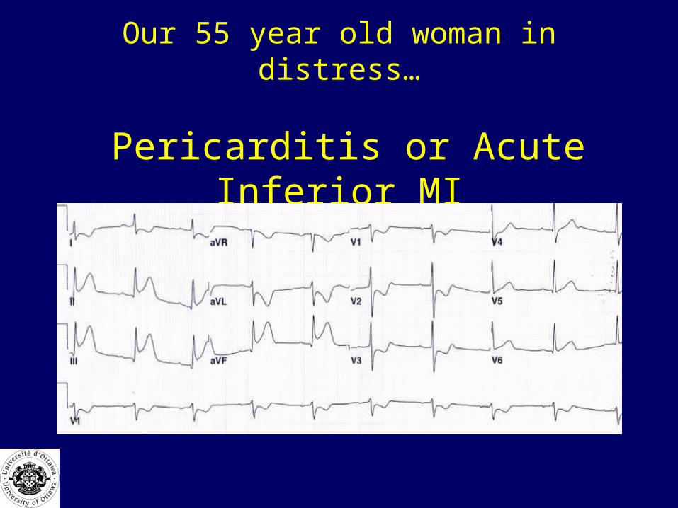
Our 55 year old woman in distress…
Pericarditis or Acute Inferior MI

Acute Inferior MI
Ischemic Symptoms in Women
• Dyspnea• Weakness• Fatigue
• Often no chest pain (vs men)

Admission Criteria for Dyspnea
• Older patient • Abnormal vitals including 02 sat• Abnormal level of consciousness• Significant illness ex. Pneumonia• Patient fatigue• No improvement despite treatment• Home situation

?
Syncope

Syncope
• http://www.blogtelevision.net/p/Videos-Watch-a-Video___1,2,,59315.html
Syncope-MCC Objectives
• Definition• Distinguish from Seizure• Causes: serious or not, cardiac or not• List and interpret ‘Targeted’Hx, Px, • List and interpret investigations,
– ex cardiac: ECG, echo
• Initial Management Plan: meds, • Who needs referral, fitness to drive
Syncope• A 73 y.o. man collapsed in the bathroom and had a 30
second episode of unresponsiveness at 0430. He awakes fully, and is brought to the Emergency Department by his wife.
• Is this a syncopal episode?• What are the causes of syncope?• What is the likelihood he had a cardiac cause of syncope?• What is your workup and management of this patient?

What is syncope?
• Sudden, transient loss of consciousness• Rapid and complete recovery
• May have minor myoclonic jerks or muscle twitching
• No postictal state
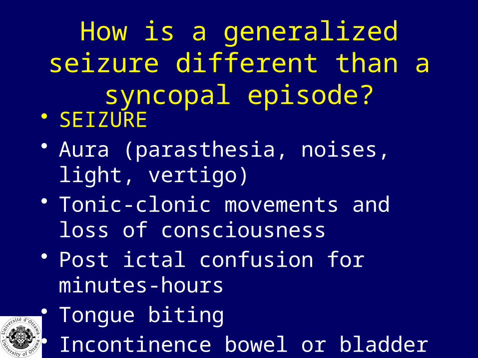
How is a generalized seizure different than a syncopal episode?
• SEIZURE• Aura (parasthesia, noises, light, vertigo)• Tonic-clonic movements and loss of
consciousness• Post ictal confusion for minutes-hours• Tongue biting• Incontinence bowel or bladder

Syncope
• Prodrome often occurs– Feeling faint, hot, lightheaded, weak, sweaty
• Brief loss of consciousness– seconds to 1-2 minutes
• Rapid and complete recovery• Speaking normally within 1 minute
– No post event confusion

What are the common causes of syncope? (MCC)
• Cardiovascular (80%)– Cardiac arrhythmia (20%)– Decreased cardiac output –MI, Ao. Stenosis– Reflex/underfill (60%) (vasovagal, orthostatic)
• Cerebrovascular (15%)• Other
– Metabolic (low BS)– Psychiatric– Meds (BP)
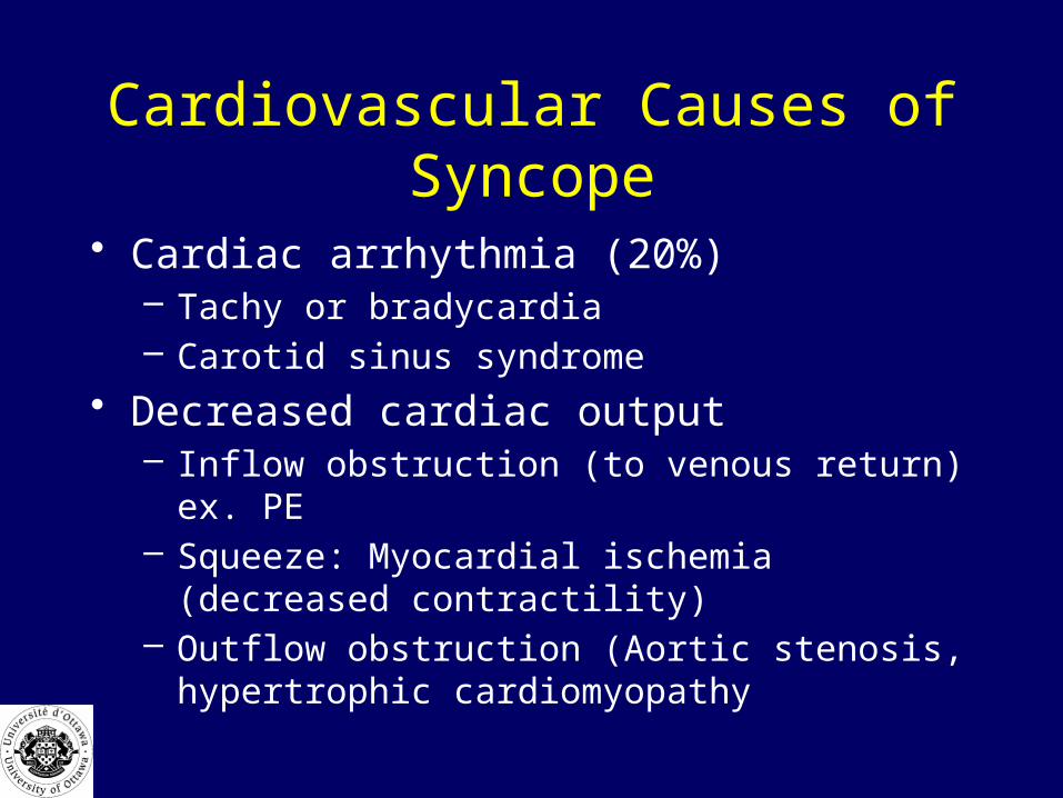
Cardiovascular Causes of Syncope
• Cardiac arrhythmia (20%)– Tachy or bradycardia– Carotid sinus syndrome
• Decreased cardiac output– Inflow obstruction (to venous return) ex. PE– Squeeze: Myocardial ischemia (decreased contractility)– Outflow obstruction (Aortic stenosis, hypertrophic
cardiomyopathy
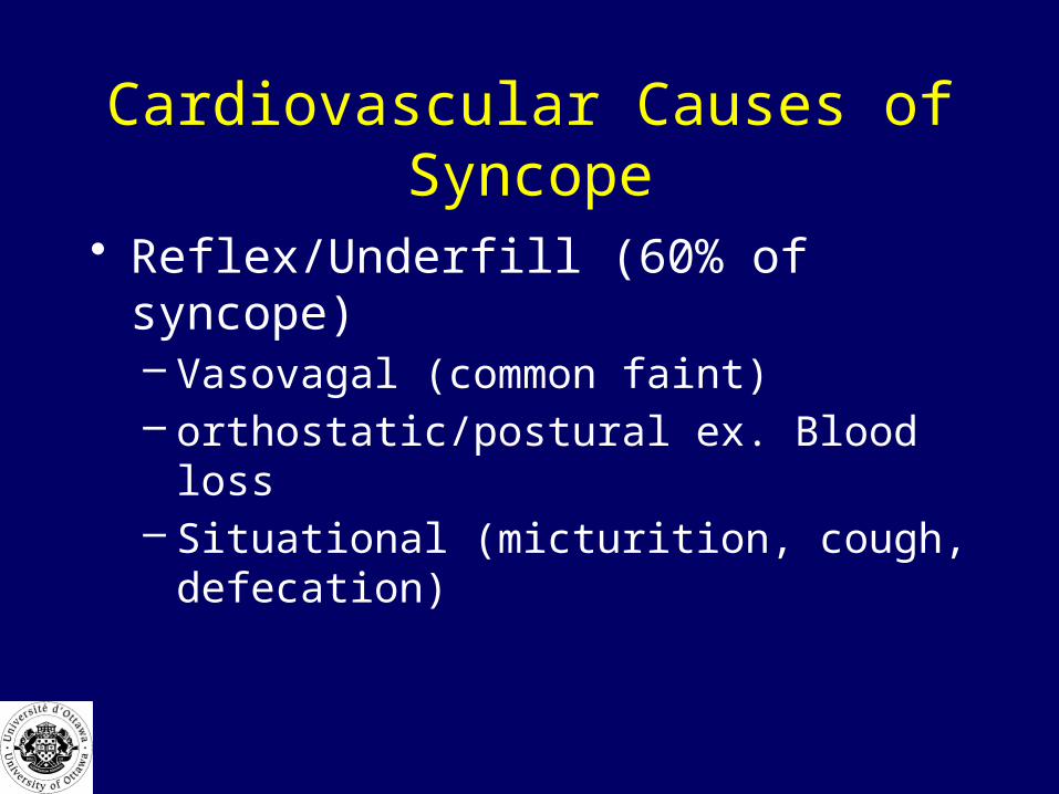
Cardiovascular Causes of Syncope
• Reflex/Underfill (60% of syncope)– Vasovagal (common faint)– orthostatic/postural ex. Blood loss– Situational (micturition, cough, defecation)
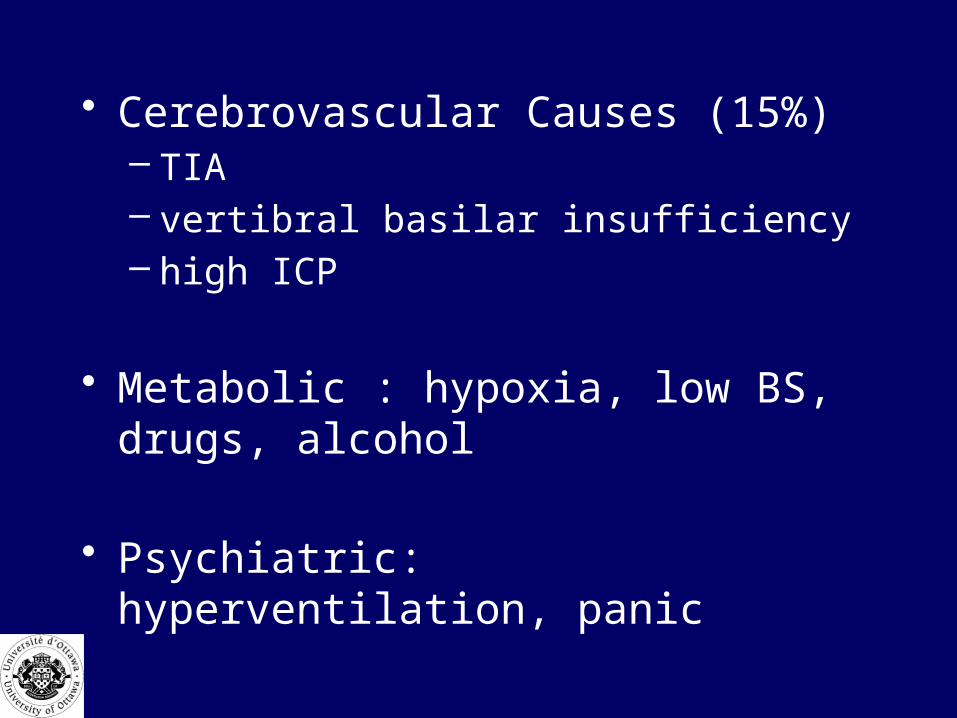
• Cerebrovascular Causes (15%)– TIA– vertibral basilar insufficiency– high ICP
• Metabolic : hypoxia, low BS, drugs, alcohol
• Psychiatric: hyperventilation, panic
What is your initial approach with your patient with syncope?
• Check ABC,s, • 5 vitals -postural• monitor, IV, ECG, blood tests• Bolus fluids if hypotensive 250-1000cc NS• glucosan• give thiamine if giving glucose• consider naloxone if patient not fully awake• history and physical
History• what happened (witnesses important)• what were you doing (ex. urination,
standing up quickly etc.)• prodrome (hot, sweaty, vomiting)• any tonic-clonic activity• postural or neck turning• recovery – long or short
– any confusion

Review of Systems
• volume status (eating, diarrhea, exercise)• recent blood loss• chest pain, palpitations, SOB, • any focal neurologic symptoms• pregnancy
PMH
• previous history of syncope• ex. occasional episodes over the years vs
several episodes recently (more sinister)• cardiac disease or medications• bleeding disorders or PUD• diabetes• medications ex. antihypertensives often
cause orthostatic syncope

Physical Exam
• ABC• Orthostatic Vitals • HEENT: trauma, papilledema, • Resp/CVS: S3, AS murmur, • Abd: aorta, pulses, peritoneal, blood PR• Pelvic: bleeding, tenderness• Neurologic: focal findings
Lab Investigations• CBC• Type and xmatch
– If suspect acute blood loss AAA, ectopic, GI bleed• Lytes, BS, BUN, Cr• D dimer• Pregnancy Test• ECG• CT Head if suspect cerebrovascular cause• Holter• EEG
Vasovagal Faint
• Common (60% all syncope)
• Increased parasympathetic tone
• Bradycardia, hypotension
Vasovagal Faint -Predisposing Factors
• Fatigue• Hunger• Alcohol• Heat• Strong smells• Noxious stimuli• Medical conditions anemia, dehydration• Valsalva (trumpet player)

Vasovagal FaintSymptoms and signs
• Warm, sweaty• Weak• Nausea• Confused• Unprotected fall• Eye rolling, myoclonic jerks, • Resolves in 1-2 min• Rarely tongue biting or incontinence• Not confused afterward
Cardiac Syncope
• 20% all syncope• Serious prognosis• Exertional syncope
– Outflow obstruction AS, IHSS
• Ischemia/MI• Conduction disorders• dysrhythmias

Orthostatic
• Decrease in systolic BP by 20-30 or increase in pulse by 20-30 on standing
• Supine• Meds -antihypertensives• Blood loss, dehydration
Syncope-When to Admit• Uncertain diagnosis• Elderly (more likely cardiac)• Suspected cardiac etiology• Abrupt onset with no prodrome (typical for
dysrhythmia)• Unstable vitals • Blood loss• Abnormal ECG
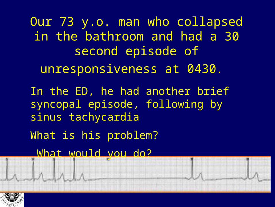
Our 73 y.o. man who collapsed in the bathroom and had a 30 second episode of
unresponsiveness at 0430.
In the ED, he had another brief syncopal episode, following by sinus tachycardia
What is his problem?
What would you do?

Our 73 y.o. man who collapsed in the bathroom and had a 30 second episode of
unresponsiveness at 0430.
• Sick sinus syndrome: need pacer
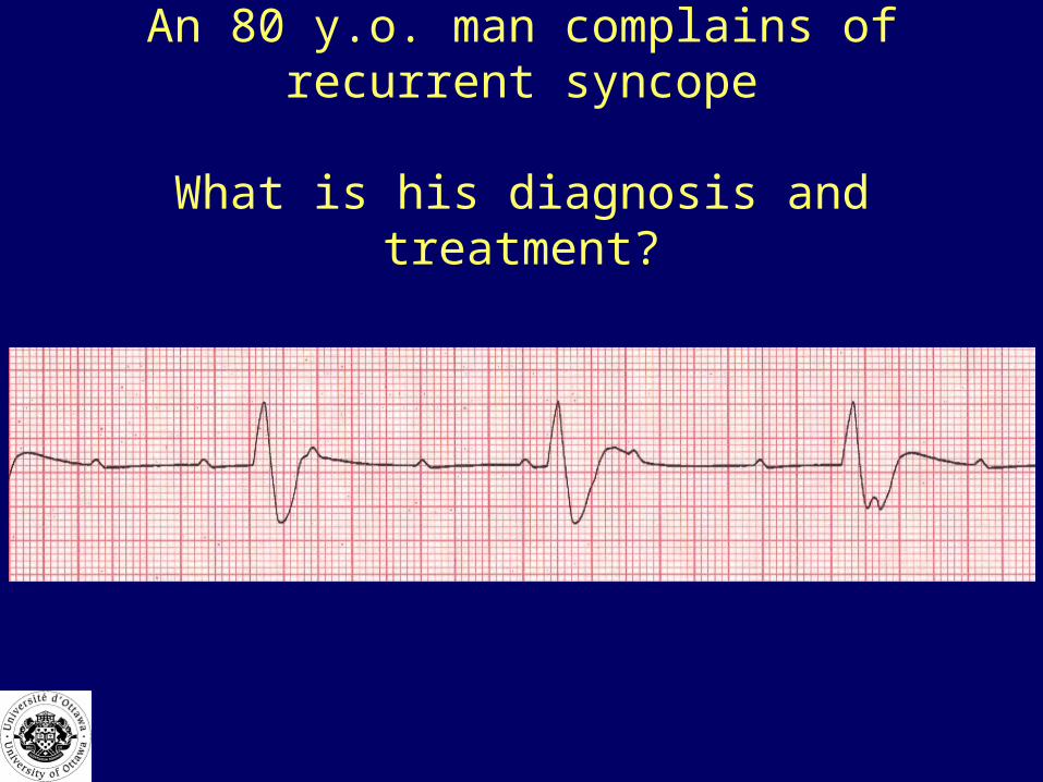
An 80 y.o. man complains of recurrent syncope
What is his diagnosis and treatment?

An 80 y.o. man complains of recurrent syncope
What is his diagnosis and treatment?
• Third degree Heart Block

A 65 y.o. man on diuretics has recurrent syncope
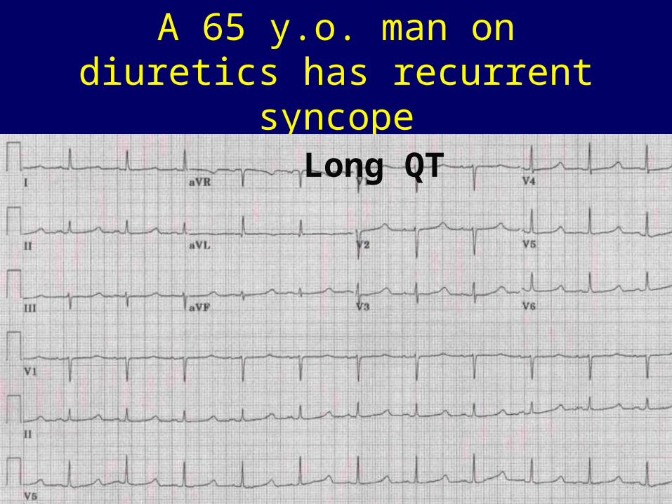
A 65 y.o. man on diuretics has recurrent syncope
Long QT

Torsades de Pointes

Treatment of Torsades
• Correct electrolytes • Magnesium 2 gm over 20 min• Isoproterenol 2-20 mcg/min• Overdrive pacing
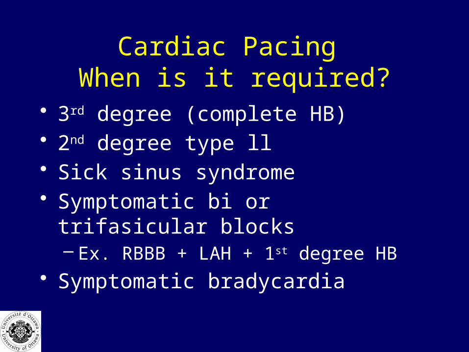
Cardiac Pacing When is it required?
• 3rd degree (complete HB)• 2nd degree type ll• Sick sinus syndrome• Symptomatic bi or trifasicular blocks
– Ex. RBBB + LAH + 1st degree HB
• Symptomatic bradycardia
Fitness to Drive• CPSO: > 16 yrs old
– Suffering from a condition that may make it dangerous to operate a motor vehicle
• Single episode of syncope that is easily explained ie. Simple faint dosen’t need reporting
• Recurrent episodes or suspected cardiac cause– needs to be reported and the patient shouldn’t
drive til a cause is determined and treated.

?Break
Coma
Coma

MCC Objectives• Definition and Causes of coma
– Vascular, infectious, trauma, metabolic, substance use/OD, seizures
• List and interpret key clinical findings: hx, px, other sources, assessment tools (GCS)
• List and interpret critical investigations: – Lab: tox screen, glucoscan, LP, – DI: CT, MRI, EEG
• Initial management plan– immediate treatment: A,B, C’s,
• Antibiotics, anticonvulsants– Empiric RX: naloxone, flumazenil, glucose, – Who needs specialized treatment
• Management of Incompetent Patients-proxy decision-making
What is Coma?
• MCC Defintion:
• state of pathologic unconsciousness (unarousable)

An 80 y.o. man is comatose 2 weeks after falling down stairs?
Why is this patient comatose?
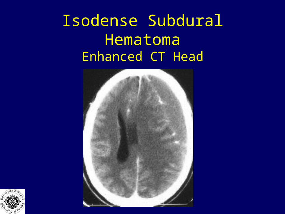
Isodense Subdural HematomaEnhanced CT Head
A diabetic patient present in a coma and is found to have a BS of 1.5
Why are they in a coma?
Rx: 1 amp (50cc) D50W
contains how much sugar?

ComaCan be induced by structural damage or chemical
depression
1) reticular activating system in brainstem, midbrain, or diencephalon (thalamic area)
• Ex. Pressure from a mass• Toxins
2) Bilateral cerebral cortices– Ex. Toxins, hypoxia, hypoglycemia
A 45 y.o. ‘street’ person is brought into the ED in a coma. What are the
causes?
Causes of Coma
• Structural– Bleed, CVA, CNS infection,
• Metabolic (medical)– A,E,I, O, U, TIPS
A 45 y.o. ‘street’ person is brought in to the ED in a coma. What are the
causes?• AEIOU TIPS• A - alcohol, anoxia• E – epilepsy, electrolytes (Na, Ca, Mg), encephalopathy (hepatic)• I - insulin (diabetes)• O - overdose• U - uremia, underdose (B12, thiamine)• • T- trauma, toxins, temperature, thyroid• I - infection• P - psychiatric• S - stroke (cardiovascular)
What is your initial approach with this comatose patient?
• A-airway protection (and c spine)• B-breathing O2 sat • C-5 vitals (pulse, BP, temp)• D-dextrose Glucoscan• Thiamine (if giving glucose)• Naloxone (should have small pupils)• IV, ECG monitor, foley, labs• Hx, Px• Determine level of consciousness

Why Thiamine if giving a bolus of glucose
• Precipitate Wernicke’s encephalopathy
• Cranial nerve palsy - ocular• Confusion• Ataxia

Level of Consciousness
• AVPU– Awake, verbal, pain , unresponsive
• Glasgow Coma Scale

GCSBest Eye Response. (4)
1. No eye opening. 2. Eye opening to pain. 3. Eye opening to verbal command. 4. Eyes open spontaneously.
Best Motor Response. (6) 8 or less = coma5. No motor response. 6. Extension to pain. 7. Flexion to pain. 8. Withdrawal from pain. 9. Localizing pain. 10.Obeys Commands
Best Verbal Response. (5) 11.No verbal response 12.Incomprehensible sounds. 13.Inappropriate words. 14.Confused 15.Orientated

History• What happened?• Symptoms: depression, Headache• Gradual or sudden LOC• Sudden = intracranial hemorrhage• Gradual more likely metabolic, could be
subdural • PMH: diabetes, thyroid, hypertension,
substance abuse, alcohol• Meds,

Physical Exam• Goal: Try and determine if a
structural lesion is present, or a metabolic cause.
How do structural lesions present differently than metabolic causes of coma?
Physical Exam• Structural lesions:
– Often have focal findings, abnormal pupils, evidence of increased ICP
• Metabolic causes:– No focal findings, pupils equal mid or small, no
evidence of increased ICP
Signs and Symptoms of Increased ICP
• Headache, N, V,• Decreased LOC• Abnormal posturing• Abnormal respiratory pattern• Abnormal cranial nerve findings• Cushing Triad: late sign of high ICP
– high BP, bradycardia, and low RR = high ICP

Physical Exam• Vitals• BP > 120 diastolic may cause encephalopathy• Hypotension uncommon with intracranial pathology • Temperature
– Infection, CNS or otherwise– Neuroleptic malignant syndrome
• antipsychotics, dopaminergic (levadopa) , or anti-dopamine (metoclopramide)
• Altered mental status, muscle rigidity, and fever

Respirations
• Cheyne stokes– Fast alternating with slow breathing
• Brain lesions, acidosis
• Apneustic – Pauses in inspiration
• Pons lesions, CNS infection, hypoxia
Physical Exam
• HEENT:– Battle’s sign, hemotympanum.– Breath odour
• Ex. Acetone = DKA

Pupils• Metabolic:
– pupils usually react
• Structural: – may be unilateral dilatation Why?
• Uncal herniation presses on CN 111, • Lose Parasympathetic tone• Unapposed sympathetic stimulation
• 10% normal people have 1-2 mm difference
Pupils• Fixed dilated pupils ominous
• Dead, central herniation, hypoxic injury
• Small pinpoint pupils– Lesion in pons (ischemic or bleed– Opiate OD

Physical Exam
• Corneal Reflex
– Sensory CN 5, and Blink is CN 7

Extraocular Movements
• Helps determine brainstem function in coma
• Doll’s eyes– Eyes move in opposite direction to head
movement– indicates functioning brainstem

Oculocephalic ReflexEnsure C spine cleared
• Awake person: – eyes look forward, some nystagmus
• Comatose patient with brainstem function: Eyes deviate completely in opposite direction to head movement
• Comatose Patient with no brainstem function– Eyes follow head movement

Oculovestibular ReflexCold Calorics
• Check eardrum• 50 cc iced saline
• Awake person: – COWS– Nytagmus away from cold– Driving a car, cerebral cortex keeps you on the
road

Oculovestibular ReflexCold Calorics
• Comatose patient, intact brainstem– Eyes deviate to cold side– Hey who’s putting ice in my ear
• Comatose patient, nonfunctioning brainstem– No reaction

Physical Exam cont.
• Disc• Nuchal rigidity• Resp/CVS/Abd/Extrem• Neuro:
level of consciousness, CN, Motor, Sensory, DTR
Motor Exam
• Is there asymmetry in response to pain• Evidence for seizures? • Withdrawing: nearly awake pt• Decorticate:
– Abnormal flexion response. Flexes elbow, wrist, and adducts shoulder
– Cerebral cortex injury
Motor Exam
• Decerebrate posture– Extends elbow with internal rotation– Lesions or metabolic effect in midbrain
• Flaccidity– Ominous sign– Toxin/OD

Labs ?
• CBC,• Lytes, Bun Cr, BS• LFT, Ca, Mg, • ABG• Alcohol, Osmolality• Tox screen• CO level
Diagnostic Tests/Imaging
• CXR• CT Head• LP• ECG• EEG

A 25 y.o. woman presents in a coma. Pupils pinpoint. RR 8. No focal
findings?
What will you do?
• ABC’s, vitals• BS• Naloxone 0.4-2 mg IV• What if she is chronically taking narcotics?

A 30 y.o. man, hit on the head, comatose with a unilateral fixed
dilated pupil?
What would you do?
• Intubate, pC02 to 30 mmHg• Mannitol .5 gm/kg• CT Head• Stat Neurosurgery consult

Uncal Herniation
Substitute Decision Maker
• Who can be a substitute decision maker in a patient who is incompetent or unable to decide for themselves?
• Who has the higher authority:– Spouse– Sibling– Parent

Substitute Decision MakingHighest of

Summary COMA
• ABC, Vitals, O2, CO2, BS, Naloxone• Metabolic vs Structural• Key to Exam
– Respiration– Pupils– EOM– Motor response

?
Hypotension Shock – MCC Objectives
• Causes• List and interpret critical findings
– Symptoms and signs of shock– Diagnose cause
• List and interpret critical investigations– Tests to confirm presence and cause
• Management strategy– Restore tissue perfusion– Specific therapy related to cause
What Is Shock
• Tissue hypoperfusion or tissue hypoxia
Shock
• Catecholamine surge• Vasoconstriction, increased CO• Renin-angiotensin, vasopressin
– Salt and water retention
Shock
• If persists– Lactic acidois, decreased CO and vasodilation– Cell membrane ion dysfunction,– cell edema– Leakage of cellular contents– Cell and organ death
Shock What are the causes?
• Pump
• Fluid
• Pipes
Cardiac

Shock What are the causes?
Cardiac
Obstructive Obstructive
Hypovolemic
Distributive

• Obstructive Shock– PE, tamponade, tension pneumothorax
• Cardiac– Pump failure: MI, ruptured cordae or septum
• Contusion, aortic value dysfunction
– Dysrhythmia

• Hypovolemic– Blood Loss
• Trauma, AAA, aneurysm, GI bleed, ectopic– Dehydration
• Gastro, DKA, Burns
• Distributive– Sepsis –most common– adrenal, neurogenic, anaphylactic– Toxins (cyanide), CO, acidosis
Initial Management
• ABC’s• Vitals• MAP = DBP + 1/3 PP (SBP-DBP)
– MAP <70 = shock (inadequate perfusion)• IV How much?
– Fill the patient up• Two, 16 ga, 500-1000cc bolus• Cardiac shock: bolus 250 cc at a time
Hx and Px
• Ask questions and examine carefully to rule in or out all of the major causes of shock
• ABC approach• Head to Toe Survey
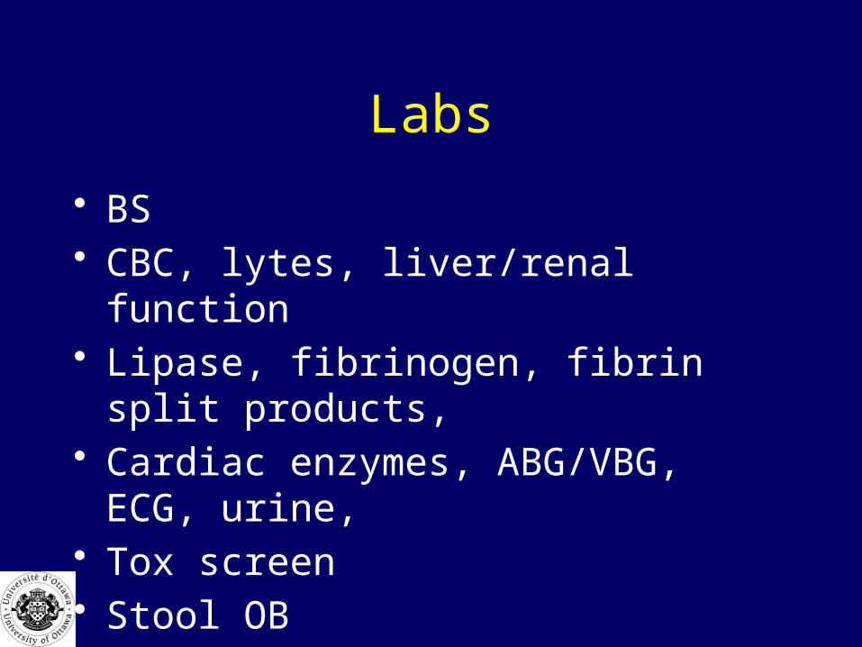
Labs
• BS• CBC, lytes, liver/renal function• Lipase, fibrinogen, fibrin split products, • Cardiac enzymes, ABG/VBG, ECG, urine, • Tox screen • Stool OB
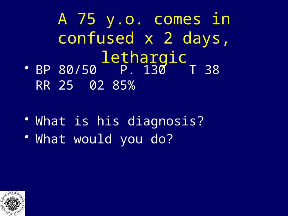
A 75 y.o. comes in confused x 2 days, lethargic
• BP 80/50 P. 130 T 38 RR 25 02 85%
• What is his diagnosis?• What would you do?
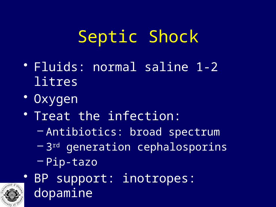
Septic Shock
• Fluids: normal saline 1-2 litres• Oxygen• Treat the infection:
– Antibiotics: broad spectrum– 3rd generation cephalosporins– Pip-tazo
• BP support: inotropes: dopamine
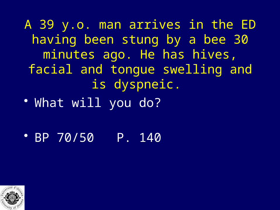
A 39 y.o. man arrives in the ED having been stung by a bee 30 minutes ago. He has hives, facial and tongue swelling and is dyspneic.
• What will you do?
• BP 70/50 P. 140

Anaphylaxis• 100 % oxygen• bolus 1-2 litres normal saline• epinephrine 0.3 mg IM q5min
• or 5-15 microgm/min IV with shock
• benadryl 50 mg IV• ranitidine 50 mg IV• solumedrol 125 mg IV• Glucagon 1mg IV if on beta blockers

?
Cardiac Arrest – MCC Objectives
• Causes– Cardiac and noncardiac
• List and interpret critical clinical findings– Pulseless state– Determine etiology
• List and interpret critical investigation• Management plan-CPR and ACLS protocols• Communicate
– Outcome– Breaking bad news– Organ donation– Autopsy request

Cardiac Arrest - Causes
• Cardiac– Coronary artery– Conduction
• Metabolic: hypo Ca, Mg, K, anorexia• Brady or tachydysrhythmia
– Myocardium• Hereditary: cardiomyopathy• Acquired: LVH, Valve disease, myocarditis
Cardiac Arrest - Causes
• Non Cardiac– Tamponade– PE– Tension– Trauma
A 72 y.o. man complains of chest pain and collapses in the ED
• What are you going to do ?

Sudden Cardiac Arrest
• electrical accident due to ischemia or reperfusion
• 80% ventricular fibrillation or ventricular tachycardia
• 20 % asystole pulseless electrical activity

Mechanism of Fibrillation• ischemia: slows conduction
• adjacent myocardium in various phases of excitation and recovery
• multiple depolarizing reentrant wave fronts
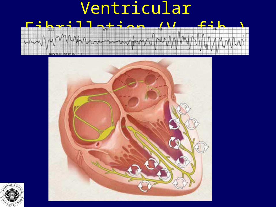
Ventricular Fibrillation (V. fib.)
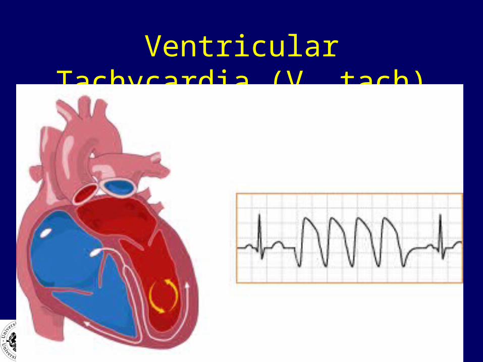
Ventricular Tachycardia (V. tach)
Cardiac Arrest
• What are the key actions that are required to improve survival from cardiac arrest?

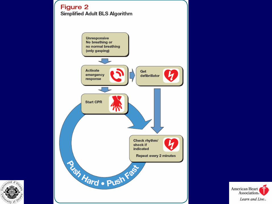
Chain of Survival
Major Changes of BLS
• Change in CPR sequence to :
–C-A-B rather than A-B-C...
• Begin with chest compressions !!!

Major Changes of BLS
• Trained Layperson or Health Care Provider– 30 compressions, 2 breaths
• Untrained layperson– Compression only CPR acceptable – ‘Hands Only’ CPR
Major Changes of BLS
• Elimination of : “Look, Listen & Feel” for breathing...
• …except for hypoxic arrest
• Pulse check for Health Care Providers < 10 sec.
High Quality C.P.R.
• Compression : Ventilation ratio (30 : 2)– Until advanced airway
• Minimize interruptions in CPR
• Push Hard & Fast : 2 inches / 100/ min.
• Full chest recoil-lift hands off chest
• Change compressors q2min
Airway Management
• BVM (Bag-Valve-Mask)– Avoid hyperventilation! – 8 – 10 breaths / min. interposed with CPR
• Secure Airway & Confirm Placement – No need to pause compressions!
• Advanced airway: LMA, ETT
– ETCO2 monitoring !

What are the only things that should interrupt CPR?
• Rhythm and pulse check• Ventilation (if advanced airway not present)• Advanced airway and intubation• Defibrillation

A patient you are talking to suddenly becomes unresponsive

Is this
A) Normal sinus rhythm
B) Ventricular tachycardia
C) Ventricular fibrillation
D) Can I call a friend?
The crash cart arrives, you grab the paddles and have a quick-look
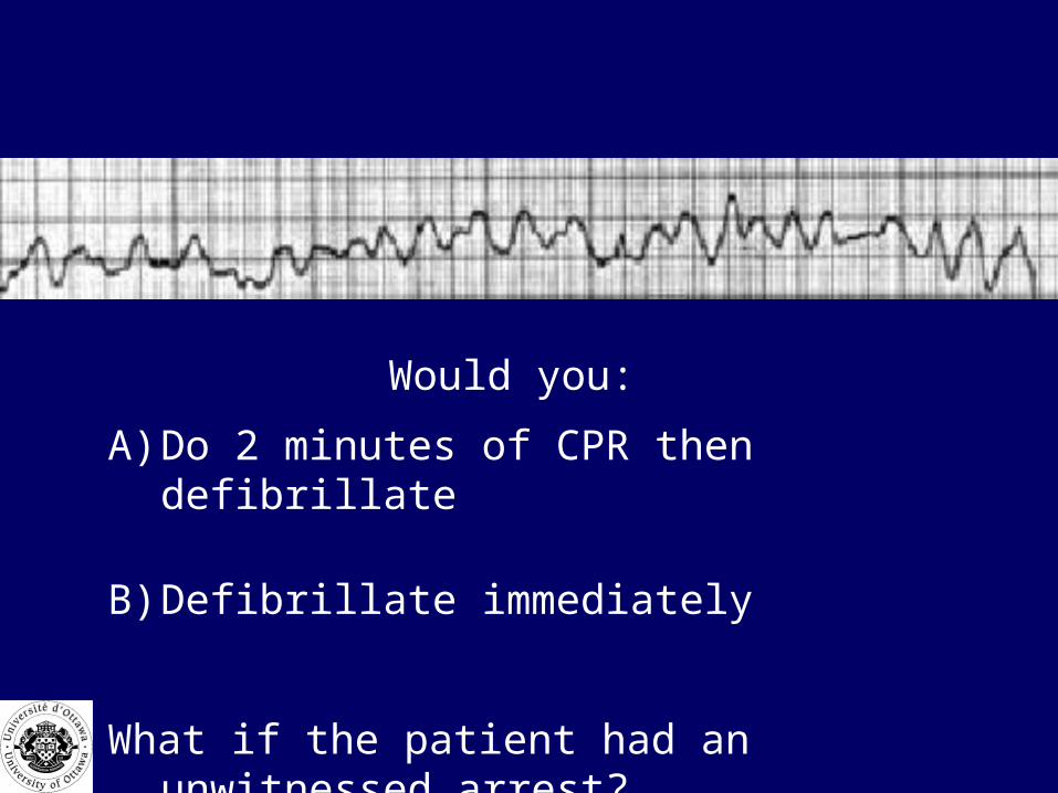
Would you:
A) Do 2 minutes of CPR then defibrillate
B) Defibrillate immediately
What if the patient had an unwitnessed arrest?
New CPR Guidelines
• Even with unwitnessed arrest….
• Once V fib is recognized…shock ASAP

Shock Protocol
• Shorten interval between compressions and shocking
– improves shock success.
• After shock delivery, resume CPR immediately– Don’t delay chest compressions for rhythm or pulse check

How many times do you defibrillate?

No Change in Recommendations
• 1 shock then resume CPR
If you can’t get an IV, what other route can you give drugs?
• Intraosseus
• Endotrachael: (not a good route)
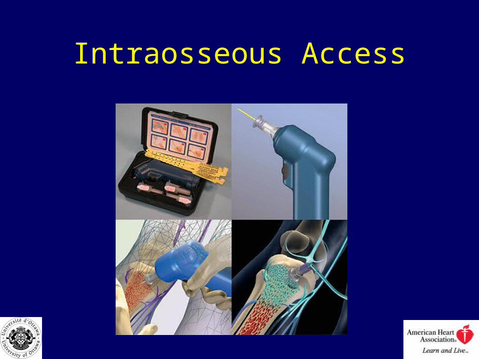
Intraosseous Access
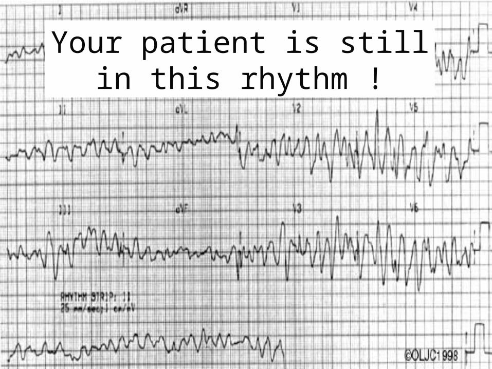
Your patient is still in this rhythm !

Cardiac Arrest MedicationsNo Significant Change in New Guidelines
• Vasopressors– Epinephrine
• 1 mg q3-5 min
– Vasopressin• 40 units• May replace 1st or 2nd dose of epinephrine
Cardiac Arrest MedicationsNo Significant Change in New Guidelines
• Antiarrythmics• Don’t revert v fib.• Work by preventing V.Fib,
– Amiodarone – – Procainamide– Lidocaine– Magnesium Sulfate

Amiodarone
• First line antidysrhymthmic
• 300 mg IV bolus
• May give 2nd dose: 150 mg

Lidocaine
• 1.5 mg/kg • Repeat x 1 prn.

• The paramedics brings in a 56 y.o. man who arrested at home, was successfully defibrillated but remains comatose and intubated. BP. 100/70, P. 75 NSR
• What other treatment options are available to you to increase survival?
Therapeutic Hypothermia for Cardiac Arrest
• Cool to 32-34°C x 24 hrs• Criteria:
– adult patient prehospital cardiac (v.fib) arrest .
– Spontaneous circulation BP > 90– Patient remains comatose and intubated

?
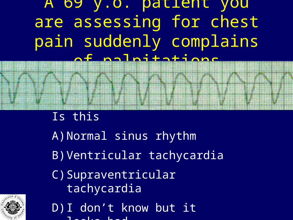
A 69 y.o. patient you are assessing for chest pain suddenly complains of
palpitations
Is this
A) Normal sinus rhythm
B) Ventricular tachycardia
C) Supraventricular tachycardia
D) I don’t know but it looks bad

A 69 y.o. patient you are assessing for chest pain suddenly complains of
palpitations
Is this
A) Normal sinus rhythm
B) Ventricular tachycardia
C) Supraventricular tachycardia
D) I don’t know but it looks bad
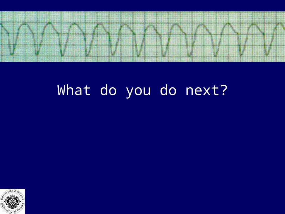
What do you do next?
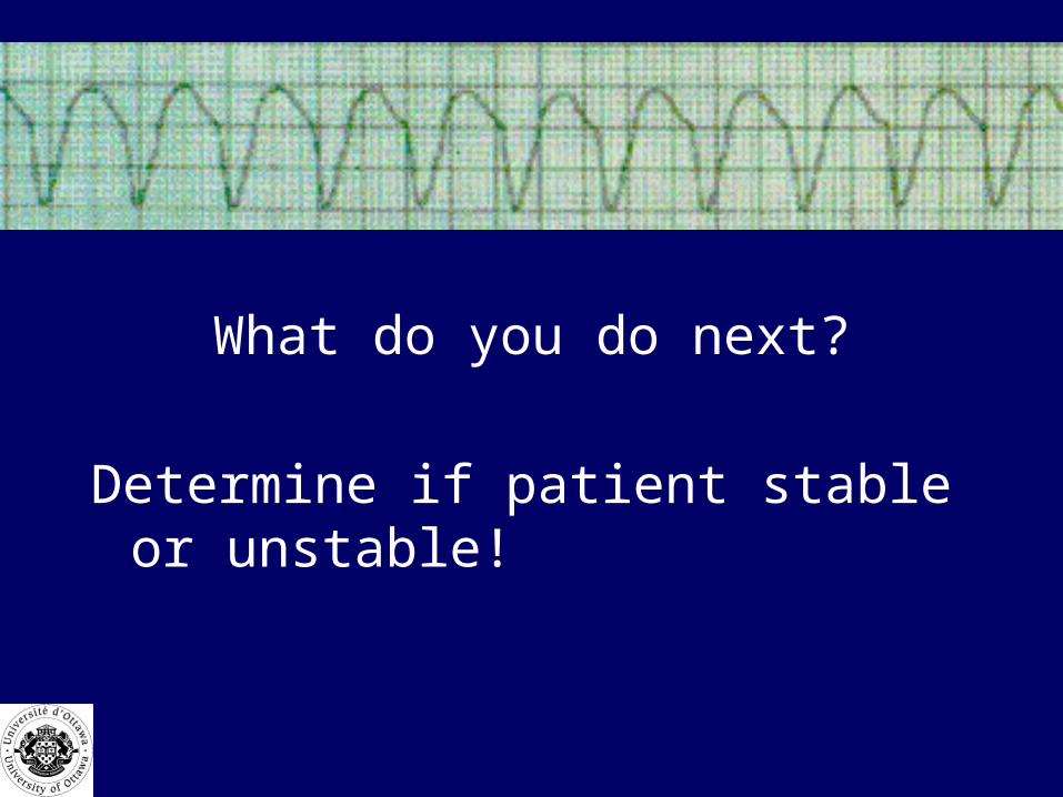
What do you do next?
Determine if patient stable or unstable!
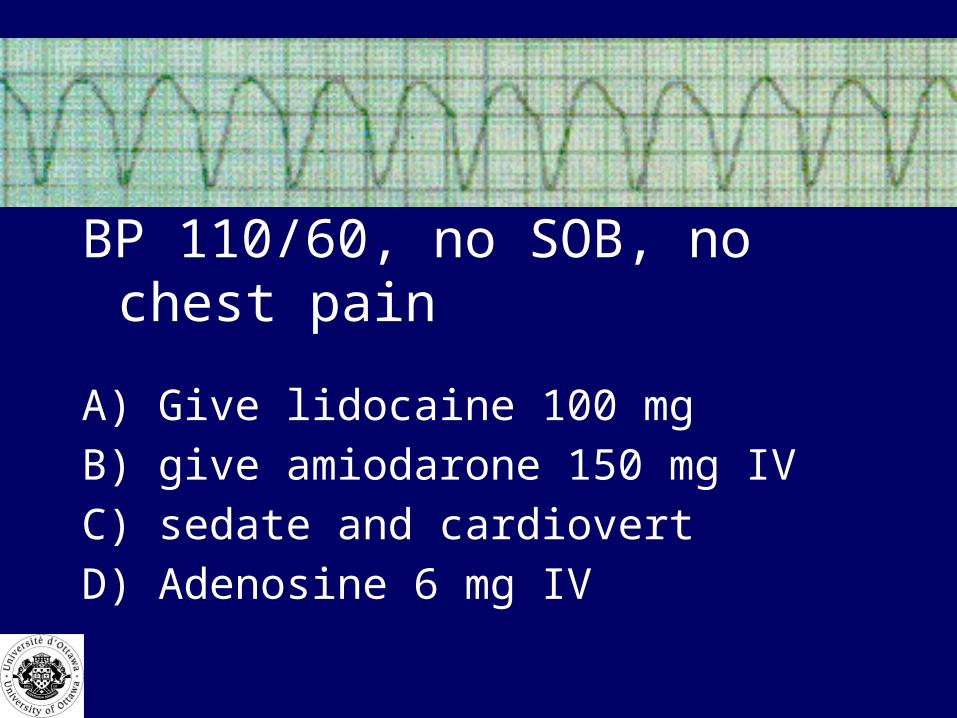
BP 110/60, no SOB, no chest pain
A) Give lidocaine 100 mg
B) give amiodarone 150 mg IV
C) sedate and cardiovert
D) Adenosine 6 mg IV
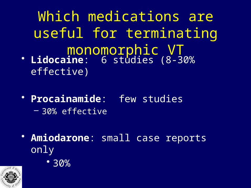
Which medications are useful for terminating monomorphic VT
• Lidocaine: 6 studies (8-30% effective)
• Procainamide: few studies– 30% effective
• Amiodarone: small case reports only• 30%

Amiodarone in V. Tach
• 150 mg over 10 min• may repeat up to 5-7mg/kg• infusion: 1 mg/min for 1st 6 hours
»then 0.5 mg/min
Lidocaine in V. Tach
• 1.5 mg/kg bolus• 2nd and 3rd dose: 0.75 mg/kg q 5 min
• Total maximum: 3 mg/kg

Ventricular Tachycardia
• Do not give multiple antidysrhythmics if one has failed (pro-arrhythmic effects)
• pick one antidysrhythmic, if it fails, go to electrical cardioversion.

Ventricular Tachycardia-Summary
• If stable: can try drugs but cardioversion best choice
• If unstable: cardiovert (synchronized)
• If pulseless: defibrillate
• Drugs rarely effective
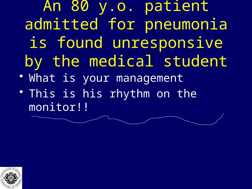
An 80 y.o. patient admitted for pneumonia is found unresponsive
by the medical student
• What is your management• This is his rhythm on the monitor!!
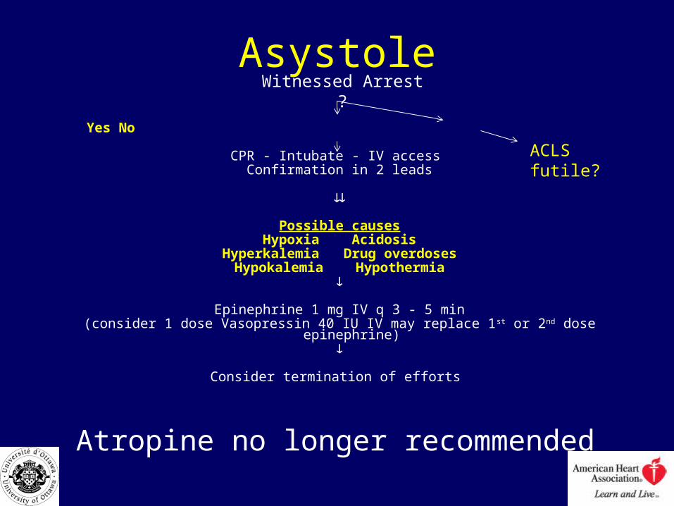
Asystole
Yes No
CPR - Intubate - IV access Confirmation in 2 leads
Possible causesHypoxia Acidosis
Hyperkalemia Drug overdosesHypokalemia Hypothermia
Epinephrine 1 mg IV q 3 - 5 min(consider 1 dose Vasopressin 40 IU IV may replace 1st or 2nd dose epinephrine)
Consider termination of efforts
Atropine no longer recommended
Witnessed Arrest ?
ACLS futile?
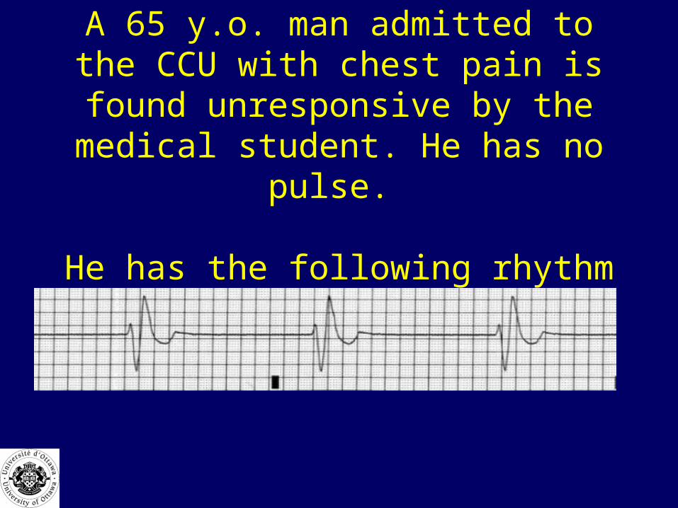
A 65 y.o. man admitted to the CCU with chest pain is found unresponsive by the
medical student. He has no pulse.
He has the following rhythm
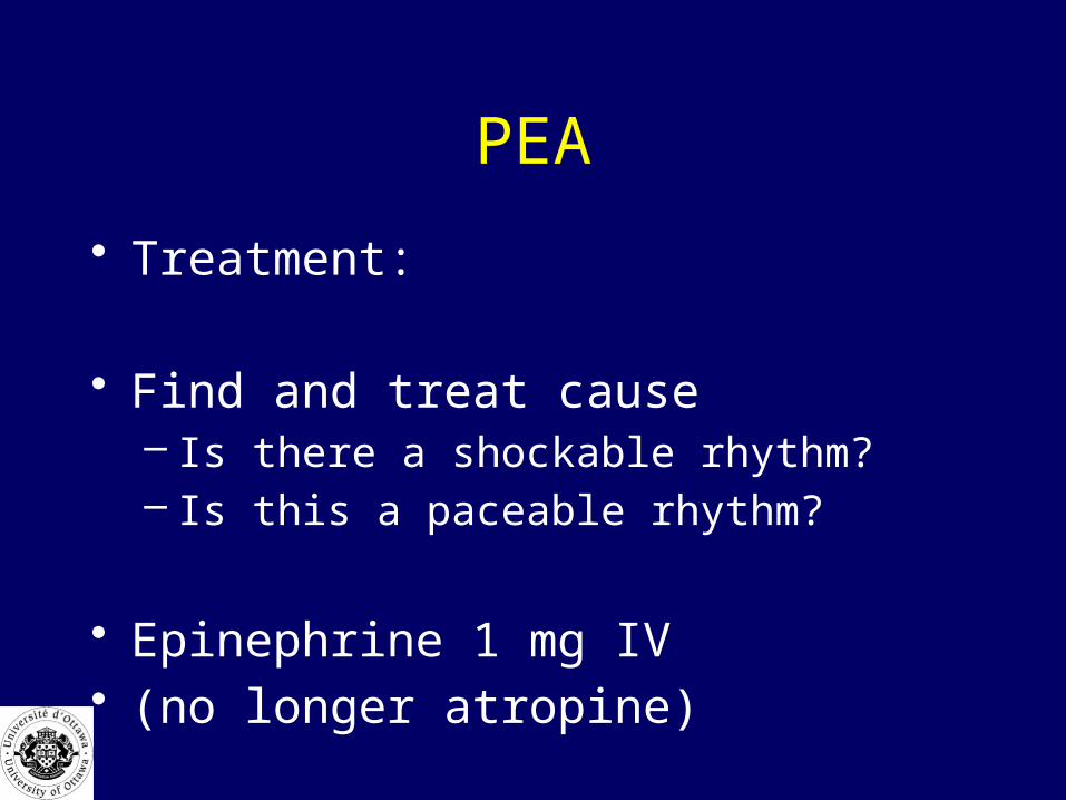
PEA
• Treatment:
• Find and treat cause– Is there a shockable rhythm?– Is this a paceable rhythm?
• Epinephrine 1 mg IV• (no longer atropine)

PEA
• Consider causes:– 5 H’s :– hypovolemia, hypoxia, H ion, hyper/hypo K,
– 5 T’s: – tamponade, tension pneumo, thrombosis-
coronary or pulmonary, tablets OD
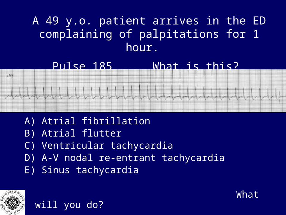
A 49 y.o. patient arrives in the ED complaining of palpitations for 1 hour.
Pulse 185 What is this?
A) Atrial fibrillationB) Atrial flutterC) Ventricular tachycardiaD) A-V nodal re-entrant tachycardiaE) Sinus tachycardia
What will you do?

SVT
STABLE UNSTABLE CARDIOVERSION VAGAL MANOEUVRES Class 1 Verapamil 2.5 – 5 MG I.V. over 2 min or Diltiazem 20 mg IV over 2 min) or Adenosine 6 mg IV then 12 mg if needed RAPID PUSH or Metoprolol 5 mg IV repeat x 2 prn
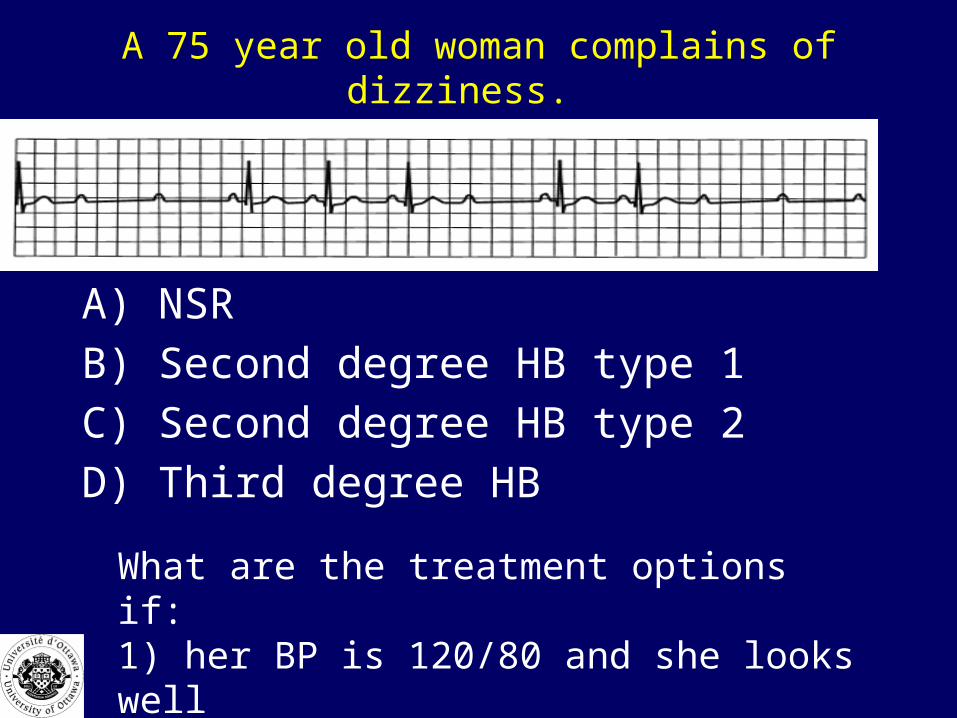
A 75 year old woman complains of dizziness.
A) NSR
B) Second degree HB type 1
C) Second degree HB type 2
D) Third degree HB
What are the treatment options if:1) her BP is 120/80 and she looks well2) her pulse was 45, BP 70/30 and she looks ill
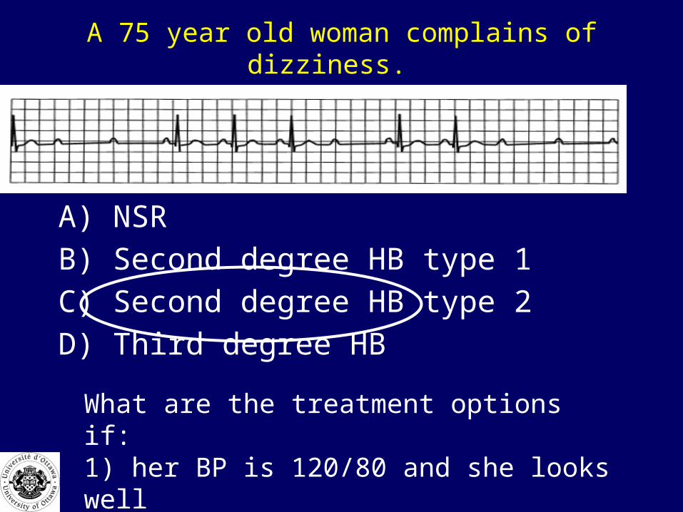
A 75 year old woman complains of dizziness.
A) NSR
B) Second degree HB type 1
C) Second degree HB type 2
D) Third degree HB
What are the treatment options if:1) her BP is 120/80 and she looks well2) her pulse was 45, BP 70/30 and she looks ill
Second degree HB type ll
• Dysfunctional His Purkinje system
can lead to complete heart block
• If stable, send to monitored bed, and arrange permanent transvenous pacer
• If unstable: external pacing, or dopamine or epinephrine infusion.
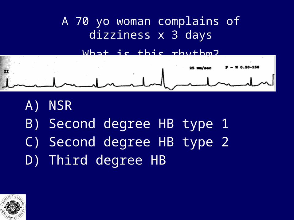
A 70 yo woman complains of dizziness x 3 days
What is this rhythm?
A) NSR
B) Second degree HB type 1
C) Second degree HB type 2
D) Third degree HB

A 70 yo woman complains of dizziness x 3 days
What is this rhythm?
A) NSR
B) Second degree HB type 1
C) Second degree HB type 2
D) Third degree HB
Would 1 mg of epinephrine be appropriate if her BP was 60/40
A) Agree
B) Disagree
BradycardiaWhen to Treat ?
• Symptomatic: chest pain, SOB, hypotension
• Therapy:– atropine 0.5-1 mg (max total 3 mg)– transcutaneous pacemaker OR– dopamine 5-20 microgm/kg/min OR– epinephrine 2-10 microgm/min
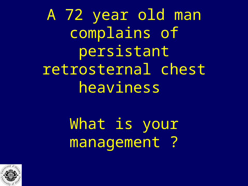
A 72 year old man complains of persistant retrosternal chest
heaviness
What is your management ?
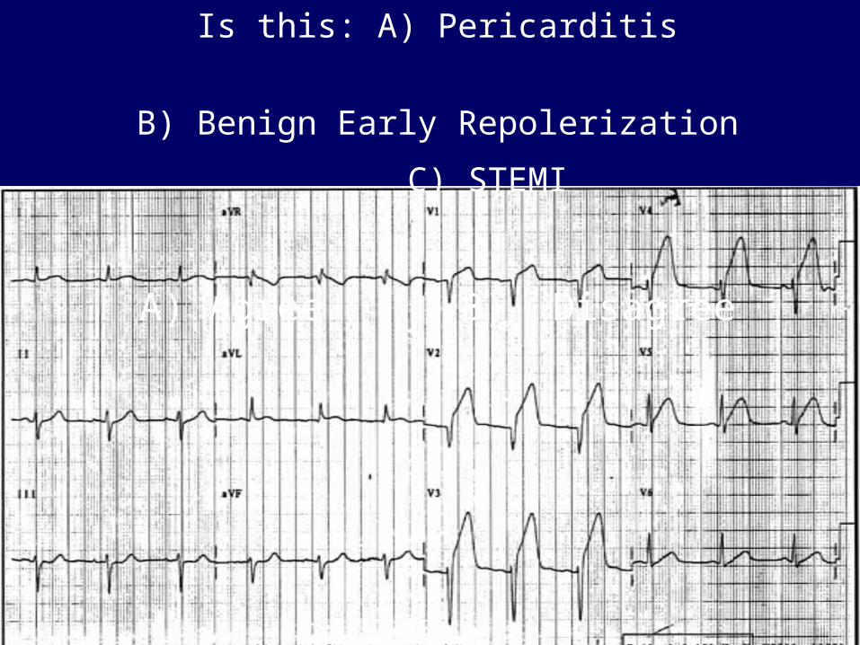
Is this: A) Pericarditis
B) Benign Early Repolerization
C) STEMI
A) Agree B) Disagree
Is this: A) Pericarditis
B) Benign Early Repolerization
C) STEMI
A) Agree B) Disagree

Myocardial InfarctionWhat can you do?
• MONA– ASA 160 mg chew– Oxygen (if sat < 95%)– nitrates sublingual or IV– morphine 2-3 mg prn
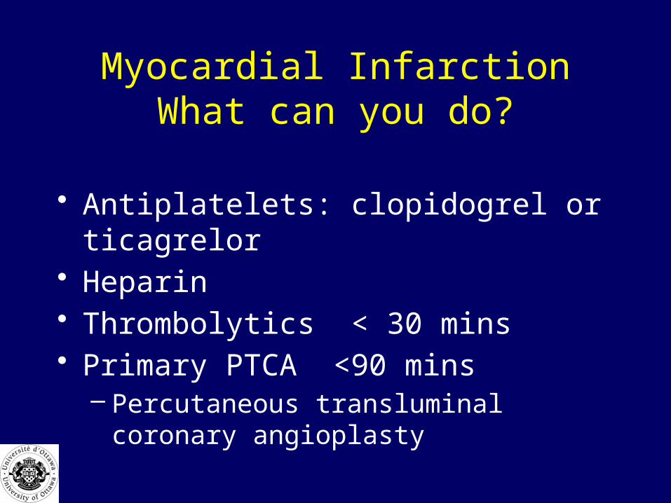
Myocardial InfarctionWhat can you do?
• Antiplatelets: clopidogrel or ticagrelor• Heparin• Thrombolytics < 30 mins• Primary PTCA <90 mins
– Percutaneous transluminal coronary angioplasty

An 80 year old man is being treated in hospital for pneumonia.
He is found VSA at 0300. His rhythm shows asystole.
How long are you required to perform CPR for?
CPR and ACLS
Purpose: treatment of sudden unexpected death.

When Not To Initiate CPR• CPR is inappropriate and ineffective for
medical problems where death is neither sudden or unexpected
• don’t offer CPR as an option to patients or families if it is not medically indicated
• communicate openly

When to Discontinue CPR
• Judgement that patient is unresuscitatable
• Variables: – down time, rhythm, age, premorbid conditions– advance directives
You have just finished a 45 minute unsuccessful resuscitation attempt on a
42 y.o. man. His wife is anxiously waiting.
How do you tell her that her husband has died?
How do you make it less stressful on the survivors when a sudden unexpected
death has occurred.
Sudden Unexpected Death
• Develop multidisciplinary approach• Develop intervention strategy
• Contacting Survivors– Avoid disclosure on the phone– meet family at a specific site
CMAJ 1993 149(10) 1445-1451

Sudden Unexpected Death
• Arrival of Survivors– met by RN, or Social Worker– updated regularly
Should the family be brought to the bedside
if the resuscitation attempt is ongoing ?
Sudden Unexpected Death
• Notificiation of Death– obtain all information prior to meeting– quiet room, have RN also there– sit next or across from closest relative– explain in lay terms sequence of events– use the words dead or died– express condolences– answer questions now or later
Sudden Unexpected Death• Grief Response
– private time
• Viewing Deceased– encourage family– clean patient and remove equipment if possible
• Conclusion– return valuables, address concerns– give family permission to leave

?