Embed Size (px)
Citation preview

JOURNAL OF MEDICALCASE REPORTS
Ball et al. Journal of Medical Case Reports 2013, 7:5http://www.jmedicalcasereports.com/content/7/1/5
CASE REPORT Open Access
Avoidance of total abdominal wall loss despitetorso soft tissue clostridial myonecrosis: a casereportChad Geoffrey Ball*, Jean-Francois Ouellet, Ian Bruce Anderson and Andrew Wallace Kirkpatrick
Abstract
Introduction: Clostridial necrotizing soft tissue infections are often fatal. Myonecrosis of the torso is a particularlylethal combination given the classic need for radical debridement of the abdominal and thoracic walls, andtherefore total exposure of the intraperitoneal and intrathoracic viscera. This case is unusual do to our ability topreserve anatomical separation between the viscera and the atmosphere.
Case presentation: We present a 42-year-old Caucasian man with obesity and diabetes who developed clostridialmyonecrosis of his right torso following a mesenteric lymph node biopsy. This required an aggressive debridement(sparing subcutaneous flaps and internal oblique aponeurosis) followed by reconstruction of his right hemi-torsowith a biologic prosthesis to prevent subsequent hernia formation.
Conclusion: Although basic principles associated with radical debridement were maintained, a full thickness torsowall resection was avoided. This provided reconstruction advantages that included endogenous subcutaneous flapcoverage, separation of the peritoneal cavity by the internal oblique aponeurosis, and prevention of a subsequenthernia below the arcuate line. This technique would be of interest to any surgeon or clinician who treats patientswith life-threatening torso soft tissue infections.
Keywords: Biologic prosthesis, Hernia, Necrotizing soft tissue infection
IntroductionNecrotizing soft tissue infections comprise a broadspectrum of infectious processes that include, but arenot limited to, necrotizing cellulitis, necrotizing fasciitis,and myonecrosis. The classification of these entities isbased on the extent of soft tissue involvement as well asthe depth of infection [1]. Although the bacteria thatcause infections often differ, the general approach totreatment is similar and includes: aggressive resuscita-tion, broad-spectrum antimicrobial therapy, physiologicsupport, and immediate radical surgical debridement [2].The extent of surgical debridement is dictated by theneed to achieve margins with normal appearing tissueand vigorous bleeding. This is particularly problematicin cases of trunk myonecrosis because of the require-ment for complete abdominal and/or thoracic wall
* Correspondence: [email protected] of Surgery, University of Calgary, Foothills Medical Centre,1403-29 St. N.W., Calgary, AB T2N 2T9, Canada
© 2013 Ball et al.; licensee BioMed Central LtdCommons Attribution License (http://creativecreproduction in any medium, provided the or
resection, and subsequent exposed intraperitoneal andintrathoracic organs.
Case presentationA 42-year-old, obese, Caucasian male plumber was re-ferred for a mesenteric lymph node biopsy to rule outlymphoma. His medical comorbidities included recenttype 2 diabetes, dyslipidemia, and chronic back pain.Physical examination and cross-sectional imaging con-firmed abnormal lymphadenopathy limited to the smallbowel mesentery. A laparoscopic procedure, convertedto 10cm laparotomy, obtained an appropriate nodal exci-sion. The patient began to exhibit increasing oxygenrequirements and abdominal discomfort 48 hours afterthe procedure. His physical examination remained other-wise unremarkable. Computed tomography identifiedmassive right torso soft tissue gas extending from thecostal margin to inguinal canal (midline to the back)(Figure 1).
. This is an Open Access article distributed under the terms of the Creativeommons.org/licenses/by/2.0), which permits unrestricted use, distribution, andiginal work is properly cited.
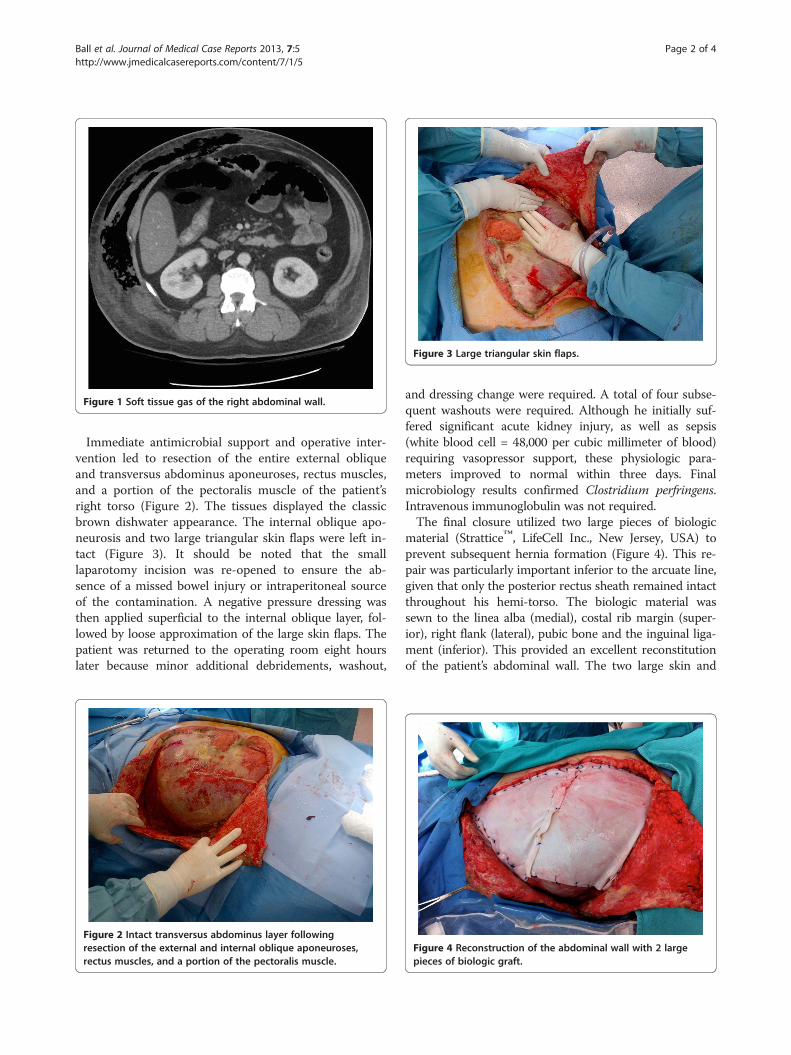
Figure 1 Soft tissue gas of the right abdominal wall.
Figure 3 Large triangular skin flaps.
Ball et al. Journal of Medical Case Reports 2013, 7:5 Page 2 of 4http://www.jmedicalcasereports.com/content/7/1/5
Immediate antimicrobial support and operative inter-vention led to resection of the entire external obliqueand transversus abdominus aponeuroses, rectus muscles,and a portion of the pectoralis muscle of the patient’sright torso (Figure 2). The tissues displayed the classicbrown dishwater appearance. The internal oblique apo-neurosis and two large triangular skin flaps were left in-tact (Figure 3). It should be noted that the smalllaparotomy incision was re-opened to ensure the ab-sence of a missed bowel injury or intraperitoneal sourceof the contamination. A negative pressure dressing wasthen applied superficial to the internal oblique layer, fol-lowed by loose approximation of the large skin flaps. Thepatient was returned to the operating room eight hourslater because minor additional debridements, washout,
Figure 2 Intact transversus abdominus layer followingresection of the external and internal oblique aponeuroses,rectus muscles, and a portion of the pectoralis muscle.
and dressing change were required. A total of four subse-quent washouts were required. Although he initially suf-fered significant acute kidney injury, as well as sepsis(white blood cell = 48,000 per cubic millimeter of blood)requiring vasopressor support, these physiologic para-meters improved to normal within three days. Finalmicrobiology results confirmed Clostridium perfringens.Intravenous immunoglobulin was not required.The final closure utilized two large pieces of biologic
material (Strattice™, LifeCell Inc., New Jersey, USA) toprevent subsequent hernia formation (Figure 4). This re-pair was particularly important inferior to the arcuate line,given that only the posterior rectus sheath remained intactthroughout his hemi-torso. The biologic material wassewn to the linea alba (medial), costal rib margin (super-ior), right flank (lateral), pubic bone and the inguinal liga-ment (inferior). This provided an excellent reconstitutionof the patient’s abdominal wall. The two large skin and
Figure 4 Reconstruction of the abdominal wall with 2 largepieces of biologic graft.

Figure 6 Abdominal wall 12 months after the reconstruction.
Ball et al. Journal of Medical Case Reports 2013, 7:5 Page 3 of 4http://www.jmedicalcasereports.com/content/7/1/5
soft tissue flaps were then closed (Figure 5). This left athree-cm central area to heal by negative suction therapyand secondary intention.This patient was discharged home 34 days after his ini-
tial excisional biopsy with no evidence of organ failure.His 12-month out-patient follow-up continues to showno obvious hernia or attenuation in his abdominal wall(Figure 6).
DiscussionAlthough up to 80% of necrotizing soft tissue infectionsare polymicrobial, Clostridium species are still associatedwith the classic presentation of gas gangrene myonecrosis[3]. Typically the presence of soft tissue gas, as observedin this patient, offers a grave prognosis [4]. In addition, gasgangrene of the torso carries an even worse outcome giventhe typical requirement for radical full thickness debride-ment that often results in open abdominal and thoraciccavities with exposed viscera [2]. Because of the tremen-dous risks associated with soft tissue coverage and even-tual reconstruction, our patient underwent an alternativestrategy. By maintaining two large, well-vascularized sub-cutaneous flaps as potential coverage, in addition to theinternal oblique aponeurosis as a barrier to the peritonealcavity, we were able to maintain significant reconstructiveoptions. Given the risk inherent in this initial approach,
Figure 5 Closure of skin flaps over the abdominalwall reconstruction.
the patient was returned to the operating room for a sec-ond tissue evaluation only eight hours after the initialdebridement. As a consequence of his radical improve-ment in physiology and tissue quality, we persisted withthis methodology.In a significant number of patients (40%), a source of
the necrotizing soft tissue infections is not readily identi-fiable [5]. In our patient, the initial lymph node biopsywas clearly the index insult; however, the specific sourceof the Clostridium is unknown. The gastrointestinal tractwas not injured and this species is atypical for our hos-pital. We postulate that the preoperative skin prepa-ration was insufficient to remove all the patient’s topicalbacteria given his employment as a sewer plumber. Thisis particularly plausible given the known ability ofC. perfringens to remain quiescent in tissues and theninitiate a clinical infection when minor trauma (lymphnode biopsy) provides an opportunity for growth(diabetes mellitus) [2]. We also believe the lack of skinchanges and crepitus in the initial phases were a resultof the patient’s general obesity. Furthermore, it is clearthat the immunosuppression associated with his diabetesmellitus represented a significant co-factor in the rapidprogression of this disease [2,6]. In addition, his finalconfirmed diagnosis of follicular lymphoma may alsohave negatively impacted his immune status [7].Although our avoidance of a complete resection of all
layers of the patient’s abdominal wall represents a signifi-cant departure from traditional dogma, the principles

Ball et al. Journal of Medical Case Reports 2013, 7:5 Page 4 of 4http://www.jmedicalcasereports.com/content/7/1/5
requiring a normal-appearing margin of tissue were main-tained. Although the use of biologic materials represents asignificant advancement in the field of surgery, this risk-adjusted technique of partial debridement has beenechoed by previous authors in the context of clostridialmyonecrosis [8,9]. Despite this technique, the risk of asubsequent massive ipsilateral abdominal wall hernia wassubstantial. As a result, we felt reconstruction of the ab-dominal wall with a prosthesis was essential. Given thereported improvements in function and durability of bio-logic materials in infected fields, two large pieces wereinserted [10]. There is no current evidence on whichto base the performance of biologics in the setting ofC. perfringens. At a 12-month follow-up however, thepatient’s abdominal wall remains well healed, with nodrainage and no torso asymmetry. This is particularlyimportant inferior to the arcuate line given the naturallythin posterior rectus sheath and aponeurosis components.
ConclusionThe management of this patient represents a significantdeparture from the classic concepts surrounding clostri-dial myonecrosis. Although basic principles associatedwith radical debridement were maintained, a full thicknesstorso wall resection was avoided. This provided recon-struction advantages that included endogenous subcutane-ous flap coverage, separation of the peritoneal cavity bythe internal oblique aponeurosis, and prevention of a sub-sequent hernia below the arcuate line. These principlesmust be individualized to any specific patient.
ConsentWritten informed consent was obtained from the patientfor publication of this case report and accompanyingimages. A copy of the written consent is available forreview by the Editor-in-Chief of this journal.
Competing interestsAll authors declare that they have no competing interest.
Authors’ contributionsCGB, JFO and IBA analyzed and interpreted all patient data. CGB, JFO, IBA,and AWK each made substantial contributions to writing of the entiremanuscript. All authors read and approved the final manuscript.
Received: 13 August 2012 Accepted: 7 November 2012Published: 8 January 2013
References1. Tilkorn DJ, Citak M, Fehmer T, Ring A, Hauser J, Al Benna S, Steinstrasser L,
Roetman B, Steinau HU: Characteristics and differences in necrotizingfasciitis and gas forming myonecrosis: a series of 36 patients.Scand J Surg 2012, 101:51–55.
2. Guo WA, Steinberg SM: Infections of the skin, muscle and soft tissue. InTextbook of Critical Care Medicine. 5th edition. Edited by Fink MP, AbrahamE, Vincent J-L, Kochanek PM. Philadelphia: Elsevier; 2005:44–98.
3. Elliot D, Kuferna JA, Myers RA: The microbiology of necrotizing soft tissueinfections. Am J Surg 2000, 179:361–366.
4. Stevens DL, Aldape MJ, Bryant AE: Life threatening clostridial infections.Anaerobe 2012, 18:254–259.
5. Park H, Copeland C, Henry S, Barbul A: Complex wounds and theirmanagement. Surg Clin North Am 2010, 90:1181–1194.
6. Sakurai J, Nagahama M, Oda M: Clostridium perfringens alpha-toxin:characterization and mode of action. J Biochem 2004, 136:569–574.
7. García-Suárez J, de Miguel D, Krsnik I, Barr-Ali M, Hernanz N, Burgaleta C:Spontaneous gas gangrene in malignant lymphoma: an underreportedcomplication? Am J Hematol 2002, 70:145–148.
8. Phillips J, DM H, Jones RC: Clostridial myonecrosis of the abdominal wall.Management after extensive resection. Am J Surg 1974, 128:436–438.
9. McSwain B, Sawyers JL, Lawler MR Jr: Clostridial infections of theabdominal wall: a review of 10 cases. Ann Surg 1966, 163:859–865.
10. Ouellet JF, Ball CG, Kortbeek JB, Mack LA, Kirkpatrick AW: Bioprostheticmesh use for the problematic thoracoabdominal wall: outcomes inrelation to contamination and infection. Am J Surg 2012, 203:594–597.
doi:10.1186/1752-1947-7-5Cite this article as: Ball et al.: Avoidance of total abdominal wall lossdespite torso soft tissue clostridial myonecrosis: a case report. Journal ofMedical Case Reports 2013 7:5.
Submit your next manuscript to BioMed Centraland take full advantage of:
• Convenient online submission
• Thorough peer review
• No space constraints or color figure charges
• Immediate publication on acceptance
• Inclusion in PubMed, CAS, Scopus and Google Scholar
• Research which is freely available for redistribution
Submit your manuscript at www.biomedcentral.com/submit


