Embed Size (px)
Citation preview

PERIODONTAL FLAPS
DR JEBIN,MDS.,D.ICOI

DEF:
“A periodontal flap is a section of gingiva and/or mucosa surgically separated from the underlying tissues to provide visibility and access to the bone and root surface.

INDICATIONS:
• Irregular bony contours
• Deep craters
• Pockets on teeth in which a complete removal of root irritants is not clinically possible
• Grade II or III furcation involvement
• Root resection / hemisection
• Intrabony pockets on distal areas of last molars
• Persistent inflammation in areas with moderate to deep pockets.
CONTRAINDICATIONS:
• Uncontrolled medical conditions such as
‐ unstable angina
‐ uncontrolled diabetes
‐ uncontrolled hypertension
‐myocardial infarction / stroke within 6 months
• Poor plaque control
• High caries rate
• Unrealistic patient expectations or desires

CLASSIFICATION OF FLAPS
Classified based on:
Bone exposure after flap reflection
• Full thickness (mucoperiosteal)
• Partial thickness (mucosal)
Placement of the flap after surgery
• Nondisplaced flaps
• Displaced flaps
Management of the papilla
• Conventional flaps
• Papilla preservation flaps

BASED ON BONE EXPOSURE AFTER REFLECTION
FULL THICKNESS FLAP
• Periosteum is reflected to expose the underlying bone.
• Indicated in resective osseous surgery.
Contraindications :
• Area where treatment for osseous defect with mucogingival problem is not required.
• Thin periodontal tissue with probable osseous dehiscence and osseous fenestration.
• Area where alveolar bone is thin.

PARTIAL THICKNESS FLAP
• Split thickness flap.
• Periosteum covers the bone.
• Indicated when the flap has to be positioned apically.
• When the operator does not desire to expose the bone.

BASED ON FLAP PLACEMENT AFTER SURGERY
• Non displaced flaps:
When the flap is returned and sutured in its original position.
• Displaced flaps:
When the flap is placed apically, coronally or laterally to their original position

DESIGN OF THE FLAP
• Split the papilla (conventional flap) or
• Preserve it (papilla preservation flap).

BASED ON MANAGEMENT OF THE PAPILLA
CONVENTIONAL FLAP
The interdental papilla is split beneath the contact point of the two approximating teeth to allow reflection of buccal and lingual flaps.

The conventional flap is used when
• The interdental spaces are too narrow
• When the flap is to be displaced.
Conventional flaps include the
• The Modified Widman flap,
• The undisplaced flap,
• The apically displaced flap, and
• The flap for regenerative procedures.

PAPILLA PRESERVATION FLAP
The papilla preservation flap incorporates the entire papilla in one of the flaps

Two basic flap designs, those with and those without vertical releasing incisions:
• Envelope Flap: A flap that is released in a linear fashion at the gingival margin but has no vertical releasing incision(s).
• Pedicle Flap: If two vertical releasing incisions are included in the flap design.
• Triangular Flap: If one vertical releasing incision is included in the flap design.
Envelope Flap Pedicle Flap Triangular Flap

• The major blood supply to a flap is at its base and travels in an apical to coronal direction.
• Recommended flap length (height)‐to‐base ratio should be no greater than 2:1

INCISIONS
• Horizontal incisions.
• Vertical incisions.

HORIZONTAL INCISIONS
• Horizontal incisions are directed along the margin of the gingiva in a mesial or a distal direction.
• Two types of horizontal incisions have been recommended:
A) The internal bevel incision, which starts at a distance from the gingival margin and is aimed at the bone crest, and
B) The crevicular incision, which starts at the bottom of the pocket and is directed to the bone margin.
C) In addition, the interdental incision is performed after the flap is elevated.

INTERNAL BEVEL INCISION
1st incision1º incisionReverse bevel incision11 or 15 surgical scalpelusedStarts at a distance from the gingival margin aiming at the bone crest.

• Removes pocket lining.
• Produces a sharp thin flap margin.
• Starts from a designated area on the gingiva and is directed to an area at or near the crest of the bone.

V arious locations and
angles of internal bevel incision

CREVICULAR INCISION
Also known as secondincision Made from the base of the pocket to the crestof the boneThis incision, togetherwith the initial reversebevel incision, forms a V-shaped wedge ending at or near the crest of bone

PROCEDURE:1) Beak-shaped #12D blade is used2) Periosteal elevator inserted into the initial internal bevel incision3) Flap separated from the bone

INTERDENTAL INCISION
Also known as third incision
To separate the collar ofgingiva that is left around the tooth
Incision made facially lingually & interdentallyconnecting the 2 segments.
Orbans knife is used

VERTICAL INCISIONS
Can be used on one or both ends of the horizontal incision
Must extend beyond the mucogingival line, reaching the alveolar mucosa, to allow for the release of the flap to be displaced
Vertical incisions in the lingual and palatal areas are avoided

This incision should be made at the line angles to prevent splitting of apapilla or incising directly over a radicular surface.

ELEVATION OF THE FLAP
Blunt dissection with periosteal elevator
For reflection of full thickness flap

Sharp dissection with surgical scalpel (#11 or #15)
For reflection of partial thickness flap

• A flap that includes only gingival tissue is referred to as a GINGIVAL FLAP.
• A flap that extends beyond the mucogingival junction to include alveolar mucosa, is a MUCOGINGIVAL FLAP.

FLAP RETRACTION
• Retraction should be passive without any tension.
• Force should not be necessary to keep the flap retracted.
• The edge of the retractor always be kept on bone.

• Continuous flap retraction for long periods is also is not advised. Such a practice will desiccate the soft tissue and bone causing a delay in wound healing.
• When the flap is retracted, the surgical assistant should frequently irrigate the surgical field with sterile saline, to keep the tissues moistened, to reduce contamination, and to improve visibility.

OPEN FLAP DEBRIDEMENT
• provide access to root surfaces and marginal alveolar bone.

FLAP POSITIONING
surgical flaps may be repositioned, apically positioned, coronally positioned or laterally positioned
• An apically positioned flap is one that is apically displaced from its original position to the level of the alveolar crest or about 1mm coronal to the crest.
• The coronally positioned flap is advanced coronal to its original position.

FLAP TECHNIQES FOR POCKET THERAPY
• Increase accessibility to root deposits
• Eliminate or reduce pocket depth by resection of the pocket wall
• Expose the area to perform regenerative methods
FLAP TECHNIQUES
• The modified widman flap
• The undisplaced flap
The palatal flap
• The apically displaced flap
• Flaps for regenerative surgery
The papilla preservation flap
Conventional flap for regenerative surgery
• Distal molar surgery

THE ORIGINAL WIDMAN FLAP
• By Leonard Widman (1918)
• Widman described a mucoperiosteal flap designed to remove the pocket epithelium and the inflamed connective tissue, there by facilitating optimal cleaning of the root surfaces.
ADVANTAGES:
• Less discomfort for the patient, since healing occurred by primary intention.
• It was possible to reestablish a proper contour of the alveolar bone in sites with angular bony defects.

STEP 1 STEP 2
STEP 3 STEP 4
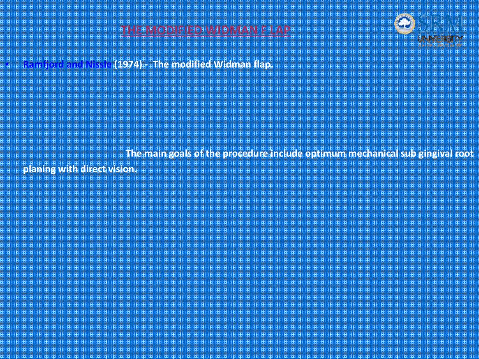
THE MODIFIED WIDMAN F LAP
• Ramfjord and Nissle (1974) ‐ The modified Widman flap.
The main goals of the procedure include optimum mechanical sub gingival root
planing with direct vision.

INDICATIONS:
• Especially effective with pocket depths of 5‐7 mm.
CONTRAINDICATIONS:
• Lack of or very thin and narrow attached gingiva can render the technique difficult, because a narrow band of attached gingiva does not permit the initial scalloped incision (internal gingivectomy)..
• osseous surgical procedure
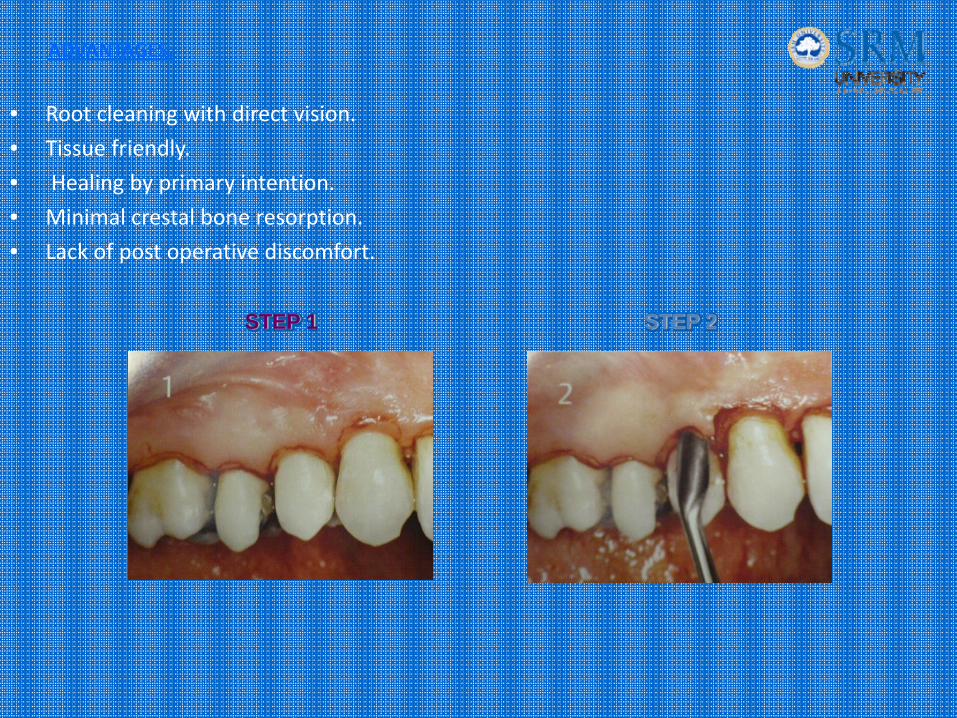
ADVANTAGES:
• Root cleaning with direct vision.
• Tissue friendly.
• Healing by primary intention.
• Minimal crestal bone resorption.
• Lack of post operative discomfort.
STEP 1 STEP 2

STEP 3 STEP 4
STEP 5 STEP 6

THE UNDISPLACED FLAP
• Currently, it is the most commonly performed type of periodontal surgery.
• It differs from the modified Widman flap in that the soft tissue pocket wall is removed with the initial incision; thus it may be considered an internal bevel gingivectomy.

Pre-operative Facial & Lingual Views
Internal Bevel Incisions – Facial & Palatal Aspects
Flap Elevated Showing Osseous Defects

Osseous surgery has been performed
Flaps Placed In Their Original Site And Sutured
Post Operative Results

PARTIAL‐THICKNESS PALATAL FLAP SURGERY
• Developed by Staffileno and improved by Corn et al.
• elimination of periodontal pockets where thick palatal tissues occur.
ADVANTAGES:
• Flap thickness may be adjusted.
• Palatal flap may be adapted to the proper position.
• Better postoperative gingival morphology is possible with a thin flap design.
• Treatments may be combined (osseous resection and wedge procedure).
• Rapid healing.
• Easy management of palatal tissue.
• Minimal damage to palatal tissue.

Outline of primary incision

. Primary incision Thin primary flap preparation.

. Secondary incision Secondary flap removal Suture

THE APICALLY DISPLACED FLAP
• It can be used for both pocket eradication as well as widening the zone of attached gingiva.
• It can be a full thickness (mucoperiosteal) or a split thickness (mucosal) flap.
ADVANTAGES:
• Eliminates periodontal pocket.
• Preserves attached gingiva and increases its width.
• Establishes gingival morphology facilitating good hygiene.
• Ensures healthy root surface necessary for the biologic width on alveolar margin and lengthened clinical crown.

DISADVANTAGES:
• May cause esthetic problems due to root exposure.
• May cause attachment loss due to surgery.
• May cause hypersensitivity.
• May increase the risk of root caries.
• Unsuitable for treatment of deep periodontal pockets.
• Possibility of exposure of furcations and roots, which complicates post operative supragingival plaque control.
CONTRAINDICATIONS:
• Periodontal pockets in severe periodontal disease.
• Periodontal pockets in areas where esthetics is critical.
• Deep intrabony defects.
• Patient at high risk for caries.
• Severe hypersensitivity.
• Tooth with marked mobility and severe attachment loss.
• Tooth with extremely unfavourable clinical crown / Root ratio.

Facial And Lingual Preoperative Views
Facial And Lingual Flaps Elevated
After Debridement Of The Areas

Sutures In Place
Healing After 1 Week Healing After 2 Months
Preoperative Postoperative

PRE-TREATMENT
POST-TREATMENT
BEFORE OSSEOUS RESECTION
AFTER OSSEOUS RESECTION FLAP APICALLY POSITIONED AND SUTURED

FLAPS FOR REGENERATIVE SURGERY
Two flap designs are available for regenerative surgery:
• The papilla preservation flap &
• The conventional flap with only crevicular incisions.
Adequate interdental space Interdental space is very narrow
Papilla preservation flap Conventional flap with only crevicular incisions

THE PAPILLA PRESERVATION FLAP
Entire papilla is incorporated into one of the flaps.
INDICATIONS:
• Where esthetics is of concern.
• Where bone regeneration techniques are attempted.


CONVENTIONAL FLAP FOR REGENERATIVE SURGERY
In the conventional flap operation, the incisions for the facial and the lingual or palatal flap reach the tip of the interdental papilla, thereby splitting the papilla into a facial half and a lingual or palatal half.
INDICATIONS:
• When the interdental areas are too narrow to permit the preservation of flap.
• When there is a need for displasing flaps.

DISTAL MOLAR SURGERY
• Treatment of periodontal pockets on the distal surface of terminal molars is often complicated by the presence of bulbous fibrous tissue over the maxillary tuberosity or prominent retromolar pads in the mandible.
• Operations for this purpose were described by Robinson and Braden
Impaction Of A Third Molar Distal To A Second Molar
Little Or No Bone Distal To The Second Molar.
Often Leads To A Vertical Osseous Defect Distal To The Second Molar.

A typical incision design for a surgical procedure distal to themaxillary second molar.

• Incision designs for surgical procedures distal to the mandibular second molar.
• The incision should follow the areas of greatest attached gingiva and underlying bone.

TRIANGULAR DISTAL WEDGE:• Triangular wedge incisions are placed creating the apex of the triangle close to the hamular notch
and the base of the triangle next to the distal surface of the terminal tooth.

LINEAR DISTAL WEDGE:
• The linear distal wedge incorporates two parallel incisions over the crest of the tuberosity that extend from the proximal surface of the terminal molar to the hamular notch area.
• The distance between the two linear incisions is determined by the thickness of the tissues

DISTAL POCKET ERADICATION PROCEDURE WITH THE INCISION DISTAL TO THE MOLAR
SCALLOPED INCISION AROUND THE REMAINING TEETH
FLAP REFLECTED AND THINNED AROUND THE DISTAL INCISION

FLAP IN POSITION BEFORE SUTURING. IT SHOULD BE CLOSELY APPROXIMATED
FLAP SUTURED BOTH DISTALLY AND OVER THE REMAINING SURGICAL AREA


HEALING AFTER FLAP SURGERY
• Immediately after suturing (0 to 24 hours), established by a blood clot, which consists of a fibrin reticulum with many polymorphonuclear leukocytes, erythrocytes, debris of injured cells, and capillaries at the edge of the wound.
• One to 3 days after flap surgery, the space between the flap and the tooth or bone is thinner, and epithelial cells migrate over the border of the flap
• One week after surgery‐ The blood clot is replaced by granulation tissue derived from the gingival connective tissue, the bone marrow, and the periodontal ligament.

• Two weeks after surgery, collagen fibers begin to appear parallel to the tooth surface. Union of the flap to the tooth is still weak, owing to the presence of immature collagen fibers, although the clinical aspect may be almost normal.
• One month after surgery, a fully epithelialized gingival crevice with a well‐defined epithelial attachment is present. There is a beginning functional arrangement of the supracrestal fibers.

THANK “U”