Embed Size (px)
Citation preview
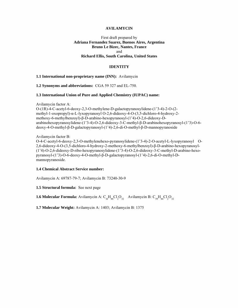
AVILAMYCIN
First draft prepared by Adriana Fernandez Suarez, Buenos Aires, Argentina
Bruno Le Bizec, Nantes, France and
Richard Ellis, South Carolina, United States
IDENTITY 1.1 International non-proprietary name (INN): Avilamycin 1.2 Synonyms and abbreviations: CGA 59 327 and EL-750. 1.3 International Union of Pure and Applied Chemistry (IUPAC) name: Avilamycin factor A: O-(1R)-4-C-acetyl-6-deoxy-2,3-O-methylene-D-galactopyranosylidene-(1’3-4)-2-O-(2- methyl-1-oxopropyl)-α-L-lyxopyranosyl O-2,6-dideoxy-4-O-(3,5-dichloro-4-hydroxy-2- methoxy-6-methylbenzoyl)-β-D-arabino-hexopyranosyl-(1’4)-O-2,6-dideoxy-D-arabinohexopyranosylidene-(1’3-4)-O-2,6-dideoxy-3-C-methyl-β-D-arabinohexopyranosyl-(1’3)-O-6-deoxy-4-O-methyl-β-D-galactopyranosyl-(1’4)-2,6-di-O-methyl-β-D-mannopyranoside Avilamycin factor B: O-4-C-acetyl-6-deoxy-2,3-O-methylenehexo-pyranosylidene-(1’3-4)-2-O-acetyl-L-lyxopyranosyl O-2,6-dideoxy-4-O-(3,5-dichloro-4-hydroxy-2-methoxy-6-methylbenzoyl)-β-D-arabino-hexopyranosyl-(1’4)-O-2,6-dideoxy-D-ribo-hexopyranosylidene-(1’3-4)-O-2,6-dideoxy-3-C-methyl-D-arabino-hexo-pyranosyl-(1’3)-O-6-deoxy-4-O-methyl-β-D-galactopyranosyl-(1’4)-2,6-di-O-methyl-D-mannopyranoside. 1.4 Chemical Abstract Service number: Avilamycin A: 69787-79-7; Avilamycin B: 73240-30-9 1.5 Structural formula: See next page 1.6 Molecular Formula: Avilamycin A: C6lH88Cl2O32 Avilamycin B: C59H84Cl2O32
1.7 Molecular Weight: Avilamycin A: 1403; Avilamycin B: 1375

2
Figure 1: Structural formula of main avilamycin components
Melting point: Avilamycin A: 166-169°C Avilamycin B: 179-182°C
OTHER INFORMATION ON IDENTITY AND PROPERTIES Pure active ingredient: Avilamycin is an orthosomycin antibiotic complex produced by the fermentation of Streptomyces viridochromogenes. Orthosomycin antibiotics are divided into two groups: those that contain an aminocyclitol residue and those that are esters of dichlorisoeverninic acid. Avilamycin is in the latter group as are the evernimicins. Avilamycin complies with the following specifications for the composition of the total factor content.
Avilamycin A: Not less than 60% Avilamycin B: Not more than 18%
Avilamycin A + Avilamycin B: Not less than 70%
Other single Avilamycin factors: Not more than 6%
Typical Avilamycin content is 260 mg activity/g.
Sixteen minor factors have been specifically identified. Their molecular and structural formulas are given below.
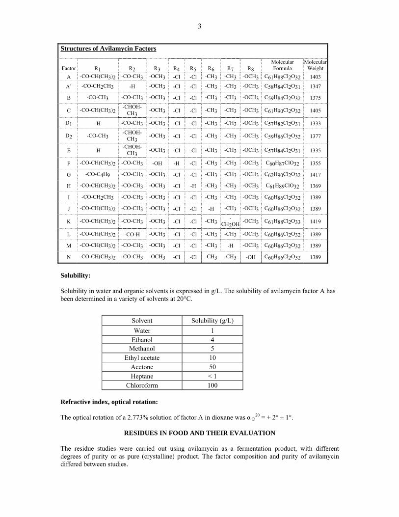
3
Structures of Avilamycin Factors
Factor
R1
R2
R3
R4
R5
R6
R7
R8
Molecular Formula
MolecularWeight
A -CO-CH(CH3)2 -CO-CH3 -OCH3 -Cl -Cl -CH3 -CH3 -OCH3 C61H88Cl2O32 1403
A’ -CO-CH2CH3 -H -OCH3 -Cl -Cl -CH3 -CH3 -OCH3 C58H84Cl2O31 1347
B -CO-CH3 -CO-CH3 -OCH3 -Cl -Cl -CH3 -CH3 -OCH3 C59H84Cl2O32 1375
C -CO-CH(CH3)2 -CHOH-CH3 -OCH3 -Cl -Cl -CH3 -CH3 -OCH3 C61H90Cl2O32 1405
D1 -H -CO-CH3 -OCH3 -Cl -Cl -CH3 -CH3 -OCH3 C57H82Cl2O31 1333
D2 -CO-CH3 -CHOH-CH3 -OCH3 -Cl -Cl -CH3 -CH3 -OCH3 C59H86Cl2O32 1377
E -H -CHOH-
CH3 -OCH3 -Cl -Cl -CH3 -CH3 -OCH3 C57H84Cl2O31 1335
F -CO-CH(CH3)2 -CO-CH3 -OH -H -Cl -CH3 -CH3 -OCH3 C60H87ClO32 1355
G -CO-C4H9 -CO-CH3 -OCH3 -Cl -Cl -CH3 -CH3 -OCH3 C62H90Cl2O32 1417
H -CO-CH(CH3)2 -CO-CH3 -OCH3 -Cl -H -CH3 -CH3 -OCH3 C61H89ClO32 1369
I -CO-CH2CH3 -CO-CH3 -OCH3 -Cl -Cl -CH3 -CH3 -OCH3 C60H86Cl2O32 1389
J -CO-CH(CH3)2 -CO-CH3 -OCH3 -Cl -Cl -H -CH3 -OCH3 C60H86Cl2O32 1389
K -CO-CH(CH3)2 -CO-CH3 -OCH3 -Cl -Cl -CH3 -CH2OH -OCH3 C61H88Cl2O33 1419
L -CO-CH(CH3)2 -CO-H -OCH3 -Cl -Cl -CH3 -CH3 -OCH3 C60H86Cl2O32 1389
M -CO-CH(CH3)2 -CO-CH3 -OCH3 -Cl -Cl -CH3 -H -OCH3 C60H86Cl2O32 1389
N -CO-CH(CH3)2 -CO-CH3 -OCH3 -Cl -Cl -CH3 -CH3 -OH C60H86Cl2O32 1389
Solubility: Solubility in water and organic solvents is expressed in g/L. The solubility of avilamycin factor A has been determined in a variety of solvents at 20°C.
Solvent Solubility (g/L) Water 1
Ethanol 4 Methanol 5
Ethyl acetate 10 Acetone 50 Heptane < 1
Chloroform 100 Refractive index, optical rotation: The optical rotation of a 2.773% solution of factor A in dioxane was α D20 = + 2° ± 1°.
RESIDUES IN FOOD AND THEIR EVALUATION
The residue studies were carried out using avilamycin as a fermentation product, with different degrees of purity or as pure (crystalline) product. The factor composition and purity of avilamycin differed between studies.

4
The Committee evaluated avilamycin to recommend MRLs in poultry, pigs and rabbits at the request of the 17th session of the Codex Committee on Residues of Veterinary Drugs in Foods. Conditions of use Avilamycin is intended for use as a veterinary medicine in chickens, turkeys, pigs and rabbits to control bacterial enteric infections. It exhibits good antimicrobial activity against important veterinary Gram-positive pathogens (e.g., Clostridium perfringens) and has no related molecules in its class in human use. Therefore, avilamycin has been developed for treating necrotic enteritis in poultry, and enteric disease in pig and rabbits. Avilamycin was previously authorised in the European Union (EU) as a feed additive for growth promotion in accordance with Council Directive 70/524/EEC; the substance was incorporated in pig feedstuffs at a concentration of 20 mg/kg feed for animals up to 6 months of age and 40 mg/kg feed for animals up to 4 months of age. It was incorporated into chicken and turkey feedstuffs at a concentration of 10 mg/kg feed. The use of the substance as a feed additive was discontinued in the EU from 1 January 2006. Dosage Table 1: Recommended doses and duration of treatment for Avilamycin in feed
Target Animal Dose in Feed
(mg/kg) Dose Rate
(mg/kg bw/day) Maximum Duration
(days) Pig 100 6-8 21 days Chicken 100 20 21 days Turkey 100 20 21 days Rabbit 80 5 28 days
PHARMACOKINETICS AND METABOLISM
Pharmacokinetics in Laboratory Animals, Humans and Food Animals No classical pharmacokinetic studies have been conducted in any species with avilamycin because avilamycin is not detectable in plasma (LOD = 0.05 mg/kg) following oral administration of avilamycin in feed. In addition, the concentration necessary for kinetic analysis would be well below the toxicologically relevant concentrations and would not be pertinent to human food safety. Metabolism and Residue studies in pigs, poultry and other species (rat) that have been conducted using radiolabelled material are presented below. Where blood, serum or plasma concentrations were measured in various species following oral doses, avilamycin concentrations were below the limits of detection. For example, in broiler chickens that were fed with a ration containing 22 mg of avilamycin/kg of feed for 25 days, no avilamycin was detected in blood measured by a bio-autographic method (LOD <0.04 mg/kg) or by GC method (LOD <0.1 mg/kg) (West, et al., 1982). Humans Avilamycin has not been developed for human use and therefore, no pharmacokinetic data in humans are available.

5
Laboratory animals Avilamycin is primarily excreted in faeces when administered orally to pig or chickens. In a GLP compliant rat study (Magnussen, 1985a), less than one percent of the oral dose was eliminated in the urine after 72 hours, while 80 to 104% was recovered in the faeces. Pigs In a balance-excretion non GLP-compliant study two cross-bred gilts were administered non-radiolabelled avilamycin at 120 mg per kg in the feed per day (Dalidowicz, et al., 1983). After 7 days administration to approximate steady state conditions, a single bolus dose of 120 mg of [U-14C]avilamycin was administered and excreta were collected at 24-hr intervals for 9 days. During the collection period, the two gilts excreted 96.9% and 99.0% of the dose, respectively, with an average of 93.4% in the faeces and 4.5% in the urine. The bulk of the radioactivity was excreted within the first four days. Another radiolabelled GLP-compliant pig study (Magnussen, et al., 1987) conducted on six crossbred pigs receiving the same dose for either ten or fourteen days showed similar results. Excreted radioactivity reached a plateau after 2-3 days and, on average comprised 8% in urine and 92% in faeces. Approximate concentration in faeces was 120 mg/kg equivalents avilamycin. Results are shown in Table 2. Table 2 : Excreted radioactivity in pigs fed ten days with avilamycin
Collection Period (day) Urine (µCi) Faeces (µCi) 1 0.76 1.12 2 1.06 18.56 3 1.45 17.57 4 1.51 18.83 5 1.53 16.96 6 1.54 18.83 7 2.04 21.09 8 1.69 21.26 9 1.94 20.29 10 1.57 18.16
Chickens A balance-excretion non-GLP-compliant study was conducted in chickens (Dalidowicz, et al., 1984a). Broiler chickens (2 males/2 females) were administered non-radiolabelled avilamycin at 20 mg of microbiological activity per kg in the feed. After 7 days administration to approximate steady state conditions, a single bolus dose of 4 mg of [U-14C]avilamycin was administered and excreta were collected at 24-hr intervals for 13 days. During the collection period, the birds excreted 92.8%, 99.2%, 96.6% and 84.4% of the dose, respectively. An average of 90% of the radioactivity was excreted within the first 6 days. Data for avilamycin in turkeys and rabbits are not available. Metabolism in Laboratory Animals Position of radiolabel (14C) in Avilamycin. The pivotal residue and metabolism studies for pig and chickens were performed using radiolabelled avilamycin. However, because avilamycin is extensively metabolized, the position of the radiolabel is important in understanding not only the metabolic profile of this substance, but also the correct

6
interpretation of the total radioactive tissue data. Therefore, a discussion of the radiolabel position is necessary prior to the assessment of the specific studies. Two types of radiolabelled avilamycin have been prepared by fermentation using S. viridochromogenes with one of two radiolabelled precursors: Figure 2: Structure of radiolabelled avilamycin
1. [U-14C]avilamycin: Using uniformly labelled glucose, [U-14C-glucose], as the precursor labels
the molecule uniformly in all rings (Donoho et al, 1987). 2. [DIA-14C]avilamycin: Using [2-14C-diethylmalonate] as the precursor places approximately
85% of the radioactivity in the dichloro-isoeverninic acid moiety (DIA; Ring A in Figure 2). The remaining 15% of the radioactivity was not conclusively identified, but is suggested to be associated with the iso-butyrate, propionate or acetate moiety at position R1 on Ring G (Dalidowicz, 1985).
Rats In a GLP- compliant study (Magnussen, 1985a), three male and three female Sprague-Dawley rats weighing 215-265 grams each were dosed by gavage for three consecutive days with [DIA14C]avilamycin (specific activity 0.246 μgCi/mg) at levels equivalent to 100 mg/kg of body weight. Following the initial dose, urine and faeces were collected separately from each animal at 24-hour intervals.. In addition, faeces collected during the 24-hour period following the third dose were extracted and assayed for avilamycin and metabolites. All radioactivity in the selected faeces samples was extractable into ethyl acetate at a neutral and acidic pH. The neutral fraction contained 85-87% of the radioactivity, while the acidic fraction contained 12-14%. TLC analysis showed avilamycins A and B to represent 40-60% of the radioactivity in the neutral fraction, while an unidentified, polar metabolite represented 10-30%. The major radioactive component in the acidic fraction was confirmed as flambalactone (previously identified as the major avilamycin derived residue in pig liver (Magnussen, 1985b). It is formed by cleavage of the ortho ester linking the C and D rings of avilamycin. Flambalactone represented 30-60% of the radioactivity in the acidic fraction. One other metabolite common to both rats and pigs in the acidic fraction representing 10-30% of the radioactivity was later identified as flambic acid (Magnussen, et al., 1987). In other studies flambalactone and flambic acid were found to be inter-convertible.
O
OO H COCH3
CH3
OHH
O
O
O
O
HO
H
O
OH OCH3
OO
O
CH3
OHO
O O
CH3
H CH3
O
OO
O OCH3
CH3
Cl
OH
Cl
CH3
O
OHCH3
OH
H3CO
R1
H3CO
A
B
C
D
E F G
H
[U-14C]Avilamycin: All rings labelled
[DIA-14C]Avilamycin 85% Ring A 15% other (possibly R1)
7
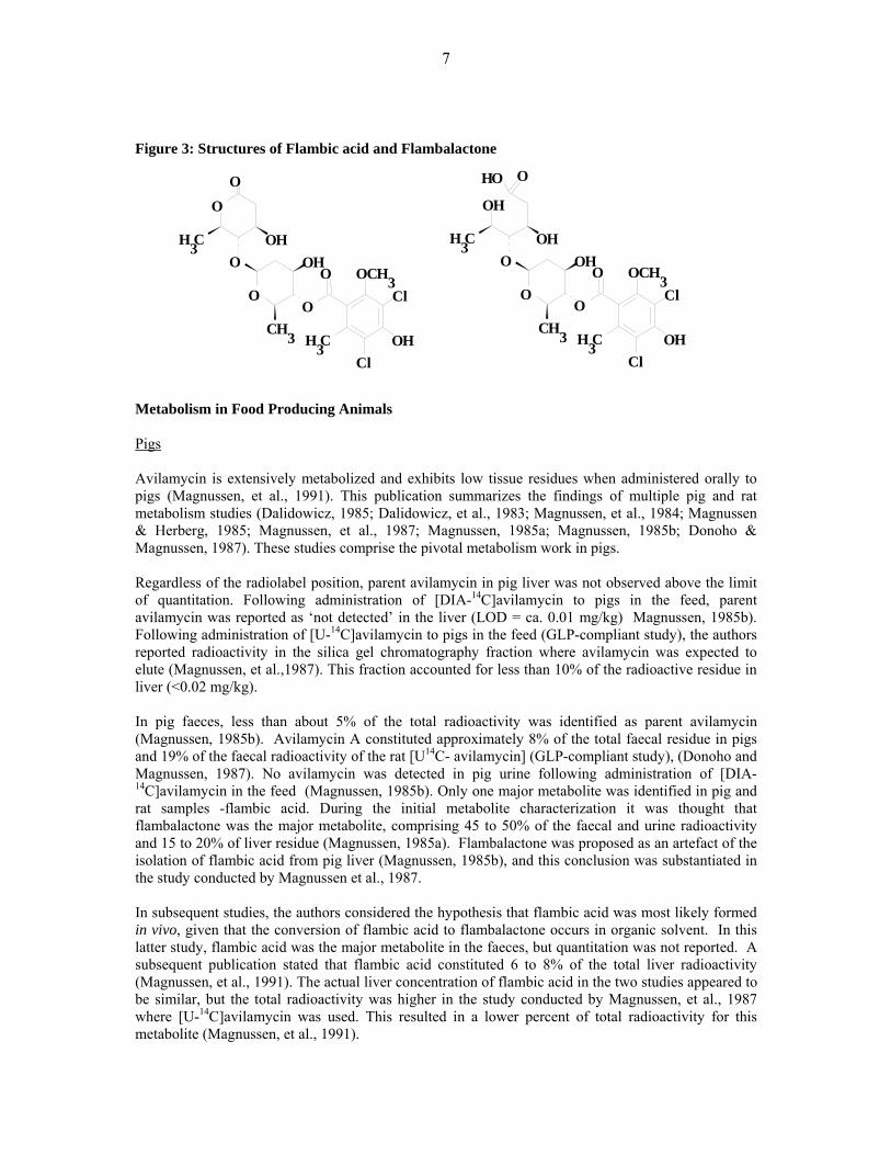
Figure 3: Structures of Flambic acid and Flambalactone Metabolism in Food Producing Animals Pigs Avilamycin is extensively metabolized and exhibits low tissue residues when administered orally to pigs (Magnussen, et al., 1991). This publication summarizes the findings of multiple pig and rat metabolism studies (Dalidowicz, 1985; Dalidowicz, et al., 1983; Magnussen, et al., 1984; Magnussen & Herberg, 1985; Magnussen, et al., 1987; Magnussen, 1985a; Magnussen, 1985b; Donoho & Magnussen, 1987). These studies comprise the pivotal metabolism work in pigs. Regardless of the radiolabel position, parent avilamycin in pig liver was not observed above the limit of quantitation. Following administration of [DIA-14C]avilamycin to pigs in the feed, parent avilamycin was reported as ‘not detected’ in the liver (LOD = ca. 0.01 mg/kg) Magnussen, 1985b). Following administration of [U-14C]avilamycin to pigs in the feed (GLP-compliant study), the authors reported radioactivity in the silica gel chromatography fraction where avilamycin was expected to elute (Magnussen, et al.,1987). This fraction accounted for less than 10% of the radioactive residue in liver (<0.02 mg/kg). In pig faeces, less than about 5% of the total radioactivity was identified as parent avilamycin (Magnussen, 1985b). Avilamycin A constituted approximately 8% of the total faecal residue in pigs and 19% of the faecal radioactivity of the rat [U14C- avilamycin] (GLP-compliant study), (Donoho and Magnussen, 1987). No avilamycin was detected in pig urine following administration of [DIA-14C]avilamycin in the feed (Magnussen, 1985b). Only one major metabolite was identified in pig and rat samples -flambic acid. During the initial metabolite characterization it was thought that flambalactone was the major metabolite, comprising 45 to 50% of the faecal and urine radioactivity and 15 to 20% of liver residue (Magnussen, 1985a). Flambalactone was proposed as an artefact of the isolation of flambic acid from pig liver (Magnussen, 1985b), and this conclusion was substantiated in the study conducted by Magnussen et al., 1987. In subsequent studies, the authors considered the hypothesis that flambic acid was most likely formed in vivo, given that the conversion of flambic acid to flambalactone occurs in organic solvent. In this latter study, flambic acid was the major metabolite in the faeces, but quantitation was not reported. A subsequent publication stated that flambic acid constituted 6 to 8% of the total liver radioactivity (Magnussen, et al., 1991). The actual liver concentration of flambic acid in the two studies appeared to be similar, but the total radioactivity was higher in the study conducted by Magnussen, et al., 1987 where [U-14C]avilamycin was used. This resulted in a lower percent of total radioactivity for this metabolite (Magnussen, et al., 1991).
O
O O
O OCH3
O
C H 3
Cl
OHCl
C H 3
O O H C H 3
O H
OH
O
OOHCH3
OH
OO
O O C H 3
CH3
Cl
O H Cl
CH3
OH
8
No other significant metabolites were identified in pigs or rats, although a few minor peaks were observed. The silica gel chromatographic profiles of extracts from the faeces, urine and livers of rats and pigs treated with [U-14C]avilamycin were qualitatively comparable and quantitatively similar by visual inspection (Donoho & Magnussen, 1987). There was a good correlation between the metabolic profiles of rats and pigs. The faeces extract from [DIA-14C]avilamycin-treated pigs exhibited the same three peaks, but the proportions were different, with the flambic acid-containing peak predominant. Additional TLC analyses of the column fractions from faeces extracts indicated that oligosaccharide-derived metabolites were present in [U-14C]avilamycin samples that were not present in [DIA-14C]avilamycin samples. These metabolites were not further characterized because the corresponding peaks were not present in liver and would thus not pose a food safety risk. The metabolic profiles in liver of treated rats and pigs were essentially the same with the flambic acid as the most abundant metabolite (Donoho & Magnussen, 1987). Parent avilamycin concentration in rat and pig liver were less that 0.05 mg/kg. The pattern of minor metabolites was similar but insufficient for identification. Data are supportive that rats treated with avilamycin have been exposed to the same metabolites that are present in edible tissues of treated pigs. Characterization of residues in fat samples from treated pigs demonstrated that essentially all of the residues in fat are due to the incorporation of radioactivity into the endogenous fatty acids, oleic and stearic acid (Dalidowicz, 1985). No DIA-related residues were detected in fat when assayed by hydrolysis and GC analysis, indicating that parent and DIA-containing metabolites such as flambic acid are not detectable (Magnussen, et al., 1984). Moreover, when the radiolabel is distributed into the carbohydrate moieties of avilamycin (i.e., [U-14C]avilamycin), the total radioactive residues are higher than those when using [DIA-14C]avilamycin, while the amounts of DIA-containing residues remain relatively constant (Magnussen, et al., 1984;, Magnussen, et al.; 1987, Magnussen, et al., 1991) The increased incorporation of carbon-14 into fatty acids when [U-14C]avilamycin was administered is consistent with the avilamycin carbohydrate moieties being extensively metabolized. Chickens, Turkeys and Rabbits No metabolism data available.
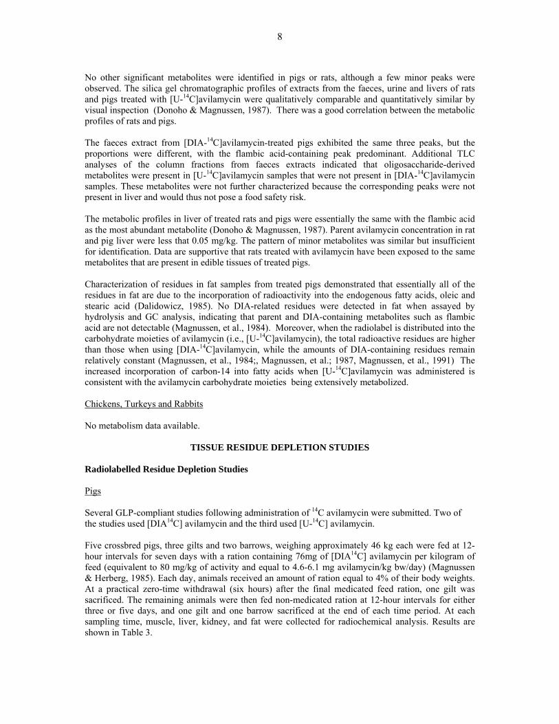
TISSUE RESIDUE DEPLETION STUDIES Radiolabelled Residue Depletion Studies Pigs Several GLP-compliant studies following administration of 14C avilamycin were submitted. Two of the studies used [DIA14C] avilamycin and the third used [U-14C] avilamycin. Five crossbred pigs, three gilts and two barrows, weighing approximately 46 kg each were fed at 12-hour intervals for seven days with a ration containing 76mg of [DIA14C] avilamycin per kilogram of feed (equivalent to 80 mg/kg of activity and equal to 4.6-6.1 mg avilamycin/kg bw/day) (Magnussen & Herberg, 1985). Each day, animals received an amount of ration equal to 4% of their body weights. At a practical zero-time withdrawal (six hours) after the final medicated feed ration, one gilt was sacrificed. The remaining animals were then fed non-medicated ration at 12-hour intervals for either three or five days, and one gilt and one barrow sacrificed at the end of each time period. At each sampling time, muscle, liver, kidney, and fat were collected for radiochemical analysis. Results are shown in Table 3.
9
Table 3: Total radiolabel residue (TRR) in pig tissues
Withdrawal TRR (mg/kg equivalents avilamycin) Days n Muscle Liver Kidney Fat
0 1 NDR1 0.15 0.08 0.07 3 2 NDR NDR 0.02 0.05 5 2 NDR NDR < 0.032 0.05
1: NDR = no detectable residue 2: one animal, 0.025 mg/kg; one animal NDR (0.017 mg/kg)
At zero-time withdrawal, no detectable residue was found in muscle, while the total radiolabel residues in liver, kidney, and fat, expressed as avilamycin equivalents, were 0.15, 0.08, and 0.07 mg/kg, respectively. After a three-day withdrawal period, no residues were detected in either liver or muscle, while residues in kidney and fat were 0.024 and 0.053 mg/kg, respectively. After five days, residue levels in fat were nearly the same as those observed at three days, while levels in kidney were from non-detectable residues to 0.025 mg/kg. Concentrations of avilamycin-related radioactivity in liver and muscle declined to non-detectable levels within three days after the termination of dosing, while concentrations in kidney declined to near non-detectable levels within five days after the termination of dosing. Radioactivity in fat showed a much slower rate of decline due to the fact that radiolabelled carbon from the 14C-avilamycin molecule had become incorporated into the fatty acid fraction as demonstrated by Dalidowicz, 1985. Authors quoted a fat turnover rate of 14-21 days but provided no evidence to support the comment. In another study conducted by Magnussen, et al, 1984, nine crossbred pigs, weighing approximately 44 kg each, were fed with a ration containing 76.2 mg of [DIA14C]avilamycin per kilogram of feed (equivalent to 80 mg/kg of avilamycin activity and equal to 4.6-6.1 mg avilamycin/kg bw/day) at 12-hour intervals for either four, seven, or ten days. Each day, animals received a ration equal to 4.0% of their body weights, equivalent to a daily dose of approximately 134 mg of DIA14C-avilamycin. All animals were sacrificed at a practical zero-time withdrawal (six hours) after the final feeding. Muscle, liver, kidney, fat, and bile were collected for radiochemical analyses by liquid scintillation counting. Selected tissues were assayed for avilamycin by bio-autography and residues containing the dichloroisoeverninic acid (DIA) moiety. Liver from each animal was extracted to determine levels of non-extractable radioactivity. For total radioactive residues (TTR) results are shown in Table 4. Table 4: Total radiolabel residue (TTR) in pig tissues.
Dosing Total Radioactivity (mg/kg equivalents avilamycin) Interval (days) Muscle Liver Kidney Fat Bile
4 0.01 0.21 0.10 0.05 18.9 7 0.01 0.23 0.10 0.08 19.9 10 0.02 0.22 0.10 0.12 19.8
After ten days dosing, total mean radiolabel residues in liver, fat, and kidney, expressed as avilamycin equivalents, were 0.22, 0.12, and 0.10 mg/kg, respectively. Residues in muscle were less than 0.016 mg/kg. Steady-state concentrations of radioactivity were attained in muscle, liver, and kidney within four days after the initiation of dosing. A steady-state concentration was not attained in fat during this study. The study mentioned in the metabolism section conducted by Dalidowicz, 1985 demonstrated that radioactivity found in fat was incorporated into the fatty acid portion of triglycerides. These non-active residues were not of toxicological concern.

10
Liver, kidney, and fat from animals dosed for ten days were assayed for avilamycin by bio-autography (Prichard et al, 2006; Method Number AM-AA-CA-R075-AB-755). This method consisted of extracting avilamycin from pig or broiler tissues with acetone. The acetone extract is purified by liquid-liquid partitioning, and the purified extract is spotted on a thin layer chromatographic plate (TLC). After development the TLC plate is subjected to bio-autographic analysis using a Micrococcus flavus overlay. The plate is sprayed to enhance the appearance of the zones of inhibition and the presence or absence of avilamycin is determined by comparison with a reference standard. The method does not determine the concentration of avilamycin, but the LOD was reported at 0.05 mg/kg. Results are presented in Table 5. Table 5: Microbiologically active avilamycin pig tissue residues
Dosing Interval (days)
Microbiological Activity (mg/kg equivalents avilamycin)
Muscle Liver Kidney Fat 10 -- < 0.05 NDR1 NDR1
NDR1: non-detectable residues. No microbiologically active residues of avilamycin were detected in kidney or fat and only traces in liver, but were considerably less than the limit of detection (LOD is < 0.05 mg/kg). Muscle was not assayed due to radioactivity concentrations less than LOD for the bio-autographic assay. Tissue residues containing DIA were analysed by gas chromatography (Formica, G and Giannone, C., 1986). Results are shown in Table 6. Table 6: DIA Residues in selected pig tissues from the 10-day withholding time
DIA Residue (mg/kg equivalents avilamycin) Animal No. Muscle Liver Kidney Fat
130 -- 0.10 < 0.1 NDR 135 -- 0.12 < 0.1 NDR 137 -- 0.17 < 0.1 NDR
Mean -- 0.13 < 0.1 NDR Approximately 50% of TRR in liver was due to DIA-related residues. DIA-related residues were detected in kidney, but below the limit of quantification (LOQ < 0.1 mg/kg). No DIA residues were observed in fat (< 0.1 mg/kg). Liver results are presented in Table 7. Table 7: Pig liver extraction results
Dosing Interval Mean (n=3) Percent of Radioactivity Days Acetone Unextracted
4 79.5 20.5 7 82.2 17.8 10 73.1 26.9
About 18 - 27% of radioactivity was not extractable into acetone for the 4, 7 and 10 day liver samples. Statistical analysis of the extraction data indicated no significant difference between un-extracted radioactivity through 10 days of treatment. A steady-state, tissue residue study using uniformly labelled 14C avilamycin was conducted by Magnussen, et al., 1987. Six crossbred pigs, four barrows and two gilts, weighing approximately 44 kg each were fed at 12-hour intervals for either ten or fourteen days with a ration containing a nominal concentration of 60 mg of 14C-avilamycin/kg of feed (equivalent to 60 mg activity/kg and to 3.6-4.8 mg/kg bw/day). Each day, animals received an amount of ration equal to 4% of their body weights.

11
Groups of three animals were killed after ten days and fourteen days on treatment. Muscle, liver, kidney, and fat were collected for radiochemical analysis. Liver from each animal was extracted to determine the concentration of non-extractable radioactivity and to characterize the extractable radioactivity. Radioactivity in fat was also characterized. Total radioactivity residues in tissues are presented in Table 8. Table 8: Total radiolabel residues (TRR) in pig tissues
Dosing Interval TTR (mg/kg equivalents avilamycin) Days Muscle Liver Kidney Fat
10 0.09 0.55 0.32 0.26 14 0.14 0.66 0.34 0.55
Radioactive tissue residues are higher in this study than the other two 14C studies because the avilamycin molecule was more uniformly labelled over all rings with 14C for this study. The 14C label in the avilamycin for the other two studies was primarily (85%) in the DIA ring. One-way analysis of variance (ANOVA) indicated no difference between 10 or 14 days for muscle, liver or kidney total radioactive residues. Only the fat radioactive residues were significantly different at 10 and 14 days (P < 0.05). Non-extractable liver residues were 33 - 37% of total liver residues and were not different in the 10- and 14-day treatment groups as are shown in Table 9. Table 9: Percent extraction of radioactivity from pig livers
10-day Group 14-day Group Animal No. 961 960 957 954 955 959 Acetone Extract 34 32 34 32 33 29 Methanol Extract 25 24 26 25 26 28 Acetone/water 7 7 6 8 6 6 Pellet 34 37 33 34 35 37
The GC analysis shown that extractable liver radioactivity consisted of several minor metabolites (<0.1 mg/kg). Flambic acid was present at concentrations up to 0.04 mg/kg. Parent 14C-avilamycin concentrations were less than 0.01-0.02 mg/kg. Chickens In a GLP-compliant conducted study (Dalidowicz, 1986), twelve seven-week-old broiler-type chickens, six male and six female, were fed a standard broiler finishing ration containing 14.16 mg of [DIA14C] avilamycin per kilogram of feed (equivalent to 15 mg of activity /kg and equal to 3 mg/kg bw/day) for either four, seven, or ten days. Medicated ration and water were provided ad libitum throughout the dosing phase. At the end of each designated dosing period, two birds of each sex were deprived of food and water for six hours and then killed. Samples of muscle, liver, abdominal fat, kidney and skin with subcutaneous fat were collected for radiochemical analysis. Results are shown in Table 10.
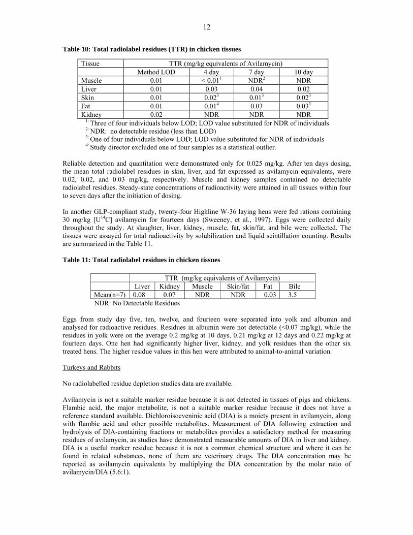
12
Table 10: Total radiolabel residues (TTR) in chicken tissues
Tissue TTR (mg/kg equivalents of Avilamycin) Method LOD 4 day 7 day 10 day Muscle 0.01 < 0.011 NDR2 NDR Liver 0.01 0.03 0.04 0.02 Skin 0.01 0.023 0.013 0.023
Fat 0.01 0.014 0.03 0.033
Kidney 0.02 NDR NDR NDR 1: Three of four individuals below LOD; LOD value substituted for NDR of individuals 2: NDR: no detectable residue (less than LOD) 3: One of four individuals below LOD; LOD value substituted for NDR of individuals 4: Study director excluded one of four samples as a statistical outlier. Reliable detection and quantitation were demonstrated only for 0.025 mg/kg. After ten days dosing, the mean total radiolabel residues in skin, liver, and fat expressed as avilamycin equivalents, were 0.02, 0.02, and 0.03 mg/kg, respectively. Muscle and kidney samples contained no detectable radiolabel residues. Steady-state concentrations of radioactivity were attained in all tissues within four to seven days after the initiation of dosing. In another GLP-compliant study, twenty-four Highline W-36 laying hens were fed rations containing 30 mg/kg [U14C] avilamycin for fourteen days (Sweeney, et al., 1997). Eggs were collected daily throughout the study. At slaughter, liver, kidney, muscle, fat, skin/fat, and bile were collected. The tissues were assayed for total radioactivity by solubilization and liquid scintillation counting. Results are summarized in the Table 11. Table 11: Total radiolabel residues in chicken tissues
TTR (mg/kg equivalents of Avilamycin) Liver Kidney Muscle Skin/fat Fat Bile Mean(n=7) 0.08 0.07 NDR NDR 0.03 3.5
NDR: No Detectable Residues Eggs from study day five, ten, twelve, and fourteen were separated into yolk and albumin and analysed for radioactive residues. Residues in albumin were not detectable (<0.07 mg/kg), while the residues in yolk were on the average 0.2 mg/kg at 10 days, 0.21 mg/kg at 12 days and 0.22 mg/kg at fourteen days. One hen had significantly higher liver, kidney, and yolk residues than the other six treated hens. The higher residue values in this hen were attributed to animal-to-animal variation. Turkeys and Rabbits No radiolabelled residue depletion studies data are available. Avilamycin is not a suitable marker residue because it is not detected in tissues of pigs and chickens. Flambic acid, the major metabolite, is not a suitable marker residue because it does not have a reference standard available. Dichloroisoeveninic acid (DIA) is a moiety present in avilamycin, along with flambic acid and other possible metabolites. Measurement of DIA following extraction and hydrolysis of DIA-containing fractions or metabolites provides a satisfactory method for measuring residues of avilamycin, as studies have demonstrated measurable amounts of DIA in liver and kidney. DIA is a useful marker residue because it is not a common chemical structure and where it can be found in related substances, none of them are veterinary drugs. The DIA concentration may be reported as avilamycin equivalents by multiplying the DIA concentration by the molar ratio of avilamycin/DIA (5.6:1).
13
Residue Depletion Studies with Unlabelled Drug Residues in Tissues Pigs A GLP-compliant residue study in young pigs was submitted (Eichmeier, et al, 2006a). Twelve crossbred commercial pigs (plus 1 male and 1 female as controls) weighing about 9 to 15 kg were fed ad libitum a commercial diet containing avilamycin at a nominal concentration of 150 mg/kg feed for 21 consecutive days (equal to 9-12 mg/kg bw/day). At the end of the 21-day exposure period, animals were euthanized at withdrawal intervals of 0, 6, and 24-hours (n=4 per group, 2 males and 2 females). Samples of liver, kidneys, muscle and skin with fat were collected. Avilamycin residues were analyzed as DIA by a LC-MS-MS validated method (Eichmeier, 2006a) and also by a microbiological assay (Eichmeier, et al, 2006, Appendix G). The LC-MS-MS method requires a tissue hydrolysis step to yield dichloroisoeverninic acid. Results were reported as avilamycin equivalents by multiplying the determined DIA concentration by the molar ratio (5.6:1). The mean residue data are summarized in the Table 12. Residues were only detected in liver tissue. Table 12: Equivalent avilamycin residues in pig tissues
Mean Equivalent Avilamycin Residues (μg/kg ) ± SD1
Liver Kidney Muscle Fat/Skin Control < LOD2 <LOD <LOD <LOD
0 hr 103 ± 26 < 28 3 <LOD <LOD 6 hr 42 ± 9 < 28 3 <LOD <LOD 24 hr < 324 ND <LOD <LOD
1: Mean ± standard deviation 2: Limit of Detection in avilamycin equivalents/kg tissue: Liver (9.0 μg/kg );
Fat/Skin (5.5 μg/kg ); Muscle (4.2 μg/kg ); Kidney (3.3 μg /kg ) 3: 28 μg/kg = 5.0 μg/kg DIA 4: n = 1, other 3 samples < 28 μg/kg
For the microbiological assay samples were extracted with acetone, purified and the organic phase analyzed by thin layer-chromatography (TLC) on silica gel plates. Antibiotic activity was assayed using Micrococcus luteus ATCC No. 10240 as the assay organism. The pig muscle, liver, and skin/fat tissues for zero hour and 6 hr withdrawal times showed no response on the assay plates at a limit of detection of 5 μg/kg tissue. The 24-hr samples were not assayed for antimicrobial activity.
The results of this study showed that DIA was quantifiable in pig liver at zero and 6 hr withdrawal time and declined by more than half in 6 hours. After 24 hours, the residues were below or near 28 μg avilamycin/kg tissue. DIA residues were detected, but not quantifiable, in kidney at 0 and 6 hr withdrawal, and were not detected after 24 hours. No residues were detected in muscle or fat/skin samples at any time. No antimicrobial activity was detected in any tissue (LOD = 5 mg/kg) indicating that DIA detected in liver and kidney was due to inactive metabolites of the drug.
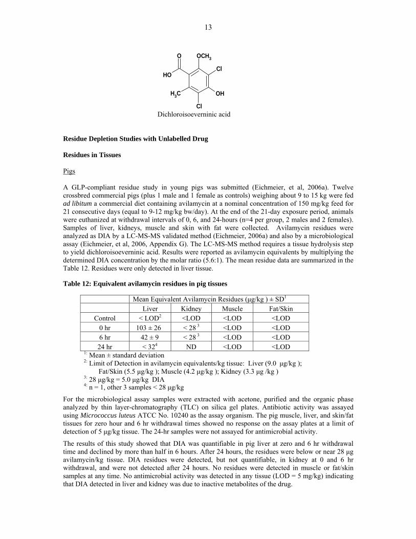
Dichloroisoeverninic acid
OH
O OCH3
Cl
OH
Cl
CH3

14
A non GLP-compliant study to determine microbiological activity of avilamycin residues was submitted (Asanuma, et al., 1987a). A preparation of 10% avilamycin/mg (EL-750) was administered to castrated male pigs from 28 to 84 days orally by medicated feed at concentrations of 40 or 400 mg/kg feed (two groups of 8 pigs, one control). Non-medicated feed was provided during withdrawal. The pigs were allowed free access to the feed. Liver, kidney, muscle, fat tissues and small intestine were collected at 42 days (two animals per group) and after 84 days (two animals at each time, 0.1 and 3 days withdrawal). The avilamycin residues were analysed by the microbiological assay method with Micrococcus flavus (LOD = 25 μg/kg). Avilamycin was not detected in major organs or tissue for all sampling points including during medication. In a previously reported non GLP-compliant study, the microbiological activity was studied by Morimoto et al., 1986a. Breeding piglets (22 boars and 22 sows) at approximately 30 days old were fed with medicated feed containing avilamycin (40, 200 or 400 mg/kg) for 12 weeks followed by a 7-day withdrawal. The avilamycin preparation was the same used in the previous mentioned study (EL-750. 10% avilamycin/mg). Plasma, liver, kidney, muscle, fat, and small intestine was collected from groups of 1 male and 1 female at 6 weeks and 12 weeks at 0-hour withdrawal and at 1, 3, 5 and 7days withdrawal time. The residues were analyzed using the TLC/microbiological assay based on inhibition of Micrococcus flavus (LOD = 25 μg/kg). No microbiologically active residues were observed in any samples of plasma, muscle, liver, kidney, or fat (25 μg/kg was detected in two small intestine samples from the 200 mg/kg treatment group, one at 6 weeks and one at 12 weeks; 27 μg/kg was detected in a small intestine sample of the 400 mg/kg group at 6 weeks and 30 μg/kg at 12 weeks). No other residues were detected. The authors concluded that EL-750 is not readily absorbed and only very small amounts of avilamycin are found in tissues, even at a dose of 400 mg/kg in the feed for 12 weeks. Two non GLP-compliant studies simulating the commercial pig industry were reported. In the first study (West and Wellenreiter, 1983), grower-finisher pigs (2 male, 4 female) were fed standard rations containing 0 or 40 mg/kg of avilamycin for a period of 99 days. The pigs were sacrificed after a zero-day (six-hour) or a one-day (30-hour) withdrawal period. Using the bio-autographic technique with Micrococcus flavus as the indicator organism, no microbiologically active residues were detected in the muscle, liver, kidney or fat tissues from any of the six treated or three control pigs analysed at a detection limit of 0.05 mg/kg. In the second study (West, et al, 1983), starter pigs (3 male, 3 female) were fed a standard ration containing 0 or 200 mg/kg of avilamycin for a period of 56 days. The pigs were sacrificed after a zero-day (six-hour) withdrawal time. Using the microbiological assay, no microbiologically active residues were detected in the muscle, liver, kidney or fat tissues from any of the pigs analysed at a detection limit of 0.05 mg/kg. Chickens In a GLP-compliant residue study, a commercial breed of broiler chickens (15 males and 15 females plus 10 males and 10 females as controls) approximately two weeks old weighing from 339-541 g were fed ad libitum a commercial diet containing avilamycin at a nominal concentration of 150 mg/kg feed for 21 consecutive days, equivalent to 30 mg/kg bw/day (Eichmeier, 2006b). At the end of the 21-day exposure period, animals were euthanized at withdrawal intervals of 0, 6, and 24-hours (n=6 per group, 3 males and 3 females). Samples of liver, kidneys, muscle and skin/fat (subcutaneous) were collected. Avilamycin residues were analyzed as DIA by the LC/MS/MS method (Eichmeier, 2006b) and also by the microbiological assay (Eichmeier, et al., 2006, Appendix G) as previously described for the study with pigs (Eichmeier, et al., 2006a). The DIA moiety of avilamycin was quantifiable in chicken liver at zero time withdrawal and declined to below or near 28 µg avilamycin/kg tissue within 6 hours. After 24 hours, the liver residues were below 28 µg avilamycin/kg. DIA residues were detected, but not quantifiable, in kidney and skin/fat at
15
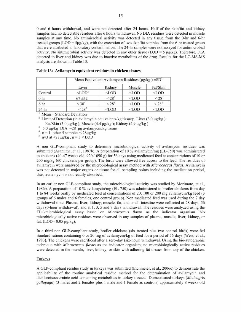
0 and 6 hours withdrawal, and were not detected after 24 hours. Half of the skin/fat and kidney samples had no detectable residues after 6 hours withdrawal. No DIA residues were detected in muscle samples at any time. No antimicrobial activity was detected in any tissue from the 0-hr and 6-hr treated groups (LOD = 5μg/kg), with the exception of two skin/fat samples from the 6-hr treated group that were attributed to laboratory contamination. The 24-hr samples were not assayed for antimicrobial activity. No antimicrobial activity was detected in any other tissue (LOD = 5 μg/kg). Therefore, DIA detected in liver and kidney was due to inactive metabolites of the drug. Results for the LC-MS-MS analysis are shown in Table 13. Table 13: Avilamycin equivalent residues in chicken tissues
A non GLP-compliant study to determine microbiological activity of avilamycin residues was submitted (Asanuma, et al., 1987b). A preparation of 10 % avilamycin/mg (EL-750) was administered to chickens (40-47 weeks old, 920-1090 g) for 56 days using medicated feed at concentrations of 10 or 200 mg/kg (60 chickens per group). The birds were allowed free access to the feed. The residues of avilamycin were analysed by the microbiological assay method with Micrococcus flavus. Avilamycin was not detected in major organs or tissue for all sampling points including the medication period, thus, avilamycin is not readily absorbed. In an earlier non GLP-compliant study, the microbiological activity was studied by Morimoto, et al., 1986b. A preparation of 10 % avilamycin/mg (EL-750) was administered to broiler chickens from day 1 to 84 weeks orally by medicated feed at concentrations of 20, 100 or 200 mg avilamycin/kg feed (3 groups of 6 males and 6 females, one control group). Non medicated feed was used during the 7 day withdrawal time. Plasma, liver, kidney, muscle, fat, and small intestine were collected at 28 days, 56 days (0-hour withdrawal), and at 1, 3, 5 and 7 days withdrawal. The residues were analyzed using the TLC/microbiological assay based on Micrococcus flavus as the indicator organism. No microbiologically active residues were observed in any samples of plasma, muscle, liver, kidney, or fat. (LOD= 0.05 μg/kg). In a third non GLP-compliant study, broiler chickens (six treated plus two control birds) were fed standard rations containing 0 or 20 mg of avilamycin/kg of feed for a period of 56 days (West, et al., 1983). The chickens were sacrificed after a zero-day (six-hour) withdrawal. Using the bio-autographic technique with Micrococcus flavus as the indicator organism, no microbiologically active residues were detected in the muscle, liver, kidney, or skin with adhering fat tissues from any of the chicken. Turkeys A GLP-compliant residue study in turkeys was submitted (Eichmeier, et al., 2006c) to demonstrate the applicability of the routine analytical residue method for the determination of avilamycin and dichloroisoeverninic acid-containing metabolites in turkey tissues. Domesticated turkeys (Melleagris gallopago) (3 males and 2 females plus 1 male and 1 female as controls) approximately 8 weeks old
Mean Equivalent Avilamycin Residues (μg/kg ) ±SD1
Liver Kidney Muscle Fat/Skin Control <LOD2 <LOD <LOD <LOD 0 hr 67 ±32 < 283 <LOD < 28 6 hr < 304 < 285 <LOD < 282
24 hr < 282 <LOD <LOD <LOD 1: Mean ± Standard Deviation 2: Limit of Detection (in avilamycin equivalents/kg tissue): Liver (3.0 μg/kg );
Fat/Skin (5.0 μg/kg ); Muscle (4.4 μg/kg ); Kidney (4.9 μg/kg ) 3: 5.0 μg/kg DIA =28 μg avilamycin/kg tissue 4 n = 1, other 5 samples < 28μg/kg 5: n=3 at <28μg/kg , n = 3 < LOD
16
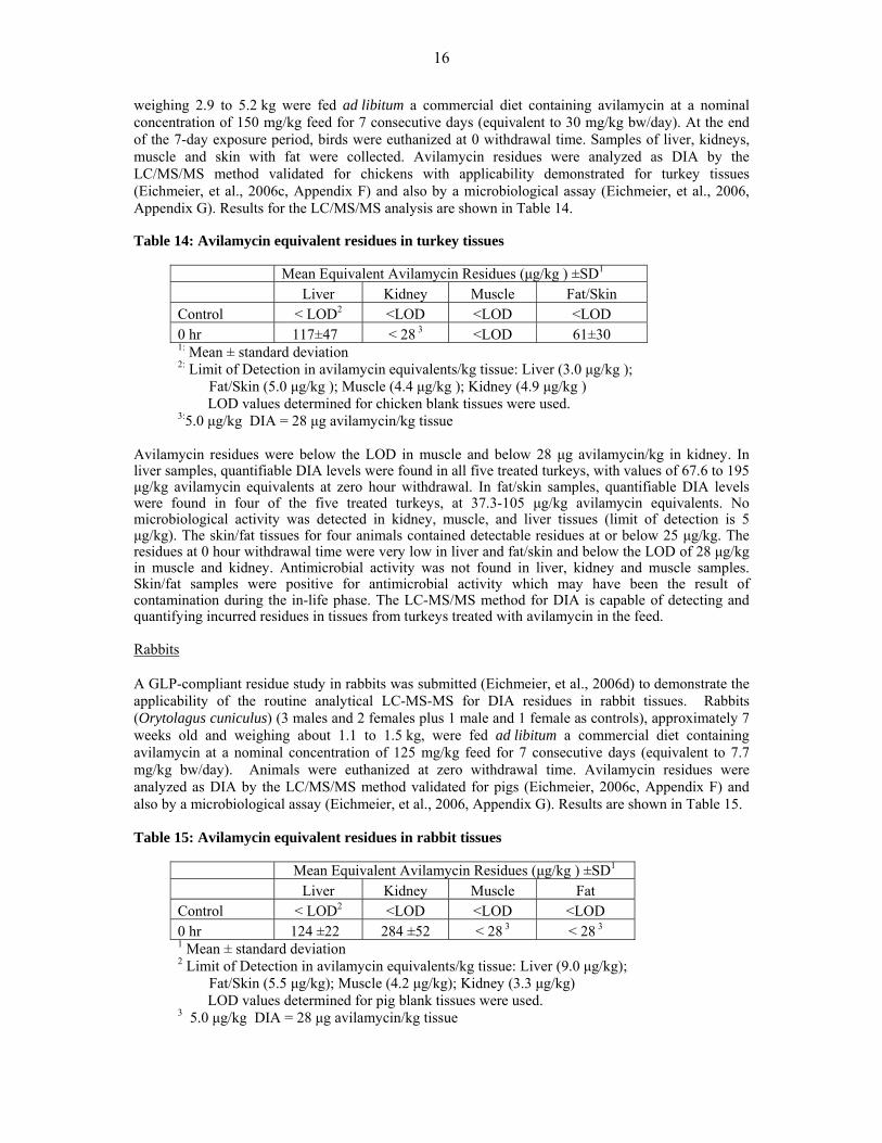
weighing 2.9 to 5.2 kg were fed ad libitum a commercial diet containing avilamycin at a nominal concentration of 150 mg/kg feed for 7 consecutive days (equivalent to 30 mg/kg bw/day). At the end of the 7-day exposure period, birds were euthanized at 0 withdrawal time. Samples of liver, kidneys, muscle and skin with fat were collected. Avilamycin residues were analyzed as DIA by the LC/MS/MS method validated for chickens with applicability demonstrated for turkey tissues (Eichmeier, et al., 2006c, Appendix F) and also by a microbiological assay (Eichmeier, et al., 2006, Appendix G). Results for the LC/MS/MS analysis are shown in Table 14. Table 14: Avilamycin equivalent residues in turkey tissues
Mean Equivalent Avilamycin Residues (μg/kg ) ±SD1
Liver Kidney Muscle Fat/Skin Control < LOD2 <LOD <LOD <LOD 0 hr 117±47 < 28 3 <LOD 61±30 1: Mean ± standard deviation 2: Limit of Detection in avilamycin equivalents/kg tissue: Liver (3.0 μg/kg );
Fat/Skin (5.0 μg/kg ); Muscle (4.4 μg/kg ); Kidney (4.9 μg/kg ) LOD values determined for chicken blank tissues were used. 3:5.0 μg/kg DIA = 28 μg avilamycin/kg tissue
Avilamycin residues were below the LOD in muscle and below 28 μg avilamycin/kg in kidney. In liver samples, quantifiable DIA levels were found in all five treated turkeys, with values of 67.6 to 195 μg/kg avilamycin equivalents at zero hour withdrawal. In fat/skin samples, quantifiable DIA levels were found in four of the five treated turkeys, at 37.3-105 μg/kg avilamycin equivalents. No microbiological activity was detected in kidney, muscle, and liver tissues (limit of detection is 5 μg/kg). The skin/fat tissues for four animals contained detectable residues at or below 25 μg/kg. The residues at 0 hour withdrawal time were very low in liver and fat/skin and below the LOD of 28 μg/kg in muscle and kidney. Antimicrobial activity was not found in liver, kidney and muscle samples. Skin/fat samples were positive for antimicrobial activity which may have been the result of contamination during the in-life phase. The LC-MS/MS method for DIA is capable of detecting and quantifying incurred residues in tissues from turkeys treated with avilamycin in the feed. Rabbits A GLP-compliant residue study in rabbits was submitted (Eichmeier, et al., 2006d) to demonstrate the applicability of the routine analytical LC-MS-MS for DIA residues in rabbit tissues. Rabbits (Orytolagus cuniculus) (3 males and 2 females plus 1 male and 1 female as controls), approximately 7 weeks old and weighing about 1.1 to 1.5 kg, were fed ad libitum a commercial diet containing avilamycin at a nominal concentration of 125 mg/kg feed for 7 consecutive days (equivalent to 7.7 mg/kg bw/day). Animals were euthanized at zero withdrawal time. Avilamycin residues were analyzed as DIA by the LC/MS/MS method validated for pigs (Eichmeier, 2006c, Appendix F) and also by a microbiological assay (Eichmeier, et al., 2006, Appendix G). Results are shown in Table 15. Table 15: Avilamycin equivalent residues in rabbit tissues
Mean Equivalent Avilamycin Residues (μg/kg ) ±SD1
Liver Kidney Muscle Fat Control < LOD2 <LOD <LOD <LOD 0 hr 124 ±22 284 ±52 < 28 3 < 28 3 1 Mean ± standard deviation 2 Limit of Detection in avilamycin equivalents/kg tissue: Liver (9.0 μg/kg);
Fat/Skin (5.5 μg/kg); Muscle (4.2 μg/kg); Kidney (3.3 μg/kg) LOD values determined for pig blank tissues were used. 3 5.0 μg/kg DIA = 28 μg avilamycin/kg tissue

17
In all five treated rabbits, the equivalent avilamycin levels were below 28 μg/kg in muscle and fat at zero hour withdrawal time. In liver and kidney samples, quantifiable DIA levels were found in all five treated rabbits at 93 – 145 μg/kg and 228 – 352 μg/kg avilamycin equivalents, respectively. No microbiological response was detected in rabbit kidney, muscle, fat and liver tissues on the assay plates at a limit of detection of 5 μg/kg tissue. The residues at zero withdrawal time were very low in liver and kidney and below the 28 μg/kg in muscle and fat. The routine analytical method (LC-MS/MS for DIA) was capable of detecting and quantifying incurred residues in tissues from rabbits treated with avilamycin in the feed. Antimicrobial activity was not found in liver, fat, kidney and muscle samples.
METHODS OF ANALYSIS Pigs An analytical method validated for the determination of avilamycin in pig tissues (muscle, fat, liver and kidney) was submitted (Eichmeier, 2006b). Avilamycin and/or its metabolites containing dichloroisoeverninic (DIA) were extracted from homogenized tissues using acetone. Following centrifugation and evaporation of the solvent, the extracted residues were hydrolyzed at about 70°C for 2 hr in 1 N NaOH. The hydrolysate was acidified to pH 1 with phosphoric acid and partitioned with ethyl acetate. An external standard was added (Dicamba) to appropriate samples and the hydrolysate was purified by alumina solid phase extraction (SPE). DIA was eluted from a SPE cartridge using 5% formic acid in acetonitrile. The eluate was evaporated to dryness and reconstituted in methanol for LC-MS-MS analysis. The DIA concentration was measured by gradient HPLC with mass spectrometric detection using negative-ion electrospray ionization mass spectrometry with selected-ion monitoring of the molecular ions of DIA and the external standard (M+H+). HPLC was performed using a Synergi Polar-RP 80A column (75 x 4.6 mm, 4μm, injection volume 10-25 μl, flow rate 0.6 ml/min) with a solvent gradient (0.1% formic acid in water-methanol, run time 8 minutes). A Phenomenex Security Guard C18 (4.0 x 3.0 mm) was employed. For the MS detection the instrument acquisitions were: Detector PE Sciex API 3000, APCI, negative mode, Ion Spray Voltage -4200, Scan Dwell time 250ms for both DIA and Dicamba, Turbo gas temperature was 550 ºC. A MRM procedure was applied and the following transitions were monitored: m/z 249.0 →190.0 and 219.2→ 175.0 for DIA and the external standard respectively. Calibration curves are constructed by weighted linear regression using the DIA peak area or the ratio of the peak area of the DIA to that of the added external standard, if used. The DIA concentration was converted to avilamycin equivalents by multiplying the DIA concentration by the molar ratio of avilamycin/DIA (5.6:1). Linearity: The linear range is from 28 to 3000 ng/ml of avilamycin equivalents (DIA nominal linear range is from 5 to 550 ng/ml). Duplicate calibration curves were use to generate the linear regression curve for each tissue with nominal calibration standards at 5, 10, 25, 50, 100, 150, 200, 350 and 550 ng/ml. The correlation coefficient (r) values were > 0.990 in all cases. Accuracy/Recovery/Precision (repeatability): Within laboratory data at three concentrations: (300 μg DIA/kg for liver, 200 μg DIA/kg for kidney, 100μg DIA/kg for skin/fat and 50μg DIA/kg for muscle) were used to determine the accuracy and precision of the measured concentration of avilamycin. Replicates (n=3) of test portions of tissue at each avilamycin concentration were analyzed each day for three days. Repeatability (n=6) was used as one of the within laboratory day runs. Within run repeatability precision was from 9.8 to 14.6%. Within laboratory precision was between 8.4 and 14.6%. Accuracy, determined as percent recovery from tissues fortified with avilamycin was between 79 and 105%. Limit of Detection: Blank matrix samples were assayed as part of the within-laboratory analytical batches and additional blank matrix samples (>10) were assayed for each tissue type from a minimum of 2 different animals. The response from 20 or more blank control test portions for each tissue plus

18
three times the standard deviation determined the limit of detection for each tissue. The claimed avilamycin limits of detection were 5.5μg/kg DIA for fat/skin, 4.2μg/kg for muscle, 3.3μg/kg for kidney and 9.0μg/kg for liver. The Committee reconsidered the data provided and calculated the LOQs considering the representative chromatograms of typical LC-MS-MS spectra of the extracted fortified samples for pigs using the criteria of signal to noise ratio equal to 10. Limit of Quantification: The sponsor adopted the LOQs as the minimum concentration in fortified samples that were shown to satisfy the criteria for recovery and precision, – i.e., the lowest concentration on the calibration curve and rounding these values (150μg DIA/kg for liver, 100μg DIA/kg for kidney, 50μg DIA/kg for skin/fat and 25μg DIA/kg for muscle). Repeatability measurements at concentrations noted below using signal to noise ratios were used to estimate LOD and LOQ at avilamycin equivalents of 100μg/kg in muscle; 750μg/kg in liver; 500μg/kg in kidney; and 250μg/kg in skin/fat. Results are tabulated in table 16. Table16: LOD and LOQ determinations for pig tissues
Tissue Avilamycin (μg/kg)
LOD (μg/kg)
LOQ (μg/kg)
Muscle 100 7.2 24 Liver 750 3.0 10
Kidney 500 1.0 3.3 Skin/Fat 250 6.7 22.4
Selectivity, Specificity and Carry-Over: These parameters were evaluated by extracting and analyzing individual blank pig liver, kidney, muscle and fat/skin samples. No significant response at the retention times of DIA or the external standard (Dicamba), were noted in the tissue blanks. Specificity was also examined by separately analyzing medicated feed additives of monensin, tylosin, tilmicosin, nicarbazin, narasin, salinomycin and clopidol. These reference compounds were processed by the avilamycin method procedure through to analysis by HPLC-MS/MS. None of the medicated feed additives showed a response at the retention time of DIA or Dicamba. Carry-over was evaluated by placing vials of solvent blank (methanol) at several locations in the analysis set after a high calibration standard sample. No carry over was observed in the solvent blank samples. Robustness: Examination of different lots of HPLC columns and the effect of variation of pH (0.2 pH units) after the method hydrolysis step in DIA extraction were used to determine the robustness using liver extracts. The mean accuracy values of the tested extracts were within 20% of each other. Stability: DIA solutions in reconstituted solvent for HPLC-MS/MS analysis for tissues are stable at 4 to 8 ºC for at least 7 days. Avilamycin fortified tissue samples are stable for at least 9 months at -70ºC. Liver, kidney and fat/skin extracts are stable for at least 7 days at room temperature and muscle extracts for at least 19 days. DIA and Dicamba in methanol stored at 4-8ºC are stable for at least 8 and 9 months respectively. Chickens The analytical validated method for the determination of avilamycin in pig tissues (muscle, fat, liver and kidney) for use with chicken tissues was submitted (Eichmeier, 2006a). Identical clean-up steps were used, HPLC/MS/MS conditions and construction of calibration curves described for pig samples were applied. Linearity: Results are described in the pig tissue method.

19
Accuracy/Recovery/Precision (repeatability): Within laboratory data at 0.5 MRL, MRL and 2xMRL of the sponsor proposed MRLs (300 μg/kg for liver, 200 μg/kg for kidney, 100 μg/kg for skin/fat and 50 μg/kg for muscle) were used to determine the accuracy and precision of the measured concentration of avilamycin. Replicates (n=3) of test portions of tissue at each avilamycin concentration were analyzed each day for three days. Repeatability (n=6) was used as one of the within laboratory day runs. Within run repeatability precision ranged from 7.9 - 13.3%. Within laboratory precision ranged between 7.5 to 20.6%. Accuracy, determined as percent recovery from tissues fortified with avilamycin ranged between 82 - 105%. Limit of Detection: Blank matrix samples were assayed as part of the within-laboratory analytical batches and additional blank matrix samples (11) were assayed for each tissue type from a minimum of 2 different animals. The response from 20 or more blank control test portions for each tissue plus three times the standard deviation determined the limit of detection for each tissue. The claimed avilamycin limits of detection were 5.0 μg/kg for fat/skin, 4.4 μg/kg for muscle, 4.9 μg/kg for kidney and 9.0 μg/kg for liver. The Committee reconsidered the data provided and calculated the LOQs considering the representative chromatograms of typical LC-MS-MS spectra of the extracted fortified samples for chickens using the criteria of signal to noise ratio equal to 10. Limit of Quantification: The sponsor adopted the LOQs as the minimum concentration in fortified samples that were shown to satisfy the criteria for recovery and precision, – i.e., the lowest concentration on the calibration curve and rounding these values (150 μg DIA/kg for liver, 100 μg DIA/kg for kidney, 50 μg DIA/kg for skin/fat and 25 μg DIA/kg for muscle). It was deemed that a more appropriate measure of limit of quantification was the comparison of signal to noise ratios of typical LC-MS-MS spectra. Repeatability measurements at concentrations noted below were used to estimate LOD and LOQ as avilamycin equivalents of 100μg/kg in muscle; 750 μg/kg in liver; 500 μg/kg in kidney; and 250 μg/kg in skin/fat. Results are tabulated in table 17. Table17: LOD and LOQ determinations for chicken tissues
Tissue Avilamycin (μg/kg)
LOD (μg/kg)
LOQ (μg/kg)
Muscle 100 5.7 18.8 Liver 750 9.1 30.4
Kidney 500 6.7 22.4 Skin/Fat 250 5.6 18.7
Selectivity, Specificity, Carry-Over and Robustness: These parameters were evaluated with identical procedures as those employed in the pig tissue method with identical results. Stability: DIA tissue extracts are stable at 4 to 8 ºC for at least 7 days in muscle, liver and fat/skin and for at least 6 days in kidney. Avilamycin - fortified tissue samples are stable at -70 ºC for at least 7.5 months in liver, muscle and skin/fat and for at least 9 months in kidney tissue. Liver and fat/skin extracts are stable for at least 7 days and kidney and muscle for at least 6 days at room temperature. Turkeys The same analytical method for the determination of avilamycin in chicken tissues was employed and its applicability in turkey tissues was demonstrated (Eichmeier, et al., 2006c, Appendix F). The method was capable of detecting and quantifying incurred residues in tissues from turkeys treated with avilamycin in feed. Linearity of the DIA calibration curves was acceptable with correlation coefficient (r) values for this study ranging from 0.9959 to 0.9989. Recovery samples were analyzed at tissue fortification levels of

20
50 μg/kg for each tissue; acceptable recoveries were from 72 to 103%. LOD and LOQ were nearly equivalent to those adopted for chicken tissues. Rabbit The same analytical method for the determination of avilamycin in pig and chicken tissues was demonstrated (Eichmeier et al., 2006d, Appendix F). The method was capable of detecting and quantifying residues incurred in tissues from rabbits treated with avilamycin in the feed. Linearity of the DIA calibration curves was acceptable and correlation coefficient (r) values for this study were 0.9957 to 0.9985. Recovery samples were analyzed at tissue fortification levels of 50 μg/kg for each tissue; acceptable recoveries ranged from 81 to 110%. The LOD and LOQ were nearly equivalent to that determined for chicken tissues
APPRAISAL Avilamycin has not been previously evaluated by the Committee. Avilamycin is an orthosomycin antibiotic complex primarily active against Gram-positive bacteria. The major fermentation product consists of avilamycin A and avilamycin B while 15 minor factors have been identified. Avilamycin is intended for use only as a veterinary medicine in chickens, turkeys, pigs and rabbits to control bacterial enteric infections at a dose of 100 mg/kg feed for 21 days. In rabbits it is administered orally at a dose of 80 mg/kg feed for 28 days. No classical pharmacokinetic studies were conducted in any species with avilamycin because avilamycin is not detectable in plasma following oral administration of avilamycin in feed. Metabolism and residue studies in pigs, poultry and the rat were conducted using radiolabelled material. Where avilamycin residues were measured in blood, serum or plasma following oral doses, they were below the limits of detection. In rats, less than one percent of the oral dose was eliminated in the urine while 80 - 104% was recovered in the faeces. Similar results were observed in food animal species. For example, when avilamycin is administered orally to pigs, 92 - 93% of the residues are recovered in the faeces and 5-8% in the urine. Similar results were found for chickens. Pharmacokinetic data in turkeys and rabbits are not available. However, pharmacokinetic data in rats, pigs and chickens are highly consistent. Owing to the similarity of species, pharmacokinetic data in chickens may be applied to turkeys. The metabolite pattern in urine and faeces of treated pigs was essentially the same as the pattern for rats. Parent avilamycin constituted less than 10% of the faecal radioactivity in pigs. Similarly, the metabolite profiles in livers of treated rats and pigs were essentially the same. Parent avilamycin concentrations in rat and pig livers were less than 0.05 mg/kg. The most abundant metabolite was flambic acid. The pattern of minor metabolites was similar, but none of the minor metabolites were sufficiently abundant for identification. Characterization of residues in fat samples from pigs demonstrated that almost all radioactivity in fat was due to its incorporation into the endogenous fatty acids. No metabolism data are available on turkeys or rabbit. Dichloroisoeverninic acid (DIA) is a moiety present in avilamycin, flambic acid and other possible metabolites that can be released by hydrolysis of avilamycin residues. DIA is proposed as the marker residue. The DIA concentration may be reported as avilamycin equivalents by multiplying the determined DIA concentration by the molar ratio of avilamycin/DIA of 5.6:1. As noted below, the only tissue with measurable residues at six hour withdrawal times is liver, and is the only possible target tissue. Three GLP-compliant radiolabelled residue studies in pigs were submitted. Two of them used avilamycin labelled in the DIA moiety [DIA-14C], and the third used uniformly labelled [U-14C] avilamycin. One GLP -compliant [DIA-14C] radiolabelled study in chickens was submitted. In all studies, animals were slaughtered at a practical zero-time withdrawal of 6 h after the final feeding of medicated ration.
21
In the first study, pigs fed a ration containing [14C-DIA]avilamycin in feed at 12 hour intervals for 7 days, the concentrations of avilamycin-related radioactivity in liver and muscle declined to non-detectable levels within 3 days after the termination of dosing, whereas concentrations in kidney declined to near non-detectable levels within 5 days after the termination of dosing (LOD = 0.025 mg/kg). Radioactivity in fat showed a much slower rate of decline due to [14C]avilamycin being incorporated into the fatty acid fraction. In the second study in pigs using the same radiolabelled compound fed at 12 hour intervals for 4, 7 or 10 days, total radioactive residues in liver, muscle, fat and kidney, expressed as avilamycin equivalents, were 0.22, 0.02, 0.12 and 0.10 mg/kg, respectively. Steady-state concentrations of radioactivity were attained in muscle, liver and kidney within 4 days after the initiation of dosing. A steady-state concentration was not attained in fat; residues were 0.12 mg/kg at 10 days. No residues of parent avilamycin were detected in pig kidney or fat analysed by thin-layer chromatography bio-autography after 10 days of treatment, and only traces were detected in liver. Muscle was not assayed because of very low amounts of radioactivity (LOQ <0.05 mg/kg). Approximately 50% of total radiolabelled residues in liver were DIA-related residues. DIA-related residues were detected in kidney, but were less than the LOQ (<0.1 mg/kg). No DIA residues were observed in fat (<0.1 mg/kg). In the third study, pigs were dosed with [U-14C]avilamycin at 12-h intervals for either 10 or 14 days. After 10 days of treatment, total radioactive residues expressed as avilamycin equivalents in liver, fat, muscle and kidney were 0.55, 0.26, 0.09 and 0.32 mg/kg, respectively. There was no statistical difference in total radioactive residues in muscle, liver or kidney at 10 or 14-day dosing times. Only the radioactive residues in fat were significantly different between 10 and 14 days (P < 0.05). The gas chromatographic analysis showed that extractable liver radioactivity consisted of several minor metabolites (<0.1 mg/kg). Flambic acid was present at concentrations up to 0.04 mg/kg. Parent [14C]avilamycin concentrations were less than 0.01-0.02 mg/kg. Total residues in broiler chickens fed a standard broiler finishing ration containing [DIA-14C] avilamycin in feed for up to 10 days. Total residues at ten days, expressed as avilamycin equivalents, in skin, liver and fat were 0.02, 0.022 and 0.03 mg/kg, respectively. Muscle and kidney samples contained no detectable radiolabel residues. Steady-state concentrations of radioactivity were attained in all tissues within 4-7 days after the initiation of dosing. No radiolabelled depletion studies on turkeys or rabbits are available. One GLP-compliant non-radiolabelled residue depletion study was provided for pigs. Pigs fed a commercial diet containing avilamycin ad libitum for 21 consecutive days. Using a LC/MS/MS validated method and also by a microbiological assay, the DIA moiety of avilamycin was quantifiable in pig liver at 0 and 6 hours withdrawal. After 24 hours, the residues were below 28 μg avilamycin equivalents/kg tissue. DIA residues were detected, but not quantifiable, in kidney at 0 and 6 hour withdrawal and were not detected after 24 hours. No residues were detected in muscle or fat/skin samples at any time. No antimicrobial activity was detected in any tissue by an inhibition assay using Micrococcus luteus as the indicator organism. Thus, DIA residues detected in liver and kidney are due to microbiologically inactive metabolites of the drug. In a non-radiolabelled broiler chickens study, birds were fed a commercial diet containing avilamycin ad libitum for 21 consecutive days (equal to 30 mg/kg bw/day). After a 21-day exposure period, DIA was quantifiable in chicken liver at 0 time withdrawal and declined to 28 µg avilamycin equivalents/kg tissue or less within 6 hours. DIA residues were detected, but not quantifiable, in kidney and skin/fat at 0 and 6 hour withdrawal and were not detected after 24 hours. Skin/fat and kidney samples did not have detectable residues after 6 hours withdrawal. No DIA residues were detected in muscle samples at any time. No antimicrobial activity was detected in any other tissue by the inhibition assay using Micrococcus luteus (LOD = 5 μg/kg), indicating that DIA residues detected in liver and kidney were due to microbiologically inactive metabolites of the drug.

22
In a similar study conducted in turkeys fed a commercial diet containing avilamycin ad libitum for 7 consecutive days, residues at zero withdrawal time were 68-195 μg avilamycin equivalents/kg in liver and 37-105 μg avilamycin equivalents/kg in fat/skin and below 28 μg avilamycin equivalents/kg tissue in muscle and kidney. Antimicrobial activity was not found in liver, kidney and muscle samples. In rabbits fed a commercial diet containing avilamycin ad libitum for 7 consecutive days the residues at zero withdrawal time were very low in liver and kidney (93-145 μg avilamycin equivalents/kg and 228-352 μg avilamycin equivalents/kg, respectively, and below 28 μg avilamycin equivalents/kg tissue in muscle and fat. Antimicrobial activity was not found in liver, fat, kidney and muscle samples. For considering MRLs, an estimate of marker residue (DIA) to total residues was calculated. For pig liver, available data indicate a ratio of 0.5. For the other pig tissues and the other species, this ratio could not be established on an experimental basis owing to the low or non-detectable residue concentrations. A conservative ratio of 0.1 was considered appropriate for recommending MRLs in other species and tissues. Analytical methods for residues of avilamycin in pig and chicken tissues (muscle, skin/fat, liver and kidney) have been developed. The applicability of the methods to turkey and rabbit tissues was demonstrated to measure DIA-avilamycin equivalents. The DIA concentration was measured by gradient HPLC using negative-ion electrospray ionization mass spectrometry and converted to avilamycin equivalents by multiplying the determined DIA concentration by the molar ratio of avilamycin to DIA (5.6:1). The method was validated by the sponsor at three concentrations for all tissues in all species. The sponsor adopted the LOQs as the minimum concentration in fortified samples shown to satisfy the criteria for recovery and precision, however, this is not always the case. The Committee reconsidered the data provided and calculated the LOQs considering the representative chromatograms of the extracted fortified samples for pigs and chickens and using the LOQ criterion of signal to noise ratio equal to 10. The LOQs expressed as DIA determined for pigs are 24, 22.4, 3.3 and 10 μg/kg for muscle, skin/fat, kidney and liver, respectively. The LOQs expressed as DIA for chickens are 18.8, 18.7, 22.4 and 30.4 μg/kg for muscle, skin/fat, kidney and liver, respectively. While the method is satisfactory for measuring avilamycin residues as DIA in a quantitative manner, it requires relatively complex instrumentation that may not be available in all regulatory laboratories. It may be necessary to use alternative methods in these situations.
MAXIMUM RESIDUE LIMITS
The following data have been taken into account in recommending MRLs for avilamycin:
• A toxicological ADI of 0–2 mg/kg bw was established, which is equivalent to a daily intake of 0-120 mg for a 60 kg person.
• Avilamycin is poorly absorbed and extensively metabolized. • Metabolism studies are available in rats and pigs. No metabolism data are available for
chickens, turkeys or rabbits. • DIA was selected as the marker residue and liver is a suitable target tissue. • Residue concentrations of the marker residue were not quantifiable or detected in muscle,
skin/fat and kidney in pigs and chickens at a withdrawal time of 0 h or greater. Low residue concentrations were present in liver of all species studied in the first hours post-treatment, but were not quantifiable or detected after 24 h withdrawal.
• For pig liver, a ratio of marker residue to total residue of 0.5 has been established. For the other pig tissues and the other species, the ratio could not be established on an experimental basis owing to the low or non-detectable residue concentrations. A conservative ratio of 0.1 was adopted.
23
• No microbiologically active residues were detected in edible tissues of pigs, chickens, turkeys or rabbits.
• A validated routine analytical method for the determination of the marker residue in edible tissues of pigs, chickens, turkeys and rabbits is available.
• A conservative estimate of approximately 10 × LOQ expressed as DIA was used to recommend MRLs for chickens. Pig MRLs have been harmonized with chicken MRLs. Chicken MRLs may be extended to turkeys based on similarity between the species. For rabbits, as a minor species, MRLs were harmonized based on the existing recommended MRLs in major species.
The recommended MRLs are expressed as the marker residue, DIA. Rounded MRL values are 200 µg/kg for muscle, 200 µg/kg for skin/fat, 200 µg/kg for kidney and 300 µg/kg for liver for pigs, chickens, turkeys and rabbits.
The EDI was not determined because of insufficient quantifiable data points with which to calculate the median values of residues (low quantities of residues or absence of quantifiable residues). Using the model diet and the ratio of avilamycin equivalents to DIA, the recommended MRLs would result in a daily intake of 5.3 mg of avilamycin, approximately 4% of the upper bound of the ADI.
REFERENCES Asanuma, K., Shimazaki, S., Wada, N., Satake, A., Igarashi, Y., Funabashi, M., Kato, C., and Tanno, K. (1987a). Residue of EL-750 (avilamycin) in pig. Research Institute for Animal Science in Biochemistry & Toxicology, 2277 Shimokuzawa, Sagamihara-shi, Kanagawa, Japan. Sponsor submitted. Asanuma, K., Fukushima, T., Wada, N., Satake, A., Igarashi, Y., Funabashi, M., Kato, C., and Tanno, K. (1987b). Residue of EL-750 (avilamycin) in chickens. Research Institute for Animal Science in Biochemistry & Toxicology, 2277 Shimokuzawa, Sagamihara-shi, Kanagawa, Japan. Sponsor submitted. Dalidowicz J.E., Thomson T.C., and Herberg, R.J. (1983). 14C Avilamycin Balance-Excretion Study in Pig (GLP-compliant study No. ABC-0229). Eli Lilly and Company, Greenfield, IN, USA. Sponsor submitted. Dalidowicz, J. E., Thomson, T. C., and Herberg, R. J. (1984a). 14C Avilamycin Balance-Excretion Study in Chickens (non-GLP-compliant study). Eli Lilly and Company, Greenfield, Indiana, USA. Sponsor submitted. Dalidowicz, J. E., Thomson, T.C., and Herberg, R.J. (1984). 14C Avilamycin Balance-Excretion Study in Chickens. Unpublished GLP-compliant Study No. ABC-0230 from Lilly Research Laboratories, Eli Lilly and Company, Greenfield, IN, USA. Sponsor submitted. Dalidowicz, J. E. (1985). Characterization of 14C Residues in Fat from Pig Fed 14C Avilamycin (GLP-compliant samples from ABC-0287). Eli Lilly and Company, Greenfield, IN, USA. Sponsor submitted. Dalidowicz, J. E. (1986). 14C Avilamycin Steady State Tissue Residue Study in Broilers Study. Submitted to FAO by Eli Lilly and Company, Greenfield, Indiana, USA. Donoho, A. L., Goebel, G. V., and Babbitt, G. E. (1987). Characterization of 14C Avilamycin Prepared from 14C Glucose Precursor. Submitted to FAO by Eli Lilly and Company, Greenfield, IN, USA.
24
Donoho, A. L., and Magnussen, J. D. (1987). Comparative Metabolism of 14C Avilamycin in Pig and Rats (GLP-compliant study). Eli Lilly and Company, Greenfield, IN, USA. Sponsor submitted. Eichmeier, L.S. (2006a). Validation of an HPLC-MS/MS Method for the Determination of Avilamycin in Chicken Liver, Kidney, Muscle, and Fat/Skin. Report No. 49783, ABC Laboratories, Inc., Columbia, MO, USA (ABC Method 49783-MI). Sponsor submitted. Eichmeier, L.S. (2006b). Validation of an HPLC-MS/MS Method for the Determination of Avilamycin in Pig Liver, Kidney, Muscle, and Fat/Skin. Report No. 49784, ABC Laboratories, Inc., Columbia, MO, USA (ABC Method 49784-MI-01). Sponsor submitted. Eichmeier, L.S., Burnett, T.J., Madsen, T., Pritchard, T.W., Rodewald, J.M., and Turberg, M.J. (2006). Avilamycin Residue Decline Study in Pig (non-clinical GLP-compliant laboratory study), ABC Laboratories, Inc., Columbia, MO, USA. Appendix G pg. 248. Determination of Avilamycin in tissue by TLC and detection using bioautographic techniques. Covance Study N0.7074-126, Eli Lilly and Company, Greenfield, IN, USA. Sponsor submitted. Eichmeier, L.S., Burnett, T.J., Madsen, T., Pritchard, T.W., Rodewald, J.M., and Turberg, M.J. (2006a). Avilamycin Residue Decline Study in Pig (non-clinical GLP-compliant laboratory study), Study 50405, ABC Laboratories, Inc., Columbia, MO, USA. Sponsor submitted. Eichmeier, L.S., Burnett, T.J., Madsen, T., Pritchard, T.W., Rodewald, J.M., and Turberg, M.J. (2006b). Avilamycin Residue Decline Study in Pig and Broiler Chickens (non-clinical GLP-compliant laboratory study), Study 50406, ABC Laboratories, Inc., Columbia, MO, USA. Sponsor submitted. Eichmeier, L.S., Burnett, T.J., Madsen, T., Pritchard, T.W., Rodewald, J.M., and Turberg, M.J. (2006c). Non-clinical laboratory study (GLP-compliant): Avilamycin Residue Decline Study in Turkey Tissues, Study 49785, ABC Laboratories, Inc., Columbia, MO, USA. Submitted to FAO by Eli Lilly and Company, Greenfield, IN, USA. Eichmeier, L.S., Burnett, T.J., Madsen, T., Pritchard, T.W., Rodewald, J.M., and Turberg, M.J. (2006d). Avilamycin Residue Decline Study in Pig (non-clinical GLP-compliant laboratory study), Study 49786, ABC Laboratories, Inc., Columbia, MO, USA. Sponsor submitted. Formica, G., and Giannone, C. (1986). Gas chromatographic determination of avilamycin total residues in pig tissues, fat, blood, faeces, and urine. Journal of the Association of Official Analytical Chemists 69(5), 763-766. Magnussen, J. D., Herberg, R. J., and Thomson, T. C. (1984). 14C-Avilamycin Steady-State Tissue Residue Study in Pig (GLP-compliant study). Submitted to FAO by Eli Lilly and Company, Greenfield, IN, USA. Magnussen, J. D., and Herberg, R. J. (1985).14C-Avilamycin Tissue Withdrawal Study in Pig (GLP-compliant study). Eli Lilly and Company, Greenfield, IN, USA. Sponsor submitted. Magnussen, J.D. (1985a). 14C-Avilamycin Rat Metabolism Study (GLP-compliant study). Eli Lilly and Company, Greenfield, IN, USA. Sponsor submitted. Magnussen, J.D. (1985b). Characterization of 14C Avilamycin Residues in Pig Liver and Excreta, (non-GLP-compliant samples from ABC-0287). Eli Lilly and Company, Greenfield, IN, USA. Sponsor submitted.

25
Magnussen, J. D., Donoho, A. L., Herberg, R. J., and Thomson, T. C. (1987). A Steady-State Tissue Residue Study in Pig Dosed with Uniformly Labeled 14C-Avilamycin (GLP-compliant). Eli Lilly and Company, Greenfield, IN, USA. Sponsor submitted. Magnussen, J. D., Dalidowicz, J.E., Thomson, T. C., and Donoho, A. L. (1991). Tissue residues and metabolism of avilamycin in swine and rats. J. Agric. Food Chem., 39, 306-310. Method Number AM-AA-CA-R075-AB-755. Bioautographic Detection Of Avilamycin Residues In Pig And Broiler Tissues. Submitted to FAO by Eli Lilly and Company, Greenfield, IN, USA. Method Number AM-AA-CA-R093-AA-755. Gas Chromatographic Determination Of Avilamycin And Its Metabolites Containing Dichloro-Isoeverninic Acid In Animal Tissues. Eli Lilly and Company, Greenfield, Indiana, USA. Sponsor submitted. Morimoto, H., Otaki, H., Takagi, H., Suga, K., and Yonemochi, C. (1986a). Investigation of safety and residue of EL-750 in pigs. Japan Scientific Feeds Association, 821 Yoshikura, Narita-shi, Chiba, Japan. Morimoto, H., Otaki, H., Takagi, H., Suga, K., and Yonemochi, C. (1986b). Investigation of safety and residue of EL-750 in broilers. Japan Scientific Feeds Association, 821 Yoshikura, Narita-shi, Chiba, Japan. Prichard, T. (2006). Non clinical laboratory study (GLP-compliant): Validation of a Bioautobiographic Method for Detection of Avilamicyn Residues in Pig and Chicken Tissues. Covance Method 70-74 113, Vol 1. S26 submitted to FDA CVM. Sponsor submitted. Sweeney, D. J., Bewley, G. W., and Fossler, S. C. (1997). Total Residues in Edible Tissues and Eggs of Highline W-36 Hens Dosed with 14C-Avilamycin in Feed (GLP-compliant study). Eli Lilly and Company, Greenfield, IN, USA. Sponsor submitted. West, S.D., Poole, G.M., and Wellenreiter, R.J. (1982). “Determination Of Residues In The Blood Of Broilers Fed Diets Containing Avilamycin.” (non-GLP-compliant study). Eli Lilly and Company, Greenfield, IN, USA. Sponsor submitted. . West, S.D., and Wellenreiter, R. J. (1983). Bioautographic Detection Of Avilamycin Residues In Pig (Grower-Finisher) Tissues (non-GLP-compliant study). Submitted to FAO by Eli Lilly and Company, Greenfield, IN, USA West, S.D., and Weston, J. H. (1983). Bioautographic Detection of Avilamycin Residue in Chicken Tissues (non-GLP-compliant study). Eli Lilly and Company, Greenfield, IN, USA Sponsor submitted. West, S.D., Weston, J. H., and Wellenreiter, R. J. (1983). Bioautographic Detection Of Avilamycin Residue In Pig (Starter Pig) Tissues (non-GLP-compliant study). Eli Lilly and Company, Greenfield, IN, USA. Sponsor submitted.