Embed Size (px)
Citation preview

7/18/2011 11
AUTOMATED
RETICULOCYTE
ANALYSIS
Fery SoedewoDept.of Clinical Pathology Airlangga University
Medical School/ Sutomo General Hospital
2011

7/18/2011 2
Reticulocyte
- The last immature erythrocyte stage
- Spend 2-3 days in marrow and 1 day in circulation → mature erythrocyte
- Contain remnant cytoplasmic RNA and organelles (mitochondria & ribosomes)
- Retics = non-nucleated erythrocyte that contains ≥ 2 particles of blue-stained granulofilamentous material after NMB-staining
2

7/18/2011 3
Fig. 1. Reticulocyte ( New Methylene Blue staining)
7/18/2011 3

7/18/2011 4
- The amount of reticulocytes in circulation
reflects erythropoietic activity .
- Retic.maturation started from extrusion of
orthochromatic normoblast nucleus , to
complete loss of ribosomes and RNA ,is thought
to take 4 days (only the last day occurs in the
circulation )
4

7/18/2011 55Fig. 2 . Erythropoiesis stages

7/18/2011 6
1930 : Heilmeyer classify 4 groups of
reticulocytes :
Grup 0 - normoblast
Grup I - Reticulum as a clumped precipitate
(0.1%)
Grup II - Reticulum as a form of wreath ( 7% )
Grup III - show an opened wreath ( 32% )
Grup IV - only shown a few granules of the
reticulum ( 61% )
7/18/2011 6

7/18/2011 7
Fig.3. Stages of
Red Cell
Maturation

7/18/2011 8
Fig. 4 . Heilmeyer’s reticulocytes maturation stages
8

7/18/2011 9
Retic.count Reporting Methods
1. Percentage :
% Retics/ 1000 red cells ( N: 1±0.5%)
2. Corrected Retics- correction made for anemic patients
- increased retic.count :
→ increased of retics in circulation
→ decreased of red cells in circulation
- observed retic.count corrected to normal
Hct / PCV (0.45)

7/18/2011 10
Corrected Retic.count =
patient’s PCV
observed retics(%) x ------------------
0.45

7/18/2011 11
3. Absolute Retic.Count
4. Retic.Production/Maturation Index :- peripheral retic.number is
combination of the rate of release of
retics from marrow and the degree of
immaturity of freshly released retics .

7/18/2011 12
In ↑ erythrop’s stimulation :
1. younger retics (shift cells) released
into circulation ( = basophilic
macroretics)
2. shortened retic.maturation time in
marrow and longer maturation time
in circulation

7/18/2011 1313
Fig. 5. Reticulocyte’s Maturation time

7/18/2011 14
Reticulation Production/Maturation Index =
Pt’s PCVobserved retic.count (%) x -----------
0.45-------------------------------------------------
maturation time in circulation

7/18/2011 15
Younger Retics (Shift Cells)
- Reticulocyte = red cell containing ≥ 2
stained intra erythrocytic particles
- Circ.Retics are not distinguishable
from mature red cells morphologically
in Wright-stained smears
- Young retics (shift cells) have the
greatest quantity of RNA

7/18/2011 16
Young retics (Shift cells)
Young retics have a bluish cast after
fixation and staining with Wright’ stain,
larger than mature red cells , called
Polychromatophilic macrocytes
( N: < 5% of total Retics)
Polychr.macrocytes = a good
indication of EPO-mediated increase in
erythropoiesis

7/18/2011 17
Young Retics (Shift Cells)
Hb level of 10.5 g/dl (Hct 31%) is the
critical threshold for increased
Polychrm.macrocytes
7/18/2011 18
Assessment of Manual Retic.Count :
- High interlab’s CV (25-48%) due to :
1. interobserver variation in retic’s
definition
2. the number of red cells evaluated
3. the types of blood film stained
4. the use of standard area-reduction
device (Miller disc)

7/18/2011 1919
Table 1. The number of counted erythrocytes and
reticulocytes’ precision
Retic.count(%) CV 2% CV 5%
1
3
5
10
20
30
40
27,700
11,100
7,750
5,000
4,000
3,500
3,000
4,500
1,350
1,100
900
650
550
500

7/18/2011 20
Manual reticulocytes count :
- imprecise
- inaccurate
- labour intensive →
↓
cannot yield a quantitative measurement .
20
7/18/2011 21
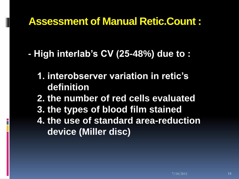
Clinical interests
↓
only on ↑ retics
(in hemorrhage, hemolysis, hematinic
therapy’s response)
↓
imprecision/inaccuracy was tolerable
21

7/18/2011 22
Modern medicine →increase of reticulocyte’s clinical utilities → need more precise & accurate retic.counts
22

7/18/2011 23
Reticulocyte’s Clinical Utilities
1. For hematological diagnosis :
- Classify anemic patients
- Assess bone marrow’s function
- Aplastic crisis
- Myelodysplastic Syndrome (MDS)
- Hemorrhages or hemolysis
23

7/18/2011 24
2. Treatment Monitoring :
- In EPO therapy
- As an indicator of Marrow’s regeneration
after chemotherapy or BMT
- Timing the Stem Cell harvest
24

7/18/2011 25
Automated reticulocytes count is more
accurate
( Manual : CV › 25% → 5-7%)
Blood cell counters permit precise
measurements of RNA content and cellular
indices (Volume, Hb-concentration and Hb-content)
25

7/18/2011 26
CV from manually Reticulocyte count &
Analyzers :
CV
Manual
CV
Automatic
Reticulocyte 1%
Reticulocyte 9%
47.3%
27.2%
6.4%
5.8%
26

7/18/2011 27
Hematology’s Automation
- 2 basic principles operation of
Hematology Analyzers :
1. Electronic Impedance / low-voltage DC
resistance (Coulter, 1950s)
2. Optical Scatter
27

7/18/2011 2828
Reticulocyte’s Automation
Earlier there were only 2 automatic methods for
counting reticulocyte :
- Computer-controlled automated
microscope for blood smear analysis (early
’80s)
- Flow cytometric methods

7/18/2011 2929
Automatic microscope → analogue with manual light-microscope, and scanned automatically NMB-stained blood films usinga pattern recognition devices .
→ a good equipment for its better reproducibility , the analysis results is as good as the manual methods . Unfortunately it is not so popular .

7/18/2011 3030
Automatic retic. Count method using
acridine-orange fluorescence (1952) →
the dye binds to retic’s ribosomal-RNA →
fluorescent in UV-light .
→ the intensity of fluorescence is
proportional to the RNA present → ~
maturity of the reticulocytes .
There is satisfactory agreement between
manual and automated methods in
categorizing the 4 Heilmeyer maturation
groups

7/18/2011 3131
Fluorescence staining combined with
flow-cytometry → led to the new automated
systems for reticulocyte counting .
Several different dyes have been used for
flow cytometric retics count .

7/18/2011 32
Flow cytometric Retic.counting
- Many fluorochromes have been used , i.e :
- Acridine orange
- Auramine O
- Di-methyloxacarbocyanide
- Ethidine bromide
- Pyronin -Y
- Thioflavine-T
- Thiazole orange
most require 30 minutes incubation → semi-
automated ?
32

7/18/2011 33
- Ethidium bromide at relatively high pH
require only few minutes to enter the cells
- Auramine O requires only a few seconds
33
7/18/2011 34
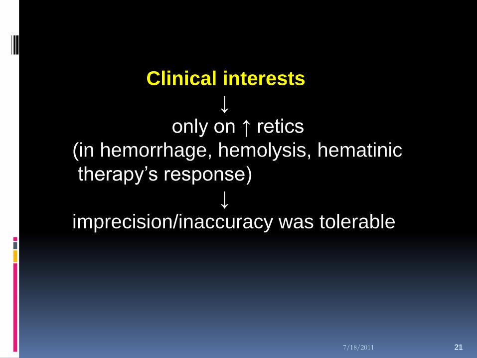
Cell Information - FCM
34
Side scatter
Information on amount of
RNA and DNA
Information on internal structure
Information on cell size
Forward scatter
Fluorescence
Laser
Beam
Dichroic mirror

7/18/2011 3535
Table 2. Evolution on parameters’ content of CBC
No Parameters Year
1
2
3
4
5
6
7
8
Hb, Hct, RBC, WBC
MCV, MCH, MCHC
PLT,RDW,PDW,MPV,Pct,
P-LCR,RDW-SD
LUC,CHCM,HDW,LI,MPXI
LYM,MONO,GRAN (%,#)
NEUT,EOS,BASO (%,#)
FLAGGING
RETICULOCYTE COUNT
1950s
1960s
1970s
1980s
1990s

7/18/2011 36
Current Options on Reticulocyte
Counting :
Method Dye Technique Usage CV-%
Microscopy, Supravital NMB/BCB Manual ± 70% 25
Flow Cytometry Thiazole O Fluorescence < 1% 15-20
Flow Cytometry NMB, spered
Optical scatter ± 10% 5-10
Flow Cytometry Oxazine 750 Optical scatter ± 4% 5-10
Flow Cytometry Auramine O Fluorescence ± 5% 5-10
36

7/18/2011 37
Retic.count methods on Hematology instruments
Coulter
LH-750
SysmexXE-2100
AbbottCD-4000
BayerAdvia 2120
Supravital staining(NMB) ; Volume, Conductivity, Optical Scatter(VCS technology)
Supravital staining (Auramin O)Fluorescent detection
CD4K530 stainMultiangle scatter and Fluorescent detection
Supravital staining (Oxazine 750)Low-angle (2-30) and High-angle (5-150, optical scatter and absorbance
37

7/18/2011 38
The improved precision for Flow
Cytometric methods arises from :
1. Removal of inter-observer variation
2. The larger number of cells counted (10000 – 30000 RBC events , compare to 1000 for the visual method)
3. The fluorescence measured is proportional to the amount of RNA present in the cell
38
7/18/2011 39
How the methods inform the maturation
of Retics ?
- The analyzers divide the retic area on the
scattergram by 2 vertical discriminators
→ producing 3 populations :
- Low Fluorescence Ratio (LFR) – the most
mature forms
- Middle Fluorescence Ratio (MFR)
- High Fluorescence Ratio (HFR) – the least
mature forms
39

7/18/2011 40
Fig. 6. Frequency’s curve of red cell maturation

7/18/2011 4141
Reticulocyte’s Automation
Fluorescence-based (Thiazole Orange,
Auramine O, fluorescence’dyes)
Absorbance-based (Oxazine O, New
Methylene Blue, nucleic acid dyes)
Interacting with RNA
Immature Retics high fluorescence
Thiazole Orange overestimated
because of DNA / RNA content of another
cells/components

7/18/2011 4242
Table 3. Reticulocyte’s parameters in various
hematology analyzers
Methods Parameters
CD 4000
(Abbott)
GEN-S
(Coulter)
SE-9000
(Sysmex)
Advia 120
(Bayer)
Fluorescen (Auramine O)
Absorbans (New
Methylene Blue)
Fluorescen (Auramine O)
Absorbans (Oxazine 750)
3
4
5
8
Retic(%,#), IRF
Retic(%,#),
IRF,RMI
Retic(%,#),
HFR,MFR,LFR
Retic(%,#),CHr,
MCVr, RDWr,
HDWr,CHCMr,
CHDWr

7/18/2011 43
Fig. 7. Reticulocyte Analysis
43
Absorbance
RNA Content
High angle detector
(5o - 15o)
Low angle detector
(2o-3o)
670nm
Laser
Diode
Oxazine 750
RNA
Stain

7/18/2011 44
Fig.8. Reticulocyte Technology
Reticulocytes are
stained with a nucleic
acid dye - Oxazine 750
Scatter and Absorption
are measured using
laser channel
RBC and Reticulocyte
indices are measured
simultaneously
448
Hig
h A
ngle
(5
-15 d
egre
es)
Oxazine 750 Absorption

7/18/2011 45

7/18/2011 46
Fig. 9. Reticulocyte Cytogram and Relationship to Maturity
46
Hig
h A
ngle
(5
-15 d
egre
es)
Oxazine 750 Absorbance

7/18/2011 47
Fig. 10. CBC results

7/18/2011 4848
- Sysmex methods (R-3000/3500) :
Sysmex use supravitally-fluorescence dye
, Auramine-O .
Forward-scatter (measuring size) and
side-fluorescence intensity(measure
RNA’s content)

7/18/2011 4949
forward light scatter & side fluorescence scattergram red cells, reticulocytes, platelets area
Reticulocytes area :
LFR (low fluorescence ratio)
MFR (middle fluorescence ratio)
HFR (high fluorescence ratio)IRF (Immature Retic.Fraction) = MFR + HFR
LFR : Heilmeyer III and IV
Flow-cytometry increase retics.count precision
Retic. 1% -- CV manl= 47.3%; automt = 6.4%
Retic. 9% -- CV manl= 27.2%; automt = 5.8%

7/18/2011 5050
- Bayer’s method (Advia-120) :
Bayer uses Oxazine-750 , a Nucleic-acid-
binding dye .
Erythrocyte changed into spherical-isovolumic
Measurement using 3 detectors :
1. Low-angle-scatter (2-3o)
2. High-angle-scatter (5-15o)
3. Absorbance

7/18/2011 5151
From these 3 detectors → 3 cytograms :
1. High-angle scatter vs Absorbans →
LAC/MAC/HAC , IRF (MAC + HAC)
2. High-angle vs Low-angle-scatter (RBC map) → Reticulocyte’s indices
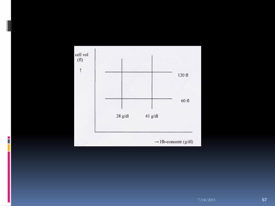
3. Volume vs Hb-concentration → CHr

7/18/2011 52
- Automated Reticulocyte’s Parameters :
Retics # and %
HAC, MAC, LAC (Absorbance/Fluorescence Ratio)
IRF (Immature Retics Fraction = HAC + MAC)
Retic’s indices (MCVr, MCHr, CHCMr, CHr, RDWr, HDWr)
52

7/18/2011 53
Fig. 11. CBC result ( normal patient )

7/18/2011 54
Fig. 12. CBC results ( Reticulocytosis )

7/18/2011 55
- Content of Retic’s Hb (CHr) :
Content of retic’s Hb never changed as long as the survival of Retics and red cells
The CHr’s mean : 28.5 pg
CHr/CH ratio ± 1 ( range : 0.96-1.03)
The meaningful of CHr :- indicate Iron availability real-time . - as a strong predictor for Iron Deficiency - Early indicator for Iron therapy in Iron Deficiency
Anemia .
55

7/18/2011 56
- Reticulocyte’s Size/Volume
(MCVr) :
Retic’s size drastically decreased along its
maturation ( Heilmeyer’s clasification )
MCVr/MCV ratio = 1.24 ( constant in normal,
microcytosis or macrocytosis)
Stress Retics : MCVr/MCV ratio = >1.5-3
Inverse MCVr/MCV ratio (=< 1) → seen in Vit.B12 therapy’s response in Megaloblastic Anemia .
56
7/18/2011 5757

7/18/2011 5858
Fig. 13. Red Cell Analisys

7/18/2011 59
Quantitation of :
- % Microcytic and % Macrocytic
- % Hypochromic and % Hyperchromic
↓
Differentiate between β-thal trait and
Iron Deficiency Anemia

7/18/2011 60
- Differential Diagnosis of β-Thal trait
and IDA :
In β-Thal trait :
The microcytosis is more significantcompared with mild hypochromia .
In Fe Deficiency Anemia :
The hypochromia is more significantcompared with mild microcytosis .
60

7/18/2011 61
Ratio % M/H (Micro/Hypo)
Ratio % M/H > 0.9 → β-Thal trait
Ratio % M/H < 0.9 → IDA
CHr combined with Ratio % M/H give
stronger differentiation ;
Ratio % M/H < 0.9 with Low CHr give
strong prediction for IDA

7/18/2011 62
Clinical Applications of Retic’s Indices
- Retics = the first erythroid cell appeared in circulation and become mature red cell 24 hours later .
- Red cell morphological begin to change in the late-stage of Fe deficiency
- Retic.Indices reflects a real-time erythropoietic activities

7/18/2011 63
- When Fe-store is low and
erythropoiesis is decreased , red cell
indicators are still normal
but marrow already release a new
retic. with low Hb-content ( low-CHr )
7/18/2011 64
- Early detection of Functional Fe
Deficiency is by measuring %-
Hypochromic and CHr
%-Hypochromic reflects Hb
concentration during 8-12 weeks .
- Fe-therapy responses have already
seen from the CHr in 4 days (1-2
weeks) when normally it’s only seen
from the increament of Hb after 1
month therapy

7/18/2011 6565

7/18/2011 6666

7/18/2011 67
- Hypochromic Macrocytic Cells ?
Reticulocyte ?
Dyserythropoiesis / Myelodysplastic
syndrome / Sideroblastic anemia
Fe-deficient megaloblastic anemia
67

7/18/2011 68

7/18/2011 69
Limitations
- Most dyes stain also other blood
components containing DNA/RNA, so
assessed as reticulocytes, i.e :
Howell-Jolly bodies, Cabot’s ring,
Malaria parasites, white cell’s
fragments in Leukemia
- Retics may “mature” during storage
especially if not refrigerated

7/18/2011 70
Thank U