Embed Size (px)
Citation preview

This article appeared in a journal published by Elsevier. The attachedcopy is furnished to the author for internal non-commercial researchand education use, including for instruction at the authors institution
and sharing with colleagues.
Other uses, including reproduction and distribution, or selling orlicensing copies, or posting to personal, institutional or third party
websites are prohibited.
In most cases authors are permitted to post their version of thearticle (e.g. in Word or Tex form) to their personal website orinstitutional repository. Authors requiring further information
regarding Elsevier’s archiving and manuscript policies areencouraged to visit:
http://www.elsevier.com/copyright
Author's personal copy
Atherosclerosis 218 (2011) 356–362
Contents lists available at ScienceDirect
Atherosclerosis
journa l homepage: www.e lsev ier .com/ locate /a therosc leros is
Asymptomatic carotid plaque rupture with unexpected thrombosis over anon-canonical vulnerable lesion
Alessandro Maurielloa,∗ , Francesca Servadeia , Giuseppe Sangiorgib , Lucia Anemonaa , Erica Giacobbia ,Doriana Liotti a, Luigi Giusto Spagnoli a
a Anatomic Pathology, University of Rome Tor Vergata, Italyb Department of Cardiology, University of Rome Tor Vergata, Italy
a r t i c l e i n f o
Article history:Received 11 May 2011Received in revised form 23 June 2011Accepted 28 June 2011Available online 12 July 2011
Keywords:CarotidThrombosisAsymptomaticsPathology
a b s t r a c t
Objective: Several studies have demonstrated that carotid plaque rupture and thrombosis represent themost important factors correlated with the onset of acute cerebrovascular symptoms. Nevertheless, rup-tured thrombotic plaques have been described also in asymptomatic patients. What still needs to beclarified is why a plaque rupture leads either to an acute ischemic syndrome or, in a minor group ofpatients, remains asymptomatic. The purpose of this study was to systematically compare the histo-logic features of thrombotic plaques both in asymptomatic and symptomatic patients in order to identifyspecific findings that could explain the peculiar clinical behavior that characterizes each of the clinicalsettings.Methods: A total of 157 thrombotic plaques from 60 asymptomatic patients and 97 with major strokewho consecutively underwent CEA were serially sectioned and studied by histology.Results: A minute cap disruption very frequently characterizes thrombotic plaques of asymptomaticpatients and it was always smaller than large ulcers observed in thrombotic symptomatic plaques(651 ± 687 �m vs. 4150 ± 3526, p = 0.001). In asymptomatics this typical feature was associated withfewer inflammatory cells (20.1 ± 8.8 vs. 33.9 ± 26.1 cells × hpf, p = 0.001), smaller lipidic–necrotic core(33.9% ± 2.9% vs. 42.0% ± 2.4%; p = 0.04) and larger calcification (16.2 ± 12.8% vs. 8.1 ± 12.2%, p = 0.02).Symptomatic patients with thrombotic plaques showed higher incidence of metabolic syndrome(p = 0.002) and moderate-high Framingham risk scores (p = 0.001) comparing to asymptomatic individu-als.Conclusion: The transformation from a stable to a vulnerable plaque is a gradual process in the naturalhistory of the disease and plaque rupture is an event not necessarily occurring at a late phase but also atearlier one. In this case, the rupture will be most likely smaller and clinically asymptomatic.
© 2011 Elsevier Ireland Ltd. All rights reserved.
1. Introduction
The importance of carotid atherosclerosis and related throm-boembolic occlusion of a large artery tributary of the brain in thepathogenesis of ischemia and cerebral infarction has been acknowl-edged for many years [1–4]. Although the grade of stenosis due toa plaque encroaching the lumen constitutes the major criterion toidentify subgroups of patients at high risk of stroke [5,6], results
∗ Corresponding author at: Cattedra di Anatomia ed Istologia Patologica, Dipar-timento di Biopatologia e Diagnostica per Immagini, Universita’ di Roma “TorVergata”, Via Montpellier 1, 00133 Roma, Italy. Tel.: +39 06 2023751;fax: +39 06 20902209.
E-mail address: [email protected] (A. Mauriello).
from several studies suggest that plaque rupture and thrombosisrepresent the most important factor correlated with the onset ofsymptoms [1,3]. In a previous study, we observed a ruptured andthrombotic plaque in 74% of patients with ipsilateral stroke and in100% of patients treated within 2 months from symptoms onset[1]. This strong correlation between the plaque thrombosis and anacute cerebrovascular disease has been confirmed by other mor-phologic and imaging-based studies [7,8]. Thus, these data suggestthat 2 types of carotid artery disease can occur: one stable form,unlikely to produce symptomatic embolization or carotid occlu-sion and a second, unstable form at high risk of embolization orcarotid occlusion, the latter is not necessarily being more stenotic.
Many efforts have been recently made using non-invasive tech-niques [8–12] for identifying plaques at high risk of disruptionleading to thrombosis, generally defined as “vulnerable plaques”
0021-9150/$ – see front matter © 2011 Elsevier Ireland Ltd. All rights reserved.doi:10.1016/j.atherosclerosis.2011.06.056

Author's personal copy
A. Mauriello et al. / Atherosclerosis 218 (2011) 356–362 357
[13,14]. These latter lesions are characterized by a large necroticcore with an overlying thin cap rich in inflammatory cells. Unlikethe stable plaque which shows a chronic inflammatory infiltrate,both vulnerable and ruptured plaques are characterized by achronic “active” inflammation [15].
Ruptured plaques have been also described in asymptomaticpatients. While ruptured symptomatic plaques have been widelyexamined, only few data are available on asymptomatic ruptures.What still needs to be clarified is why a plaque rupture can lead toan acute ischemic syndrome or, alternatively, in a minor group ofpatients, remain asymptomatic.
The aim of this study was to systematically compare the his-tologic features of thrombotic plaques both in asymptomatic andsymptomatic patients in order to identify specific findings thatcould explain the peculiar clinical behavior that characterizes eachof the clinical settings.
2. Materials and methods
2.1. Selection of cases
From the 486 consecutive carotid endarterectomies present inour database – The Interinstitutional Carotid Tissue Bank [1] – andcollected from 2006 to 2010, a total of 157 thrombotic plaquesfrom 60 asymptomatic patients and 97 with major stroke formedthe basis of the study. A routine pre-operative evaluation in allpatients included: clinical assessment of risk factor profile, a cere-bral CT scan study, a selective angiographic examination of extraand intracranial carotid arteries and their branches.
Major stroke was defined as a clinical syndrome characterizedby rapidly developing focal or at times global symptoms withoutsignificant clinical improvement within 7 days in the distributionof symptomatic carotid artery, not hemorrhagic and with no causeother than vascular origin, assessed by brain CT study as a corti-cal or deep white matter or basal ganglia lesion bigger than 1 cm[1]. Asymptomatic patients had never suffered from neurologicalsymptoms and had no cerebral lesions assessed by CT.
The time interval between symptom onset and CEA in strokepatients was from 1 to 6 months. Aspirin was administered to allpatients prior to surgery.
The study was approved by the institutional review boards andall patients gave consent for entry into the study. Histopathologicexamination was performed in the pathology core lab of the Uni-versity of Rome Tor Vergata by three different pathologists (AM, FS,LGS) blinded to the clinical data.
2.2. Histologic sampling and light microscopy
Carotid plaques were removed en bloc during surgery to entirelypreserve plaque structure. Surgical samples were fixed for 24 h in10% buffered formalin immediately upon removal. The specimenswere sectioned transversally every 5 mm and paraffin embedded.Haematoxylin–eosin and Movat pentachrome stains were per-formed for morphological study. Each segment was sequentiallynumbered to reconstruct the entire plaque length. For each plaque,three to ten sections were examined according to the plaque length(mean 5 sections per specimen).
The presence of acute thrombosis, cap rupture or erosion, min-imum cap thickness, cross-sectional area of the lipidic–necroticcore, intraplaque hemorrhage and calcification were evaluated pereach plaque. The inflammatory infiltrate was also studied using thefollowing monoclonal antibodies: CD68 (anti human macrophages;Dakopatts, Denmark), CD163 (anti M2 macrophages, NovocastraLeica, United Kingdom) and CD3 (anti-human T cell; Dakopatts)monoclonal antibodies.
An ultrastructural study was also performed in selected cases.Tissue samples were postfixed in 1% phosphate-buffered osmiumand embedded in epoxy resin (Epon 812). Ultrathin sections wereexamined by means of Hitachi H-7100FA electron microscope.
2.3. Atherosclerotic plaque types and histologic definitions
Plaques were classified, according to the modified AmericanHeart Association (AHA) atherosclerosis classification [14] intothrombotic and non-thrombotic.
Thrombotic plaques were characterized by the presence of anacute thrombus associated to plaque rupture, erosion or calcifiednodule. Plaque rupture was defined by an area of fibrous cap dis-ruption whereby the overlying acute thrombus was in continuitywith the underlying necrotic core [14]. Ruptures were further clas-sified into two subgroups according to their dimension: (a) ≤1 mm(also called fissure) and (b) >1 mm (called ulceration). Plaque ero-sion was identified when serial sectioning of a thrombosed arterialsegment failed to reveal fibrous cap rupture [14]. Calcified nod-ule referred to a lesion with luminal thrombus associated with aneruptive, dense area of calcium and underlying calcific plate [14].Some plaques were classified as thrombotically active (TAP) if anacute thrombus was present over an organized or an organizingthrombus [1].
Non-thrombotic plaques included vulnerable plaques or thincap fibroatheromata, fibrous cap atheromata, fibrocalcific lesionsand healed plaque ruptures.
As vulnerability criteria for carotid plaque the presence of athin fibrous cap (<165 �m), heavily infiltrated by macrophages(>25 cells per high power field – hpf, done at a magnification of40× using a test grid with an area of 0.22 mm2) associated to alarge lipidic core (30–40% of plaque area) were considered [13,16].
2.4. Risk factors
The following risk factors were evaluated: hypertension, dia-betes mellitus, cigarette smoking (former smokers who hadstopped smoking for <5 years were considered as smokers andpatients who had not smoked for >5 years were considered as non-smokers), hypercholesterolemia, elevated triglyceridemia (TG),low HDL-cholesterol (HDL-C), abdominal obesity (patients with awaist circumference ≥102 cm in men or ≥88 cm in women [5]).In addition, we evaluated the presence of metabolic syndromeand the Framingham risk score [17]. The metabolic syndrome wasdefined, according to the scientific statement from the AHA andthe National Heart, Lung, and Blood Institute (NHLBI) [18], if any 3of the following 5 abnormalities were present: (1) elevated waistcircumference, (2) elevated TG, (3) elevated blood pressure (BP)(≥130 mmHg systolic BP or ≥85 mmHg diastolic BP or drug treat-ment), (4) elevated fasting glucose (>100 mg/dL or drug treatment),and (5) low HDL-C. The Framingham risk score [17] was calculatedutilizing an algorithm which includes age, gender, smoking status,presence of diabetes as dichotomous parameters, as well as totalcholesterol, HDL-C, diastolic and systolic BP using the categoriesdefined in the Framingham equation.
2.5. Statistical analysis
Data were analyzed by SPSS 13.0 (Statistical Package for theSocial Sciences) software. Continuous and categorical variables areexpressed as mean ± SD and as frequency values and proportions,respectively. Pearson’s chi-square test was utilized to assess pos-sible differences of dichotomous variables between plaques of thevarious groups examined. The means of normally distributed datawere compared with Student’s t test. In the other cases the groupswere compared with Mann–Whitney’s U test. Multivariate analy-

Author's personal copy
358 A. Mauriello et al. / Atherosclerosis 218 (2011) 356–362
sis using stepwise logistic regression (using the “enter” method forvariable selection) was utilized to identify independent risk fac-tors which significantly correlate with plaque thrombosis. Receiveroperating characteristic curves (ROC; for ulcer size, number ofinflammatory cells, lipidic–necrotic core % size, and calcification% size) with corresponding area under the curve (AUC) have beenevaluated to describe the optimal cut-offs for distinction betweenasymptomatic and symptomatic plaques. In addition, the AUCs ofROCs from different plaque features were compared according toVergara et al. [19]. The ROC curves provided several cutoff pointsto show the trade-off between sensitivity and specificity at differ-ent cutoff values. Sensitivity and specificity values were calculatedwith confidence interval analysis according to the Wilson method.A probability value <0.05 was considered statistically significant.
3. Results
3.1. Thrombotic plaque characteristics
Out of 157 thrombotic plaques, 95 (38 asymptomatic patientsand 57 with stroke) showed an acute thrombosis, while in theremaining 62 plaques an organizing thrombus (TAP) was found(Table 1). The incidence of thrombosis was significantly higher inpatients affected by stroke, compared to asymptomatic patients(p = 0.02). The grade of luminal narrowing of the thrombotic seg-ment was significantly lower in asymptomatic than in symptomaticpatients (58.1 ± 11.9% vs. 68.0 ± 12.8%, p = 0.01, Table 1). In about90% of asymptomatic patients thrombosis was found in segmentswith <70% stenosis.
In 24 out of 38 asymptomatic patients with acute thrombosis(63.1%) a ruptured plaque was found. It is noteworthy that asymp-tomatic plaque ruptures were significantly smaller than thosefound in stroke patients (651 ± 687 �m vs. 4150 ± 3526 respec-tively, p = 0.001) (Fig. 1). In fact, in 20 out of 24 cases the rupturewas characterized by only a small fissure (<1 mm).
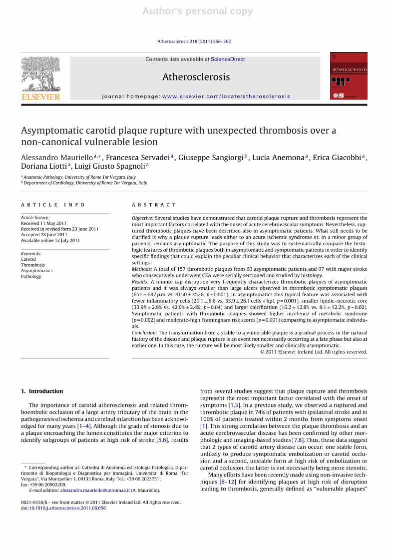
Moreover 10.5% of asymptomatic patients (4/38) showeda plaque erosion associated with the presence of scatteredmacrophages and numerous mast cells (Fig. 2). In all these casesa calcification underneath the eroded area was observed.
Finally, in 26.3% of asymptomatic patients with thromboticplaques (10/38) a calcified nodule was present (Fig. 2).
The location of acute thrombotic plaques was in the commoncarotid in 11 cases (11.6%, 5 asymptomatic and 6 symptomatic), atlevel of bifurcation in 59 (62.1%; 22 asymptomatic and 37 symp-tomatic) and in the remaining 25 cases in the internal carotid(26.3%, 11 asymptomatic and 14 symptomatic), with no signi-ficative differences (p = 0.67). Moreover, no statistical differenceswere observed in the incidence of various types of plaques rupturebetween patients treated and non-treated with statins (p = 0.78).
3.2. Features of vulnerability
The thickness of fibrous cap near the rupture site in asymp-tomatic plaques was about twice that of plaques of strokepatients (183.2 ± 67.1 vs. 84.4 ± 38.6, p = 0.001) and showed amild macrophagic infiltrate, mainly of M2 phenotype (Fig. 1D).The inflammatory infiltrate in asymptomatic plaques was abouthalf of that observed in symptomatic ones (20.1 ± 7.2 vs.33.9 ± 14.1 cells × hpf, p = 0.001).
Asymptomatic ruptured plaques showed also a significantlyhigher extension of calcification as compared to stroke patients.In fact, the relative area occupied by calcium deposits in asymp-tomatic ruptured plaques was 16.2 ± 12.8%, while in symptomaticthrombotic lesions it was 8.1 ± 12.2% (p = 0.02). Similarly, in asymp-tomatic thrombotic plaques the relative area of lipidic–necrotic
core was smaller than that of symptomatic ones (33.9% ± 2.9% vs.42.0% ± 2.4%; p = 0.04).
Intraplaque hemorrhage is another finding that we frequentlyobserved both in non-ruptured and in thrombotic plaques ofasymptomatic patients. We cannot establish if the hemorrhage is aconsequence of the blood flowing from the lumen through the caprupture or if it represents, on the contrary, the cause of the cap rup-ture. This latter hypothesis is supported by the observation of manysmall and thin-walled vessels within the fibrous cap. Moreover, theultrastructural examination showed that these microvessels lackinterendothelial junctions, explaining the fragility of these vascularchannels (Fig. 1E and F).
We also constructed ROC curves by plotting sensitivity (theproportion of true-positive results) vs. 1-specificity (the propor-tion of false-positive results) and determined the correspondingAUC to show the ability of some plaque morphological featuresto discriminate symptomatic from asymptomatic rupture. Bothminimum cap thickness (AUC = 0.90, 95% CI 0.61–1.00) and calcifi-cation (AUC = 0.70, 95% CI 0.59–0.81) showed a moderate diagnosticaccuracy, AUC being in the interval 0.7–0.9 [20]. The ROC curvesalso provided several cutoff points to show the tradeoff betweensensitivity and specificity at different cutoff values. For minimumcap thickness, the optimal threshold was 122 �m correspondingto 86% sensitivity and 82% specificity, whereas for calcificationarea the optimal threshold was 7.5% with 65% sensitivity and 72%specificity. Regarding other plaque characteristics the optimal cut-off was 715 �m for ulcer size (sensitivity = 44%, specificity = 86%),27 cells × hpf for inflammatory infiltrate (sensitivity = 21%, speci-ficity = 56%) and 42.5% for the relative area of lipidic–necrotic core(sensitivity = 50%, specificity = 41%).
In symptomatic plaques no significative correlation wasobserved between plaque morphologic features and time fromsymptom onset and endarterectomy.
3.3. Correlation with risk factors
The univariate analysis showed that among the single riskfactors only hypertension and low HDL-C were more frequentlyobserved in symptomatic than in asymptomatic patients (p = 0.04and p = 0.02, respectively) (Table 2). Moreover, symptomaticpatients with thrombotic plaques showed higher incidence ofmetabolic syndrome comparing to asymptomatic individuals(47.4% vs. 15.8% respectively, p = 0.002). Similarly, moderate-highFramingham risk scores were more common in symptomaticpatients (68.4%), while in the asymptomatic group low-mediumscores were prevalent (p = 0.001). Multivariate analysis showedsimilar results, confirming that metabolic syndrome and Fram-ingham risk score were independent predictive factors for thesymptomatic carotid thrombosis.
4. Discussion
Our results demonstrate that asymptomatic ruptures andthrombosis frequently occur in the carotid district. Asymp-tomatic thrombotic plaques differ from symptomatic ones for thepresence of a small disruption of a thicker cap, fewer inflamma-tory cells mainly represented by M2 type macrophages, smallerlipidic–necrotic core and larger calcification. These features are notcompletely consistent with the morphological definition of a vul-nerable plaque, mainly as regarding cap thickness and amount ofinflammatory infiltrate (Table 1).
The present study suggests that carotid plaques could showseveral levels of vulnerability correlated to different histologicalfeatures. The acquisition of vulnerability is a gradual process andplaque rupture is an event not necessarily occurring at the end

Author's personal copy
A. Mauriello et al. / Atherosclerosis 218 (2011) 356–362 359
Table 1Morphological findings of thrombotic plaques.
Thrombotic plaques p
Asymptomatic (60 patients) Stroke (97 patients)
Plaque types(A) Acute thrombosis 38 (63.3) 57 (58.8) 0.02Fissure (<1 mm) 20 (33.3) 5 (5.1)Ulceration (>1 mm) 4 (6.7) 42 (43.3)Erosion 4 (6.7) 3 (3.1)Calcified nodule 10 (16.6) 7 (7.2)(B) Organizing thrombosis 22 (36.7) 40 (41.2)Rupture dimension (�m ± SD) 651 ± 687 4150 ± 3526 0.001Stenosis of thrombotic segment (% + SD) 58.1 ± 11.9 68.0 ± 12.8 0.01
Features of vulnerabilityCap thickness (�m ± SD) 183.2 ± 67.1 84.4 ± 38.6 0.001Cap macrophagic infiltrate (no. of cells/hpf ± SD) 20.1 ± 7.2 33.9 ± 14.1 0.001Lipidic–necrotic core (area % ± SD) 33.9 ± 2.9 42.0 ± 2.4 0.04Calcification (area % ± SD) 16.2 ± 12.8 8.1 ± 12.2 0.02
of the natural history of the disease but also in an earlier phase.The accepted criteria for the definition of a carotid vulnerableplaque are the presence of a thin fibrous cap (<165 �m), heav-ily infiltrated by macrophages (>25 cells per hpf) associated to alarge lipidic core (30–40% of plaque area) [13,16]. This type plaqueis at high risk of wide symptomatic ruptures and thrombosis.Our study demonstrates that there are “non canonical” vulnera-ble plaques characterized by mild inflammation, thicker fibrouscap, that will undergo small asymptomatic ruptures and repair.Prospective studies using imaging methods could determine ifa plaque type represents an earlier vs. later stage of vulnera-bility. Today it is impossible to say whether each patient willprogress through different stages of vulnerability. It is possiblethat asymptomatic patients will always be asymptomatic, and thatsymptomatic patients never had asymptomatic ruptures.
In asymptomatic lesions the presence of few macrophagic cells,mainly of M2 subtype, as compared to symptomatic plaques,suggests that not only a smaller infiltration, but also a differ-ent pattern of inflammation correlate with asymptomatic plaquerupture. It has been established that monocytes, in response todifferent cytokine pattern and immunological microenvironment,can differentiate in two major subpopulations, referred to as M1and M2 macrophages. M1 macrophages are considered to pro-mote atherosclerosis development and plaque rupture, secretingcytokines and lytic enzymes involved in the thinning of fibrouscap, while M2 macrophages are expected to inhibit plaque growthand to promote reparative processes, mediating plaque stabil-ity [21,22]. Since in asymptomatic plaque ruptures we observed
an intense M2 response, it could be hypothesized that in theselesions a reparative response rapidly occurs. Nevertheless we can-not establish if M2 macrophages were already present in theplaque before the rupture occurred or if they were attractedfollowing cap disruption as a consequence of thrombosis andhemorrhage. In fact, it has been recently demonstrated that M2macrophages are able to bind and clear haemoglobin–haptoglobincomplexes from the vessel wall using the scavenger receptor CD163[23].
Also in asymptomatic erosions we observed a modest inflam-matory infiltrate. Nevertheless, several mast cells were found(Fig. 2E and F). It has been demonstrated that subendothelial mastcells, by inducing apoptosis of endothelial cells [24], may inducedetachment of endothelium from the cap thus determining plaqueerosion.
It has been widely accepted that major coronary and cerebrovas-cular risk factors increase vascular inflammation, thus mediatingplaque instability [16]. Although most risk factors have an inde-pendent effect, there may be important interactions betweenthem, amplifying their action. Our observation that patientswith symptomatic thrombotic plaques, characterized by a greaterinflammatory activity, had a significantly higher Framingham riskscore as compared to that of asymptomatic individuals, seemsto confirm this hypothesis. Similarly, it is not surprising thatsymptomatic patients with highly inflamed plaques and wide capruptures and thrombosis tend to be more frequently affected bymetabolic syndrome. Metabolic syndrome is a common and com-plex disorder combining obesity, dyslipidemia, hypertension and
Table 2Risk factors and thrombotic plaques.
Risk factors Thrombotic plaque Univariate analysis (p) Multivariate analysis (p)
Asymptomatic (38 patients) Stroke (57 patients)
Age, mean (±SD) 70.2 ± 7.5 70.1 ± 7.8 0.97 0.80Sex, N (%)Male
30 (78,9) 47 (82.5) 0.93 0.62
Female 8 (21.1) 10 (17.5)Hypertension, N (%) 23 (60.5) 47 (82.5) 0.04 0.17Diabetes, N (%) 5 (13.2) 13 (22.8) 0.27 0.76Cigarette smoking, N (%) 21 (55.3) 40 (70.2) 0.21 0.47Hypercholesterolemia, N (%) 23 (60.5) 40 (70.2) 0.46 0.82Low HDL-C, N (%) 13 (34.2) 35 (61.4) 0.02 0.12Hypertriglyceridemia, N (%) 13 (34.2) 29 (50.9) 0.15 0.56Abdominal obesity, N (%) 4 (10.5) 9 (15.8) 0.57 0.86Metabolic syndrome, N (%) 6 (15.8) 27 (47.4) 0.002 0.005Framingham risk score, N (%)
Low–medium 26 (68.4) 18 (31.6) 0.001 0.001Moderate–high 12 (31.6) 39 (68.4)
In bold statistically significant differences are reported.

Author's personal copy
360 A. Mauriello et al. / Atherosclerosis 218 (2011) 356–362
Fig. 1. Histopathology of an asymptomatic carotid plaque rupture and thrombosis. Panels A–C: micrographs showing different histologic plaque rupture samples fromasymptomatic patients submitted to surgical carotid endarterectomy. Rupture sites (Rup) are characterized by a small disruption (arrow) of the thin fibrous cap withoverlying acute thrombus in continuity with necrotic core debris. In the inset a magnification of rupture site is reported with its dimension. In the panel C a large calcificationis present under the small cap rupture (A–B: Movat pentachrome stains, 4×; C: Haematoxylin–eosin stain, 2×). Panel D: the fibrous cap near the rupture site showed amild macrophagic infiltrate, mainly of M2 phenotype, positive to CD163 antibody (10×). Panel E: an intramural hemorrhage associated to the presence of many small andthin-walled vessels within the fibrous cap was frequently observed in plaques of asymptomatic patients. The ultrastructural examination showed that these microvesselslack interendothelial junctions, explaining the fragility of these vascular channels. A magnification of (E) is reported in the Panel F.
insulin resistance. Hyperinsulinemia and impaired glycaemic con-trol were associated with an increase of in vivo LDL oxidationand inflammation [25,26]. Dyslipidemia, associated with high lev-els of triglycerides and low HDL concentrations, contributes to aproinflammatory state [27,28]. Moreover, increasing experimentalevidences support the hypothesis that also hypertension induces anincrement of oxidative stress in the arterial wall, favoring thereforethe recruitment of the inflammatory cells [16,29,30].
It is noteworthy that 26.3% of acute thrombosis in asymptomaticpatients has been observed in correspondence with a calcified nod-ule. This kind of lesion has been described in coronary circulation[14] and it is rarely observed in carotids. Subendothelial calcifiednodules in asymptomatic patients are usually localized in the con-text of thin cap plaques and mildly stenotic segments. Both theseconditions, according to Laplace law, make these plaques highlysusceptible to rupture [13,31]. Moreover, it has to be emphasized
Author's personal copy
A. Mauriello et al. / Atherosclerosis 218 (2011) 356–362 361
Fig. 2. Histopathology of asymptomatic carotid plaque erosions and calcified nodule. Panels A and B: micrographs showed two plaque erosions (er) associated to the presenceof broad calcific plates (ca) (Movat pentachrome stains, 4×). Panel C: transmission electron micrograph of a mast cell in the subepithelial space under the plaque erosion. Amagnification of (C) is reported in the Panel D. Panels E and F: micrographs showed three asymptomatic carotid plaques constituted by a luminal thrombus associated withan eruptive, dense area of calcium and underlying calcific plate (Movat pentachrome stains, 4×).
that these calcified nodules, protruding into the vascular lumen,can induce flow disturbances, increase the stiffness and reduce theelasticity of the vessel wall [32].
In our cases, broad calcific plates have been also observed inthe other subtypes of asymptomatic thrombotic plaques, such asrupture (Fig. 1C) and erosion (Fig. 2A and B). This finding is onlyapparently in contradiction with previous data reported in theliterature showing that in the carotid the calcium deposition is asso-ciated with stable lesions [33,34]. In fact, in our cases symptomaticthrombotic plaques have little calcifications, while wide calcificplates occur in the small asymptomatic plaque ruptures.In con-clusion, our results demonstrate that two types of plaque rupturecan occur in the carotid, probably related to different pathogeneticmechanisms. When plaque rupture is associated with the onsetof an acute cerebrovascular event, a wide cap disruption can befound in the context of a lesion showing the classic features ofvulnerable plaque (high active inflammation, thin fibrous cap). On
the contrary, when small-sized plaque ruptures occur, those arecharacterized by mild inflammation, thicker fibrous cap, broad cal-cification and mural hemorrhage, and are most likely correlatedwith an increased parietal stress. This rupture is likely to undergorepair and organization. The organization of these small asymp-tomatic ruptures could represent a frequent mechanism of plaquegrowth, as suggested by the fact that in 20% of asymptomaticpatients a healed lesion has been observed.
Financial support
This study was supported by grant from MURST (MURST did notparticipate in the design and conduct of the study, in the collec-tion, analysis, and interpretation of the data, or in the preparation,review, or approval of the manuscript).
Author's personal copy
362 A. Mauriello et al. / Atherosclerosis 218 (2011) 356–362
Conflict of interest
There are no potential conflicts of interest that relate to themanuscript.
References
[1] Spagnoli LG, Mauriello A, Sangiorgi G, et al. Extracranial thrombotically activecarotid plaque as a risk factor for ischemic stroke. JAMA 2004;292:1845–52.
[2] Redgrave JN, Lovett JK, Gallagher PJ, Rothwell PM. Histological assessmentof 526 symptomatic carotid plaques in relation to the nature and tim-ing of ischemic symptoms: the Oxford plaque study. Circulation 2006;113:2320–8.
[3] Virmani R, Ladich ER, Burke AP, Kolodgie FD. Histopathology of carotidatherosclerotic disease. Neurosurgery 2006;59:S219–27, discussion S213.
[4] Golledge J, Greenhalgh RM, Davies AH. The symptomatic carotid plaque. Stroke2000;31:774–81.
[5] Sacco RL, Adams R, Albers G, et al. Guidelines for prevention of stroke in patientswith ischemic stroke or transient ischemic attack: a statement for healthcareprofessionals from the American Heart Association/American Stroke Asso-ciation Council on Stroke: co-sponsored by the Council on CardiovascularRadiology and Intervention: the American Academy of Neurology affirms thevalue of this guideline. Stroke 2006;37:577–617.
[6] Goldstein LB, Adams R, Alberts MJ, et al. Primary prevention of ischemic stroke:a guideline from the American Heart Association/American Stroke AssociationStroke Council: cosponsored by the Atherosclerotic Peripheral Vascular DiseaseInterdisciplinary Working Group; Cardiovascular Nursing Council; Clinical Car-diology Council; Nutrition, Physical Activity, and Metabolism Council; and theQuality of Care and Outcomes Research Interdisciplinary Working Group: theAmerican Academy of Neurology affirms the value of this guideline. Stroke2006;37:1583–633.
[7] Takaya N, Yuan C, Chu B, et al. Association between carotid plaque characteris-tics and subsequent ischemic cerebrovascular events: a prospective assessmentwith MRI-initial results. Stroke 2006;37:818–23.
[8] Chu B, Ferguson MS, Chen H, et al. Magnetic resonance imaging features ofthe disruption-prone and the disrupted carotid plaque. JACC Cardiovasc Imag2009;2:883–96.
[9] Saam T, Cai J, Ma L, et al. Comparison of symptomatic and asymptomaticatherosclerotic carotid plaque features with in vivo MR imaging. Radiology2006;240:464–72.
[10] Fabiano S, Mancino S, Stefanini M, et al. High-resolution multicontrast-weighted MR imaging from human carotid endarterectomy specimens to assesscarotid plaque components. Eur Radiol 2008;18:2912–21.
[11] Coli S, Magnoni M, Sangiorgi G, et al. Contrast-enhanced ultrasound imaging ofintraplaque neovascularization in carotid arteries: correlation with histologyand plaque echogenicity. J Am Coll Cardiol 2008;52:223–30.
[12] Underhill HR, Hatsukami TS, Fayad ZA, Fuster V, Yuan C. MRI of carotidatherosclerosis: clinical implications and future directions. Nat Rev Cardiol2010;7:165–73.
[13] Falk E, Shah PK, Fuster V. Coronary plaque disruption. Circulation1995;92:657–71.
[14] Virmani R, Kolodgie FD, Burke AP, Farb A, Schwartz SM. Lessons from sud-den coronary death: a comprehensive morphological classification scheme foratherosclerotic lesions. Arterioscler Thromb Vasc Biol 2000;20:1262–75.
[15] Libby P, Ridker PM, Hansson GK. Inflammation in atherosclerosis: from patho-physiology to practice. J Am Coll Cardiol 2009;54:2129–38.
[16] Mauriello A, Sangiorgi GM, Virmani R, et al. A pathobiologic link between riskfactors profile and morphological markers of carotid instability. Atherosclerosis2010;208:572–80.
[17] Wilson PW, D’Agostino RB, Levy D, et al. Prediction of coronary heart diseaseusing risk factor categories. Circulation 1998;97:1837–47.
[18] Grundy SM, Cleeman JI, Daniels SR, et al. Diagnosis and management of themetabolic syndrome: an American Heart Association/National Heart, Lung, andBlood Institute Scientific Statement. Circulation 2005;112:2735–52.
[19] Vergara IA, Norambuena T, Ferrada E, Slater AW, Melo F. StAR: a simple tool forthe statistical comparison of ROC curves. BMC Bioinformatics 2008;9:265.
[20] Akobeng AK, Thomas AG. Enteral nutrition for maintenance of remission inCrohn’s disease. Cochrane Database Syst Rev 2007 [CD005984].
[21] Mantovani A, Sica A, Sozzani S, et al. The chemokine system in diverse formsof macrophage activation and polarization. Trends Immunol 2004;25:677–86.
[22] Stoger JL, Goossens P, de Winther MP. Macrophage heterogeneity: rele-vance and functional implications in atherosclerosis. Curr Vasc Pharmacol2010;8:233–48.
[23] Fabriek BO, Dijkstra CD, van den Berg TK. The macrophage scavenger receptorCD163. Immunobiology 2005;210:153–60.
[24] Kovanen PT. Mast cells: multipotent local effector cells in atherothrombosis.Immunol Rev 2007;217:105–22.
[25] Moreno PR, Fuster V. New aspects in the pathogenesis of diabetic atherothrom-bosis. J Am Coll Cardiol 2004;44:2293–300.
[26] Ceriello A, Motz E. Is oxidative stress the pathogenic mechanism underly-ing insulin resistance, diabetes, and cardiovascular disease? The common soilhypothesis revisited. Arterioscler Thromb Vasc Biol 2004;24:816–23.
[27] Adams MR, Kinlay S, Blake GJ, et al. Atherogenic lipids and endothelial dys-function: mechanisms in the genesis of ischemic syndromes. Annu Rev Med2000;51:149–67.
[28] Barter PJ, Nicholls S, Rye KA, Anantharamaiah GM, Navab M, Fogelman AM.Antiinflammatory properties of HDL. Circ Res 2004;95:764–72.
[29] Schiffrin EL. Beyond blood pressure: the endothelium and atherosclerosis pro-gression. Am J Hypertens 2002;15:S115–22.
[30] Spagnoli LG, Mauriello A, Palmieri G, et al. Relationships between risk fac-tors and morphological patterns of human carotid atherosclerotic plaques. Amultivariate discriminant analysis. Atherosclerosis 1994;108:39–60.
[31] Li ZY, Howarth SP, Tang T, Gillard JH. How critical is fibrous cap thick-ness to carotid plaque stability? A flow–plaque interaction model. Stroke2006;37:1195–9.
[32] Davies PF. Hemodynamic shear stress and the endothelium in cardiovascularpathophysiology. Nat Clin Pract Cardiovasc Med 2009;6:16–26.
[33] Kwee RM. Systematic review on the association between calcification in carotidplaques and clinical ischemic symptoms. J Vasc Surg 2010;51:1015–25.
[34] Wahlgren CM, Zheng W, Shaalan W, Tang J, Bassiouny HS. Human carotidplaque calcification and vulnerability. Relationship between degree of plaquecalcification, fibrous cap inflammatory gene expression and symptomatology.Cerebrovasc Dis 2009;27:193–200.