Embed Size (px)
Citation preview
Author Version: Dev. Comp. Immunol., vol.55; 2016; 241-252 The zebrafish galectins Drgal1-L2 and Drgal3-L1 bind in vitro to the infectious hematopoietic
necrosis virus (IHNV) glycoprotein and reduce viral adhesion to fish epithelial cells*
Mihai Nita-Lazar1**, Justin Mancini1§**, Núria González-Montalbán1¥, Chiguang Feng1, Chinnarajan Ravindran1¶, Shawn Jackson1∆, Ana de las Heras-Sánchez1∂, Barbara Giomarelli1, Hafiz Ahmed2, Stuart M. Haslam3, Gang Wu3, Anne Dell3, Arun Ammayappan4, Vikram Vakharia4, and Gerardo R. Vasta1*** 1Department of Microbiology and Immunology, and 2Department of Biochemistry and Molecular Biology, University of Maryland School of Medicine, Institute of Marine and Environmental Technology, Baltimore, Maryland, USA; 3Division of Molecular Biosciences, Faculty of Natural Sciences, Imperial College, London, UK; and 4Department of Marine Biotechnology, University of Maryland Baltimore County, Catonsville, Maryland, USA § Pre-doctoral Fellow supported by grant 5T32AI095190-03 from the National Institutes of Health ¶ Visiting Scientist from the Department of Marine Biotechnology, National Institute of Oceanography (CSIR), GOA, India ¥ Post-doctoral Fellow supported by grant 5T32A195190-02 from the National Institutes of Health ∆ Supported by a Ruth L. Kirschstein National Research Service Award from the National Institutes of Health ∂ Pre-doctoral fellow from the Ministry of Education and Science (MEC), Spain * Supported by grant R01 GM070589-01 to G.R.V. from the National Institutes of Health, and grants BB/K016164/1 and BB/F008309/1 to A.D. and S.M.H. from the Biotechnology and Biological Sciences Research Council. ** Contributed equally to this study *** Corresponding author: [email protected] (Gerardo R. Vasta)
Abstract The infectious hematopoietic necrosis virus (IHNV; Rhabdoviridae, Novirhabdovirus) infects teleost fish, such as salmon and trout, and is responsible for a severe systemic disease that causes catastrophic losses in the aquaculture industry and wild fish populations. Rhabdoviruses are characterized by a single-stranded, negative-sense RNA genome, and an envelope consisting of trimers of a glycoprotein that displays N-linked oligosaccharides, and plays an integral role in the pathogenesis of viral infection. Although it has been recently proposed that IHNV enters the host through the skin at the base of the fins, the viral adhesion and entry mechanisms are not fully understood. In recent years, evidence has accumulated in support of the key roles played by protein-carbohydrate interactions between host lectins and virion envelope glycoproteins in modulating viral adhesion and infectivity. Galectins, which constitute a conserved and ubiquitous lectin family defined by a unique amino acid sequence motif and their preferential binding to galactosyl moieties, have been described as mediators of developmental processes and key immunoregulatory factors. However, they can also recognize glycans on the surface of potential pathogens and function as pattern recognition receptors in innate immunity. In this study, we used a zebrafish (Danio rerio) proto type galectin-1 (Drgal1-L2) and a chimera galectin-3 (Drgal3-L1) as a model to assess in vitro the potential role(s) of the host’s galectins in viral infectivity for epithelial cells. Our results suggest that exposure to IHNV transiently upregulates galectin expression in fish epithelial cells during the early stages of infection, and that the extracellular Drgal1-L2 and Drgal3-L1 interact directly with the IHNV glycosylated envelope in a carbohydrate-dependent manner significantly reducing viral adhesion. A preliminary glycosylation study of the IHNV envelope glycoprotein identified oligosaccharides that may potentially constitute the galectin ligands responsible for the observed inhibition of viral adhesion to the host.
Introduction
Viral infections cause severe diseases in farmed and wild fish populations and have a detrimental impact
in the aquaculture industry and the environment. Both rhabdoviruses, such as the infectious
hematopoietic necrosis virus (IHNV), snakehead rhabdovirus (SHRV) and viral hemorrhagic septicemia
virus (VHSV), and birnaviruses such as the infectious pancreatic necrosis virus (IPNV) are responsible
for massive losses of fish in both wild and farmed salmon and trout species by highly pathogenic local
and systemic infections that lead to significant morbidity and mortality (Wolf 1988; Crane and Hyatt
2011; Purcellet et al. 2012).
Rhabdoviruses are characterized by a single-stranded, negative-sense RNA genome, and an envelope
consisting of trimers of a glycoprotein that displays multiple N-linked oligosaccharides and plays an
integral role in the pathogenesis of viral infection. IHNV (family Rhabdoviridae, genus Novirhabdovirus
type species) infects teleost fish, such as salmon and trout, and is responsible for catastrophic losses in
the aquaculture industry. The genome of IHNV encodes for six gene products, the nucleocapsid (N),
phosphoprotein (P), matrix protein (M), envelope glycoprotein (G), non-virion protein (NV) and
polymerase or large protein (L), and is organized in the following order: 3'-leader-N-P-M-G-NV-L-
trailer-5'. Three major genetic groups of North American IHNV isolates have been defined based on
their envelope protein sequence similarity, designated as the U, M, and L genogroups. The M genogroup
is endemic in the rainbow trout farming region in Idaho, where phylogenetically distinct sub-groups,
designated MA-MD have been reported. The MB, MC, and MD sub-groups are the three most prevalent
and widely distributed types of IHNV in the virus-endemic region, and they have been shown to co-
circulate in the field for over 20 years (Troyer and Kurath 2003).
IHNV infections spread throughout fish populations by waterborne transmission (Batts et al. 1991), as
the virus shed by the infected animals into the water enters the new host, and soon infections can be
detected in gills, esophagus and cardiac stomach (Chilmonczyk and Monge 1980; Helmick et al. 1995)
The primary infection site(s) of IHNV have not been rigorously identified but it has been proposed that
the virion enters the host through the skin at the base of the fins (Harmache et al. 2006). The viral
adhesion and entry mechanisms, however, are not fully understood but a unique fibronectin has been
identified as an IHNV attachment factor present at the cell surface (Liu and Collodi 2002).
In recent years, evidence has accumulated in support of the key roles played by protein-carbohydrate
interactions between host lectins and virion envelope glycoproteins in modulating viral adhesion and
infectivity. Glycans on the viral surface can be recognized by host lectins that function as pattern
recognition receptors (PRRs) that signal to activate and regulate the appropriate innate and adaptive
immune responses (Barrionuevo et al. 2007; Jeon et al. 2010). However, multiple examples reveal that
co-evolution of host-pathogen consortia has led to subversion of the immune recognition roles of lectins
to facilitate adhesion and entry of the pathogens into the host cells (Kamhawi et al. 2004; Ouellet et al.
2005; Okumura et al. 2008; Yang et al. 2011). Galectins are good examples of these opposing
recognition functions of lectins that may be either beneficial for the host in their roles as PRRs, but also
detrimental by functioning as facilitators of viral, bacterial, and parasitic infection (Tasumi and Vasta
2007; Nieminen et al. 2008; Stowell et al. 2008; Vasta 2009; St-Pierre et al. 2011; Yang et al. 2011;
Vasta 2012)
Galectins comprise an evolutionary conserved family of ß-galactoside binding proteins, ubiquitous in
mammals and other vertebrate taxa, invertebrates, and fungi. Galectins are defined by a unique sequence
motif in their carbohydrate-recognition domains (CRDs), and are classified into three major structural
types: (i) proto-type galectins, which contain one CRD and form homodimers; (ii) chimera-type
galectins, which have a single CRD and can oligomerize forming trimers and pentamers; (iii) tandem-
repeat-type galectins, which are characterized by two CRDs joined by a linker peptide (Vasta and
Ahmed 2008). Since their discovery in the 1970s, their biological roles, initially understood as limited to
the recognition of cell surface glycans in embryogenesis and early development (Camby et al. 2006),
have expanded in recent years by the discovery of their immunoregulatory activities (Rabinovich and
Toscano 2009). A gradual paradigm shift has taken place in the past few years through the realization
that galectins also bind glycans on the surface of potentially pathogenic microbes, and function as
recognition and effector factors in innate immunity (Stowell et al. 2010). Galectins can function as
PRRs, recognizing various pathogen-associated molecular patterns (PAMPs) such as glycans, like
lipopolysaccharides (LPS) and peptidoglycan on the surface of pathogenic microbes, parasites, and fungi
(Vasta 2012). Most galectins are either bivalent or multivalent with regard to their carbohydrate-binding
activities, which enable the simultaneous recognition of multiple binding partners (“self” or “non self”).
This suggests that galectins can act as bridging molecules between immune cells such as phagocytes and
their targets, acting as opsonins (Karlsson et al. 2009) or between microbes and other host cells
modulating the immune response (Rabinovich and Toscano 2009; Vasta 2009; Davicino et al. 2011).
Galectins of all three structural types have been identified and characterized in various tissues, plasma
and mucus of teleost fish, and their roles in recognition of bacterial pathogens has been reported for
multiple fish species (Vasta et al. 2011).
In this study, we use the zebrafish (Danio rerio) proto type galectin-1 (Drgal1-L2) and chimera galectin-
3 (Drgal3-L1) as a model to assess in vitro the potential role(s) of the host’s galectins in viral infectivity
for epithelial cells. Our results suggest that exposure to IHNV upregulates galectin expression in fish
epithelial cells, and that the extracellular Drgal1-L2 and Drgal3-L1 interact directly with the IHNV
glycosylated envelope in a carbohydrate-dependent manner, as well as with galactosyl moieties on the
epithelial cell surface, significantly reducing viral adhesion. A preliminary glycosylation study of the
IHNV envelope glycoprotein identified oligosaccharides that may potentially constitute the galectin
ligands responsible for the observed inhibition of viral adhesion to the host.
Materials and Methods
Cell cultures: The epithelial EPC (epithelioma papulosum cyprini) cell line (CRL-2872, ATCC) from
the fathead minnow Pimephales promelas was grown in minimal essential medium (MEM; Cellgro)
supplemented with 10% fetal bovine serum (FBS; Quality Biological), 2 mM L-glutamine and
penicillin-streptomycin at 26 °C. The ZFL (zebrafish liver) cell line was grown in Leibovitz’s L-15
medium supplemented with penicillin-streptomycin at 28 °C. All cell lines were grown in an atmosphere
of 100% air with humidification.
Virus purification and labeling: The infectious clone of IHNV strain 220-90 was propagated by
multiple passages on confluent layers of EPC cells. In brief, confluent EPC cells grown in 75 cm2 flasks
at 26 °C were infected at a multiplicity of infection (MOI) of 0.1 in the presence of MEM and 2% FBS.
The infection was allowed to proceed at 16 °C until extensive cytopathic effect (CPE) was observed.
The supernatant was harvested 3-4 days post-infection (dpi), clarified by low-speed centrifugation, then
separated on a discontinuous cesium chloride gradient by ultracentrifugation at 28,000×g. The banded
virus was resuspended in TEN buffer (1 mM EDTA, 10 mM Tris-HCl, 1 M NaCl, pH 7.4) and stored at
−80°C. Virus titers were measured by endpoint dilution on EPC cells (Burke and Mulcahey 1980). For
biotinylation, the 1× TEN buffer was exchanged with phosphate-buffered saline (PBS) by overnight
dialysis at 4-8 ºC prior to the purified recombinant virus being reacted with 1 mg/mL EZ-Link Sulfo-
NHS-Biotin (Pierce) in accordance with the manufacturer’s recommendations.
Expression and purification of recombinant zebrafish galectins: The cloning of the zebrafish galectins
has been described elsewhere (Ahmed et al. 2004). Histidine-tagged recombinant Drgal1-L2 (Genbank

accession no. AY421704) and Drgal3-L1 (Genbank accession no. AY421705) were separately
expressed from the pET-28b(+) vectors in transformed Rosetta (DE3)pLysS competent cells (Novagen).
Plasmid-carrying cells were grown in Luria-Bertani (LB) broth with antibiotics (chloramphenicol, 34
μg/ml; kanamycin, 15 μg/ml) to an OD600 of 0.600 at which time recombinant protein expression was
induced with the addition of 0.1 mM isopropyl D-thiogalactoside (IPTG) for 16 h at 23 °C. After
pelleting, the soluble protein fraction was extracted with BugBuster Protein Extraction Reagent
(Novagen) containing 1 mM phenylmethylsulfonyl fluoride (PMSF), 1× Protease Inhibitor Cocktail Set
I (Calbiochem), 20 μg/ml lysozyme, 25 U/ml Benzonase Nuclease (Novagen) and 0.07% β-
mercaptoethanol (BME). The clarified soluble fraction was loaded onto a pre-equilibrated
chromatography column packed with 4 ml of divinyl sulfone-conjugated lactosyl-Sepharose slurry.
After rinsing the column thoroughly with wash buffer (1:10 PBS, 0.07% BME), the galectins were
eluted with elution buffer (1:10 PBS, 0.07% BME, 100 mM lactose). Glycerol was added to a final
concentration of 50% (v/v); stocks were made and stored at −20 ºC.
Binding of zebrafish galectins to IHNV: Binding of recombinant zebrafish galectins (Drgal1-L2 and
Drgal3-L1) to IHNV was detected by ELISA. A 96-well microtiter plate was coated with 1 μg/well of
recombinant Drgal1-L2 or Drgal3-L1 in coating buffer (15 mM sodium carbonate, 85 mM sodium
bicarbonate, pH 9.6) overnight at 4-8 °C overnight in the presence of 100 mM lactose to stabilize the
CRD binding pocket and 10 mM BME to prevent oxidation. As a negative control, the remaining wells
were coated with 1 μg of BSA in PBS. The plate was washed with cold PBS/lactose/BME and
incubated with blocking buffer diluted in PBS/lactose/BME for 8 h at 4-8 °C. The plate was then
washed 6 times with PBS/10 mM BME (PBS/BME) and incubated with serial dilution of the purified
viral preparation overnight at 4-8 °C. After three washes with PBS/BME, the plate was fixed with 2%
paraformaldehyde for 1.5 h at room temperature. The wells were washed extensively with PBS to
remove formaldehyde. Binding of the virus to galectins was detected by incubation with a 1:1000
dilution of anti-IHNV rabbit antiserum followed by horseradish peroxidase (HRP)-conjugated anti-
rabbit IgG. The plate was developed using TMB substrate (KPL) for HRP and the reaction was stopped
by adding 1 M hydrochloric acid (HCl). Absorbance measurements were read on a SpectraMax plate
analyzer at a 450 nm wavelength. The inhibition of IHNV binding to recombinant zebrafish galectins by
free monosaccharides and disaccharides was carried out in presence of sugars prepared at 100 mM
(lactose, sucrose, glucose or N-acetyllactosamine). After blocking, the plate was incubated with

increasing amounts of purified virus with or without sugars. The assay was then conducted as described
above.
Assessment of IHNV infection on galectin levels and secretion in fish cell lines: Confluent monolayers
of EPC cells grown in L-15 medium were infected with purified IHNV at MOI of 10 in the presence of
10% FBS. Infections were allowed to proceed for 48 h at 16 ºC. At designated time points, the
inoculums were removed and RNA was harvested with TRI Reagent (Sigma-Aldrich) and further
purified following the directions to the RNeasy Mini Kit (Qiagen). Purified RNA was stored at −80°C.
Relative quantification of gene expression by real-time PCR: Complementary DNA (cDNA) was
synthesized from 1 µg of total cellular RNA by reverse transcription using the RevertAid First Strand
cDNA Synthesis kit (Thermo Scientific) following the manufacturer’s instructions. For quantitative
PCR, 0.1-100 ng of cDNA was used in a reaction containing 1× RT2 SYBR Green ROX qPCR
Mastermix (Qiagen) and 500 nM of the following oligonucleotide primer sets designed to match both
Danio rerio and Pimephales promelas gene sequences (Sigma-Aldrich): Gal1-L2, forward, 5′-
CCAGTGCACTATAGTGTGCAATTC-3′; reverse, 5′-TCATTGGTGAATGTGATTTTTATCTG-3′;
Gal3-L1, forward, 5′-GAGGCAATGACATTGCATTTC-3′; reverse, 5′-
GATCTTCATCTCAAAAGGCTTTCC-3′; 18S ribosomal RNA, forward, 5′-
AATGTCTGCCCTATCAACTTTC-3′; reverse, 5′-TGGATGTGGTAGCCGTTTC-3′. Amplification
was carried out on a Fast 7500 Real-Time PCR System (Applied Biosystems). The thermal cycling
conditions were as follows: 95 ºC for 10 min; 40 cycles of 95ºC for 15 s, 57ºC for 30 s, 72ºC for 30s.
Western blot: Cell lysates and supernatant precipitates were fractionated on a 10% SDS-PAGE gel and
transferred to a PVDF membrane. The membranes were blocked with 5% non-fat dry milk for 1 h at
room temperature, then incubated with a 1:1000 dilution of purified polyclonal rabbit immunoglobulins
specific for zebrafish galectins Drgal1-L2 and Drgal3-L1 (Open Biosystems) overnight at 4-8 ºC.
Indirect detection was carried out with a 1:1000 dilution of HRP-conjugated anti-rabbit IgG. All
membranes were developed using Western Lightning Plus-ECL reagent (PerkinElmer) followed by
exposure to autoradiography film.
Detection and identification of viral proteins by Western blot: Whole purified IHNV was fractionated
on a 10% SDS-PAGE gel and transferred to PVDF membranes as described above. The membranes
were blocked with 5% non-fat dry milk for 1 h at room temperature, then incubated with a 1:1000
dilution of polyclonal rabbit antisera specific for IHNV strain 220-90 overnight at 4-8 ºC. Indirect
detection and development were carried out as described above.
Glycotyping of viral glycoprotein(s) by a lectin overlay binding assay: For the characterization of the
glycosylation of viral components, whole purified IHNV was separated on a 10% SDS-PAGE gel and
transferred to PVDF membranes as described above. Glycosylated viral components were detected with
the Glycoprotein Staining Kit (Thermo Scientific). The detection and preliminary characterization of
specific carbohydrates on the IHNV envelope were carried out using digoxigenin (DIG)-labeled lectins
from the DIG Glycan Differentiation Kit (Roche).
Fractionation and isolation of the viral glycoprotein: The IHNV envelope glycoprotein was isolated by
affinity chromatography on a column consisting of Datura stramonium agglutinin conjugated to agarose
(DSA-agarose, EY Laboratories), subsequently subjected to immunoprecipitation using the Classic IP
Kit (Pierce) in conjunction with polyclonal anti-IHNV antibody, and finally analyzed by SDS-PAGE
and Western blot.
Galectin binding to the isolated viral glycoprotein: The binding of the zebrafish galectins Drgal1-L2
and Drgal3-L1 to the isolated viral glycoprotein fraction was assessed by the lectin overlay binding
assay as described above, with the PVDF membrane strips displaying the isolated viral glycoprotein
probed with recombinant Drgal1-L2 and Drgal3-L1, followed by anti-Drgal1-L2 and anti-Drgal3-L1
antibodies. Binding of DSA was assessed similarly using a DSA-HRP conjugate (EY Laboratories).
N-glycosidase F and endoglycosidase H treatment of the viral glycoprotein: In order to release the N-
linked glycans from the sample, whole purified IHNV was digested with 100 U of either PNGase F or
EndoH (New England Biolabs) for 1 h at 37 oC and further analyzed for lectin binding after separation
on a 10% SDS-PAGE gel and transfer to a PVDF membrane. Control samples were incubated without
enzymes.
Glycomics analysis of the EPC cell line: This was carried out as described elsewhere (Jang-Lee et al.
2006). Briefly, cells were homogenized in an extraction buffer of 1% CHAPS (v/v) in 25 mM Tris, 150
mM NaCl, 5 mM EDTA in water, pH 7.4 and subsequently dialyzed against a 50 mM ammonium
bicarbonate buffer. Glycoproteins were reduced, carboxymethylated and tryptic-digested prior to the
release of protein linked N-glycans by PNGase F (Roche Applied Science) digestion. Released N-
glycans were permethylated prior to MS analysis.

Glycoproteomic analysis: The IHNV glycoproteins were first isolated by DSA-agarose affinity followed
by a second round of purification by immunoprecipitation with antibody against IHNV as described
above. The viral glycoprotein interacting with zebrafish galectins was cut out for glycopeptide MS
identification carried out as described elsewhere (Harrison, Hitchen et al. 2012). Briefly, gel bands were
excised and destained using 50 mM ammonium hydrogen carbonate (Sigma-Aldrich) mixed with an
equal amount of acetonitrile (Romil). The samples in the gel pieces were reduced and
carboxymethylated by dithiothreitol (Roche Applied Science, East Sussex, UK) and iodoacetic acid
(Sigma-Aldrich). They were then digested by trypsin (Promega) and the supernatant, which contained
(glyco)peptides, was transferred to a new tube. The (glyco)peptides remaining in the gel were extracted
using 0.1% (v/v) trifluoroacetic acid (Romil), followed by acetonitrile. The supernatants were combined
and concentrated for mass spectrometric analysis. Online nanoLC-MS was run on an API QSTAR
Pulsar Hybrid LC/MS/MS system (Applied Biosystems/MDS Sciex). The data were analyzed using
Analyst QS Software (Applied Biosystems).
Binding of zebrafish galectins to EPC cells: Each well of a 96-well microtiter plates was seeded with
5×10⁴ EPC cells and incubated overnight, then incubated with various concentrations (15 μg/ml or 30
μg/ml) of recombinant Drgal1-L2 or Drgal3-L1 for 1 h at room temperature. After blocking with a 1%
BSA solution for 1 h at room temperature, galectin binding to EPC cells was indirectly detected by
incubation with polyclonal rabbit anti-Drgal1-L2 (1: 500) or Drgal3-L12 (1:500) antiserum followed by
HRP-conjugated anti-rabbit IgG. The plate was developed using TMB substrate, and the reaction was
stopped by adding 1 M HCl. Absorbance values were read at 450 nm.
IHNV binding to EPC cells in the presence or absence of zebrafish galectins: EPC cells were grown in
a 96-well microtiter plate to 100% confluence, and incubated with biotinylated IHNV (MOI of 3 or 6)
previously incubated with 30 μg/ml of recombinant Drgal1-L2 or Drgal3-L1. The IHNV binding was
detected with HRP-conjugated streptavidin (Thermo Scientific). The plate was developed using TMB
substrate and the reaction was stopped by adding 1 M HCl. Absorbance values were read at 450 nm.
Statistical analysis: The mRNA and protein expression levels were quantified using Image J software to
estimate the relative band intensities. Comparison of two groups was performed by Student’s T-test for
the comparison of non-paired samples. All results with p<0.05 were considered statistically significant.

Results
Our prior studies have shown that the zebrafish represents a suitable model system to elucidate the
functional aspects of galectins in both development and immunity (Vasta et al. 2004; Vasta and Ahmed
2008; Ahmed et al. 2009; Feng et al. 2015). As studies in mammals have shown that galectin-1 and
galectin-3 (Gal1 and Gal3) are involved in interactions with potential bacterial and viral pathogens, we
selected the Gal1 and Gal3 homologs in zebrafish, Drgal1-L2 and Drgal3-L1, to examine their role in
pathogen recognition and entry. Thus, to gain insight into the possible participation of galectins as PRRs
or innate immune effectors in IHNV infection in teleost fish, we examined the in vitro expression and
secretion of selected proto and chimera type galectins in cultured fish cell lines, their binding to both the
epithelial cell surface and IHNV, identified their ligands on the virion envelope, and experimentally
tested their effects on viral infectivity.
The ZFL and EPC cell lines express and secrete homologous galectin-like proteins: As the major
entry portal of IHNV has been identified as the epithelium at the base of the fins, and given the lack of
established zebrafish epithelial cell lines, we selected the EPC epithelial cell line for further study.
Interestingly, IHNV is rapidly propagated in vitro in EPC cells but, although IHNV can infect adult and
larval zebrafish, in vitro propagation of IHNV in ZFL (hepatic cells) has been mostly unsuccessful. To
confirm that the epithelial EPC cell line would be a suitable model for the zebrafish galectin functions,
however, we compared the expression and secretion of the zebrafish Drgal1-L2 and Drgal3-L1 in the
ZFL with those from the EPC epithelial cell line (Fig 1). Our study by PCR on total RNA, and by
Western blot on cell lysates and the culture supernatants not only confirmed that the EPC cell line
expresses galectins of similar sequence and cross-reactive with the zebrafish Drgal1-L2 and Drgal3-L1
(Fig. 1A, B), but also that these galectins are similarly secreted to the extracellular environment at
comparable levels by the two cell lines (Fig. 1C).
Exposure of fish epithelial cells (EPC) to IHNV modulates of galectin expression: Based on our
observations that galectins of similar sequence are synthesized and secreted by the ZFL and EPC cells,
we examined the potential effects of IHNV exposure on their transcription. Exposure of EPC cells to
IHNV revealed only slight to moderate increases in DrGal1-L2 and DrGal3-L1 expression during the
first 12h, followed by a downregulation of expression at 24h (70-80% of the unexposed controls),
reaching about 10-15% of the controls at 48h, a time point in the experimental exposure that coincides
with the full-fledged infection, in which all cells in the field show expression of viral proteins (Fig. 2).

Recombinant zebrafish galectins Drgal1-L2 and Drgal3-L1 bind to IHNV envelope in a dose-
dependent and carbohydrate-specific manner: Given that the epithelial cells express and secrete proto-
and chimera type galectins to the extracellular space, we investigated whether these galectins could bind
to the IHNV glycosylated envelope. For this, we tested the binding of the recombinant Drgal1-L2- and
Drgal3-L1 to intact IHNV virions in a “capture” format ELISA. Our results revealed that both Drgal1-
L2- and Drgal3-L1 recognized the IHNV virus in a dose-response manner, although the latter showed a
significantly stronger binding at the three concentrations tested, and greatest difference in IHNV binding
with Drgal1-L2 at 0.25 μg protein/well (Fig. 3A). Based on these results we tested the carbohydrate
specificity of the galectin-virion interactions by a binding-inhibition capture ELISA, in which the
potential inhibitory effects of the disaccharides N-acetyllactosamine (LacNAc), lactose, and sucrose
were examined (Fig. 3B-D). The most effective inhibitor for the interactions of both Drgal1-L2 and
Drgal3-L1 with IHNV was N-acetyllactosamine, although it was significantly more effective for Drgal1-
L2 than for Drgal3-L1: while only 0.5 mM was required for 50% inhibition for Drgal1-L2 binding to
IHNV, a 3-fold higher concentration (1.5 mM) was required for Drgal3-L1 (Fig. 3B). In contrast, lactose
concentrations of 30 mM and 60 mM were required for 50% inhibition of Drgal3-L1 and Drgal1-L2,
respectively (Fig. 3C), while sucrose had a negligible inhibitory effect on both galectins (Fig. 3D).
These results confirmed that the binding of the zebrafish galectins Drgal1-L2 and Drgal3-L1 to IHNV is
carbohydrate-dependent and can be specifically blocked in a concentration-dependent manner by
disaccharides that we have previously shown as high (LacNAc) or moderate (lactose) affinity ligands for
these galectins (Ahmed et al. 2004).
Identification of galectin ligands on the IHNV envelope: To elucidate the structural basis of the
interactions between Drgal1-L2 and Drgal3-L1 with IHNV, we first characterized the carbohydrates
present on IHNV proteins via the binding of a set of DIG-labeled plant lectins of well-established
carbohydrate specificity. These included GNA, specific for terminally linked mannose, MAA, that binds
sialic acid terminally linked in α2-3 to galactose, PNA, that binds galactose in β(1-3)-GalNAc, SNA,
that is specific for sialic acid terminally linked in α2-6 to galactose or GalNAc, and Galβ(1-4)-GlcNAc,
and DSA, that binds GlcNAc oligosaccharides and Galβ(1-4)-GlcNAc. The results revealed that, except
for PNA, all lectins tested recognized the envelope glycoprotein (not shown), indicating that it displays
complex or hybrid type N-linked oligosaccharides that are sialylated via both 2-3 or 2-6 linkages to the
non-reducing terminal sugar moiety, most likely galactose.

To confirm our findings from the IHNV glycotyping, we subjected the virions to treatment with the
glycosidases PNGase F and Endo H and assessed the results by analyzing DSA binding and potential
shifts in the mobility of the envelope glycoprotein. While PNGase F cleaves the bonds between the basal
GlcNAc and asparagine residues of high mannose, hybrid, and complex oligosaccharides from N-linked
glycoproteins, Endo H cleaves glycosidic linkages within the chitobiose core of high mannose and
hybrid oligosaccharides. The PNGase F treatment abolished the recognition of the IHNV glycoprotein
by DSA (Fig. 4A, lane “F”), and increased the electrophoretic mobility of the IHNV glycoprotein, most
visibly in the band corresponding to the glycoprotein monomer (58 kDa band) (Fig. 4B, lane “F”). In
contrast, Endo H treatment of the virions did not affect the interactions between DSA and the viral
glycoprotein (Fig. 4A, lane “H”), and only marginally increased the electrophoretic mobility of the
bands corresponding to the glycoprotein monomer, dimer or trimer (Fig. 4B, lane “H”). These results
suggest that mostly complex N-glycans carrying galactose-β(1-4)-N-acetylglucosamine moieties are
present on the viral glycoprotein and are involved in the IHNV-lectin interactions.
Finally, we examined by Western blot analysis the recognition of the affinity/immunoprecipitation-
glycoprotein purified IHNV envelope glycoprotein by the zebrafish galectins Drgal1-L2 and Drgal3-L1.
Indeed, the IHNV glycoprotein was not only strongly recognized by the anti-IHNV antibodies and DSA,
but also by Drgal1-L2 and Drgal3-L1 (Fig. 4C), indicating that the aforementioned terminal galactose-
β(1-4)-N-acetylglucosamine moieties in complex N-linked oligosaccharides decorating the IHNV
envelope are the most likely Drgal1-L2 and Drgal3-L1 ligands responsible for the galectin-IHNV
specific interactions previously identified (Fig. 3).
Glycosylation analysis of the EPC cell line and the IHNV envelope glycoprotein revealed potential
galectin ligands: To obtain information on the types of oligosaccharide that are expressed on the IHNV
envelope, we carried out glycomic profiling of the EPC cell line together with a glycoproteomics
analysis of an IHNV glycoprotein sample. Although the dominant glycans expressed by the EPC cells
were found to be high mannose in our glycomics experiment, a significant minority of the glycans are
complex-type and some of the latter have N-acetyllactosamine antennae that are potential galectin
ligands (Fig. 5A). Importantly, our glycoproteomics analysis of the IHNV glycoprotein sample, revealed
a glycopeptide which was solely glycosylated with complex-type glycans (Fig. 5B). Seven glycoforms
of this peptide were observed as doubly charged molecular ions at m/z 1211, 1292, 1313, 1373, 1394,
1446 and 1475. With the exception of m/z 1211 and 1313, which have truncated antennae (see

annotations on Fig. 5B), all the glycans have compositions consistent with biantennary structures
carrying one or two potential galectin ligands (N-acetyllactosamine).
Extracellular galectins bind to the epithelial cell surface in a dose-dependent manner: As our results
revealed the specific binding of Drgal1-L2 and Drgal3-L1 to the IHNV envelope glycoprotein, and
tentatively identified the galectin ligands as complex biantennary N-glycan structures containing non-
reducing terminal LacNAc, it became of interest to investigate if these galectins could also bind to the
surface glycans on the epithelial cells. For this, we first tested the binding of recombinant Drgal1-L2 to
EPC cells grown in ELISA plates, using asialofetuin-coated wells as a positive control for galectin
binding. The results showed curves of similar binding profiles confirming the recognition of glycans on
the epithelial cell surface by the zebrafish galectin (Fig. 6A). A comparative analysis of the binding of
Drgal1-L2 and Drgal3-L1 at two concentrations (15 µ g/ml and 30 µ g/ml) confirmed that both
recombinant Drgal1-L2 and Drgal3-L1 bound in a dose-response manner to the surface of the epithelial
cells (Fig. 6B). A preliminary study to examine whether endogenous galectins secreted by the EPC cells
into the culture medium could bind to the cell surface was carried out by exposing the cells to culture
medium containing 100 mM lactose to displace any cell surface-bound endogenous galectins prior to
adding the exogenous recombinant Drgal1-L2 to the culture. The results showed a 10-20% increase in
binding of the exogenous Drgal1-L2 (not shown), suggesting that as the cells grow in culture and secrete
galectins to the extracellular space, these may remain bound to the cell surface.
Pre-incubation of recombinant Drgal-1 or Drgal-3 with IHNV inhibits viral adhesion to the host cell
surface. Finally, the observations that both Drgal1-L2 and Drgal3-L1 recognized the IHNV envelope
glycoprotein, and strongly bound to the epithelial cell surface led us to inquire whether the zebrafish
galectins either facilitate IHNV infection by cross-linking the virus to the cell surface or prevent
infection by hindering viral adhesion. Thus, we investigated the effect of binding of zebrafish galectins
to the IHNV on adhesion of the virions to the surface of epithelial cells by pre-incubating the
biotinylated IHNV with either Drgal1-L2 or Drgal3-L1 for 1 hour, before adding the virus to the EPC
culture. Our results indicate that pre-exposure of IHNV to extracellular Drgal1-L2 or Drgal3-L1
significantly decreased the viral adhesion to the EPC cells (Fig. 7). Pre-incubation of IHNV with
30μg/ml recombinant Drgal1-L2 decreased IHNV binding to EPC cells by over 40% relatively to the
unexposed controls, whereas Drgal3-L1 had a more dramatic effect, decreasing IHNV binding to EPC
cells by approximately 65% (Fig. 7).
Discussion
Although some galectin family members have been shown to participate in early embryogenesis and
developmental processes (Camby et al. 2006), limited information is currently available concerning their
potential roles in infectious disease (Vasta 2009; Vasta et al. 2012). In recent years, however, evidence
has accumulated not only in support of the role(s) of some galectins as PRRs (Vasta 2009; Vasta 2012),
but also as effectors displaying bacteriocidal activity (Stowell et al. 2010). Furthermore, some pathogens
have co-evolved with their hosts to take advantage of the recognition functions of their galectins to
facilitate entry and infection (Pelletier et al. 2003; Tasumi and Vasta 2007; Okumura et al. 2008; Feng et
al. 2013). Fragmentary evidence suggests that in mammals, selected galectin types can mediate very
different mechanisms to elicit protective functions during microbial infections (Farnworth et al. 2008;
Yang et al. 2011). For example, Gal3 mediates neutrophil recruitment into the lung in an integrin-
independent manner upon pneumococcal infection (Sato et al. 2002; Nieminen et al. 2008). In contrast,
Gal1 directly inhibits hemagglutination activity and infectivity of influenza A virus by binding its
envelope glycoproteins and suppresses virus-induced cell death (Yang et al. 2011).
In this study we investigated the potential role(s) of galectins as PRRs in IHNV infection using the
zebrafish as a model system. The initial studies for the identification and characterization of galectins in
zebrafish (Ahmed et al. 2004) had revealed that this teleost species expresses a diverse galectin
repertoire that consists of the same proto, chimera and tandem repeat structural types that have been
defined in the mammals (Cooper 2002). In this study we tentatively selected their zebrafish homologues,
Drgal1-L2 and Drgal3-L1, to examine their potential role(s) in pathogen recognition and entry. The
expression and secretion of zebrafish Drgal1-L2 and Drgal3-L1 galectin homologues by the epithelial
cell line EPC and the zebrafish also confirmed its suitability for in vitro studies aimed at investigating
the potential effect of fish galectins secreted by epithelial cells on IHNV infectivity. The skin of teleost
fish is covered by mucus secreted by specialized mucous glands, and galectins secreted by these glands
as well as epithelial cells have been identified and their binding activity demonstrated in several studies
(Suzuki et al. 2003; Tasumi et al. 2004; Takayama et al. 2009). Our observation that exposure of the fish
epithelial cells to IHNV modulates the expression of galectin-like proteins, is suggestive of potential
interactions between these proteins and the virions. Recently, human galectins have been shown to be
induced in vivo in response to several different viral infections, including influenza (Katoh et al. 2014),
Nipah (Garner et al. 2014), and human immunodeficiency virus (Tandon et al. 2014).
The finding that both Drgal1-L2 and Drgal3-L1 recognized and bound the IHNV virion glycosylated
envelope was strongly suggestive of a role in the teleost antiviral defensive mechanisms, and we focused
subsequent studies on the structural basis of the galectin-ligand recognition. The results confirmed that
the binding of the zebrafish galectins Drgal1-L2 and Drgal3-L1 to IHNV is carbohydrate-dependent and
can be specifically blocked in a concentration-dependent manner by the disaccharides LacNAc or
lactose, but not with sucrose. In a previous study we had shown that LacNAc behaves as a high affinity
ligands for zebrafish galectins, whereas lactose is less effective (Ahmed et al. 2004).
Glycotyping with plant lectins and enzymatic treatment with glycosidases revealed that the IHNV
envelope glycoprotein displays complex or hybrid type N-linked oligosaccharides that are sialylated via
both 2-3 or 2-6 linkages to the non-reducing terminal sugar moiety, most likely Gal. The lack of binding
by PNA, which preferentially recognizes Galβ(1-3)GalNAc, together with strong binding by DSA,
which in addition to chitobiose binds to Galβ(1-4)GlcNAc provided the structural basis for the
observation that the envelope glycoprotein is effectively recognized by both Drgal1-L2 and Drgal3-L1,
and that these interactions can be most effectively inhibited by LacNAc. The preliminary glycosylation
analysis of the IHNV purified envelope glycoprotein by mass spectrometry revealed the presence of
complex biantennary N-glycan structures containing non-reducing terminal LacNAc. These structures
were also present in the glycome of the uninfected fish epithelial cells, suggesting that like other viruses
(Mercier et al. 2008; St-Pierre et al. 2011; Yang et al. 2011), IHNV takes advantage of the glycosylation
systems of the host cells in which it replicates.
Upon the observations that both Drgal1-L2 and Drgal3-L1 bind to the IHNV envelope glycoprotein, and
also effectively bind to the epithelial cell surface it became of interest to investigate if these galectins
could either prevent infection by hindering viral adhesion as shown for the influenza virus (Yang et al.
2011) or facilitate viral adhesion by cross-linking the virus to the cell surface as proposed for HIV (Sato
and Nieminen 2004). At the concentrations tested, both Drgal1-L2 and Drgal3-L1 inhibited IHNV
infection of the epithelial cells, suggesting that both may function by coating the virus envelope and
interfering with the viral adhesion mechanisms, such as that mediated by the truncated fibronectin
identified as the IHNV co-receptor on the zebrafish cell surface (Liu and Collodi 2002). However, it is
also possible that by partially coating the host cell surface, the galectins also contribute to hinder,
sterically or specifically, the cell surface sites to which the IHNV virions adhere. The relatively higher
degree of inhibition of viral adhesion observed for Drgal3-L1 as compared to Drgal1-L2, was consistent
with Drgal3-L1’s more effective binding to the epithelial cell surface. Further, fibronectin is a
glycoprotein that is recognized as an effective galectin ligand (Ozeki et al. 1995). Thus, our results
suggest the possibility that both galectins may inhibit IHNV adhesion by coating the virions surface
glycoproteins, as well as the truncated fibronectin co-receptor. In some teleost fish species, galectins
have been shown to participate in innate immune mechanisms against eukaryotic parasites, bacteria, and
viruses. In conger eel (Congeer myriaster) both isotypes of congerin, a proto-type galectin, participate in
cellular encapsulation of the parasitic nematode (Cucullanus spp.) via the induction of cellular adhesion
to the parasites through galectin-glycan recognition (Nakamura et al. 2012; Watanabe et al. 2012). Other
examples have revealed direct and specific interactions of fish galectins with the pathogens (Tasumi et
al. 2002; Tanguy et al. 2004; Dutta et al. 2005) and by controlling virulence and viral replication rate by
modulating the viral adhesion to the host (Poisa-Beiro et al. 2009; Liu et al. 2013). Current studies in our
laboratory are aimed at elucidating the mechanisms involved in the modulation of viral adhesion by
Drgal1-L2 and Drgal3-L1, and other galectins from the zebrafish repertoire.
References
Ahmed, H., S.-J. Du, et al. (2004). "Biochemical and molecular characterization of galectins from zebrafish (Danio rerio): notochord-specific expression of a prototype galectin during early embryogenesis." Glycobiology 14(14693912): 219-232.
Ahmed, H., S.-J. Du, et al. (2009). "Knockdown of a galectin-1-like protein in zebrafish (Danio rerio) causes defects in skeletal muscle development." Glycoconj J 26(18763034): 277-283.
Ammayappan, A., S. E. LaPatra, et al. (2010). "Molecular characterization of the virulent infectious hematopoietic necrosis virus (IHNV) strain 220-90." Virol J 7: 10.
Barrionuevo, P., M. Beigier-Bompadre, et al. (2007). "A novel function for galectin-1 at the crossroad of innate and adaptive immunity: galectin-1 regulates monocyte/macrophage physiology through a nonapoptotic ERK-dependent pathway." J Immunol 178(1): 436-445.
Batts, W. N., M. L. Landolt, et al. (1991). "Inactivation of infectious hematopoietic necrosis virus by low levels of iodine." Appl Environ Microbiol 57(5): 1379-1385.
Burke, J. A. and D. Mulcahy. (1980). "Plaquing procedure for infectious hematopoietic necrosis virus." Appl Environ Microbiol 39(4): 872–876.
Camby, I., M. Le Mercier, et al. (2006). "Galectin-1: a small protein with major functions." Glycobiology 16(11): 137R-157R.
Chilmonczyk, S. and D. Monge (1980). "Rainbow trout gill pillar cells: demonstration of inert particle phagocytosis and involvement in viral infection." J Reticuloendothel Soc 28(4): 327-332.

Cooper, D. N. W. (2002). "Galectinomics: finding themes in complexity." Biochim Biophys Acta 1572(2-3): 209-231.
Davicino, R. C., R. J. Elicabe, et al. (2011). "Coupling pathogen recognition to innate immunity through glycan-dependent mechanisms." Int Immunopharmacol 11(10): 1457-1463.
Dutta, S., B. Sinha, et al. (2005). "Characterization of a galactose binding serum lectin from the Indian catfish, Clarias batrachus: possible involvement of fish lectins in differential recognition of pathogens." Comp Biochem Physiol C Toxicol Pharmacol 141(1): 76-84.
Farnworth, S. L., N. C. Henderson, et al. (2008). "Galectin-3 reduces the severity of pneumococcal pneumonia by augmenting neutrophil function." Am J Pathol 172(2): 395-405.
Feng, C., A. Ghosh, et al. (2013). "The galectin CvGal1 from the eastern oyster (Crassostrea virginica) binds to blood group A oligosaccharides on the hemocyte surface." J Biol Chem 288(34): 24394-24409.
Feng, C., M. Nita-Lazar, et al. (2015). "Manipulating galectin expression in zebrafish (Danio rerio)." Methods Mol Biol 1207: 327-341.
Garner, O.B., T. Yun, et al. (2014). "Timing of galectin-1 exposure differentially modulates Nipah virus entry and syncytia formation in endothelial cells." J Virol #(#): ###-###.
Harmache, A., M. LeBerre, et al. (2006). "Bioluminescence imaging of live infected salmonids reveals that the fin bases are the major portal of entry for Novirhabdovirus." J Virol 80(7): 3655-3659.
Helmick, C. M., J. F. Bailey, et al. (1995). "The esophagus/cardiac stomach region: site of attachment and internalization of infectious hematopoietic necrosis virus in challenged juvenile rainbow trout Oncorhynchus mykiss and coho salmon O. kisutch." Dis Aquat Org 23: 189-199.
Jang-Lee, J., S. J. North, et al. (2006). "Glycomic profiling of cells and tissues by mass spectrometry: fingerprinting and sequencing methodologies." Methods Enzymol 415: 59-86.
Jeon, S. B., H. J. Yoon, et al. (2010). "Galectin-3 exerts cytokine-like regulatory actions through the JAK-STAT pathway." J Immunol 185(11): 7037-7046.
Kamhawi, S., M. Ramalho-Ortigao, et al. (2004). "A role for insect galectins in parasite survival." Cell 119(3): 329-341.
Karlsson, A., K. Christenson, et al. (2009). "Galectin-3 functions as an opsonin and enhances the macrophage clearance of apoptotic neutrophils." Glycobiology 19(1): 16-20.
Katoh, S., M. Ikeda, et al. (2014). "Increased levels of plasma galectin-9 in patients with influenza virus infection." Tohoku J Exp Med 232(4): 263-267.
Liu, S., G. Hu, et al. (2013). "Anti-viral activity of galectin-1 from flounder Paralichthys olivaceus." Fish Shellfish Immunol 34(6): 1463-1469.
Liu, X. and P. Collodi (2002). "Novel form of fibronectin from zebrafish mediates infectious hematopoietic necrosis virus infection." J Virol 76(2): 492-498.

Livak, K. J. and T.D. Schmittgen. (2001). "Analysis of relative gene expression data using real-time quantitative PCR and the 2(-ΔΔCT) method." Methods 25(4): 402–408.
Mercier, S., C. St-Pierre, et al. (2008). "Galectin-1 promotes HIV-1 infectivity in macrophages through stabilization of viral adsorption." Virology 371(1): 121-129.
Nakamura, O., M. Watanabe, et al. (2012). "Galectins in the abdominal cavity of the conger eel Conger myriaster participate in the cellular encapsulation of parasitic nematodes by host cells." Fish Shellfish Immunol 33(4): 780-787.
Nieminen, J., C. St-Pierre, et al. (2008). "Role of galectin-3 in leukocyte recruitment in a murine model of lung infection by Streptococcus pneumoniae." J Immunol 180(4): 2466-2473.
Okumura, C. Y., L. G. Baum, et al. (2008). "Galectin-1 on cervical epithelial cells is a receptor for the sexually transmitted human parasite Trichomonas vaginalis." Cell Microbiol 10(10): 2078-2090.
Ouellet, M., S. Mercier, et al. (2005). "Galectin-1 acts as a soluble host factor that promotes HIV-1 infectivity through stabilization of virus attachment to host cells." J Immunol 174(7): 4120-4126.
Ozeki Y, Matsui T, Yamamoto Y, Funahashi M, Hamako J, Titani K. (1995) Tissue fibronectin is an endogenous ligand for galectin-1. Glycobiology. 15(2): 255-261.
Pelletier, I., T. Hashidate, et al. (2003). "Specific recognition of Leishmania major poly-beta-galactosyl epitopes by galectin-9: possible implication of galectin-9 in interaction between L. major and host cells." J Biol Chem 278(12684513): 22223-22230.
Poisa-Beiro, L., S. Dios, et al. (2009). "Nodavirus infection of sea bass (Dicentrarchus labrax) induces up-regulation of galectin-1 expression with potential anti-inflammatory activity." J Immunol 183(19846879): 6600-6611.
Rabinovich, G. A. and M. A. Toscano (2009). "Turning 'sweet' on immunity: galectin-glycan interactions in immune tolerance and inflammation." Nat Rev Immunol 9(5): 338-352.
Sato, S. and R. C. Hughes. (1992). "Binding specificity of a baby hamster kidney lectin for H type I and II chains, polylactosamine glycans, and appropriately glycosylated forms of laminin and fibronectin." J Biol Chem 267(10), 6983-6990.
Sato, S. and J. Nieminen (2004). "Seeing strangers or announcing "danger": galectin-3 in two models of innate immunity." Glycoconj J 19(7-9): 583-591.
Sato, S., N. Ouellet, et al. (2002). "Role of galectin-3 as an adhesion molecule for neutrophil extravasation during streptococcal pneumonia." J Immunol 168(4): 1813-1822.
St-Pierre, C., H. Manya, et al. (2011). "Host-soluble galectin-1 promotes HIV-1 replication through a direct interaction with glycans of viral gp120 and host CD4." J Virol 85(22): 11742-11751.
Stowell, S. R., C. M. Arthur, et al. (2010). "Innate immune lectins kill bacteria expressing blood group antigen." Nat Med 16(3): 295-301.
Suzuki, Y., S. Tasumi, et al. (2003). "Molecular diversity of skin mucus lectins in fish." Comp Biochem Physiol B Biochem Mol Biol 136(4): 723-730.

Takayama, S., E. Saitoh, et al. (2009). "Effect of eel galectin AJL-1 on periodontopathic bacterial biofilm formation and their lipopolysaccharide-mediated inflammatory cytokine induction." Int J Antimicrob Agents 34(4): 355-359.
Tandon, R., G. M. Chew, et al. (2014). "Galectin-9 is rapidly released during acute HIV-1 infection and remains sustained at high levels despite viral suppression even in elite controllers." AIDS Res Hum Retroviruses 30(7): 654-664.
Tanguy, A., X. Guo, et al. (2004). "Discovery of genes expressed in response to Perkinsus marinus challenge in Eastern (Crassostrea virginica) and Pacific (C. gigas) oysters." Gene 338(15302413): 121-131.
Tasumi, S., T. Ohira, et al. (2002). "Primary structure and characteristics of a lectin from skin mucus of the Japanese eel Anguilla japonica." J Biol Chem 277(30): 27305-27311.
Tasumi, S. and G. R. Vasta (2007). "A galectin of unique domain organization from hemocytes of the Eastern oyster (Crassostrea virginica) is a receptor for the protistan parasite Perkinsus marinus." J Immunol 179(5): 3086-3098.
Tasumi, S., W. J. Yang, et al. (2004). "Characteristics and primary structure of a galectin in the skin mucus of the Japanese eel, Anguilla japonica." Dev Comp Immunol 28(4): 325-335.
Troyer, R. M. and G. Kurath (2003). "Molecular epidemiology of infectious hematopoietic necrosis virus reveals complex virus traffic and evolution within southern Idaho aquaculture." Dis Aquat Organ 55(3): 175-185.
Vasta, G. R. (2009). "Roles of galectins in infection." Nat Rev Microbiol 7(6): 424-438.
Vasta, G. R. (2012). "Galectins as pattern recognition receptors: structure, function, and evolution." Adv Exp Med Biol 946: 21-36.
Vasta, G. R. and H. Ahmed (2008). Animal Lectins: A Functional View, CRC Press.
Vasta, G. R., H. Ahmed, et al. (2004). "Galectins in teleost fish: Zebrafish (Danio rerio) as a model species to address their biological roles in development and innate immunity." Glycoconj J 21(8-9): 503-521.
Vasta, G. R., H. Ahmed, et al. (2012). "Galectins as self/non-self recognition receptors in innate and adaptive immunity: an unresolved paradox." Front Immunol 3: 199.
Vasta, G. R., M. Nita-Lazar, et al. (2011). "Structural and functional diversity of the lectin repertoire in teleost fish: relevance to innate and adaptive immunity." Dev Comp Immunol 35(12): 1388-1399.
Watanabe, M., O. Nakamura, et al. (2012). "Allosteric regulation of the carbohydrate-binding ability of a novel conger eel galectin by D-mannoside." J Biol Chem 287(37): 31061-31072.
Wolf K. (1988). "Infectious hematopoietic necrosis." Fish Viruses and Fish Viral Diseases. Cornell University Press, Ithaca, New York, USA, 83-114.
Yang, M. L., Y. H. Chen, et al. (2011). "Galectin-1 binds to influenza virus and ameliorates influenza virus pathogenesis." J Virol 85(19): 10010-10020.

Figure legends
Fig 1. EPC cells express and secrete galectin homologous to Drgal-1 and Drgal-3. Total RNA and cell lysates of epithelioma papulosum cyprini cells (EPC) or zebrafish liver cells (ZFL) and their spent culture media were collected to analyze relative levels of galectin-1-L2 (Drgal1-L2), Drgal3-L1 (Drgal3-L1) transcripts (A), expressed protein (B), and proteins secreted to the extracellular space (C). Fig. 2. Exposure of fish epithelial cells (EPC) to IHNV modulates galectin expression. Relative levels of Drgal1-L2 and Drgal3-L1 mRNA transcript were measured at various timepoints, indicated as hours post-infection (hpi) with IHNV. All threshold cycle (CT) values were normalized to 18S rRNA endogenous controls. Relative fold changes in gene expression were calculated using the −ΔΔCT method (Livak and Schmittgen 2001). The log(10)-transformed fold changes were evaluated in a two sided t test. The derived p values less than 0.05 were considered to be significantly different. Fig. 3. Recombinant zebrafish galectins Drgal1-L2 and Drgal3-L1 bind to the IHNV envelope in a carbohydrate-specific manner. A capture ELISA was performed to detect and analyze the binding properties of exogenous zebrafish galectins to IHNV envelope. Increasing concentrations (from 0.5 to 0.125 μg/well; 200 μl/well) of purified IHNV were incubated with recombinant Drgal1-L2 or Drgal3-L1 immobilized on a microtiter plate (1 μg/well) (A); or with recombinant Drgal1-L2 or Drgal3-L1 immobilized on a microtiter plate (1 μg/ml) and pre-incubated with N-acetyllactosamine (B), lactose (C) or sucrose (D). Binding was detected with antibodies specific for IHNV. Nonspecific background absorbance (no virus) was subtracted from the absorbance values. Percent inhibition was calculated as
1− Absorbance with sugarAbsorbance without sugar
⎛
⎝⎜
⎞
⎠⎟×100%(B-D). Average of triplicate data (+/-) standard deviation is
represented. Fig 4. Identification of galectin ligands on the IHNV envelope. A. Viral samples were treated with enzymes Endo H (H) or PNGase F (F). Enzyme-treated and untreated IHNV (C) were loaded onto a 10% SDS-PAGE and the protein and glycoprotein components detected either by HRP-conjugated Datura stramonium agglutinin (DSA) (A) or by anti-IHNV antibody (IHNV) followed by an HRP-conjugated anti-rabbit antibody. B. The (m) enriched fraction was characterized in WB with anti-IHNV, HRP-conjugated DSA, or biotinylated zebrafish galectins -1 (Drgal1-L2), and -3 (Drgal3-L1). N, nucleocapsid; P, phosphoprotein; M, matrix protein; G, surface glycoprotein (m: monomer; d: dimer; t: trimer, based on Anderson et al., 2008.) Fig 5. Glycosylation analysis of the EPC cell line and the IHNV envelope glycoproteins. (A) MALDI-TOF mass spectra of permethylated N-glycans of EPC cell line. Annotated structures are according to the Consortium for Functional Glycomics guidelines. All molecular ions are [M+Na]+. Putative structures are based on composition, tandem MS, and the biosynthetic knowledge

(B) Electrospray mass spectrum of a glycopeptide from IHNV envelope glycoproteins. EPC viral glycoproteins were purified on a DSA-agarose column followed by immunoprecipitation with anti-IHNV antibodies. Purified viral glycoproteins were separated by SDS-PAGE and the glycoproteins that were recognized by Drgal1-L2 and Drgal3-L1 in a Western blot were excised from the gel for glycoproteomics analysis. Glycopeptide molecular ions are annotated with sugar sequences that are inferred from their compositions and biosynthetic knowledge. Signals marked with a cross are not glycopeptides. Fig 6. Recombinant zebrafish galectins Drgal-1 and Drgal-3 bind to the surface of EPC cells. The binding of Drgal1-L2 to the EPC cell surface was quantified by ELISA, with Drgal1-L2 binding to asialofetuin as a reference. EPC cultured in ELISA plates (EPC) or asialofetuin coated plates (ASF) were incubated with Drgal1-L2 (0-30 μg/ml) (A); or EPC cultured in ELISA plates were incubated with 15 or 30 μg/ml of recombinant Drgal1-L2 and Drgal3-L1 (B). The binding of by Drgal1-L2 and Drgal3-L1 was detected with rabbit antibodies against specific galectins followed by HRP-conjugated anti-rabbit antibody. Nonspecific background absorbance (cells only) was subtracted from the absorbance values. The fold change in binding of recombinant Drgal1-L2 and Drgal3-L1 to EPC is shown relative to the unexposed cells (B). Data shown and the bar graphs are representative data from at least three independent experiments. *p <0.05; **p<0.001. Fig 7. Pre-incubation of IHNV with recombinant Drgal1-L2 and Drgal3-L1 inhibits viral adhesion to the host cell surface. Biotinylated IHNV virus was pre-incubated with Drgal1-L2 or Drgal3-L1 (30 μg/ml) for 1 hour, or without galectin incubation (IHNV), and applied to EPC cells cultured in ELISA plates (MOI 1). IHNV binding to the cell surface was detected by HRP-conjugated streptavidin. Nonspecific background (EPC cells only) was subtracted. Bar graph shows the fold change in adhesion to EPC cells of IHNV that had been pre-incubated with recombinant Drgal1-L2 or Drgal3-L1 relative to the unexposed IHNV. Representative data from at least three independent experiments are shown. *p<0.05, **p<0.001

Fig. 1

Fig. 2
Fig.3
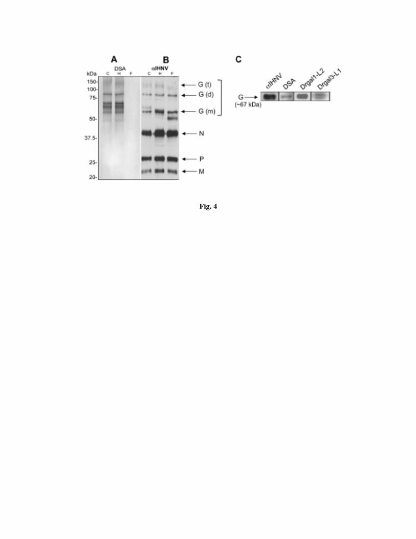
Fig. 4

Fig.5

Fig.6
Fig. 7