Embed Size (px)
Citation preview
Atractylenolide-I Sensitizes HumanOvarian Cancer Cells to Paclitaxel byBlocking Activation of TLR4/MyD88-dependent PathwayJian-Ming Huang1,2, Guo-Nan Zhang1,3, Yu Shi1, Xiao Zha1,2, Yi Zhu1,4, Miao-Miao Wang1, Qing Lin1,Wen Wang1, Hai-Yan Lu1, Shi-Qi Ma2, Jia Cheng2 & Bi-Fang Deng2
1Department of Gynaecologic Oncology, Sichuan Cancer Hospital, No. 55, Section 4, South People’s Road, Chengdu 610041,Sichuan, P. R. China, 2Department of Biochemistry & Molecular Biology, Sichuan Cancer Institute, Chengdu 610041, Sichuan, P. R.China, 3Graduate School, Guangxi Medical University, Nanning 530021, Guangxi, P. R. China, 4Department of Ultrasound,Sichuan Cancer Hospital, Chengdu 610041, Sichuan, P. R. China.
Paclitaxel, a known TLR4 ligand, leads to activation of TLR4/MyD88-dependent pathway that mediateschemoresistance and tumor progression in epithelial ovarian carcinoma (EOC). Atractylenolide-I (AO-I), anovel TLR4-antagonizing agent, inhibits TLR4 signaling by interfering with the binding of LPS or paclitaxelto membrane TLR4 of human leukocytes. In this study, AO-I was found to attenuate paclitaxel-inducedprotein expression of IL-6, VEGF and survivin, and to enhance early apoptosis and growth inhibition inMyD881 EOC cells; AO-I was shown to fit into the hydrophobic pocket of human MD-2 and to partiallyoverlap with the binding site of paclitaxel by docking simulations, suggesting that AO-I may block theMD-2-mediated TLR4/MyD88-dependent paclitaxel signaling in MyD881 EOC cells. Therefore, AO-I couldsignificantly sensitize the response of MyD881 EOC cells to paclitaxel by blocking MD-2-mediated TLR4/MyD88 signaling, and that AO-I-paclitaxel combination could be a promising strategy for the treatment ofEOC with a functional TLR4/MyD88/NF-kB pathway.
Epithelial ovarian cancer (EOC) is the leading cause of death among gynecological malignancies worldwideand usually has a poor prognosis1,2. Recent studies revealed that EOC cells expressing TLR4 and MyD88constitutively secrete pro-inflammatory cytokines and are resistant to the paclitaxel, and directly contri-
butes to their own survival and tumor progression3–6. Our previous study reported that MyD88 expression wasobserved in 77.1% of patients with EOC, which is an independent prognostic factor for poor disease-free survivaland overall survival for EOC4.
TLR4, the receptor for lipopolysaccharide, is unique in that it activates both MyD88-dependent and TRIF-dependent or MyD88-independent pathways. MyD88 is an adaptor protein for TLR4 signaling known to hyper-activate NF-kB, MAPK and PI3K pathways driving tumor survival and paclitaxel chemoresistance in EOC cells7-11.Paclitaxel is an important chemotherapeutic agent against EOC which acts by microtubule over-stabilization.However, paclitaxel elicits both cytotoxic and pro-survival responses in tumor cells. The likely mechanism forpaclitaxel-dependent tumor-activating effects is the ability of paclitaxel to activate TLR4/MyD88 signaling path-way. Paclitaxel, a known TLR-4 ligand, enhances NF-kB activity and up-regulates expression of X-linked inhibitorof apoptosis (XIAP) and pro-inflammatory cytokines known to promote tumor survival and progression in EOC.The expression of pro-inflammatory cytokines by MyD881 EOC cells are lost upon the knockdown of MyD88,suggesting that an active MyD88-dependent TLR4 signaling is responsible for MyD88/NF-kB-mediated cytokinesecretion, proliferation and paclitaxel resistance of EOC cells. TLR4/MyD88 signaling has become prominentlyimplicated as a means by which EOC cells can acquire the ability to invade, disseminate and resist paclitaxel-induced apoptosis3,12,13.
The TLR4 accessory protein, myeloid differentiation protein 2 (MD-2), is known to be an essential componentfor the initiation of TLR4/MyD88 signaling. The binding of LPS or paclitaxel to human MD-2 is required fordimerization of human TLR4 leading to activation of the MyD88-dependent pathway. Formation of the TLR4/MD-2 complex by paclitaxel may suggest important new mechanisms for paclitaxel-resistant tumors14 and
OPEN
SUBJECT AREAS:HEALTH SCIENCES
CANCER THERAPY
TRANSLATIONAL RESEARCH
Received15 July 2013
Accepted6 January 2014
Published23 January 2014
Correspondence andrequests for materials
should be addressed toG.-N.Z. (zhanggn@
hotmail.com)
SCIENTIFIC REPORTS | 4 : 3840 | DOI: 10.1038/srep03840 1
blocking the binding of paclitaxel to MD-2 may reduce its pro-sur-vival response, then enhance its cytotoxic response. It is also import-ant to identify alternative chemotherapy options that would benefitMyD881 EOC patients.
Atractylenolide-I (AO-I) is a naturally occurring sesquiterpenelactone isolated from Atractylodes macrocephala Koidz [Family:Compositae], and has been used for anti-inflammatory purposesand the treatment of cancers15–20. Anti-inflammatory effect of AO-Idisplays a potent inhibitory effect on angiogenesis by a set of down-regulation of NO, TNF-a, IL-1b, IL-6 and VEGF in monocytes andmacrophages stimulated with LPS21,22. It has been reported that AO-Ihas a binding site similar to LPS or paclitaxel by dissociating LPS orpaclitaxel from TLR4 in the model of white blood cell membranechromatography (WB-CMC), and is a novel TLR4-antagonizingagent18–20.
In the study, we determined if AO-I can block paclitaxel-inducedexpression of pro-inflammatory cytokines and anti-apoptotic pro-tein survivin, and potentiate paclitaxel–induced apoptosis andgrowth inhibition of EOC cells. We also performed a preliminarydocking of AO-1 and paclitaxel to human MD-2 by computationalsimulation. We demonstrated, for the first time, that AO-I is able tosensitize EOC cells to paclitaxel by blocking MD-2-mediated TLR4/MyD88-dependent signaling pathway. The combination of AO-1with paclitaxel elicites significantly greater inhibition of cell growthand more apoptosis, compared with paclitaxel alone.
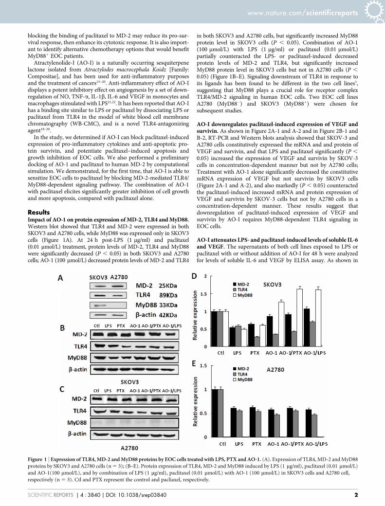
ResultsImpact of AO-1 on protein expression of MD-2, TLR4 and MyD88.Western blot showed that TLR4 and MD-2 were expressed in bothSKOV3 and A2780 cells, while MyD88 was expressed only in SKOV3cells (Figure 1A). At 24 h post-LPS (1 mg/ml) and paclitaxel(0.01 mmol/L) treatment, protein levels of MD-2, TLR4 and MyD88were significantly decreased (P , 0.05) in both SKOV3 and A2780cells; AO-1 (100 mmol/L) decreased protein levels of MD-2 and TLR4
in both SKOV3 and A2780 cells, but significantly increased MyD88protein level in SKOV3 cells (P , 0.05). Combination of AO-1(100 mmol/L) with LPS (1 mg/ml) or paclitaxel (0.01 mmol/L)partially counteracted the LPS- or paclitaxel-induced decreasedprotein levels of MD-2 and TLR4, but significantly increasedMyD88 protein level in SKOV3 cells but not in A2780 cells (P ,
0.05) (Figure 1B–E). Signaling downstream of TLR4 in response toits ligands has been found to be different in the two cell lines3,suggesting that MyD88 plays a crucial role for receptor complexTLR4/MD-2 signaling in human EOC cells. Two EOC cell linesA2780 (MyD882) and SKOV3 (MyD881) were chosen forsubsequent studies.
AO-I downregulates paclitaxel-induced expression of VEGF andsurvivin. As shown in Figure 2A-1 and A-2 and in Figure 2B-1 andB-2, RT-PCR and Western blots analysis showed that SKOV-3 andA2780 cells constitutively expressed the mRNA and and protein ofVEGF and survivin, and that LPS and paclitaxel significantly (P ,
0.05) increased the expression of VEGF and survivin by SKOV-3cells in concentration-dependent manner but not by A2780 cells;Treatment with AO-1 alone significantly decreased the constitutivemRNA expression of VEGF but not survivin by SKOV3 cells(Figure 2A-1 and A-2), and also markedly (P , 0.05) counteractedthe paclitaxel-induced increased mRNA and protein expression ofVEGF and survivin by SKOV-3 cells but not by A2780 cells in aconcentration-dependent manner. These results suggest thatdownregulation of paclitaxel-induced expression of VEGF andsurvivin by AO-I requires MyD88-dependent TLR4 signaling inEOC cells.
AO-I attenuates LPS- and paclitaxel-induced levels of soluble IL-6and VEGF. The supernatants of both cell lines exposed to LPS orpaclitaxel with or without addition of AO-I for 48 h were analyzedfor levels of soluble IL-6 and VEGF by ELISA assay. As shown in
Figure 1 | Expression of TLR4, MD-2 and MyD88 proteins by EOC cells treated with LPS, PTX and AO-1. (A). Expression of TLR4, MD-2 and MyD88
proteins by SKOV3 and A2780 cells (n 5 3); (B–E). Protein expression of TLR4, MD-2 and MyD88 induced by LPS (1 mg/ml), paclitaxel (0.01 mmol/L)
and AO-1(100 mmol/L), and by combination of LPS (1 mg/ml), paclitaxel (0.01 mmol/L) with AO-1 (100 mmol/L) in SKOV3 cells and A2780 cell,
respectively (n 5 3). Ctl and PTX represent the control and pacliaxel, respectively.
www.nature.com/scientificreports
SCIENTIFIC REPORTS | 4 : 3840 | DOI: 10.1038/srep03840 2

Figure 2C-1 and C-2, LPS- or paclitaxel-treated SKOV3 cells secreteda wide range of IL-6 and VEGF (P , 0.05), but no or lower signal forsoluble IL-6 and VEGF were detectable in A2780 cells; Interestingly,SKOV3 cells treated with AO-1 alone significantly decreased thelevel of VEGF (P , 0.05). Following treatment with AO-I, asignificant decrease in paclitaxel-induced levels of soluble IL-6 andVEGF was observed in SKOV3 cells but not in A2780 cells (P , 0.05)(Figure 2C).
AO-I enhances paclitaxel–induced growth inhibition. As shown inTable 1 and Figure 3A and 3B, SKOV3 cells showed more resistanceto paclitaxel than A2780 cells (P , 0.05). AO-I or LPS and their
combination did not markedly impact the proliferation of bothSKOV3 and A2780 cells, the IC50 values of AO-I for SKOV3 andA2780 cells were far more than 100 mmol/L, suggesting that AO-Ihave no distinct cytotoxic activity against SKOV3 and A2780 cellswhen used alone. We also found that AO-I significantly enhancedpaclitaxel-induced growth inhibition in SKOV3 cells, and inparticular, decreased paclitaxel IC50 value by up to 3.5-fold (from0.038 to 0.011 mmol/L) (P , 0.05), but had no sensitizing effect onA2780 cells when A2780 cells were treated with paclitaxel. Inaddition, LPS (0.1 mg/ml) greatly increased the resistance ofSKOV3 cells to paclitaxel (P , 0.05). These results suggest thatAO-I, as a potent antagonist of TLR4/MD-2 complex, could
Figure 2 | Expression of VEGF and survivin and levels of soluble IL-6 and VEGF. (A1–A2). The mRNA expression of VEGF and survivin induced by
LPS, paclitaxel and AO-1 in SKOV3 cells and A2780 cells, respectively (n 5 3); (B1–B2). The protein expression of VEGF and survivin induced by LPS,
paclitaxel and AO-1 in SKOV3 cells and A2780 cells, respectively (n 5 3); (C1–C2). Levels of soluble IL-6 and VEGF, induced by combination of
paclitaxel with AO-1 in SKOV3 cells and A2780 cells, respectively (n 5 3). * P , 0.05, compared to Control.
Table 1 | Drug-induced growth inhibition in EOC cells in 4 treatment groups
Treatment group n
SKOV3 cells A2780 cells
IC50 (mmol/L) P* IC50 (mmol/L) P*
AO-1 3 .100 - .100 -Paclitaxel 3 0.038 6 0.004 - 0.024 6 0.004 -Paclitaxel/AO-I 3 0.011 6 0.004* 0.027 0.029 6 0.006 0.500Paclitaxel/LPS 3 0.082 6 0.002* 0.044 0.033 6 0.004 0.100
EOC cells were treated with vehicle or paclitaxel plus AO-I (100 mmol/L) or paclitaxel plus LPS (1 mg/ml) for 72 hours. Relative number of viable cells was assayed using MTT. IC50 values were measured asa curvilinear regression equation for each survival curve. Data represent mean values 6 S.D. from three independent experiments.*P values, compared with paclitaxel alone.
www.nature.com/scientificreports
SCIENTIFIC REPORTS | 4 : 3840 | DOI: 10.1038/srep03840 3

significantly potentiate the growth inhibitory effect of paclitaxel inMyD881 EOC cells.
AO-I potentiates paclitaxel-induced early apoptosis. To determinewhether the decrease in cell viability is due to the increase ofpaclitaxel-induced early apoptosis, we measured Annexin V/PIbinding responses of EOC cells for 24 hours. As shown in Table 2,paclitaxel induced the early apoptosis of both A2780 and SKOV3cells. Following co-treatment with paclitaxel and AO-I, anapproximate 2.5-fold increase in paclitaxel-induced apoptosis(Annexin V1 from 5.4% to 13.6%) was observed in SKOV3 cells,but the level of paclitaxel-induced early apoptosis of A2780 cellsremained unchanged, suggesting that AO-I inhibits pro-survivalresponse of paclitaxel that is MyD88-dependent in SKOV3 cells.
Docking of AO-1 and paclitaxel to human MD-2. We docked AO-1and paclitaxel to the crystal structure of human MD-2 bycomputational simulation. AO-1 could be fitted into thehydrophobic pocket of human MD-2 and overlap with the bindingsite of paclitaxel (Figure 4), and competitively displace paclitaxelfrom MD-2 under the most energetically favorable simulation,suggesting that AO-1, as an antagonist, may be responsible forinhibition of TLR4-mediated paclitaxel signaling by binding tohuman MD-2.
DiscussionThe expression of TLR4/MyD88 signaling pathway linked to tumorprogression and resistance to paclitaxel has been reported recently in
EOC cells3,7,11. The expression of MyD88 in more than 70% ofpatients with EOC and has been identified as an indicator of tumormetastasis, paclitaxel chemoresistance and a significantly poor pro-gnosis factor9,11. In respect to functioning TLR4/MyD88 signaling,evidence supporting its involvement in carcinogenesis and sensitivityto apoptosis mediated by paclitaxel is available for ovarian can-cer3,23–25. Evidence implicates paclitaxel and LPS share a TLR4/MyD88-dependent pathway leading to activation of MAPK andNF-kB in generating pro-inflammatory cytokines12,26,27 and anti-apoptostic proteins in EOC cells. High level expression of IL-6 andVEGF and survivin are characterized by increasing survival andproliferation in EOC cells that express TLR4/MyD88, suggesting thatactivation of this pathway in EOC cells maintain chronic inflam-mation and promote cancer growth, metastasis and paclitaxelchemoresistance.
Ligand-induced MD-2-mediated dimerization of TLR4 isrequired for the activation of TLR4/MyD88-dependent signalingpathways. The MyD88-dependent response occurs on the dimeriza-tion of TLR4 which leads to the recruitment of MyD88 to the intra-cellular domain of TLR4, initiating the intracellular signal cascadethat culminates in activation and nuclear translocation of transcrip-tion factors AP-1 and NF-kB leading to the induction of the express-ion of various inflammatory gene products28. MD-2 is a part of theTLR4 signaling complex with an indispensable role in activation ofthe TLR4 pathway and thus, the formation of TLR4/MD-2 complexmay be one of the first lines of regulation in activating TLR4-mediated responses. It has been confirmed that LPS or paclitaxelbinding to TLR4 results in dimerization of the TLR4 associated with
Figure 3 | Proliferation of tumor cells in response to AO-I, paclitaxel or LPS. (A). Cell viabilty (%) of SKOV3 and A2780 cells treated with LPS
(0.1 emsp14;mg/ml) alone or combined with AO-1 for 72 hours (n 5 3); (B). Cell viabilty (%) of SKOV3 and A2780 cells treated with paclitaxel alone or
combined with AO-1(100 mmol/L) or with LPS (0.1 mg/ml) (n 5 3).
Table 2 | Drug-induced early apoptosis in EOC cells in 6 treatment groups
Treatment group
SKOV3 cells A2780 cells
% AnnexinV1 cells P* % AnnexinV1 cells P*
Control 4.0 6 0.57 - 3.5 6 0.40 -AO-1(1 mmol/L) 4.3 6 0.48 - 3.7 6 0.38 -AO-1(100 mmol/L) 5.1 6 0.34 - 3.2 6 0.44 -Paclitaxe l (0.01 mmol/L) 5.8 6 0.45 - 8.5 6 0.65 -Paclitaxel/AO-I (1 mmol/L) 9.9 6 0.82* 0.002 9.1 6 0.51 0.336Paclitaxel/AO-I (100 mmol/L) 13.6 6 1.76* 0.001 9.8 6 1.01 0.593
EOC cells were treated with paclitaxel (0.01 mmol/L), AO-I (1 mmol/L, 100 mmol/L), and with paclitaxel (0.01 mmol/L) plus AO-I (1 mmol/L, 100 mmol/L) for 24 hours. FCM assay for AnnexinV binding asdescribed in Materials and methods. Data represent mean values 6 S.D. from three independent experiments.*P values, compared with paclitaxel group, AO-1 groups and control group. AO-I significantly potentiates paclitaxel-induced early apoptosis of SKOV-3 cells in a concentration-dependent manner.
www.nature.com/scientificreports
SCIENTIFIC REPORTS | 4 : 3840 | DOI: 10.1038/srep03840 4

MD-214,29. MD-2 silencing has been reported to decrease LPS-induced cytokine production and TLR4/MyD88 pathway activity30.MD-2 undergoes a ligand-dependent conformational change that inturn induces or blocks the homotypic aggregation of TLR4/MD-2,followed by the recruitment of MyD88. The demonstration thatpaclitaxel can bind to TLR4/MD-231 and therefore activate NF-kBcould explain why tumor growth was observed during paclitaxeltreatment of patient with advanced EOC4. Recent data attest to a roleof MD-2 activity in colon cancer epithelial cell proliferation andmigration, which may be important in the general correlationbetween innate immune response, chronic inflammation, and can-cer31. Recent studies revealed that several small molecular includingamitriptyline, curcumin and Eritoran, a structural analogue of LPS,exhibited TLR4 inhibition, possibly by binding pocket on interac-tions with binding pocket on MD-2, in a TLR4/MyD88-dependentmanner32–35.
In this study, the expression of MD-2 and TLR4 were observed inboth SKOV3 and A2780 cells but MyD88 only in SKOV3 cells.Indeed, our results showed that in SKOV3 cells, LPS and paclitaxeldecreased protein levels of MD-1, TLR4 and MyD88. It is possiblethat the endocytosis of receptor complex TLR4/MD-2 followingligand-receptor interaction results in lysosomal degradation andE3-mediated degradation of Syk-phosphorylated MyD8836,37, lead-ing to the control of intensity and duration of TLR4 signaling.Interestingly, in addition to reducing protein levels of MD-2 andTLR4, AO-1 also significantly increased protein levels of MyD88,suggesting that the inhibition of AO-1 on the TLR4/MyD88 signal-ing by blocking the binding of MD-2 to TLR4 may lead to the reduc-tion of TGF-b1-mediated down-regulation of MyD88 expression37.
In response to paclitaxel, AO-1 can attenuate paclitaxel-inducedincreased expression of IL-6, VEGF and survivin, potentiate pacli-taxel-induced growth inhibition and early apoptosis in MyD881
EOC cells, suggesting that the binding of paclitaxel to MD-2 mediatesdimerization of TLR4/MD-2 to activate MyD88-dependent signal-ing. But we found no significant cytotoxic effect in MyD881 EOCcells treated with AO-I alone at less than 100 mmol/L, suggesting that
AO-I, as an antagonist of TLR4/MyD88 signaling with low cytotoxi-city, can sensitize MyD881 EOC cells to paclitaxel. However, theunderlying mechanisms of AO-I blocking TLR4 signaling are poorlyunderstood. A triggering event on TLR4 is involved in the molecularrearrangement of the receptor complex and its homodimeriza-tion14,38,39. The assembly of the TLR4/MD-2 complex initiates aMyD88-dependent signaling cascade, which relocates NF-kB fromthe cytoplasm to the nucleus40.
Our preliminary docking analysis of AO-I and paclitaxel bindingto the crystal structure of human MD-2 showed that AO-1 couldpreferentially fit into the hydrophobic binding pocket of humanMD-2, which binds paclitaxel, suggesting that AO-1 induce the con-formational changes of MD-2 that may obstruct the formation of anactive TLR4/MD-2 complex, and then inactivate TLR4 signaling.Inhibition of MD-2-mediated active dimerization of TLR4 may bea promising therapic strategy for overcoming TLR4/MyD88 signal-ing-mediated resistance of EOC cells to paclitaxel. We demonstrated,for the first time, that the antagonizing effect of AO-I is TLR4/MD-2-mediated MyD88-dependent signaling pathway in EOC cells.
Our preliminary docking analysis of AO-I and paclitaxel bindingto the crystal structure of human MD-2 showed that AO-1 couldpreferentially fit into to the hydrophobic binding pocket of humanMD-2, suggesting that AO-1 induce the conformational changes ofMD-2 that may obstruct the formation of an active TLR4/MD-2complex, and then inactivate TLR4 signaling. We determined thatAO-I binds to MD-2 at submicromolar affinity and competes func-tional cellular TLR4 signaling pathway stimulated by paclitaxel, andwe demonstrated, by using FCM assay, that AO-1 can prevent pacli-taxel- or LPS-induced formation of TLR4/MD-2 complex andcellular stimulation by interfering with TLR4/MD-2 binding (datanot shown). Therefore, Inhibition of TLR4/MD-2 dimerizationmay be a promising therapic strategy for overcoming TLR4/MyD88 signaling-mediated resistance of EOC cells to paclitaxel.We demonstrated for the first time that the antagonizing effect ofAO-I is TLR4/MD-2-mediated MyD88-dependent signaling path-way in EOC cells.
Figure 4 | Molecular model of docking of paclitaxel and AO-I to the molecular model of MD-2. (A and B). 3D structures of AO-I and paclitaxel were
drawn as a ball-and-stick representation: AO-I in purple, paclitaxel in blue-green. (C). 3D structure of human MD-2. Protein surface showing
hydrophobic and hydrophilic properties. Green and red represent hydrophobicity and hydrophilicity, respectively, and the surface center of MD-2 with a
potential binding-pocket (iron-gray); (D). AO-I binding to the hydrophobic pocket of MD-2, which partially overlaps with the binding site of paclitaxel.
www.nature.com/scientificreports
SCIENTIFIC REPORTS | 4 : 3840 | DOI: 10.1038/srep03840 5
Taken together, these data indicate that the AO-1 inhibits TLR4/MyD88 signaling mediated pro-survival of paclitaxel in EOC cellsand the combined use of AO-I with paclitaxel could improve tumorresponse to paclitaxel chemotherapy in patients with EOC by block-ing MD-2-mediated activation of TLR4/MyD88 signaling.
MethodsCompounds and reagents. LPS, paclitaxel and MTT (3-[4,5-dimethylthiazol-2-yl]-2,5-diphenyl tetrazolium bromide) were purchased from Sigma Chemical Co. (St.Louis, MO, USA); AO-I, (4aS,8aS)-3,8a-dimethyl-5-methylidene-4a,6,7,8-tetrahydro-4H-benzo[f][1]benzofuran-2-one (CAS Number 73069-13-3, MFC15H18O2, MW (g/mol) 230.3022, HPLC $ 98%) was purchased from ChengduBest-Reagent Co. Ltd. (Chengdu, Sichuan, China); the rabbit polyclonal antibodies toTLR4, MD-2 and MyD88 were purchased from Epitomics, Inc. (Burlingame, CA,USA) and Abcam plc. (Cambridge, MA, USA); the rabbit polyclonal antibodies to IL-6, VEGF, survivin and horseradish peroxidase conjugated secondary antibodies werepurchased from Santa Cruz Biotechnology, Inc. (Santa Cruz, CA, USA).
Cell lines and culture. Human EOC cell lines SKOV3 (TLR41/MyD881, derivedfrom the ascites of a patient with advanced, metastatic EOC and resistant to mostcytotoxic drugs)41 and A2780 (TLR41/MyD88-, derived from a primary untreatedand paclitaxel-sensitive cancer)42 were purchased from the Committee on TypeCulture Collection of Chinese Academy of Sciences (CTCCCAS, Shanghai, China).Cell lines were maintained in RPMI 1640 medium (GIBCO) supplemented 10% heat-inactivated fetal calf serum (FCS), 2 mM L-glutamine, 100 U/ml penicillin and40 IU/ml gentamicin at 37uC in a humidified atmosphere of 5% CO2 and 95% air.Subconfluent cells (80%) were passaged with a solution containing 0.25% trypsin and0.5 mmol/L EDTA. Cell lines were tested for Mycoplasma and confirmed to benegative.
Docking of AO-I or paclitaxel to the MD-2 structural model. Docking simulationof AO-I (CID: 5321018) and paclitaxel (CID: 36314) were carried out with theprogram AutoDock4 (open source, Scripps Research Institute), and the crystalstructure of human MD-2 was cited from Protein Data Bank (2E56)14,18. The ligand-binding groove on MD-2 was kept rigid, whereas all torsible bonds of AO-I orpaclitaxel were set free to perform flexible docking to produce more than 100structures. Final docked conformations were clustered within the tolerance of 1 Aroot-mean-square deviation.
RNA isolation and RT-PCR. Total RNA was isolated from cell culture (1 3 106 cells)using Trizol reagent (Invitrogen Co., CA, USA) according to the manufacturer’sprotocol. 2 mg of total RNA was reverse transcribed to cDNA. cDNA then amplifiedusing the following primers specific for VEGF: F:59-CACATAGGAGAGATGAGCTTC-39, R: 59-CCTCGGCTTGTCACATCTG-39; for survivin: F: 59-TCAAGGACCACCGCATC-39, R: 59-CAATCCATGGCAGCCAG-39; for b-actin: F: 59-AAGAGATGGCCACGGCTGCT-39, R: 59-GACTCGTCATACTCCTGCTTGCT-39.PCR reaction was performed using the following conditions: Pre-denaturation at 95uCfor 5 min. 35 cycles of amplification at 95uC for 15 sec. 56uC for 45 sec. and 72uC for45 sec, and final extension at 72uC for 5 min. b-actinwas used as an internal control.
Western blot analysis for protein expression. EOC cells were treated simultaneouslywith LPS or paclitaxel with or without addition of AO-I for different periods of timeand lysed in RIPA buffer [1% Triton X-100, 150 mmol/L NaCl, 1 mmol/L EGTA,50 mmol/L Tris–HCl, 0.1% sodium dodecyl sulfate (SDS), 1% sodium desoxycholateand phenylmethylsuphonyl fluoride (PMSF)]. Proteins separated by SDS-PAGE wereelectrotransfered to polyvinylidene difluoride (PVDF) membranes. The followingAbs: anti-TLR4, anti-MD-2 and anti-MyD88 and anti-survivin were used fordetection, and horseradish peroxidaseconjugated secondary antibodies (15100,000dilution) for development of reactions in a chemiluminescent detection system(ChemiDoc XRS1, Bio-Rad). b-actin antibodies were used as controls for equalprotein loading.
ELISA assay for cytokines. VEGF and IL-6 production was determined usingEnzyme-linked immunosorbent assay (ELISA). Cells were plated in 12-well plates at 13 105 cells/well in 1 mL of medium and treated simultaneously with LPS or paclitaxelwith or without addition of AO-I for 24 h. The supernatants were collected, then thelevels of IL-6 and VEGF were measured using ELISA kits (R&D Systems). The assaysensitivity varied from 5 to 15 pg/ml.
Annexin V/PI binding for early apoptosis. A flow-based Annexin V/Propidiumiodide (ANX-V/PI) assay was used to measure EOC cell early apoptosis. Briefly, cellswere treated simultaneously with paclitaxel (0.01 mmol/L) with or without addition ofAO-I (100 mmol/L) for 24 h, trypsinized with 0.25% trypsin, washed in PBS,resuspended in ANX V/PI-binding buffer and stained with 1 mg/mL FITC-conjugated ANX V and 1 mg/mL PI (keyGentec BioTHEC) for 15 min on ice in thedark. Cell apoptosis was evaluated by flow cytometry (BD FACS Canto II).
Cell viability assay. To a 96-well plate, 5 3 103 cells/well were pre-cultured for 24hours, and then treated simultaneously with paclitaxel or LPS (1 mg/ml) with orwithout addition of AO-I, and 0.1% ethaol was used as a vehicle in triplicate. After 72
hours, 20 ml of MTT (5 mg/ml in PBS) was added to each well. The plates were gentlyshaken and incubated for 4 hours at 37uC in 5% CO2 atmosphere. The supernatantwas removed and 200 ml of dimethyl sulfoxide (DMSO) was added and the plateswere gently shaken to solubilize the formed formazan. Cell viability was determinedby absorbance readings with ELISA Microplate Reader.
Statistical analysis. Data was summarized using descriptive statistics of mean andstandard deviation. ANOVA was used as a statistical test with SPSS 17.0 software, andP value , 0.05 was considered significant.
1. Siegel, R., Ward, E., Brawley, O. & Jemal, A. Cancer statistics, 2011: the impact ofeliminating socioeconomic and racial disparities on premature cancer deaths. CAcancer J Clin 61, 212–236 (2011).
2. Davidson, B., Reich, R., Trope, C. G., Wang, T. L. & Shih Ie, M. New determinatesof disease progression and outcome in metastatic ovarian carcinoma. Histol andhistopathol 25, 1591–1609 (2010).
3. Szajnik, M. et al. TLR4 signaling induced by lipopolysaccharide or paclitaxelregulates tumor survival and chemoresistance in ovarian cancer. Oncogene 28,4353–4363 (2009).
4. Zhu, Y., Huang, J. M., Zhang, G. N., Zha, X. & Deng, B. F. Prognostic significanceof MyD88 expression by human epithelial ovarian carcinoma cells. J Transl Med10, 77 (2012).
5. Kim, K. H. et al. Expression and significance of the TLR4/MyD88 signalingpathway in ovarian epithelial cancers. World J Surg Oncol 10, 193 (2012).
6. Huang, B., Zhao, J., Unkeless, J. C., Feng, Z. H. & Xiong, H. TLR signaling bytumor and immune cells: a double-edged sword. Oncogene 27, 218–224 (2008).
7. Chen, R., Alvero, A. B., Silasi, D. A., Steffensen, K. D. & Mor, G. Cancers take theirToll--the function and regulation of Toll-like receptors in cancer cells. Oncogene27, 225–233 (2008).
8. Kelly, M. G. et al. TLR-4 signaling promotes tumor growth and paclitaxelchemoresistance in ovarian cancer. Cancer Res 66, 3859–3868 (2006).
9. Zhou, M. et al. Toll-like receptor expression in normal ovary and ovarian tumors.Cancer Immunol Immunother: CII 58, 1375–1385 (2009).
10. Sato, Y., Goto, Y., Narita, N. & Hoon, D. S. Cancer Cells Expressing Toll-likeReceptors and the Tumor Microenvironment. Cancer Microenviron: officialjournal of the International Cancer Microenvironment Society 2 Suppl 1, 205–214(2009).
11. Wang, A. C., Su, Q. B., Wu, F. X., Zhang, X. L. & Liu, P. S. Role of TLR4 forpaclitaxel chemotherapy in human epithelial ovarian cancer cells. Eur J Clin Invest39, 157–164 (2009).
12. Byrd-Leifer, C. A., Block, E. F., Takeda, K., Akira, S. & Ding, A. The role of MyD88and TLR4 in the LPS-mimetic activity of Taxol. Eur J Immunol 31, 2448–2457(2001).
13. Kawasaki, K., Gomi, K., Kawai, Y., Shiozaki, M. & Nishijima, M. Molecular basisfor lipopolysaccharide mimetic action of Taxol and flavolipin. J Endotoxin Res 9,301–307 (2003).
14. Zimmer, S. M., Liu, J., Clayton, J. L., Stephens, D. S. & Snyder, J. P. Paclitaxelbinding to human and murine MD-2. J Biol Chem 283, 27916–27926 (2008).
15. Endo, K. et al. Antiinflammatory principles of Atractylodes rhizomes. Chemical &pharmaceutical bulletin 27, 2954–2958 (1979).
16. Wang, K. T. et al. Analysis of the sesquiterpenoids in processed AtractylodisRhizoma. Chem Pharm Bull 55, 50–56 (2007).
17. Wang, H. X., Liu, C. M., Liu, Q. & Gao, K. Three types of sesquiterpenes fromrhizomes of Atractylodes lancea. Phytochemistry 69, 2088–2094 (2008).
18. Li, C. Q., He, L. C., Dong, H. Y. & Jin, J. Q. Screening for the anti-inflammatoryactivity of fractions and compounds from Atractylodes macrocephala koidz.J Ethnopharmacol 114, 212–217 (2007).
19. Zhang, Y. Q. et al. Antagonistic effects of 3 sesquiterpene lactones fromAtractylodes macrocephala Koidz on rat uterine contraction in vitro. ActaPharmacol Sin 21, 91–96 (2000).
20. Li, C. & He, L. Establishment of the model of white blood cell membranechromatography and screening of antagonizing TLR4 receptor component fromAtractylodes macrocephala Koidz. Sci China C Life Sci 49, 182–189 (2006).
21. Wang, C., Duan, H. & He, L. Inhibitory effect of atractylenolide I on angiogenesisin chronic inflammation in vivo and in vitro. EurJ Pharmacol 612, 143–152(2009).
22. Li, C. Q., He, L. C. & Jin, J. Q. Atractylenolide I and atractylenolide III inhibitLipopolysaccharide-induced TNF-alpha and NO production in macrophages.Phytother Res: PTR 21, 347–353 (2007).
23. Silasi, D. A. et al. MyD88 predicts chemoresistance to paclitaxel in epithelialovarian cancer. The Yale J Biol Med 79, 153–163 (2006).
24. Killeen, S. D., Wang, J. H., Andrews, E. J. & Redmond, H. P. Exploitation of theToll-like receptor system in cancer: a doubled-edged sword? Br J cancer 95,247–252 (2006).
25. Chen, R., Alvero, A. B., Silasi, D. A. & Mor, G. Inflammation, cancer andchemoresistance: taking advantage of the toll-like receptor signaling pathway. AmJ Reprod Immunol 57, 93–107 (2007).
26. Perera, P. Y., Qureshi, N. & Vogel, S. N. Paclitaxel (Taxol)-induced NF-kappaBtranslocation in murine macrophages. Infect Immun 64, 878–884 (1996).
www.nature.com/scientificreports
SCIENTIFIC REPORTS | 4 : 3840 | DOI: 10.1038/srep03840 6

27. Kawasaki, K. et al. Mouse toll-like receptor 4.MD-2 complex mediateslipopolysaccharide-mimetic signal transduction by Taxol. J Biol Chem 275,2251–2254 (2000).
28. Laird, M. H. et al. TLR4/MyD88/PI3K interactions regulate TLR4 signaling.J Leukoc Biol 85, 966–977 (2009).
29. Peri, F., Piazza, M., Calabrese, V., Damore, G. & Cighetti, R. Exploring the LPS/TLR4 signal pathway with small molecules. Biochem Soc Trans 38, 1390–1395(2010).
30. Ren, W. et al. Myeloid differentiation protein 2 silencing decreases LPS-inducedcytokine production and TLR4/MyD88 pathway activity in alveolar macrophages.Immunol Lett 141, 94–101 (2011).
31. Grondin, V. et al. Regulation of colon cancer cell proliferation and migration byMD-2 activity. Innate immun 17, 414–422 (2011).
32. Hutchinson, M. R. et al. Evidence that tricyclic small molecules may possess toll-like receptor and myeloid differentiation protein 2 activity. Neuroscience 168,551–563 (2010).
33. Gradisar, H., Keber, M. M., Pristovsek, P. & Jerala, R. MD-2 as the target ofcurcumin in the inhibition of response to LPS. J Leukoc Biol 82, 968–974 (2007).
34. Slivka, P. F. et al. A peptide antagonist of the TLR4-MD2 interaction.Chembiochem: a European journal of chemical biology 10, 645–649 (2009).
35. Kim, H. M. et al. Crystal structure of the TLR4-MD-2 complex with boundendotoxin antagonist Eritoran. Cell 130, 906–917 (2007).
36. Husebye, H., Halaas, Ø., Stenmark, H., Tunheim, G., Sandanger, Ø., Bogen, B.,Brech, A., Latz, E. & Espevik, T. Endocytic pathways regulate Toll-like receptor 4signaling and link innate and adaptive immunity. EMBO J 25, 683–92 (2006).
37. Lee, Y. S., Park, J. S., Kim, J. H., Jung, S. M., Lee, J. Y., Kim, S. J. & Park, S. H.Smad6-specific recruitment of Smurf E3 ligases mediates TGF-b1-induceddegradation of MyD88 in TLR4signalling. Nat Commun 2, 60 (2011).
38. Visintin, A., Iliev, D. B., Monks, B. G., Halmen, K. A. & Golenbock, D. T. Md-2.Immunobiology 211, 437–447 (2006).
39. Park, B. S. et al. The structural basis of lipopolysaccharide recognition by theTLR4-MD-2 complex. Nature 458, 1191–1195 (2009).
40. Takeda, K. & Akira, S. TLR signaling pathways. Semin Immunol 16, 3–9 (2004).41. Cuello, M., Ettenberg, S. A., Nau, M. M. & Lipkowitz, S. Synergistic induction of
apoptosis by the combination of trail and chemotherapy in chemoresistantovarian cancer cells. Gynecol Oncol 81, 380–390 (2001).
42. Behrens, B. C. et al. Characterization of a cis-diamminedichloroplatinum(II)-resistant human ovarian cancer cell line and its use in evaluation of platinumanalogues. Cancer Res 47, 414–418 (1987).
AcknowledgmentsThis work was supported by Grant-in-Aid for Scientific Research from SichuanGovernment (No. 407).
Author contributionsJ.M.H.: Study design, manuscript editing; G.N.Z.: Study concepts, manuscript review; Y.S.and X.Z.: Quality control of data and algorithms; Y.Z.: Data analysis and interpretation,manuscript preparation; M.M.W., Q.L., W.W. and H.Y.L.: statistical analysis; S.Q.M., J.C.and B.F.D.: Data acquisition. All authors reviewed the manuscript.
Additional informationCompeting financial interests: The authors declare no competing financial interests.
How to cite this article: Huang, J.-M. et al. Atractylenolide-I Sensitizes Human OvarianCancer Cells to Paclitaxel by Blocking Activation of TLR4/MyD88-dependent Pathway. Sci.Rep. 4, 3840; DOI:10.1038/srep03840 (2014).
This work is licensed under a Creative Commons Attribution 3.0 Unported license.To view a copy of this license, visit http://creativecommons.org/licenses/by/3.0
www.nature.com/scientificreports
SCIENTIFIC REPORTS | 4 : 3840 | DOI: 10.1038/srep03840 7