Embed Size (px)
Citation preview
ATA Trainees’ Educational Track Case Studies – Thyroid Nodules
Case Study #1 Ruchi Gaba, MD
Baylor College of Medicine Houston, TX
51 year old Asian female with H/o hypothyroidism on levothyroxine 50 mcg daily presented with painful progressively enlarging goiter over the course of 3 months ( R>L) associated with compressive symptoms like difficulty breathing and swallowing, no change in voice. Levothyroxine held for three weeks and CT soft tissue neck done which showed marked bilateral enlarged thyroid lobes and isthmus, R/L 9 cm/3.5-4.5cm displacing the trachea and mass effect of posteriorly displaced esophagus.
TSH 26 uIU/mL and FT4 0.7 ng/dl, TPO 21885 IU/mL and TSI 39 .
Thyroid ultrasound showed heterogeneous echogenicity suspicious for autoimmune thyroiditis with overall normal vascularity. Also two right sided solid and one left sided nodules, each > 1 cm found. FNA of the nodules- Both the right sided were negative for malignancy but the left one was indeterminate- cellular micro follicular proliferation indicating follicular neoplasm; probably adenoma but cannot exclude follicular carcinoma.
Differential at that time included- thyroid cancer/ lymphoma vrs thyroiditis. Patient was started on levothyroxine and prednisone taper with some improvement in symptoms. Surgery referral for thyroidectomy was done given the rapidity of the enlarging goiter, indeterminate finding on left thyroid FNA, and prior report of compressive symptoms (improved after prednisone taper)
After total thyroidectomy with parathyroid auto transplantation, initial pathology results consistent with Hashimoto’s Thyroiditis with fibrous variant, but lots of plasma cells; immunohistochemistry showed presence of light chains. Free kappa, lambda light chains, Kappa to Lamda ratio, urine and serum electrophoresis within normal limits.
But the final pathology results were later addended as follows- Immuno histochemical stains for CD10 and CD117 interpreted as negative within the plasma cell component, and kappa and lambda double stain interpreted as polytypic with variable kappa-to-lambda ratios of 4-5:1 to 2-3:1.
IgG4 was positive in > 20 plasma cells per high power field meeting a criterion outlined by Li, et al for IgG4 Hashimoto's Thyroiditis.
A form of Hashimoto’s thyroiditis with lymphoplasmacytic sclerosing changes and increased numbers of IgG4-positive plasma cells has recently been reported in the literature. These histo pathological features suggest that this subtype of Hashimoto’s thyroiditis may be closely related to IgG4-related disease. Therefore, this unique form of IgG4-related Hashimoto’s thyroiditis, which is referred to as IgG4 thyroiditis, has its own clinical, serological, and sonographic features that are distinct from those associated with non-IgG4 thyroiditis. It is associated with rapid progression,

subclinical hypothyroidism, higher level of circulating antibodies and diffuse low echogenicity and male predominance.
1
ATA Trainees’ Educational Track Case Studies – Thyroid Nodules
Case Study #2 Elizabeth McAninch, MD
University of Miami Miami, FL
Ms. A is a 42-year-old Nicaraguan woman with autoimmune hepatitis status-post liver transplant four months prior to admission presented with a three week history of fevers and severe anterior neck pain. Home medications included tacrolimus, trimethoprim/sulfamethoxazole, lamivudine for donor +hepatitis B, and ganciclovir for donor +CMV. Symptoms persisted despite one week of empiric broad-spectrum antibiotics; endocrinology was consulted for consideration of thyroiditis. Physical exam revealed a normotensive female in moderate distress with fever, tachycardia and tachypnea. There was no exophthalmos. The thyroid was exquisitely tender to palpation, diffusely enlarged, smooth, and mobile without distinct nodule. The perithyroidal tissue demonstrated bogginess. There was left cervical lymphadenopathy. Labs on admission showed leukopenia, normal liver enzymes, TSH 1.68 μIU/ml (0.27-4.2 μIU/ml), free t4 0.93 ng/dL (0.93-1.7 ng/dL) and T3 77.4 ng/dL (80-200 ng/dL). On US, the thyroid was enlarged and heterogeneous without definite nodules. Noncontrast CT showed normal thyroid, bilateral innumerable lung nodules and an ill-defined hepatic hypodensity. Extensive infection workup including BAL, liver biopsy, and thyroid FNA was performed; the patient tolerated FNA poorly due to pain such that cytology sample was not obtained from the scant serosanguinous aspirate. Empiric voriconazole was initiated with resolution of fever and neck pain within 48 hours. Repeat CT showed improvement in the lung nodules. All cultures were negative except for the thyroid specimen, which was positive for Coccidioides immitis. Antifungal therapy was changed to oral fluconazole; tacrolimus dose was decreased to achieve about half of the prior target while staying within the therapeutic range. Analysis of serum from the organ donor was consistent with coccidiomycosis infection; the other organ recipients were without infectious signs or symptoms. One month later she remained clinically well but developed two enlarging, extremely firm, left thyroid masses. In the left upper pole there was a hypoechoic, avascular nodule measuring 1.1 x 1.2 x 1.0cm and in the left mid pole a 2.9 x 1.8 x 2.0cm, hyperechoic , avascular nodule. These yielded insufficient cytology sample from FNA; fungal culture was negative. What is the etiology of these new masses? What should you do next? Due to overall improvement of her clinical status immune reconstitution syndrome was considered as an etiology for these new thyroid masses. After consultation with colleagues in surgery and infectious disease the decision was made to closely monitor these lesions. Serial imaging of the thyroid revealed evolution into abscesses which paralleled a deterioration in her overall clinical status. She developed a diffuse eruption of subcutaneous skin nodules which

2
were culture positive for C. immitis, developed mastoid osteomyelitis, and the lung lesions progressed in both size and number. Primary hypothyroidism also developed and levothyroxine was initiated. Parenteral amphotericin B was added to her regimen of oral fluconazole but the thyroid masses enlarged further and began spontaneously draining from a cutaneous fistula with continued development of subcutaneous nodules. Renal function deteriorated as a consequence of the antifungal therapies. Given the dramatic progression of the thyroid masses in parallel with deterioration of her overall clinical status, the etiology of the thyroid masses was thought to be less consistent with immune reconstitution syndrome and more likely continued coccidiomycosis thyroiditis. Decision was made to pursue total thyroidectomy to remove a presumed nidus of infection. Surgery was performed six months after initial presentation; the operation was complex as the left thyroid lobe was virtually indistinguishable from surrounding inflammatory tissue in the paratracheal and paraesophageal areas. A cuff of strap muscle was removed in the area of the cutaneous fistula. Recurrent laryngeal nerves and parathyroids were preserved on the right. Pathology revealed necrotizing granulomatous thyroiditis extending to adjacent subcutis and skin; staining consistent with Coccidioides. She remains on antifungal therapies while her clinical status is being closely monitored.
ATA Trainees’ Educational Track Case Studies – Thyroid Cancer
CASE STUDY #5 Ms. Lina Zahra Benamira, MS
University of Montreal Montreal, QC
CANADA
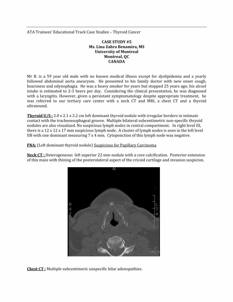
Mr B. is a 59 year old male with no known medical illness except for dyslipidemia and a yearly followed abdominal aorta aneurysm. He presented to his family doctor with new onset cough, hoarsness and odynophagia. He was a heavy smoker for years but stopped 25 years ago; his alcool intake is estimated to 2-3 beers per day. Considering the clinical presentation, he was diagnosed with a laryngitis. However, given a persistant symptomatology despite appropriate treatment, he was referred to our tertiary care center with a neck CT and MRI, a chest CT and a thyroid ultrasound. Thyroid U/S : 2.0 x 2.1 x 2.2 cm left dominant thyroid nodule with irregular borders in intimate contact with the tracheoesophageal groove. Multiple bilateral subcentimetric non-specific thryoid nodules are also visualized. No suspicious lymph nodes in central compartment. In right level III, there is a 12 x 12 x 17 mm suspicious lymph node. A cluster of lymph nodes is seen in the left level IIB with one dominant measuring 7 x 4 mm. Cytoponction of this lymph node was negative. FNA: (Left dominant thyroid nodule) Suspicious for Papillary Carcinoma Neck CT : Heterogeneous left superior 22 mm-nodule with a core calcification. Posterior extension of this mass with thining of the posterolateral aspect of the cricoid cartilage and invasion suspicion.
Chest CT : Multiple subcentimeric unspecific hilar adenopathies.
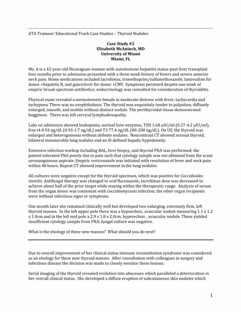
Neck MRI : Left superior pole thyroid lesion of 2.7 x 1.7 cm with cricoid invasion and suspected esophagal extension. At his visit in July, he had no more symptoms of odynophagia but chronic cough and hoarsness were still an important complaint. The patient denied hypo/hyperthyroidism symptoms, weight loss and asthenia. Flexible laryngoscopy proved left vocal cord paralysis. A pulmonary endoscoscopy performed by an experimented thoracic surgeon showed no tumoral extension in the trachea. Esophageal endosonography, however, revealed tumoral invasion of the muscularis propria distally to the superior esophageal sphincter. We performed a total thyroidectomy with bilateral central and lateral neck dissection. During the surgery, the right thyroid lobe was easily dissected. The difficulty resided mostly in dissecting the left lobe which seemed completely adherent to the laryngotracheal structure. We started by resecting the inferior pharyngeal constrictor muscles as well as esophageal muscles at its left cervical portion. The bougie proved to be a useful tool in performing the latter without penetrating the mucosa. Once this was done, the left lobe was easier to mobilize and was further dissected from the larynx and cricoid cartilage. The inferior horn of the left thyroid cartilage as well as ½ cm of the left cricoid showed signs of gross invasion. Thus, we performed a left thyroid cartilage horn resection and a shaving of the left cricoid cartilage at its external portion. The excised specimen was sent to pathology (details of pathology not yet available but should be for further discussion during the meeting). Questions :
1- What would have been the optimal preoperative investigations for this patient? Esophagoscopy? Esophagoscopy and esophageal endosonography ?
2- What would be the most appropriate treatment modality of the cricoid cartilage lesion? Shaving vs laryngectomy ?
3- Is adjuvent radiotherapy indicated in this case?
ATA Trainees’ Educational Track Case Studies – Thyroid Cancer
CASE STUDY #6 Johnson Thomas, MD
Nassau University Medical Center East Meadow, NY
Ms. AB is a 19 year old African American female. At the age of 14, in 2008, she had a benign left ovarian cystic teratoma resected. In 2010, a benign cystic teratoma with predominant thyroid tissue was resected from the right ovary. In September 2012 she presented to the ER with worsening abdominal pain and was admitted to the GYN service. Sonogram revealed a mixed solid and cystic pelvic mass measuring 10.6X7.9X7.6 cm. She underwent exploratory laporatomy in September 2012 and was found to have a large right adnexal mass with widespread nodules involving the peritoneum, omentum, spleen, bladder, uterus and rectal and abdominal walls. The intra-operative surgical impression was of widespread metastatic ovarian carcinoma and consequently the procedure was terminated by the GYN surgeon. However, Pathology reported follicular tissue with colloid in the widespread nodules and their impression was Peritoneal Strumosis. The Endocrinology service was consulted and the working diagnosis became Metastatic Struma Ovarii. In December 2012, CT revealed a large complex mass measuring 12.0 X 8.1 X 11 cm arising from the right adnexal area. Multiple enhancing soft tissue peritoneal nodules were noted including a lesion anterior to the spleen measuring 2.3 X 2.9 cm. She then underwent total hysterectomy, bilateral oophorectomy, omentectomy and splenectomy. Surgical pathology revealed well differentiated follicular thyroid carcinoma of ovarian origin. The tissue was positive for thyroglobulin and TTF-1. The diagnosis of metastatic Malignant Struma Ovarii was thus confirmed. Molecular marker testing was negative for BRAF, RAS, RET/PTC and TP53 mutations. Prior to surgery in December 2012, the patient was clinically and biochemically euthyroid (TSH 0.998 uIU/ml, T4 9.2 ug/dl, free T4 ng/dl 1.11, T3 122 ng/dl) and had a serum thyroglobulin of 229 ng/ml in the absence of antiTG antibodies. In April 2013, prior to total thyroidectomy, TSH and free T4 remained normal at 0.928 and 1.28 respectively while serum TG was 49.1 ng/ml with antiTGab undetectable. No lesions were found in the thyroidectomy specimen. Post-thyroidectomy 131I ablation treatment was given. Dosimetry evaluation revealed whole body isotope retention of 32.27% at 6 days. Photodense areas in the thyroid bed, abdomen and pelvis were seen on scan. The calculated maximum safe treatment dose of 131I was 200 mCi. An initial dose of 62.8 mCi was administered. Three months post-thyroidectomy, without thyroxine supplementation, TSH and T4 remained normal at 1.16 uIU/ml and 8.8 ug/dl respectively. Four months post-thyroidectomy, TSH rose to 4.82 uIU/ml while T4, free T4 and T3 were each normal at 7.3 ug/dl, 1.2 ng/dl and 99 ng/dl respectively. Serum thyroglobulin was 69.7 ng/ml in the continued absence of antiTGab. L-thyroxine was begun. It is planned to perform a followup whole body isotope scan with dosimetry and a second 131I ablation in October (six months post thyroidectomy).
1. Does the entity of Benign Peritoneal Strumosis exist or is this always metastatic well differentiated thyroid cancer?
2. If we see significant uptake in the second nuclear scan what are our options?


ATA Trainees’ Educational Track Case Studies – Thyroid Cancer
Case Study #7 Sarah Catherine Oltmann, MD
University of Wisconsin Madison, WI
HA is an 88 y.o. male who initially presented with complaints of voice hoarseness. During the work up of these complaints, he was found to have a left sided vocal cord paresis, along with vocal fold leukoplakia. This led to a CT scan of his neck to evaluate for any masses causing compression or disruption of his recurrent laryngeal nerve, and bilateral thyroid nodules were discovered. The largest was on the left, measuring 2.5 cm. A fine needle aspiration of this nodule was suspicious for papillary thyroid cancer. Further ultrasound examination of his neck demonstrated a suspicious lymph node in the left lateral chain. Given its location behind the carotid artery, it was not amenable to preoperative FNA biopsy. Based on this constellation of findings, he underwent a total thyroidectomy with central and lateral neck dissection. Incidentally, at time of surgery, nerve monitoring was used, and a strong signal was noted in bilateral recurrent laryngeal nerves. His voice hoarseness resolved immediately post-op.
His final pathology revealed a diagnosis of multi-focal medullary thyroid cancer, without lymphovascular invasion, and with 0 out of 9 lymph nodes involved. Tumor markers were sent off in the immediate post-operative period, with a calcitonin of 20.3, and a CEA of 37. Since that time they have normalized and remained stable.
This prompted referral for genetic testing, which returned positive for a RET mutation at Exon 16 (Arg912Pro). Testing of his adult children revealed that 2 out of the 4 were also positive. None of the grandchildren were noted to be positive.
With over a year of follow up, he is disease free and doing well.
ATA Trainees’ Educational Track Case Studies – Thyroid Cancer
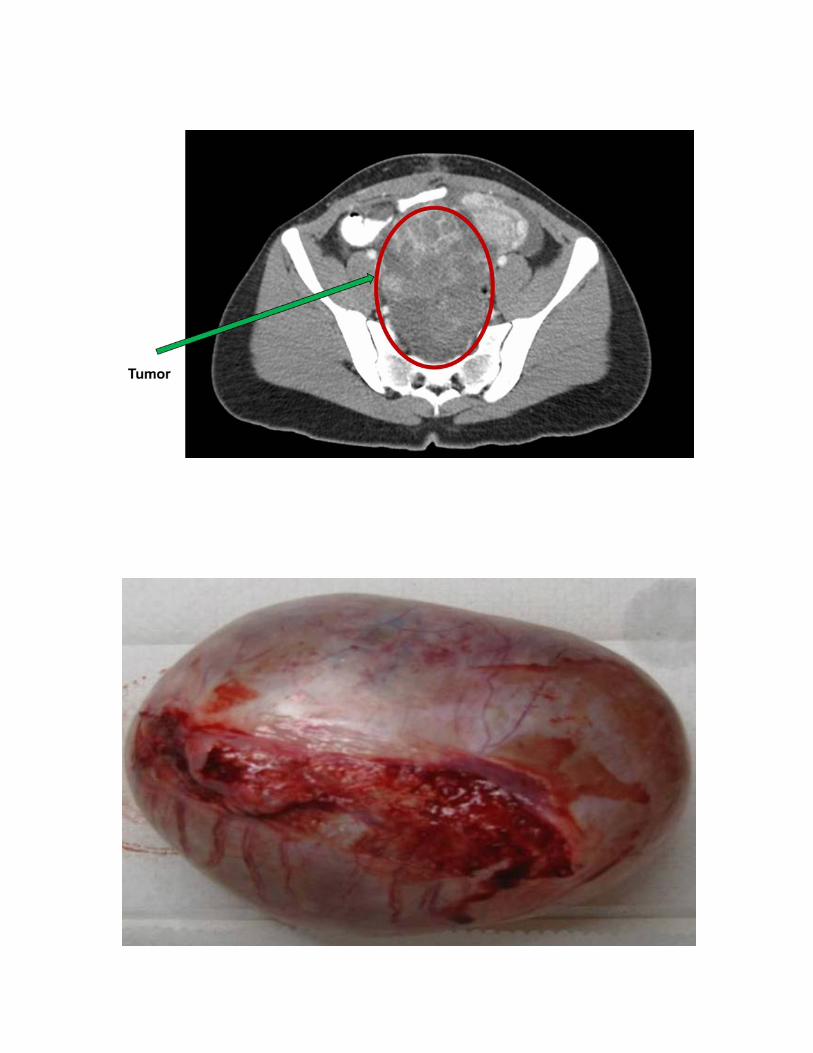
Case Study #8 Chhaya D. Makhija, MD
University of Nebraska Medical Center Omaha, NE
69 y.o. male seen followup of his papillary thyroid carcinoma T4 N1b M1, stage IVc with known pulmonary metastasis and newly diagnosed right choroidal mets, while on Pazopanib systemic therapy. Of note, he has history of melanoma in past. Papillary thyroid cancer: initially diagnosed in 2006, s/p total thyroidectomy and central neck dissection. Pathology showed multifocal tumor with extrathyroidal extension. Largest nodule focus was 1.7 cm. He received his first dose of radioactive iodine with 340 millicuries at that time. He had recurrence in his left neck and underwent left lateral neck dissection. He received a second dose of radioactive iodine of 313 millicuries at that time and then he had external beam radiation therapy because of extension of the tumor. He had a third dose of radioactive iodine with 192.7 millicuries of I-131 in 2009 because of rising thyroglobulin and no resectable disease. A post treatment scan showed foci of increased activity in the left lower lobe of the lung, superior mediastinum, and right clavicular region, and he has known pulmonary metastasis, however, not all of the pulmonary lesions were RAI avid. Pulmonary lesions were continuing to slowly increase in size along with his thyroglobulin. We started pazopanib, tyrosine kinase inhibitor, in August 2011. radiographically his disease had been relatively stable, however, his thyroglobulin continued to rise with the highest being 139 with negative ab. In Feb 2013 CT of his neck and chest showed essentially stable disease in his lungs without clear progression. At his last visit in April 2013, a new right retinal mass was identified. He was seen at University of Iowa for biopsy of the mass since the etiology was unclear (met thyroid cancer versus melanoma). The biopsy was consistent with metastatic papillary thyroid cancer. Unfortunately, he developed panendophthalmitis and required surgical removal of his right eye in May 2013. The choroidal mass was also completely excised and measures 3.5 x 9.5 mm. PAS positive, the tumor demonstrated staining for pancytokeratin, ck7, thyroglobulin, and TTF. CK20 negative. Near the apex of the tumor, tumor can be seen extending from the choroid into the subretinal space where there is a breach in Bruch’s membrane likely from the previous FNA. In addition, repeat CT of the chest prior to his choroid biopsy identified a left infrahilar node that had dramatically increased in size from Feb, 2013. It measured 3.8 x 3.2 cm (previously 2.4 x 2.3 cm). There was also mass effect along the left paraspinal region and post obstructive atelectasis and infiltrate in the left lower lobe and left pleural effusion. He received 3000 cGy in 10 fraction to the gross disease in lower mediastinum, left hilum and left lower lobe. This was completed in June 2013.
In regards to his post-surgical hypothyroidism, he is taking Synthroid 250mcg daily, with 2 additional 125 mcg pills during the week. After resection of choroidal metastasis and failure to respond to pazopanib, what would be the next treatment option? Patient was started on vandetanib (off label use).
ATA Trainees’ Educational Track Case Studies – Thyroid Function
Case Study #9 Georgiana Alina Dobri, MD
Cleveland Clinic Foundation Cleveland, OH
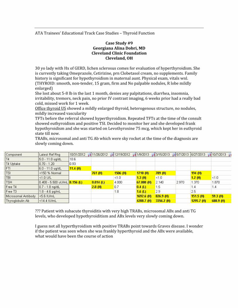
30 yo lady with Hx of GERD, lichen sclerosus comes for evaluation of hyperthyroidism. She is currently taking Omeprazole, Cetirizine, prn Clobetasol cream, no supplements. Family history is significant for hypothyroidism in maternal aunt. Physical exam, vitals wnl. (THYROID: smooth, non-tender, 15 gram, firm and No palpable nodules, R lobe mildly enlarged) She lost about 5-8 lb in the last 1 month, denies any palpitations, diarrhea, insomnia, irritability, tremors, neck pain, no prior IV contrast imaging, 6 weeks prior had a really bad cold, missed work for 1 week. Office thyroid US showed a mildly enlarged thyroid, heterogenous structure, no nodules, mildly increased vascularity TFTs before the referral showed hyperthyroidism. Repeated TFTs at the time of the consult showed euthyroidism and positive TSI. Decided to monitor her and she developed frank hypothyroidism and she was started on Levothyroxine 75 mcg, which kept her in euthyroid state till now. TRABs, microsomal and anti TG Ab which were sky rocket at the time of the diagnosis are slowly coming down.
??? Patient with subacute thyroiditis with very high TRABs, microsomal ABx and anti TG levels, who developed hypothyroiditism and ABx levels very slowly coming down. I guess not all hyperthyroidism with positive TRABs point towards Graves disease. I wonder if the patient was seen when she was frankly hyperthyroid and the ABx were available, what would have been the course of action
ATA Trainees’ Educational Track Case Studies – Thyroid Function
Case Study #11 Jelena Maletkovic, MD
University of California - Los Angeles Los Angeles, CA
The patient is a 52 year old woman with history of severe CAD, status post multiple stents, CABG, also hypertension, hyperlipidemia, diabetes mellitus, ESRD on dialysis, paroxismal A fib, recently diagnosed with AIT type 1, now admitted with chest pain and hypotension (56/26), diagnosed with ACS, managed conservatively.
Endocrine consult is called for management of thyrotoxicosis in the setting of elevated LFT’s on methimazole.
Prior to this admission the patient was diagnosed on AIT type1 and started on methimazole with TSH 0.014 (nl 0.35-4.94), Free T3 11.4 (nl 1.71-3.71), Free T4 4.35 (nl 0.7-1.48). Thyroid-Stimulating Immunoglobulin 106 (nl <122%), TPO 2.9 (neg), and thyroglobulin <0.9 (neg). Thyroid ultrasound shows two cystic nodules around 1cm each.
On admission the patient has abnormal LFT’s: AST 81, ALT 547, TB 5.3 and methimazole is stopped. With liver failure, kidney failure, heart failure on pressors this patient is not a surgical candidate. The patient is also not a candidate for any more methimazole or PTU. Not a candidate for radioactive iodine treatment with history of 3 years treatment with amiodarone.
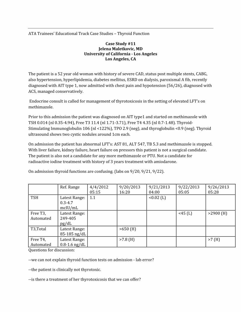
On admission thyroid functions are confusing. (labs on 9/20, 9/21, 9/22).
Ref. Range 4/4/2012 05:15
9/20/2013 16:20
9/21/2013 04:00
9/22/2013 05:05
9/26/2013 05:28
TSH Latest Range: 0.3-4.7 mcIU/mL
1.1 <0.02 (L)
Free T3, Automated
Latest Range: 249-405 pg/dL
<45 (L) >2900 (H)
T3,Total Latest Range: 85-185 ng/dL
>650 (H)
Free T4, Automated
Latest Range: 0.8-1.6 ng/dL
>7.8 (H) >7 (H)
Questions for discussion:
--we can not explain thyroid function tests on admission - lab error?
--the patient is clinically not thyrotoxic.
--is there a treatment of her thyrotoxicosis that we can offer?
ATA Trainees’ Educational Track Case Studies – Thyroid Function
Case Study #12 Washington University in St. Louis
Cynthia Joan Herrick, MD St. Louis, MO
A 71-year-old woman with a history of well-controlled type 2 diabetes, hypertension, hyperlipidemia, hypothyroidism and remote history of breast cancer status post lumpectomy, chemotherapy and radiation, presents with a one-month history of heat intolerance, palpitations, tremors, dyspnea on exertion and chest pain on exertion. She had noted a weight loss of about 46 pounds over the last year with 20 pound loss in the last three months. She went to a new primary care provider and was noted to have tachycardia to the 120s in the office and was advised to go the ED for further evaluation. She reports a diagnosis of hypothyroidism at age 18, on thyroid replacement since that time. She never had a diagnosis of Graves’ disease and never received a radioactive iodine treatment. Pharmacy records confirm that she was on 200 micrograms per day of Levothyroxine for many years in the past. This was decreased to 175 mcg six times per week in 2010 and increased to 175 mcg daily 3 months prior to admission (~15% increase in dose). Thyroid studies at the times of these dose changes were not available. She was noted on admission to have a thyroid-stimulating hormone of 0.04 mcIU/ml (0.35-5.5), free T4 of 9.37 ng/dl (0.9-1.8) and a free T3 of greater than 15 pg/ml (2.3-4.2). Troponin was elevated at 1.14 ng/ml (0.00-0.24). She denies any preceding history of a viral illness or neck pain at any point. She denies taking any supplements and denies that her levothyroxine pills looked any different. She has taken all of her pills in the morning and nothing has changed in the way she takes her medication. She was not taking and had never taken Amiodarone. She lives in a rural area but denies any recent ingestion of meat from a local uncertified butcher. Her last dose of Levothyroxine was 24 hours prior to admission. She has no thyroid or other endocrine disease in the family. Exam was notable for normal temperature, heart rate of 100, blood pressure of 135/45. Weight is currently160 lbs. She had no proptosis, stare or lid lag, and extraocular movements were intact. She had no conjunctival injection. The thyroid was small but palpable without nodularity. Cardiovascular exam was normal. Extremities were notable for trace edema bilaterally. Reflexes were 2+ with normal relaxation. She was noted to have a fine tremor. She had a CT chest/abd/pelvis with contrast in the ED prior to the return of the thyroid labs looking for malignancy, which precluded doing an RAI uptake. Given her initial presentation of NSTEMI, in addition to stopping Levothyroxine, she was started on Propranolol 40 mg TID and Methimazole 20 mg daily given the possibility of endogenous overproduction of thyroid hormone. A thyroid stimulating immunoglobulin and thyroglobulin level were sent to further evaluate this possibility. When these returned normal, the Methimazole was stopped. The patient’s troponin trended down to 0.15. She

had an echocardiogram showing a normal EF and no regional wall motion abnormalities, and she was discharged with plan for outpatient evaluation. She had a repeat FT4 of 1.9 ng/dl (0.9-1.8) after one week off of Levothyroxine and Methimazole. She had a repeat TSH and FT4 after being off of Levothyroxine and Methimazole for 6 weeks that were normal - TSH 1.25 mcIU/ml (0.35-5.5) and FT4 1.04 ng/dl (0.9-1.8). What accounts for this apparent return of thyroid function after 50 years on replacement? How frequently would you monitor thyroid function going forward?