Embed Size (px)
Citation preview
APPLIED AND ENVIRONMENTAL MICROBIOLOGY, Jan. 1989, p. 137-1410099-2240/89/010137-05$02.00/0Copyright © 1989, American Society for Microbiology
Associated Bacterial Flora, Growth, and Toxicity of CulturedBenthic Dinoflagellates Ostreopsis lenticularis
and Gambierdiscus toxicusT. R. TOSTESON,* D. L. BALLANTINE, C. G. TOSTESON, V. HENSLEY, AND A. T. BARDALES
Department of Marine Sciences, University of Puerto Rico at Mayaguez, Mayaguez, Puerto Rico 00709-5000
Received 19 July 1988/Accepted 20 October 1988
The growth, toxicity, and associated bacterial flora of 10 clonal cultures of the toxic benthic dinoflagellatesOstreopsis lenticularis and Gambierdiscus toxicus isolated from the coastal waters of southwest Puerto Rico havebeen examined. Clonal cultures of 0. lenticularis grew more rapidly and at broader temperature ranges thanthose of G. toxicus. All five Ostreopsis clones were toxic, while only one of the five Gambierdiscus clones was
poisonous. The degree of toxicity among poisonous clones was highly variable. The number of associatedbacterial genera and their frequency of occurrence were quite variable among clones of both dinoflagellategenera. Bacterial isolates represented six genera (Nocardia, Pseudomonas, Vibrio, Aeromonas, Flavobacterium,and Moraxella) in addition to coryneform bacteria. Extracts of dinoflagellate-associated bacteria grown in pureculture were not toxic. Gambierdiscus clones were characterized by the frequent presence of Pseudomonas spp.
(four of five clones) and the absence of coryneforms. In 0. lenticularis, only one of five clones showed thepresence of Pseudomonas spp., and Moraxella sp. was absent altogether. Detailed analyses of toxicity andassociated microflora in a selected Ostreopsis clone, repeatedly cultivated (four times) over a period of 160 days,showed that peak cell toxicities developed in the late static and early negative culture growth phases. PeakOstreopsis cell toxicities in the stationary phase of culture growth were correlated with significant increases inthe percent total bacteria directly associated with these cells. Changes in the quantity of bacteria directlyassociated with microalgal cell surfaces and extracellular matrices during culture growth may be related tovariability and degree of toxicity in these laboratory-cultured benthic dinoflagellates.
Toxins produced by marine dinoflagellates are among themost potent nonproteinaceous poisons known (4, 30, 31).Twenty-two dinoflagellate species are known to producetoxins (31, 34). These toxins include both water- and lipid-soluble moieties, which have hemolytic, neurotoxic, andgastrointestinal inflammatory activities. Toxins produced byGambierdiscus toxicus Adachi and Fukuyo (2, 38, 39),Ostreopsis lenticularis Fukuyo (40), and other benthic dino-flagellates (32) have been proposed to be linked with cigua-tera fish poisoning.
Fluctuations in natural populations of G. toxicus and itsecological associate, 0. lenticularis, in Puerto Rico havebeen documented for several years (5, 5a). Both speciesform occasional blooms, with 0. lenticularis reaching den-sities of greater than 100,000 cells per g (wet weight) of algalhost tissue. Puerto Rican G. toxicus and 0. lenticularisproduce toxins that are lethal to mice. Recently reportedtoxicity in 0. lenticularis is the first found for this species(34). While toxicity in G. toxicus has been recognized forsome time, questions remain as to the number of toxinsactually produced and their precise chemical nature. Chem-ical or pharmacological similarities between toxins producedby these sources have not been determined.
In nature, most microorganisms are found in heteroge-neous aggregations with suspended detrital material or ad-hered to macroalgal, animal, and inanimate surfaces. Thesesurface interactions are dynamic in nature and are importantfactors in microbial proliferation and survival. Aquatic algaein situ as well as in laboratory culture are often found to beassociated with a variety of bacterial strains (6, 8, 17, 24, 25).The inter- and intraspecific interactions of marine microbial
* Corresponding author.
cells are frequently mediated by macromolecular surfacecomponents, suggesting the presence of specific receptor-ligand binding sites on interactive surfaces (16, 33, 35).Recently reported analyses of bacterial-microalgal interac-tions in a consortium of three bacteria associated with a
chlorella-like green alga illustrate the complexity of theserelationships (19). Bacteria are universally associated withalgae in the ocean. The fact that many algae grow more
slowly, if at all, in axenic rather than bacterized culturessuggests that the associations constitute a form of symbiosis.
Production of toxins by marine macrobiota and microalgalcells, associated with variable microbial consortia, posesimportant questions regarding the precise origin and mech-anism of synthesis of the toxins in question. Thus, it hasbeen suggested that palytoxin, one of the most potent marine
toxins, may well be produced by a Vibrio bacterial symbiontof the zooanthid Palythoa sp., originally thought to be thesole source of this toxin (22, 23). The highly variable toxicityof Palythoa specimens taken at different geographical sitesinitially suggested the possible involvement of other factorsin the elaboration of this toxin. Large variations in toxinproduction by strains of the dinoflagellate Protogonyalauxtamarensis (Lebour) Taylor from different geographical lo-cations have also been reported (27). Four pure, distincttoxins were isolated from this dinoflagellate. Variability inclone toxin potency and the increasing numbers of dinofla-gellate species associated with toxins suggest that dinofla-gellate toxin production may be linked to symbiotic or
contaminant bacteria (30, 31). Grown in bacterium-free(axenic) culture, Ptychodiscus brevis (Davis) Steidinger hasbeen reported to retain toxicity, while Gymnodinium vene-
ficum Ballantine lost toxicity (1, 26). Toxicity of Prorocen-trum minimum Schiller and Protogonyalaux tamarensis has
137
Vol. 55, No. 1
on July 9, 2018 by guesthttp://aem
.asm.org/
Dow
nloaded from
138 TOSTESON ET AL.
been proposed to be due to the presence of bacteria associ-ated either with the medium or dinoflagellate endosymbionts(28). These investigators isolated a bacterial strain, a Pseu-domonas sp., from cultures of Protogonyalaux tamarensisthat induced toxicity when inoculated into cultures of apreviously nontoxic Gyrodinium strain.The work reported here was undertaken to identify the
bacteria associated with benthic dinoflagellates 0. lenticu-laris and G. toxicus originally isolated from coastal waters ofsouthwest Puerto Rico and grown in clonal laboratory cul-ture. Studies were conducted to ascertain the relationship ofdiversity, quantity, and direct microalgal cell association ofthese bacterial strains with dinoflagellate culture growth andtoxicity.
MATERIALS AND METHODS
Dinoflagellate culture. Clonal cultures were obtained bythe methods of Lewin (20) as adapted for benthic dinoflagel-late isolation by Yasumoto et al. (39) and Ballantine et al. (5).Clonal isolates were obtained by transferring individual cellsthrough three sterile seawater washes with drawn Pasteurpipettes. After the third wash, a single cell was similarlytransferred into a test tube containing 10 ml of sterile f/2medium (14, 21) with added germanium dioxide (5 mg/liter).Germanium dioxide is an inhibitor of silicon metabolism, andits presence in culture medium controls diatom contamina-tion (20). Test tubes were then placed under reduced lightconditions and were left undisturbed for approximately 1month. In our experience, approximately 20% of the inocu-lated test tubes yielded viable clonal cultures.
Stock cultures of dinoflagellates were grown in standardf/2 medium prepared with artificial seawater. A water-sol-uble extract (0.1% by volume) of the red alga Acanthophoraspicifera was added (Withers, personal communication), asit has been shown to increase the growth rate of both G.toxicus and 0. lenticularis in culture. Optimal conditions forsupport of growth of 0. lenticularis and G. toxicus withrespect to light and temperature were evaluated with across-gradient culture apparatus (13). Experiments wereconducted in stoppered 50-ml flasks containing initial con-centrations of 75 dinoflagellate cells per ml. Light regimensof 20, 60, and 100 microeinsteins/m2 per s combined withtemperature regimens ranging from 20 to 30°C were tested.On the basis of these analyses, subsequent cultures weregrown at 27°C in a light-to-dark regimen of 16:8 and at a lightflux of 50 microeinsteins/m2 per s.For purposes of establishing base-line information con-
cerning clonal toxicity and associated bacterial flora, G.toxicus and 0. lenticularis clones were grown in batchcultures (3 to 6 liters) under the culture conditions describedabove. Cells were harvested by screening (35-,um mesh)after appropriate periods of growth. One Ostreopsis clone(no. 116) was selected for analyses of growth and toxicity ina series of experiments each lasting from 28 to 49 days.Experiments were initiated at dinoflagellate concentrationsof 75 cells per ml in 2.5-liter Fernbach flasks containing 1liter of culture. Growth was evaluated microscopically byusing a Sedgewick Rafter counting cell after 4 and 7 days andthen at weekly intervals until the termination of the partic-ular experiment. On each of these sampling days, a variablenumber of flasks (1 to 4) were selected for harvest.
Associated bacterial flora. Bacteria associated with alldinoflagellate clones were isolated in pure culture by re-peated streaking on nutrient seawater agar, and isolates wereidentified to the generic level (18, 34). Classifications were
made by using API 20E and other biochemical and antibiotictests. Bacteria of the genus Vibrio were distinguished fromnonfermenting Pseudomonas spp. by using 0/129 (2,4-dia-mino-6,7-diisopropyl-pteridine phosphate) disks at concen-trations of 150 and 10 p.g.
In 0. lenticularis culture flasks selected for toxicity anal-yses, densities of the total bacterial populations relative todinoflagellate cells (total bacterial cell/dinoflagellate cell ra-tio) and proportions of these bacteria directly associatedwith dinoflagellate surfaces and extracellular matrices wereestimated by epifluorescence microscopy (15). The totalbacterial cell/dinoflagellate cell ratio was taken to be equal tothe sum of the unassociated free bacteria in the medium/dinoflagellate cell ratio and the bacteria closely associatedwith the dinoflagellates/dinoflagellate cell ratio. Thus, thetotal bacterial cell/dinoflagellate cell ratio (A) minus thebacteria recovered free in the dinoflagellate culture medium/dinoflagellate cell ratio (B) equalled C, the fraction of bacte-ria directly associated with dinoflagellate cell surface perdinoflagellate cell. Samples (10 ml) of culture suspensionswere sonicated (Megason; Ultrasonic Instruments Interna-tional, Inc.) to disrupt bacterium-dinoflagellate cell attach-ments. The resulting suspensions were stained with acridineorange and layered on filters (Nuclepore Corp.) for countingand subsequent determination of the ratios of the totalbacterial cells to dinoflagellate cells (ratio A) in the respec-tive cultures. In addition, unsonicated samples (50 to 100 ml)from these culture flasks were gently passed through filters(Gelman A-E, 1.0-pLm pore size) which retained intact dino-flagellate cells while allowing unattached free bacterial cellsto pass into the filtrate. Concentrations of these bacteria inthe dinoflagellate-free filtrates were determined by usingepifluorescence techniques, and ratios of these unassociatedbacterial cells to dinoflagellate cells (ratio B) were subse-quently determined. These ratios were used to calculatepercentages of bacteria directly associated with Ostreopsiscells as follows: %C/A = [1 - (B/A)] x 100.
Toxicity analyses. Harvested dinoflagellate cells werebriefly rinsed with distilled water and were sonicated inredistilled methanol. Extracts (final volumes, approximately100 ml) were kept at laboratory temperatures (22°C) for 48 h.Extract suspensions were filtered (Whatman no. 1 filterpaper), and filtrate solvent was removed by flash evapora-tion (Buchi, Rotavapor). The resulting residues were driedunder nitrogen and were stored in a vacuum desiccator forsubsequent toxicity analyses. These procedures and themethods on which they are based have been reportedelsewhere (34). Similar extracts were prepared from purecultures of the dinoflagellate-associated bacterial strains.Isolates were individually grown in suspension culture for 48h at laboratory temperatures (22°C) in a modified Zobell2216E medium (5.0 g of yeast extract-1.0 g of peptone-0.01g of FePO4 in 500 ml of artificial seawater and 500 ml ofdistilled water). Following incubation, the medium wascentrifuged (1 h at 27,000 x g in a Sorvall RC2-B centrifugeat 4°C) and the pellet was suspended in redistilled methanoland sonicated for extraction as described above.
Extracts were assayed for their toxicity in 20-g whiteSwiss CF1 mice. Known quantities of dried extracts to betested were suspended in 0.15 M phosphate buffer solution(pH 7.4) containing 5% Tween 80 (34). Inocula of between0.2 and 0.5 ml were administered by intraperitoneal injec-tion. Control animals received injections of equal volumes ofTween-phosphate-buffered saline medium. Mice were ob-served for 48 h, and 50% lethal dose values were calculatedby standard methods (42). Dinoflagellate extracts were ex-
APPL. ENVIRON. MICROBIOL.
on July 9, 2018 by guesthttp://aem
.asm.org/
Dow
nloaded from
BACTERIAL FLORA OF TOXIC BENTHIC DINOFLAGELLATES
amined at concentrations of 0.01 to 5 mg per animal (0.5 to
250 mg per kg of body weight). Each extract was tested atfrom four to eight concentrations, decreasing geometricallyfrom the highest levels tested. Three or four mice were
inoculated at each extract concentration in a given experi-ment. Toxicities of the extracts reported here are expressedin terms of "mouse units" (MU) per cell extracted. An MUwas defined as the amount (in milligrams) of toxic extract
inoculated into a single 20-g mouse that resulted in the deathof 50% of the mice injected with this quantity of material.The total MU in an extract (i.e., milligrams of toxic extract
per MU) was calculated and toxicity was expressed in termsof MU per cell (total MU per total number of cells ex-
tracted).
RESULTS AND DISCUSSION
Laboratory-cultured dinoflagellates. 0. lenticularis demon-strated optimal growth (shortest mean generation time, 1.03days) at 29°C and light fluxes of 60 and 100 microeinsteins/m2per s. In general, at temperatures less than 27°C, the mean
generation time was shorter at light fluxes of 20 microein-steins/m2 per s than at 100 microeinsteins/m2 per s. Thereverse was true at temperatures greater than 29°C. Rapidgrowth could nevertheless be elicited at both high and lowlight levels by changes in temperature. For Gambierdiscuscultures, temperatures below 23°C and above 30°C were notsuitable for growth. The shortest mean generation timeobtained with G. toxicus was 4.40 days. Thus, cultures of 0.
lenticularis were capable of considerably more rapid growthat broader temperature ranges than those of G. toxicus.
Dinoflagellate-associated bacterial flora. A total of 41 bac-terial isolates were recovered from the dinoflagellate clonesexamined in this study and were classified by genera. The 10dinoflagellate clones examined had associated bacteria of sixgenera as follows: Nocardia, 20%; Pseudomonas, 50%;Vibrio, 30%; Aeromonas, 20%; Flavobacterium, 20%; andMoraxella, 10%. Coryneform bacteria were associated with10% of the clones. Pseudomonas was the most frequentlypresent genus. On the basis of the methods employed here,this genus may have included bacteria of the genus Altero-monas. An endoceflular Pseudomonas species has beenreported to play a role in determining toxicity in otherdinoflagellates (28).
Dinoflagellate toxicity and associated bacteria. Thirty tox-icity assays of dinoflagellate clone extracts were conducted.The results are summarized in Table 1. Of five isolates ofPuerto Rican G. toxicus, only one clone proved to be toxic,while all five clones of Puerto Rican 0. lenticularis were
poisonous. Toxicity of Gambierdiscus clone 105 (162 MU/106 cells) was within the range of Ostreopsis clone toxicities(16 to 651 MU/106 cells) determined. Ostreopsis clonesshowed considerable variability in their toxicity. Assays ofmethanolic extracts made of pure cultures of the dinoflagel-late-associated bacterial genera indicated that none of themwere toxic.
Bacterial genera and their frequency of occurrence were
quite variable among clones of both dinoflagellate genera.Gambierdiscus clones were characterized by the frequentpresence of Pseudomonas spp. (four of five clones) and theabsence of coryneforms. Only one of five Ostreopsis clonesshowed the presence of Pseudomonas spp., and Moraxellaspp. were absent altogether. In toxic Gambierdiscus clone105, the only bacterial genus found was Pseudomonas. It isof interest that the most toxic Ostreopsis clone (116-5) was
the only clone of this dinoflagellate genus tested that had an
TABLE 1. Associated bacteria and dinoflagellate toxicity
Associated DinoflagellateDinoflagellate bacterial toxicity
clone genus or (MU/106 cells)type"
G. toxicus94-13 N, V, A 0101-16 P, M 0105-13 P 162106-13 P, V 0107-24 P, F 0
0. lenticularis95-49 N, A 42116-5 P, F 651117-5 None found 45119-4 V 53120-2 c 16
a N, Nocardia; V, Vibrio; A, Aeromonas; P, Pseudomonas; M, Moraxella;F, Flavobacterium; c, coryneform bacteria.
associated Pseudomonas species. All Ostreopsis cloneswere toxic, and three of five clones had only one associatedbacterial strain. No bacteria were found associated withOstreopsis clone 117.
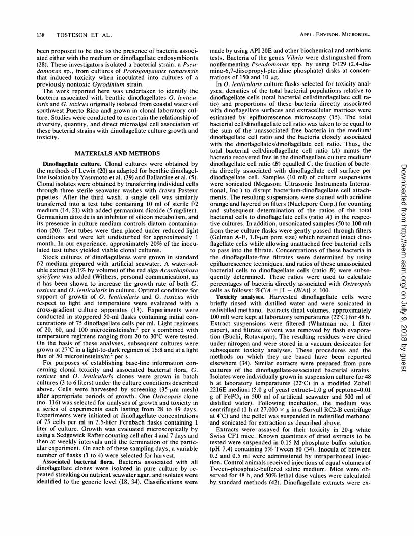
Dinoflagellate culture growth, toxicity, and bacterial flora.0. lenticularis clone 116 was transferred 48 times betweenthe first and fourth experiments. With increasing numbers oftransfers, there was a reduction in the observed toxicity.Peak toxicity in the first experiment was 5,785 MU/106 cellsand was only 49 MU/106 cells in the final experiment of theseries, at the end of 1 year during which these experimentswere conducted. These results differ from those of Durand-Clement (12), who reported little change in the toxicity ofcultured G. toxicus over a period of 3 years. While peak celldensities did not show systematic change, the time requiredto achieve maximum cell densities increased from 14 (exper-iments 1 and 2) to 28 days (experiments 3 and 4). Despitethese changes in toxicity and growth rate, Ostreopsis cellsshowed maximum toxicity on the same day of culture growthin all four experiments. The average relative growth rate ofthe cultures in all four experiments (the relative growth ratein each expressed as the ratio of the growth rate [reciprocalof the mean generation time x 102] for any given interval ofculture growth to that of the growth rate over the initial4-day period of that experiment) is summarized in Fig. 1. Ineach experiment, cultures grew most rapidly during theinitial 4-day period. The average relative toxicities of cells(expressed as the ratio of toxicity of the cells sampled at agiven time to the toxicity of the cells used to initiallyinoculate the cultures in each experiment) showed peakvalues after 28 days of culture growth (Fig. 1). Peak celltoxicities were found in the late static and early negativegrowth phases of these cultures. Maximum cell toxicitieshave been reported elsewhere for cultures in exponentialgrowth phase (9) as well as during stationary phase and theinitial stages of negative growth (3, 7, 28, 29, 31, 40).The genera of bacteria found with Ostreopsis clone 116 did
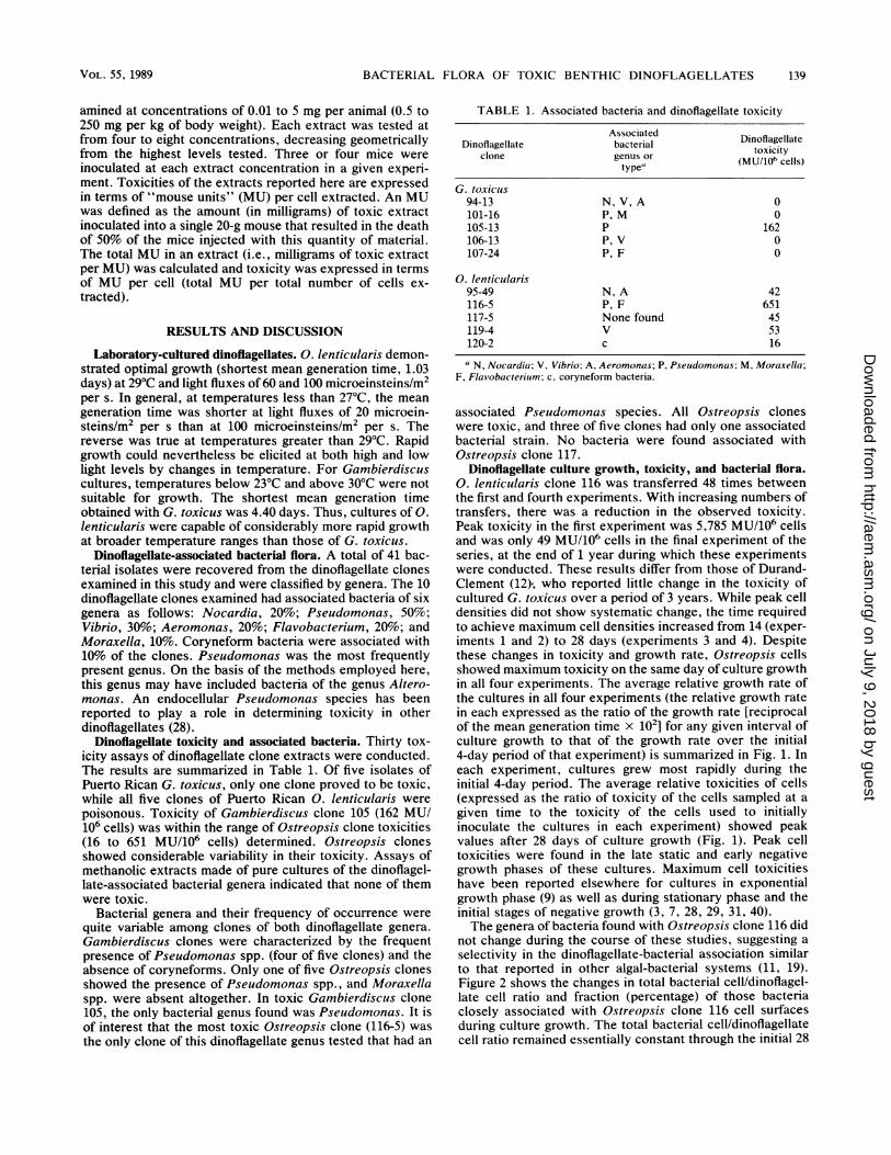
not change during the course of these studies, suggesting aselectivity in the dinoflagellate-bacterial association similarto that reported in other algal-bacterial systems (11, 19).Figure 2 shows the changes in total bacterial cell/dinoflagel-late cell ratio and fraction (percentage) of those bacteriaclosely associated with Ostreopsis clone 116 cell surfacesduring culture growth. The total bacterial cell/dinoflagellatecell ratio remained essentially constant through the initial 28
VOL. 55, 1989 139
on July 9, 2018 by guesthttp://aem
.asm.org/
Dow
nloaded from

140 TOSTESON ET AL. APPL. ENVIRON. MICROBIOL.
RELATIVE 0.4_ \ 4 RELATIVE
GROWTH TOXICITY
0.2- 2
-0.24 -2
-04 I40 10 20 30 40 50
TIME (DAYS)
FIG. 1. Relative growth of 0. lenticularis (E), expressed as theratio of the growth rate (reciprocal of the generation time) for a givenperiod to the growth rate of the culture during the first 4 days ofculture growth, and the relative toxicity of the Ostreopsis cells (0),expressed as the ratio of the toxicity of the dinoflagellate cells (MU/106 cells) during a given period of culture growth to the toxicity ofthe cells used to initiate the cultures in this study, versus time ofculture growth.
days of culture growth. Following this period, there was a
steady, significant increase in the total bacterial cell/dinofla-gellate cell ratio through 49 days of culture growth. Thepercent total bacteria directly associated with the dinoflagel-late cells was high (above 70%) in the inocula used to initiatethe dinoflagellate cultures in this study. This percentagedecreased significantly (to values below 10%) during the first7 days, followed by sharp increases (60 to 80%) at 21 to 35days of culture growth. Chrost reported a general decline in
t0 .-00
/ y./ RELATIVE ..X TOXICITY
400~~~~~~~~~
100- 50
SCELL
TOTAL 9ACT,10'I
ASSOC.
CELL BACC.10T
/3
~~~~~~~~~~~~~~~~0
FIG. 2. The total bacterial cell/0. lenticularis cell ratio and thepercent bacterial cells directly associated with these dinoflagellatecells, versus time of culture growth. Shaded area indicates thechanges in the relative toxicity of Ostreopsis cells during the periodof culture growth.
bacterial populations during blooms of mixed phytoplank-ton, which he attributed to the production of allelopathicsubstances (10).The dinoflagellate cells appeared to exercise some control
on both the total density and distribution of the bacterialpopulations present in the respective culture flasks. Peakdinoflagellate culture growth rates (first 4 to 7 days ofculture, Fig. 1) were associated with reduced numbers ofbacteria directly associated with the dinoflagellate cells (Fig.2), while peak relative dinoflagellate cell toxicity (Fig. 2,shaded area) was associated with a significantly increasedfraction of closely associated bacteria. Later stages of cul-ture growth (35 to 49 days) were marked by reductions indinoflagellate cell toxicity and relatively uncontrolled in-creases in the total bacterial cell/dinoflagellate cell ratio.Increases in bacterial population densities associated withthe decline of phytoplankton blooms have been reportedelsewhere (10, 36).
Results presented here show that bacterial genera associ-ated with 0. lenticularis grown in clonal laboratory cultureare not toxic when grown individually in pure culture.Marked increases in the proportion of these bacteria directlyassociated with the surfaces or extracellular matrices ofthese microalgal cells were correlated with the developmentof peak dinoflagellate toxicity during the static phase of theirculture growth. Subsequent declines in dinoflagellate culturedensity and toxicity corresponded to uncontrolled increasesin the total bacterial cell/dinoflagellate cell ratio and decreas-ing proportions of bacteria directly associated withOstreopsis cells. These results suggest that associated bac-terial flora may play a role in the phasic development oftoxicity in laboratory growth cycles of these algal-bacterialconsortia.
ACKNOWLEDGMENTS
This work was supported by grants R/LR-08-1 and L/LR-08-87-TT02 from the Sea Grant Program of the University of Puerto Rico,grant RII-8610677 from the National Science Foundation, PuertoRico EPSCoR Program, and funds from the Office of ResearchCoordination, Faculty of Arts and Sciences, University of PuertoRico-Mayaguez.
LITERATURE CITED1. Abbott, B. C., and D. Ballantine. 1957. The toxin from Gymno-
dinium i'eneficum Ballantine. J. Mar. Biol. Assoc. U.K. 36:169-189.
2. Adachi, R., and Y. Fukuyo. 1979. The thecal structure of amarine toxic dinoflagellate Gambierdiscus toxicus gen. et sp.nov. collected in a ciguatera endemic area. Bull. Jpn. Soc. Sci.Fish. 45:67-71.
3. Aldrich, D. V., S. M. Ray, and W. B. Wilson. 1967. Gonyaulaxmonilata: population growth and development of toxicity incultures. J. Protozool. 14:636-639.
4. Anderson, D. M., and P. S. Lobel. 1987. The continuing enigmaof ciguatera. Biol. Bull. 172:89-107.
5. Ballantine, D. L., A. T. Bardales, T. R. Tosteson, and H. D.Durst. 1985. Seasonal abundance of Gambierdiscus toxicus andOstreopsis sp. in coastal waters of southwest Puerto Rico. FifthInt. Coral Reef Congr. 4:417-422.
Sa.Ballantine, D. L., T. R. Tosteson, and A. T. Bardales. 1988.Population dynamics and toxicity of natural populations ofbenthic dinoflagellates in southwestern Puerto Rico. J. Exp.Mar. Biol. Ecol. 119:201-212.
6. Bell, W., and R. Mitchell. 1974. Selective stimulation of marinebacteria by algal extracellular products. Limnol. Oceanogr. 19:833-839.
7. Bergmann, J. S., and M. Alam. 1981. On the toxicity of theciguatera producing dinoflagellate, Gambierdiscuis toxicus Ada-
on July 9, 2018 by guesthttp://aem
.asm.org/
Dow
nloaded from
BACTERIAL FLORA OF TOXIC BENTHIC DINOFLAGELLATES
chi and Fukuyo isolated from the Florida Keys. J. Environ.Health Sci. A16(5):493-500.
8. Berland, B. R., D. J. Bonin, and S. Y. Maestrini. 1970. Study ofbacteria associated with marine algae in culture. III. Organicsubstrates supporting growth. Mar. Biol. (Berlin) 5:68-76.
9. Boyer, G. L., J. J. Sullivan, P. J. Andersen, P. J. Harrison, andF. J. R. Taylor. 1985. Toxin production in three isolates ofProtogonyaulax sp., p. 281-286. In D. M. Anderson, A. W.White, and D. G. Baden (ed.), Toxic dinoflagellates. ElsevierScience Publishing, Inc., New York.
10. Chrost, R. S. 1975. Inhibitors produced by algae as an ecologicalfactor affecting bacteria in water ecosystems. I. Dependencebetween phytoplankton and bacterial development. Acta Micro-biol. Pol. Ser. B 7:125-133.
11. Dimanlig, M. N. V., and F. J. R. Taylor. 1985. Extracellularbacteria and toxin production in Protogonyaulax species, p.103-108. In D. M. Anderson, A. W. White, and D. G. Baden(ed.), Toxic dinoflagellates. Elsevier Science Publishing, Inc.,New York.
12. Durand-Clemedt, M. 1987. Study of production and toxicity ofcultured Gambierdiscus toxicus. Biol. Bull. 172:108-121.
13. Edwards, P. 1970. An apparatus for the culture of benthicmarine algae under varying regimes of temperature and lightintensity. Bot. Mar. 13:42-43.
14. Guillard, R. R. L., and J. H. Ryther. 1962. Studies of marineplanktonic diatoms. I. Cyclotella nana Hustedt and Detonulaconfervacea (Cleve) Gran. Can. J. Microbiol. 8:229-239.
15. Hobbie, J. E., R. J. Daley, and S. Jasper. 1977. Use ofNuclepore filters for counting bacteria by fluorescence micros-copy. Appl. Environ. Microbiol. 33:1225-1228.
16. Imam, S. H., R. F. Bard, and T. R. Tosteson. 1984. Specificity ofmarine microbial surface interactions. Appl. Environ. Micro-biol. 48:833-839.
17. Jolley, E. T., and A. K. Jones. 1977. The interaction betweenNavicula muralis Grunow and an associated species of Flavo-bacterium. Br. Phycol. J. 12:315-328.
18. Krieg, N. R., and J. G. Holt (ed.). 1984. Bergey's manual ofsystematic bacteriology, vol. 1. The Williams & Wilkins Co.,Baltimore.
19. Lee, Y. K., and S. J. Pirt. 1981. Interactions between an algaand three bacterial species in a consortium selected for photo-synthetic biomass and starch production. 1. Chem. Technol.Biotechnol. 31:295-305.
20. Lewin, J. C. 1966. Silicon metabolism in diatoms. V. Germa-nium dioxide, a specific inhibitor of diatom growth. Phycologia6:1-12.
21. Lewin, R. 1959. The isolation of algae. Rev. Algol. 3:181-197.22. Moore, R. E. 1982. Toxins, anticancer agents, and tumor
promoters from marine prokaryotes. Pure Appl. Chem. 54:1919-1934.
23. Moore, R. E., P. Helfrich, and G. M. L. Patterson. 1982. Thedeadly seaweed of Hana. Oceanus 25:54-63.
24. Paerl, H. W. 1976. Specific association of the blue-green algaeAnabaena aphanizomenon with bacteria in fresh water blooms.J. Phycol. 12:431-435.
25. Paerl, H. W., and P. E. Kellar. 1978. Significance of bacterial
Anabaena (Cyanophyceae) association with respect to N2 fixa-tion in fresh water. J. Phycol. 14:254-260.
26. Ray, S. M., and W. B. Wilson. 1957. Effect of unialgal andbacteria-free cultures of Gymnodinium breve on fish. Fisheries.U.S. Fish and Wildlife Service special report 211. U.S. Fish andWildlife Service, Washington, D.C.
27. Shimizu, Y. 1982. Recent progress in marine toxin research.Pure Appl. Cheni. 54:1973-1980.
28. Silva, E. S., and I. Sousa. 1981. Experimental work on thedinoflagellate toxin production. Arq. Inst. Nac. Saude 6:381-387.
29. Spikes, J. J., S. M. Ray, D. V. Aldrich, and J. B. Nash. 1968.Toxicity variations of Gymnodinium breve cultures. Toxicon 5:171-174.
30. Steidinger, K. A. 1983. A re-evaluation of toxic dinoflagellatebiology and ecology, p. 147-188. In F. E. Round and D5. J.Chapman (ed.), Progress in phycological research, vol. 2. Else-vier Science Publishing, Inc., New York.
31. Steidinger, K. A., and D. G. Baden. 1984. Toxic marine dino-flagellates, p. 201-261. In D. L. Spector (ed.), Dinoflagellates.Academic Press, Inc., New York.
32. Tindall, D. R., R. W. Dickey, R. D. Carlson, and G. Morey-Gaines. 1984. Ciguatoxigenic dinoflagellates from the CaribbeanSea, p. 225-240. In E. P. Ragelis (ed.), Seafood toxins. Amer-ican Chemical Society Symposium Series 262. American Chem-ical Society, Washington, D.C.
33. Tosteson, T. R. 1985. The regulation and specificity of marinemicrobial surface interactions. p. 78-114. In R. R. Colwell,E. R. Pariser, and A. J. Sinskey (ed.), Biotechnology of marinepolysaccharides. Hemisphere Publishing Corp., New York.
34. Tosteson, T. R., D. L. Ballantine, C. G. Tosteson, A. T. Bar-dales, H. l). Durst, and T. B. Higerd. 1986. Comparative toxic-ity of Gambierdiscus toxicus, Ostreopsis cf. lenticularis andassociated microbial flora. Mar. Fish. Rev. 48:57-59.
35. Tosteson, T. R., R. Revuelta, B. R. Zaidi, S. H. Imam, and R. F.Bard. 1984. Aggregation-adhesion enhancing macronmoleculesand the specificity of marine microbial surface interactions.Coll. Inter. Sci. 104:60-71.
36. VanWambeke, F., and M. A. Bianchi. 1985. Dynamics ofbacterial communities and qualitative evolution of heterotrophicbacteria during the growth and decomposition process of phy-toplankton in an experimental marine ecosystent. J. Exp. Mar.Biol. Ecol. 86:119-137.
37. Weil, C. S. 1952. Tables for convenient calculation of median-effective dose (LD50 or ED50) and instructions in their use.Biometrics 8:249-263.
38. Yasumoto, T., A. Inoue, R. Bagnis, and M. Garson. 1979.Ecological survey on a dinoflagellate possibly responsible forthe induction of ciguatera. Bull. Jph. Soc. Sci. Fish. 45:395-399.
39. Yasumoto, T., I. Nakajima, R. Bagnis, and R. Adachi. 1977.Finding of a dinoflagellate as a likely culprit of ciguatera. Bull.Jpn. Soc. Sci. Fish. 43:1021-1026.
40. Yasumoto, T., I. Nakajima, Y. Oshima, and R. Bagnis. 1979. Anew toxic dinoflagellate found in association with ciguatera, p.65-70. In F. J. R. Taylor and H. Seliger (ed.), Toxic dinoflagel-late blooms. Elsevier Science Publishing, Inc., New York.
VOL. 55, 1989 141
on July 9, 2018 by guesthttp://aem
.asm.org/
Dow
nloaded from


![Specific Roles of a-andg-Tocopherol in Abiotic Stress · Specific Roles of a-andg-Tocopherol in Abiotic Stress Responses of Transgenic Tobacco1[W][OA] Ali-Reza Abbasi, Mohamad Hajirezaei,](https://img.dokumen.tips/doc/110x75/5e8507981618ad218954811a/speciic-roles-of-a-andg-tocopherol-in-abiotic-speciic-roles-of-a-andg-tocopherol.jpg)


