Embed Size (px)
Citation preview

VotL 60, No 4, OCTOBER 1979
21. Burn JH: Action of nicotine on the heart. Ann NY Acad Sci 90:70, 1960
22. Jain AC, Bowyer AF, Marxhall RJ, Asato H: Left ventricularfunction after cigarette smoking by chronic smokers: com-parison of normal subjects and patients with coronary arterydisease. Am J Cardiol 39: 27, 1976
23. Aronow WS, Cassidy J, Vangrow JS, March H, Kem JC,Goldsmith JR, Khemka M, Pagano J, Vawter M: Effects ofcigarette smoking and breathing carbon monoxide on car-diovascular hemodynamics in anginal patients. Circulation 50:340, 1974
24. Hirshleifer J, Crawford M, O'Rourke RA, Karliner JS:
Influence of acute alterations in heart rate and systemic arterialpressure on echocardiographic measures of left ventricular per-formance in normal human subjects. Circulation 52: 835, 1975
25. Goldbarg AN, Krone RJ, Resnekov, L: Effects of cigarettesmoking on hemodynamics at rest and during exercise. I. Nor-mal subjects. Chest 60: 531, 1971
26. Krone RJ, Goldbarg AN, Balkoura M, Schuessler R,Resnekov L: Effects of cigarette smoking at rest and during ex-ercise. II. Role of venous return. J Appl Physiol 32: 745, 1972
27. Tachmes L, Fernandez RJ, Sackner MA: Hemodynamic effectsof smoking cigarettes of high and low nicotine content. Chest74: 243, 1978
Assessment of Left Ventricular Ejection Fractionand Volumes by Real-time, Two-dimensional Echocardiography
A Comparison of Cineangiographicand Radionuclide Techniques
EDWARD D. FOLLAND, M.D., ALFRED F. PARISI, M.D., PAUL F. MOYNIHAN, B.S.,D. RAY JONES, M.S., CHARLES L. FELDMAN, D.SC., AND DONALD E. Tow, M.D.
SUMMARY Five different algorithms for determining left ventricular (LV) ejection fraction (EF) andvolumes from two-dimensional echocardiographic examination (TDE) were compared with standard methodsfor obtaining EF and volume from x-ray cineangiography (cine) and EF from radionuclide ventriculography(RVG) in 35 patients. Although all methods correlated positively, the degree of correlation varied with thealgorithm used. For EF determination, TDE algorithms (especially those using multiple planes of section)were superior to unidimensional algorithms commonly used with M-mode echocardiography. The bestalgorithm (modified Simpson's rule) correlated well enough with cine EF (r = 0.78; SEE 0.097) and RVG EF(r = 0.75; SEE 0.087) to make clinically useful estimates. TDE volumes also correlated meaningfully with cineend-diastolic and end-systolic volumes (r = 0.84; n = 70) but were associated with a large standard error ofthe estimate (43 ml) and offered less advantage over unidimensional volume estimations. Quantitative applica-tion of TDE appears to be a useful noninvasive method of evaluating LVEF, but is not as useful for estimatingLV volumes.
LEFT VENTRICULAR PERFORMANCE is oneof the most important factors for the prognosis of ac-quired heart disease, whether treated medically or sur-gically.1-4 Measures of left ventricular performancesuch as chamber volume and ejection fraction (EF) areusually deemed most reliable when derived from car-diac catheterization data. Recently, several in-vestigators have shown that EFs derived fromradionuclide ventriculograms (RVG) correlate wellwith EFs derived from x-ray cineangiograms. Two-dimensional echocardiography (TDE) has been usedqualitatively to identify regional abnormalities of left
From the Departments of Medicine and Radiology (NuclearMedicine), West Roxbury Veterans Administration and Peter BentBrigham Hospitals and Harvard Medical School, Boston,Massachusetts.
Supported by the Medical Research Service of the U.S. VeteransAdministration. Address for reprints: Alfred F. Parisi, M.D.,Veterans Administration Hospital, 1400 VFW Parkway, West Rox-bury, Massachusetts 02132.
Received July 31, 1978; revision accepted April 17, 1979.Circulation 60, No. 4, 1979.
ventricular function,9 10 and there is preliminary datasuggesting that it has promise in assessing global leftventricular performance."16 We compared EF andventricular volumes obtained from TDE images withcorresponding determinations derived from RVGsand from x-ray cineangiograms made at cardiaccatheterization in the same series of patients.
MethodsPatient Selection
Fifty patients scheduled for diagnostic cardiaccatheterization underwent examination by TDE andRVG during the same hospitalization. Thirty-five ofthese patients were selected because they had studiesof adequate quality for quantitative interpretation.Specific reasons for exclusion were technically inade-quate echo (13 patients) and cineangiograms with ex-cessive premature complexes (two patients). Theclinical status of all patients was unchanged betweenstudies. Twenty-six patients had coronary artery dis-ease, four had valvular heart disease, four had com-
760 ClIRCULATION
by guest on May 10, 2018
http://circ.ahajournals.org/D
ownloaded from
EF BY 2D ECHO/Folland et al.
bined valvular and coronary disease, and one had con-strictive pericarditis. Twenty of '35 patients hadcineangiographic segmental contraction abnor-malities. All were in normal sinus rhythm.
Imaging Methods and Processing
Images from the three methods were processed byindependent observers who were unfamiliar with eachother's results. In all instances, echocardiographicimages were traced before cineangiography. Beatsimmediately succeeding premature ventricular depo-larizations were excluded from consideration.
Echocardiographic ImagesTDEs were recorded using a commercially
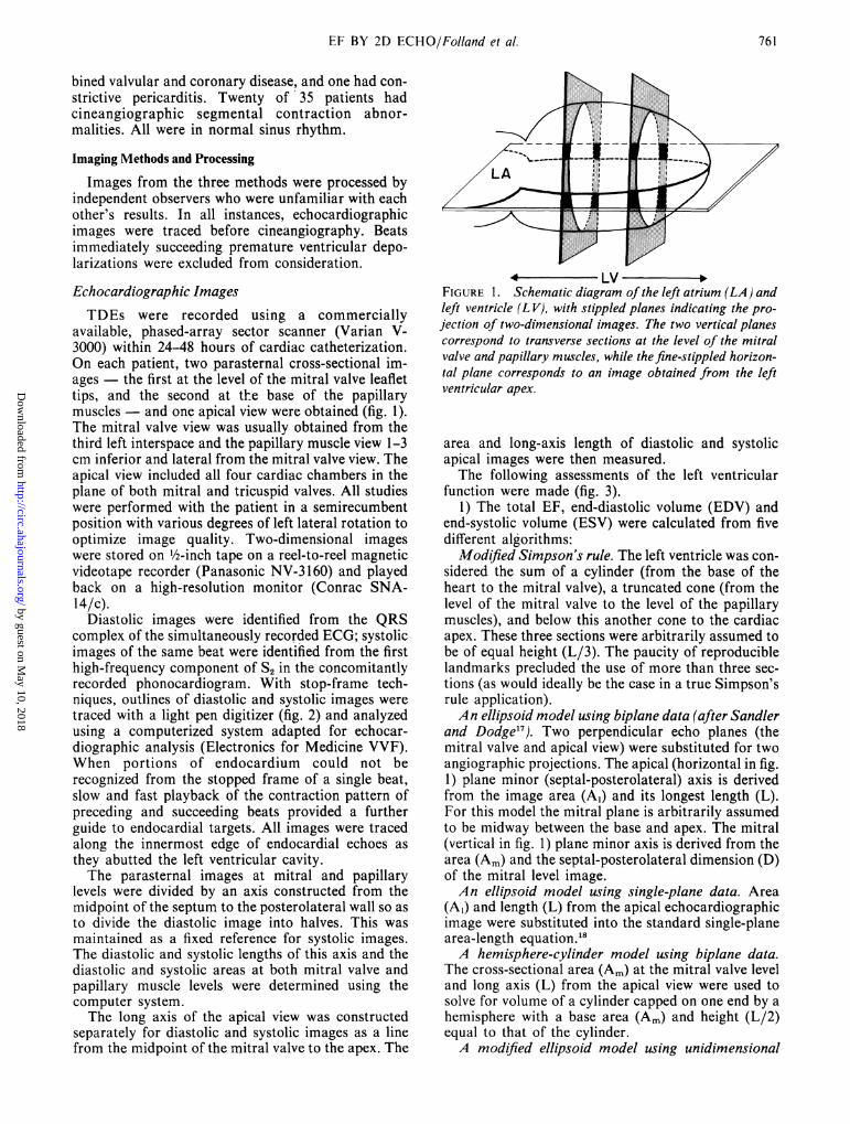
available, phased-array sector scanner (Varian V-3000) within 24-48 hours of cardiac catheterization.On each patient, two parasternal cross-sectional im-ages - the first at the level of the mitral valve leaflettips, and the second at the base of the papillarymuscles - and one apical view were obtained (fig. 1).The mitral valve view was usually obtained from thethird left interspace and the papillary muscle view 1-3cm inferior and lateral from the mitral valve view. Theapical view included all four cardiac chambers in theplane of both mitral and tricuspid valves. All studieswere performed with the patient in a semirecumbentposition with various degrees of left lateral rotation tooptimize image quality. Two-dimensional imageswere stored on ½/2-inch tape on a reel-to-reel magneticvideotape recorder (Panasonic NV-3160) and playedback on a high-resolution monitor (Conrac SNA-14/c).
Diastolic images were identified from the QRScomplex of the simultaneously recorded ECG; systolicimages of the same beat were identified from the firsthigh-frequency component of S2 in the concomitantlyrecorded phonocardiogram. With stop-frame tech-niques, outlines of diastolic and systolic images weretraced with a light pen digitizer (fig. 2) and analyzedusing a computerized system adapted for echocar-diographic analysis (Electronics for Medicine VVF).When portions of endocardium could not berecognized from the stopped frame of a single beat,slow and fast playback of the contraction pattern ofpreceding and succeeding beats provided a furtherguide to endocardial targets. All images were tracedalong the innermost edge of endocardial echoes asthey abutted the left ventricular cavity.The parasternal images at mitral and papillary
levels were divided by an axis constructed from themidpoint of the septum to the posterolateral wall so asto divide the diastolic image into halves. This wasmaintained as a fixed reference for systolic images.The diastolic and systolic lengths of this axis and thediastolic and systolic areas at both mitral valve andpapillary muscle levels were determined using thecomputer system.The long axis of the apical view was constructed
separately for diastolic and systolic images as a linefrom the midpoint of the mitral valve to the apex. The
4 LVFIGURE 1. Schematic diagram of the left atrium (LA) andleft ventricle (LV), with stippled planes indicating the pro-jection of two-dimensional images. The two vertical planescorrespond to transverse sections at the level of the mitralvalve and papillary muscles, while the fine-stippled horizon-tal plane corresponds to an image obtained from the leftventricular apex.
area and long-axis length of diastolic and systolicapical images were then measured.The following assessments of the left ventricular
function were made (fig. 3).1) The total EF, end-diastolic volume (EDV) and
end-systolic volume (ESV) were calculated from fivedifferent algorithms:
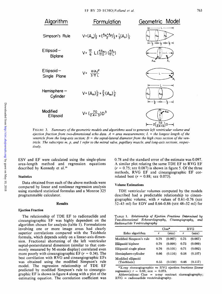
Modified Simpson's rule. The left ventricle was con-sidered the sum of a cylinder (from the base of theheart to the mitral valve), a truncated cone (from thelevel of the mitral valve to the level of the papillarymuscles), and below this another cone to the cardiacapex. These three sections were arbitrarily assumed tobe of equal height (L/3). The paucity of reproduciblelandmarks precluded the use of more than three sec-tions (as would ideally be the case in a true Simpson'srule application).An ellipsoid model using biplane data (after Sandler
and Dodge17). Two perpendicular echo planes (themitral valve and apical view) were substituted for twoangiographic projections. The apical (horizontal in fig.1) plane minor (septal-posterolateral) axis is derivedfrom the image area (A1) and its longest length (L).For this model the mitral plane is arbitrarily assumedto be midway between the base and apex. The mitral(vertical in fig. 1) plane minor axis is derived from thearea (Am) and the septal-posterolateral dimension (D)of the mitral level image.An ellipsoid model using single-plane data. Area
(A1) and length (L) from the apical echocardiographicimage were substituted into the standard single-planearea-length equation.'8A hemisphere-cylinder model using biplane data.
The cross-sectional area (Am) at the mitral valve leveland long axis (L) from the apical view were used tosolve for volume of a cylinder capped on one end by ahemisphere with a base area (Am) and height (L/2)equal to that of the cylinder.A modified ellipsoid model using unidimensional
761
by guest on May 10, 2018
http://circ.ahajournals.org/D
ownloaded from
Vot, 60. No 4, 0( t-]()Bl1q5R 1979
I -- --PM Ap
DIASTOLE
SYSTOLtE
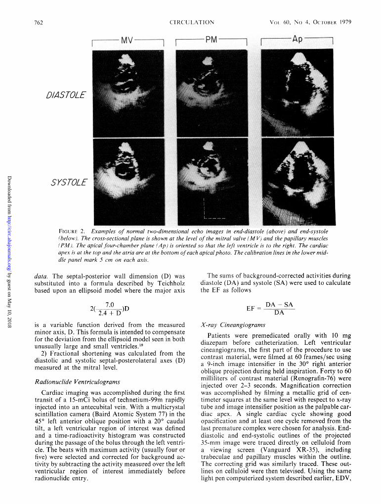
FIGURE 2. Examples of normal two-dimensional echo images in end-diastole (above) and end-systole(below). The cross-sectional plane is shown at the level of the mitral valve (MV) and the papillary muscles(PM). The apical four-chamber plane (Ap) is oriented so that the left ventricle is to the right. The cardiacapex is at the top and the atria are at the bottom ofeach apicalphoto. The calibration lines in the lower mid-dle panel mark 5 cm on each axis.
data. The septal-posterior wall dimension (D) was
substituted into a formula described by Teichholzbased upon an ellipsoid model where the major axis
2( 7.0 )D2.4 + D
is a variable function derived from the measuredminor axis, D. This formula is intended to compensatefor the deviation from the ellipsoid model seen in bothunusually large and small ventricles."9
2) Fractional shortening was calculated from thediastolic and systolic septal-posterolateral axes (D)measured at the mitral level.
Radionuclide Ventriculograms
Cardiac imaging was accomplished during the firsttransit of a 15-mCi bolus of technetium-99m rapidlyinjected into an antecubital vein. With a multicrystalscintillation camera (Baird Atomic System 77) in the450 left anterior oblique position with a 200 caudaltilt, a left ventricular region of interest was definedand a time-radioactivity histogram was constructedduring the passage of the bolus through the left ventri-cle. The beats with maximum activity (usually four orfive) were selected and corrected for background ac-
tivity by subtracting the activity measured over the leftventricular region of interest immediately beforeradionuclide entry.
The sums of background-corrected activities duringdiastole (DA) and systole (SA) were used to calculatethe EF as follows
DA-SAEF- D
X-ray Cineangiograms
Patients were premedicated orally with 10 mgdiazepam before catheterization. Left ventricularcineangiograms, the first part of the procedure to usecontrast material, were filmed at 60 frames/sec usinga 9-inch image intensifier in the 300 right anterioroblique projection during held inspiration. Forty to 60milliliters of contrast material (Renografin-76) wereinjected over 2-3 seconds. Magnification correctionwas accomplished by filming a metallic grid of cen-timeter squares at the same level with respect to x-raytube and image intensifier position as the palpable car-diac apex. A single cardiac cycle showing goodopacification and at least one cycle removed from thelast premature complex were chosen for analysis. End-diastolic and end-systolic outlines of the projected35-mm image were traced directly on celluloid froma viewing screen (Vanguard XR-35), includingtrabeculae and papillary muscles within the outline.The correcting grid was similarly traced. These out-lines on celluloid were then televised. Using the samelight pen computerized system described earlier, EDV,
762 CIlRCULATION
by guest on May 10, 2018
http://circ.ahajournals.org/D
ownloaded from
FT BY 2D ECHO/Folland et al.
Algorithm
Simpson's Rule
Ellipsoid -Biplane
Formulation
V=(Am)L (L+1)(Ap)L)
V= 6E L(fR TL
Geometric Modeli. - L b
H- L_U L _t_ L ,43 3 3
Am AI
El lipsoid -Single Plane
V= 8 (A ,)23IT L
Hemisphere -Cylinder
Al
F. L
2 2
Modified V= ( _)D_3El Iipsoid V(2.42D)D D
FIGURE 3. Summary of the geometric models and algorithms used to generate left ventricular volume andejection fraction from two-dimensional echo data. A = area measurements; L = the longest length of theventricle from the long-axis section; D = the septal-lateral diameterfrom the high cross section of the ven-
tricle. The subscripts m, p, and I refer to the mitral valve, papillary muscle, and long-axis sections, respec-
tively.
ESV and EF were calculated using the single-planearea-length method and regression equationsdescribed by Kennedy et al.18
Statistics
Data obtained from each of the above methods were
compared by linear and nonlinear regression analysisusing standard statistical formulas and a Monroe 325programmable calculator.
Results
Ejection Fraction
The relationship of TDE EF to radionuclide andcineangiographic EF was highly dependent on thealgorithm chosen for analysis (table 1). Formulationsinvolving one or more image areas had clearlysuperior correlations compared with the Teichholzformula, which depends solely on a linear-axis dimen-sion. Fractional shortening of the left ventricularseptal-posterolateral dimension (similar to that com-
monly measured by M-mode display) correlated even
more poorly with cineangiographic EF (r = 0.34). Thebest correlation with RVG and cineangiographic EFswas obtained using the modified Simpson's rulemodel. The regression relationship of TDE EFpredicted by modified Simpson's rule to cineangio-graphic EF is shown in figure 4 along with a plot of theestimating equation. The correlation coefficient was
0.78 and the standard error of the estimate was 0.097.A similar plot relating the same TDE EF to RVG EF(r = 0.75; SEE 0.087) is shown in figure 5. Of the threemethods, RVG EF and cineangiographic EF cor-
related best (r = 0.88; SEE 0.073).
Volume Estimations
TDE ventricular volumes computed by the modelsdescribed had a predictable relationship to cinean-giographic volume, with r values of 0.61-0.76 (SEE52-43 ml) for EDV and 0.64-0.86 (SEE 48-32 ml) for
TABLE 1. Relationship of Ejection Fractions Determined byTwo-dimensional Echocardiography, Cineangiography, andRadionuclide Ventriculography
Cine* RVGEcho algorithm r (SEE) r (SPE)
Modified Simpson's rule 0.78 (0.097) 0.75 (0.087)Ellipsoid biplane 0.78 (0.098) 0.73 (0.089)Ellipsoid single plane 0.76 (0.101) 0.71 (0.092)Hemisphere-cylinder 0.66 (0.116) 0.58 (0.107)Modified ellipsoid
(Teichholz) 0.55 (0.130) 0.46 (0.117)
*X-ray cineangiographic vs RVG ejection fractions (linearregression): r = 0.88; SEE = 0.073.
Abbreviations: Cine = x-ray contrast cineangiography;RVG = radionuclide ventriculography.
763
V= (A,)L+2(A,,)L2 3 2
by guest on May 10, 2018
http://circ.ahajournals.org/D
ownloaded from

Voi- 60, No 4. 0CTOLR 1979
. 0
0
.60- .
0
C3 40
LL
.20-
I 1 1 1 -.20 .40 .60 .80
Ejection Fraction - EchoFIGURE 4. Relationship of left ventricular ejection frac-tion determined by two-dimensional echocardiography,modified Simpson's rule method (horizontal axis), com-
pared with ejection fraction determined by x-raycineangiography (vertical axis). A regression line corre-
sponding to the equation = l.Ox + 0.04 is shown(r 0. 78, SEE= 0.097).
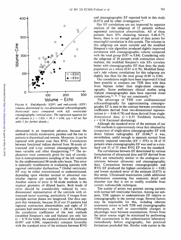
ESV (table 2). The modified Simpson's rule modelproduced the best correlation (fig. 6). This approxima-tion systematically underestimated ventricular vol-umes, as did the other algorithms.
Discussion
TDE, radionuclide and x-ray cineangiographictechniques are fundamentally different approaches tomeasuring left ventricular function. X-ray cinean-giography derives volume and EF from a two-dimen-sional silhouette of the left ventricle. Volume can besimilarly derived from a radionuclide image; however,radionuclide EF is most accurately measured by thetime-activity method described earlier and used in thisstudy.5 TDE is a tomographic method, producingslice-like images of the ventricle along the plane oforientation of the ultrasonic beam. Theoretically, in-tegration of an appropriate selection of "slices" wouldproduce a composite, three-dimensional reconstruc-tion of left ventricular geometry. The approach chosenhere is a compromise between limitations of ultrasonicmethods and practicality. The 1-cm-wide beamgenerated by the phased-array scanner allows only afinite number of slices to be taken in transverse orapical orientations. Ventricular landmarks are few,allowing reproducible sampling at a limited number ofsites. Imperfect lateral resolution distorts endocardialtargets as a function of both depth of field and beamorientation. Accordingly, our approach is the simplestand most pragmatic we could find for this problem.Further refinement of ultrasonic imaging, different
a)
U
-50 .6C
O .4C
0
0Ut-
C.20
0
-4--
a)
0
0
0
0
0
0
0
4 */X 0
.
.20
.
.40
I.I.60 .80
Ejection Fraction-EchoFIGURIE 5. Left ventricular ejection fraction determined bytwo-dimensional echocardiography, modified Simpson'srule method (horizontal axis), and ejection fraction deter-mined by radionuclide angiography (vertical axis) in thesame patients. The regression equation is y = 0.75x + 0.07(r = 0.75; SEE= 0.087).
"slice" orientations and different algorithms may bemore satisfactory in the future.The models used rest upon arbitrary and sometimes
different assumptions about ventricular anatomy. Forinstance, two of three models using biplane dataassume that the mitral level echo plane is midwaybetween the apex and base of the ventricle. The thirdmodel, modified Simpson's rule, assumes that themitral and papillary muscle echo levels trisect the ven-tricle equally. The models also assume that therelative position and/or geometry of the involvedplanes is constant in systole and diastole. Therefore,the value of these models is best judged by the extentof agreement between information derived from eachmodel and analogous information derived from in-dependent standards such as x-ray contrast and RVG.Accurate assessment of left ventricular function by
TABLE 2. Relationship of Two-dimensional EchocardiographicVolumes to Cineangiographic Volumes
Cine EDV* Cine ESVEcho algorithm r (SEE) r (SEE)
Modified Simpson's rule 0.76 (43 ml) 0.86 (32 ml)Ellipsoid biplane 0.67 (49 ml) 0.72 (44 ml)Ellipsoid single planeHemisphere-cylinderModified ellipsoid
(Teichholz)
0.61 (52 ml) 0.64 (48 ml)0.68 (49 ml) 0.75 (42 ml)
0.72 (46 ml) 0.81 (37 ml)
*Cine vs modified Simpson's rule echo (all volumes):r = 0.84; SEE = 43 ml.
Abbreviations: EDV = end-diastolic volume; ESV =end-systolic volume; cine = x-ray contrast cineangiography.
1 I t iI I t
764 ClIRCUL'LATION
)4
by guest on May 10, 2018
http://circ.ahajournals.org/D
ownloaded from
EF BY 2D ECHO/Folland et al.
U)
c
E
0
0
0
0
0
0
0
0
0
0
0
0
100
o = EDV=ESVl
200
Volume - EchoFIGURE 6. End-diastolic (EDV) and end-systolic (ESV)volumes determined by two-dimensional echocardiography(horizontal axis) compared with left ventricularcineangiography (vertical axis). The regression equation forall volumes is y = 1.02x + 34 (r = 0.84; SEE = 43 ml. Seetable 2 for further details.)
ultrasound is an important advance, because themethod is totally noninvasive, painless and the risk topatients is theoretical and remote. Moreover, it can berepeated with greater ease than RVG. Correlationsbetween functional indices derived from M-mode ul-trasound and x-ray contrast cineangiography havebeen variable and often disappointing.19-23 The ex-
planation most commonly given for lack of correla-tion is nonrepresentative sampling of the left ventricleby the unidimensional M-mode echo beam. This erroris especially troublesome in evaluating patients withregional ventricular dysfunction.19 Global ventricularEF may be either overestimated or underestimated,depending upon whether normal or abnormal ven-
tricular regions are sampled by the echo beam.Another source of error in M-mode methods is theatypical geometry of dilated hearts. Both kinds oferror should be considerably reduced by two-dimensional representation of the ventricle with ul-trasound sector scanning, particularly if images frommultiple section planes are integrated. Our data sup-port this viewpoint, because 20 of our 35 patients hadregional contraction abnormalities and eight of 35patients had ventricular volumes > 200 ml. Althoughthe correlation coefficients of the best methods(modified Simpson's rule and biplane) are only fair(r = 0.78 for both), the standard errors of the estimate(0.097 and 0.098, respectively) compare favorablywith the standard error of the estimate between RVG
and cineangiographic EF reported both in this study(0.073) and by other investigators.5-8Our EF correlations are not improved by separate
analysis of the subgroup of 15 patients withoutsegmental contraction abnormalities. All of thesepatients have EFs clustering between 0.48-0.77;hence, there is not enough spread of data points formeaningful correlation in this sample. The volumes inthis subgroup are more variable and the modifiedSimpson's rule algorithm produced slightly improvedcorrelation with cineangiographic volume comparedwith the total group (0.87 vs 0.84). Considering onlythe subgroup of 20 patients with contraction abnor-malities, the modified Simpson's rule EFs correlatebetter with cineangiographic EF than does the studypopulation as a whole (0.83 vs 0.78). Modified Simp-son's rule volume correlations for this subgroup areslightly less than for the total group (0.80 vs 0.84).
Our correlations might have been improved if it hadbeen possible to compare our TDE data with datafrom biplane rather than single-plane cineangi-ography. Some preliminary clinical studies usingbiplane cineangiographic data have reported closercorrelations, 12, 1,5 but not consistently.'6The advantage of TDE over unidimensional
echocardiography for approximating cineangio-graphic EF is seen in the contrast between correlationcoefficients derived from two-dimensional represen-tations (r = 0.66-0.78) and those derived from single-dimensional data (r = 0.55 Teichholz formula;r = 0.34 fractional shortening).
Although the standard error of the estimate of ourTDE methods is approximately twice that obtained bycomparison of single-plane cineangiographic EF withdirect biplane radiographic EF (0.04),2o it was,nevertheless, useful enough to separate normal fromseverely impaired ventricles (EF < 35%) in 34 of 35patients when cineangiographic EF was used as a stan-dard and 33 of 35 when RVG EF was the standard.The correlations between EF determined by various
formulations of ultrasound data and EF derived fromRVG are remarkably similar to the analogous cor-relations between ultrasonic and cineangiographicdata. Comparison between cineangiographic andRVG EF produced the highest correlation (r = 0.88)and lowest standard error of the estimate (0.073) inthis series. Ultrasound examination yields additionalinformation concerning fine anatomic detail andchamber size that is not so readily available fromcurrent radionuclide techniques.The scatter of points was greatest among patients
with normal left ventricular function. Among our sub-jects, TDE appears to underestimate EF relative tocineangiography in the normal range. Several factorsmay be responsible for this, including inherentsystematic errors in both TDE and cineangiographictechniques, as well as unavoidable changes inphysiologic state between examinations. Errors fromthe latter source might be minimized by performingTDE examinations in the catheterization laboratoryimmediately before angiography, but practicallimitations precluded this. Similar wide scatter in the
765
by guest on May 10, 2018
http://circ.ahajournals.org/D
ownloaded from

VOLt 60, No 4, OCTOBER 1979
normal range has been noted in other studies whereEFs obtained invasively and noninvasively were com-pared.5Volumes derived from TDE and cineangiographic
methods correlate well, especially when modifiedSimpson's rule is used to derive TDE volume(r = 0.76 EDV and r = 0.86 ESV). The scatter ofmeasurements about the regression line is wide,however, and produces a rather large standard error ofthe estimate (at best 43 ml for EDV and 32 ml forESV), which is considerably greater than the standarderror of the estimate noted when volumes derivedfrom single-plane cineangiography are compared withthose derived from direct biplane radiography (21 mlfor EDV and 13 ml for ESV).20 A 95% confidence limitfor EDV of ± 86 ml limits the value of TDE volumedeterminations for patients with normal to moderatelyincreased ventricular size. However, the reproduc-ibility of these measurements in the same subject hasnot been tested. Should they prove highly repro-ducible, these measurements could be useful in thelongitudinal follow-up of patients in spite of theirvariability compared with x-ray cineangiography.A consistent advantage ofTDE data over unidimen-
sional data in approximating cineangiographicvolumes was not demonstrated. Cineangiographic cor-relations of volumes derived from the Teichholz modelwere exceeded only by the modified Simpson's rulecorrelations, perhaps indicating a need for furtherrefinement of the formulations.
In conclusion, the data presented indicate that TDEcan be used as a reasonable predictor of left ven-tricular EF, particularly when patients with moderateor severe dysfunction are being separated from thosewith normal or less severely depressed EFs. Volumedeterminations are more problematic. Though there isa statistically significant correlation between invasiveand noninvasive data, the scatter of our results in-dicates that the current approach is not likely to behighly accurate in estimating ventricular volumeswhen single-plane x-ray contrast cineangiograms areused as the standard.
AcknowledgmentsWe thank Rosemary Phillips for her assistance in the preparation
of the illustrations; Stephanie Karaffa and Carolyn Dilts for theirtechnical assistance, and Betty Gillam, Clare Smith and CarolKrasauskis for their typing assistance.
References
I. Cohn PF, Gorlin R, Cohn LH, Collins JJ Jr: Left ventricularejection fraction as a prognostic guide in surgical treatment ofcoronary and valvular heart disease. Am J Cardiol 34: 136,1974
2. Hammermeister KE, Kennedy JW: Predictors of surgical mor-tality in patients undergoing direct myocardial revasculariza-tion. Circulation 50 (suppl II): 11- 1 12, 1974
3. Nelson GR, Cohn PF, Gorlin R: Prognosis in medically treatedcoronary artery disease. Influence of ejection fraction comparedto other parameters. Circulation 52: 408, 1975
4. Murray JA, Chinn N, Peterson DR: Influence of left ven-
tricular function on early prognosis in atherosclerotic heart dis-ease. Am J Cardiol 33: 159, 1974
5. Folland ED, Hamilton GW, Larson SM, Kennedy JW,Williams DL, Ritchie JL: The radionuclide ejection fraction: acomparison of three radionuclide techniques with contrastangiography. J Nucl Med 18: 1159, 1977
6. Schelbert MR. Verba JW, Johnson AD, Brock GW, AlazrakiNP, Rose FJ, Ashburn WL: Nontraumatic determination ofleft ventricular ejection fraction by radionuclide angiocar-diography. Circulation 51: 902, 1975
7. Steele P, Kirch D, Matthews M, Davies M: Measurement ofleft ventricular ejection fraction and end-diastolic volume by acomputerized scintigraphic technique using a wedgedpulmonary arterial catheter. Am J Cardiol 34: 179, 1974
8. Marshall RC, Berger HJ, Costin JC, Freedman GS, Wolberg J,Cohen LS, Gottschalk A, Zaret BL: Assessment of cardiac per-formance with quantitative radionuclide angiocardiography.Sequential left ventricular ejection fraction, normalized leftventricular ejection rate, and regional wall motion. Circulation56: 820, 1977
9. Weyman AE, Peskoe SM, Williams ES, Dillon JC, Feigen-baum H: Detection of left ventricular aneurysms by cross-sectional echocardiography. Circulation 54: 936, 1976
10. Kisslo JA, Robertson D, Gilbert BW, von Ramm 0, Behar VS:A comparison of real-time, two-dimensional echocardiographyand cineangiography in detecting left ventricular asynergy. Cir-culation 55: 134, 1977
11. Wyatt HL, Heng M, Murbaum S, Davidson R, Lee S, CordayE: Quantitative left ventricular analysis in dogs with the phased-array sector scan. (abstr) Circulation 56 (suppl III): 111-152,1977
12. Schiller N, Botvinick E, Cogan J, Greenberg B, Acquatella H,Glantz S: Noninvasive methods are reliable predictions ofangiographic left ventricular volumes. (abstr) Circulation 56(suppl III): 111-221, 1977
13. Chaudry KR, Ogawa S, Pauletto FJ, Hubbard FE, DreyfussLS: Biplane measurements of left ventricular volumes usingwide-angle, cross-sectional echocardiography. (abstr) Am JCardiol 41: 391, 1978
14. Silverman NM, Schiller NB, Yaeger RL, Ports TA: Left ven-tricular volume analysis by two-dimensional echocardiographyin children. Circulation 58 (suppl II): 11-202, 1978
15. Carr K, Engler R, Forsythe J, Johnson A, Gosink B: Measure-ment of left ventricular ejection fraction by mechanical cross-sectional echocardiography and comparison with angiography.(abstr) Circulation 58 (suppl II): 11-40, 1978
16. Nixon JV, Saffer SI: Three-dimensional echoventriculography.(abstr) Circulation 58 (suppl II): 11-157, 1978
17. Dodge NT, Sandier H, Ballew AM, Lord JA Jr: Use of biplaneangiocardiography for the measurement of left ventricularvolume in man. Am Heart J 60: 762, 1960
18. Kennedy JW, Trenholme SE, Kasser IS: Left ventricularvolume and mass from single-plane cineangiocardiogram. Acomparison of anteroposterior and right anterior obliquemethods. Am Heart J 80: 343, 1970
19. Teichholz LE, Kreulen T, Herman MV, Gorlin R: Problems inechocardiographic volume determinations: echocardiographic-angiographic correlations in the presence or absence ofasynergy. Am J Cardiol 37: 7, 1976
20. Fortuin NW: Evaluation of left ventricular size and function byechocardiography. In Cardiac Ultrasound, edited by GramiakR, Waag RC. St. Louis, CV Mosby Co, 1975, p 121
21. Pombo JF, Troy BL, Russell RO Jr: Left ventricular volumesand ejection fraction by echocardiography. Circulation 43: 480,1971
22. Feigenbaum H, Popp RL, Wolfe SB, Haine CL, Dodge HT:Ultrasound measurements of the left ventricle: a correlativestudy with angiocardiography. Arch Intern Med 129: 461, 1972
23. Fortuin NJ, Hood WP Jr, Sherman ME, Craige E: Determina-tion of left ventricular volumes by ultrasound. Circulation 44:575, 1971
24. Kasser IS, Kennedy JW: Measurement of left ventricularvolumes in man by single-plane cineangiography. Invest Radiol4: 83, 1969
766 ClIRCU LATI ON
by guest on May 10, 2018
http://circ.ahajournals.org/D
ownloaded from
E D Folland, A F Parisi, P F Moynihan, D R Jones, C L Feldman and D E Towtechniques.
two-dimensional echocardiography. A comparison of cineangiographic and radionuclide Assessment of left ventricular ejection fraction and volumes by real-time,
Print ISSN: 0009-7322. Online ISSN: 1524-4539 Copyright © 1979 American Heart Association, Inc. All rights reserved.
is published by the American Heart Association, 7272 Greenville Avenue, Dallas, TX 75231Circulation doi: 10.1161/01.CIR.60.4.760
1979;60:760-766Circulation.
http://circ.ahajournals.org/content/60/4/760the World Wide Web at:
The online version of this article, along with updated information and services, is located on
http://circ.ahajournals.org//subscriptions/
is online at: Circulation Information about subscribing to Subscriptions:
http://www.lww.com/reprints Information about reprints can be found online at: Reprints:
document. Permissions and Rights Question and Answer information about this process is available in the
located, click Request Permissions in the middle column of the Web page under Services. FurtherEditorial Office. Once the online version of the published article for which permission is being requested is
can be obtained via RightsLink, a service of the Copyright Clearance Center, not theCirculationpublished in Requests for permissions to reproduce figures, tables, or portions of articles originallyPermissions:
by guest on May 10, 2018
http://circ.ahajournals.org/D
ownloaded from