Embed Size (px)
Citation preview

2/17/11
1
Why’N’How!
Arterial-Spin Labeling
Jean Chen
Postdoctoral Fellow A. A. Martinos Center for Biomedical Imaging
Massachusetts General Hospital, Harvard Medical School
02/17/2011
Why? How?
Why is it called “arterial-spin labeling”? Why are there PASL, CASL and pCASL?
How to choose which ASL to use? How to know what parameters to use?
How can ASL go wrong? How to get cerebral blood flow maps? How long is this talk going to be?
…
2/45

2/17/11
2
Arterial-Spin Labeling (ASL)
“Labeled” spins in arterial blood water act as an endogenous tracer
At the time of imaging, tagged spins have arrived in the regions of interest
Water is exchanged between blood and tissue --- resulting tissue longitudinal relaxation (T1) is proportional to flow
CASL: continuous ASL PASL: pulsed ASL
3/45
How do you “label”?
CASL: flow-driven adiabatic fast-passage - Continuous and constant RF wave (1-2 sec) - Applied in a plane, no off-resonance behaviour - Relies on blood flow to achieve adiabaticity
PASL: adiabatic RF pulses - Pulsed RF (a few msec) - Applied to a slab, ampl and freq-modulated - Using gradients to enhance spatial selectivity
while reducing power deposition
4/45

2/17/11
3
Courtesy: WM Luh 5/45
CASL PASL Body-coil labeling
Courtesy: WM Luh
CASL (tag)
PASL (tag)

2/17/11
4
Lower labeling efficiency
Continuous RF not supported on most clinical scanners
Requires separate coil to minimize MT (magnetization transfer) effects
Higher SNR
Minimally sensitive to tag dispersion
Higher labeling efficiency
Easily implemented on clinical systems
Does not require additional hardware
Lower SNR
Sensitive to dispersion
Most commonly used
7/45
CASL PASL
Pseudo-Continuous ASL (pCASL) Tag
A series of Hanning RF pulses, shaped to avoid aliased labeling planes
Can be implemented on clinical scanners
Dai, W et al. Magn Reson Med 2008 8/45

2/17/11
5
Pseudo-Continuous ASL (pCASL)
Analogous to flow-driven adiabatic fast-passage, but using pulsed instead of continuous wave
- Builds up pseudo steady-state to imitate CASL labeling
Higher SNR than PASL
Higher labeling efficiency than CASL
Not adiabatic inversion
Sensitive to flow velocity, gradient strength, RF timing and duty cycle
Best performance in conjunction with angiography and flow phase mapping
9/45
FAIR PASL (fBIRN)
Labeling Region &
Imaging Region
Control Region
Time
labeled
control Mz_
blo
od
ΔM TI
Blood Inflow
-image-slab tag, global control -ascending & descending flow -potential radiation damping
10/45

2/17/11
6
PICORE PASL (Siemens)
Labeling Region
Control Region
Time
labeled
control
Mz_
blo
od
ΔM TI
• - Proximal tag, global control • - Magnetization transfer cancellation • - Background signal attenuation • - Sensitive to ascending flow only
Blood Inflow
Imaging Region
11/45
12/45
pCASL (UPenn)
Time
labeled
control
Mz_
blo
od
ΔM TI
Labeling Region
Control Region
Blood Inflow
Imaging Region
- Proximal tag, global control - Magnetization transfer cancellation - Sensitive to ascending flow only

2/17/11
7
ASL Assumptions
Tag is fully delivered to imaging region Rapid water exchange between blood
and tissue Intact blood-brain barrier
Negligible tag washout at time of imaging Negligible partial-volume effects in voxel Tagged blood has completely washed out
before the subsequent measurement
13/45
Arterial-Spin Labeling: the Math
Modified Bloch’s equation
- M0,t: Tissue equilibrium magnetization - T1,t: Tissue T1
- In plain terms: the change of tissue magnetization (Mt) due to the
tagged arterial blood (Ma) is proportional to CBF
14/45

2/17/11
8
Cerebral Blood Flow (CBF)
Quantitative CBF in humans:
GM CBF ≅ 60 ml/100 g/min WM CBF ≅ 20 ml/100 g/min
voxel
15/45
ASL Confounds: Intravascular Signal
Prominent intravascular signal
With crushers (Typically at 100cm/s, fBIRN & Siemens)
TI (100 – 2000 ms)
Yang Y et al, Magn Reson Med 1998
Intravascular signal contributes to CBF overestimation Add crusher gradients to attenuate macrovascular contribution
OR, adjust TI to permit macrovascular flow to wash out
16/45

2/17/11
9
ASL Confounds: Relaxation
T1 relaxation reduces signal from the tag
Compensate for decay incurred during acquisition delay 17/45
Time (s)
Time (s)
Time (s)
Time (s)
ASL Confounds: Transit Delay
Courtesy: H-L A. Liu
For a given TI, slower spins will not be able to reach imaging region, resulting in lower measured CBF
Worse in white matter, and may be exacerbated in aging and disease
18/45

2/17/11
10
ASL Confounds: Transit Delay
Solution: Insert saturation pulse to cut off slower tail of tag, creating a well-defined tag width
Wong EC et al, Magn Reson Med 1998 19/45
ASL Confounds: Transit Delay Saturation Schemes
Q2TIPS (Siemens)
QUIPSS II (fBIRN)
EPI readout
TI2
TI1
20/45

2/17/11
11
ASL Acquisition Considerations
Inversion time TI1 - Long enough to permit tag to leave tagging region
- Short enough to ensure “QUIPSS II” effectiveness
- More slices, longer TI1
Inversion time TI2 - Long enough to avoid intravascular signal and ensure
tag exchange with tissue water
- Short enough to reduce loss of tag
- Slower flow, longer TI2
Tailor parameters for the aims of your study
21/45
ASL Acquisition Considerations
TR must allow tag washout and refreshing (2 – 4 s)
TE should be short to minimize T2 contamination
Tag width should be large, especially for multislice
Labeling gap should be as small as possible
Signal drop-out (if using gradient-echo EPI) - Increase bandwith, reduce slice thickness, use non-EPI
T2 “shine-through” (static tissue & CSF)? - Use background suppression (not in fBIRN or Siemens)
Do not angulate > 45° relative to main feeding arteries
- To maintain control of tag width and tagging efficiency
22/45
2/17/11
12
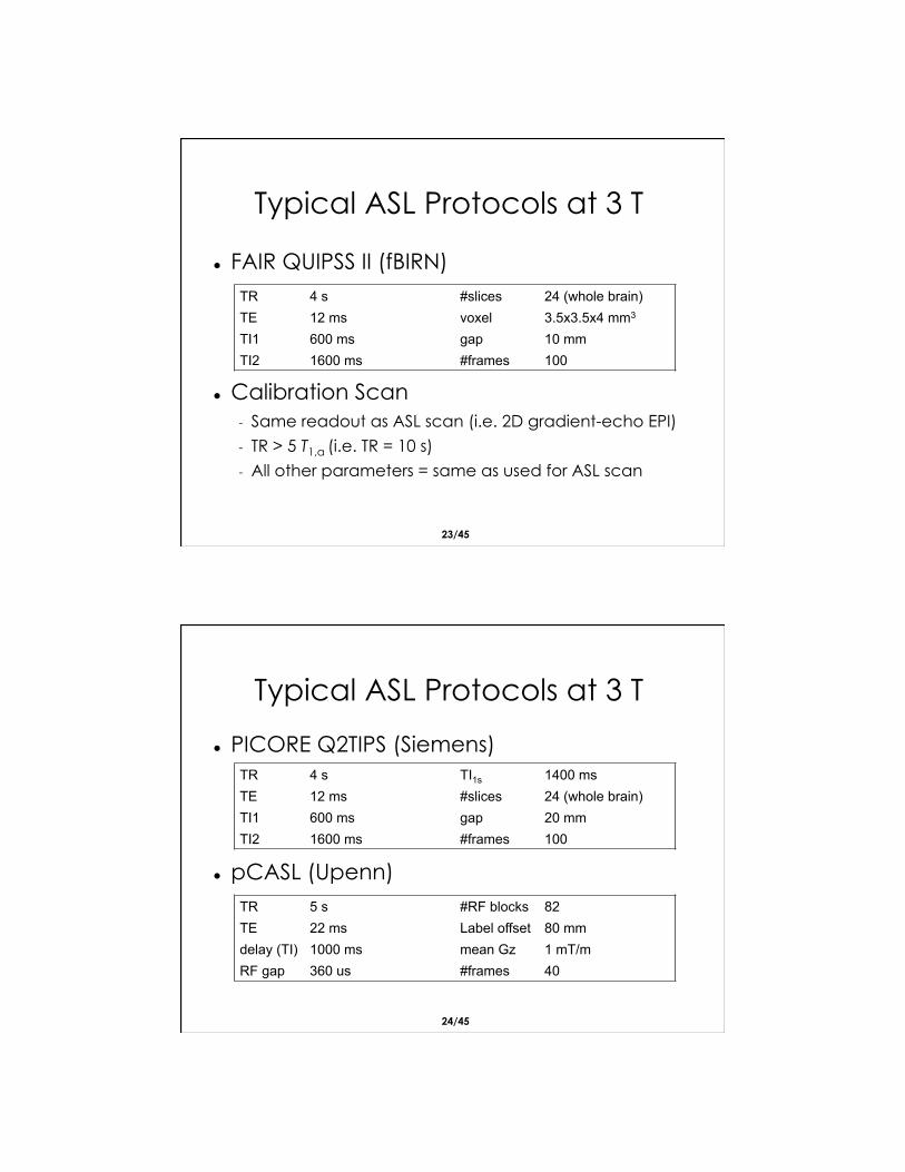
Typical ASL Protocols at 3 T
FAIR QUIPSS II (fBIRN)
Calibration Scan - Same readout as ASL scan (i.e. 2D gradient-echo EPI)
- TR > 5 T1,a (i.e. TR = 10 s)
- All other parameters = same as used for ASL scan
TR 4 s #slices 24 (whole brain) TE 12 ms voxel 3.5x3.5x4 mm3
TI1 600 ms gap 10 mm TI2 1600 ms #frames 100
23/45
PICORE Q2TIPS (Siemens)
pCASL (Upenn)
TR 4 s TI1s 1400 ms TE 12 ms #slices 24 (whole brain) TI1 600 ms gap 20 mm TI2 1600 ms #frames 100
TR 5 s #RF blocks 82 TE 22 ms Label offset 80 mm
delay (TI) 1000 ms mean Gz 1 mT/m RF gap 360 us #frames 40
24/45
Typical ASL Protocols at 3 T

2/17/11
13
CBF Quantification
1. Difference image (ΔM) calculation - Surround subtraction (minimize contamination) - Average across frames (maximize SNR)
25/45
ΔM Calculation
ΔM is typically ~1% of the control signal
Control Label ΔM − =
26/45

2/17/11
14
“CBF α diff(control, label)”
E1: running subtraction
E2&E3: pairwise subtraction
- Timing mismatch = incomplete cancellation of BOLD effects
E4: surround subtraction
E5: Sinc-interpolated subtraction
- Matched BOLD effect
[Lu, H et al, MRM 2006]
27/45
“BOLD α mean(control, label)”
Pros - Time savings - Increased temporal
resolution - No need for ASL-
BOLD cross-registration
Cons - Lower SNR
- Long echo-time needed for optimal BOLD contrast (causing ASL-BOLD cross contamination)
- T1-weighting
28/45

2/17/11
15
ΔM
CBF Changes
Resting CBF
29/45
ΔM Calculation
CBF Quantification
1. Difference image calculation - Surround subtraction (minimize contamination) - Average across frames (maximize SNR)
2. Arterial magnetization (M0,a) estimation - Intensity non-uniformity compensation - Blood-tissue partition coefficient, T1, T2*
30/45

2/17/11
16
M0,a Estimation ASL Calibration Scan
Did not acquire a separate calibration scan? It’s common to use the 1st control image of the ASL
dataset, if magnetization fully relaxed.
31/45
M0,a will vary depending on - Static field (B0) inhomogeneities - RF field (B1) inhomogeneities - Receive coil sensitivity nonuniformity
Calibration methods: 1. CSF (cerebrospinal fluid) based 2. White matter based 3. Local tissue based: intrinsic normalization for
nonuniformities
32/45
M0,a Estimation

2/17/11
17
Calibration scan
M0,t M0,a
X 1/λeTE/T2t =
Distinguish between grey matter and white matter when assuming values for T2,t and λ.
33/45
M0,a Estimation
Ideally, one should Measure T1 and T2 of tissue (grey matter and
white matter separately)
However, it’s more common to assume:
Courtesy: WM Luh
M0,a: local-tissue calibration
34/45

2/17/11
18
CBF Quantification
1. Difference image (ΔM) calculation - Surround subtraction (minimize contamination) - Average across frames (maximize SNR)
2. Arterial magnetization (M0,a) estimation - Intensity non-uniformity compensation - Blood-tissue partition, T1, T2*
3. Decay correction
35/45
Decay Correction
T1 decay of tag incurred during transit must be compensated for acquisition delay
This delay is dependent on the order of slice acquisition
€
TIcorrected = TInominal + slice_order* slice_ time
36/45
€
exp(−TI /T1a )

2/17/11
19
CBF Quantification
1. Difference image (ΔM) calculation - Surround subtraction (minimize contamination) - Average across frames (maximize SNR)
2. Arterial magnetization (M0,a) estimation - Intensity non-uniformity compensation - Blood-tissue partition, T1, T2*
3. Decay correction 4. Plug into ASL signal model
37/45
Standard Kinetic Model
PASL
- M0,a: Arterial blood equilibrium magnetization
- λ: Tissue blood-water partition coefficient
- α: Labeling efficiency
€
CBF =ΔM × λ
2α ×M0,a ×TI1e−TI 2 T1a
38/45

2/17/11
20
Standard Kinetic Model
CASL & pCASL
- M0,a: Arterial blood equilibrium magnetization
- T1,a: Arterial blood T1
- λ: Tissue blood-water partition coefficient
- α: Labeling efficiency
- τ: post-labeling delay
- w: label width
39/45
CBF Quantification
1. Difference image calculation - Surround subtraction (minimize contamination) - Average across frames (maximize SNR)
2. Arterial magnetization estimation - Intensity non-uniformity compensation - Blood-tissue partition, T1, T2*
3. Decay correction 4. Plug into ASL signal model
5. Scaling: CBF [ml/100g/min] ≅ CBF[1/s] x 6,000
40/45

2/17/11
21
ASL
ΔM
T1t
CBF
CBF Quantification
Courtesy: H-L A. Liu 41/45
Sample Quantitative CBF Map
0
90
CBF [ml/100 g/min]
42/45

2/17/11
22
Sample Quantitative CBF Maps
Chen et al., NeuroImage 2011
43/45
Effect of Normal Aging
ASL at Higher Field
Pros - Greater M0: higher available signal - Longer T1: less tag decay
Cons - Shorter T2*: more BOLD contamination, signal drop-
out and geometric distortion
- Higher ΔB0 and ΔB1: harder to achieve adiabaticity - Shorter body coil: more limited tag width - Higher power deposition: do fewer slices per unit time
44/45

2/17/11
23
ASL Applications
Cerebral blood flow - White matter CBF measurement still challenging
Cardiac, pulmonary and renal perfusion ASL processing methods:
- SPM-compatible Perfusion Toolbox: http://www.cfn.upenn.edu/~zewang/ASLtbx.php
- In-house method: www.nmr.mgh.harvard.edu/~jjchen/ASL.html
45/45