Embed Size (px)
Citation preview
17742-v6-2010Jan Page 1
Meso Scale Discovery®
MULTI-SPOT
® Assay Sys tem Argutus AKI Test™ (rat) Assay Kit
1-Plate Kit K15156C-1 5-Plate Kit K15156C-2 25-Plate Kit K15156C-4
This product insert should be used with kit lots: K0040045 K0040052 K0040055
Meso Scale Discovery Meso Scale Discovery Meso Scale Discovery Meso Scale Discovery Meso Scale Discovery Meso Scale Discovery Meso Scale Di
www.mesoscale.com ®

17742-v6-2010Jan Page 2
MSD Toxicology Assays Argutus AKI Test™ (rat) Assay Kit αGST, GSTYb1, RPA-1 This package insert must be read in its entirety before using this product. FOR RESEARCH USE ONLY. NOT FOR USE IN DIAGNOSTIC OR THERAPEUTIC PROCEDURES. Meso Scale Discovery, Meso Scale Diagnostics, www.mesoscale.com, MSD, MSD (design), Discovery Workbench, Quickplex, Multi-Array, Multi-Spot, Sulfo-Tag and Sector are trademarks of Meso Scale Diagnostics, LLC. AKI Test is a registered trademark of Argutus Medical Limited. © 2010 Meso Scale Discovery a division of Meso Scale Diagnostics, LLC. All rights reserved.

17742-v6-2010Jan Page 3
Table of Contents Introduction................................................................................................................................ 4 Principle of the Assay ................................................................................................................ 5 Reagents Supplied .................................................................................................................... 6 Required Material and Equipment – not supplied ...................................................................... 6 Safety ........................................................................................................................................ 7 Reagent Preparation ................................................................................................................. 7 Assay Protocol........................................................................................................................... 9 Analysis of Results .................................................................................................................... 9 Typical Standard Curve ........................................................................................................... 10 Sensitivity ................................................................................................................................ 11 Precision.................................................................................................................................. 11 Spike Recovery ....................................................................................................................... 12 Samples................................................................................................................................... 13 Calibrators ............................................................................................................................... 14 References .............................................................................................................................. 14 Summary Protocol ................................................................................................................... 15 Plate Diagrams ........................................................................................................................ 17
Ordering Information
MSD Customer Service Phone: 1-301-947-2085 Fax: 1-301-990-2776 Email: [email protected]
Meso Scale Discovery A division of Meso Scale Diagnostics, LLC. 9238 Gaither Road Gaithersburg, MD 20877 USA www.mesoscale.com
t a b l e o f c o n t e n t s
o r d e r i n g i n f o r m a t i o n
17742-v6-2010Jan Page 4
Introduction
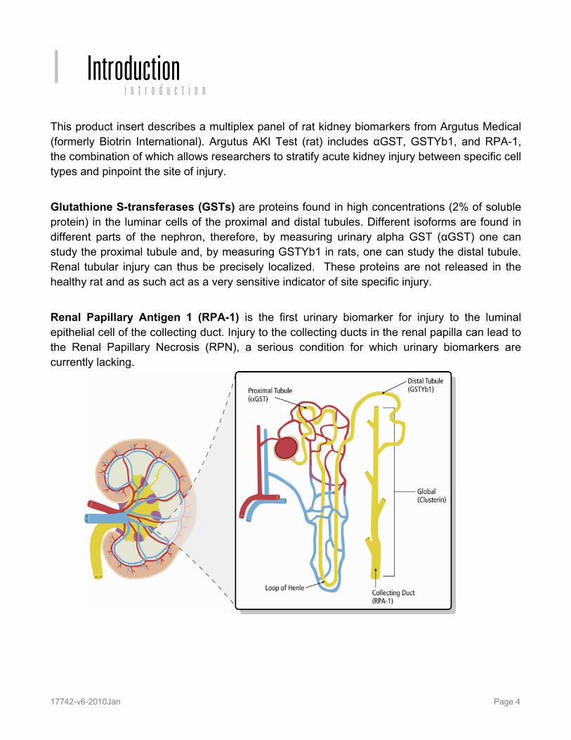
This product insert describes a multiplex panel of rat kidney biomarkers from Argutus Medical (formerly Biotrin International). Argutus AKI Test (rat) includes αGST, GSTYb1, and RPA-1, the combination of which allows researchers to stratify acute kidney injury between specific cell types and pinpoint the site of injury. Glutathione S-transferases (GSTs) are proteins found in high concentrations (2% of soluble protein) in the luminar cells of the proximal and distal tubules. Different isoforms are found in different parts of the nephron, therefore, by measuring urinary alpha GST (αGST) one can study the proximal tubule and, by measuring GSTYb1 in rats, one can study the distal tubule. Renal tubular injury can thus be precisely localized. These proteins are not released in the healthy rat and as such act as a very sensitive indicator of site specific injury. Renal Papillary Antigen 1 (RPA-1) is the first urinary biomarker for injury to the luminal epithelial cell of the collecting duct. Injury to the collecting ducts in the renal papilla can lead to the Renal Papillary Necrosis (RPN), a serious condition for which urinary biomarkers are currently lacking.
I i n t r o d u c t i o n

17742-v6-2010Jan Page 5
Principle of the Assay
MSD® toxicology assays provide a rapid and convenient method for measuring the levels of protein targets within a single small-volume sample. The assays are available in both singleplex and multiplex formats. In a singleplex assay, an antibody for a specific protein target is coated on one electrode (or “spot”) per well. In a multiplex assay, an array of capture antibodies against different targets is patterned on distinct spots in the same well. Our Argutus AKI Test (rat) Assay is a multiplexed sandwich immunoassay (Figure 1). MSD provides a plate that has been pre-coated with three capture antibodies on spatially distinct spots—antibodies for αGST, GSTYb1, and RPA-1. The user adds the sample and a solution containing the labeled detection antibodies—Anti-rat αGST, Anti-rat GSTYb1, and Anti-rat RPA-1 labeled with an electrochemiluminescent compound, MSD SULFO-TAG™ label—over the course of one or more incubation periods. Analytes in the sample bind to capture antibodies immobilized on the working electrode surface; recruitment of the labeled detection antibodies by bound analytes completes the sandwich. The user adds an MSD read buffer that provides the appropriate chemical environment for electrochemiluminescence and loads the plate into an MSD SECTOR® instrument for analysis. Inside the SECTOR instrument, a voltage applied to the plate electrodes causes the labels bound to the electrode surface to emit light. The instrument measures intensity of emitted light to afford a quantitative measure of αGST, GSTYb1, and RPA-1 present in the sample.
Figure 1. Spot diagram showing placement of analyte capture antibody. The numbering convention for the different spots is maintained in the software visualization tools, on the plate packaging, and in the data files. Any spot that is not coated with a specific capture antibody is blocked with BSA to reduce non-specific binding to that spot. A unique bar code label on each plate allows complete traceability back to MSD manufacturing records.
p r i n c i p l e o f t h e a s s a yII
Labeled-Ab
Analyte
Capture-Ab
αGST GSTYb1
RPA-1

17742-v6-2010Jan Page 6
Reagents Supplied Quantity per Kit Product Description Storage K15156C-1 K15156C-2 K15156C-4
MULTI-SPOT 96-well Argutus AKI Test (rat) Plate N45156A
2-8°C 1 plate 5 plates 25 plates
SULFO-TAG Anti-rat αGST Antibody (50X)1
2-8°C 1 vial (75 µL)
1 vial (375 µL)
5 vials (375 µL ea)
SULFO-TAG Anti-rat GSTYb1 Antibody (50X)1
2-8°C 1 vial (75 µL)
1 vial (375 µL)
5 vials (375 µL ea)
SULFO-TAG Anti-rat RPA-1 Antibody (50X)1
2-8°C 1 vial (75 µL)
1 vial (375 µL)
5 vials (375 µL ea)
Argutus AKI Test (rat) Calibrator Blend Lot A0040015 αGST: 1500 ng/mL GSTYb1: 430 ng/mL RPA-1: 870 U/L
< -70°C 1 vial (200 µL)
5 vials (200 µL ea)
25 vials (200 µL ea)
Blocker B RT 1 vial (1 g)
1 vial (1 g)
5 vials (1 g ea)
Diluent 31 R50IA-4 (15 mL), R50IA-9 (60 mL)
< -10°C 1 bottle (15 mL)
1 bottle (60 mL)
5 bottles (60 mL ea)
Read Buffer T (with surfactant), 4X R92TC-3 (50 mL)
RT 1 bottle (50 mL)
1 bottle (50 mL)
5 bottles (50 mL ea)
Required Materials and Equipment - not supplied
Deionized water for diluting concentrated buffers 50 mL tubes for reagent preparation 15 mL tubes for reagent preparation Microcentrifuge tubes for preparing serial dilutions Phosphate-buffered saline (PBS) for making Blocker B solution Phosphate buffered saline plus 0.05% Tween-20 (PBS-T) for plate washing Appropriate liquid handling equipment for desired throughput, capable of dispensing
10 to 150 µL into a 96-well microtiter plate Plate washing equipment: automated plate washer or multichannel pipette Adhesive plate seals Microtiter plate shaker
1 Some SULFO-TAG labeled detection antibodies may be light-sensitive, so they should be stored in the dark.
r e a g e n t s s u p p l i e dIII
r e q u i r e d m a t e r i a l s a n d e q u i p m e n t — n o t s u p p l i e dIV

17742-v6-2010Jan Page 7
Safety
Safe laboratory practices and personal protective equipment such as gloves, safety glasses, and lab coats should be used at all times during the handling of all kit components. All hazardous samples should be handled and disposed of properly, in accordance with local, state, and federal guidelines.
Reagent Preparation
Bring all reagents to room temperature.
Important: Upon first thaw, separate Diluent 31 into aliquots appropriate to the size of your assay needs. The diluent can go through up to three freeze-thaw cycles without significantly affecting the performance of the assay.
Prepare Blocking Solution Prepare a solution of 0.5% (w/v) Blocker B in 1X PBS to be used as a blocking solution. For one plate, mix 20 mL of PBS with 0.1 g of Blocker B. Mix at room temperature until all the Blocker B has dissolved. 0.15 mL per well of blocking solution will be required.
Prepare Calibrator and Control Solutions Calibrators for the Argutus AKI Test (rat) are supplied at the concentration of the highest calibrator. No dilution is required for the highest calibrator. For each assay, an 8-point standard curve is recommended with 3-fold serial dilution steps and a Zero Calibrator. The Calibrators are supplied as a blend. The concentrations provided are for Calibrator Lot A0040015. Calibration curves are lot specific. The table below shows the concentrations of the 8-point standard curve:
Standard αGST (ng/mL)
GSTYb1 (ng/mL)
RPA-1 (U/L)
Dilution Factor
STD-01 1500 430 870 STD-02 500 143 290 3 STD-03 167 47.8 96.7 3 STD-04 55.6 15.9 32.2 3 STD-05 18.5 5.3 10.7 3 STD-06 6.2 1.8 3.6 3 STD-07 2.1 0.6 1.2 3 STD-08 0 0 0 n/a
s a f e t y V
r e a g e n t p r e p a r a t i o nVI

17742-v6-2010Jan Page 8
To prepare this 8-point standard curve: 1) The highest Calibrator is the undiluted calibrator blend. 2) Prepare the next Calibrator by transferring 60 µL of the highest Calibrator to 120 µL of
Diluent 31. Repeat 3-fold serial dilutions 6 additional times to generate 7 Calibrators. 3) The recommended 8th Standard is Diluent 31 alone (i.e. Zero Calibrator).
Calibrators should be prepared no more than 20 minutes before use.
Dilution of Samples Some rat samples may need to be diluted prior to the assay in order to get the analyte levels into the detection range. If this is the case, Diluent 31 should be used to dilute samples. For rat urine samples, 5-fold dilution is recommended. Samples with high analyte abundance may need further dilution. For serum, plasma, or tissue homogenates a 5–5000-fold dilution may be required.
Prepare Detection Antibody Solution The Detection Antibodies are provided as a 50X stock solution. The working Detection Antibody Solution should contain 1X as final concentration of each antibody. In a 15 mL tube combine (per plate):
60 µL of 50X SULFO-TAG Anti-rat αGST Antibody 60 µL of 50X SULFO-TAG Anti-rat GSTYb1 Antibody 60 µL of 50X SULFO-TAG Anti-rat RPA-1 Antibody 2820 µL of Diluent 31
Prepare Read Buffer The Read Buffer should be diluted in deionized water to make a final concentration of 1X Read Buffer T. Add 5 mL of stock Read Buffer T (4X) to 15 mL of deionized water for each plate.
Prepare MSD Plate This plate has been pre-coated with antibodies for the analytes shown in Figure 1. The plate can be used as delivered; no additional preparation (e.g., pre-wetting) is required. The plate has also been exposed to a proprietary stabilizing treatment to ensure the integrity and stability of the immobilized antibodies.

17742-v6-2010Jan Page 9
Assay Protocol
1. Addition of Blocking Solution: Dispense 150 µL of 0.5% Blocker B Solution into each well. Seal the plate with an adhesive plate seal and incubate for 1 hour with vigorous shaking (300–1000 rpm) at room temperature.
2. Wash and Addition of the Sample or Calibrator: Wash the plate 3X with PBS-T. First, dispense 25 µL of Diluent 31 into each well of the MSD plate. Then, dispense 25 µL of sample or calibrator into separate wells of the MSD plate. Seal the plate with an adhesive plate seal and incubate for 2 hours with vigorous shaking (300–1000 rpm) at room temperature.
3. Wash and Addition of the Detection Antibody Solution: Wash the plate 3X with PBS-T. Dispense 25 µL of the 1X Detection Antibody Solution into each well of the MSD plate. Seal the plate and incubate for 2 hours with vigorous shaking (300–1000 rpm) at room temperature.
4. Wash and Read: Wash the plate 3X with PBS-T. Add 150 µL of 1X Read Buffer T to each well of the MSD plate. After adding Read Buffer, keep the plate at room temperature without shaking for 5 minutes before analyzing the plate on the SECTOR Imager.
Analysis of Results The calibrators should be run in duplicate to generate a standard curve. The standard curve is modeled using least squares fitting algorithms so that signals from samples with known levels of the analyte of interest can be used to calculate the concentration of analyte in the sample. The assays have a wide dynamic range (3–4 logs) which allows accurate quantitation in many samples without the need for dilution. The MSD DISCOVERY WORKBENCH® analysis software utilizes a 4-parameter logistic model (or sigmoidal dose-response) and includes a 1/Y2 weighting function. The weighting functionality is important because it provides a better fit of data over a wide dynamic range, particularly at the low end of the standard curve.
Notes Shaking a 96-well MSD MULTI-SPOT plate typically accelerates capture at the working electrode.
Bubbles in the fluid will interfere with reliable reading of MULTI-SPOT plate. Use reverse pipetting techniques to insure bubbles are not created when dispensing the Read Buffer.
a n a l y s i s o f r e s u l t sVIII
a s s a y p r o t o c o lVII

17742-v6-2010Jan Page 10
Typical Standard Curve
The following standard curve is an example of the dynamic range of the assay. The actual signals may vary and a standard curve should be run for each set of samples and on each plate for the best quantitation of unknown samples. The data shown is obtained using Calibrator lot A0040015. Calibration curves are lot specific since they are made using papilla- medulla extracts.
αGST GSTYb1 RPA-1 Conc.
(ng/mL) Average Counts %CV Conc.
(ng/mL) Average Counts %CV
Conc.(U/L)
Average Counts %CV
0 270 6.2 0 419 3.7 0 263 4.5 2.1 371 4.1 0.6 451 3.8 1.2 280 4.0 6.2 568 4.2 1.8 519 3.4 3.6 320 4.2 18.5 1122 3.7 5.3 706 3.2 10.7 467 3.0 55.6 2573 2.8 15.9 1271 3.3 32.2 908 4.2 167 5583 2.6 47.8 2906 3.1 96.7 2676 4.7 500 9299 3.1 143 6630 1.6 290 8346 2.9
1500 11394 2.7 430 13167 2.9 870 22886 3.7
t y p i c a l s t a n d a r d c u r v eIX

17742-v6-2010Jan Page 11
Sensitivity
The lower limit of detection (LLOD) is the calculated concentration of the signal that is 2.5 standard deviations over the zero calibrator. A multi-plate, multi-day study was performed to measure the reproducibility of the assay. The lower limit of quantitation (LLOQ) and upper limit of quantitation (ULOQ) were established from the multiple plate run. The LLOQ is determined as the lowest concentration where the %CV of the calculated concentration is less than 20% and the percent recovery of the standard is between 80% and 120%. For RPA-1, the percent recovery is between 75% and 125%. The ULOQ is determined as the highest concentration where the %CV of the calculated concentration is less than 20% and the percent recovery of the standard is between 80% and 120%. For RPA-1, the percent recovery is between 75% and 125%.
αGST (ng/mL)
GSTYb1(ng/mL)
RPA-1 (U/L)
LLOD 0.87 0.71 2.0 LLOQ 6.6 12.4 30 ULOQ 196 333 800
Precision
A set of four control samples was measured on each plate. Controls were made by spiking papilla medulla extract into rat urine.
Intra-plate Inter-plate
Control Plates Avg Conc. Average % CV % CV
3 8 79.1 2.9 6.5 αGST (ng/mL) 4 8 61.7 3.3 6.6
2 8 86.5 4.1 5.1 GSTYb1 (ng/mL) 3 8 28.4 4.3 5.7
1 8 471 4.7 5.6 RPA-1 (U/L) 2 8 89.6 5.5 6.3
s e n s i t i v i t y X
p r e c i s i o n XI

17742-v6-2010Jan Page 12
Spike Recovery
Papilla medulla extract was spiked into 5X diluted rat urine and tested on the Argutus AKI Test. The concentrations of the spikes were distributed throughout the linear range of the assay. All of the spiked samples above the LLOQ had acceptable recoveries (between 80% and 120%). % Recovery = measured / expected x 100
SpikeLevel Conc. Conc.
%CV %
Recovery0 7.8 1.5
13.9 22.8 8.6 105 41.7 46.7 1.0 94
αGST (ng/mL)
125 132 7.0 100 0 2.9 111
27.8 28.8 8.6 94 83.3 90.2 22.2 105
GSTYb1 (ng/mL)
250 239 4.4 95 0 114 8.5
27.8 138 7.2 98 83.3 202 6.1 102
RPA-1 (U/L)
250 345 6.2 95
s p i k e r e c o v e r yXII

17742-v6-2010Jan Page 13
Samples Rat urine samples from known injury inducing drugs were run at a 5-fold dilution. We confirmed high levels of RPA-1 in samples from animals treated with NPAA. Tenidap has been shown to induce elevated levels of Clusterin, another emerging biomarker of injury to the collecting duct. The MSD multiplex panel found that RPA-1 is also elevated upon treatment with Tenidap, supporting the expectation that RPA-1 is related to injury of the collecting duct. Our panel confirmed elevated levels of αGST in Cisplatin treated animals. The measurements made with our multiplex were in agreement with the concentrations determined from the Argutus EIA kits. Measurements in italics were below the assay LLOQ at a 5-fold dilution. Measurements in bold were made at a 20-fold dilution of the urine samples.
MSD Assay Kits αGST GSTYb1 RPA-1 Nephro-toxicant
Associated Biomarker
Sample ID
Conc. (ng/mL)
Conc. %CV
Conc. (ng/mL)
Conc. %CV
Conc. (U/L)
Conc. %CV
B671 11.7 2.9 13.4 35.6 714 1.4 Control None B672 26.8 16.0 16.7 20.5 1382 2.6 B673 7.1 3.1 29.0 13.1 8700 9.8 NPAA RPA-1 B674 7.3 13.3 14.9 34.7 5482 5.5 B675 20.3 7.6 23.4 7.2 4363 9.1 Tenidap Clusterin B676 7.8 7.8 19.5 37.6 1657 5.2 B680 69.2 3.9 38.0 36.6 757 6.3 Cisplatin αGST B681 176 2.6 43.3 30.2 704 4.4
Argutus EIA Kits αGST GSTYb1 RPA-1 Nephro-toxicant
Associated Biomarker
Sample ID
Conc. (ng/mL)
Conc. (ng/mL)
Conc. (U/L)
B671 14.0 4.0 831 Control None B672 30.0 3.0 1429 B673 35.0 7.0 5588 NPAA RPA-1 B674 12.0 3.0 4499 B675 14.0 4.0 2939 Tenidap Clusterin B676 12.0 6.0 1950 B680 138 12.0 668 Cisplatin αGST B681 246 18.0 663
s a m p l e s XIII

17742-v6-2010Jan Page 14
Calibrators
Papilla-medulla extract from rat is combined with recombinant GSTYb1 to make the Argutus AKI Test (rat) Calibrator Blend. The papilla-medulla extract contains all three analytes. Each lot of the Calibrator Blend is assigned a concentration for each analyte based on a multi-day test. Because of variability between batches of the Papilla-medulla extract, the concentration values of the calibrator are lot-specific.
References
1. Campbell, J.A.H., et al. (1991) Immunohistologic localization of alpha, mu and pi class glutathione S-transferase in human tissues. Cancer. 67:1608-1613.
2. Hassett, B. and Doyle, S. (1995) Biotrin International internal research.
3. Falkenberg F. W., et al. (1996) Urinary antigens as markers of papillary toxicity identification and characterization of rat kidney papillary antigens with monoclonal antibodies. Arch Toxicol. 71:80-92. Note: the anti-RPA-1 monoclonal was called PAP X5C10 and RPA-1, PAP1.
c a l i b r a t o r s
XIV
r e f e r e n c e s
XV

17742-v6-2010Jan Page 15
Summary Protocol MSD 96-well MULTI-SPOT Argutus AKI Test™ (rat) Assay Kit
MSD provides this summary protocol for your convenience. Please read the entire detailed protocol prior to performing
the Argutus AKI Test (rat) Assay. Step 1 : Sample and Reagent Preparation
Bring appropriate diluents and plates to room temperature. Rat urine samples should be diluted 5-fold in Diluent 31. Rat serum, plasma and tissue homogenates may require 5–5000X fold dilution in Diluent 31. Prepare Blocking Solution by diluting Blocker B to 0.5% (w/v) in PBS. Prepare an 8-point standard curve using supplied calibrators:
The Calibrator Blend should be diluted in Diluent 31. Calibrator is supplied as 1X solution, ready to be used as the highest Calibrator of the
standard curve. Reserve 120 µL of the Calibrator Blend as Calibrator 1 and dilute the remaining 60 µL by six, 3-fold serial dilution steps.
Prepare a Detection Antibodiy Solution by diluting the supplied Detection Antibodies stock to 1X concentration of each antibody in Diluent 31 (per plate). Each Detection Antibody is supplied as 50X stock solution. Prepare 20 mL of 1X Read Buffer T by diluting MSD Read Buffer T with deionized water.
Step 2 : Add Blocking Solution Dispense 150 µL/well Blocking Solution (0.5% Blocker B). Incubate at room temperature with vigorous shaking (300–1000 rpm) for 1 hour.
Step 3 : Wash and Add Sample or Calibrator Wash plate 3X with PBS-T. Dispense 25 µL/well Diluent 31. Dispense 25 µL/well Calibrator or Sample. Incubate at room temperature with vigorous shaking (300–1000 rpm) for 2 hours.
Step 4 : Wash and Add Detection Antibody Solution
Wash plate 3X with PBS-T. Dispense 25 µL/well 1X Detection Antibody Solution. Incubate at room temperature with vigorous shaking (300–1000 rpm) for 2 hours.
Step 5 : Wash and Read Plate Wash plate 3X with PBS-T. Dispense 150 µL/well 1X Read Buffer T. Wait 5 minutes. Analyze plate on SECTOR instrument.

17742-v6-2010Jan Page 16

17742-v6-2010Jan Page 17