Embed Size (px)
Citation preview
132 Molaei et al.
Archives of Insect Biochemistry and Physiology
Archives of Insect Biochemistry and Physiology 59:132–149 (2005)
© 2005 Wiley-Liss, Inc.DOI: 10.1002/arch.20067Published online in Wiley InterScience (www.interscience.wiley.com)
Isolation, Cloning, and Tissue Expression of a PutativeOctopamine/Tyramine Receptor from Locust VisceralMuscle Tissues
Goudarz Molaei,1 Jean-Paul Paluzzi,1 William G. Bendena,2 and Angela B. Lange1*
Octopamine has been shown to play major roles in invertebrate nervous systems as a neurotransmitter, neuromodulator, andneurohormone. Tyramine is the biochemical precursor of octopamine and its neuromodulatory role is now being investigatedand clarified in invertebrates, particularly in insects. Both octopamine and tyramine mediate their actions via G protein-coupled receptors (GPCRs) and are believed to play important functions in the regulation of physiological processes in locustoviduct. Here we report the isolation, cloning, and tissue expression of a putative octopamine/tyramine receptor from thelocust, Locusta migratoria. Degenerate oligonucleotides in PCR reactions were first used to obtain partial cDNA sequences andthen these partial sequences were used in screens to obtain a full-length cDNA. The cloned cDNA is about 3.1 kb long andencodes a protein of 484 amino acid residues with typical characteristics of GPCRs including seven transmembrane domainsand many signature residues. The amino acid sequence of the cloned cDNA displays sequence similarities with known GPCRs,particularly octopamine/tyramine receptors. Screening of the locust genomic DNA library resulted in isolation of a genomicDNA with the same size as the cDNA, indicating that the gene is intronless. RT-PCR and Northern blot analyses revealed theexpression of the receptor mRNA in brain, ventral nerve cord, oviduct, and midgut tissues. Southern blot analyses using EcoRIand HindIII restriction endonucleases recognized at least two distinct gene bands. Arch. Insect Biochem. Physiol. 59:132–149,2005. © 2005 Wiley-Liss, Inc.
KEYWORDS: Locusta migratoria; amine; G protein-coupled receptors; oviduct; midgut
1Department of Biology, University of Toronto at Mississauga, Mississauga, Canada2Department of Biology, Queen’s University, Kingston, Canada
Presented at the XXII International Congress of Entomology Symposium: Cellular Actions of Biogenic Amines, Brisbane, Australia, August 2004.
Contract grant sponsor: Natural Sciences and Engineering Research Council of Canada.
*Correspondence to: Angela B. Lange, Department of Biology, University of Toronto at Mississauga, 3359 Mississauga Rd., Mississauga, ON, Canada, L5L 1C6.E-mail: [email protected]
INTRODUCTION
Receptor characterization and transductionmechanism studies will prove vital in gaining in-sight into the complexity of physiological pro-cesses. Most receptors mediate their intracellularactions in a pathway that involves activation of oneor more guanine nucleotide-binding regulatoryproteins or G proteins, the largest group of pro-teins in animals. There are predicted to be approxi-mately 700, 1,100, 160, and 276 G protein-coupledreceptors (GPCRs) in the Homo sapiens, Caenor-habditis elegans, Drosophila melanogaster, and Anoph-
eles gambiae genomes, respectively (Hill et al., 2002;Gaillard et al., 2004). There is, however, no com-mon consensus on the relationship among GPCRs.These receptors may be derived from very divergentancestors or, alternatively, they may have indepen-dently undergone functional convergence in orderto maintain interaction with G proteins (Kola-kowski, 1994). However, GPCRs share considerablestructural similarity, which suggests a commonmechanism of action. Cloning and sequence analy-ses have indicated that transmembrane domains(TMs) in GPCRs, including biogenic amine recep-tors, are much conserved whereas their extracellu-

Tyramine Receptor in Locust Visceral Muscle 133
July 2005
lar and intracellular loops show diversity in se-quences (Wess, 1998).
Biogenic amines, as a group of neuroactivechemicals that mediate their actions via activationof GPCRs, play functional roles as neurotransmit-ters, neuromodulators, and neurohormones byregulating or modulating many signaling pathwaysin the central as well as peripheral nervous system.These include the integration of sensory informa-tion, control of muscular and glandular activities,and complex processes such as learning, memory,and behavior (Blenau and Baumann, 2001). Octo-pamine and tyramine are two biogenic aminespresent in insect nervous and peripheral tissues(Roeder, 1994). Octopamine is known as the fightor flight hormone in insects (Roeder, 1999). In ad-dition, octopamine is involved in the modulationof many other physiological and behavioural pro-cesses such as proboscis extension, sting response,and juvenile hormone release from the corpora al-lata (Roeder, 1999; Roeder et al., 2003). It is alsoinvolved in olfactory learning since the discrimi-nation of nest mates from unrelated bees has beenattributed to octopamine (Grohmann et al., 2003).
In the locust, Locusta migratoria, octopamine andtyramine inhibit spontaneous and induced-contrac-tions and relax the basal tonus of the oviductmuscle tissue (Lange and Tsang, 1993; Lange andNykamp, 1996; Donini and Lange, 2004). Studieson the relaxation of the locust oviduct by octo-pamine indicate that this biogenic amine acts as aneurotransmitter and binds to octopamine-2 recep-tors leading to an increase in cyclic AMP (Langeand Nykamp, 1996). Dorsal unpaired median(DUMOV1 and DUMOV2) neurons located in theposterior region of the seventh abdominal ganglionin the locust, L. migratoria, have been shown tocontain axons projecting to the oviducts (Langeand Orchard, 1984). The presence of octopaminein cell bodies of DUMOV, in the oviducal nerve,and in the innervated oviduct muscle has beendocumented (Orchard and Lange, 1985).
The biological precursor of octopamine istyramine, and, although its biosynthesis, release,uptake, and distribution within the locust centralnervous system have been reported, its physiologi-
cal role is not well understood (Downer et al.,1993; Roeder et al., 2003). Tyramine has been re-cently shown to have multiple and complex func-tions in the locust oviduct; at low doses, tyramineis capable of attenuating the forskolin-induced in-crease in cyclic AMP levels in a dose-dependentmanner, suggesting the presence of specific tyraminereceptor(s) in this tissue (Donini and Lange, 2004).Nerve processes containing tyramine-like immu-noreactive materials are present on the locust ovi-duct. These processes also originate from DUMneurons in the posterior region of the seventh ab-dominal ganglion (Donini and Lange, 2004). Recentphysiological studies suggest a neurotransmitter/neuromodulator role for tyramine in the locust ovi-duct with potential binding to two putativetyramine receptors (Donini and Lange, 2004), andwith molecular cloning of specific tyramine recep-tors, including the one involved in cocaine sensi-tization in D. melanogaster (McClung and Hirsh,1999), and the idea that tyramine might act as anindependent neuroactive substance is being recon-sidered (Roeder, 1999).
cDNA clones encoding octopamine receptors(Arakawa et al., 1990; von Nickisch-Rosenegk etal., 1996; Gerhardt et al., 1997a,b; Han et al., 1998;Baxter and Barker, 1999; Chang et al., 2000;Grosmaitre et al., 2001; Grohmann et al., 2003)and tyramine receptors (Saudou et al., 1990; VandenBroeck et al., 1995; Blenau et al., 2000; Ohta et al.,2003) have been isolated from a variety of inverte-brate species. Pharmacological studies have beenperformed to examine the characteristics of theoctopamine and tyramine receptors in insects. Thepreferential binding of yohimbine to the tyraminereceptor has been a distinguishing factor of thetyramine receptor from other biogenic amine re-ceptors (Hiripi et al., 1994). Pharmacological stud-ies indicate the presence of both octopamine andtyramine receptors in locust brain with tyramine-binding sites showing higher affinity to yohimbine(Hiripi et al., 1994). Cloned tyramine receptors infunctional expression studies negatively couple toadenylate cyclase and reduce forskolin-induced cy-clic AMP increase in most cases (Saudou et al.,1990; Vanden Broeck et al., 1995; Blenau et al.,

134 Molaei et al.
Archives of Insect Biochemistry and Physiology
2000; Poels et al., 2001; Rex and Komuniecki, 2002;Ohta et al., 2003). In contrast, activation ofoctopamine receptors, in physiological prepara-tions and in cell lines, has been shown to lead toincreases in cyclic AMP and/or [Ca2+] (Roeder,1999; Blenau and Baumann, 2003). Tyramine re-ceptors in D. melanogaster and L. migratoria ex-pressed in various cell lines also have been shownto increase intracellular calcium levels (Robb et al.,1994; Reale et al., 1997; Poels et al., 2001). Sincecloned tyramine receptors activate second messen-ger pathways such as cyclic AMP via Gi-protein toreduce intracellular cyclic AMP levels, it is reason-able to speculate that tyramine, as a neuroactiveamine, has a function that differs from that ofoctopamine (Ohta et al., 2003).
To understand the molecular mechanisms con-trolled by octopaminergic/tyraminergic systems, wedescribe molecular cloning and tissue distributionof a putative octopamine/tyramine receptor in lo-cust muscle tissues. The cloned cDNA encodes foran open reading frame (ORF) of 484 amino acidresidues and shares considerable sequence similar-ity with known octopamine/tyramine receptors.
MATERIALS AND METHODS
Animals
Experiments were conducted on mature adultfemale locusts, L. migratoria. The locusts were raisedin a crowded laboratory colony at the Universityof Toronto at Mississauga, Canada. They were kepton a 12/12-h light/dark regime at 30°C, and fedfresh wheat seedlings supplemented with bran andcarrots.
Isolation of the mRNAs From the Locust Midgut andOviduct Tissues
QuickPrep mRNA Purification Kit (Pharmacia,Uppsala, Sweden) was used for mRNA isolationaccording to the manufacturer’s recommendation.Locust tissues (oviducts and midgut) were dissectedout in physiological locust saline (150 mM NaCl,10 mM KCl, 4 mM CaCl2, 2 mM MgCl2, 4 mMNaHCO3, 5 mM HEPES (pH 7.2), 90 mM sucrose,
and 5 mM trehalose) via a mid-ventral incision,through the abdomen and thorax, after removalof the head, legs, and wings. Following removal ofthe luminal contents of the midgut and the fat andtrachea on oviduct and midgut, the tissues werefrozen in liquid nitrogen and stored separately at–70°C. About 0.5 g (wet weight) of the dissectedtissues was homogenized over liquid nitrogen in apre-chilled manual homogenizer and subjected tothe mRNA isolation procedure.
cDNA Synthesis
SuperScript™ Choice System Kit (Gibco BRL,Rockville, MD) was used for cDNA synthesis accord-ing to the manufacturer’s recommendation. Follow-ing the synthesis of the second strand cDNA, thereaction mixture was purified by phenol/chloroformextraction, precipitated, and then reconstituted inan appropriate volume of diethylpyrocarbonate(DEPC)-treated water. The Eco RI (Not I) adapteraddition procedure was carried out followed by col-umn chromatography according to the manu-facturer’s recommendation. The cDNAs from thefractions that contained appropriate sizes and concen-trations for λ vector ligation were pooled, precipi-tated, washed, and reconstituted in an appropriatevolume of either DEPC-treated water or TEN buffer(40 mM Tris-HCl [pH 7.5], 1 mM EDTA [pH 8.0],and 150 mM NaCl). The synthesized cDNA wasthen used as a template to construct the locust mid-gut and oviduct cDNA libraries using the λ ZAP IIsystem (Stratagene, La Jolla, CA).
Screening of the cDNA Libraries by PCR
Isolation and purification of the λ DNA was car-ried out using λ Quick Spin Kit (Bio 101, Vista,CA) according to the manufacturer’s recommen-dation. The isolated λ DNA was used as DNA tem-plate in PCR reactions. Two degenerate PCRprimers corresponding to DNA sequences withinthe conserved regions of TM III and TM VI ofknown GPCRs, 5’- CCG GAT CCG YSA TYR SSITKG ACN GST A-3’, and 5’-ACG AAT TCG GSMICC ARC AGA ISR YRA A-3’ were used to amplify

Tyramine Receptor in Locust Visceral Muscle 135
July 2005
fragments of the template cDNA. After one dena-turation step at 95°C for 5 min, 32 cycles of dena-turation at 94°C for 40 s, annealing at 50°C for2 min, extension at 72°C for 3 min, and the finalcycle of extension at 72°C for 10 min were per-formed using the GeneAmp PCR System 9700 (Ap-plied Biosystems, Foster City, CA) at the rampspeed of 3–5°C/sec. Multiple PCR products weregenerated and those of expected sizes (400–1,000bp) were recovered from low melting temperatureagarose gels using Sephaglas™ Band Prep Kit(Pharmacia, Uppsala, Sweden). PCR products werecloned into the Eco RI sites of the pCR® 2.1 vector(Invitrogen, San Diego, CA), and plasmid DNAswere isolated from the overnight cell cultures andsequenced at the Core Molecular Biology and DNASequencing Facility (York University, Toronto,Canada) or at the DNA Sequencing Facility (TheCentre for Applied Genomics, Hospital for SickChildren, Toronto, ON, Canada) using the se-quencer Model 377, Version 3.3, SemiAdaptive Ver-sion 3.2, ABI Prism (Applied Biosystems).
Screening of the cDNA Libraries by theConventional Method
Following the titer determination, screening ofthe library was carried out by using a probe basedon a partial cDNA obtained from initial screeningof the libraries. In order to prepare the probe, plas-mid vector, pCR® 2.1 (TA, Invitrogen) containinginsert was digested with Eco RI restriction endonu-clease followed by gel purification of the insert.The gel purified insert was subjected to randomprime labeling procedure (Sambrook et al., 1989)using Klenow enzyme, hexanucleotide mix (Roche,Laval, QC, Canada) and [α-32P] dCTP (activity at3,000 ci/mmol (10 mci/ml) Amersham Pharmacia,Uppsala, Sweden). Membranes were incubated inpre-hybridization solution [6× SSC (saline sodiumcitrate; 20× SSC is 3 M NaCl and 0.3 M sodiumcitrate, pH 7.4), 5× Denhardt’s medium, 0.5% SDS,10 µg/ml salmon sperm DNA] at 65°C for 1–2 h.This was followed by hybridization at 65°C for 18h in a solution that contained 6× SSC, 0.5% SDS,and the denatured 32P labeled probe. Following the
hybridization, membranes were washed 2–3 timesin 0.1× SSC, 0.1% SDS at 65°C. Exposures weremade at –80°C for 48 h using X-OMAT AR films(Kodak, Rochester, NY). Positive clones were iden-tified and subjected to two additional screens asabove. To isolate the corresponding full-lengthcDNA clones, cDNA inserts from these clones wereexcised in vivo as phagemids (λ ZAP II systemmanual, Stratagene) and sequenced. DNA se-quences were determined from both strands andsequence analysis was performed using the BLASTsearch at the National Center for BiotechnologyInformation (www.ncbi.nlm.nih.gov /BLAST).
Isolation and Cloning of the Genomic DNA
Genomic DNA was isolated from locust tissuesusing Wizard® Genomic DNA Purification Kit(Promega, Madison, WI) according to the manu-facturer’s recommendation. A PCR-based approachwas used to screen the genomic DNA. PCR prim-ers were 5’-CTG CGC ACA TCG AGT TCT AAT ATC-3’ and 5’-CAG TCA GTC TCC CAA TCT GCC-3’(located at nucleotide positions of 120–143 and3,028–3,048, respectively) designed based on thefull-length gcr3 cDNA sequence. After one dena-turation step at 95°C for 10 min, 35 cycles of de-naturation at 94°C for 10 s, annealing at 61°C for1 min, and extension at 68°C for 3:30 min wereperformed. The final cycle was completed with10 min of extension at 68°C. PCR amplified prod-uct was gel purified using QIAEX® II Gel Extrac-tion Kit (QIAGEN, Mississauga, ON, Canada) andsubcloned to the pCR® 2.1 cloning vector (Invi-trogen). Plasmid DNA minipreparation was per-formed on the overnight culture and sequenced.
Reverse Transcription (RT)-PCR
Locust tissues were dissected in physiologicallocust saline and frozen in liquid nitrogen. TotalRNA was isolated from frozen tissues using guani-dinium isothiocyanate-acid phenol, TRIZOL® Re-agent (GIBCO BRL, Rockville, MD) according tothe manufacturer’s recommendation. Isolated RNAwas digested with RNase-free DNase I (Sigma-
136 Molaei et al.
Archives of Insect Biochemistry and Physiology
Aldrich, Mississauga, ON, Canada) and quantifiedusing spectrophotometer analysis and gel electro-phoresis. RT-PCR was performed using the OneStep RT-PCR kit (QIAGEN, Mississauga, ON,Canada) in 50 µl reaction mixture containing 1.5µg total RNA, 400 µM of each dNTP, 0.6 µM ofeach primer, 1× RT-PCR buffer, and 2 µl of en-zyme mix. RT-PCR primers were 5’-CTG CGC ACATCG AGT TCT AAT ATC-3’ and 5’-GCA CGT CAGCCA CAT CTT G-3’ (located at nucleotide positionsof 120–143 and 1,103–1,121, respectively) de-signed on the full-length gcr3 (Tyr-Loc2) cDNA.Primers for positive control were 5’-CTA GTG GAAAGG CAG CCA AG-3’ and 5’-GTG TCA GGA TGGACC TGC TT-3’ designed on the histone H2B gene.The absence of DNA in the RNA samples was veri-fied by running PCR directly without reverse tran-scription. Thermocycling reaction included reversetranscription at 50°C for 30 min, PCR reaction acti-vation at 95°C for 15 min, denaturation at 94°Cfor 1 min, annealing at 60°C for 1 min, and ex-tension at 72°C for 1:30 min. The final cycle wascompleted with 10 min of extension at 72°C. PCRreactions of 24, 27, 30 cycles were performed inorder to assure measurement in the exponentialphase of the reaction. PCR amplified products weresubjected to electrophoresis on a 1.3% agarose geland visualized by staining with ethidium bromide.The specificity of the amplified fragments was veri-fied by sequence analysis. RT-PCR experimentswere repeated 4–5 times.
Northern Blot Analysis
Total RNA was isolated from the locust tissuesas above. Aliquots (20 µg) of RNA were size-frac-tionated on a 1% agarose gel containing formal-dehyde and transferred overnight in 20× SSC to apositively charged nylon membrane (Roche Mo-lecular Biochemicals, Laval, QC, Canada) by cap-illary blotting and then immobilized by baking ina vacuum oven at 80°C for 2 h. Probe was a frag-ment amplified from the full-length gcr3 (Tyr-Loc2) cDNA by PCR using primers, 5’-CCT GGAAGT TGA TGA AGT GGT AGA C-3’ and 5’-GCACGT CAT CGT GCG ACA CAC-3’ (located at nucle-
otide positions of 425–449 and 633–653, respec-tively) as forward and reverse primers. The probewas labeled with [α-32P] dCTP (Amersham Pharm-acia Biotech., Montreal, QC, Canada) by randompriming. Blots were prehybridized at 42°C in hy-bridization solution (DIG-Easy Hyb buffer,Roche, Laval, QC, Canada) for 60 min, followedby hybridization using 107 cpm/mL–1 of heat de-natured 32P-labeled probes at 42°C for 18 h. Blotswere then rinsed twice in 2× SSC/0.1% SDS atroom temperature for 15 min, and once in 1× SSC/0.1% SDS at 50°C for 30 min, respectively. Expo-sures were made at –80°C for 48 h using X-OMATAR films (Kodak, Rochester, NY). To strip off theprobe, blots were treated in 0.1% SDS at 80°Cfor 30 min and reprobed with a histone H2BcDNA probe to monitor the integrity and quan-tity of RNA.
Southern Blot Analysis
Genomic DNA was isolated from adult femalelocusts as above. Genomic DNA (30 µg) was di-gested with Eco RI or Hind III endonucleases, trans-ferred onto a positively charged nylon membrane(Roche, Laval, QC, Canada) and fixated by UVcrosslinking using the Hyperlinker HL 2000 (UVPLaboratory Products, Upland, CA) at autocrosslinksetting of 120,000 µJ for about 30 s. A set of primerpair, 5′-GAA GTG CTT GTT GAC GTC ATA AGG-3′and 5’-CAG TCA GTC TCC CAA TCT GCC-3’ de-signed on the full-length gcr3 (Tyr-Loc2) cDNA wasused to amplify a region of the clone in order touse as a probe. The resulting fragment was gel pu-rified and 25 ng of the purified DNA was random-primed using the Rediprime II DNA LabelingSystem (Amersham Biosciences, Baie d’Urfé, QC,Canada) in the presence of [α-32P] dCTP. Hybrid-ization was carried out using the radio-labeledprobe at 68°C for ~16 h using DIG-Easy Hybbuffer. The blot was then washed under stringencyconditions with two washes of 2× SSC, 0.1% SDSat room temperature for 10 min, one wash of 1×SSC, 0.1% SDS at room temperature for 10 min,followed by a final brief wash in 0.2× SSC, 0.1%SDS at 50°C (Sambrook et al., 1989). Exposures

Tyramine Receptor in Locust Visceral Muscle 137
July 2005
were made at –80°C for 72 h using X-OMAT ARfilms (Kodak).
RESULTS
Molecular and Structural Properties of theCloned Octopamine/Tyramine Receptor cDNAand Genomic DNA
A PCR-based cloning strategy was implementedfor screening of the locust oviduct and midgutcDNA libraries by taking advantage of the highlyconserved amino acid sequences located in thetransmembrane regions (between TM III and TMVI) of GPCRs, specifically biogenic amine receptors,using degenerate oligonucleotide primers. Severalpartial cDNA sequences with sequence similaritiesto known GPCRs, particularly to biogenic aminereceptors including octopamine/tyramine recep-tors, were obtained. Further screening of the librar-ies using one of the partial cDNA sequences as aprobe resulted in cloning of a putative octopamine/tyramine receptor subtype that we have named gcr3(Tyr-Loc2). The nucleotide and predicted aminoacid sequence of the cloned receptor cDNA areshown in Figure 1. Sequence analysis revealed a1,452-bp open reading frame (ORF) encoding 484amino acid residues. The predicted major ORF con-tains a translation initiation (ATG, methionine)codon and is terminated by a translation termina-tion (stop) codon (TGA) at nucleotide positions1,453–1,455. The 5’ and 3’ nontranslated regionsof the cloned cDNA consist of 731 and 867 nucle-otides, respectively (Fig. 1). The molecular massof the cloned receptor protein is predicted to be53.6 kDa, with a calculated PI of 9.41. A hydropa-thy plot of the deduced amino acid sequence ofthe clone revealed the presence of seven transmem-brane domains connected by extracellular and in-tracellular loops in accordance with all GPCRs. Thededuced amino acid sequence of the cloned recep-tor shows other characteristic features common tobiogenic amine receptors. There is a highly con-served DRY domain immediately downstream ofTM III that is thought to be important for bindingto and activation of G proteins. Several serine and
threonine residues in the carboxy-terminal loop aretargets for phosphorylation by cyclic AMP-depen-dent Protein Kinase A (PKA) and Protein Kinase C(PKC). In addition, conserved cysteine (C) residues(C124, 203) predicted to form a disulfide bond arealso found in this receptor (Fig. 1).
Screening of a locust genomic library by PCRusing primers designed at the beginning of the 5’and the end of 3’ of the gcr3 cDNA sequence asforward and reverse primers, respectively, resultedin the isolation of a genomic clone. Sequenceanalysis of the genomic DNA revealed that thereis no intron at least in the region amplified by thedesigned primers (results not shown). This is inaccordance with the findings of studies indicatingthat some GPCRs are either intronless or containsmall introns (Bryson-Richardson et al., 2004).
Characterization of the ClonedOctopamine/Tyramine Receptor
A BLAST search of GenBank with the deducedamino acid sequence revealed sequence similari-ties with several known vertebrate and invertebrateGPCRs including the biogenic amine receptors. Themost similar insect and other invertebrate recep-tors were aligned by CLUSTAL W (Fig. 2). The high-est sequence similarity was between the clonedreceptor and a previously cloned tyramine recep-tor (gcr1, Tyr-Loc1, Vanden Broeck et al., 1995)from L. migratoria. The cloned receptor in thepresent study (gcr3, Tyr-Loc2) showed differencesin at least 15 amino acid residues (97% sequencesimilarity) with gcr1 (Tyr-Loc1). In the open read-ing frame, 10 amino acid replacements are non-conservative substitutions. Sequence similaritiesamong the cloned receptor and invertebrate bio-genic amine receptors ranged from 30% for theserotonin receptor from Lymnaea stagnalis (Suga-mori et al., 1993) to 59% for the tyramine recep-tor from Apis mellifera (Blenau et al., 2000) (Table1). The BLAST search of the GenBank also re-vealed high sequence similarity (97.3% at theamino acid level) to gcr2 (GenBank accession no.X69521). No functional analysis has been re-ported for gcr2.

138 Molaei et al.
Archives of Insect Biochemistry and Physiology
Figure 1.

Tyramine Receptor in Locust Visceral Muscle 139
July 2005
Fig. 1. Nucleotide sequence of the locust, L. migratoria,octopamine/tyramine receptor cDNA, and deduced aminoacid sequence of the coding region. Putative transmem-brane domains are underlined and numbered from I to
VII. Potential sites for N-linked glycosylation are shownin open boxes. Consensus sites for phosphorylation byProtein Kinase C and Protein Kinase A are indicated byshaded boxes.

140 Molaei et al.
Archives of Insect Biochemistry and Physiology
Figure 2.

Tyramine Receptor in Locust Visceral Muscle 141
July 2005
Figure 2. (continued)

142 Molaei et al.
Archives of Insect Biochemistry and Physiology
Tissue Expression of the Receptor Gene Determinedby Reverse Transcriptase (RT)-PCR and NorthernHybridization
The expression pattern of gcr3 in locust tissueswas analyzed by RT-PCR. The receptor transcriptwas detected in all tissues examined includingbrain, ventral nerve cord (VNC), oviduct, and mid-gut. It appeared that the expression of the receptortranscript was the highest in brain tissue followedby VNC. The expression levels of receptor mRNA
in oviduct and midgut tissues were relatively simi-lar as examined under a semi-quantitative RT-PCRcondition (Fig. 3).
Northern blot analysis using total RNA isolatedfrom locust tissues including brain, VNC, oviduct,and midgut was performed to determine the pres-ence of mRNA encoding the receptor. The gcr3transcript estimated to be 4.3 kb long, is longerthan the cDNA sequence that has been determined,indicating that the transcript may have an exten-sive 5’ non-coding region. The mRNA was detected
Fig. 2. Amino acid sequence alignment of the locustoctopamine/tyramine receptor with other biogenic aminereceptors. L. migratoria tyramine receptor (gcr1 or Tyr-Loc1and gcr2; Vanden Broeck et al., 1995), L. migratoriaoctopamine/tyramine receptor (gcr3, Tyr-Loc2), A. melliferatyramine receptor (Tyr-Am; Blenau et al., 2000), B. morioctopamine receptor (OAR-Bm; von Nickisch-Rosenegk etal., 1996), D. melanogaster tyramine receptor (Tyr-Dm;Saudou et al., 1990), C. elegans tyramine receptor (Tyr-Ce; Rex and Komuniecki, 2002), D. melanogaster octo-pamine receptor (OAR-Dm or OAMB; Han et al., 1998),L. stagnalis octopamine receptor (OAR-Ls1; Gerhardt et al.,
1997a), and A. californica octopamine receptor (OAR-Ac;Chang et al., 2000). Stars (*) indicate conservation of iden-tical residues in all 10 sequences and the colons (:) andperiods (.) indicate the residues are “strongly” or “weakly”conserved, respectively. Those residues that differ in gcr1,gcr2, and gcr3 are shown in boxes. Seven transmembranedomain sequences are shaded in grey and numbered in Ro-man numerals. The non-conserved residues are not shown.Multiple sequence alignment was performed using the Eu-ropean Molecular Biology Laboratories-European Bio-informatics Institute (EMBL-EBI) CLUSTAL W alignmentprogram at http://www.ebi.ac.uk/clustalw/index.html.

Tyramine Receptor in Locust Visceral Muscle 143
July 2005
in all tissues examined indicating a broad range ofdistribution for the receptor in various locust tis-sues including both nervous and visceral muscletissues (Fig. 4).
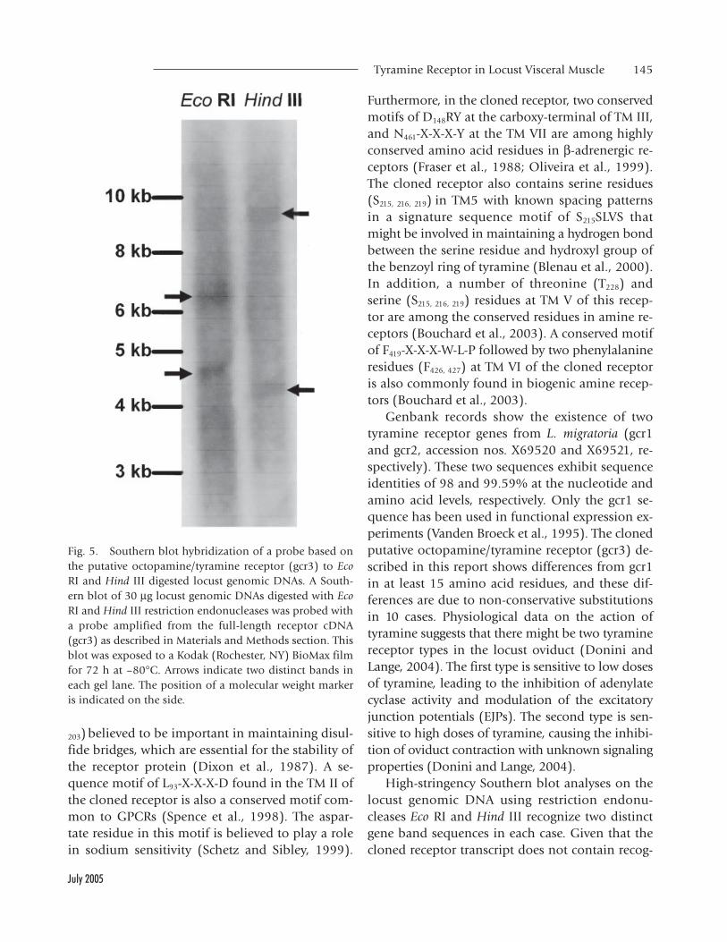
Southern Blot Analysis of the Receptor Gene
Southern blot analysis was carried out using ge-nomic DNA isolated from individual locusts. Forthis purpose, locust genomic DNA was digested withEco RI and Hind III restriction endonucleases andhybridized to a gcr3 probe (Fig. 5). Digests witheither Eco RI or Hind III restriction endonucleasesresulted in two bands. Since there is no recognitionsite for these enzymes within the full-length gcr3receptor sequence and the gene is intronless, theresults indicate that the probe utilized in thesestudies identifies two DNA fragments that sharesequence similarity to the receptor probe.
Phylogenetic Relationship of the Cloned Receptor WithOther Octopamine/Tyramine Receptors
Phylogenetic analysis of the cloned receptor andoctopamine/tyramine receptors from other inver-tebrates using the complete amino acid sequencessuggest that relatedness at the primary sequencelevel among octopamine/tyramine receptors can beused to group them into three major groups (Fig.6). Octopamine receptors from Aplysia californica(OAR-Ac) and A. kurodai (OAR-Ak) form the first
group in this analysis. Octopamine receptors fromBombyx mori (OAR-Bm), Heliothis virescens (OAR-Hv), L. stagnalis (OAR-Ls2) and tyramine receptorsfrom L. migratoria (Tyr-Loc1 (gcr1) and Tyr-Loc2
Fig. 3. Tissue-specific expression of the locust octo-pamine/tyramine receptor examined by RT-PCR. Locusttissues including (lane 1) brain, (lane 2) VNC, (lane 3)oviduct, and (lane 4) midgut were used to generate RNAfor RT-PCR as described in Materials and Methods. A no-template negative control (lane 5) ensuring the specificityof the amplicons did not generate any products (A). PCRreactions without reverse transcription step were also per-formed. PCR primers designed on the gcr3 cDNA sequencewere used in RT-PCR reaction. Positive control productswere amplified using primer pairs based on the histone H2Bgene (180 bp) and numbers indicate the source of RNAsfrom locust tissues as above (B). Amplified products werevisualized under UV illumination after staining withethidium bromide. The position of the molecular weightmarkers (MW, GeneRuler 100 bp DNA Ladder Plus, MBI,Fermentas, Hanover, MD) are indicated on both sides.
TABLE 1. Percentage Amino Acid Sequence Similarity of the LocustOctopamine/Tyramine Receptor With Other Biogenic Amine Receptors asDetermined by Multiple Sequence Alignment Using the CLUSTAL WProgram (Higgins and Sharp, 1988)
Receptor Species % similarity Reference
Tyramine L. migratoria 97 Vanden Broeck et al. (1995)Tyramine A. mellifera 59 Blenau et al. (2000)Tyramine D. melanogaster 52 Saudou et al. (1990)Tyramine C. elegans 35 Rex and Komuniecki (2002)Octopamine D. melanogaster 52 Arakawa et al. (1990)Octopamine M. brassicae 52 Grosmaitre et al. (2001)Octopamine B. microplus 51 Baxter and Barker (1999)Octopamine H. virescens 51 von Nickisch-Rosenegk
et al. (1996)Octopamine B. mori 50 von Nickisch-Rosenegk
et al. (1996)Serotonin L. stagnalis 30 Sugamori et al. (1993)Serotonin A. californica 30 Barbas et al. (2002)

144 Molaei et al.
Archives of Insect Biochemistry and Physiology
(gcr3), A. mellifera (Tyr-Am), C. elegans (Tyr-Ce),and D. melanogaster (Tyr-Dm) form the secondmajor group. The cloned tyramine receptor (gcr3,Tyr-Loc2) clusters with the first tyramine receptorfrom L. migratoria (gcr1 or Tyr-Loc1, Vanden Broecket al., 1995) exclusively within this group. Finally,octopamine receptors from D. melanogaster (OAR-Dm1 or OAMB and OAR-Dm2) and L. stagnalis(OAR-Ls1) form the third distinct group. Interest-ingly, two of the major groups are comprised en-tirely of octopamine receptors. This is in contrastto the second major group, which is comprised ofboth receptor types. However, it is worth notingthat the octopamine receptors from B. mori andH. virescens (von Nickisch-Rosenegk et al., 1996)that cluster within the mostly tyramine receptors’group could now be considered as tyramine re-ceptors based on the new evidence (Ohta et al.,
2003). A serotonin receptor from D. melanogaster(5-HT2A-Dm) has been used as an out-group inthis analysis.
DISCUSSION
The present study identifies a GPCR that is ex-pressed in the L. migratoria nervous system and inthe visceral muscle tissues, oviduct, and midgut.The presence of certain amino acid residues in thereceptor sequence is important in transducing theextracellular response to effector enzymes and in-teraction of the receptor with G proteins. In addi-tion to seven transmembrane domains commonto all GPCRs, this receptor displays a series of con-served signature residues specific to octopamine/tyramine receptors in insects. There are a numberof cysteine residues in the cloned receptor (C124,
Fig. 4. Tissue distribution of locust octopamine/tyraminereceptor determined by Northern blot analysis. A North-ern blot of 20 µg of total RNA from locust tissues includ-ing (lane 1) brain, (lane 2) ventral nerve cord (VNC),(lane 3) oviduct, and (lane 4) midgut probed with a probebased on the sequence of the cloned receptor cDNA (gcr3)as described in Materials and Methods. This blot was ex-
posed to a Kodak (Rochester, NY) BioMax film for 2 daysat -80°C. After stripping, each blot was hybridized with ahistone H2B cDNA probe for normalization, shown be-low the corresponding blot. The position of the RNA mo-lecular weight markers (MW, Invitrogen, San Diego, CA)are indicated on both sides.
Tyramine Receptor in Locust Visceral Muscle 145
July 2005
203) believed to be important in maintaining disul-fide bridges, which are essential for the stability ofthe receptor protein (Dixon et al., 1987). A se-quence motif of L93-X-X-X-D found in the TM II ofthe cloned receptor is also a conserved motif com-mon to GPCRs (Spence et al., 1998). The aspar-tate residue in this motif is believed to play a rolein sodium sensitivity (Schetz and Sibley, 1999).
Furthermore, in the cloned receptor, two conservedmotifs of D148RY at the carboxy-terminal of TM III,and N461-X-X-X-Y at the TM VII are among highlyconserved amino acid residues in β-adrenergic re-ceptors (Fraser et al., 1988; Oliveira et al., 1999).The cloned receptor also contains serine residues(S215, 216, 219) in TM5 with known spacing patternsin a signature sequence motif of S215SLVS thatmight be involved in maintaining a hydrogen bondbetween the serine residue and hydroxyl group ofthe benzoyl ring of tyramine (Blenau et al., 2000).In addition, a number of threonine (T228) andserine (S215, 216, 219) residues at TM V of this recep-tor are among the conserved residues in amine re-ceptors (Bouchard et al., 2003). A conserved motifof F419-X-X-X-W-L-P followed by two phenylalanineresidues (F426, 427) at TM VI of the cloned receptoris also commonly found in biogenic amine recep-tors (Bouchard et al., 2003).
Genbank records show the existence of twotyramine receptor genes from L. migratoria (gcr1and gcr2, accession nos. X69520 and X69521, re-spectively). These two sequences exhibit sequenceidentities of 98 and 99.59% at the nucleotide andamino acid levels, respectively. Only the gcr1 se-quence has been used in functional expression ex-periments (Vanden Broeck et al., 1995). The clonedputative octopamine/tyramine receptor (gcr3) de-scribed in this report shows differences from gcr1in at least 15 amino acid residues, and these dif-ferences are due to non-conservative substitutionsin 10 cases. Physiological data on the action oftyramine suggests that there might be two tyraminereceptor types in the locust oviduct (Donini andLange, 2004). The first type is sensitive to low dosesof tyramine, leading to the inhibition of adenylatecyclase activity and modulation of the excitatoryjunction potentials (EJPs). The second type is sen-sitive to high doses of tyramine, causing the inhibi-tion of oviduct contraction with unknown signalingproperties (Donini and Lange, 2004).
High-stringency Southern blot analyses on thelocust genomic DNA using restriction endonu-cleases Eco RI and Hind III recognize two distinctgene band sequences in each case. Given that thecloned receptor transcript does not contain recog-
Fig. 5. Southern blot hybridization of a probe based onthe putative octopamine/tyramine receptor (gcr3) to EcoRI and Hind III digested locust genomic DNAs. A South-ern blot of 30 µg locust genomic DNAs digested with EcoRI and Hind III restriction endonucleases was probed witha probe amplified from the full-length receptor cDNA(gcr3) as described in Materials and Methods section. Thisblot was exposed to a Kodak (Rochester, NY) BioMax filmfor 72 h at –80°C. Arrows indicate two distinct bands ineach gel lane. The position of a molecular weight markeris indicated on the side.

146 Molaei et al.
Archives of Insect Biochemistry and Physiology
nition sites for either of these endonucleases, andthe fact that this gene is intronless, the probe uti-lized in this analyses is identifying two distinctDNA fragments that share sequence similarity tothe cloned receptor probe. It is also known thatseveral genes are duplicated within the L. migratoriagenome. Therefore, it is conceivable to expect sev-eral fragments hybridize to the probe. Furthermore,
sequence alignment of the residues flanking TM5and TM6 located in the intracellular loop 3 of thecloned receptor and tyramine receptors revealed ahigh degree of sequence conservation among thesereceptors. Amino acid residues flanking TM5 andTM6 are important for functional coupling of thereceptors (Blenau and Baumann, 2003). Basedupon this evidence, we suggest that there might
Fig. 6. Dendrogram shows the phylogenetic relationshipof the cloned GPCR with other octopamine/tyramine re-ceptors from invertebrates. Amino acid sequence align-ment was carried out according to the CLUSTAL Walgorithm and the dendrogram was constructed by Neigh-bour-Joining using Phylip program at http://www.genebee.msu.su/services/ phtree_reduced.html. The lengthsof horizontal lines are inversely proportional to the se-quence homology between two sequences or betweengroups of sequences. Abbreviations and GenBank acces-sion numbers (#) for the receptor sequences are: OAR-Ac,octopamine receptor from the sea slug, A. californica (#AAF37686); OAR-Ak, octopamine receptor from the seaslug, A. kurodai (# AAF28802); OAR-Bm octopamine re-ceptor from the silkworm, B. mori (# Q17232); OAR-Hv,
octopamine receptor from the tobacco budworm, H.virescens (# Q25188); Tyr-Loc1 (gcr1), tyramine receptorfrom the locust, L. migratoria (# Q25321); Tyr-Loc2 (gcr3),putative octopamine/tyramine receptor cloned from thelocust, L. migratoria in our studies; Tyr-Am, tyramine re-ceptor from the honeybee A. mellifera (# AJ245824); Tyr-Ce, tyramine receptor from the nematode, C. elegans (#NM171978); Tyr-Dm, tyramine receptor from D. melano-gaster (# S12004); OAR-Ls2, octopamine receptor from L.stagnalis (# O01670); OAR-Dm1, octopamine receptorfrom D. melanogaster (# AF065443); OAR-Dm2, octopaminereceptor from D. melanogaster (# AJ007617); and OAR-Ls1octopamine receptor from L. stagnalis (# O77408). 5-HT2A-Dm, serotonin receptor from D. melanogaster (# S19155)has been used as an out-group.

Tyramine Receptor in Locust Visceral Muscle 147
July 2005
be at least two, if not more receptor subtypes andthat the cloned receptor in the present study andthe previously cloned tyramine receptor (Tyr-Loc;Vanden Broeck et al., 1995) may represent twotyramine receptor isoforms or subtypes. The pres-ence of different receptor subtypes for biogenicamines has been indicated in insects and these re-ceptor subtypes may bring about a variety of ef-fects and activate different groups of secondmessenger systems (Roeder, 2002).
High-sequence similarity between the clonedreceptor in our studies and gcr1 (Vanden Broecket al., 1995) is not surprising. For instance, theamino acid sequences of two octopamine/tyraminereceptors isolated from B. mori and H. virescensshowed 96.3% homology to each other (vonNickisch-Rosenegk et al., 1996). More recently, theoccurrence of an octopamine receptor (AmOA1)from the brain of the honeybee, A. mellifera, wasexamined by Western blot analysis using an anti-serum raised against a peptide selected from theAmOA1 sequence (Farooqui et al., 2004). In addi-tion to an expected band of 78 kDa, 5 additionalbands were identified. There was no significant se-quence identity between the peptide sequence usedto generate the antiserum and other biogenicamine receptor sequences from honeybee or otherinsects. Furthermore, the expression of these pro-teins was reduced by AmOA1 dsRNA treatment.Based on the above evidence, it was concluded thatthese proteins may be different subtypes, differentsplice variants, and/or post-translational covalentmodifications of the AmOA1 receptor (Farooquiet al., 2004).
Little expression data are available on gcr1 (Tyr-Loc1) to serve as a basis for comparison. ThemRNA expression of the Tyr-Loc1 receptor hasbeen detected in the locust nervous system withwidespread distribution in the brain and the ven-tral nerve cord (Vanden Broeck et al., 1995). Thetissue distribution of our cloned receptor mRNAindicates that the expression of the receptor tran-script was the highest in brain tissue followed byVNC. The expression levels of receptor mRNA inoviduct and midgut tissues was relatively similaras examined under a semi-quantitative RT-PCR and
Northern blot analysis. Furthermore, the expressionof the receptor mRNA was detected by in situ hy-bridization in a small number of cells likely beingendocrine-like cells in the locust midgut and in asmall group of spindle-shape cells in the commonoviduct (preliminary observations). Further workwill be aimed at assigning a functional role for thereceptor and its respective ligand in regulating/modulating physiological processes such as theperistaltic movements and the passage of foodthrough the midgut lumen, digestive breakdownand uptake of dietary nutrients in midgut, andmodulating physiological processes such as con-traction and facilitating the process of egg deposi-tion in the oviduct. It is also worth noting that theprobe(s) utilized in expression experiments prob-ably can not discriminate between gcr1, 2, or 3 inthe above studies.
In conclusion, a novel GPCR from the locustvisceral muscle has been cloned and shown to beexpressed in the central nervous system and vis-ceral muscle tissues. Sequence analysis strongly sug-gests that the receptor belongs to the biogenicamine receptor family and specifically to theoctopamine/tyramine receptor group. Phylogeneticanalysis supports this receptor as being a tyraminereceptor. Our data suggest that this receptor is dif-ferent from the previously identified locust tyra-mine receptor and may represent a sub-type of thisfamily. The distribution of the receptor mRNA inlocust tissues, particularly oviduct, verifies thephysiological responsiveness of these tissues tooctopamine and tyramine (Lange and Nykamp,1996; Donini and Lange 2004). Together thesestudies suggest that the cloned receptor and its pos-sible ligand, tyramine, may assume important func-tions in mediating physiological processes in locustvisceral muscles, oviduct, and midgut.
ACKNOWLEDGMENTS
The authors express their gratitude to Dr. IanOrchard and Dr. Tim Westwood for their adviceand support and to Dr. Joseph Vanden Broeck forthe initial cDNA library and his helpful sugges-tions. This work was supported by grants to A.B.L.

148 Molaei et al.
Archives of Insect Biochemistry and Physiology
from the Natural Sciences and Engineering Re-search Council of Canada.
LITERATURE CITED
Arakawa S, Gocayne JD, McCombie WR, Urquhart DA, Hall
LM, Fraser CM, Venter JC. 1990. Cloning, localization, and
permanent expression of a Drosophila octopamine recep-
tor. Neuron 4:343–354.
Barbas D, Zappulla JP, Angers S, Bouvier M, Castellucci VF,
DesGroseillers L. 2002. Functional characterization of a
novel serotonin receptor (5-HTap2) expressed in the CNS
of Aplysia californica. J Neurochem 80:335–345.
Baxter GD, Barker SC. 1999. Isolation of a cDNA for an
octopamine-like, G-protein coupled receptor from the
cattle tick, Boophilus microplus. Insect Biochem Mol Biol
29:461–467.
Blenau W, Baumann A. 2001. Molecular and pharmacologi-
cal properties of insect biogenic amine receptors: Lessons
from Drosophila melanogaster and Apis mellifera. Arch In-
sect Biochem Physiol 48:13–38.
Blenau W, Baumann A. 2003. Aminergic signal transduction
in invertebrates: Focus on tyramine and octopamine re-
ceptors. Recent Res Dev Neurochem 6:225–240.
Blenau W, Balfanz S, Baumann A. 2000. Amtyr1: character-
ization of a gene from honeybee (Apis mellifera) brain en-
coding a functional tyramine receptor. J Neurochem
74:900–908.
Bouchard C, Ribeiro P, Dube F, Anctil M. 2003. A new G
protein-coupled receptor from a primitive metazoan shows
homology with vertebrate aminergic receptors and displays
constitutive activity in mammalian cells. J Neurochem
86:1149–1161.
Bryson-Richardson RJ, Logan DW, Currie PD, Jackson IJ. 2004.
Large-scale analysis of gene structure in rhodopsin-like
GPCRs: evidence for widespread loss of an ancient intron.
Gene 338:15–23.
Chang DJ, Li XC, Lee YS, Kim HK, Kim US, Cho NJ, Lo X,
Weiss KR, Kandel ER, Kaang BK. 2000. Activation of a het-
erologously expressed octopamine receptor coupled only
to adenylyl cyclase produces all the features of presynap-
tic facilitation in Aplysia sensory neurons. Proc Natl Acad
Sci USA 97:1829–1834.
Dixon RA, Sigal IS, Candelore MR, Register RB, Scattergood
W, Rands E, Strader CD. 1987. Structural features required
for ligand binding to the β-adrenergic receptor. EMBO J
6:3269–3275.
Donini A, Lange AB. 2004. Evidence for a possible neurotrans-
mitter/neuromodulator role of tyramine on the locust ovi-
ducts. J Insect Physiol 50:351–361.
Downer RG, Hiripi L, Juhos S. 1993. Characterization of the
tyraminergic system in the central nervous system of the
locust, Locusta migratoria migratorioides. Neurochem Res
18:1245–1248.
Farooqui T, Vaessin H, Smith BH. Octopamine receptors in
the honeybee (Apis mellifera) brain and their disruption
by RNA-mediated interference. J Insect Physiol 50:701–713.
Fraser CM, Chung F-Z, Wang C-D, Venter JC. 1988. Site-di-
rected mutagenesis of human beta-adrenergic receptors:
substitution of aspartic acid-130 by asparagine produces
a receptor with high-affinity agonist binding that is un-
coupled from adenylate cyclase. Proc Natl Acad Sci USA
85:5478–5482.
Gaillard I, Rouquier S, Giorgi D. 2004. Olfactory receptors.
Cell Mol Life Sci 61:456–469.
Gerhardt CC, Bakker RA, Piek GJ, Planta RJ, Vreugdenhil E,
Leysen JE, van Heerikhuizen H. 1997a. Molecular cloning
and pharmacological characterization of a molluscan
octopamine receptor. Mol Pharmacol 51:293–300.
Gerhardt CC, Lodder HC, Vincent M, Bakker RA, Planta RJ,
Vreugdenhil E, Kits KS, van Heerikhuizen H. 1997b. Clon-
ing and expression of a complementary DNA encoding a
molluscan octopamine receptor that couples to chloride
channels in HEK293 cells. J Biol Chem 272:6201–6207.
Grohmann L, Blenau W, Erber J, Ebert PR, Strunker T,
Baumann A. 2003. Molecular and functional characteriza-
tion of an octopamine receptor from honeybee (Apis
mellifera) brain. J Neurochem 86:725–735.
Grosmaitre X, Marion-Poll F, Renou M. 2001. Biogenic amines
modulate olfactory receptor neurons firing activity in
Mamestra brassicae. Chem Senses 26:653–661.
Han KA, Millar NS, Davis RL. 1998. A novel octopamine re-
ceptor with preferential expression in Drosophila mush-
room bodies. J Neurosci 18:3650–3658.
Hill CA, Fox AN, Pitts RJ, Kent LB, Tan PL, Chrystal MA,

Tyramine Receptor in Locust Visceral Muscle 149
July 2005
Cravchik A, Collins FH, Robertson HM, Zwiebel LJ. 2002.
G protein-coupled receptors in Anopheles gambiae. Science
298:176–178.
Hiripi L, Juhos S, Downer RG. 1994. Characterization of
tyramine and octopamine receptors in the insect (Locusta
migratoria migratorioides) brain. Brain Res 633:119–126.
Kolakowski LF Jr. 1994. GCRDb: a G-protein-coupled recep-
tor database. Recept Chan 2:1–7.
Lange AB, Nykamp DA. 1996. Signal transduction pathways
regulating the contraction of an insect visceral muscle. Arch
Insect Biochem Physiol 33:183–196.
Lange AB, Orchard I. 1984. Dorsal unpaired median neu-
rons, and ventral bilaterally paired neurons, project to a
visceral muscle in an insect. J Neurobiol 15:441–453.
Lange AB, Tsang PKC. 1993. Biochemical and physiological
effects of octopamine and selected octopamine agonists
on the oviducts of Locusta migratoria. J Insect Physiol
39:393–400.
McClung C, Hirsh J. 1999. The trace amine tyramine is es-
sential for sensitization to cocaine in Drosophila. Curr Biol
9:853–860.
Ohta H, Utsumi T, Ozoe Y. 2003. B96Bom encodes a Bombyx
mori tyramine receptor negatively coupled to adenylate cy-
clase. Insect Mol Biol 12:217–223.
Oliveira L, Paiva ACM, Vriend G. 1999. A low resolution
model for the interaction of G protein with G protein-
coupled receptors. Prot Eng 12:1087–1095.
Orchard I, Lange AB. 1985. Evidence for octopaminergic
modulation of an insect visceral muscle. J Neurobiol
16:171–181.
Poels J, Suner MM, Needham M, Torfs H, De Rijck J, De Loof
A, Dunbar SJ, Vanden Broeck J. 2001. Functional expres-
sion of a locust tyramine receptor in murine erythro-
leukaemia cells. Insect Mol Biol 10:541–548.
Reale V, Hannan F, Midgley JM, Evans PD. 1997. The expres-
sion of a cloned Drosophila octopamine /tyramine recep-
tor in Xenopus oocytes. Brain Res 769:309–320.
Rex E, Komuniecki RW. 2002. Characterization of a tyramine
receptor from Caenorhabditis elegans. J Neurochem 82:1352–
1359.
Robb S, Cheek TR, Hannan FL, Hall LM, Midgley JM, Evans
PD. 1994. Agonist-specific coupling of a cloned Drosophila
octopamine/tyramine receptor to multiple second messen-
ger systems. Brain Res 633:119–126.
Roeder T. 1994. Biogenic amines and their receptors in in-
sects. Comp Biochem Physiol 107C:1–12.
Roeder T. 1999. Octopamine in invertebrates. Prog Neurobiol
59:533–561.
Roeder T. 2002. Biochemistry and molecular biology of re-
ceptors for biogenic amines in locusts. Micros Res Tech
56:237–247.
Roeder T, Seifert M, Kahler C, Gewecke M. 2003. Tyramine
and octopamine: antagonistic modulators of behavior and
metabolism. Arch Insect Biochem Physiol 54:1–13.
Sambrook J, Fritsch EF, Maniatis T. 1989. Molecular cloning:
a laboratory manual. Cold Spring Harbor, NY: Cold Spring
Harbor Laboratory Press.
Saudou F, Amlaiky N, Plassat JL, Borrelli E, Hen R. 1990.
Cloning and characterization of a Drosophila tyramine re-
ceptor. EMBO J 9:3611–3617.
Schetz JA, Sibley DR. 1999. Meta-ions as atomic scale probes
of G protein-coupled receptor structure. In: Wess J, editor.
Structure-function analysis of G protein-coupled receptors.
New York: Wiley-Liss, Inc. p 121–140.
Spence P, Bard J, Jones P, Betty M. 1998. The identification
of G protein-coupled receptors in sequence databases. Exp
Opin Ther Patents 8:235–247.
Sugamori KS, Sunahara RK, Guan HC, Bulloch AG, Tensen
CP, Seeman P, Niznik HB, Van Tol HH. 1993. Serotonin
receptor cDNA cloned from Lymnaea stagnalis. Proc Natl
Acad Sci USA 90:11–15.
Vanden Broeck J, Vulsteke V, Huybrechts R, De Loof A. 1995.
Characterization of a cloned locust tyramine receptor
cDNA by functional expression in permanently trans-
formed Drosophila S2 cells. J Neurochem 64:2387–2395.
von Nickisch-Rosenegk E, Krieger J, Kubick S, Laage R, Strobel
J, Strotmann J, Breer H. 1996. Cloning of biogenic amine
receptors from moths (Bombyx mori and Heliothis virescens).
Insect Biochem Mol Biol 26:817–827.
Wess J, 1998. G protein-coupled receptors: molecular mecha-
nisms involved in receptor activation and selectivity of G
protein recognition. FASEB J 11:346–354.