Embed Size (px)
Citation preview

Units 2, 7, 8 Molecular biology, Nucleic acids and Metabolism
Concept Learning Objective Required contento Define the meaning of
molecular biology Describe molecular biology as the study
of biology on a molecular basis Discuss the formation of ionic bonds Discuss the formation of covalent bonds
Molecular Biology is the study of Biology on a molecular basis. This means that it looks at all the interactions that occur between molecules.
In order to form compounds atoms must form bonds, these bonds may involve the sharing of electrons (covalent) or the creation of electrically charged particles which are then attracted to each other (ionic). A weak or temporary bond may form between some electrically charged particles- we call these hydrogen bonds
o Discuss the bonding properties of carbon
Define the term organic molecule Discuss the atomic structure and
bonding properties of carbon Discuss the versatility of carbon
Any chemical that has been produced by a living organisms is referred to as organic.
One exception to this statement is carbon dioxide. Organic molecules tend to contain carbon. Carbon is considered to be a versatile molecule as it can form up to
4 covalent bonds and therefore can form a large number of stable compounds
o Discuss the importance of carbon in the formation of organic molecules
Discuss the importance of carbon in the formation of biological molecules
Recognise the outline form for common organic molecules
Discuss the role of organic molecules in molecular biology
Carbon along with hydrogen and oxygen are essential components of many organic molecules.
Nitrogen is also essential for amino acids. Organic molecules are important as an energy source, for growth
and repair and for energy storage and insulation.
o Define metabolism, anabolism and catabolism
o Define and describe metabolic pathways
o Give an example of an enzyme that is part of a metabolic pathway and link to lower activation energy
Define metabolism as, the sum of the biochemical reactions that are needed to sustain life
Outline anabolic reactions Outline catabolic reactions Explain what is meant by metabolic
pathway Discuss the importance of metabolic
pathways in organisms Give examples of metabolic pathways in
a cell include specific enzymes and describe how they lower activation energy
Metabolism refers to the chemical reactions that take place in the cells.
An anabolic reaction is one that requires energy to make larger molecules from smaller ones eg the synthesis of protein.
Catabolism is a reaction that gives out energy when larger molecules are broken down in to smaller ones, eg the breakdown of sugar in respiration.
Some reactions need to be controlled by the body and therefore will happen in steps or cycles. This is referred to as a metabolic pathway- eg photosynthesis or respiration. These reactions are generally controlled by enzymes
Enzymes speed up metabolic reactions by reducing the activation energy needed for the reaction to occur. Examples of enzymes include amylase released by the salivary gland and by the pancreas

Units 2, 7, 8 Molecular biology, Nucleic acids and Metabolism
which breaks down starch in to maltoseo Outline vitalism and
urea synthesis Define vitalism Discuss the process of urea synthesis Discuss the idea that synthetic urea has
allowed us to falsify theories
Vitalism is a belief that living things are driven by a vital internal force.
This suggests that only living things can make organic molecules. This was disproved by the accidental (serendipitous) discovery of
synthetic urea by Friedrich Wohler 1828, who was able to rearrange the molecular structure of ammonium citrate to form urea.
Concept Objective Required contento Describe the chemical
properties of water molecules
Describe the structure of a water molecule
Discuss the polarity of water
Describe the formation of hydrogen bonds
Water is made from one oxygen and two hydrogen atoms, it is a polar molecule (di-pole) which means that one end (Hydrogen) has a positive charge and the other (Oxygen) has a negative charge.
It therefore forms a ‘V’ shape. A water molecule will form an attractive bond (hydrogen bond- each water molecule can form 3H bonds) with neighboring water molecules, we call this cohesion, it will also form hydrogen bonds with other polar molecules which we call adhesion. These properties are essential for water transport in the xylem (Transpiration stream- a continuous column of water molecules).
o Discuss the properties of water and link to biological importance
Identify the key properties of water
Discuss the biological importance of water
Link the absence of water to the collapse of biological systems
Water is an essential component of life and as a habitat. It is important due to its properties as a solvent due to its polar properties, many
other polar substances will dissolve in water making it good for transport and as a place for metabolic reactions.
Water is needed for temperature regulation (sweat) due to a high specific heat capacity (amount of energy needed to raise water by 1oC).
Hydrogen bonds form between the O- and H+ of adjacent molecules, each water molecule can form 3H bonds. Cohesion between water molecules provides surface

Units 2, 7, 8 Molecular biology, Nucleic acids and Metabolism
tension for transpiration and also adhesion between the water molecules and the xylem.
Surface tension also allows organisms to move on the surface of water. Cohesion also gives water a high latent heat of vaporization (high boiling point) -
meaning it takes a high amount of energy to break the hydrogen bonds and it is therefore good as a coolant whist at the same time a high boiling point means that the water is unlikely to boil in a natural habitat and its SHC means that it will maintain the temperature of the environment.
One vital property of water is that it can be found in its liquid form over a wide global temperature range therefore water provides a thermally stable habitat.
As water reaches its maximum density at 4oC and the fact that ice float means that it can act as an insulator allowing life to survive in cold waters.
The transparency of water allows light to penetrate allowing the survival of hydrophytes and this transparency allows animals to see under water.
o Discuss the meaning of hydrophilic and hydrophobic
Define hydrophilic and give examples
Define hydrophobic and give examples
Define amphipathic and give examples
Hydrophilic molecules such as glucose are water loving Hydrophobic molecules such a fats are water hating. Amphipathic means a molecule has both hydrophilic and hydrophobic parts, eg a
phospholipid
o Memory of water Discuss the ‘memory of water’
Some scientists believe the theory that water can remember the arrangement of the molecules when something was dissolved in it. We call this water memory; this concept is useful in homeopathy as a beneficial solution can be diluted many times yet still have the same benefit.
o Describe the structure of carbohydratesDiscuss the genetic modification of potatoes to reduce amylose content so that adhesives can be extracted
Describe the importance of carbohydrates
Draw the ring structure for alpha glucose and beta glucose
Draw the structure of ribose and fructose
Carbohydrates are essential for energy storage eg starch, energy release eg glucose, structure eg chitin and cellulose.
Glucose exists in different formats, alpha and beta. Glucose is made up of a 6 membered ring called a pyranose ring.
Ribose is made up of a 5 member ring called a furanose ring.
o Describe the formation of polysaccharides
Explain condensation and hydrolysis reactions
When two monosaccharaides (monomers) are joined together a bond is formed between them. This involves the removal of water; we call this a condensation reaction.

Units 2, 7, 8 Molecular biology, Nucleic acids and Metabolism
State that glycosidic bonds form between monomers during a condensation reaction
Show with examples the formation of a disaccharide and polysaccharide
When a molecule is split water is used to do this, we call this hydrolysis. Sucrose is a disaccharide formed from the two monosaccharaides glucose and
fructose, the bond between them is called a glycosidic bond. Other disaccharides are lactose and maltose. Starch is formed from many glucose molecules being joined together, we call this a
polysaccharide Other polysaccharides include cellulose, glycogen and chitin.
Concept Objective Required contento Describe the structure of
fatty acids and explain what is meant by mono/poly/unsaturated
Draw the general structure of a fatty acid and glycerol
Show the formation of an ester bond
Identify saturated and unsaturated fatty acid chains
Fatty acids are made up of a glycerol backbone with three fatty acid tails. We call this structure a triglyceride.
When a fatty acid joins to the glycerol an ester bond is formed. Fatty acids may be saturated or unsaturated. In saturated fatty acids all of the bonds
are occupied with hydrogen and there are no C=C double bonds- it forms a straight chain. In unsaturated fatty acids there are C=C double bonds, these bonds cause a bend (kink) in the chain. An unsaturated fatty acid with more than one C=C double bond is said to be polyunsturated
o Discuss the role of lipids in the body
State the key function of lipids in the body
Lipids are important as an energy store and respiratory substrate. They are needed for heat insulation, the protection of internal organs, water
proofing, buoyancy, they form a key component of cell membranes and are needed for the formation of the insulating myelin sheath in neurons.
Lipids can be used to form steroid hormones and are important for cell transport though the formation of lipoproteins.
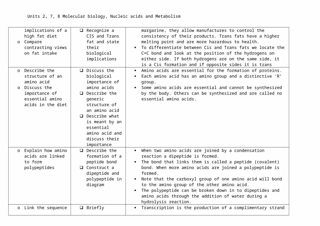
o Explain the difference between cis and trans fats
o Discuss the implications of a high fat diet
o Compare contrasting views on fat intake
Define CIS and Trans fat
Give examples of CIS and Trans fats
Recognize a CIS and Trans fat and state their biological implications
Cis fats such as cis-oleic acid have a characteristic kink in their tail and are found naturally.
Trans fats such as trans-oleic acid do not have a kink and are produced artificially through hydrogenation.
Trans fats are found in fried foods and also in margarine, they allow manufactures to control the consistency of their products. Trans fats have a higher melting point and are more hazardous to health.
To differentiate between Cis and Trans fats we locate the C=C bond and look at the position of the hydrogens on either side. If both hydrogens are on the same side, it is a Cis formation and if opposite sides it is trans

Units 2, 7, 8 Molecular biology, Nucleic acids and Metabolism
o Describe the structure of an amino acid
o Discuss the importance of essential amino acids in the diet
Discuss the biological importance of amino acids
Describe the generic structure of an amino acid
Describe what is meant by an essential amino acid and discuss their importance
Amino acids are essential for the formation of proteins. Each amino acid has an amino group and a distinctive ‘R’ group. Some amino acids are essential and cannot be synthesized by the body. Others can
be synthesized and are called no essential amino acids.
o Explain how amino acids are linked to form polypeptides
Describe the formation of a peptide bond
Construct a dipeptide and polypeptide in diagram
When two amino acids are joined by a condensation reaction a dipeptide is formed. The bond that links them is called a peptide (covalent) bond. When more amino acids
are joined a polypeptide is formed. Note that the carboxyl group of one amino acid will bond to the amino group of the
other amino acid. The polypeptide can be broken down in to dipeptides and amino acids through the
addition of water during a hydrolysis reaction.o Link the sequence of
amino acids in a protein to genetics
o Link the amino acid sequence of a polypeptide to primary protein structure
o Describe the stages of protein formation
o Link the amino acid sequence to the 3D structure of a protein
o Describe protein structure
Briefly describe the process of transcription and translation- linking to amino acid sequence
Discuss the importance of the correct amino acid sequence
Give an example of the consequences of a malformed protein
Discuss the primary protein structure
Describe how the secondary stage of protein structure arises
Transcription is the production of a complimentary strand of mRNA for a section of DNA (gene) it is a similar process for both prokaryotes and eukaryotes.
The molecule of mRNA is a copy (transcript) of a gene, mRNA is smaller than the DNA and can pass through the pores in the nuclear membrane.
This molecule then passes to the ribosomes where it is then translated. Translation uses the mRNA as a template to make a protein.
Primary proteins are a long chain of amino acids it is the order of these amino acids that determine the function of the protein,
Secondary proteins have hydrogen bonds between C=O and N-H groups of different amino acids (every 4 amino acids may form a H bond) which then form an alpha helix and beta pleated sheet, these proteins tend to be structural such as collagen.
Tertiary proteins have disulfide bonds (S-bridges), hydrogen bonds and ionic bonds, they have a 3D shape (globular) eg enzymes, the 3D shape is due in part to the hydrophobic and hydrophilic interactions, hydrophobic amino acids will face inwards, causing folds in the molecule, this helps to form the shape of the active site in an enzyme polar and non-polar amino acids also add to the shape of the active site. These proteins are often globular and soluble.
Quaternary proteins are formed from more than one peptide or may be conjugated

Units 2, 7, 8 Molecular biology, Nucleic acids and Metabolism
Describe the tertiary and quaternary stages of protein structure
Define conjugated protein
by the addition of a prosthetic group (haemoglobin and iron). Haemoglobin is a quaternary protein formed from multiple peptide chains (2α chains and 2β). It is a globular protein; it also has a prosthetic heme group (Fe2+) to which oxygen will bind. Each molecule of haemoglobin therefore has four protein chains and each heme group will bind to one oxygen molecule (O2) therefore each molecule of haemoglobin will bind with 4 molecules of oxygen.
Note that there is a difference between the proteins produced by free ribosomes and those produced by ribosomes in the RER, free ribosomes synthesize proteins for use inside of the cell and RER produces proteins for export.
o Discuss the influence of polar and non-polar amino acids and the formation of bonds between the R groups
o
Define polar and non-polar in relation to amino acids
Identify polar and non-polar amino acids
Discuss the bonds that form between non-polar and polar R groups
Polar molecules are water loving and will dissolve easily, non-polar molecules are water hating and will not dissolve in water easily.
The R groups of amino acids may be polar or non-polar. Positively charged R groups may form hydrogen bonds with negatively charged ones.
Non polar amino acids will tend to fold inwards while polar amino acids may be on the outer side of the protein, these properties are important in the shaping of the protein
o Discuss the universality of protein formation
Explain the importance of proteins in the body
Describe the optimal working conditions of proteins
Discuss the uses of fibrous and globular proteins
Proteins are needed for growth and repair, for enzymes and hormones, for the clotting of blood and for cell transport- amongst other things.
Proteins have an optimum working temperature and pH. Above this optimum temperature or at the wrong pH the proteins will be denatured.
Fibrous proteins such as collagen are structural and are insoluble in water. They are made from long thin chains of repeating amino acids.
Globular proteins are more likely to dissolve in water and include enzymes, they are made from nonrepeating amino acid sequences and have a rounded/spherical shape. They are usually involved in metabolic reactions and include haemoglobin, pepsin and amylase. They tend to be affected by temperature, pH and salt concentration which may cause them to denature.
Units 2, 7, 8 Molecular biology, Nucleic acids and Metabolism
o Discuss the practicality of proteomics
Define proteome Discuss the
individuality of each proteome
Link a person’s proteome to their genome
Each organism has a unique genetic code (genome), this is based upon the genetic base sequence and triplet codons. As a result, they also have a unique proteome- the proteome is the sum of all the proteins produced by an organism.
Each triplet codon codes for a specific amino acid, however there are also start and stop codons which are important for translation.
o Describe the structure of enzymes including their active site
Describe with examples the functions of enzymes in the body
Draw and label the structure of an enzyme
Discuss allosteric enzymes
Describe the function of co-enzymes
Enzymes have many functions in the organism including digestion, respiration and photosynthesis.
All enzymes have an active site which is a specific shape to fit a particular substrate it may also have an electrical charge which attracts the substrate. This active site is found on the surface of the enzyme and it is here where the substrate binds and the reaction is catalyzed.
Allosteric enzymes have an additional binding point/s which is different to the active site and which other substances may bind to. This may change the shape (conformation) of the active site making the enzyme active or inactive.
Chemicals need to activate the enzyme are called co-enzymes.
o Describe the kinetics of enzyme activity
Describe an enzyme as a substance that reduces activation energy
Draw a chart plotting the energy changes that take place during an enzyme reaction
Discuss the action of an enzyme being to bring about a conformational shape of a molecule
All chemical reactions need energy to start them off. An enzyme will reduce the amount of activation energy needed- without the enzymes reactions would take far too long to occur at body temperature.
The enzyme brings the substrates closer together and in the correct orientation. The substrate binds with the active site forming an enzyme substrate complex, this puts the substrate under stress causing a slight change in its structure and weakening molecular bonds. We call this a conformational change.
o Describe the activity of enzymes
Describe the formation of an enzyme/substrate complex
Describe the theory of induced fit
One theory of enzyme activity is that of the lock and key theory, the substrate has a complimentary shape to the rigid shape of the active site and the two fit exactly to form the enzyme-substrate complex.
The second theory of enzyme activity used by the IB is the theory that the enzyme is not rigid and will change the shape of its active site to fit the substrate, we call this the induced fit model. The active site and the substrate do not match exactly, the enzyme will mold its active site to compliment the shape of the substrate as the

Units 2, 7, 8 Molecular biology, Nucleic acids and Metabolism
enzyme substrate complex is formed. The enzyme and substrate may have opposite electrical charges to assist with this. This change in configuration of the enzyme puts strain on the bonds of the substrate and weakens them, lowering the activation energy needed to break the bonds. The enzyme therefore catalyses the reaction.
Once the reaction has occurred the products will leave the active site, leaving the enzyme unchanged and available for the next substrate.
Some enzymes are able to act upon more than one substrate and this is explained by the induced fit method.
o Discuss the factors that influence enzymes and their rates of reaction
Discuss the optimum conditions needed for enzyme activity
Draw sketch graphs showing the relationship between enzyme activity and the controlling factors
Use a graph of enzyme activity to extract data relating to activity
Enzymes work best at an optimum temperature, as temperature increase towards the optimum, the rate of reaction will also increase due to an increase in motion of the molecules and an increase in the number of collisions between the enzyme and substrate, more complexes will be formed and more of the substrates will obtain their activation energy.
Above this temperature the active site will change shape and the tertiary structure of the protein is altered, due to the breaking of hydrogen bonds (due to vibrations inside the enzyme disrupting the intermolecular forces). The substrate will no longer be able to bind with the active site and the enzyme substrate complex will not form we say that the enzyme has denatured.
Each enzyme also has an optimum pH outside of which there will be lower activity and if the pH is too high or low the enzyme may go through a change in shape or may denature, this may be as a result of ionization or changes in electric charge due to the acidity. Salivary amylase produced in the salivary glands found in the mouth breaks starch down in to maltose, this enzyme prefers a pH or around 7/8. Pepsin in the stomach breaks protein in to amino acids and prefers a pH of 2.
The concentration of the substrate will also affect the activity of the enzyme, the increased concentration will mean a greater number of collisions between the enzyme and substrate and therefore more reactions take place. The rate of reaction is directly proportional to substrate concentration, however this will eventually plateau as all the active sites on the enzymes are utilized, therefore further increases in concentration will see no further increase.
o Explain what happens to an enzyme when it is denatured
Describe the specificity of the active site
Explain why changes occur to the active site as it denatures
When an enzyme denatures the bonds between the different amino acids that maintain the 3D shape will break. This means that the active site loses its specific shape. We say that the active site has gone through a conformational change. This change is irreversible and will prevent the enzyme from working.

Units 2, 7, 8 Molecular biology, Nucleic acids and Metabolism
Describe these changes as conformational and irreversible
o Discuss the action of competitive and non-competitive enzyme inhibitors
Discuss what is meant by enzyme inhibition
Describe with examples the process of competitive enzyme inhibition
State how to overcome competitive inhibition
Describe with examples non-competitive enzyme inhibition
Discuss the types of enzyme inhibition which are reversible and those which are irreversible
Discuss, with examples, what is meant by end product inhibition
Interpret graphs showing enzyme inhibition
In a metabolic pathway the end product (metabolites) in the chain may switch off an allosteric enzyme found earlier in the chain by binding to its allosteric site- some enzymes have multiple allosteric sites.
The metabolites act as allosteric inhibitors. This causes a temporary change in the shape (conformational change) of the active site preventing the enzyme from working, it is a form of non-competitive inhibition as the metabolite does not compete for the active site.
It is a form of negative feedback as the buildup of the product switches off the production process.
It is reversible as when the volume of the product falls the metabolite will detach from the allosteric site and the enzyme will once again activate.
We call this end product inhibition (allosteric inhibition), allostery is the process when the binding of substance to one part of an enzyme causes a conformational change to another part of the enzyme.
An example of end-product inhibition is the regulation of ATP formation by phosphofructokinase (PFK) an enzyme in glycolysis that is inhibited by ATP binding to its allosteric site.
Sometimes a chemical may have a similar shape to the substrate and will temporary bind to the active site preventing the substrate from binding, this results in competitive inhibition. An example of competitive inhibition could be malonic acid which competes with succinate for active sites of succinic dehydrogenase, an important enzyme in the Krebs cycle.
Some substances such as a heavy metals (eg mercury and silver) may bind with a part of the enzyme that is not the active site or with the allosteric site, this causes an irreversible conformational change (change in shape) at the active site and stops the enzyme from working we call this non-competitive inhibition An example of non-competitive inhibition could be cyanide (or potassium cyanide – KCN) which inhibits Cytochrome C Oxidase responsible for the transfer of hydrogen atoms during cellular respiration preventing the production of ATP.

Units 2, 7, 8 Molecular biology, Nucleic acids and Metabolism
o Compare competitive and non-competitive inhibition
o Discuss the functions of enzymes in industry- including the importance of immobilized enzymes
State uses of enzymes in industry
Describe how enzymes are manufactured and harvested in industry
Discuss the advantages of using immobilized enzymes
Enzymes are biological catalysts and speed up biochemical reactions. They have many uses in the body and in industry.
Pectinase is used in industry to breakdown the pectin in the cell walls of fruits into smaller more soluble carbohydrates to make the extraction of the juice easier, increasing clarity and yield. DNA Nuclease is used in genetic engineering to extract fragments of DNA, washing powders use protease enzymes to breakdown insoluble protein in to soluble amino acids that can be washed out of the clothes.
Enzymes can be produced by a fermenter, bacteria are genetically modified to produce the required enzyme and are grown at the optimum conditions, eg temperature, nutrients, pH and oxygen levels. The enzymes are then harvested by filtration methods.
It is advantageous to attach enzymes to a non-soluble component as it makes them easier to retrieve.

Units 2, 7, 8 Molecular biology, Nucleic acids and Metabolism
o Discuss the role of enzyme inhibitors in medicine- include the use of ethanol to combat methanol poisoning
o Discuss the use of fomepizole in the treatment of antifreeze poisoning
o Discuss methods in which enzyme inhibitors are used to treat alcoholism
An example of the use of enzyme inhibitors is in methanol poisoning, ethanol is used as a competitor to reduce the effects of methanol on the nervous system.
Antifreeze contains the chemical Ethylene glycol, this is broken down by the enzyme alcohol dehydrogenase in the liver and causing glycol poisoning. Fomepizole is a competitor for the Ethylene glycol and therefore a competitive inhibitor for alcohol dehydrogenase.
Alcoholics may be treated with Disulfiram this inhibits the enzyme aldehyde dehydrogenase, and, as a result the production of acetaldehyde (a bi-product of the breakdown of ethanol). This leads to nausea, hypotension, and flushing.
o Discuss the production of lactose free milk and how advances in enzyme technology benefits some more than others
Note that there are higher occurrences of lactose intolerance in Asia than Europe or America.
Lactose is an important disaccharide as it provides energy for young mammals. However, many people throughout the world are intolerant to the lactose as they do
not produce the enzyme lactase. Lactose is found in milk, dairy and processed foods It causes bloating and abdominal discomfort in those who are intolerant.
It is possible to breakdown the disaccharide lactose in to the monosaccharaides glucose and galactose, during the food manufacturing process by adding the enzyme lactase- this is extracted from yeast which naturally ferments milk.
The yeast is added to foods containing lactose, extracted enzymes can be immobilized in order that the enzyme can be retrieved and reused.
The hydrolysis of lactose by lactase can make the food taste sweeter, reducing the need to add sweeteners to the food, the glucose and galactose are more soluble than lactose so this avoids crystallization in products such as ice-cream.
This process tends to be expensive and is only done in where the costs can be offset by the purchase price. This often excludes people in poorer countries from purchasing the lactose free products.
There is a high occurrence of lactose intolerance in Asia. Bacteria utilize galactose and glucose faster than lactose by fermenting them, this
spoils food much quicker so reduces shelf life and profit. During the production process lactase is often used at low temperatures (5oC) as this
prevents the enzyme from denaturing, and also slows down bacterial growth, reducing the rate at which the product spoils.
o Is it appropriate to study A person’s proteome is unique and is directly related to their DNA sequence.

Units 2, 7, 8 Molecular biology, Nucleic acids and Metabolism
a person’s proteome? Studying the proteome will allow the identification of certain diseases and give a view of the person’s future susceptibility to a disease.
o Many metabolic pathways have been traced one reaction at a time- how can looking at part of a reaction give us an understanding of the whole reaction
Many metabolic pathways are quite complex trying to understand the whole process in a single step may lead to confusion and misunderstanding. By breaking the process down in to smaller steps will allow the process to be mapped more effectively. If we look at individual steps, then then we can deduce the product of previous reactions and predict the products of reactions in the subsequent steps.
o Describe the structure of nucleic acids
o Compare and contrast DNA with RNA
Describe the structure of a nucleotide
Describe complimentary base pairings
Identify purines and pyrimidines
Nucleic acids consist of a phosphate group, sugar (deoxyribose/ribose) and base (covalent bonds). There are 4 bases for DNA Adenine, Guanine, Thymine, Cytosine, and they form complimentary pairs A-T (2 H bonds) and C-G (3 H bonds).
A purine (A, G) is a double ringed structure and a pyrimidine (T-C) is a single ringed structure.
Describe the structure of RNA
Compare the structure of DNA with that of RNA
Compare the uses or DNA and RNA
Both DNA and RNA are nucleic acids made up from a phosphate group, pentose (pentagonal) sugar and a base.
The base is covalently bonded to C1 of the sugar and the phosphate group to C5. The C3 of the sugar will bond to the phosphate on the next nucleotide - forming a
polynucleotide. Uracil substitutes for thymine in RNA, but both have adenine, guanine and cytosine. RNA differs from DNA as it is a smaller single stranded molecule with a ribose sugar
rather than the deoxyribose possessed by DNA. DNA is a double stranded helical molecule.
mRNA is transcribed DNA, tRNA is a carrier molecule used to transport amino acids in the translation process.
Each molecule of tRNA consists of a looped strand of RNA and each differs in structure. Each tRNA carries a different amino acid, at the 3’ end of the tRNA molecule there is a triplet code CCA, it is here where the amino acid will bind. To assist with the binding a specific tRNA-activating enzyme is needed along with a molecule of ATP. The enzyme recognizes the specific shape of the tRNA.
o Describe the structure of DNA
o Discuss the work of
Discuss the double helix, antiparallel structure of DNA
DNA is formed from 2 antiparallel strands of nucleic acid (5’3, 3’5’) that are intertwined to form a double helix. The structure of DNA was officially first identified by the scientists Watson and Crick in 1953.

Units 2, 7, 8 Molecular biology, Nucleic acids and Metabolism
Watson and Crick and their elucidation of DNA structure
o Discuss the work of Rosalind Franklin and Maurice Wilkins and their contribution to our knowledge of DNA structure through x-ray diffraction
o Discuss the work of Hershey and Chase and how they provided evidence that DNA is genetic material
Discuss how DNA is formed
Produce a timeline leading to the discovery of the structure of DNA
Describe what is meant by 5’ and 3’
DNA is formed by this addition nucleotides (sub-units) using enzymes. Each nucleotide is made from a deoxyribose sugar, phosphate linked to C5 and base liked to C1- the sugar and phosphate are linked with a sugar phosphate bond (covalent). There are 4 bases Adenine, Thymine, Cytosine and Guanine. A pairs with T and C pairs with G. This complimentary base pairing allows the formation of hydrogen bonds between bases and thus the linking of two strands base to base.
When DNA is replicated the complimentary copy will be made in the 5’ and 3’ direction and the template will be read in the 3’ 5’ direction.
o Describe the structure and function of a nucleosome
o Use molecular visualization software to analyse the association between histone protein and DNA within a nucleosome
1. Define nucleosome2. state the importance
of histone proteins in the formation of a nucleosome
3. State why the packaging of DNA is important in eukaryotes
DNA exists in very long strands and therefore needs to be packaged. If not packaged then mitosis and meiosis would prove difficult. The DNA is packaged by wrapping it around a core of 8 histone proteins (octomer) to form nucleosomes, the nucleosomes are then stacked to condense the strand further.

Units 2, 7, 8 Molecular biology, Nucleic acids and Metabolism
o Discuss the semi-conservative replication of DNA
o Describe the role of helicase on DNA replication
o Describe the role of DNA polymerase on DNA replication
o Discuss how DNA polymerase is only able to add nucleotides to the 3’ end of the primer
o Discuss continuous and discontinuous DNA replication
o Describe how the structure of DNA has led to our understanding of DNA replication
o Describe the action of the enzymes involved in DNA replication
Recap the stage of the cell cycle in which DNA is replicated
Define what is meant by complimentary base pairing
State that DNA replication is semiconservative
Suggest evidence to support that DNA replication is semiconservative
State that DNA replication is an enzyme led process
State the role of helicase in DNA replication
State the role of DNA polymerase I and III in DNA replication
Discuss the action of DNA polymerase
Discuss the role of DNA gyrase State the role of RNA primase in
replication Explain the formation of Okazaki
fragments and the role of ligase in discontinuous DNA replication
State the role of single strand binding proteins in DNA replication
Discuss the connections between the structure of DNA and our understanding of DNA replication
Draw a flow chart to show the action of, Helicase, DNA Polymerase I and III, Primase, single strand binding proteins and Ligase in DNA replication
DNA is replicated in the S phase of the cell cycle. Each strand of existing DNA is replicated semi-conservatively through complimentary base pairing. This means that in each DNA molecule there will be one of the original strands and one new complimentary strand.
We can use radioactive isotopes of nitrogen and observing the strands of DNA during mitosis. DNA replication is an enzyme led process and requires the presence of Helicase, RNA Primase and Polymerase.
Leading strand. DNA helicase unwinds the DNA double helix by breaking the hydrogen bonds between complimentary bases. Each DNA strand will act as a blueprint for a complimentary new strand. RNA Primase adds an RNA nucleic acid (primer) to the point where replication will begin- this primer will later be removed. DNA Polymerase III binds to the DNA and moves along it from the 3’ to the 5’ direction BUT builds the new complimentary strand of DNA in the 5’ and 3’ direction. Free nucleotides form complimentary base pairs by forming hydrogen bonds (A-T, C-G). The nucleotides are covalently linked by the polymerase III and the new DNA strand forms. Once formed the double helix will again form the result is an identical copy of the original DNA strands.
Lagging strand. The DNA is replicated in sections- still reading in the 3’5’ direction. The fragments are called Okazaki fragments these fragments are still formed by Polymerase III, however need to be joined together by DNA ligase once the RNA primers have been removed.
Enzymes in prokaryotic DNA replication DNA Polymerase III elongates the DNA by adding a new
nucleotide to the 3’ end of the chain building the DNA in the 5’3’dorection. This enzyme also proofreads the new DNA strand.

Units 2, 7, 8 Molecular biology, Nucleic acids and Metabolism
DNA Polymerase I removes the RNA primer and replaces it with DNA nucleotide
DNA Polymerase II is believed to repair damaged DNA
DNA Gyrase acts to relieve the strain on the DNA strand as it unwinds.
Helicase- unwinds the double helix by breaking the hydrogen bonds between complimentary base pairs.
Ligase- an enzyme that joins together the Okazaki fragments from the lagging strands, forms sugar phosphate bonds between the nucleotides.
Single stranded binding proteins are used to stabilize the unwound single strands of DNA and prevents them from annealing (rejoining to form a double strand).
o Discuss the importance of introns, exons, telomeres, regulators and tRNA genes. State the functions of each
State that not all of the DNA on a chromosome will code for a protein
Define introns and exons and discuss their relevance
Explain what is meant by a telomere
Explain the role of regulator genes
Explain the role of tRNA genes
Not all of the DNA will code for protein synthesis there are sections called introns and exons. Exons provide the code for protein synthesis and introns a sections of DNA that do not code for proteins but are important for the regulation of gene expression.
A telomere is a section of DNA that is found at each end of the chromosome, it is believed that this portion of DNA protects the integrity of the DNA structure.
tRNA genes are sections of DNA that code for the reproduction of tRNA molecules- these are in turn are important for the synthesis of protein.
o Describe the processes involved in transcription
o State that transcription builds a molecule in the 5’-3’ direction
State the need for transcription Describe the process of
transcription State the direction of
transcription as being in the 5’ 3’ direction
State the key enzymes involved in
The molecules of DNA are long and only certain sections are expressed in certain cells leading to cell differentiaation. The molecules are too big to pass through the pores in the nuclear membrane. Therefore, shorter chains of mRNA are used for gene expression, these molecules are produced through the process of transcription.
During transcription RNA polymerase controls the process, it

Units 2, 7, 8 Molecular biology, Nucleic acids and Metabolism
transcription unwinds the DNA strands and breaks the H bonds, exposing 10-20 bases at a time. RNA polymerase uses one strand as a template (antisense). It starts complimentary base pairing at a binding site called the promoter and will build the mRNA molecule in the 5’ 3’ whilst reading the antisense strand in the 3’ 5’ direction.
As the mRNA is constructed new hydrogen bonds form between the complimentary bases. RNA polymerase will stop transcribing the DNA when it reaches the terminator codon.
After which the mRNA and RNA polymerase will detach completely and be released. The DNA double helix will reform (anneal).
Energy is required for transcription and this is attained by the hydrolysis of phosphate compounds (eg ATP).
Note that the sense strand is the DNA strand that has the correct code for the gene. The antisense strand which has the complimentary base pairs and acts as the template so that the gene can be transcribed.
Remember that in RNA Thymine is substituted by Uracil.o Explain the role of the promoter
and terminator in transcription Identify promoters and
terminators as non-coding genes State the importance of
promoters and terminators in transcription
A promoter is the base code that will initiate the transcription process. It may consist of between 1000-2000 base sequences.
Transcription factors (enhancers) will bind to the DNA to initiate transcription.
A terminator is the point at which transcription ends. o Explain how nucleosomes help
to regulate transcriptiono Explain how mRNA is modified
after transcription
Discuss the need to regulate transcription
Describe the role played by nucleosomes in the regulation of transcription
Explain how mRNA is modified after transcription
Nucleosomes consist of DNA tightly wound around a histone protein core. The protein blocks the transcription of the DNA by covering the promoter until it is needed.
It is important that the RNA produced through transcription is first modified before it leaves the nucleus. The noncoding introns are removed and the exons joined in a process known as splicing.
The 5’ end of the molecule is capped and the 3’ end is cleaved and a tail added.
o Explain how the splicing mRNA State what is meant by the Splicing DNA involves the removal of noncoding introns and

Units 2, 7, 8 Molecular biology, Nucleic acids and Metabolism
increases protein production splicing of mRNA Explain how the splicing of DNA
leads to increased protein production
the splicing of or even exclusion of exons. The alternative splicing of the mRNA allows for a single gene to code for more than one protein. This process takes place in the nucleus of the cell.
o Explain how gene expression is regulated
o Link the environment of a cell to gene expression
State what is meant by gene expression
Explain how gene expression is regulated
State the impact that the environment of cells and organisms impact upon gene expression
Certain chemicals act as enhancers and bind to promoters on the DNA enhancing the rate of transcription.
Other chemicals act as silencers, they bind to the promoters and reduce transcription.
The nucleosomes are also important in the regulation of transcription; they will unravel to expose the section of DNA to be transcribed.
If the DNA is exposed to certain chemicals or radiation form the environment the rate of transcription can be effected.
o Discuss the structure of a ribosome
The ribosomes are synthesized in the nucleolus each ribosome has a small subunit and large subunit
The mRNA binding sites are located on the small subunit; there are three tRNA binding sites (E,P,A).
Ribosomes are formed from protein and RNA.o Explain the process of
translation State why mRNA needs to be
translated State the role of ribosomes and
tRNA in translation Identify the key molecules that
are involved in translation Outline the process of translation
Mature mRNA needs to be translated in order for proteins to be produced.
The mRNA is a transcript of a gene and will leave the nucleus by passing through the nuclear pores. Once in the cytoplasm it will locate a ribosome.
Ribosomes are made from two subunits, proteins and ribosomal RNA.
The 5’ end (cap) of the mRNA will bind to a binding site on the small subunit of the ribosome. The small subunit of the ribosome will move along the mRNA towards the 3’ end until it locates the start triplet codon AUG.
At this point the large subunit of the ribosome will bind along with a specific molecule of tRNA- we call this initiation.
The ribosome subunits will then move along the mRNA molecule to the next triplet codon/base triplet (translocation), here a specific tRNA molecule with the correct anticodon will pair with the triplet codon on the mRNA forming hydrogen bonds.

Units 2, 7, 8 Molecular biology, Nucleic acids and Metabolism
The enzyme peptidyl transferase will join the amino acids that are attached to each molecule of tRNA to form peptide bonds (condensation reaction) note that a peptide bond is a type of covalent bond and that two tRNA molecules will be bound to the ribosome with one of them at the P site holding the peptide chain.
This will continue forming a primary protein chain (polypeptide)- we call this elongation.
When the ribosomal complex reaches a stop codon (UAG, UGA and UAA), a release factor will bind with the codon and the peptide chain will be released from the ribosomes (termination).
The peptide chain will then go through further modifications until it has the correct structure. Note that the small subunit of the ribosome has three binding points E P and A, the tRNA first binds to the A site and then is moved to the P site during translocation and finally the E site where it exits, leaving behind the amino acid. Remember that the mRNA is read in the 5’ 3’ direction and the polypeptide is constructed from the amino to the carboxyl terminus.
All living organisms use the same genetic code. The genetic code is therefore universal and determines the expression of the DNA base sequence and therefore the amino acid sequence during the synthesis of peptides. Each base sequence forms a triplet codon (3 bases), many of these codons code for a specific amino acid. There are 64 possible triplet codons, as a result each amino acid has more than on triplet codon we say that the genetic code is degenerate.
o Explain how translation is initiatedo Describe how mRNA is translatedo State the role of free ribosomes in
translationo Explain the function of stop codons
State how translation is initiated Describe the action of the start
codon and stop codon State the processes involved in
translation and describe the action of free ribosomes, tRNA and anticodons
Identify the key enzymes for translation
Translation is initiated by the mRNA binding to the small subunit of the ribosome.
Once the start codon AUG (this codes for the amino acid methionine) is located translation will begin. Each tRNA molecule has an anticodon and will be attached to a specific amino acid.
The tRNA and amino acid with the correct anticodon will bind to the ‘A’ site of the small subunit on the ribosome complex the tRNA will then move to the ‘P’ site and another tRNA binds to the ‘A’

Units 2, 7, 8 Molecular biology, Nucleic acids and Metabolism
site. The amino acids bound to the tRNA molecules will be joined with peptide bonds. The key enzyme involved in this process will be peptidyl transferase.
The tRNA will then move along to the ‘E’ site from where it will be released.
o State the role of membrane bound ribosomes and link to secretions
State that translation also occurs using the membrane bound ribosomes of the endoplasmic reticulum
State the different destinations and functions of the proteins
Ribosomes are found free floating in the cytoplasm and bound to the rough endoplasmic reticulum. Proteins that are formed by free ribosomes are usually used within the cell. Those that are formed and modified by the RER are often secreted from the cell to be used outside of the cell, eg digestive enzymes.
o Explain how translation in eukaryotes differs from that in prokaryotes
Identify the fact that translation must occur in all living organisms
Compare translation in prokaryotes with that of eukaryotes
Discuss differences in the type of ribosomes in prokaryotes and eukaryotes
Discuss why mitochondria may contain the same type of ribosomes as prokaryotes (endosymbiosis)
All organisms require proteins for growth, repair and metabolic function. These proteins are coded for by the DNA. Therefore, in order for the proteins to be synthesized the DNA must first be transcribed and the translated.
In prokaryotes translation id done by 70s ribosomes as opposed to the 80s ribosomes in the eukaryotes. mRNA in the prokaryote translation is polycistronic (codes for several proteins) in eukaryotes it is monocistronic. The process is much faster in prokaryotes, the lifespan of the mRNA is short and unstable. In Eukaryotes the lifespan of the mRNA is stable and longer. In addition, they have different amino acids that are used at the initiation points.
o Discuss the relevance of triplet codons and anticodons in determining amino acid sequence
Define triplet codon and anticodon
Discuss the relevance of codons and triplet codons
Discuss how amino acids are joined during the formation of proteins during translation
Each 3 bases form a triplet codon. Each triplet codon codes for a specific amino acid. The tRNA molecule with the complimentary codon (anticodon) will bind to the ribosome mRNA complex during the elongation stage of translation. The enzyme peptidyl transferase is used to join the amino acids, forming a polypeptide chain.
o What is meant by junk DNA and explain how this type of understanding might influence our approach to knowledge
We do not yet know all the functions of the different sections of DNA. There are sections that do not code for proteins and the functions are unknown. We call these non-coding sections of DNA ‘Junk DNA’. Some scientists believe that mutation may occur and make the noncoding DNA functional. We call this process exaptation.

Units 2, 7, 8 Molecular biology, Nucleic acids and Metabolism
o Identify the differences between DNA replication in prokaryotes and eukaryotes?
Prokaryote: DNA Replication takes place in the protoplasm, chromosomes are circular, single stranded, without telomeres, one origin of replication, no histones (Naked DNA), one replication fork
Eukaryote: Replication takes place in the cytoplasm, chromosomes are linear, double stranded, with telomeres, several origins of replication, histone proteins, several replication forks
o Comparing transcription and translation
DNA is transcribed to produce mRNA. mRNA, is used to produce polypeptides. Both involve the use of ATP and enzymes. Transcription uses RNA Polymerase, translation uses peptidyl transferase and ribosomes and tRNA. Transcription takes place in the nucleus of the eukaryote whereas translation takes place in the cytoplasm. Both new molecules are synthesized in the 5’3’direction.
o Discuss the work of Meslson and Stahl
o
Meslson and Stahl used isotopic Nitrogen to monitor the replication of DNA,
Bacteria were grown in a medium containing 15N, the bacteria were then transferred to a medium containing 14 N, after several generations the bacteria were harvested and the DNA extracted. The DNA was placed in to cesium chloride and centrifuged.
The DNA fromed bands at three different levels in the solution 15N, 14N and a mixture of the two in the middle. This proved that DNA was replicated semi-conservatively
o Discuss the PCR and its relevance
The polymerase chain reaction is a technique that is used to replicate a small sample of DNA several times over. It is useful in forensic science when trying to identify a perpetrator from a small sample of DNA, during the process DNA goes through cycles of heating and annealing this is done in a thermal cycler.
o Discuss how we have been able to artificially produce human insulin
Insulin is essential to regulate blood glucose levels. Some people are unable to produce sufficient amounts of insulin and therefore need to inject it. In order to meet the global

Units 2, 7, 8 Molecular biology, Nucleic acids and Metabolism
demand for insulin it can be produced by using genetically modified bacteria. In this procedure the DNA from healthy pancreas cells is removed and the gene for insulin removed through the use of restriction enzymes.
Sticky ends are then added to the gene and it is inserted in to a plasmid, which is a small loop of DNA, that has been extracted from an e coli bacterium and cut with the same restriction enzymes and complimentary sticky ends added.
Ligase enzymes are used to splice (join) the host and donor DNA sections together. The recombinant plasmid is then reinserted in to the bacterium and a culture of clones is grown in a fermenter. The insulin is then harvested and purified.
Alternatively reverse transcriptase, can be used to make copies of required DNA by transcribing mRNA back into a strand of DNA which can be inserted in to a plasmid using restriction enzymes, sticky ends and ligase. The plasmid can be reinserted in to a bacteria which can then be cloned in a fermenter, the product can then be harvested. The clotting factor IX needed to treat hemophilia can be produced in this way, by removing the mRNA for the clotting factor from healthy cells.
o Discuss the role of tRNA activating enzymes and the role of phosphorylation
Each tRNA molecule binds to a specific amino acid. The amino acid first binds to a molecule of ATP. The amino acid is phosphorylated (phosphate added) and the ATP converted to AMP. The ‘charged’ amino acid now binds to the tRNA. When the tRNA binds to the relevant codon on the ribosome mRNA complex the energy from the ‘charged’ amino acid is used to form the peptide bond.
o Identify polysomes in electron micrographs for prokaryotes and eukaryotes
Several ribosomes may act upon the same molecule of mRNA at the same time. We call these polysomes. We can see them on electron micrographs as clusters attached to an mRNA molecule
o Identify the structure of a tRNA molecule with its binding sites
tRNA is made up from a single strand of RNA with three loops, one loop has an anticodon which will bind with the complimentary triplet codon on the mRNA. At one end of

Units 2, 7, 8 Molecular biology, Nucleic acids and Metabolism
the molecule is a receptor site with the codon CCA it is here where an amino acid will bind with an ester bond.
o Describe cell respiration as the controlled release of energy
State the cell respiration is the controlled release of energy in the cell
Discuss the need for a controlled release of energy
Discuss the different energy values of food and relate this to the respiratory quotient
Respiration is a biochemical pathway that slowly releases the energy found in food (controlled release). If the release of energy was not controlled there is a risk that there will be too much energy released, which may result in overheating and wasted energy.
Different food substances release different amounts of energy during respiration, we call this the respiratory quotient (RQ). RQ = CO2 eliminated / O2 consumed
The RQ values we need to know are fats 0.7, proteins 0.9 and carbohydrates 1. The study of the RQ allows us to determine which substrates are being used for respiration.
Lipids have about twice the energy as carbohydrates per unit mass. In animals much energy is stored in the form of glycogen- although this is generally a short term storage solution.
When the energy is required, enzymes convert the glycogen to soluble glucose which is then used as a substrate for respiration.
In plants the energy is stored in the form of starch. Lipids are a longer term energy store, when needed they are
broken down to form fatty acids and glycerol, enzymes breakdown triglycerides further to form acetyl CoA, which enters the Krebs cycle.
o Compare carbohydrates and lipids as substrates for respiration.
Both lipids and carbohydrates are primary sources of energy for organisms; lipids store more energy per gram than carbohydrates, lipids generally provide 2 to 3 times the energy of carbohydrates for a given mass; carbohydrates are easier to transport (than lipids) making their energy more accessible this is because lipids are insoluble (in water) whereas small carbohydrates are soluble in water; carbohydrates are more easily taken

Units 2, 7, 8 Molecular biology, Nucleic acids and Metabolism
out of storage making their energy more quickly available; carbohydrates are short-term storage molecules, whereas lipids provide long-term storage;
o Describe ATP as an immediate energy source in the cell
State the role of ATP in the body Describe the structure of ATP and
relate this to its function Define coupling reaction and link
this to ATP
ATP is considered the body’s energy currency. It is made from up from the genetic base adenine the sugar ribose and three phosphate groups. The bonds between the phosphate groups are considered to be high energy bonds and will release their energy when the bond is broken.
This energy can be used in molecular synthesis (anabolism) and for cell transport. The ATP molecule is manufactured from ADP and inorganic phosphate through cellular respiration.
ATP is often involved in a coupling reaction where the energy released from one reaction (exergonic) is used to drive a second reaction (endergonic). ATP is in limited supply in the body and needs to be constantly recycled. The average person has about 50g of ATP and will recycle their own body weight in ATP per day.
o Describe the process of aerobic respiration
Discuss the need for aerobic respiration
Write word and chemical equations for aerobic respiration
Outline the main stages involved in aerobic respiration
State the products of aerobic respiration
During respiration energy is released from an organic substrate. The energy that is released needs to be controlled. Therefore, respiration is the controlled release of energy from an organic substrate (glucose). If the release of energy was not controlled there would be too much energy released during a chemical reaction and much of this would be converted to heat.
Aerobic respiration uses oxygen; this type of respiration is important as it has a higher net energy output (38 ATP) than anaerobic respiration (2ATP).
The main stages involved in aerobic respiration are, glycolysis, the link reaction, Krebs cycle and oxidative respiration. During aerobic respiration glucose and oxygen are converted in to water and carbon dioxide.
o Describe the structure and function of mitochondria
Sketch a diagram of the mitochondrion
Identify the components and
The mitochondrion is a double membrane bound organelle containing granules, 70s ribosomes and naked mitochondrial DNA. The inner membrane is folded to form cristae which

Units 2, 7, 8 Molecular biology, Nucleic acids and Metabolism
functions of the mitochondrion State the chemical reactions that
take place in the different components of the mitochondrion
increases surface area, the fluid inside of the mitochondrion is called the matrix.
The outer membrane separates the contents of the mitochondrion from the contents of the cytoplasm. The inner membrane contains electron transport chains and the enzyme ATP synthase- oxidative phosphorylation takes place here. Between the membranes is the inter-membrane space, protons are pumped in to this space for the process of chemiosmosis. The matrix is the place where the Krebs cycle occurs.
o State the key processes involved in cell respiration
o Identify the key steps in glycolysis and state where in the cell it takes place
o State the end products of glycolysis
Identify glycolysis as the initial stage of respiration taking place in the cytoplasm
Outline the breakdown of glucose to pyruvate
Identify the products of Glycolysis Describe the role of oxygen in
aerobic respiration
Organic molecules such as glucose release energy in stages. Glycolysis is the breakdown of a glucose molecule to produce pyruvate (2 molecules of pyruvate per molecule of glucose). This process takes place in the cytoplasm and requires an investment of 2 ATP molecules to phosphorylate the glucose. The reaction results in the formation of several intermediary molecules and 4 molecules of ATP are released. The net products of this reaction are 2 molecules of pyruvate and 2 molecules of ATP along with 2 molecules of NADH + H+. If oxygen is present, then the pyruvate will enter the mitochondrion
o State the key steps involved in the link reaction
Describe the conversion of pyruvate to acetyl CoA
The next stage is the link reaction, where each molecule of the pyruvate has CO2 removed (oxidative decarboxylation) and NAD is reduced to form NADH + H this forms an oxidized acetyl group (2C) which is then combined with the molecule CoA forming the molecule acetyl CoA (2C). This process takes place in the matrix of the mitochondria. Net product per pyruvate- 1 Acetyl CoA, 1 CO2 and 1 NADH + H
o Discuss the key stages of the Krebs cycle
Outline the stages of the Krebs cycle
Identify the products of the Krebs cycle
The newly formed acetyl groups (2C) enter the Krebs cycle which is also known as the citric acid cycle (TCA) and takes place in the matrix of the mitochondrion, the molecule of acetyl CoA (2C) combine with the 4 carbon molecule oxaloacetate, to form a 6 carbon molecule (citrate) and a molecule of CoA.

Units 2, 7, 8 Molecular biology, Nucleic acids and Metabolism
The citrate goes through a series of steps, with oxidative decarboxylation occurring twice and CO2 being released each time, the oxaloacetate is eventually reformed.
During the process NAD and FAD (co-enzymes) are reduced and act as hydrogen acceptors these then enter the electron transport chain.
The products of one turn of the Krebs cycle are. CoA, oxaloacetate, 2 x CO2, 1 x ATP, 3 x NADH + H+, FADH2. Remember that these products are per molecule of acetyl CoA- each glucose molecule produces 2 acetyl CoA molecules during glycolysis.
o Explain the role of NAD and FAD as electron carriers
o Discuss the key stages involved in the electron transport chain
Define oxidative phosphorylation Describe the role of NADH+H and
FADH2 as electron carriers Describe the electron transport
chain
NAD and FAD are referred to as electron carriers, they transport hydrogen ions and electrons to the electron transport chain found in the cristae of the mitochondrial inner membrane. The reduced electron carrier donates electrons to the proteins (NAD Dehydrogenase) in the electron transport chain and the hydrogen ions are pumped in to the inter-membrane space by proton pumps.
The electrons move through the chain and release energy as they pass between molecules (Cytochromes) this causes more H+ ions to be pumped in to the inter-membrane space of the mitochondria.
The last step of this chain involves the use of the hydrogen and a terminal electron acceptor (oxygen) to form water (O + H2) in the matrix.
The loss of electrons from the electron carriers is an oxidation reaction and the energy changes as the electrons pass along the chain is used to join inorganic phosphate to ADP,
This process is referred to as oxidative phosphorylation. Note that in the electron chain there are two more electron carriers ubiquinone (q) and cytochrome c.
Each pair of electrons from NADH + H results in the transfer of 9 protons and the generation of 3ATP, each FADH2 pair of electrons result in the formation of 2ATP.
o Describe the process of Describe the process of As the H+ ions are pumped in to the mitochondrial inner

Units 2, 7, 8 Molecular biology, Nucleic acids and Metabolism
chemiosmosis chemiosmosis membrane space, a H+ concentration gradient is formed. The H+ ions (protons) flow down their concentration gradient through the enzyme ATP synthase, found in the inner membrane.
The change in position of the protons releases energy which is used to form ATP from ADP and inorganic phosphate (oxidative phosphorylation), in the matrix of the mitochondria.
This movement of H+ is referred to as chemiosmosis. This is an example of a coupling reaction. Some textbooks will state that 32 ATP are produced from chemiosmosis and others 34. It depends upon the molecule used as the mitochondrial shuttle. This means that the NADH + H that is produced in the cytoplasm cannot pass through the mitochondrial membrane and therefore a shuttle molecule is needed to transport the electrons to the transport chain. If the shuttle is NAD then more ATP is produced than if the shuttle is FAD.
o Describe the process of anaerobic respiration
Write word and chemical equations for anaerobic respiration
Discuss the need for anaerobic respiration
Describe the key stages of anaerobic respiration in humans and in yeast
Describe the effects of the bi-products from anaerobic respiration
If oxygen is not available as the final electron acceptor in the electron transport chain, then the electron transport chain will stop. And the pyruvate from glycolysis will no longer be absorbed by the mitochondria.
The pyruvate is instead converted to lactic acid, lactic acid is a harmful chemical but can be quickly converted to lactate by the enzyme lactate dehydrogenase, which can be used as respiratory substrate by the heart (lactate shuttle).
This overall process is referred to as anaerobic respiration and is due the incomplete oxidation of the glucose molecule. It is important in the body as it maintains the level of NAD. (NADH is needed to form the lactate).
o Describe what happens during anaerobic respiration in humans and in yeast
Describe the chemical reactions that take place during anaerobic respiration in yeast
Describe the conversion of pyruvate to ethanal and then ethanol in yeast
In yeast the pyruvate is converted in to ethanol and carbon dioxide. In muscle cells the pyruvate is converted to lactic acid.

Units 2, 7, 8 Molecular biology, Nucleic acids and Metabolism
o Compare anaerobic and aerobic respiration
Compare the products of aerobic and anaerobic respiration in humans
Compare anaerobic respiration in humans and in yeast
o Describe the pathway taken by alternative substrates to glucose
Compare the energy values of different substrates
State where fatty acids and glycerol enter the respiratory chain
State where amino acids enter the respiratory chain
Discuss the implications of using proteins as a respiratory substrate
The respiratory quotient (RQ) is a measure of the conversion rate between oxygen and carbon dioxide. It can be used to determine the substrate that is being used for respiration. Carbohydrates have the highest RQ followed by proteins and then fats.
One reason for this is that the different substrates join the respiratory process at different stages, Deaminated amino acids can form pyruvate and therefore enter at the link reaction stage, fats form acetyl groups when broken down and can then form acetyl CoA, they join the Krebs cycle.
The body will use proteins as a substrate for respiration as a last resort as these proteins are important for other functions in the body such as growth, repair, hormones and enzymes.
o Discuss the ethics of using invertebrates in respirometers
Define respirometer and discuss the ethics of using invertebrates.
A respirometer is a device used to measure the rate of respiration by measuring the consumption of oxygen it uses potassium hydroxide solution to absorb any carbon dioxide

Units 2, 7, 8 Molecular biology, Nucleic acids and Metabolism
that is produced. Traditionally invertebrates such as maggots have been used
for this process as they provide a quick a recordable result. This is now seen as unethical as it causes stress for the animal, they may be harmed when being transferred to the apparatus and can be harmed if they make contact with the potassium hydroxide solution. Germinating peas can be used as an alternative.
o Define energy investment and payout
Energy needs to be invested in order to initiate the glycolysis reaction, however the amount of ATP produced by this reaction (pay out) exceeds the investment. (4ATP v 2ATP)
o Explain how the formation of the chemiosmotic theory led to a paradigm shift
o Explain the difficulties that Peter Mitchell’s chemiosmotic theory faced before it was expected and discuss why the falsification of a theory does not gain immediate acceptance
Chemiosmosis is the production of ATP by the diffusion of H+ ions across the inner membrane of the mitochondrion, using ATP synthase. It was discovered by Peter D Mitchel; in 1961. This this discovery/theory challenged the conventional views towards ATP synthesis.
The publishing of his findings caused a change in understanding within the scientific world. We call this change a view a paradigm shift.
Initially it was difficult for Mitchel to convince the scientific community to accept his research as it took a completely different approach to conventional theories. Eventually chemiosmosis was accepted and in 1978 Mitchel was awarded the Nobel prize for Chemistry.
o Explain the use of electron tomography to produce images of active mitochondria
Electron tomography is a technique that uses a transmission electron microscope to obtain 3D images of active mitochondria.
Electron tomography has revealed that cristae are connected with the inter-membrane space between the inner and outer membranes via narrow openings. The shape and volume of the cristae change when mitochondria are active in ways that are still being investigated.
o Describe the structure of a leaf
Sketch a diagram of a leaf Identify the function of each layer
The leaf is a flattened organ that absorbs light for photosynthesis. The blade is the flattened part and the

Units 2, 7, 8 Molecular biology, Nucleic acids and Metabolism
found in the leaf Identify the location of
chloroplasts within the leaf Explain how the leaf is adapted
for the efficiency of photosynthesis
petiole is the part that holds the leaf away from the stem. The leaf is made up or several layers, including the waxy
cuticle to reduce water loss, the epidermis is a tough layer of cells for protection. The palisade mesophyll layer contains photosynthetic cells that have chloroplasts. The spongy mesophyll cells have some chloroplasts. The air spaces allow the diffusion of gasses. The lower epidermis is again for protection.
The guard cells contain chloroplasts they open and close the stomata- tiny pores to allow gas exchange. Collenchyma tissue found at the midrib of the leaf has cells with thickened walls and provides the leaf with extra structural support, the leaf vein contains the xylem and the phloem.
o Define photosynthesis as the production of carbon compounds in cells using light energy
State the photosynthesis is the production carbon compounds using light energy
Write a word equation and chemical equation for photosynthesis
State the biological importance of photosynthesis
Describe how the plant utilizes the product of photosynthesis
Photosynthesis is the production of carbon compounds using light energy. It is very important as it fixes the carbon from carbon dioxide forming organic molecules such as glucose. 6H2O +6CO2 C6H12O6 + 6O2, we call this carboxylation.
The glucose can then be converted in to starch, fructose, cellulose and amino acids. The rate of photosynthesis can be measured through the uptake of CO2 or the production of O2 and increase in biomass.
o Explain how photosynthesis can be measured
O2 is a waste gas produced by photosynthesis, this can be collected using a gas syringe to measure the volume, equally the number of oxygen bubbles can be counted or rising leaf disks can be counted.
CO2 uptake can be measured by monitoring the CO2 content of air and water or the pH of water as CO2 is an acidic gas therefore lower the pH the higher the CO2.
Fixation (carboxylation); increase in biomass would be an indirect measure of photosynthesis or a measure of the net photosynthesis you can measure starch production by monitoring the dry organic

Units 2, 7, 8 Molecular biology, Nucleic acids and Metabolism
mass o Explain how to
calculate biomass The organism/sample is cleaned of any other material
(e.g. plant is pulled free of soil and roots washed); measure the wet mass of the organism/sample; organism/sample is dried in an incubator/drying oven to the point where a constant mass is obtained; biomass calculated based on wet mass of sample; biomass is expected to be higher at lower trophic levels.
o Describe the range of wavelengths found in visible light
o Describe the absorption spectrum for chlorophyll
Define absorption spectrum (400- 700nm)
Define action spectrum Compare the absorption
spectrum with the action spectrum
Note that most of the action spectrum is within the violet/blue range of wavelengths
Light exists in several wavelengths not all of these are absorbed by the plant. Those which are form the absorption spectrum. Out of the wavelengths that are absorbed only some of them are used for photosynthesis- we call this the action spectrum.
The violet/blue (400-525nm peaking at 460nm) and orange/red (625-700 nm peaking at 670nm) are the two main wavelengths that make up the action and absorption spectrum, green (600nm) is mostly reflected.
It is important to note that not all of the light absorbed by the plant falls on the chloroplasts and will not be used for photosynthesis, however in diagrams for chlorophyll, the action spectrum will be slightly higher than the absorption spectrum particularly for red and blue light, and this is due to accessory pigments (eg carotene).
Chlorophyll is made up of the following pigments- Chlorophyll a, chlorophyll b and carotenoids. Some organisms (eg kelp) have additional pigments that allow them to absorb additional frequencies of light improving efficiency. We call these accessory pigments. The pigments in a leaf can be separated by chromatography.

Units 2, 7, 8 Molecular biology, Nucleic acids and Metabolism
o Describe the structure and function of the chloroplast
Sketch the structure of a chloroplast and label the components
State the function of the key components
Identify the reactions that take place in each part of the chloroplast
A chloroplast is an organelle surrounded by a double membrane. It consists of flattened sacks bound by pigmented membranes, within the chloroplasts are photosystems.
The thylakoids are stacked to form grana. There may be connecting channels between the thylakoids which are called thylakoid lamella. It is in the thylakoid membranes where the light dependent reactions of photosynthesis take place these membranes provide a large surface area.
The inner liquid of the chloroplast is called the stroma, it is here where the light independent (Calvin cycle) takes place.
Starch grains, lipid droplets and 70s ribosomes are also found in the stroma.
o Outline the key stages involved in photosynthesis
Briefly outline the stages of photosynthesis
The key stages in photosynthesis are photosystem I, photosystem II, the Calvin cycle and chemiosmosis-Note PSI and PSII refer to the order that they were discovered. - PSII actually happens first. PSI and PSII are light dependent.
o State the key processes of the light dependent reactions and identify the end products
Define light dependent reaction and state the location of this reaction
Define photo activation and electron excitation
Describe the reactions that occur
Everything below takes place either in either the thylakoid membrane (PSII, PSI and chemiosmosis) or the fluid of the thylakoid (photolysis) there is a buildup of H+ in the thylakoid lumen. Groups of pigments are found in the thylakoid membrane we call these photosystems. The thylakoid membrane is folded to bring the photosystems

Units 2, 7, 8 Molecular biology, Nucleic acids and Metabolism
o Identify the thylakoid membranes as being the site of the light dependent reactions
in photosystem 1 Describe the reactions that occur
in photosystem 2 Discuss the role of electron
transport molecules State the products of PS1 and PS2
closer together. Photosystem II (P680- light 680nm), Photons of light are
absorbed by pigments and the energy is passed to electrons in the reaction centre (P680) of the photosystem, these electrons become excited (photo-activated), as they are in this excited state electrons jump from one orbital to another. The excited electrons are then passed along a transport chain of carrier molecules (electron transport chain) this flow of electrons causes protons to be pumped in to the thylakoid. At the end of the chain electrons are removed from water leaving behind oxygen and H+ ions, these electrons replace those lost from the pigment P680 at the reaction centre. The electrons that passed along the electron chain are now transported to PSI this flow of electrons causes H+ ions to be pumped in to the stroma.
Photosystem I - P700 (700nm). Electrons are received from PSII via an electron carrier (plastoquinone Pq and plastocyanin Pc), additional photons of light activate the electrons further. Two electrons then pass on through the photosystem until- along with hydrogen from the stroma, they are used to reduce a molecule of NADP to form NADPH, the electron carrier ferredoxin Fd and the enzyme NADP reductase assist with this
A H+ concentration gradient forms between thylakoid and the stroma, H+ ions move down the concentration gradient from the thylakoid through ATP synthase- embedded in the thylakoid membrane and into the stroma. This movement causes the formation of ATP from ADP and inorganic phosphate (chemiosmosis) as the energy for this process comes from sunlight, we also refer to the formation of ATP as photophosphorylation. This process may be cyclic (without water) or non-cyclic (with water), cyclic phosphorylation refers to the fact that the electron that is lost from Photosystem 1 returns PS1 and there is no need for water to be used as an electron

Units 2, 7, 8 Molecular biology, Nucleic acids and Metabolism
acceptor, in this case no NADPH is generated. In non-cyclic water acts as an electron acceptor, oxygen gas is produced and NADP is reduced to form NADPH the electrons lost from PS1 are replaced by those removed by the photolysis of water. Cyclic is more common in bacteria and non-cyclic in green plants.
o Describe the production of oxygen by the photolysis of water
Define photolysis Describe the process of the
photolysis of water Explain how electrons obtained
from the photolysis of water replace those lost by the reaction center in PSII
Photolysis is the splitting of water using light. Two photons of light are needed for the removal of 2 electrons from water forming oxygen and 2 hydrogen ions. The electrons are used to replace the ones that were lost from PSII from the pigment P680 during photo-activation. The hydrogen may be used to form organic molecules and reduce NADP to NADPH in PS1
o State the role of reduced NADP
o State how a proton gradient is formed and link this to ATP formation
State the role of NADP in photosynthesis
State of a proton gradient is established between the inner thylakoid space and the thylakoid membrane
Describe the process of ATP generation through chemiosmosis involving the enzyme ATP synthase
NADP is reduced at the end of PSI (in the stroma of the chloroplast) to form NADPH. This molecule is then used in the Calvin cycle. The formation of this molecule in the stroma and the production of H+ ions in the thylakoid by photolysis maintains a H+ concentration gradient. This concentration gradient results in the flow of H+ ions though a molecule of ATP synthase located in the thylakoid membrane, the energy changes caused by this movement allows the formation of ATP from ADP and inorganic phosphate through photophosphorylation
Chemiosmosis is the synthesis of ATP coupled to electron transport and the movement of protons.
o Describe the use of energy to produce carbohydrates from carbon dioxide
Briefly outline the stages in the Calvin cycle
identify where energy is invested in to the Calvin cycle
Identify where carbon dioxide is used in the Calvin cycle
The Calvin cycle is part of the light independent reaction and takes place in the stroma of the chloroplast. The purpose of the reaction is to produce glycerate-3-phosphate (GP) a triose sugar that is reduced to form triose phosphate (TP) and then glucose.
The enzyme rubisco is used to fix (carboxylation) a molecule of carbon dioxide onto an existing molecule of RuBP (5C), this forms a temporary 6C compound. The 6C compound is quickly broken down to form two molecules of phosphoglycerate (PGA), these molecules are then

Units 2, 7, 8 Molecular biology, Nucleic acids and Metabolism
converted to GP and then TP - this requires an energy investment of 2ATP and the use of 2 NADPH.
Some of these triose phosphate molecules are used to form glucose. Others are converted back in to RuBP, an additional ATP molecule needs to be invested at this point.
It is important to note that a single cycle will not produce the required products of glucose and RuBP, several cycles are taking place at the same time and the products of 6 cycles are combined to produce the glucose (1/6 of product) and the RuBP (5/6 of product). Note- an additional energy investment is needed to reform the RuBP.
Although this reaction does not require light, it is dependent upon the presence of ATP and NADPH, both of which are produced in the light dependent reaction.
o Explain the effects of limiting factors on the rate of photosynthesis
Discuss the factors that may affect photosynthesis
Produce a sketch graph showing the effect of each factor on the rate of photosynthesis
Identify the optimal conditions for photosynthesis and link this to productivity
Anything that can affect the rate of photosynthesis is referred to as a limiting factor. Light, temperature and the availability of CO2 are all limiting factors. Each factor may effect different parts of photosynthesis, as photosynthesis occurs in steps the rate of photosynthesis is determined by the slowest step (rate-limiting step).
Low light intensity results in lower ATP synthesis and less NADPH, thus slows the formation of GP in the Calvin cycle. As light intensity increases so does the rate of photosynthesis until it reaches a plateau.
At low CO2 levels carbon cannot be fixed to RuBP so the Calvin cycle cannot proceed. As CO2 levels increase, so does the rate of photosynthesis until an optimum is reached.
At low temperatures enzymes such as rubisco are less active, at high temperatures the enzyme may be denatured and therefore stop working thus slowing or even stopping photosynthesis, and therefore the Calvin cycle will stop. As the temperature increases so does the rate of photosynthesis until it reaches an optimum.

Units 2, 7, 8 Molecular biology, Nucleic acids and Metabolism
Ideally plants should be kept in optimum conditions to maximize photosynthesis and therefore growth. Note that light is a key limiting factor closer to the poles of the planet as the days tend to be shorter during the winter months.
o Link photosynthesis to changes in the Earth’s atmosphere and fossilization
o
Photosynthesis has changed the composition of the atmosphere through the addition of oxygen and the removal of carbon dioxide. More photosynthesis means more biomass. When plants die they may over many years fossilize to form fossil fuels.
o Produce a chromatograph of photosynthetic pigments
Grind a leaves using a pestle and mortar and methanol. Place a sample of the extracted pigment on to chromatography paper and place the chromatogram in to a beaker with a small amount of methanol. Observe the separation of chlorophyll a, chlorophyll b and carotenoids.
o Compare paper chromatography with thin layer chromatography
Thin layer chromatography (TLC) uses a glass plate coated in silica gel, it works similar to paper chromatography however can be faster and givers a sharper (clearer) result.
o Discuss the lollipop experiment as performed by Benson and Calvin to elucidate the carboxylation of RuBP
The Calvin cycle- now called the Calvin Benson cycle, was discovered using the lollipop experiment. Calvin/Benson wanted to track the carbon in CO2 as it was fixed to from a carbohydrate (glucose).
They used a flattened flask that looked like a lollipop. The shape of the flask allowed lots of light to pass through to the contents. Inside the flask they used the algae chlorella these are single celled organisms with a single chloroplast, they absorb CO2 quickly and photosynthesize quickly.
Calvin/Benson used radioactive 14C as the carbon source, they added it to the flask and after a short time they removed a sample. They placed the sample in alcohol to kill the algae. They then used two dimensional chromatography to track to position of the 14C.
o Recreate the work of Robert Hill, looking at the
In 1938 Robert Hill performed an investigation to prove that oxygen is released during photosynthesis form the splitting of water (photolysis).

Units 2, 7, 8 Molecular biology, Nucleic acids and Metabolism
decolourisation of DCPIP
He used the chemical DCPIP. Chloroplasts are removed from a plant and placed into a blue solution of DCPIP, this chemical acts as a reducing agent and replaces the role of NADP. The DCPIP accepts the electrons from photosystem I and will decolorize when it does. He concluded from the investigation that it was water and not CO2 that is split to form oxygen during photosynthesis.
o Perform an investigation on photosynthesis using hydrogen carbonate indicator and alginate beads with algae (Scenedesmus)
Algae can be immobilized in alginate beads. This allows us to monitor the rate of photosynthesis using hydrogen carbonate indicator. This indicator changes colour in the presence of CO2. It is Purple in low levels of CO2, red in equilibrium with the atmosphere and yellow in high levels of CO2. Placing alginate beads containing algae into yellow indicator will allow us to monitor the rate of colour change.
o Discuss the invention of the artificial leaf and the potential global implications that may arise from this
If we were able to recreate the efficiency of a photosynthesizing leaf to produce electricity, the world energy problems would be solved. The generation of hydrogen using an artificial leaf would provide unlimited amounts of hydrogen that when burned in oxygen will release energy and produce clean water. Imagine the implications of this if we were able to do it efficiently.