Embed Size (px)
Citation preview

Find us on
at the leading edge
Anwar Keratoplasty (DALK)
1. Use a16 Blade Ring Marker on the cornea.
2. Partial trephination (300 microns) with pre-set depth.
3. Place a dab of visco elastic on the corneal surface.
4. Insert a bent 27G needle bevel down deep in the corneal groove.
5. A needle is advanced deep in the paracentral direction deep in the stroma at about 80% depth.
6. The needle is already attached with an air filled syringe.
7. Press the plunger with some force.
8. The bubble appears instantly and is recognized by a white circular band.
9. The stroma anterior to the bubble is removed with a blade.
10. A paracentesis is done peripheral to the bubble and the aqueous fluid egressed.
11. The cavity of the bubble is penetrated with the sharp blade (30°).
12. The knife is withdrawn and the bubble collapses.
13. The Anwar Keratoplasty Spatula (ref: 6-099-3) is inserted into the cavity of the bubble through the opening
created by the sharp blade.
14. The stroma above the spatula is sliced with the blade. The Descemet’s membrane is now exposed.
15. Using the Anwar Keratoplasty Left/Right Scissors (ref: 1-218/1-219) the residual stroma is removed in two halves.
16. The Descemets membrane is stripped off the donor button.
17. The donor is sutured into place with 10/0 nylon sutures and the tension adjusted using a keratoscope.
Big Bubble Deep Lamellar Technique, Dr Mohammad Anwar, FRCS
Dr Anwar’s deep lamellar technique, ‘Big Bubble’, involves generating a big air bubble between the stroma and Descemet’s membrane.
Removal of the stroma exposes the smooth Descemet’s membrane. The following instruments have been developed with Dr Anwar by Duckworth & Kent for his technique.
16 blade corneal marker and hook (ref: 6-112)
16 regularly spaced peripheral radial marks on the cornea
Spatula (ref: 6-099-3) allows tenting up of final corneal lamella for a safe split by a
sharp metal blade
Scissors (ref: 1-218) being used for removal of final corneal
lamella
2 Reusable Titanium
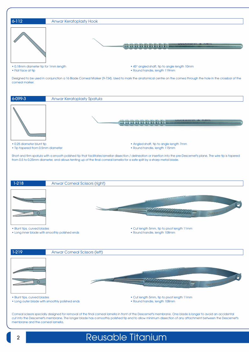
Anwar Corneal Scissors (right)
• Blunt tips, curved blades • Long inner blade with smoothly polished ends
1-218
• Cut length 5mm, tip to pivot length 11mm • Round handle, length 108mm
Anwar Keratoplasty Hook
• 0.18mm diameter tip for 1mm length • Flat face at tip
6-112
• 45° angled shaft, tip to angle length 10mm • Round handle, length 119mm
Designed to be used in conjunction a 16 Blade Corneal Marker (9-734). Used to mark the anatomical centre on the cornea through the hole in the crossbar of the corneal marker.
Anwar Keratoplasty Spatula
• 0.25 diameter blunt tip • Tip tapered from 0.5mm diameter
6-099-3
• Angled shaft, tip to angle length 7mm • Round handle, length 115mm
Short and firm spatula with a smooth polished tip that facilitates lamellar dissection / delineation or insertion into the pre-Descemet's plane. The wire tip is tapered from 0.5 to 0.25mm diameter, and allows tenting up of the final corneal lamella for a safe split by a sharp metal blade.
Anwar Corneal Scissors (left)
• Blunt tips, curved blades • Long outer blade with smoothly polished ends
1-219
• Cut length 5mm, tip to pivot length 11mm • Round handle, length 108mm
Corneal scissors specially designed for removal of the final corneal lamella in front of the Descemet's membrane. One blade is longer to avoid an accidental cut into the Descemet's membrane. The longer blade has a smoothly polished tip end to allow minimum dissection of any attachment between the Descemet's membrane and the corneal lamella.

3Surgical Instruments
Additional Instruments
www.duckworth-and-kent.com
For more information please contact us via our online enquiry form.
Or visit our website:
Daya Lamellar Spear
• 1.3mm width curved blade • 0.15mm thickness at tip • 35° angled curved shaft, tip to angle length 10.5mm • Round handle, length 130mm
6-604
• 1.8mm width curved blade • 0.15mm thickness at tip, 1mm at base of shaft • 35° angled curved shaft, tip to angle length 12mm
Morlet Lamellar Knife / Dissector
Dissector • 0.35mm x 2mm curved • Angled shafts 12mm tip to curve
6-607
Knife • 0.1mm x 1.5mm with sharp edges • Tip to angle length 3mm • Round handle, length 111mm
Combined Paufique knife and lamellar corneal dissector. Paufique knife used for starting lamellar corneal dissection and also to extend while peeling back superficial corneal tissue. Also used for undermining the periphery of host lamellar corneal bed which helps to prevent development of a step at the anterior host donor junction when implanting a donor lamellar that is thicker than excised host lamellar. Paufique knife also used to for removing lamellar host tissue down to level of Descemet's membrane. Lamellar corneal dissector is used to create a lamellar corneal plane via a peripheral corneal pocket or to widely extend a lamellar dissection that has been started with Paufique knife. Designed to separate lamellae and to stay within a plane. Corneal lamellae can be rapidly seperated with this instrument without the need for lifting and turning back lamellar flap. After separating the layers, lamellar corneal button may be excised with scissors.

D&K® is a registered trademark. All other brand names are trademarks or registered trademarks of their respective owners. All schematic line drawings, photographs and copy in this leaflet are fully protected by copyright. No part of this leaflet may be reproduced in any form without prior written permission. We reserve the right to make changes at any time, without notice, in product specifications and availability. Descriptive, typographic, or photographic errors are subject to correction. Name(s) of instruments are often comprised of surgeon’s name, combination of surgeons’ names or by the category of the instrument.
Email: [email protected] Tel: +44 (0)1462 893254Fax: +44 (0)1462 896288
www.duckworth-and-kent.com
If you are interested in any of our product range or have a general enquiry please contact anyone of our dedicated customer service team members who are ready to help.
Customer Services Customer Services Supervisor
NickyKellyMaria
Find us on
duckworth-and-kent.co.uk
nickyguthrie@
kellyobrien@
mariagentle@
© September 2006 Duckworth & KentRevised 21.03.18
Find us on
Visit : www.duckworth-and-kent.com to view over 800 ophthalmic titanium instruments
As well as product information for over 800 instruments, you will also �nd detailed literature and informative videos showing some of our instruments in action.