Embed Size (px)
Citation preview
Optimising deep anterior lamellar keratoplasty(DALK) using intraoperative online optical coherencetomography (iOCT)Philipp Steven,1 Carolin Le Blanc,1 Eva Lankenau,2 Marc Krug,2 Stefan Oelckers,3
Ludwig M Heindl,1 Uta Gehlsen,1 Gereon Huettmann,4 Claus Cursiefen1
1Department of Ophthalmology,University of Cologne, Cologne,Germany2OptoMedical TechnologiesGmbH, Luebeck, Germany3Moeller-Wedel GmbH, Wedel,Germany4Institute of Biomedical Optics,University of Luebeck, Luebeck,Germany
*Correspondence to DrPhilipp Steven, Department ofOphthalmology, UniversityHospital of Cologne, KerpenerStrasse 62, Cologne 50937,Germany; [email protected]
Received 5 November 2013Revised 6 January 2014Accepted 19 January 2014
To cite: Steven P, LeBlanc C, Lankenau E, et al.Br J Ophthalmol PublishedOnline First: [please includeDay Month Year]doi:10.1136/bjophthalmol-2013-304585
ABSTRACTBackground/aims To describe the use ofintraoperative online optical coherence tomography(iOCT) for improving deep anterior lamellar keratoplasty(DALK) surgery.Methods Retrospective case series of 6 eyes of 6 malepatients with keratokonus, corneal dystrophy or herpeticstromal scars undergoing DALK were investigated usingintraoperative optical coherence tomography andpostsurgical image/video analysis. Main outcomemeasures were: visibility of surgical steps, especially,assessment of placement depth of injection needle,preparation of bare Descemet’s membrane and drainageof interface fluid.Results iOCT enables real-time visualisation of allsurgical steps of DALK procedure in all patients.Placement of air injection needle above Descemet’smembrane was reliably monitored as was presence ofbare Descemet’s membrane and potential interface fluid.Conclusions iOCT assists with visualisation of injectionneedle placement and with assessment of bareDescemet’s membrane as well as interface fluid duringthe DALK procedure. Overall iOCT may be a helpfuldevice that supports surgeons in all steps of DALKprocedure.
INTRODUCTIONDeep anterior lamellar keratoplasty (DALK) usingthe big-bubble technique was first described byAnwar and Teichmann.1 DALK uses a stromal air-injection for dissection of stromal tissue fromDescemet’s membrane (DM) followed by engraft-ment of a stromal donor button, and is performedfor treatment of corneal stromal diseases such as ker-atokonus, hereditary corneal degeneration or cornealscarring. This technique has demonstrated advan-tages over perforating keratoplasty (PKP), such asabsent endothelial transplant rejections2–4 andreduced postoperative complications. The mostimportant limiting factor for visual outcome inDALK is incomplete stromal dissection.5
Nonetheless, the American Academy ofOphthalmology in a recent technology assessmentreport suggests DALK to be as good as PKP in termsof visual acuity, but better in terms of safety becauseof absent risk of endothelial immune reactions.6
Adaption of DALK worldwide is slow, partlybecause of the technical challenges of this surgeryand its non-standardised nature. That is partlyrelated to the limited visualisation of key surgicalsteps during surgery which is based on tissue trans-parency and hindered estimation of depth ratio due
to en-face view onto the cornea through the oper-ation microscope. Therefore, optical cross-sectionswould allow the surgeon to better navigate andcontrol the procedure, especially crucial steps, suchas assessment of depth of intrastromal needle place-ment, achievement of pure DM preparation andabsence of interface fluid at the end of surgery.Recently, optical coherence tomography (OCT)
has been modified to be used in an intraoperativesetting.7 Hereby, two main approaches are possible:(1) using hand-held devices8 9 or (2) integration ofthe OCT into the operation microscope.10 Thelatter has several advantages, such as online visual-isation of all surgical steps without the necessity ofinterrupting the procedure and alignment of theOCT-image to any given zoom and focus-step ofthe microscope. This study is the first to evaluateintraoperative OCT (iOCT) technology to visualiseDALK surgery online without the necessity of dis-continuing the procedure for OCT measurements.
METHODSIntraoperative online OCT technologyFor intraoperative online OCT evaluation, amicroscope-mounted, commercially availablespectral-domain OCT camera (iOCT, OptoMedicalTechnologies GmbH, Luebeck, Germany) using a840 nm central wavelength and performing10 000 A-scans/s was used. The iOCT was con-nected to the camera port of an OCT-compatibleMOELLER Hi-R 900 A near infrared microscope(Moeller-Wedel, Wedel, Germany).11 The OCTimage was displayed on a separate touch screen infront of the surgeon’s visual field to enable easyexchange between microscopic and OCT images.iOCT imaging included recording of high-resolution videos and images of approximately10 μm axial resolution in air. Image size was4.2 mm axially in air, respectively, 3.2 mm axiallyin water, and between 5 and 29 mm in lateral direc-tion, depending on the microscopic zoom factorused.
PatientsiOCTwas used in six consecutive male patients aged25–64 years, suffering from keratokonus (3 cases),stromal scarring (2 cases) or hereditary cornealstromal dystrophy Francois (1 case) (table 1).In two of the cases, DALK had to be converted
to penetrating keratoplasty due to larger DM per-foration during lamellar preparation.Evaluation parameters included iOCT-visualisation
of trephination depth, deep stromal placement of air
Steven P, et al. Br J Ophthalmol 2014;0:1–5. doi:10.1136/bjophthalmol-2013-304585 1
Clinical science BJO Online First, published on March 3, 2014 as 10.1136/bjophthalmol-2013-304585
Copyright Article author (or their employer) 2014. Produced by BMJ Publishing Group Ltd under licence.
on 18 July 2018 by guest. Protected by copyright.
http://bjo.bmj.com
/B
r J Ophthalm
ol: first published as 10.1136/bjophthalmol-2013-304585 on 3 M
arch 2014. Dow
nloaded from
injection needle, preparation of bare DM, interface fluid and inter-face alignment of the graft.
ProcedureDALK was performed as described previously12 13 Briefly, therecipient cornea was trephined to 90% of minimal cornealthickness at the 8 mm paracentral region using aHessberg-Barron trephine (Domilens, Hamburg) of 7.75 mm.Thickness was preoperatively assessed using Pentacam. A30-gauge injection needle was placed in deep tissue directlyabove DM, and the placement of the needle tip was observedusing intraoperative OCT. Then, air was injected until theborder of the whitening expanded towards the trephinationinterface. Following lamellar dissection of the superficial andmid-stromal tissue a pre-Descemet large air cavity (big-bubble)was opened and the remaining tissue was dissected from DM toobtain a bare DM situation under viscoelastic protection(injected into the space). The donor tissue was placed endothe-lial side up into a Hanna punch block (Moria, Doylestown,Pennsylvania, USA) and DM was stripped from the cornea. DMgrafts could then be used for Descemet membrane endothelialkeratoplasty (DMEK) surgery in a split cornea transplantation.13
An 8 mm diameter graft was cut (using a Hessberg-Barron tre-phine) and transferred onto the recipient’s cornea. The graftwas fixated by 16 single sutures or two double running continu-ous sutures, and remaining interface fluid was drained by gentlemassage of the corneal surface and opening of interface using ablunt spatula.
Donor tissueFour grafts were organ-cultured in minimal essential medium(MEM) (Biochrom, Berlin, Germany) at 32°C and deswelled inMEM containing 5% dextran (Biochrom, Berlin, Germany) at32°C.
Two grafts were organ-cultured in minimal essential medium(Cornea max R, Eurobio Laboratoires, Les Ulis, France) at 31°Cand deswelled in minimal essential medium containing 5–6%dextran (Cornea Jet R, Eurobio Laboratoires, Les Ulis, France).
No information was obtainable on exact postmortem timefrom the provider eye bank, however, due to legal requirements,all donor eyes were collected within 24 h postmortem. Donorage range from 18–72 years, (4 male, 2 female), no previous eyediseases were reported.
Retrospective image analysisFor retrospective image analysis, and to conduct depth measure-ments, the corresponding pixel size of the x-axis of the OCTimage was set to 840 pixels, according to the axial opticalwindow depth of 4.2 mm. Therefore, the axial pixel size was set
to 5 mm. To correct the index of refraction inside corneal tissueof 1.35, the pixel size was divided by this factor resulting in avalue of 3.7 mm. Using Image J, the distances between thecorneal epithelium, endothelium and the centre of the air-injection needle were measured, and the numbers of pixels con-verted to the true distance in mm.
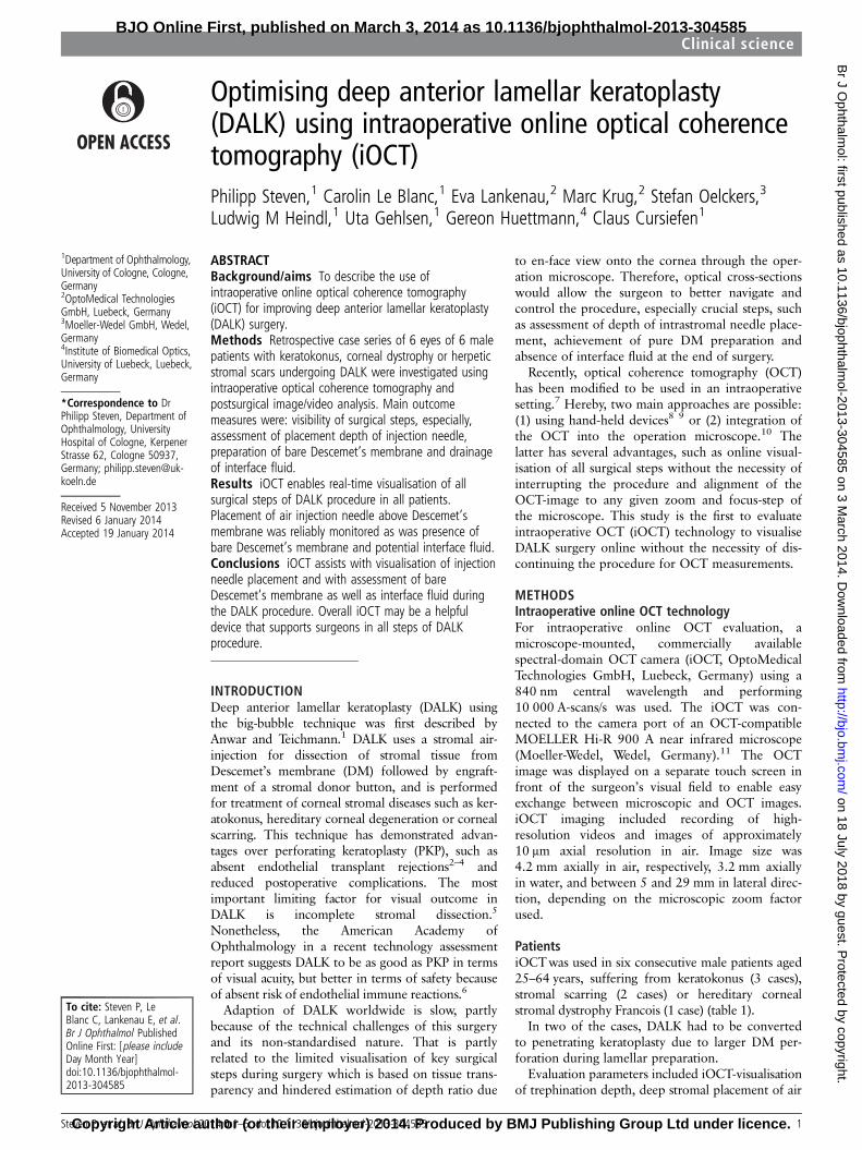
RESULTSIntraoperative OCT enables monitoring of all surgical stepsof DALK in all patients analysed (6/6: 100%)Trephination depth was accurately imaged (Figure 1A), andneedle insertion could be reliably monitored in close proximityto DM (Figure 1B). Air injection into the posterior stroma wasobserved and resulted in whitish staining of the OCT image dueto increased tissue scattering by air bubbles.14 In two out of sixcases, a big bubble formation was visible (Figure 1C) in deepstromal location. Superficial and deep stromal tissue preparation(Figure 1D,E) was monitored reliably until bare DM wasobtained (Figure 1F). Graft insertion and suturing was moni-tored, and interface congruence was controlled by intraoperativeOCT (Figure 1G). These steps were reliably monitored in 6/6patients, however, in two cases, rupture of DM during deepstromal preparation led to conversion into PKP. In these twocases, no initial big-bubble formation was achieved.
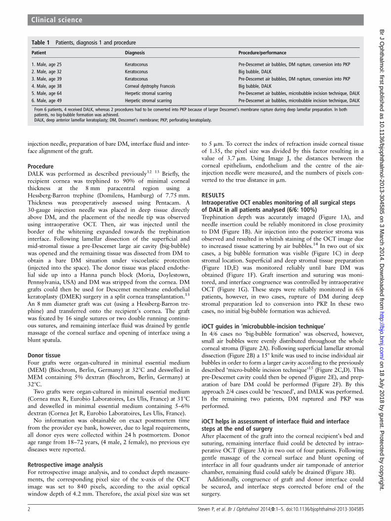
iOCT guides in ‘microbubble-incision technique’In 4/6 cases no ‘big-bubble formation’ was observed, however,small air bubbles were evenly distributed throughout the wholecorneal stroma (Figure 2A). Following superficial lamellar stromaldissection (Figure 2B) a 15° knife was used to incise individual airbubbles in order to form a larger cavity according to the previouslydescribed ‘micro-bubble incision technique’15 (Figure 2C,D). Thispre-Descemet cavity could then be opened (Figure 2E), and prep-aration of bare DM could be performed (Figure 2F). By thisapproach 2/4 cases could be ‘rescued’, and DALK was performed.In the remaining two patients, DM ruptured and PKP wasperformed.
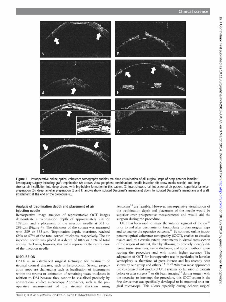
iOCT helps in assessment of interface fluid and interfacesteps at the end of surgeryAfter placement of the graft into the corneal recipient’s bed andsuturing, remaining interface fluid could be detected by intrao-perative OCT (Figure 3A) in two out of four patients. Followinggentle massage of the corneal surface and blunt opening ofinterface in all four quadrants under air tamponade of anteriorchamber, remaining fluid could safely be drained (Figure 3B).
Additionally, congruence of graft and donor interface couldbe secured, and interface steps corrected before end of thesurgery.
Table 1 Patients, diagnosis 1 and procedure
Patient Diagnosis Procedure/performance
1. Male, age 25 Keratoconus Pre-Descemet air bubbles, DM rupture, conversion into PKP2. Male, age 32 Keratoconus Big bubble, DALK3. Male, age 39 Keratoconus Pre-Descemet air bubbles, DM rupture, conversion into PKP4. Male, age 38 Corneal dystrophy Francois Big bubble, DALK5. Male, age 64 Herpetic stromal scarring Pre-Descemet air bubbles, microbubble incision technique, DALK6. Male, age 49 Herpetic stromal scarring Pre-Descemet air bubbles, microbubble incision technique, DALK
From 6 patients, 4 received DALK, whereas 2 procedures had to be converted into PKP because of larger Descemet’s membrane rupture during deep lamellar preparation. In bothpatients, no big-bubble formation was achieved.DALK, deep anterior lamellar keratoplasty; DM, Descemet’s membrane; PKP, perforating keratoplasty.
2 Steven P, et al. Br J Ophthalmol 2014;0:1–5. doi:10.1136/bjophthalmol-2013-304585
Clinical science
on 18 July 2018 by guest. Protected by copyright.
http://bjo.bmj.com
/B
r J Ophthalm
ol: first published as 10.1136/bjophthalmol-2013-304585 on 3 M
arch 2014. Dow
nloaded from
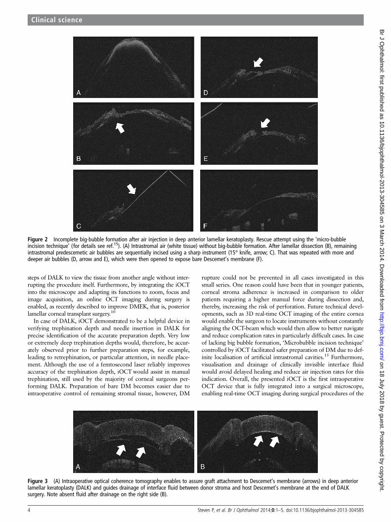
Analysis of trephination depth and placement of airinjection needleRetrospective image analyses of representative OCT imagesdemonstrate a trephination depth of approximately 270 or198 mm, and a placement of the injection needle at 311 or296 mm (Figure 4). The thickness of the cornea was measuredwith 389 or 333 mm. Trephination depth, therefore, reached69% or 67% of the total corneal thickness, respectively. The airinjection needle was placed at a depth of 80% or 88% of totalcorneal thickness; however, this value represents the centre coreof the injection needle.
DISCUSSIONDALK is an established surgical technique for treatment ofstromal corneal diseases, such as keratoconus. Several prepar-ation steps are challenging such as localisation of instrumentswithin the stroma or estimation of remaining tissue thickness inrelation to DM because they cannot be visualised precisely byconventional en-face microscopy. Approaches, such as the pre-operative measurement of the stromal thickness using
Pentacam16 are feasible. However, intraoperative visualisation ofthe trephination depth and placement of the needle would besuperior over preoperative measurements and would aid thesurgeon during the procedure.
OCT has been used to image the anterior segment of the eye17
prior to and after deep anterior keratoplasty to plan surgical stepsand to analyse the operative outcome.18 By contrast, online intrao-perative optical coherence tomography (iOCT), enables to visualisetissues and, to a certain extent, instruments in virtual cross-sectionof the region of interest, thereby allowing to precisely identify dif-ferent tissue structures, tissue thickness, and so on, without inter-rupting the procedure and with much higher accuracy. Theadaptation of OCT for intraoperative use, in particular, in lamellarkeratoplasty is, therefore, of great interest and has recently beenshown by our group and others.7 8 19 20 Whereas most approachesuse customised and modified OCT systems so far used in patientsbefore or after surgery18 or slit beam imaging21 during surgery withthe necessity to interrupt the procedure, this iOCT-system is thefirst device that was specifically developed to be mounted on a sur-gical microscope. This allows especially during delicate surgical
Figure 1 Intraoperative online optical coherence tomography enables real-time visualisation of all surgical steps of deep anterior lamellarkeratoplasty surgery including graft trephination (A; arrows show peripheral trephination), needle insertion (B; arrow marks needle) into deepstroma, air insufflation into deep stroma with big-bubble formation in this patient (C; inset shows small intrastromal air pocket), superficial lamellarpreparation (D), deep lamellar preparation (E and F; arrows show isolated Descemet’s membrane) down to isolated Descemet’s membrane and graftattachment at the end of the procedure (G).
Steven P, et al. Br J Ophthalmol 2014;0:1–5. doi:10.1136/bjophthalmol-2013-304585 3
Clinical science
on 18 July 2018 by guest. Protected by copyright.
http://bjo.bmj.com
/B
r J Ophthalm
ol: first published as 10.1136/bjophthalmol-2013-304585 on 3 M
arch 2014. Dow
nloaded from
steps of DALK to view the tissue from another angle without inter-rupting the procedure itself. Furthermore, by integrating the iOCTinto the microscope and adapting its functions to zoom, focus andimage acquisition, an online OCT imaging during surgery isenabled, as recently described to improve DMEK, that is, posteriorlamellar corneal transplant surgery.10
In case of DALK, iOCT demonstrated to be a helpful device inverifying trephination depth and needle insertion in DALK forprecise identification of the accurate preparation depth. Very lowor extremely deep trephination depths would, therefore, be accur-ately observed prior to further preparation steps, for example,leading to retrephination, or particular attention, in needle place-ment. Although the use of a femtosecond laser reliably improvesaccuracy of the trephination depth, iOCTwould assist in manualtrephination, still used by the majority of corneal surgeons per-forming DALK. Preparation of bare DM becomes easier due tointraoperative control of remaining stromal tissue, however, DM
rupture could not be prevented in all cases investigated in thissmall series. One reason could have been that in younger patients,corneal stroma adherence is increased in comparison to olderpatients requiring a higher manual force during dissection and,thereby, increasing the risk of perforation. Future technical devel-opments, such as 3D real-time OCT imaging of the entire corneawould enable the surgeon to locate instruments without constantlyaligning the OCT-beam which would then allow to better navigateand reduce complication rates in particularly difficult cases. In caseof lacking big bubble formation, ‘Microbubble incision technique’controlled by iOCT facilitated safer preparation of DM due to def-inite localisation of artificial intrastromal cavities.15 Furthermore,visualisation and drainage of clinically invisible interface fluidwould avoid delayed healing and reduce air injection rates for thisindication. Overall, the presented iOCT is the first intraoperativeOCT device that is fully integrated into a surgical microscope,enabling real-time OCT imaging during surgical procedures of the
Figure 2 Incomplete big-bubble formation after air injection in deep anterior lamellar keratoplasty. Rescue attempt using the ‘micro-bubbleincision technique’ (for details see ref.15). (A) Intrastromal air (white tissue) without big-bubble formation. After lamellar dissection (B), remainingintrastromal predescemetic air bubbles are sequentially incised using a sharp instrument (15° knife, arrow; C). That was repeated with more anddeeper air bubbles (D, arrow and E), which were then opened to expose bare Descemet’s membrane (F).
Figure 3 (A) Intraoperative optical coherence tomography enables to assure graft attachment to Descemet’s membrane (arrows) in deep anteriorlamellar keratoplasty (DALK) and guides drainage of interface fluid between donor stroma and host Descemet’s membrane at the end of DALKsurgery. Note absent fluid after drainage on the right side (B).
4 Steven P, et al. Br J Ophthalmol 2014;0:1–5. doi:10.1136/bjophthalmol-2013-304585
Clinical science
on 18 July 2018 by guest. Protected by copyright.
http://bjo.bmj.com
/B
r J Ophthalm
ol: first published as 10.1136/bjophthalmol-2013-304585 on 3 M
arch 2014. Dow
nloaded from

eye. As shown for posterior lamellar keratoplasty (DMEK).10
iOCTalso demonstrates benefits for anterior lamellar keratoplasty(DALK) and may aid surgeons, in particular, by steepening thelearning curve at the beginning of surgical training in lamellar ker-atoplasty. In comparison with other imaging approaches, intrao-perative OCT still has some limitations, such as higher costs, fixedangle and single scan direction with only limited degree of rotation.However, in challenging situations such as reduced visibility, uncer-tain tissue depth during lamellar preparation or failed big-bubbleformation iOCTenables precise evaluation of the situation and pos-sible ‘rescue-measures’ for successful completion of the procedurewithout the need of additional staff for handling hand-held devicesand without the need to interrupt the procedure. Future develop-ments such as 3D images of the entire surgical field and automatedintraoperative depths measurements, together with larger prospect-ive trials, will have to evaluate how much this technology canimprove outcome measures in big-bubble DALK surgery.
Acknowledgements The authors would like to acknowledge the superb technicalwork of S Hackbarth and G Simons from the Cornea Bank, University Hospital ofCologne.
Contributors All authors have contributed either in the design or conduct of thestudy or performed the retrospective data analysis and writing of the manuscript.
Funding Deutsche Forschungsgemeinschaft (German Research Council: SFB 643(B10), STE 1928/2-1, CU 47/4-1). Ruth und Helmut Lingen Stiftung, Köln. EuropeanUnion FP7: COST BM 1302. Koeln Fortune Research Funding, Faculty of Medicine,University of Cologne. The sponsor or funding organisation had no role in thedesign or conduct of this research.
Competing interests EL and MK are employees of OptoMedical TechnologiesGmbH, SO is employee of Moeller-Wedel GmbH, EL, MK and SO are shareholders ofOptoMedical Technologies.
Ethics approval
Provenance and peer review Not commissioned; externally peer reviewed.
Data sharing statement Data will be available on request by the correspondingauthor.
Open Access This is an Open Access article distributed in accordance with theCreative Commons Attribution Non Commercial (CC BY-NC 3.0) license, whichpermits others to distribute, remix, adapt, build upon this work non-commercially,and license their derivative works on different terms, provided the original work isproperly cited and the use is non-commercial. See: http://creativecommons.org/licenses/by-nc/3.0/
REFERENCES1 Anwar M, Teichmann KD. Big-bubble technique to bare Descemet’s membrane in
anterior lamellar keratoplasty. J Cataract Refract Surg 2002;28:398–403. Epub2002/04/26.
2 Cursiefen C, Heindl LM. [Perspectives of deep anterior lamellar keratoplasty]. DerOphthalmologe: Zeitschrift der Deutschen Ophthalmologischen Gesellschaft2011;108:833–9.
3 Shimmura S, Tsubota K. Deep anterior lamellar keratoplasty. Curr Opin Ophthalmol2006;17:349–55.
4 Steven P, Hos D, Heindl LM, et al. [Immune reactions after DMEK, DSAEK andDALK]. Klinische Monatsblatter fur Augenheilkunde. 2013;230:494–9.
5 Fontana L, Parente G, Sincich A, et al. Influence of graft-host interface on thequality of vision after deep anterior lamellar keratoplasty in patients withkeratoconus. Cornea. 2011;30:497–502.
6 Reinhart WJ, Musch DC, Jacobs DS, et al. Deep anterior lamellar keratoplasty as analternative to penetrating keratoplasty a report by the american academy ofophthalmology. Ophthalmology 2011;118:209–18.
7 Mueller M, Steven P, Lankenau E, et al. Intraoperative OCT (iOCT) for Anterior andPosterior Segment Surgery. ARVO: IOVS; 2010.
8 Scorcia V, Busin M, Lucisano A, et al. Anterior segment optical coherencetomography-guided big-bubble technique. Ophthalmology 2013;120:471–6.
9 Wykoff CC, Berrocal AM, Schefler AC, et al. Intraoperative OCT of a full-thicknessmacular hole before and after internal limiting membrane peeling. Ophthalmic SurgLasers Imaging 2010;41:7–11.
10 Steven P, Le Blanc C, Velten K, et al. Optimizing descemet membrane endothelialkeratoplasty using intraoperative optical coherence tomography. JAMAophthalmology 2013;131:1135–42.
11 Lankenau E, Klinger D, Winter C, et al. Combining Optical Coherence Tomography(OCT) with an operating micoscope. In: Buzug TM, Holz D, Bongartz J, Kohl-BareisM, Hartmann U, Weber S, eds. Advances in medical engineering. Berlin, Heidelberg,New York: Springer; 2007;343–8.
12 Heindl LM, Riss S, Bachmann BO, et al. Split cornea transplantation for 2 recipients:a new strategy to reduce corneal tissue cost and shortage. Ophthalmology2011;118:294–301.
13 Heindl LM, Riss S, Laaser K, et al. Split cornea transplantation for 2 recipients—review of the first 100 consecutive patients. Am JOphthalmol 2011;152:523–32 e2.
14 Braun JM, Hofmann-Rummelt C, Schlotzer-Schrehardt U, et al. Histopathologicalchanges after deep anterior lamellar keratoplasty using the ‘big-bubble technique’.Acta Ophthalmol 2013;91:78–82.
15 Riss S, Heindl LM, Bachmann BO, et al. Microbubble incision as a new rescuetechnique for big-bubble deep anterior lamellar keratoplasty with failed bubbleformation. Cornea. 2013;32:125–9.
16 Riss S, Heindl LM, Bachmann BO, et al. Pentacam-based big bubble deep anteriorlamellar keratoplasty in patients with keratoconus. Cornea 2012;31:627–32.
17 Huttmann G, Lankenau E, Schulz-Wackerbarth C, et al. [Optical coherencetomography: from retina imaging to intraoperative use—a review]. KlinischeMonatsblatter fur Augenheilkunde 2009;226:958–64.
18 Yeh RY, Quilendrino R, Musa FU, et al. Predictive value of optical coherencetomography in graft attachment after Descemet’s membrane endothelialkeratoplasty. Ophthalmology. 2013;120:240–5.
19 Steven P, Heindl LM, Hos D, et al. Immune reactions after DMEK, DSAEK andDALK. Klin Monbl Augenheilkd 2013;230:494–9.
20 Lankenau E, Krug M, Oelckers S, et al. iOCT with surgical microscopes: a newimaging during microsurgery. Adv Opt Technol 2013;2:233–9.
21 Burkhart ZN, Feng MT, Price MO, et al. Handheld slit beam techniques to facilitateDMEK and DALK. Cornea. 2013;32:722–4.
Figure 4 Quantitative measurements of corneal thickness, depth of the air injection needle and trephination depth (A) Representative paracentraloptical coherence tomography (OCT) image of patient 5 with indicators for corneal thickness (1=389 mm), depth of the air injection needle(2=311 mm) and trephination depth (3=270 mm) (B) Representative paracentral OCT image of patient 2 with indicators for corneal thickness(1=333 mm), depth of the air injection needle (2=296 mm) and trephination depth (3=198 mm). The dotted line represents the posterior surface ofthe cornea.
Steven P, et al. Br J Ophthalmol 2014;0:1–5. doi:10.1136/bjophthalmol-2013-304585 5
Clinical science
on 18 July 2018 by guest. Protected by copyright.
http://bjo.bmj.com
/B
r J Ophthalm
ol: first published as 10.1136/bjophthalmol-2013-304585 on 3 M
arch 2014. Dow
nloaded from