Embed Size (px)
Citation preview
Turkish J. Pharm. Sci. 3 (1), 7-18, 2006
ANTIDIABETIC and ANTIOXIDANT EFFECTS of Vitis vinifera L. LEAVES in STREPTOZOTOCIN-DIABETIC
RATS
Nilüfer ŞENDOĞDU, Mustafa ASLAN,
Didem DELİORMAN ORHAN, Fatma ERGUN*, Erdem YEŞİLADA
Gazi University, Faculty of Pharmacy, Department of Pharmacognosy,
06330 Etiler-Ankara, TURKEY
Abstract In the present study the acute and subacute antidiabetic activities of the ethanolic extract of Vitis
vinifera L. leaves were investigated. The acute effect was studied on the normoglycaemic, glucose hyperglycaemic and streptozotocin-induced d iabetic rats; and the subacute effect was studied on same diabetic rats for 15 days. The blood glucose levels were measured by using blood glucose measuring strips based on glucose-oxidase method. After the subacute administration, the antioxidant activity of the extracts was investigated on the liver, kidney and the heart tissues of the experimental animals by measuring the tissue GSH and MDA levels. All of the antioxidant parameters were compared with the diabetic control group. According to the obtained data, the ethanolic extract of V. vinifera leaves at 250 mg/kg dose was found to possess a high antidiabetic and antioxidant activity. Mainly condensed tannins and flavonoids were suggested to contribute in the activities.
Key Words: Vitis vinifera, antidiabetic, hypoglycaemia, antioxidant, lipid peroxidation, MDA, GSH
Vitis vinifera L. Yapraklarının Streptozotosin ile Diabet Yapılmış Sıçanlarda
Antidiabetik ve Antioksidan Etkileri
Bu çalışmada Vitis vinifera L. yapraklarının etanolik ekstresinin akut ve subakut aktivitesi incelenmiştir. Akut etkinin değerlendirilebilmesi için normoglisemik, glukoz hiperglisemik ve streptozotosin ile diyabet oluşturulmuş sıçanlar kullanılmıştır. Subakut etki ise 15 gün boyunca streptozotosin ile diyabet oluşturulmuş sıçanlar üzerinde incelenmiştir. Kan glukoz seviyeleri kan glikoz test çubukları kullanılarak glukoz oksidaz metodu ile ölçülmüştür. Subakut uygulama sonrası ekstrelerin antioksidan aktiviteleri karaciğer, böbrek ve kalp dokularının GSH ve MDA seviyeleri ölçülerek değerlendirilmiştir. Antioksidan parametrelerin hepsi diyabetik kontrol grubu ile karşılaştırılmıştır. V. vinifera yapraklarının etanol ekstresi 250 mg/kg dozda antidiyabetik ve antioksidan aktivite göstermiştir. Aktiviteden başlıca kondanse tanenlerin ve flavonoitlerin sorumlu olduğu düşünülmektedir.
Anahtar Kelimeler: Vitis vinifera, antidiyabetik, hipoglisemi, antioksidan, lipit peroksidasyon, MDA,
GSH.
* Corresponding author: Fax: + 90-312-2235018, e-mail: [email protected]
�
N. Şendoğdu, M. Aslan, D. Deliorman, F. Ergun, E. Yeşilada
INTRODUCTION
Vitis vinifera L. (Vitaceae) is a perennial, woody vine, usually climbing by tendrils (1). Since
ancient times, the different parts of this plant have been used because of many biological activities
in folk medicine. The leaves of plant are rich in tannins, flavonoids and procyanidins. Additionally,
the leaves also contain organic acids, lipids, enzymes and vitamins (2-4). Grape leaves have been
used to stop bleeding, inflammation, and pain, such as the kind brought on by hemorrhoids in the
traditional medicine (2, 5).
It is observed that in some regions of Anatolia, the leaves of V. vinifera have been used for
reducing the blood glucose levels in diabetics.
Diabetes is a major worldwide health problem predisposing to markedly increased
cardiovascular mortality and serious morbidity and mortality related to the development of
nephropathy, neuropathy and retinopathy. Diabetes mellitus is characterized by abnormal insulin
secretion, derangement in carbohydrate and lipid metabolism, and is diagnosed by presence of
hyperglycemia (6).
Oxidative stress which is defined as an imbalance between the generation of oxidants and
antioxidant defence capacity of the body is suggested as a mechanism underlying diabetes and
diabetic complications like many other diseases (7). Several mechanisms seem to be involved in
the genesis of oxidative stress in both diabetic patients and diabetic experimental animals like
glucose autooxidation, protein glycation, formation of advanced glycation products and the polyol
pathway (8). There is also an evidence that higher glucose concentration causes a depression on
natural antioxidant defence agents such as glutathione or vitamin C (9). The increase in the levels
of reactive oxygen species and free radicals cause damage in the biological structures such as cell
wall, genetic material and enzymes. These oxidants also cause microvascular and macrovascular
complications, cardiovascular diseases, kidney and nerve damage (10).
The aim of this study was to evaluate both the antidiabetic activity of the ethanolic extract
of V. vinifera leaves on normoglycaemic, hyperglycaemic, streptozotocin-induced diabetic rats,
and the antioxidant activity by measuring the levels of non-protein sulphydryl groups (GSH) and
malondialdehyde (MDA) in the liver, kidney and heart tissues of the diabetic rats.
EXPERIMENTAL
Material
The plant material was collected from the Viticultural Research Institute, Faculty of Agriculture,
Ankara University (Kalecik) before blooming. The plant was identified and the voucher specimens
(GÜEF 2320, GÜEF 2321) are deposited in the Herbarium of Gazi University Faculty of
Pharmacy. Plant materials were dried under shade and powdered coarsely before extraction.
�
Turkish J. Pharm. Sci. 3 (1), 7-18, 2006
Preparation of Extract
The dried and powdered leaves of V. vinifera (500 g) were extracted with ethanol 80 % at room
temperature (5 L X 6 times). Combined ethanol extract was evaporated to dryness under reduced
pressure below 40oC (EtOH Extract, yield 16.07 %).
TLC Analysis of Resveratrol
The ethanol extract of V.vinifera was administrated to TLC plate covered with Kieselgel 60F254
(Merck, 1.05554.001) to evaluate the presence of resveratrol in the ethanolic extract. Chloroform:
methanol (8:2) system was used as mobile phase. Sulphuric acid solution prepared in methanol
(5 %) was used to determine resveratrol (11).
Preparation of the test samples
The dried extract was suspended in 0.5% aqueous carboxymethylcellulose (CMC) suspension
in distilled water prior to oral administration to animals (10 ml/kg, b.w) [b.w.: body weight].
Tolbutamide (100 mg/kg, b.w.) was used as the reference drug. Animals in the control group
received only the vehicle (10 ml/kg, b.w.).
Animals
Male Wistar-albino rats (150-200 g) purchased from the Laboratories of Refik Saydam Central
Institute of Health (Ankara, TURKEY) were used in the experiments. Prior to the experiments,
rats were fed with standard food for one week in order to adapt to the laboratory conditions. 16 h
before the experiments, they were fasted overnight, but allowed free access to water. Six animals
were used for each group of study.
Determination of the blood glucose levels
Blood glucose concentrations (mg/100 ml) were determined using a Glucometer-elite
commercial test (Bayer), based on the glucose oxidase method. Blood samples were collected
from the tip of tail at the defined time patterns.
Studies with normal animals
Animals were divided into three groups and fasting blood sugar level of each was determined
at zero-time, after overnight fasting with free access to water. Control group of animal were given
0.5% CMC.
The other two groups were treated with the increasing doses of V. vinifera ethanolic extract,
250 mg/kg and 500mg/kg. Blood samples were collected at 0, 1/2, 1, 2, 4 h. after the oral
administration of test samples.
Studies with glucose-hyperglycaemic animals (Glucose loaded model, Oral glucose tolerance
test OGTT)
�
N. Şendoğdu, M. Aslan, D. Deliorman, F. Ergun, E. Yeşilada
Fasting blood sugar level of each rat was determined at zero-time, after overnight fasting
with free access to water. Glucose (2 mg/kg b.w) was orally administered 30 min after an oral
administration of the test sample. Blood glucose concentrations were measured just before and
1/2, 1, 2, 4 h. after the oral administration of the extract.
Effects on Streptozotocin-induced Diabetic Rats (Non insulin dependent diabetes model -
NIDDM)
Induction of diabetes
Diabetes was induced in rats by the intraperitoneal (i.p.) injection of streptozotocin (STZ) at
a dose of 55 mg/kg b.w. dissolved in citrate buffer (1 M, pH 4.5) (1 ml/kg) (12,13). Seven days
after the injection, the blood glucose levels were measured. Each animal with a blood glucose
level above 250 mg/dl was considered to be diabetic. In order to overcome the hypoglycemia
which occurred during the first 24 h following the STZ administration, diabetic rats were given
5% glucose solution orally. In all experiments, rats were fasted for 16 h prior to STZ injection.
Acute effect of test samples
The EtOH extract was administered orally by using a gastric gavage needle. Blood glucose
levels were determined at 30, 60, 120, and 240 min after the administration of the test samples.
Subacute effect of test samples
The EtOH extract was administered fifteen days consecutively. Blood glucose levels were
determined at 1st, 5th, 10th and 15th days after the administration of the test samples. The effect of
each test sample on body weight was also monitored at the same days. On 15th day, all animals
were sacrificed and then the kidney, liver and heart of each animal were removed for measurement
of tissue MDA and GSH levels.
Lipid peroxidation in liver, kidney and heart tissues
The method of Ohkawa et al. as modified by Jamall and Smith was used to determine lipid
peroxidation in tissue samples (14,15). Rats were sacrificed by an overdose of diethylether. The
liver of each rat was immediately excised and chilled in ice-cold 0.9% NaCl. After washing with
0.9% NaCl, 1.0 g of wet tissue was weighted exactly and homogenized in 9 ml of 0.25 M sucrose
using a Teflon homogenizer to obtain a 10% suspension. The cytosolic fraction was obtained by
a two-step centrifugation first at 1000x g for 10 min and then at 2000x g for 30 min at 4 0C. A
volume of the homogenate (0.20 ml) was transferred to a vial and was mixed with 0.2 ml of an
8.1% (w/v) Sodium dodecyl sulphate solution, 1.50 ml of a 0.8% (w/v) solution of TBA and the
final volume was adjusted to 4.0 ml with distilled water. Each vial was tightly capped and heated
in boiling water bath for 60 min. The vials were then cooled under running water.
Equal volumes of tissue blank or test sample and 10% TCA were transferred into a centrifuge
tube and centrifuged at 1000x g for 10 min. The absorbance of the supernatant fraction was
measured at 532 nm (Beckman DU 650 Spectrometer). Control experiment was processed using
10
Turkish J. Pharm. Sci. 3 (1), 7-18, 2006
the same experimental protocol except the TBA solution was replaced with distilled water due
to the peroxidative effect of streptozotocin on tissue: livers, kidneys, and hearts of STZ-diabetic
rats were used as positive control. 1,1,3,3-tetraethoxypropan was used as standard for calibration
of the curve.
Non-protein sulfhydryl groups (Cellular GSH) in liver, kidney, and heart tissues (16)
Liver (200 mg), heart (400 mg), and kidney (400 mg) were homogenized in 8.0 ml of 0.02 m
EDTA in an ice bath. The homogenates were kept in the ice bath until used. Aliquots of 5.0 ml of
the homogenates were mixed in 15.0 ml test tubes with 4.0 ml distilled water and 1.0 ml of 50%
trichloroacetic acid (TCA). The tubes were centrifuged for 15 min at approximately 3000x g 2.0
ml of supernatant was mixed with 4.0 ml of 0.4 M Tris buffer, pH 8.9, 0.1 ml Ellman’s reagent
[5,5’-dithiobis-(2-nitro-benzoic acid)] (DTNB) added, and the sample shaken. The absorbance
was read within 5 min of the addition of DTNB at 412 nm against a reagent blank with no
homogenate. Results were expressed as µmol GSH/g tissue.
Statistical analysis
Values are presented as means ± SEM. Statistical differences between the treatments and the
controls were tested by one-way analysis of variance (ANOVA) followed by the Student-Newman-
Keuls test using the “Instat” statistic computer program. A difference in the mean values of p<
0.05 was considered to be statistically significant.
RESULTS AND DISCUSSION
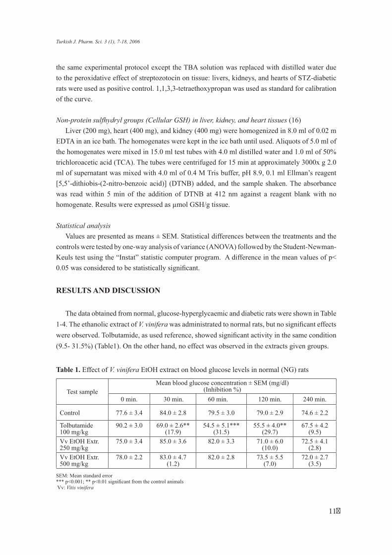
The data obtained from normal, glucose-hyperglycaemic and diabetic rats were shown in Table
1-4. The ethanolic extract of V. vinifera was administrated to normal rats, but no significant effects
were observed. Tolbutamide, as used reference, showed significant activity in the same condition
(9.5- 31.5%) (Table1). On the other hand, no effect was observed in the extracts given groups.
Table 1. Effect of V. vinifera EtOH extract on blood glucose levels in normal (NG) rats
Test sample Mean blood glucose concentration ± SEM (mg/dl)
(Inhibition %)
0 min. 30 min. 60 min. 120 min. 240 min.
Control 77.6 ± 3.4 84.0 ± 2.8 79.5 ± 3.0 79.0 ± 2.9 74.6 ± 2.2
Tolbutamide 90.2 ± 3.0 69.0 ± 2.6** 54.5 ± 5.1*** 55.5 ± 4.0** 67.5 ± 4.2 100 mg/kg (17.9) (31.5) (29.7) (9.5) Vv EtOH Extr. 75.0 ± 3.4 85.0 ± 3.6 82.0 ± 3.3 71.0 ± 6.0 72.5 ± 4.1 250 mg/kg (10.0) (2.8) Vv EtOH Extr. 78.0 ± 2.2 83.0 ± 4.7 82.0 ± 2.8 73.5 ± 5.5 72.0 ± 2.7 500 mg/kg (1.2) (7.0) (3.5)
SEM: Mean standard error *** p<0.001; ** p<0.01 significant from the control animalsVv: Vitis vinifera
11
N. Şendoğdu, M. Aslan, D. Deliorman, F. Ergun, E. Yeşilada
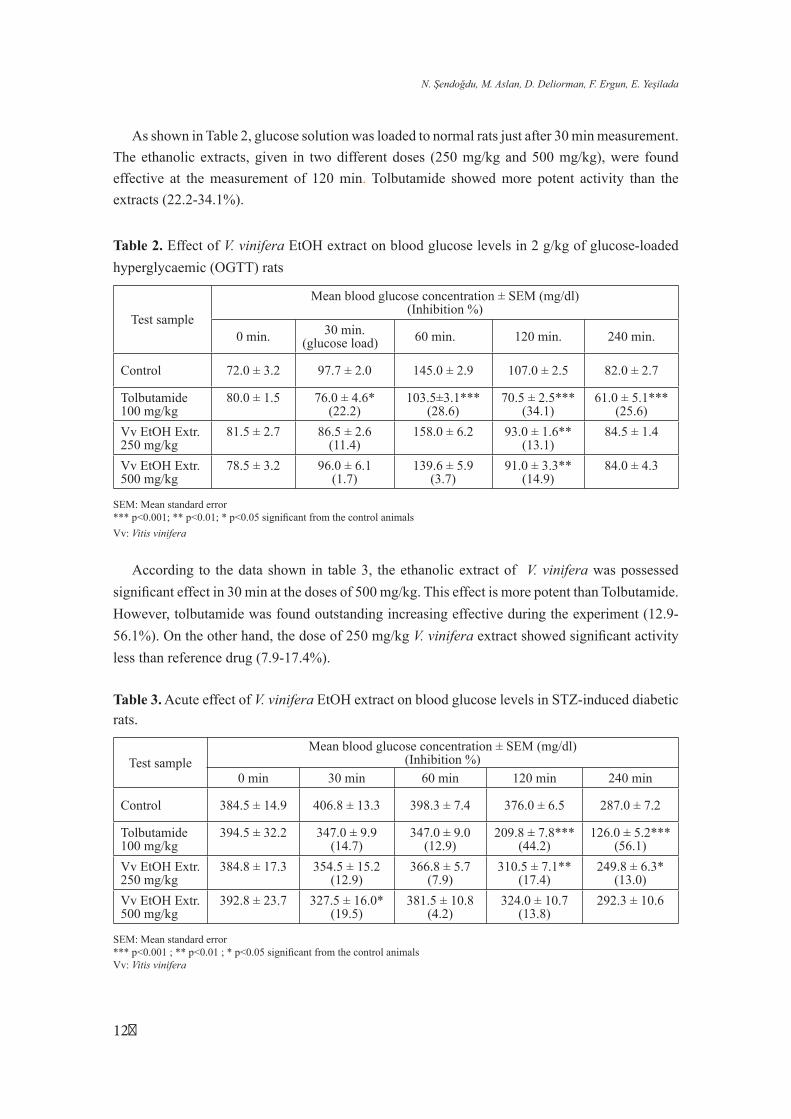
As shown in Table 2, glucose solution was loaded to normal rats just after 30 min measurement.
The ethanolic extracts, given in two different doses (250 mg/kg and 500 mg/kg), were found
effective at the measurement of 120 min. Tolbutamide showed more potent activity than the
extracts (22.2-34.1%).
Table 2. Effect of V. vinifera EtOH extract on blood glucose levels in 2 g/kg of glucose-loaded
hyperglycaemic (OGTT) rats
Mean blood glucose concentration ± SEM (mg/dl)
Test sample (Inhibition %)
0 min. 30 min. (glucose load) 60 min. 120 min. 240 min.
Control 72.0 ± 3.2 97.7 ± 2.0 145.0 ± 2.9 107.0 ± 2.5 82.0 ± 2.7
Tolbutamide 80.0 ± 1.5 76.0 ± 4.6* 103.5±3.1*** 70.5 ± 2.5*** 61.0 ± 5.1*** 100 mg/kg (22.2) (28.6) (34.1) (25.6)
Vv EtOH Extr. 81.5 ± 2.7 86.5 ± 2.6 158.0 ± 6.2 93.0 ± 1.6** 84.5 ± 1.4 250 mg/kg (11.4) (13.1)
Vv EtOH Extr. 78.5 ± 3.2 96.0 ± 6.1 139.6 ± 5.9 91.0 ± 3.3** 84.0 ± 4.3 500 mg/kg (1.7) (3.7) (14.9)
SEM: Mean standard error *** p<0.001; ** p<0.01; * p<0.05 significant from the control animals
Vv: Vitis vinifera
According to the data shown in table 3, the ethanolic extract of V. vinifera was possessed
significant effect in 30 min at the doses of 500 mg/kg. This effect is more potent than Tolbutamide.
However, tolbutamide was found outstanding increasing effective during the experiment (12.9-
56.1%). On the other hand, the dose of 250 mg/kg V. vinifera extract showed significant activity
less than reference drug (7.9-17.4%).
Table 3. Acute effect of V. vinifera EtOH extract on blood glucose levels in STZ-induced diabetic rats.
Test sample Mean blood glucose concentration ± SEM (mg/dl)
(Inhibition %) 0 min 30 min 60 min 120 min 240 min
Control 384.5 ± 14.9 406.8 ± 13.3 398.3 ± 7.4 376.0 ± 6.5 287.0 ± 7.2
Tolbutamide 394.5 ± 32.2 347.0 ± 9.9 347.0 ± 9.0 209.8 ± 7.8*** 126.0 ± 5.2*** 100 mg/kg (14.7) (12.9) (44.2) (56.1)
Vv EtOH Extr. 384.8 ± 17.3 354.5 ± 15.2 366.8 ± 5.7 310.5 ± 7.1** 249.8 ± 6.3* 250 mg/kg (12.9) (7.9) (17.4) (13.0)
Vv EtOH Extr. 392.8 ± 23.7 327.5 ± 16.0* 381.5 ± 10.8 324.0 ± 10.7 292.3 ± 10.6 500 mg/kg (19.5) (4.2) (13.8)
SEM: Mean standard error *** p<0.001 ; ** p<0.01 ; * p<0.05 significant from the control animalsVv: Vitis vinifera
12
Turkish J. Pharm. Sci. 3 (1), 7-18, 2006
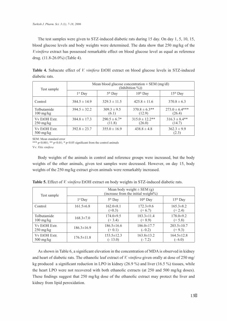
The test samples were given to STZ-induced diabetic rats during 15 day. On day 1, 5, 10, 15,
blood glucose levels and body weights were determined. The data show that 250 mg/kg of the
V.vinifera extract has possessed remarkable effect on blood glucose level as equal as reference
drug. (11.8-26.0%) (Table 4).
Table 4. Subacute effect of V. vinifera EtOH extract on blood glucose levels in STZ-induced
diabetic rats.
Test sample Mean blood glucose concentration ± SEM (mg/dl)
(Inhibition %))
1st Day 5th Day 10th Day 15th Day
Control 384.5 ± 14.9 329.3 ± 11.5 425.8 ± 11.6 370.8 ± 6.3
Tolbutamide 394.5 ± 32.2 309.3 ± 9.5 370.8 ± 6.3** 273.0 ± 6.4*** 100 mg/kg (6.1) (12.9) (26.4)
Vv EtOH Extr. 384.8 ± 17.3 290.5 ± 6.7* 315.0 ± 12.2** 316.3 ± 8.4** 250 mg/kg (11.8) (26.0) (14.7)
Vv EtOH Extr. 500 mg/kg
392.8 ± 23.7 355.0 ± 16.9 438.8 ± 4.8 362.3 ± 9.9 (2.3)
SEM: Mean standard error *** p<0.001; ** p<0.01; * p<0.05 significant from the control animals
Vv: Vitis vinifera
Body weights of the animals in control and reference groups were increased, but the body
weights of the other animals, given test samples were decreased. However, on day 15, body
weights of the 250 mg/kg extract given animals were remarkably increased.
Table 5. Effect of V. vinifera EtOH extract on body weights in STZ-induced diabetic rats.
Test sample
Mean body weight ± SEM (g) (increase from the initial weight%)
1st Day 5th Day 10th Day 15th Day
Control 161.5±6.8 162.0±8.1 172.3±9.6 165.3±8.2 (+0.3) (+ 6.7) (+ 2.4)
Tolbutamide 100 mg/kg 168.3±7.0 174.0±9.5
(+ 3.4) 183.3±11.4
(+ 8.9) 178.0±9.2
(+ 5.8)
Vv EtOH Extr. 250 mg/kg 186.3±16.9 186.5±16.6
(+ 0.1) 186.0±17.7
(- 0.2) 203.5±10.7
(+ 9.3)
Vv EtOH Extr. 500 mg/kg 176.5±11.8 153.5±12.3
(- 13.0) 163.8±13.2
(- 7.2) 164.5±12.8
(- 6.0)
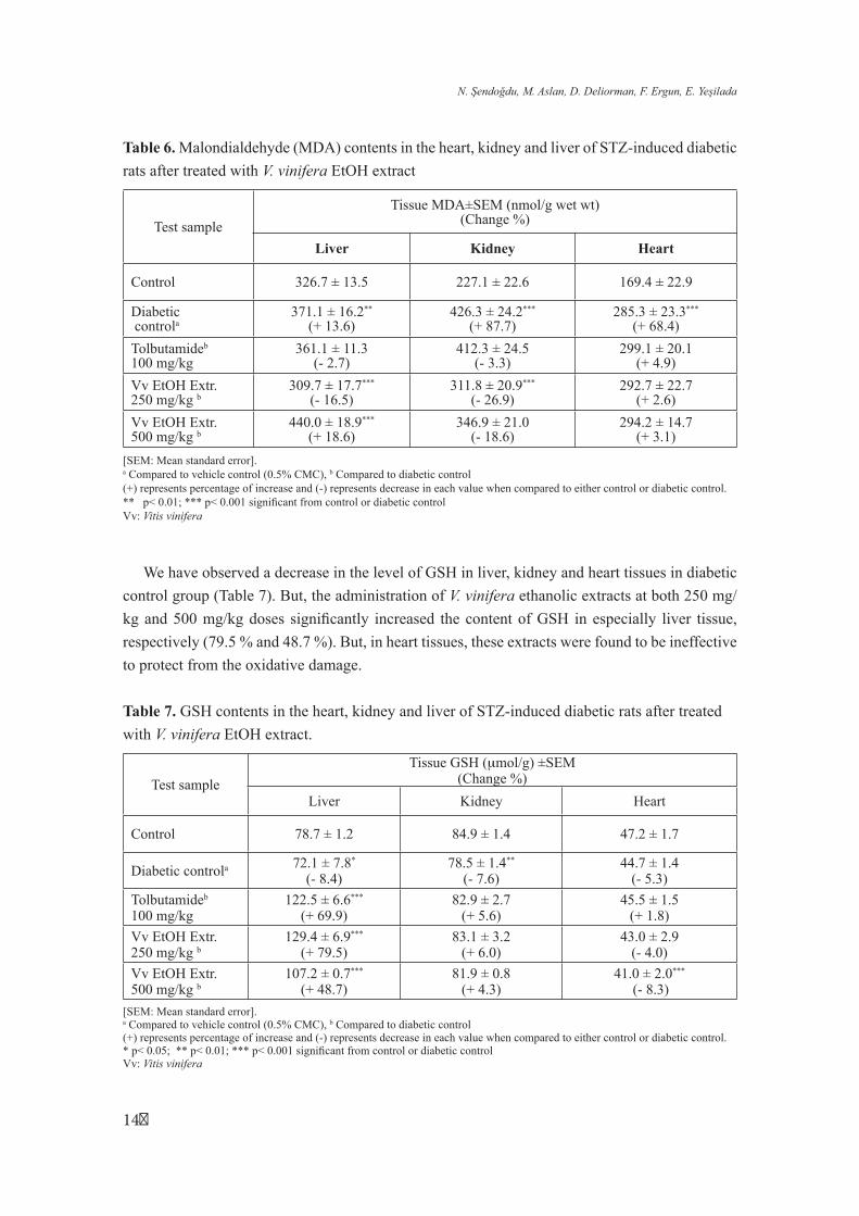
As shown in Table 6, a significant elevation in the concentration of MDA is observed in kidney
and heart of diabetic rats. The ethanolic leaf extract of V. vinifera given orally at dose of 250 mg/
kg produced a significant reduction in LPO in kidney (26.9 %) and liver (16.5 %) tissues, while
the heart LPO were not recovered with both ethanolic extracts (at 250 and 500 mg/kg doses).
These findings suggest that 250 mg/kg dose of the ethanolic extract may protect the liver and
kidney from lipid peroxidation.
13
N. Şendoğdu, M. Aslan, D. Deliorman, F. Ergun, E. Yeşilada
Table 6. Malondialdehyde (MDA) contents in the heart, kidney and liver of STZ-induced diabetic
rats after treated with V. vinifera EtOH extract
Tissue MDA±SEM (nmol/g wet wt)
Test sample (Change %)
Liver Kidney Heart
Control 326.7 ± 13.5 227.1 ± 22.6 169.4 ± 22.9
Diabetic 371.1 ± 16.2** 426.3 ± 24.2*** 285.3 ± 23.3***
controla (+ 13.6) (+ 87.7) (+ 68.4)
Tolbutamideb 361.1 ± 11.3 412.3 ± 24.5 299.1 ± 20.1 100 mg/kg (- 2.7) (- 3.3) (+ 4.9)
Vv EtOH Extr. 309.7 ± 17.7*** 311.8 ± 20.9*** 292.7 ± 22.7 250 mg/kg b (- 16.5) (- 26.9) (+ 2.6)
Vv EtOH Extr. 440.0 ± 18.9*** 346.9 ± 21.0 294.2 ± 14.7 500 mg/kg b (+ 18.6) (- 18.6) (+ 3.1)
[SEM: Mean standard error].a Compared to vehicle control (0.5% CMC), b Compared to diabetic control(+) represents percentage of increase and (-) represents decrease in each value when compared to either control or diabetic control.** p< 0.01; *** p< 0.001 significant from control or diabetic control Vv: Vitis vinifera
We have observed a decrease in the level of GSH in liver, kidney and heart tissues in diabetic
control group (Table 7). But, the administration of V. vinifera ethanolic extracts at both 250 mg/
kg and 500 mg/kg doses significantly increased the content of GSH in especially liver tissue,
respectively (79.5 % and 48.7 %). But, in heart tissues, these extracts were found to be ineffective
to protect from the oxidative damage.
Table 7. GSH contents in the heart, kidney and liver of STZ-induced diabetic rats after treated
with V. vinifera EtOH extract.
Test sample
Tissue GSH (µmol/g) ±SEM (Change %)
Liver Kidney Heart
Control 78.7 ± 1.2 84.9 ± 1.4 47.2 ± 1.7
Diabetic controla 72.1 ± 7.8*
(- 8.4) 78.5 ± 1.4**
(- 7.6) 44.7 ± 1.4
(- 5.3)
Tolbutamideb 122.5 ± 6.6*** 82.9 ± 2.7 45.5 ± 1.5 100 mg/kg (+ 69.9) (+ 5.6) (+ 1.8)
Vv EtOH Extr. 129.4 ± 6.9*** 83.1 ± 3.2 43.0 ± 2.9 250 mg/kg b (+ 79.5) (+ 6.0) (- 4.0)
Vv EtOH Extr. 107.2 ± 0.7*** 81.9 ± 0.8 41.0 ± 2.0***
500 mg/kg b (+ 48.7) (+ 4.3) (- 8.3)
[SEM: Mean standard error].a Compared to vehicle control (0.5% CMC), b Compared to diabetic control(+) represents percentage of increase and (-) represents decrease in each value when compared to either control or diabetic control.* p< 0.05; ** p< 0.01; *** p< 0.001 significant from control or diabetic control Vv: Vitis vinifera
14
Turkish J. Pharm. Sci. 3 (1), 7-18, 2006
Qualitative analysis revealed that flavanoid, tannin, cardioactive glucoside, reducing or
nonreducing sugar, organic acids, terpenes, lipids, coumarine, anthocyanin glucosides are found
as active principle in leaves (17).
In previously, several chromatographic studies established the presence of flavan-3-ols
like (+)-catechin, (-)-epicatechin- (±)-epigallocatechin and (-)-epicatechin-3-O-gallate and of
flavonols like quercitrin, traces of quercetin and kaempferol; rutin, isoquercitrin and luteolin
were also identified in leaves of plant. Flavanols and flavanol oligomers and proanthocyanidins
(condensed tannins) have been proven to possess powerfull antioxidant activities (free radical
scavenging activity and the inhibiting effect of the lipid peroxidation). The radical scavenging
properties of condensed tannins have been demonstrated in vitro on different biochemical models
mimicking preconditions of pathological situations such as ischemia, inflammation and diabetic
conditions (18). Therefore, in this study, mainly condensed tannins and flavonoids were suggested
to contribute in the antilipoperoxidant activity of V.vinifera leaves.
On the other hand, the phytoalexin resveratrol which is known as antioxidant, can also be found
in V.vinifera leaves. But, healthy leaves of V.vinifera are free from resveratrol and derivatives (2).
Indeed, TLC analysis indicated that V.vinifera leaves do not contain resveratrol. Therefore, in this
study, it can be proposed that resveratrol is not the major antioxidant active constituent of the
ethanolic extract .
Additionally, studies have found that a crude alcohol extract of Pterocarpus marsupium that
contains epicatechin and catechin derivatives, has regenerated functional pancreatic beta cells
in diabetic rats. Also the antidiabetic activity of these compounds has been confirmed by some
studies on different experimental models (19).
It is known that V.vinifera leaves contain catechin and catechin derivates (2) which may be
responsible for antidiabetic effect and further studies should be focused on this goal.
As a conclusion, this study shows that the ethanolic extract of V. vinifera leaves at 250 mg/kg
dose has a remarkable antidiabetic effect. In liver and kidney tissues, 250 mg/kg dose of the
ethanolic extract also was found effective to protect from the oxidative damage. But, the ethanolic
extract increased the GSH content of especially liver tissue at 250 and 500 mg/kg doses.
ACKNOWLEDGEMENT
This study was financially supported by the Research Fund of Gazi University (02/2004-14).
We are acknowledge for providing us with experimental animals to DROGSAN Tic. A.Ş. and
İlsan İltaş İlaçları.
15
N. Şendoğdu, M. Aslan, D. Deliorman, F. Ergun, E. Yeşilada
REFERENCES
1. Davis, P.H. Flora of Turkey and East Eagean Islands, Edinburgh University Press, Edinburgh,Vol:2, pp. 521-522, 1997.
2. Bombardelli, E., Morazzonni, P., “Vitis vinifera L.” Fitoterapia 66, 291-317, 1995.
3. Hebash, K.A.H., Fadel, H.M. and Soliman, M.M.A., “Volatile components of grape leaves” JIAS 4(1), 26-28, 1991.
4. Felicio, J.D., Santos, R.S. and Goncalez, E., “Chemical constituents from Vitis vinifera (Vitaceae)” Arq. Inst. Biol. 68(1), 47-50, 2001.
5. Baytop, T. Bitkiler İle Tedavi (Geçmişte ve Bugün), 2. Baskı, Nobel Tıp Kitabevleri, İstanbul, pp. 357-358, 1999.
6. Zimmet, P.Z., McCarty, D.J. and Courten, M.P., “The global epidemiology of non-insulin dependent diabetes mellitus and the metabolic syndrome” J. Diabetes Complicat. 11, 60-68, 1997.
7. Atalay, M. and Laaksonen, D.E., “Diabetes, oxidative stres and physical exercise” J. Sports Sci. & Med. 1, 1-14, 2002.
8. West, I.C., “Radicals and oxidative stress in diabetes” Diabet Med. 17, 171-180, 2000.
9. Gumieniczek, A., Hopkala, H., Wojtowicz, Z. and Nikolajuk, J., “Changes in antioxidant status of heart muscle tissue in experimental diabetes in rabbits” Acta Biochem. Pol. 49(2), 529-535, 2002.
10. Aydın, A., Orhan, H., Sayal, A., Özata, M., Şahin, G. and Işımer, A., “Oxidative stres and nitric oxide related parameters in type II diabetes mellitus: effects of glycemic control” Clin. Biochem. 34, 65-70, 2001.
11. Landcake, P., Cornford, C.A., and Pryce, R.J., “Identification of Pterosilbene as a phytoalexin from Vitis vinifera leaves.” Phytochemistry 18, 1025-1027, 1979.
12. Aslan, M., Sezik, E.,Yeşilada, E., “Antihyperglycaemic effect of Cannabis sativa L. fruits” J. Fac. Pharm. Gazi University-GUEDE 17, 89-93, 2000.
13. Aslan, M., Sezik, E.,Yeşilada, E., “Effect of Hibiscus esculentus L. seeds on blood glucose levels in normoglycaemic, glucose-hyperglycaemic and streptozotocin induced diabetic rats” J. Fac. Pharm. Gazi University-GUEDE 20, 1-7 , 2003.
14. Jamall, I.S., Smith, J.C.,”Effects of cadmium on glutathione peroxidase, superoxide dismutase, and lipid peroxidation in the rat heart: a possible mechanism of cadmium cardiotoxicity” Toxicol. Appl. Pharmacol. 80, 33-42, 1985.
15. Ohkawa, H., Ohishi, N., Yagi, K., “Assay for lipid peroxides in animal tissues by thiobarbituric acid reaction” Anal. Biochem. 95, 351-358, 1979.
16
Turkish J. Pharm. Sci. 3 (1), 7-18, 2006
16. Sedlak, J., Lindsay, R.H., “Estimation of total protein-band and nonprotein sulfhydryl group in tissue with Ellman’s reagent” Anal. Biochem. 25, 192-205, 1968.
17. Şendoğdu, N., “Vitis vinifera L. yapraklarının şeker hastalığı üzerine etkilerinin araştırılması”, Yüksek Lisans Tezi, Gazi Üniversitesi Sağlık Bilimleri Enstitüsü, Farmakognozi Anabilim Dalı, Ankara (2004).
18. Elstner, E.F., Kleber, E., in “Flavonoids in Biology and medicine III: Current issues in flavanoid research”, N.P. Das (Ed.), National University of Singapore 227-235, 1990.
19. Perez, R.M., Zavala, G.M.A., Perez, S.G., Perez, C.G., “Antidiabetic effect of compounds isolated from plants”, Phytomed. 5, 55-75, 1998.
received: 03.07.2005
accepted: 24.03.2006
1�