Embed Size (px)
Citation preview

PROTECTIVE EFFECTS OF ANTIOXIDANT PRETREATMENT IN VPA-TREATED
NRF2 KNOCKOUT MICE
by
JANACE J. GIFFORD
A thesis submitted to the
School of Graduate Studies
Rutgers, The State University of New Jersey
In partial fulfillment of the requirements
For the degree of
Master of Science
Graduate Program in Psychology
Written under the direction of
Dr. George C. Wagner
And approved by
____________________________
____________________________
____________________________
New Brunswick, New Jersey
January, 2018

ii
ABSTRACT OF THE THESIS
Protective effects of antioxidant pretreatment in VPA-treated Nrf2 knockout mice
By JANACE GIFFORD
Thesis Director:
George C. Wagner
Genetic and environmental factors associated with oxidative stress have been
implicated in the etiology of autism. The present study attempted to mimic the factors in
an animal model of autism. Specifically, mice with a deletion of the Nrf2 gene, a master
regulator for downstream enzymes associated with management of toxicant-generated
reactive oxygen species, were administered valproic acid (VPA), a toxicant known to
engender oxidative stress and one that has been associated with autism in humans. Prior
studies revealed that VPA treatment induces functional and pathological changes in mice
akin to autism. Further, previous work has established that pretreatment with
antioxidants has the ability to protect mice from these VPA-induced functional deficits.
The present study extended these observations to mice with alterations in NRF2
expression. On postnatal day 14 knockout (KO) and wild type (WT) mice were exposed
to VPA (400 mg/kg) or saline and pretreated with either Trolox, a water-soluble form of
vitamin E, or saline 1 hour prior to VPA. The behavioral tasks employed assessed
maturation of normal social, cognitive, and motor skills and classified toxicant-induced
deficits along a developmental timeline. Treatment with VPA resulted in deficits in mid-
air righting and Morris water maze learning. Further, Trolox pretreatment prior to VPA

iii
provided partial protection from deficits associated with VPA treatment and this
protective effect was more apparent in the Nrf2 KO mice. The results of the present
study, at least in part, indicate the importance of Nrf2 during development as it might
relate to autism and, more generally, the effects of oxidative stress during early
development as well as the potential protective effects of antioxidant pretreatment.

iv
ACKNOWLEDGEMENTS
I would like to thank the members of my thesis committee, Dr. Alexander
Kusnecov and Dr. Ben Samuels. Additionally, I would like to thank both current and
former members of the Wagner lab. I would also like to thank my family and friends for
their love and support. Finally, I would like to thank Dr. George Wagner, who has been a
wonderful mentor, for his indispensable mentoring and guidance in designing and
conducting this project.
v
TABLE OF CONTENTS
Abstract of Thesis ............................................................................................................ii
Acknowledgements ..........................................................................................................iv
List of Figures ..................................................................................................................vi
Introduction ......................................................................................................................1
Materials and Methods .....................................................................................................9
Results ..............................................................................................................................15
Discussion ........................................................................................................................20
References ........................................................................................................................26
Figure Legends .................................................................................................................29

vi
LIST OF FIGURES
Figure 1A: Mid-air righting performance trend lines .....................................................33
Figure 1B: Mid-air righting performance from P13-19 ..................................................33
Figure 2A: Rotarod performance from P25-27 trend lines .............................................34
Figure 2B: Rotarod performance from P25-27 by treatment ...........................................34
Figure 3A: Adult rotarod performance curve ..................................................................35
Figure 3B: Adult rotarod performance ............................................................................35
Figure 4A: Morris water maze acquisition curves (Day 1-4) ..........................................36
Figure 4B: Morris water maze acquisition performance (Day 1-4) .................................36
Figure 4C: Morris water maze acquisition by treatment .................................................37
Figure 5A: MWM acquisition path length trend ..............................................................38
Figure 5B: MWM acquisition path length .......................................................................38
Figure 6: Time spent in the target quadrant on probe trial ..............................................39
Figure 7A: Social Approach behavior with stranger 1 ....................................................40
Figure 7B: Social approach behavior in the test phase ....................................................40
Figure 8: Distance traveled in open field .........................................................................41
Figure 9A: Days to acquisition in Y maze .......................................................................42
Figure 9B: Errors to acquisition in Y maze .....................................................................42
Figure 9C: Days to reversal in Y maze ............................................................................43
Figure 9D: Errors to reversal in Y maze ..........................................................................43
Figure 10A: Example NRF2 blot .....................................................................................44
Figure 10B: Example H3 blot ..........................................................................................44

vii
Figure 11A: P14 NRF2 expression 2-hours post VPA exposure .....................................45
Figure 11B: Adult NRF2 expression 2-hours post VPA exposure ..................................45
1
INTRODUCTION
Background
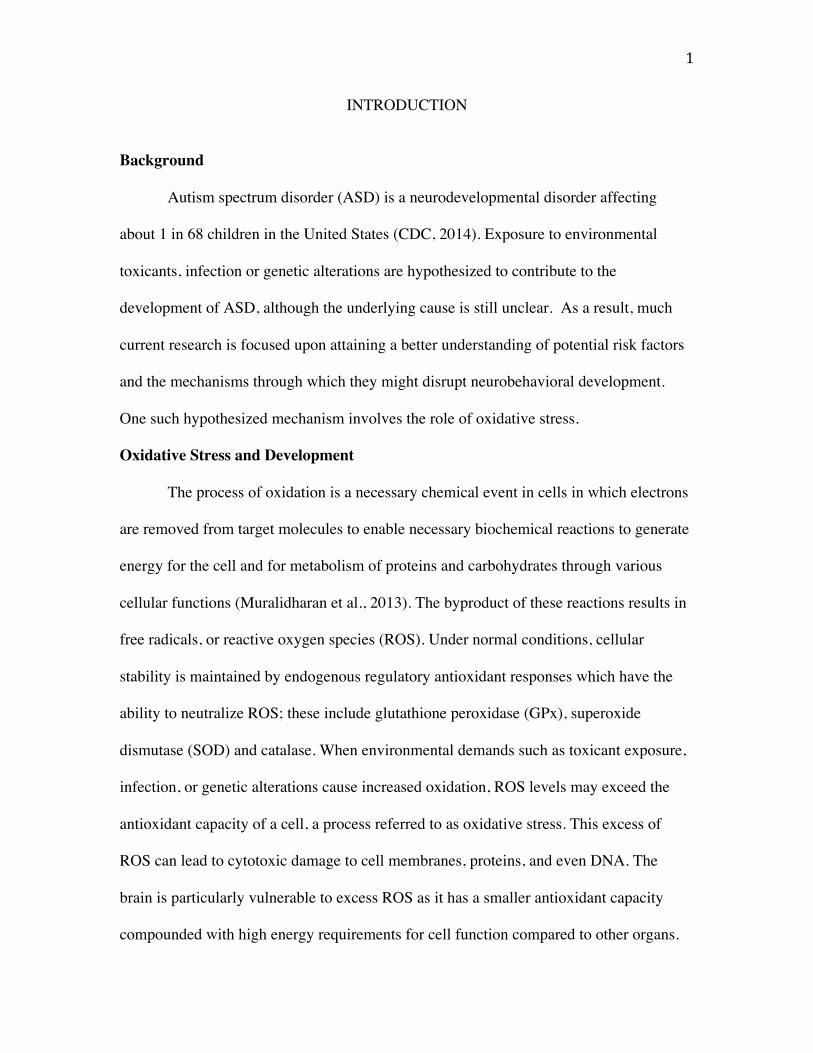
Autism spectrum disorder (ASD) is a neurodevelopmental disorder affecting
about 1 in 68 children in the United States (CDC, 2014). Exposure to environmental
toxicants, infection or genetic alterations are hypothesized to contribute to the
development of ASD, although the underlying cause is still unclear. As a result, much
current research is focused upon attaining a better understanding of potential risk factors
and the mechanisms through which they might disrupt neurobehavioral development.
One such hypothesized mechanism involves the role of oxidative stress.
Oxidative Stress and Development
The process of oxidation is a necessary chemical event in cells in which electrons
are removed from target molecules to enable necessary biochemical reactions to generate
energy for the cell and for metabolism of proteins and carbohydrates through various
cellular functions (Muralidharan et al., 2013). The byproduct of these reactions results in
free radicals, or reactive oxygen species (ROS). Under normal conditions, cellular
stability is maintained by endogenous regulatory antioxidant responses which have the
ability to neutralize ROS; these include glutathione peroxidase (GPx), superoxide
dismutase (SOD) and catalase. When environmental demands such as toxicant exposure,
infection, or genetic alterations cause increased oxidation, ROS levels may exceed the
antioxidant capacity of a cell, a process referred to as oxidative stress. This excess of
ROS can lead to cytotoxic damage to cell membranes, proteins, and even DNA. The
brain is particularly vulnerable to excess ROS as it has a smaller antioxidant capacity
compounded with high energy requirements for cell function compared to other organs.

2
Therefore, the brain seems to be quite susceptible to the deleterious effects of oxidative
stress, particularly during development when there is a high rate of cell proliferation
(Chauhan et al., 2006).
Antioxidants are necessary for survival of neurons during critical periods early in
life (Perry et al., 2004). Children from conception throughout infancy may be more
susceptible to the effects of oxidative stress as they have lower levels of antioxidants
including glutathione (Erden-Inal et al., 2002). Additionally, evidence suggests
antioxidant ability is acquire over time (Bernhardt et al., 2017). This lack of defense
mechanisms in addition to the fact that environmental toxicants found in the mother are
also detectable in the fetus, indicates the brains of developing children are at increased
risk of the deleterious effects of oxidative stress.
Oxidative Stress and Autism
The link between oxidative stress and autism has been identified through
examination of markers of oxidative stress in individuals with autism along with
increased body burdens of environmental toxicants associated with induction of oxidative
stress (Edelson& Cantor, 1998; Dietert & Dietert, 1997). ASD has been linked to low
levels of glutathione peroxidase, superoxide oxidase and catalase activities and total
glutathione and cysteine levels (James et al., 2004; Sogut et al., 2003; Yorbik et al., 2010;
Zoroglu et al., 2004). Further, decreased antioxidant levels were present in plasma of
individuals with ASD and there is evidence of impaired energy metabolism and
mitochondrial dysfunction as well (Filipek et al., 2003; Rossignol and Frey, 2014).
Finally, children with autism have increased levels of oxidative stress biomarkers in their
urinary excretions indicating they are experiencing increased oxidative stress relative to

3
age-matched controls (Ming et al., 2005). Collectively, these data indicate that
individuals with autism may have difficulty managing oxidative stress due to
environmental demands such as toxicant exposure and/or genetic alterations.
Oxidative Stress and NRF2
Excessive ROS exert a cellular burden that can cause functional decline. Since
ROS accumulation is inevitable given the energy demands of the developing organism,
its removal or limits on oxidation are controlled by endogenous antioxidant mechanisms.
A master regulator for the induction of antioxidant events is nuclear factor erythroid 2-
related factor 2 (NRF2) (Wakabayashi et al., 2010; Sandberg et al., 2013). This is a
transcription factor that is normally restrained in the cytoplasm by the protein KEAP1.
However, in response to metabolic or toxic stimuli, NRF2 migrates to the nucleus and
binds to an antioxidant response element (ARE) on DNA, initiating downstream
induction of antioxidant genes (Wakabayashi et al., 2010; Sandberg et al., 2013). This
effect is a critical response to environmental demands (including toxicants), which
necessitate its activation and the downstream induction of antioxidant genes. Our
preliminary data confirm this at the behavioral level using Nrf2-deficient mice, and
suggest that NRF2 may serve to protect the developing animal from oxidative stress
(Furnari et al., 2014).
Valproic Acid, Oxidative Stress and Autism
Valproic acid (VPA) is a GABAergic anticonvulsant that has been shown to
increase the risk of autism-like behaviors in offspring if taken during pregnancy
(Christensen et al., 2013; Singh et al., 2014). VPA acts as an indirect GABA agonist as it
non-specifically blocks the enzymatic degradation of GABA, thereby resulting in GABA

4
buildup. Recent work by Ben-Ari (2014) suggests a shift in GABA action, which is
regulated by development. This work suggests an excitatory action of GABA during fetal
development and into early postnatal life followed by a shift to inhibitory action around
the third week of life in rodents (Ben-Ari, 2014). This initial excitatory role of GABA
may underlie excitotoxicity during this critical window of development.
We, and others, developed an animal model of autism involving exposure to
VPA, which results in behavioral and neuroanatomical deficits similar to autism in
humans, both pre- and postnatally, targeting the time window of GABA shift (Wagner et
al., 2006; Yochum et al., 2008). Additionally, when VPA is metabolized it is known that
oxidative stress ensues, as measured by a decrease in cellular glutathione peroxidase and
increased ROS (Jurima-Romet at al., 1996). This connection to oxidative stress makes
VPA a prime model for examination of autism prevention strategies. That is, the
hypothesis that antioxidant pretreatment may protect mice against VPA-induced
oxidative stress may be tested.
Antioxidant Treatment of Symptoms
Improvement in behaviors following antioxidant administration to individuals
with autism further elucidates the role of oxidative stress in contributing to the etiology of
autism. Double-blind, placebo controlled trials have indicated the efficacy of high-dose
vitamin C or carnosine on improving behaviors associated with autism (Chez et al., 2002;
Dolske et al., 1993). Additionally, a three-week supplement of betaine and folinic acid
administered to twenty children with autism who displayed low levels of GSH and
cysteine showed an improvement in blood plasma levels of antioxidants (Roberts et al,
2010). Recent work has shown pretreatment of astaxanthin, an antioxidant with the

5
ability to cross the blood brain barrier, significantly improved behavioral deficits
following prenatal VPA exposure in humans (Al-Amin et al., 2013). Additionally,
sulfrophane, an antioxidant found in broccoli sprout extract, has been shown to induce
NRF2 expression and to lessen the symptoms of autism in young boys (Singh et al.,
2014). Taken together, these lines of evidence support the hypothesis that at least some
children with autism exhibit enhanced oxidative stress and antioxidant pretreatment may
lessen the severity of symptoms.
Antioxidant Pretreatment: Prevention of Symptoms
Previous work in rodents found that vitamin E treatment prior to VPA exposure
during gestation reduced the likelihood of fetal toxicity (Deeb et al., 2000). Additionally,
work in our lab has shown Trolox (6-hydroxy-2,5,7,8-tetramethyl-chroman-2-carboxylic
acid), a water-soluble vitamin E derivative, when used as the antioxidant pretreatment to
MeHg challenge, protected mice against behavioral deficits induced by the MeHg.
Specifically, Trolox effectively attenuated deficits in the mid-air righting reflex
maturation, a behavior disrupted with MeHg challenge alone (Cheh et al., 2010). MeHg,
like VPA, is known to exert its neural damage through oxidative stress. Further, work in
our lab has shown vitamin E in a corn oil vehicle can also attenuate behavioral deficits
associated with postnatal VPA treatment (Ming et al., 2008). Our lab has yet to examine
the effectiveness of Trolox, a water-soluble form of vitamin E in a VPA model. There is
also evidence of a therapeutic action of green tea extract following VPA exposure. Green
tea extract ameliorated behavioral changes associated with VPA treatment including
motor delays and anxiety as well as neutralization of free radicals and decreased lipid
peroxidation levels following treatment (Banji et al., 2011). Collectively, these results

6
indicate a protective role of antioxidant pretreatment in both MeHg and VPA-induced
behavioral deficits suggesting a role of oxidative stress in the development of behavioral
deficits in both these animal models of autism.
Animal Model of Autism
An animal model has been developed in which mice are exposed to any of a
number of toxicants early in life (i.e. pre- or early post-natal exposure) and are then
assessed for their behavioral maturation as well as for neuropathological damage. The
behavioral tasks assess the maturation of social, cognitive, emotional and motor skills;
deficits are further classified as retardations (i.e. the toxicant slows and/or eliminates the
development of a skill), regressions, (i.e. a skill matures at the same rate as control-
treated pups but then the toxicant exposure results in a loss of those skills), or intrusions
(i.e. the skills do mature on schedule but their appearance is overshadowed by stereotypic
or self-injurious behaviors). Further, this model incorporates genetic alterations by
assessment of toxicant-induced behavioral and neuropathological damage in knockout
versus wild type mice. These mice are not autistic; rather the toxicants selected have
been associated with human autism as are the genetic alterations. Thus, the use of this
model allows for the demonstration of toxicant-induced deficits in neurobehavioral
development with critical variables including time of toxicant exposure as well as
genotype and sex of the littermate offspring (Furnari et al., 2014; Wagner et al., 2006;
Yochum et al., 2008).
Previous work has indicated that toxicant exposure on postnatal day 14 (P14)
results in a regression of behaviors such as the mid-air righting reflex. P14 was selected
for postnatal treatment as the behavioral skills have now matured by that day and, hence,

7
it is possible to demonstrate regression. Further, as noted above, pretreatment with the
antioxidant, Trolox, was shown to protect pups against methylmercury challenge (Cheh
et al., 2010) and vitamin E pretreatment protected pups against VPA-induced regression
(Ming et al., 2008). Both toxicants have been associated with autism and are known to
generate oxidative stress. Finally, recent studies examined postnatal VPA administration
delivered to P14 pups with an Nrf2 deletion resulting in enhanced sensitivity to the
neurodevelopmental toxicity induced by the VPA (Furnari et al., 2014). This provides a
convincing demonstration of the importance of NRF2 in managing toxicant-induced
damage during early development.
Summary and hypotheses
The present study aims to determine whether NRF2 is necessary to maintain
stable brain and behavioral development in the face of early postnatal challenge with
VPA. If VPA stress induction shows a reliance on NRF2, this will show that NRF2 may
be a critical regulator of neuroprotection in early postnatal development. Specifically,
since developmental disorders including ASD have been linked to oxidative stress, we
hypothesize that pups lacking NRF2 will not have sufficient antioxidant neuroprotection
against physiological perturbations that result early postnatal challenge and this will
result in a pronounced deficit in postnatal development of social, cognitive, and
emotional behavior. The present study will assess mid-air righting, rotarod, social
approach, Morris water maze, and Y maze performance. It is hypothesized that Nrf2-/-
mice exposed to VPA will show the greatest impairments in the described tasks.
Additionally, previous work in our lab has shown antioxidants (including Trolox) were
fully effective against post-natal challenge with toxicants such as methylmercury but

8
effectiveness of Trolox is unknown in NRF2 deficient mice challenged with VPA. We
predict that antioxidant pretreatment will protect mice against the behavioral deficits
induced by VPA in the Nrf2 mice and the magnitude of this difference will be most
evident in the Nrf2 knockout mice.
9
MATERIALS AND METHODS
Subjects: All animals were maintained under standard vivarium conditions with free
access to food and water and a 12:12 hour light:dark cycle. All procedures were approved
by the Animal Care Committee and our facility meets AAALAC standards. From our
original colony of Nrf2-/- mice on a C57Bl/6J background (obtained from Dr. Tony Kong
of Rutgers University) we have derived wildtype and knockout Nrf2 female mice that
were mated with male wildtype and knockout Nrf2 mice generating litters that contain
either +/+ or -/- genotypes. Breeding in this way allowed for adequate time for breeding
and only produced necessary animals and removed the need for additional genotyping.
Group Design: One male (KO or WT) was placed with two receptive females (KO or
WT) together for 8 days to breed KO and WT litters. Day of birth was noted as postnatal
day 0 (P0). Pups were weighed and sexed on P13 and weighed daily until P19. On P14,
one set of pups was injected with either saline or valproic acid (400 mg/kg) delivered in a
saline vehicle in a volume of 1.0 ml/100 g body weight. In order to determine if a one-
hour pretreatment with Trolox protected mice against the toxicity induced by the P14
VPA, a second set of mice was injected with 25 mg/kg of Trolox in a saline vehicle in a
volume of 1.0 ml/100g body weight administered one hour before either the VPA or
saline treatment on P14.
Behavioral Tests:
Motor Tasks: Although autism usually is not associated with severe motor disturbances,
we and others have characterized deficits in children with autism such as delays in motor
milestone development, disturbances in reach-to-grasp, deficiencies in gross and fine
motor movement, and underdevelopment of postural control (Wagner et al., 2006; Ming
10
et al., 2007). In addition, stereotypic or repetitive motor behaviors often appear in autism.
In mice, a wide variety of motor tests are available for assessing behavioral disturbances
across development. Mid-air righting was utilized in this study. In this task animals are
dropped dorsal side down from 18 cm above a padded surface and their ability to right
themselves (land on all four paws) was assessed. The timing of this test is based on
known developmental milestones, with mid-air righting taking place on P13 – P19.
Previous research has shown that in postnatal VPA treatment produces significant deficits
in mid-air righting on P15 in both NRF2 WT and KO animals (Furnari et al, 2014).
Additionally, previous work has demonstrated the effectiveness of vitamin E
pretreatment on mid-air righting behavior in a MeHg model of autism (Ming et al., 2008).
The present study sought to determine if pups lacking NRF2 treated with both Trolox and
VPA on P14 will display fewer deficits than what is seen with VPA treatment alone.
Performance was measured by the correct responses across three trials per day.
Rotarod: We tested mice on P25-27 and again between P90-200 for their performance on
the rotarod. Mice were placed on the rotating rod for three test trials each day for three
consecutive days and latency to fall was recorded, with a 60-sec maximum. The rotarod
consists of a 6.0 cm diameter cylinder covered in textured material and run at 12 RPM.
Spatial Learning and Memory: The present study employed a scaled down Morris water
maze for pups (MWM; Wagner et al. (2006)) to test for spatial memory. The water maze
use was a circular galvanized steel tub 61 cm in diameter and 29 cm in height, filled ¾ of
the way with room-temperature water, made opaque with non-toxic white paint powder.
A white circular escape platform, measuring eight cm in diameter, was placed in one
quadrant of the maze, two cm below the surface of the water, its position held constant.

11
The water maze is a learning paradigm in which an animal must make use of spatial cues
to navigate the maze; this skill does not mature in rodents until about day 22 of life.
Additionally, there were various extra-maze cues in the testing room to facilitate mapping
of the spatial environment. Mice were tested on five consecutive days, beginning on
postnatal day 30 and continuing through day 34. For acquisition, which occurred on days
one through four, five trials were given each day. Each trial started from a randomly
assigned quadrant of the maze and each mouse was allowed a maximum of 60 seconds to
find the escape platform on each trial. For these studies, the hidden platform remained in
the same location each day. If the animal did not escape, it was placed on the platform
for a 15 second inter-trial interval (ITI). If the animal successfully found the platform,
there was a 15 second ITI, during which the animal remained on the platform. At the
completion of this phase of testing, we used one, 60 second probe trial (day 5) where
there was no platform available. We monitored the performance of the subject for time
spent in the proper quadrant (compared to time spent in other quadrants). Performance
was measured by latency to find the platform, distance travelled, and mean speed.
Social Approach: Social deficits are a core sign of autism. Therefore, we assessed social
interactions using our standard social measure. For this test, each set of mice were of
same genotype, sex, and age (P40). We used an automated chamber and tracking
software to quantify social approach in mice. There were two wire cylinder cage
enclosures (11 cm diameter) on opposite sides of an open field (36.5 cm x 36.5 cm). This
test consisted of three 10 minute phases. Phase one (stranger phase) acted as a period of
habituation with both wire cages empty. In phase two, a target mouse (same treatment,
age, sex, and genotype) was enclosed in one wire cage and an empty wire cage on the
12
opposite side of the open field. The test mouse was placed in the center of the open field
and time spent with the stranger and empty cages were automatically recorded. This was
followed by phase three (test phase) in which a new stranger (same treatment, age, sex,
and genotype) were placed in the once empty wire cage. The test mouse was placed in the
center of the open field and time spent with the novel and habituated stranger cages were
automatically recorded.
Y maze: Perseverative behaviors are another core deficit in autism. Use of the Y-maze
reversal task allowed examination of perseverative behaviors expected to be exhibited by
VPA treated animals and attenuated by antioxidant pretreatment between P42-P60. This
task employs a water filled Y shaped maze, which was made opaque using nontoxic
white paint powder. Additionally, there were various extra-maze cues placed on the walls
for animals to use to map the spatial environment. The Y maze consists of three arms,
one of which is designated the start arm while the other two are target arms. Within the
maze, there is a hidden escape platform (10 cm in diameter) placed in one of the target
two arms. There were two phases of Y maze testing; acquisition and reversal. Each phase
consisted of 6 trials per day with a 5-minute inter-trial interval. During acquisition,
animals were placed in the start arm and allowed to make a choice between one of the
two target arms. Once an animal had chosen an arm (defined by full body entrance into
an arm), a guillotine was placed at the entrance of the chosen arm, forcing the animal to
stay in the chosen arm. If this was the correct arm (arm with the hidden platform) animals
were allowed 10 seconds on the platform to gather spatial cues before the trial ended. If
the animal chose the incorrect target arm (arm without the hidden platform), this was
considered an error and animals were allowed 10 seconds to swim the incorrect arm

13
before the trial ended. Completion of acquisition was defined as 11 correct responses in
two consecutive days (ie. one error in two days of testing). Immediately following
acquisition (following day) animals began reversal training. During reversal, the same
protocol for acquisition was used except the hidden platform was switched to the
opposite target arm. Completion of reversal is also defined by 11 correct responses in two
consecutive days. Animals were considered to have “failed” the task if either acquisition
or reversal lasted more than 10 days. Data collected consisted of performance on each
trial as well as total number of errors and total number of days to both acquisition and
reversal.
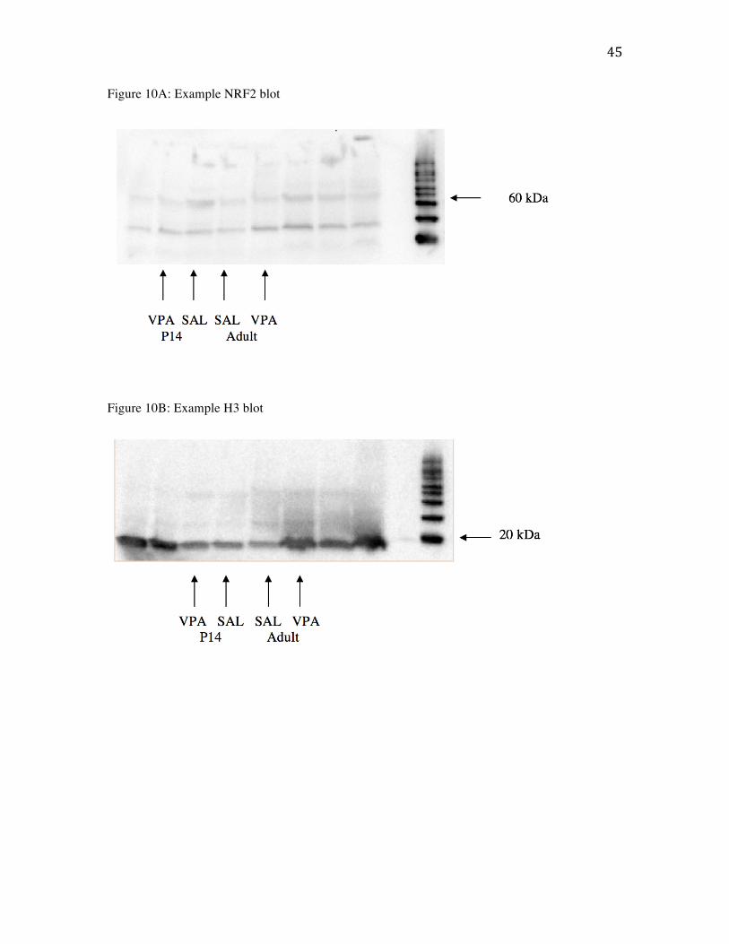
Measures of NRF2: To determine if postnatal VPA induces the expression of NRF2 in
early postnatal brains, wild type males and females were administered VPA or saline on
P14 or ~P90 and their brains were harvested two-hours post injection. Western blotting
was used to measure NRF2 expression. Tissue samples were homogenized and protein
extracted through NER-CER kits (Thermofisher) to separate cytosolic and nuclear protein
samples. A protein assay was conducted to determine amount of protein in each sample
using a Bradford coomassie protein assay kit (Pierce). Nuclear protein samples were run
on SDS-PAGE Tris-glycine-SDS gels to separate protein samples, which were then
transferred to a PDVF membrane (Bio-rad) for immunoblotting. The PDVF membrane
was be incubated with primary antibodies for NRF2 and H3 (Cell Signal Technologies)
with 1:1000 dilution. Data are presented as protein target ratio to general housekeeping
gene.
Data Analysis: Behavioral tests required repeated measures ANOVAs or univariate
ANOVAs followed by Tukey’s post hoc tests at p<0.05. Data was collapsed across sex

14
when no sex differences were observed. Similarly, if no differences were found between
Trolox+saline and saline treatment, these groups were collapsed. Western blotting data
required analysis by image-j software followed by t-tests at p<0.05.

15
RESULTS
Body Weight P13-19
Weight was measured daily from P13-19 and no effect of treatment was observed
(F(2,36) =1.279, p =0.29) indicating there was no effect of VPA on body weight
compared to saline-treated animals.
Body Weight P40
At postnatal day 40 there were still no observed differences in weight by genotype
or treatment (F(2,55) =1.431, p=.248).
Mid-air righting
Ability to right in mid-air was examined on postnatal days 13 to 19. A two-way
mixed ANOVA indicated a significant two-way interaction between day and treatment,
F(12,468)=3.798, p<.01 (Fig. 1). All other interactions were found to be not statistically
significant (p>.05). Additionally, there was a significant between subjects effect of
treatment (F(2,72)= 10.539, p<.01) with further Tukey post hoc analyses revealing a
significant difference between saline and VPA treated animals (p<.01) and saline and
trolox+VPA treated animals (p=.02). Results warranted further investigation on a day by
day basis in which a series of two way ANOVAs were run. There were no significant
differences between treatment groups on P13 or 14 before the VPA administration
(p>.05) while P15-P19 showed significant differences between the VPA treatment groups
(p<.05).
Rotarod - Adolsecence
The present study expected to examine performance of KO and WT animals
treated with VPA or saline and/or pretreated with trolox on rotarod in adolescence. Issues

16
were encountered with the rotarod during the running of the pretreated animals and as a
result, only data for VPA and saline treated KO and WT animals were analyzed. A two
way repeated measures ANOVA (genotype x treatment) revealed a significant between
subjects effect of treatment (F(1,40)=3.858, p=.056) with VPA treated animals spending
significantly less time on the rotarod compared to saline treated animals (Fig. 2).
Rotarod - Adulthood
Rotarod performance, a measure of motor ability, was averaged for each day
across three consecutive days. A two way repeated measures ANOVA (genotype x
treatment) was run on time spent on the rotarod and revealed no significant difference
between groups on genotype (F(1, 29)=1.152, p=.232) or treatment (F(2,29)=.635, p=
.639). Results are visualized in figure 3 which shows no difference in motor abilities
across groups.
Morris water maze
Morris water maze acquisition (postnatal days 30 to 33) was assessed by the
latency to find the platform, distance traveled, and swim speed (Fig. 4). A two-way
mixed ANOVA was run to assess the latency to find the platform during acquisition
trials. There were no statistically significant interactions (p>.05). Results indicate a
significant between subject effect of treatment on latency to find the platform
(F(2,72)=1.058, p=.003) (Fig. 4C). Further Tukey post hoc analyses revealed a
significant difference between SAL and VPA-treated animals (p=.028) with saline-
treated animals finding the platform significantly faster than VPA-treated animals. There
was no effect of genotype (p>.05) on latency to find the platform. No significant

17
differences in acquisition were found for treatment or genotype for path length (Fig. 5) or
swim speed (p>.05).
Performance on the probe test (postnatal day 34) was assessed by the number of
entries, time spent in the target quadrant, and distance traveled in the target quadrant (the
quadrant that contained the hidden platform during acquisition). A two way ANOVA for
entries revealed no differences between groups on genotype (F(1,72)=58.582, p=.012) or
treatment (F(2,72)=.378, p=.687). There were no differences in time spent in the target
quadrant or distance traveled for treatment or genotype (p>.05) (Fig. 6).
Social Approach
Social approach behavior was quantified in two stages (Fig. 7). The first stage
consisted of a stranger in one zone of the apparatus (stranger phase). Time spent in each
of the zones was scored by a trained observer and results examined time spent in the
“stranger 1 zone” compared to time spent in all other zones (Fig. 8A). A two way
ANOVA revealed no significant difference between genotype (F(1,63)=.190, p=.665) or
treatment (F(2,63)=.884, p=.418) with all groups spending more time with the stranger
compared to the empty zones.
The second stage consisted of placing a second (novel) stranger into one of the
previously empty zones (test phase). Videos were scored for time spent in “stranger 1
zone”, “stranger 2 zone” and the empty zone. This allowed for comparison of time spent
interacting with the habituated stranger, the novel stranger and the empty zones (figure
8B). Preference between stranger 1 and stranger 2 was expected to diverge, with VPA-
treated animals preferring the familiar animal and saline and trolox+saline-treated
animals preferring the stranger animal. A two way ANOVA indicated no group
18
differences in time spent with the habituated stranger based on genotype (F(1,63)=.206,
p=.651) or treatment (F(2,53)=1.213, p=.304). Additionally there were no differences in
time spent with the novel stranger for genotype (F(1,63)=.222, p=.639) or treatment
(F(2,63)=1.213, p=.304) indicating VPA treatment did not affect social behavior.
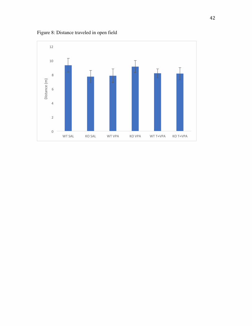
Distance traveled
Motor activity was monitored prior to social testing during a 5 minute habituation
period (Fig. 8). A two-way ANOVA was run to assess differences in treatment or
genotype on distance traveled. Results showed no effect of treatment (F(2,63)=.080,
p=.923) or genotype (F(1,63)=.024, p=.879) indicating VPA exposure did not affect
motor ability.
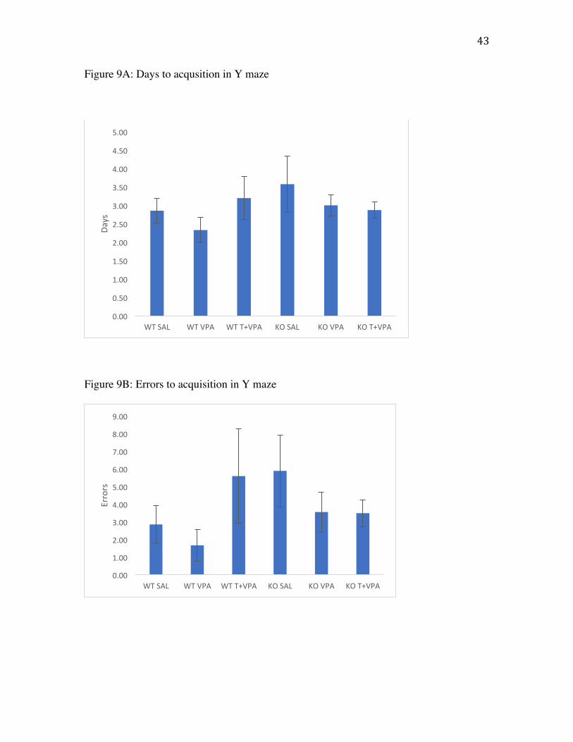
Y maze
Y maze data was examined for number of days to acquisition, errors to
acquisition, days to reversal and errors to reversal (Fig. 9). A two-way ANOVA
(treatment x genotype) yielded no significant differences between treatment groups or
genotype in any measure of Y maze performance. Figure 10a shows no differences
between groups on days to acquisition (F(2,46)=.712 , p=.496) indicating all animals
acquired the task at the same rate. Figure 10b shows errors to acquisition in which there
were no differences between groups (F(1,46)=.443 , p=.645). There were no differences
between groups in days to reversal (F(1,46)= .712, p=.496) or errors to reversal (F(1,46)=
.588, p=.560) as depicted in figure 10c and d respectively. This suggests there was no
perseveration difference evident by genotype or treatment.
NRF2 expression 2 hours post VPA exposure

19
NRF2 expression was examined through Western blot analysis in which nuclear
fractionations were probed for NRF2 and H3 (housekeeping) two hours following VPA
exposure in both young animals and adult animals (Figure 10). Results indicated a trend
towards increased nuclear NRF2 expression in P14 VPA treated animals compared to
saline treated controls (N=4/group) (t(7)=-1.923, p=.09) (figure 11A). No differences
between VPA and saline treated adult animals (~P90) were observed (p>.05) (Fig.11B).
There were no differences in amount of protein in each sample (p>.05).

20
DISCUSSION
Although the etiology of autism is not well understood, environmental toxicants,
genetics and infection have all been implicated in its development. More specifically,
oxidative stress has been a hypothesized as a potential mechanism underlying
neurobehavioral development disruptions associated with autism (Edelson & Cantor,
1998). The results presented here suggest VPA, when administered early postnatally, may
induce oxidative stress which in turn may cause the development of some autistic-like
behaviors.
The present study was able to successfully replicate previous work by Wagner et
al. (2006) to model autistic regression in which a skill matures at the same rate as control-
treated pups but then the toxicant exposure results in a loss of those skills. Previous work,
along with the present study, demonstrate postnatal VPA administration results in
regression in ability to mid-air right with VPA treated animals performing significantly
worse than saline treated controls following exposure. Similar to Wagner et al. (2006),
there were no observed effects of VPA treatment on general motor behavior or body
weight throughout testing as observed in the open field habituation phase of social
testing, Morris water maze path length or swim speeds. Taken together these results
indicate it is possible to model autistic regression with no adverse effects of VPA
treatment on motor development or body weight.
VPA acts as a GABA agonist which when metabolized is known to induce
oxidative stress, as observed through a decrease in endogenous regulatory antioxidant
enzymes (Jurima-Romet at al., 1996). Not only is VPA associated with an oxidative
stress response, its role as a GABA agonist may act as a mechanism to induce oxidative

21
stress. Recent work by Ben-Ari (2014) suggests GABA may shift from an excitatory
action during fetal development and into early postnatal to inhibitory role around the
third week of life in rodents. As such, by administering VPA at postnatal day 14 the
present study targets the window of hypothesized excitatory GABA action. If GABA is
indeed excitatory at this time, VPA administration would be expected to result in GABA
buildup resulting in excitotoxicity during a critical developmental period.
The present study further extends previous findings on oxidative stress induction
following VPA exposure through examination of NRF2 protein levels in VPA-treated
WT animals. Reactive oxygen species (ROS), the byproduct of oxidative stress, are
neutralized by endogenous antioxidant mechanisms, of which NRF2 is a master regulator
(Wakabayashi et al., 2010; Sandberg et al., 2013). When NRF2, which is normally found
bound to KEAP1 in the cytoplasm, is activated by ROS, it enters the nucleus and initiates
downstream induction of antioxidant genes (Wakabayashi et al., 2010; Sandberg et al.,
2013). As such, nuclear NRF2 protein expression through Western blotting analysis, was
used as a measure of oxidative stress response as NRF2 observed in the nucleus can be
thought of as “activated” NRF2. The present study observed increased nuclear NRF2
levels in VPA treated animals compared to controls two-hours post-exposure,
demonstrating that VPA induces oxidative stress. Further, the present study examined
VPA treatment on both young animals (P14) as a means to target the critical window of
GABA shift in which GABA is hypothesized to be excitatory in comparison to adult
animals (~P90) to target inhibitory GABA. Increased NRF2 was observed in the VPA-
treated pups but the effect was not present in the VPA-treated adult animals, supporting
the GABA shift hypothesis. Together, Western blotting results support the hypothesis

22
that VPA induces oxidative stress and that GABA has a shift in action from early
postnatal life to adulthood.
Nrf2 knockout mice have been shown to have enhanced susceptibility to various
environmental toxicants as well as behavioral deficits following postnatal VPA exposure
(Itoh et al., 1997; Liu et al., 2010; Ishii et al., 2005; Innamorato et al., 2010; Furnari et al.
2014). The present study examined mice lacking the NRF2 gene and results supported
previous findings by Furnari et al (2014) further supporting oxidative stress as a
mechanism for behavioral deficits observed following VPA treatment. Children with
autism have been found to have increased body burdens of environmental toxicants
associated with oxidative stress as well as increased levels of oxidative stress biomarkers
indicated increased susceptibility to oxidative stress (Edelson & Cantor, 1998; Dietert &
Dietert, 1997; Ming et al., 2005). This potential inability to manage high levels of
oxidative stress following toxicant exposure compounded with genetic susceptibility may
offer a potential mechanism for the development of autism.
The identified connection of oxidative stress to VPA exposure makes it a prime
model for examining antioxidant pretreatment as a potential mechanism for protection
from the previously identified deleterious effects of VPA. Previous work has indicated
pretreatment of high levels of antioxidants reduces the behavioral effects associated with
toxicant exposure (Cheh et al., 2010; Deeb et al., 2000). Specifically, previous work by
Ming et al. (2008) suggests vitamin E can protect postnatally VPA treated animals from
behavioral deficits while work by Cheh et al. (2010) suggests Trolox, a water-soluble
form of vitamin E, can protect animals from MeHg challenge. The present study
extended upon this work by examining Trolox as a pretreatment serving to protect

23
animals from VPA exposure. Results revealed VPA treated animals pretreated with
Trolox were partially protected from behavioral deficits observed in mid-air righting and
Morris water maze learning in VPA-alone treated animals. Collectively, these findings
confirm VPA acts through the generation of ROS.
Although the present study provided evidence of autistic like regression as well as
impaired learning in the Morris water maze, some expected behavioral deficits were not
observed. A plausible explanation for the lack of evidence to support deficits in social
approach behavior may be due to the fact that the social deficits associated with postnatal
VPA treatment are subtle and our ability to measure social behavior may not have been
sensitive enough to detect deficits. To further explore this, a follow up study is currently
being conducted examining social play behavior in these animals. Social play is a basic
measure of social behavior in which two previously isolated animals are placed in a
chamber and allowed to interact for a 30-minute session on two consecutive days (Cheh
et al., 2006). Videos are then scored by trained observers for a variety of social behaviors
including crawl over/under, anogenital sniffs, allogrooming, self-grooming, chases, and
aggression.
Additionally, no treatment or genotype differences were observed in Y maze
testing for perseverative behaviors or rotarod testing in adulthood. Y maze testing is a
novel measure for examining the effects of VPA treatment and Nrf2 genotype effects as
well as Trolox pretreatment protection and as such a more basic measure such as marble
burying may be warranted in further studies (Sungur et al., 2014). Differences between
treatment and genotype were identified in VPA and saline-treated WT and KO animals in
adolescence but pretreatment was not able to be examined at this time point. Rotarod

24
testing in adulthood revealed no difference between groups which may be due to the fact
testing took place between 80 and 175 days post exposure.
To extend the present findings, further work should be conducted to examine
VPA treatment at varying time points, particularly prenatal, P7, and P21 in hopes to
identify the critical window of GABA shift and then replicated with Trolox pretreatment.
Further, future Western blotting studies should examine the protective effects of Trolox
pretreatment on nuclear NRF2 expression at P14 and extend the findings to the additional
time points. It may also be beneficial to examine the effects of repeated VPA
administration during early postnatal development and to build upon previously described
behavioral deficits recently reported by Bath & Pimental (2017). Further, examining
repeated VPA administration in combination with Trolox pretreatment may elucidate its
efficacy over time.
The mechanism underlying neurobehavioral development disruptions associated
with autism is far from understood but the results of the present study, at least in part,
support the hypothesis that oxidative stress induction may result in behavioral
abnormalities akin to autism. The use of VPA as a method to induce oxidative stress was
successful in the present study as revealed by Western blot analysis following VPA
exposure. Behavioral results indicated an effect of VPA treatment in mid-air righting
ability, Morris water maze learning, and rotarod performance. Further, Trolox
pretreatment showed protection in the mid-air righting task as well as MWM testing. No
effects of treatment or genotype were observed in social approach or Y maze testing but
the methods used for these behaviors may need modification for more subtle
identification of deficits in future research. Collectively, the results of the present study
25
warrant further investigation into the link between oxidative stress and autism as well as
antioxidants as a protective mechanism from behavioral deficits. Further, this study
confirms postnatal VPA as a valid model of autistic regression as well as VPAs potential
mechanism of action through oxidative stress induction and the ability of antioxidants to
act as a protective mechanism.

26
References Al-Amin, M., Rahman, M., Kahn, F., Zaman., Reza, H. (2015). Astaxanthin improves
behavioral disorder and oxidative stress in prenatal valproic acid-induced mice model of autism. Behavioral Brain Research, 1, 112-121.
Banji, D., Banji, O.J.F., Abbagoni, S., Hayath, MS., Kambam, S., Chiluka, VL. (2011). Amelioration of behavioral aberrations and oxidative markers by green tea extract in valproate induced autism in animals. Brain Research, 1410, 121-151. doi:10.1016/k.brainres.2001.06.063
Ben-Ari Y. (2014). The GABA excitatory/inhibitory developmental sequence: a personal journey. Neuroscience 279, 187-219, doi:10.1016/j.neuroscience.2014.01.001
Bernhardt, L.K., Madhyastha, S., Bairy, L., Kishore, A. (2017). Folia Neuropathology, 55(1), 38-48.
Center for Disease Control and Prevention. (2014). Autism Prevalence. Chauhan, A, Chauhan, V. Oxidative stress in autism. (2006). Journal of pathophysiology
13, 171-181.doi: 10.1016/j.pathphys.2006.05.007 Cheh, M.A., Halladay, A.K., Yochum, C.L., Reuhl, K.R., Polunas, M., Ming, X. and
Wagner, G.C. (2010). Autism and Oxidative Stress: Evidence From an Animal Model. In: Autism: Oxidative Stress, Inflammation and Immune Abnormalities, Eds: Chauhan, A., Chauhan, V. and Brown, T. Taylor & Francis/CRC Press, New York, Chapter 8, 133-153.
Cheh, M.A., Millonig J.H., Roselli, L.M., Ming, X., Jacobsen, E., Kamdar, S., Wagner, G.C. (2006). En2 knockout mice display neurobehavioral and neurochemical alterations relevant to autism spectrum disorder. Brain Research, 1116, 166-176. doi:10.1016/j.brainres.2006.07.086
Chez MD, Buchanan CP, Aimonovitch MC., Becker, M., Schaefer, K., Black, C., Komen, J. (2002). Double-blind, placebo-controlled study of l-carnosine supplementation in children with autistic spectrum disorders. J Child Neurol 17, 833-837.
Christensen, J., Gronborg, TK., Sorensen, MJ., Schendel, D., Parner, ET., Pedersen, LH., Vestergaard, M. (2013). Prenatal valproate exposure and risk of autism spectrum disorders and childhood autism. JAMA : the journal of the American Medical Association 309, 1696-1703, doi:10.1001/jama.2013.2270
Deeb, SA, Moutaery, KA, Arshaduddin, M, & Tariq, M. (2000). Vitamin E decreases valproic acid induced neural tube defects in mice. Neuroscience Letters 292, 179-182.
Dietert, R. R. & Dietert, J. M. (2008). Potential for early-life immune insult including developmental immunotoxicity in autism and autism spectrum disorders: focus on critical windows of immune vulnerability. Journal of toxicology and environmental health. Part B, Critical reviews 11, 660-680. doi:10.1080/10937400802370923
Dolske MC, Spollen J, McKay S, Lancashire, E., Tolbert, L. (1993). A preliminary trial of ascorbic acid as supplemental therapy for autism. Prog Neuro-Psychopharmacol & Biol Psychiat 17, 765-774.
Edelson, S. B. & Cantor, D. S. (1998). Autism: xenobiotic influences. Toxicology and industrial health 14, 799- 811.

27
Erden-Inal, M, Sunal, E., Kanbak, G. (2002). Age related changes in the glutathione redox system. Journal of Cellular Biochemistry, 20, 61-66.
Filipek, P. A., Juranek, J., Smith, M., Mays, LZ., Ramos, ER., Boasica, M., Masser-Frye, D., Laulhere, TM., Modahl, C., Spence, MA., Gargus, JJ. (2003). Mitochondrial dysfunction in autistic patients with 15q inverted duplication. Annuals of neurology 53, 801-804, doi:10.1002/ana.10596
Furnari, M., Saw, C., Kong, A, Wagner, G.C. (2014). Altered behavioral development in Nrf2 knockout mice following early postnatal exposure to valproic acid. Brain Research Bulletin. 109, 132-142.
Innamorato NG, Jazwa A, Rojo AI, García C, Fernández-Ruiz J, Grochot-Przeczek A, Stachurska A, Jozkowicz A, Dulak J, Cuadrado A. (2010). Different susceptibility to the Parkinson’s toxin MPTP in mice lacking the redox master regulator Nrf2 or its target gene heme oxygenase-1. PLoS ONE, 5(7).
Ishii Y, Itoh K, Morishima Y, Kimura T, Kiwamoto T, Iizuka T, Hegab AE, Hosoya T, Nomura A, Sakamoto T, Yamamoto M, Sekizawa K.(2005). Transcription factor Nrf2 plays a pivotal role in protection against elastase-induced pulmonary inflammation and emphysema. J. Immunology, 175(10):6968–6975.
Itoh K, Chiba T, Takahashi S, Ishii T, Igarashi K, Katoh Y, Oyake T, Hayashi N, Satoh K, Hatayama I, Yamamoto M, Naveshima Y. An Nrf2/small maf heterodimer mediates the induction of phase II detoxifying enzyme genes through antioxidant response elements. Biochem. Biophys. Res. Commun. 1997; 236:313–322. [PubMed: 9240432]
James, S. J., Cutler, P., Melnyk, S., Jernigan, S., Janak, L., Gaylor, DW, Neubrander, JA. (2004). Metabolic biomarkers of increased oxidative stress and impaired methylation capacity in children with autism. The American journal of clinical nutrition 80, 1611-1617.
Jurima-Romet, M., Abbott, F. S., Tang, W., Huang, H. S. & Whitehouse, L. W. (1996). Cytotoxicity of unsaturated metabolites of valproic acid and protection by vitamins C and E in glutathione-depleted rat hepatocytes. Toxicology 112, 69-85.
Liu F, Ichihara S, Valentine WM, Itoh K, Yamamoto M, Sheik Mohideen S, Kitoh J, Ichihara G. Increased susceptibility of Nrf2-null mice to 1-bromopropane-induced hepatotoxicity. Toxicol. Sci. 2010; 115(2):596–606.
Ming, X., Cheh, M., Yochum, C., Halladay, A., Wagner, GC. (2008). Evidence of Oxidative Stress in Autism Derived from Animal Models. American Journal of Biochemistry and Biotechnology 4, 218-225.
Ming, X., Brimacombe, M., Wagner, G.C. (2007). Prevalence of motor impairments in autism spectrum disorders. Brain & Development 29, 565-570. Doi:10.1016/j.braindev.2007.03.002
Ming, X., Stein, T.P., Brimacombe, M., Johnson, W.G., Lambert, G.H., Wagner, GC. (2005). Increased excretion of a lipid peroxidation biomarker in autism. Prostagladins, Leukotrienes, and Essential Fatty Acids 73, 379-384. doi:10.1016/j.plefa.2005.06.002.
Muralidharan, S. & Mandrekar, P. (2013). Cellular stress response and innate immune signaling: integrating pathways in host defense and inflammation. Journal of Leukocyte Biology 94, 1167-1184. doi:10.1189/jlb.0313153

28
Perry, SW., Norman, J.P., Litzburg, A., Gelbard, H.A. (2004). Antioxidants are required during the early critical period, but not later, for neuronal survival. Journal of Neuroscience, 78, 485-492.
Roberts, R. A., Smith, RA., Safe, S., Szabo, C., Tjalkens, RB., Roberston, FM. (2010). Toxicological and pathophysiological roles of reactive oxygen and nitrogen species. Toxicology 276, 85-94. doi:10.1016/j.tox.2010.07.009
Rossignol, D. A. & Frye, R. E. (2012). A review of research trends in physiological abnormalities in autism spectrum disorders: immune dysregulation, inflammation, oxidative stress, mitochondrial dysfunction and environmental toxicant exposures. Molecular psychiatry 17, 389-401. doi:10.1038/mp.2011.165
Sandberg, M., Patil, J., D'Angelo, B., Weber, S. G. & Mallard, C. (2013). NRF2-regulation in brain health and disease: Implication of cerebral inflammation. Neuropharmacology 79C, 298-306. doi:10.1016/j.neuropharm.2013.11.004
Singh, K., Conners, S., Macklin, E., Smith, K., Fahey, J., Talalay, P. & Zimmerman, A. (2014). Sulforaphane treatment of autism spectrum disorder (ASD). PNAS, 111, 15550-15555. doi:10.1073/pnas.1416940111
Sogut, S., Zoroglu, S., Ozyurt, H., Yilmas, H., Ozugurlu, F., Sivasli, F., Yetkin, O, Yanik, M., Tutkun, H., Savas, H., Tarakcioglu, M, Akyol, O. (2003). Changes in nitric oxide levels and antioxidant enzyme activities may have a role in the pathophysiological mechanisms involved in autism. Clinical chimica acta; international journal of clinical chemistry 331, 111-117.
Sungur, A., Vörckel, K., SChwarting, R., Wöhr, M. (2014). Reptitive behaviors in the Shank1 knockout mouse modle for autism spectrum disorder: Developmental aspects and effects of social context. Journal of Neuroscience Methods, 234, 92-100.
Wagner, G. C., Reuhl, K. R., Cheh, M., McRae, P. & Halladay, A. K. (2006). A new neurobehavioral model of autism in mice: pre- and postnatal exposure to sodium valproate. Journal of autism and developmental disorders 36, 779-793. doi:10.1007/s10803-006-0117-y
Wakabayashi, N., Slocum, S. L., Skoko, J. J., Shin, S. & Kensler, T. W. (2010). When NRF2 talks, who's listening? Antioxid Redox Signal 13, 1649-1663. doi:10.1089/ars.2010.3216
Yochum, C., Dowling, P., Reuhl, K., Wagner, GC., Ming, X. (2008). VPA-induced apoptosis and behavioral deficits in neonatal mice. Brain Research, 1203, 126-132.
Yorbik, O., Sayal, A., Akay, C., Akbiyik, D. I. & Sohmen, T. (2010). Investigation of antioxidant enzymes in children with autistic disorder. Prostaglandins, leukotrienes, and essential fatty acids 67, 341-343
Zoroglu, S. S., Armutcu, F., Ozen, S, Gurel. A., Sivasli, E., Yetkin, O, Meram, I. (2004). Increased oxidative stress and altered activities of erythrocyte free radical scavenging enzymes in autism. European archives of psychiatry and clinical neuroscience 254, 143- 147. doi:10.1007/s00406-004-0456-7

29
FIGURE LEGENDS Figure 1A: Mid-air righting ability from P13-19 as a line graph to depict trend. Pups
received VPA following testing on P14. Values are mean correct responses per day.
* indicates significant difference between VPA and saline groups (p <.05)
Figure 1B: Mid-air righting ability from P13-19 depicted as a histogram to convey
standard error. Pups received VPA following testing on P14. Error bars represent SEM.
* indicates significant difference between VPA and saline groups (p <.05)
Figure 2A: Rotarod performance from P25-P27 as a line graph to depict trend. Values are
mean time spent on the rotarod.
Figure 2B: Rotarod performance from P25-P27 collapsed across genotypes. Values are
mean time spent on the rotarod. Error bars represent SEM.
* indicates significant difference between VPA and saline groups (p <.05)
Figure 3A: Rotarod performance of adult animals (~P90-200) as a line graph to depict
trend. Values are mean time spent on the rotarod.
Figure 3B: Rotarod performance of adult animals (~P90-200) depicted as a histogram to
convey standard error. Values are mean time spent on the rotarod. Error bars represent
SEM Error bars represent SEM.
30
Figure 4A: Morris water maze acquisition as a line graph to depict trend. Values are
mean latency (seconds) to find the platform across 5 trials per day.
Figure 4B: Morris water maze acquisition depicted as a histogram to depict trend. Values
are mean latency (seconds) to find the platform across five 60 second trials per day. Error
bars represent SEM.
Figure 4C: Morris water maze acquisition (day 1-4) collapsed across genotype. Values
are mean latency (seconds) to find the platform across five 60 second trials per day. Error
bars represent SEM.
* indicates significant difference between VPA and saline groups (p <.05)
Figure 5A: Path length to find the platform in acquisition phase of the Morris water maze
task as a line graph to depict trend. Values are mean path length (m).
Figure 5B: Path length to find the platform in acquisition phase of the Morris water maze
task as a histogram to depict error. Values are mean path length (m). Error bars represent
SEM.
Figure 6: Time spent in the target quadrant during the probe trial (day 5). Values are
mean time spent (sec) during a 60 second probe trial. Error bars represent SEM.

31
Figure 7A: Time spent interacting with stranger 1 in social approach testing. Values are
mean time spent in proximity of the stranger animal compared to the areas without a
stranger (empty) in a 10-minute test session. Error bars represent SEM.
Figure 7B: Time spent interacting with habituated stranger (stranger 1) and novel stranger
as compared to time spent in empty zones across 10-minute test. Values are mean time
spent in each zone. Error bars represent SEM.
Figure 8: Distance traveled (m) in open field across a 5 minute test. Values are average
distance traveled. Error bars represent SEM.
Figure 9A: Average days to successfully complete acquisition in the Y maze task. Values
are mean days. Error bar represent SEM.
Figure 9B: Average errors to successfully complete acquisition in the Y maze task.
Values are mean errors. Error bar represent SEM.
Figure 9C: Average days to successfully complete reversal in the Y maze task. Values are
mean days. Error bar represent SEM.
Figure 9D: Average errors to successfully complete reversal in the Y maze task. Values
are mean errors. Error bar represent SEM.

32
Figure 10A: Example blot probed with NRF2 (68 kDa). Ladder labeled for nearest
relevant band (60 kDa).
Figure 10B: Example blot probed with H3 (15 kDa). Ladder labeled for nearest relevant
band (20 kDa).
Figure 11A: NRF2 expression levels 2 hours post VPA exposure in pups (P14)
+ indicates trend toward significant difference between VPA and saline (p=.09)
Figure 11B: NRF2 expression levels 2 hours post VPA exposure in adult animals (~P90)

33
Figure 1A: Mid-air righting trend lines
Figure 1B: Mid-air righting performance from P13-19
0
0.5
1
1.5
2
2.5
3
P13 P14 P15 P16 P17 P18 P19
CorrectR
espo
nses
Dayoflife
KOSAL WTSAL KOT+VPA WTT+VPA KOVPA WTVPA
*
**
* *
0
0.5
1
1.5
2
2.5
3
P13 P14 P15 P16 P17 P18 P19
Correctrespo
nses
Dayoflife
KOSAL WTSAL KOT+VPA WTT+VPA KOVPA WTVPA
* *
* * *

34
Figure 2A: Rotarod performance from P25-27 trend lines
Figure 2B: Rotarod performance from P25-27 by treatment
0.00
5.00
10.00
15.00
20.00
25.00
30.00
35.00
Day1 Day2 Day3
Time(sec)
WTSAL KOSAL WTVPA KOVPA
0.00
5.00
10.00
15.00
20.00
25.00
30.00
35.00
Day1 Day2 Day3
Time(sec)
SAL VPA
*

35
Figure 3A: Adult rotarod performance curve
Figure 3B: Adult rotarod performance
0.0000
5.0000
10.0000
15.0000
20.0000
25.0000
30.0000
35.0000
Day1 Day2 Day3
Time(sec)
WTSAL KOSAL WTVPA KOVPA WTT+VPA KOT+VPA
0.00
5.00
10.00
15.00
20.00
25.00
30.00
35.00
40.00
45.00
Day1 Day2 Day3
Time(sec)
WTSAL KOSAL WTVPA KOVPA WTT+VPA KOT+VPA

36
37
Figure 4A: Morris water maze acquisition curves (Day 1-4)
Figure 4B: Morris water maze acquisition performance (Day 1-4)
0.00
5.00
10.00
15.00
20.00
25.00
30.00
35.00
40.00
Day1 Day2 Day3 Day4
Latency(sec)
WTSAL KOSAL WTVPA KOVPA WTT+VPA KOT+VPA
*

38
Figure 4C: Morris water maze acquistion by treatment
0.00
5.00
10.00
15.00
20.00
25.00
30.00
35.00
40.00
Day1 Day2 Day3 Day4
Latency(sec)
SAL VPA T+VPA
*
* * *
39
Figure 5A: MWM acquistion path length trend
Figure 5B: MWM acquistion path length
0.0000
1.0000
2.0000
3.0000
4.0000
5.0000
6.0000
Day1 Day2 Day3 Day4
PathLen
gth(m
)
WTSAL KOSAL WTVPA KOVPA WTT+VPA KOT+VPA
0.0000
1.0000
2.0000
3.0000
4.0000
5.0000
6.0000
7.0000
Day1 Day2 Day3 Day4
PathLen
gth(m
)
WTSAL KOSAL WTVPA KOVPA WTT+VPA KOT+VPA
*

40
Figure 6: Time spent in the target quadrant on probe trial
0.00
5.00
10.00
15.00
20.00
25.00
WTSAL KOSAL WTVPA KOVPA WTT+VPA KOT+VPA
TimeSpe
nt(sec)

41
Figure 7A: Social Approach behavior with stranger 1
Figure 7B: Social approach behavior in the test phase
0.0000
50.0000
100.0000
150.0000
200.0000
250.0000
300.0000
WTSAL KOSAL WTVPA KOVPA WTT+VPA KOT+VPA
Time(sec)
Stranger1 Empty
0.00
50.00
100.00
150.00
200.00
250.00
300.00
350.00
WTSAL KOSAL WTVPA KOVPA WTT+VPA KOT+VPA
Time(sec)
Habituated Novel EmptyArea
*
* *
* * *
42
Figure 8: Distance traveled in open field
0
2
4
6
8
10
12
WTSAL KOSAL WTVPA KOVPA WTT+VPA KOT+VPA
Distance(m
)
43
Figure 9A: Days to acqusition in Y maze
Figure 9B: Errors to acquisition in Y maze
0.00
0.50
1.00
1.50
2.00
2.50
3.00
3.50
4.00
4.50
5.00
WTSAL WTVPA WTT+VPA KOSAL KOVPA KOT+VPA
Days
0.00
1.00
2.00
3.00
4.00
5.00
6.00
7.00
8.00
9.00
WTSAL WTVPA WTT+VPA KOSAL KOVPA KOT+VPA
Errors

44
Figure 9C: Days to reversal in Y maze
Figure 9D: Errors to reversal in Y maze
0.00
1.00
2.00
3.00
4.00
5.00
6.00
7.00
8.00
WTSAL WTVPA WTT+VPA KOSAL KOVPA KOT+VPA
Days
0.00
5.00
10.00
15.00
20.00
25.00
WTSAL WTVPA WTT+VPA KOSAL KOVPA KOT+VPA
Errors
45
Figure 10A: Example NRF2 blot
Figure 10B: Example H3 blot

46
Figure 11A: P14 NRF2 expression 2 hours post VPA exposure
Figure 11B: Adult NRF2 expression 2 hours post VPA exposure
0
0.5
1
1.5
2
2.5
SAL VPA
NRF2:H3
*+
0
0.2
0.4
0.6
0.8
1
1.2
1.4
1.6
1.8
SAL VPA
NRF2:H3