Embed Size (px)
Citation preview

Available online at www.worldscientificnews.com
WSN 33 (2016) 1-14 EISSN 2392-2192
Antibacterial activity of modified zinc oxide
nanoparticles against Pseudomonas aeruginosa isolates of burn infections
Noor Hadi Aysaa, Halah Dawood Salmanb
Clinical and Laboratory Science Department, College of Pharmacy, University of Babylon, Babylon, Iraq
a,bE-mail address: [email protected] , [email protected]
ABSTRACT
In this research antimicrobial activity of nanoparticles ZnO on perilous bacteria such as
Pseudomonas aeruginosa was evaluated. P. aeruginosa is important pathogen that caused burn wound
infections as it is multi-drug resistant and has several virulence factors. Fifteen samples of P.
aeruginosa were collected from patients who suffering from Burn infections in Al-Hilla teaching
hospital burn unit with the age range between (7-80) years old for both genders. After collecting burn
samples, the diagnosis and characterization were performed by culturing and biochemical tests. ZnO
NPs were synthesized by chemical method, Zinc oxide nanoparticles are well-known to be one of the
multifunctional inorganic compounds which are widely used in medical applications. This study aims
to prepare ZnO nanoparticles with particle size ranging from 23-29 nm. In the present study, surface
modification of ZnO nanoparticles was performed, and influence of modification of the structure and
morphological properties was investigated. The resulting nanoparticles were characterized by X-ray
diffraction (XRD), scanning electron microscopy (SEM) and atomic force (AFM). Zinc oxide
nanoparticles with the average diameter of about 29 nm were modified with an oleic acid to exert
more compatibility. From the results obtained it is suggested that modified ZnO-nanoparticles could
be used effectively in safety environmental and medical applications. Antibacterial activity for
nanoparticle ZnO against P. aeruginosa isolates was measured by: Agar Diffusion Technique and
Minimum Inhibitory Concentration (MIC)/Minimum bactericidal Concentration (MBC) with
microdilution. The best zone of inhibition was (35.5mm) at a concentration of 40 μg/ml of nano-ZnO
in one strain of P. aeruginosa while the lowest inhibition zone was (16 mm) at a concentration of 20
World Scientific News 33 (2016) 1-14
-2-
μg/ml of nano ZnO in one strain also. In addition, all P. aeruginosa isolates were completely inhibited
at the concentration of 3.7 μg/ml of nano-ZnO (MIC) but no significant antibacterial activity was
observed at concentrations less than 1.8 μg/ml of nano-ZnO and the (MBC) was same as MIC (3.7
μg/ml) for all P.aeruginosa isolates.
Keywords: ZnO; nanoparticles; surface modification; burn infection; Pseudomonas aeruginosa;
Antimicrobial activity
1. INTRODUCTION
Despite the increased knowledge of microbial pathogenesis and applications of modern
therapeutics, the morbidity and mortality associated with the microbial infections still remain
high; Therefore, there is an increasing in the infectious diseases and in the drug resistance in
the pathogenic bacteria at an alarming rate is a matter of serious concern [1]. From that, there
is a needing to discover novel strategies and identify new antimicrobial agents from natural
and inorganic substances to develop the next generation of drugs or agents to control
microbial infections. In the recent times, the advances in the field of nano sciences and
nanotechnology has brought to the fore the nano sized inorganic and organic particles leading
to increasing applications in industry, medicine and therapeutics, synthetic textiles and food
packaging products [2].
Greater effectiveness on resistant strains of microbial pathogens, less toxicity and heat
resistance are the characteristic of metal oxide nanoparticles, which make them the selective
candidates for eradicating bacteria [3,4]. The small size of nano-ZnO referred which is 250
times smaller than a bacterium the might be giving it the antimicrobial ability, this makes
them easier to adhere with the cell wall of the microorganisms causing its destruction and
leads to the death of the cell, in addition to that metal nano-particles are harmful to bacteria
[5]. Nano ZnO can disrupt the bacterial cell membrane integrity (the particles interact with the
building elements of the outer membrane and might cause structural changes), reduce cell
surface hydrophobicity and down-regulate the transcription of oxidative stress-resistance
genes in bacteria, then degradation and finally cell death [6]. ZnO has recently achieved a
special attention regarding potential electronic application due to its unique optical, electrical
and chemical properties [7].
One of the most frequent and major complications in patients with burn injuries is the
infection and is the main cause for prolonged in-hospital stay and death in cases of wide-
spread burns despite marked progress in the development of treatment methods for these
patients. The development of multi-resistant organisms complicate burn infections, besides
the infected wound may be a potential source of spreading of antibiotic-resistant
microorganisms. The colonization and infection of these wounds are a dual clinical problem.
The infected wound is a cause of pain and discomfort for patients, as well as life-threatening
septic conditions. As a result, the treatment cost and the medical care increase, respectively
[8-10].
P. aeruginosa is a Gram-negative, rod-shaped bacterium, facultative anaerobe, it
belongs to the group of γ-Proteobacteria. This bacteria is an opportunistic pathogen and a
ubiquitous organism that present in soil and water and can be isolated from plants, animals
and humans [11,12]. P. aeruginosa able to tolerate a variety of physical conditions and
World Scientific News 33 (2016) 1-14
-3-
survive on minimal nutritional requirements [13,14]. P. aeruginosa is one of the important
pathogen that caused nosocomial infection, especially in immune suppressed patients like
severe burns, and in patients suffered from cancer [15,16]. The mortality rate of P. aeruginosa
infections were reported from 18% to 61% in hospital-acquired infections [17-19]. Selective
antibiotic pressure led to emerging of acquired multidrug resistance in several countries in the
past; and some multidrug resistant P. aeruginosa infections have been untreatable [20]. The
antibacterial activity of zinc oxide nano particles were probed by many researchers. Zinc
oxide nano particles are better antibacterial agent [21-22]. Therefore, we aimed in this study
to test the antibacterial activity of ZnO nanoparticles on important and perilous bacteria such
as P. aeruginosa isolated from burn infections.
2. MATERIALS AND METHODS
2. 1.Patients
Fifteen (15) samples are collected from patients who suffering from burn infections
with the age range between (7-80) years old for both genders. The period extended from
(February to April-2015) and the test isolates were obtained from samples taken from patients
submitted to Al-Hilla teaching hospital in the burn unit.
2. 2. Collection of specimens and bacterial identification:
Sterile swap samples collected from patients suffering from burn infections and did not
receive any antibiotic treatment before swabbing, moisten swaps with sterile saline were
passed over the infected area in a zigzag motion while twisting, swapped firmly from the
center of infection site outward to the edge (this may be painful for the patient). The samples
were transported as quickly as possible to the laboratory [23].
Then the swab samples had been inoculated on the culture media P. aeruginosa and
were identified by biochemical differentiation tests, including growth on cetrimide agar,
oxidase and catalase tests, motility, growth at 42 °C, growth in oxidation fermentation (OF)
medium, TSI agar and Simon's citrate Then the confirmed P. aeruginosa samples were
cloned three successive times on nutrient agar and stored on a nutrient agar slant at 4 ºC
[24,25]. Also, bacterial samples were maintained in the brain, heart infusion broth containing
15% glycerol at -75 °C during the research period.
2. 3. Preparation of ZnO Nanoparticles
Dissolve acetate, zinc in a mixture of methanol and Mono ethanol secretary at room
temperature and then mixed with a magnetic mixer for one hour until a homogenous solution
occur left for 24 hours and then the solution is heated for 3 hours at 200 Celsius the black
material precipitate calcined at 500 Celsius and then we get a white powder (nano zinc oxide).
2. 4. Preparation of Nano ZnO concentration for Agar Diffusion Method:
Nano zinc oxide was weighed as 10 mg of then dissolved in 10 ml dimethyl sulfoxide
(DMSO) yielding stock solutions of (1 mg/ml) concentration after that (1 ml) of this solution
was diluted to (10 ml) with DMSO again giving a solution of 100 μg/ml concentration, then
from this solution, the required concentration which include: 20-30-40 μg/ml had been

World Scientific News 33 (2016) 1-14
-4-
prepared for agar diffusion method and dilutions: 30-15-7.5-3.7-1.8 had been prepared for the
determination of Minimum Inhibitory Concentration (MIC) and Minimum Bactericidal
Concentration (MBC) [26].
2. 5. Preparation of bacterial samples
From each bacterium sample of the 15 P. aeruginosa isolates, small portion by using
sterilized loop was transferred to 3 ml of nutrient broth media previously prepared and
sterilized then incubated at 37 °C for 24 hrs. After that 0.1 ml from these cultures of each 15
P. aeruginosa isolates were transferred to fifteen sterilized test tubes containing 0.9% NaCl
solution.
2. 6. Determination of activity by agar diffusion method
Antimicrobial activities of nano-ZnO were tested in vitro against all P. aeruginosa
strains by the agar diffusion technique, seeded with a 24 hr- old culture of the microrganism
strains (by sterile cotton swab dipped into the broth of these microorganisms). After
solidification of 25 ml nutrient agar in Petri plates, hollows of three wells (5 millimetr
diameter) were cut into the agar by cork borer and the pathogenic bacterial strains of P.
aeruginosa were tested on this agar, 0.1 ml of nano ZnO solutions dissolved in (DMSO)
prepared earlier in different concentrations which include: 20- 30-40 μg/ml was applied in
these wells. The inoculums size was adjusted so as to deliver final inoculums of
approximately 108 colony forming unit (CFU)/ml, compared with the turbidity of a sample of
the 0.5 McFarland standards. The Petri dishes were incubated at 5-8 °C for 2-3 h to permit
good diffusion and then incubated for 24 h at 37 °C. The assessment of antibacterial was
based on measuring the diameter of the inhibition zone (mm) formed around the well. In
addition to that the activity of (DMSO) alone without nano ZnO was tested on P. aeruginosa
and it had been found there was no any effect of it on this bacteria [26,27].
2. 7. Determination of Minimum inhibitory concentration and Minimum Bactericidal
concentration (MIC/ MBC) as antimicrobial activity Nano-ZnO:
The antimicrobial activities of nano-sized zinc oxide were evaluated, showing
antimicrobial against all P. aeruginosa strains (15 strains) by serial dilution method through
the determination of the minimum inhibitory concentration (MIC and MBC) in culture broth.
The method of twofold serial dilutions (28) was used in this study for determination of the
minimum inhibitory concentration (MIC) values, 1 ml of media was taken in a test tube, to
which, 1ml of test solution (100 μg/ml) was added, thereafter, 0.1ml of the bacterial strains
(P.aeruginosa ) prepared in 0.9% NaCl was added to the test tube containing media and test
solution. Serial dilution was done five times giving concentrations of 30-15-7.5-3.7-1.8
μg/ml. The Nutrient Broth, which contained tested samples and controls were incubated for
24 h at 37 °C. Also control test was performed to ensure that the solvent had no effect on
bacterial growth, this control test was containing inoculated broth supplemented with only
DMSO at the same dilutions used in this research and found inactive in culture medium. The
MIC values were taken as the lowest concentration required to arrest the growth of the
bacteria in the test tube after incubation (showed no turbidity) while the minimum bactericidal
concentration (MBC) was determined by sub culturing 50 μl from each test tube showing no
apparent growth (clear), if there was no growth this concentration was taken as MBC.

World Scientific News 33 (2016) 1-14
-5-
3. RESULT AND DISCUSSION
3. 1. Analysis Results of ZnO Nanoparticles
3. 1. 1. Scanning Electron Microscopy (SEM)
Figure (1) shows different magnifications of ZnO nanoparticles. The results of this
analysis showed the highly homogeneity distribution of ZnO nanoparticles and this agrees
with the results of [29].
Figure 1. SEM images of different magnification of nano-ZnO.
World Scientific News 33 (2016) 1-14
-6-
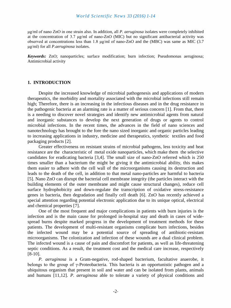
3. 1. 2. SEM/Energy Dispersive X-Ray Spectroscopy (EDS)
Figure (2) shows that spectrum of crystalline ZnO nanoparticles. One can conclude
from the Figure (2) that the purity of ZnO is 100% since there is no elements appears in the
spectrum and element analysis agrees with the research [30].
Figure 2. EDS spectrum of ZnO modified nanoparticles.
3. 1. 3. Atomic Force Microscopy
Figure (3) shows the AFM (3-D) images of ZnO nanoparticles. AFM images prove that
the grains are distributed homogeneously within the scanning area (1518x1514) nm. The
average diameter of synthesized ZnO is measured from AFM analysis using software and is
found to be around (47.69) nm.

World Scientific News 33 (2016) 1-14
-7-
Figure 3. AFM image of the ZnO nanoparticles.
The surface morphology of the ZnO unmodified nanoparticles obtained from the AFM
analysis in Figure (3) shows the surface is very smooth, the average roughness of modified
ZnO is 1.33 nm. This result agrees with [31].
3. 1. 4. X-ray Diffraction Analysis (XRD)
Figure 4. Analysis of X-ray diffraction for ZnO nanoparticles.

World Scientific News 33 (2016) 1-14
-8-
From the X-ray test Figure (4A) of ZnO nanoparticles at a diffracted angle (30° to 70°),
a crystalline peak appeared which indicate crystalline structure at (2θ = 34). This indicates
that crystalline material is prepared and were still in accordance with the standards ZnO XRD
which agrees with the results of [32].
3. 2. Agar Diffusion Technique
Researchers find new compounds that have anti-bacterial effect may provide strategies
for the control of infections and problems related to P. aeruginosa especially burn infections
caused by it. In our research, we have tested the activity of nano ZnO on the identified P.
aeruginosa bacteria isolated from hospitalized patients.
The uses of nano ZnO as antibacterial agent against (P. aeruginosa) isolated from burn
infections in this study. The results of antimicrobial activity of nano-ZnO tested using agar
diffusion technique against all 15 strains of P. aeruginosa represented in (Table-1) showed
that the best zone of inhibition was (35.5mm) at a concentration of 40 μg/ml of nano-ZnO in
one strain of P. aeruginosa while the lowest inhibition zone was (16 mm) at a concentration
of 20 μg/ml of nano ZnO in one strain also.
Table 1. Measured inhibition zone diameters (mm) of nano-ZnO against
Pseudomonas aeruginosa samples.
Nano
ZnO
Conc.
Pseudomonas aeruginosa
Samples
1 2 3 4 5 6 7 8 9 10 11 12 13 14 15
20
μg
/ml
18
16
16
.5
20
20
21
19
20
20
.5
22
22
18
.5
19
.5
21
22
30
μg
/ml
23
25
23
.5
25
24
24
.5
24
24
24
25
23
.5
22
23
.5
25
25
40
μg
/ml
30
30
.5
35
35
35
.5
34
34
34
.5
33
30
.5
30
33
35
35
33
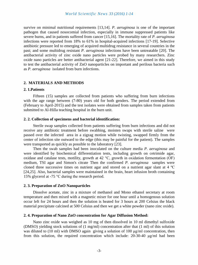
From agar diffusion method results of our study, it is obvious that the relationship
between the concentration of nano ZnO agent and the inhibition zones of P. aeruginosa is
exponentially proportional as shown in (Figure 5). The concentration of ZnO (50, 60) μg/ml
in our knowledge, it is first applied, by us and gave different results about inhibition zone, so
it's not included in the results that is there is poor search related with it to compare with.
World Scientific News 33 (2016) 1-14
-9-
Figure 5. Agar diffusion method.
As the increasing in nano-ZnO concentration lead to increasing in the inhibition zones
and this finding is in accordance with the results of (Sangani MH, et al) [33] because they
found that the effect of ZnO nanoparticles against P. aeruginosa isolates was gradually
increased. On the other hand our results were near to the results of Yousef, et al. [34] as they
found that the inhibition zone of nano ZnO at a concentration of 20 μg/ml (they use only one
concentration of ZnO) against Pseudomonas aeruginosa isolates was (22 mm), while the
average of inhibition zone of nano ZnO at a concentration of 20 μg/ml in our research was
(19.7 mm), they use only one sample of P. aeruginosa but in our research we tested fifteen
strain of the mentioned bacteria and three of them gave the inhibition zone of (22 mm).
In other study done by (Chauhan, et al) [35] they found that the antibacterial activity of
extracellular biosynthesized ZnO nanoparticles against P. aeruginosa estimated about 10.33
mm and 12.66 mm as inhibition zone at a concentration of 25 μl 50 μl respectively, while in
our result the average of inhibition zone at a concentration of 30 μg/ml and at 40 μg/ml were
(22.4 mm) and (33.2 mm) respectively and in other study done by (Voicu, et, al.) [36] used
nano ZnO with average particle size of 19 nm, the inhibition zone of ZnO nanoparticles
against P. aeruginosa at a concentration of 40 μg/ml was 2 mm, these results not in agreement
with our results and that may be due to using of completely different preparation method of
nano ZnO and different size of nanoparticles, it has been demonstrated that the size, shape,
surface area, solubility, chemical composition and dispersion factor of nanoparticles play
exceptional roles in determining their biological responses (Oberdorster et al.) [37].
There are some variations in the results of inhibition zones between strains of P.
aeruginosa such as 16 mm in one strain while the other was 22mm at a concentration of 20
μg/ml of nano-ZnO and 30 mm in one strain, but it was 35 in another strain at a concentration
of 40 μg/ml of nano-ZnO, these differences may be due to constitutional variation between
these strains such as genetic composition and enzymes possession, antibiotic resistance
mechanisms acquisition especially when isolated from hospital.

World Scientific News 33 (2016) 1-14
-10-
3. 3. Determination of Minimum inhibitory concentration and Minimum Bactericidal
concentration (MIC/ MBC) as antimicrobial activity Nano-ZnO
In our study, the relative antimicrobial activity of nano-ZnO suspensions against
pathogenic P. aeruginosa fifteen isolates were studied in nutrient broth quantitatively by
determination of the MIC and MBC.
Here, five nano-ZnO suspensions with different concentrations were tested (of 30-15-
7.5-3.7-1.8 μg/ml) and the results are given in Table 2. The data showed that all P. aeruginosa
isolates were completely inhibited at the concentration of 3.7 μg/ml of nano-ZnO (Minimum
inhibitory concentration -MIC) but no significant antibacterial activity was observed at
concentrations less than 1.8 μg/ml of nano-ZnO. The data also showed that the minimum
Bactericidal concentration (MBC) was same as MIC (3.7 μg/ml) for all P. aeruginosa
isolates. Our results identical to the results of (Nagarajan P. and Rajagopalan V.) [38] in that
ZnO nanoparticles have bactericidal activity in addition they interpreted in their study that
once hydrogen peroxide is generated by ZnO nanoparticles, the nanoparticles remains in
contact with the deadly bacteria to prevent further bacterial action and continue to generate
and discharge hydrogen peroxide to the medium.
In our study, the MIC of nano ZnO on P. aeruginosa isolates was 3.7 μg/ml for all
isolates and that differ from (Saadat, et al) [39] who found that MIC of nano-ZnO on P.
aeruginosa isolates was 300 μg/ml, in other study done by (Yousef, et al.) [34] they found
that MIC was 0.5 μg/ml and that near to our result if we compare it with the result of (Saadat,
et al) [39], these differences from our result may be due to use of preserved bacterial strain of
P. aeruginosa that not isolated from burn or isolation of clinical isolates that were able to
form an effective biofilm, other factors that may lead to different results may be the
differences in the preparation methods of nano ZnO, other probable cause may refer to the
size of ZnO nanoparticles such as (30-90) nm in (Saadat, et al.) study [39]. While we used
ZnO nanoparticles of 40-nm. The antimicrobial property of nanoparticles depends on the
synthesized method, concentration and size of them [40-41], the activity was affected by
particle size, which is controlled by processing parameters [38].
Table 2. Antimicrobial activity MIC/MBC (μg/ mL) of nano-ZnO against
Pseudomonas aeruginosa amples.
Antimicrobi
al activity of
nano-ZnO
Pseudomonas aeruginosa
Samples
1 2 3 4 5 6 7 8 9 10 11 12 13 14 15
MIC ≥ 3.7 μg/ml
MBC ≥ 3.7 μg/ml
World Scientific News 33 (2016) 1-14
-11-
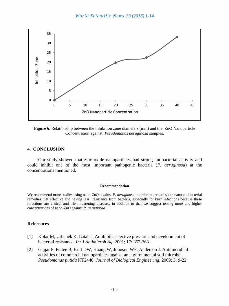
Figure 6. Relationship between the Inhibition zone diameters (mm) and the ZnO Nanoparticle
Concentration against Pseudomonas aeruginosa samples.
4. CONCLUSION
Our study showed that zinc oxide nanoparticles had strong antibacterial activity and
could inhibit one of the most important pathogenic bacteria (P. aeruginosa) at the
concentrations mentioned.
Recommendation
We recommend more studies using nano-ZnO against P. aeruginosa in order to prepare some nano antibacterial
remedies that effective and having less resistance from bacteria, especially for burn infections because these
infections are critical and life threatening diseases, in addition to that we suggest testing more and higher
concentrations of nano-ZnO against P. aeruginosa.
References
[1] Kolar M, Urbanek K, Latal T. Antibiotic selective pressure and development of
bacterial resistance. Int J Antimicrob Ag. 2001; 17: 357-363.
[2] Gajjar P, Pettee B, Britt DW, Huang W, Johnson WP, Anderson J. Antimicrobial
activities of commercial nanoparticles against an environmental soil microbe,
Pseudomonas putida KT2440. Journal of Biological Engineering. 2009; 3: 9-22.
0
5
10
15
20
25
30
35
0 5 10 15 20 25 30 35 40 45
Inh
ibit
ion
Zo
ne
ZnO Nanoparticle Concentration

World Scientific News 33 (2016) 1-14
-12-
[3] Reddy KM, Feris K, Bell J, Wingett DG, Hanley C, Punnoose A. Selective toxicity of
zinc oxide nanoparticles to prokaryotic and eukaryotic systems. Applied physics letters.
2007; 90(21): 213902-3.
[4] Sawai J. Quantitative evaluation of antibacterial activities of metallic oxide powders
(ZnO, MgO and CaO) by conductimetric assay. Journal of Microbiological Methods.
2003; 54(2): 177-82.
[5] Chwalibog A., Sawosz E., Hotowy A., Szeliga J., Mitura S., Mitura K., Grodzik M.,
Orlowski P. and Sokolowska, A. (2010): Visualization of interaction between inorganic
nano-particles and bacteria or fungi. International Journal of Nanomedicine, 5, 1085-
1094.
[6] Pati R, Mehta RK, Mohanty S, Padhi A, Sengupta M, Vaseeharan B, et al. Topical
application of zinc oxide nanoparticles reduces bacterial skin infection in mice and
exhibits antibacterial activity by inducing oxidative stress response and cell membrane
disintegration in macrophages. Nanomedicine. 2014; 10(6): 1195-208.
[7] Baxter J. B. and Aydil E.S. ( 2005): Nanowire based dye sensitized solar cells. Appl.
Phys. Lett. 86, 53114, 2005.
[8] Klasen, H. (2000). A Historical Review of the Use of Silver in the Treatment of Burns.
II. Renewed Interest for Silver, Burns, 26, pp. 131-138.
[9] Landsdown, A.B. (2006). Silver In Health Care: Antimicrobial Effects and Safety in
Use, Curr. Probl. Dermatol, 33, pp. 17-34.
[10] Fong, J. & Wood, F. (2006). Nanocrystalline Silver Dressing in Wound Management: A
Review, International Journal of Nanomedicine, 1(4), pp. 441-449.
[11] Kung VL, Ozer EA, Hauser AR. The accessory genome of Pseudomonas aeruginosa.
Microbiology and Molecular Biology Reviews. 2010; 74: 621-641.
[12] Moreau-Marquis S, Stanton BA, O'Toole GA. Pseudomonas aeruginosa biofilm
formation in the cystic fibrosis airway. Pulmonary Pharmacology & Therapeutics.
2008; 21: 595-599.
[13] Lister PD, Wolter DJ, Hanson ND. Antibacterial-resistant Pseudomonas aeruginosa:
clinical impact and complex regulation of chromosomally encoded resistance
mechanisms. Clinical Microbiology Reviews. 2009; 22(4): 582-610.
[14] Kerr KG, Snelling AM. Pseudomonas aeruginosa: a formidable and ever-present
adversary. Journal of Hospital Infection. 2009; 73: 338-344.
[15] Aloush V, Navon-Venezia S, Seigman-Igra Y, Cabili S, Carmeli Y. Multidrug-resistant
Pseudomonas aeruginosa: risk factors and clinical impact. Antimicrob Agents
Chemother. 2006; 50(1): 43-8.
[16] Gad GF, El-Domany RA, Zaki S, Ashour HM. Characterization of Pseudomonas
aeruginosa isolated from clinical and environmental samples in Minia, Egypt:
prevalence, antibiogram and resistance mechanisms. J Antimicrob Chemother. 2007;
60(5): 1010-7.
World Scientific News 33 (2016) 1-14
-13-
[17] Chatzinikolaou I, Abi-Said D, Bodey GP, Rolston KV, Tarrand JJ, Samonis G. Recent
experience with Pseudomonas aeruginosa bacteremia in patients with cancer:
Retrospective analysis of 245 episodes. Arch Intern Med. 2000; 160(4): 501-9.
[18] Hirsch EB, Tam VH. Impact of multidrug-resistant Pseudomonas aeruginosa infection
on patient outcomes. Expert Rev Pharmacoecon Outcomes Res. 2010; 10(4): 441-51.
[19] Maschmeyer G, Braveny I. Review of the incidence and prognosis of Pseudomonas
aeruginosa infections in cancer patients in the 1990s. Eur J Clin Microbiol Infect Dis.
2000; 19(12): 915-25.
[20] Tsukayama DT, van Loon HJ, Cartwright C, Chmielewski B, Fluit AC, van der Werken
C, et al. The evolution of Pseudomonas aeruginosa during antibiotic rotation in a
medical intensive care unit: the RADAR-trial. Int J Antimicrob Agents. 2004; 24(4):
339-345.
[21] Sangeetha Gunalan, Rajeshwari Sivaraj, and Venckatesh Rajendran, Progress in Natural
Science: Materials International. 22(6) (2012) 693.
[22] Mohammad Reza Arefi, Saeed Rezaei-Zarchi, Saber Imani, African Journal of
Biotechnology. 11(34) (2012) 8520.
[23] Siddiqui A, Bernstein J. Chronic wound infection: Facts and controversies. Clin
Dermatol. 2010; 28: 516-26.
[24] Krieg, Noel. Bergey's Manual of Systematic Bacteriology. Volume 1. Baltimore:
Williams & Wilkins. 1984.
[25] Forbes BA, Sahm DF, Weissfeld AS. Bailey and Scotts' Diagnostic microbiology 12th
ed. Elsevier 2007.
[26] Perez C., Pauli M. and Bazevque P. (1990) An antibiotic assay by the agar well
diffusion method. Acta Biologiae et Medicine Experimentalis 15, 113-115.
[27] NCCLS (National Committee for Clinical Laboratory Standards): Methods for dilution
antimicrobial susceptibility tests of bacteria that grow aerobically. Approved Standard
M100-S12. Wayne. PA, NCCLS; 2002.
[28] Xi JH, Yeo SY, Lee HJ, Jeong SH," Preparation of nano composite fibres for permanent
antibacterial effect", Journal of Material. Science 38 (2003) 2143-2147.
[29] Chwalibog A., Sawosz E., Hotowy A., Szeliga J., Mitura S., Mitura K., Grodzik M.,
Orlowski P. and Sokolowska, A. (2010): Visualization of interaction between inorganic
nano-particles and bacteria or fungi. International Journal of Nanomedicine; 5, 1085–
1094.
[30] Kamellia Nejati, Zolfaghar Rezvani, and Rafat Pakizevand," Synthesis of ZnO
Nanoparticles and Investigation of the Ionic Template Effect on Their Size and Shape",
Int. Nano Lett., 1(2) (2011) 75-81.
[31] Spolenak, R.; Ludwig, W.; Buffiere, J. Y. and Michler, J.,"In-situ elastic strain
measurements – diffraction and spectroscopy", MRS Bulletin, (2010), 35(5): 368-374
[32] Hong R.Y., Pan T.T., Qian J.Z., Li H.Z., "Synthesis and surface modification of ZnO
nanoparticles, Chem. Eng. J. 119 (2006) 71-81.

World Scientific News 33 (2016) 1-14
-14-
[33] Sangani MH, Nakhaei Moghaddam M, Forghanifard M. Inhibitory effect of zinc oxide
nanoparticles on Pseudomonas aeruginosa biofilm formation, Nanomed J, (2015); 2(2):
121-128.
[34] Yousef Jehad M., Enas N. Danial. In Vitro Antibacterial Activity and Minimum
Inhibitory Concentration of Zinc Oxide and Nano-particle Zinc oxide Against
Pathogenic Strains, Journal of Health Sciences, (2012), 2(4): 38-42.
[35] Chauhan R., A. Reddy, J. Abraham. Biosynthesis of silver and zinc oxide nanoparticles
using Pichia fermentans JA2 and their antimicrobial property, Appl Nanosci (2015) 5:
63-71.
[36] Voicu G., O. Oprea, B. S. Vasile, E. Anderonescu, " Antibacterial activity of Zinc
Oxide– Gentamicin hybrid material", Digest Journal of Nanomaterials and
Biostructures 8(3) (2013) 1191-1203.
[37] Oberdorster G, Maynard A, Donaldson K, Castranova V Principles for characterizing
the potential human health effects from exposure to nanomaterials: elements of a
screening strategy. Part Fibre Toxicol 2 (2005) 8.
[38] Nagarajan P. and Rajagopalan V. (2008). Enhanced bioactivity of ZnO nano-particles in
an antimicrobial study. Environ. Sci. Technol. 9 (035004), 7-15
[39] Saadat M, S.R. Mohammadi and Mehdi Eskandari. "Evaluation of Antibacterial
Activity of ZnO and TiO2 Nanoparticles on Planktonic and Biofilm Cells of
Pseudomonas aeruginosa", BIOSCIENCES BIOTECHNOLOGY RESEARCH ASIA,
10(2) (2013) 629-635.
[40] Yuan Z ZL. Influence of ZnO+Fe2O3 additives on the anatase-to-rutile transformation
of nanometer TiO2 powders. Nano Struc Mater. 10 (1998) 1127-33.
[41] Jiang JO, G.; Elder, A.; Gelein, R.; Mercer, P.; Biswas," Does nanoparticle activity
depend upon size and crystal phase? ", Nanotoxicology, 2 (2008) 33-42.
( Received 04 December 2015; accepted 14 December 2015 )