Embed Size (px)
Citation preview

ANTI-DIABETIC AND TOXICOLOGICAL STUDIES OF
GONGRONEMA LATIFOLIUM
By
BASSEL AL HINDI
Thesis submitted in fulfillment of the requirements
of the degree of
Master’s of Science (Pharmacology)
May 2016

ii
ACKNOWLEDGEMENT
This work could not have been carried out without the guidance of Allah SWT.
To my lovely wife, lab partner and mentor for all the guidance – and for making me
always eager to go to the lab every day – thank you! My heartfelt gratitude also goes to
my brother and to those friends who assisted me through this journey financially and by
their advice and counsel.
Special thanks to my supervisors for their support, especially Dr Yam Mun Fei, Assoc.
Prof. Mariam Ahmad, and Prof. Dr. Zaini Asmawi. Thank you for your patience and for
always pushing my colleagues and me to approach excellence.
Moreover, I wish to express my gratitude to Assoc. Prof. Dr. Anees Janee Ali, director
of USM International Office, for encouraging me to engage in USM student community
projects, and, by so doing, allowed me to have cherished experiences beyond academia
with people of all walks of life from other universities and tongues.
A special thank you goes to encik Rosli for being ever so kind.
Last but not the least, I wish to acknowledge the support of the Institute of Postgraduate
Studies (IPS) at USM, which awarded me USM Postgraduate Fellowship for two
consecutive years and, hence, facilitated my journey to become the person I am today.

iii
TABLE OF CONTENTS
Page
ACKNOWLEDGEMENTS ii
TABLE OF CONTENTS iii
LIST OF TABLES vii
LIST OF FIGURES viii
TABLES OF PLATES ix
LIST OF ABBREVIATIONS x
ABSTRAK xi
ABSTRACT xiii
CHAPTER ONE: INTRODUCTION 1
1.1 Diabetes overview 1
1.1.1 Terminology 1
1.1.2 Classification 2
1.1.2 (a) Insulin-Dependent Diabete Mellitus (IDDM) 2
1.1.2 (b) Non-Insulin-Dependent Diabetes Mellitus (NIDDM) 3
1.1.2 (c) Gestational Diabetes Mellitus (GDM) 4
1.1.3 Classes of Conventional Hypoglycemic Agents 4
1.1.3 (a) Insulin-replacement therapy 5
1.1. 3 (b) Pramlintide 6
1.1.3 (c) Biguanides 6
1.1.3 (d) Sulfonylureas 7
1.1.3 (e) Metiglinides 7
1.1.3 (f) α-Glucosidase Inhibitors 8
1.1.3 (g) Thiazolidinediones 9
1.1.3 (h) Dipeptidyl Peptidase-4 (DPP-4) Inhibitors 9

iv
1.1.3 (i) GLP-1 Receptor Agonists 10
1.1.3 (j) SGLT2 inhibitors 10
1.1.4 Diagnosis 10
1.1.5 Worldwide burden 11
1.1.6 Malaysian Burden 11
1.2 Clarifying ambiguous terminology 12
1.3 Trend to herbal therapy 12
1.4 Gongronema latifolium Overview 13
1.4.1 Morphological description 14
1.4.2 Taxonomy 14
1.4.3 Chemical composition 15
1.4.4 GL use in DM treatment 16
1.5 Problem statement 16
1.6 Tested hypothesis 17
1.7 Study objectives 17
CHAPTER TWO: GENERAL METHODOLOGY 19
2.1 Plant material 19
2.2 Preparation of plant extract 19
2.3 Experimental animals 20
2.4 Induction of diabetes 20
2.5 Preparation of metformin for oral dosing 21
2.6 Statistical analysis 21
2.6 Flow chart of study protocols 22

v
CHAPTER THREE: ANTI-DIABETIC STUDY OF GONGRONEMA
LATIFOLIUM 23
3.1 Background 23
3.2 Experimental design 25
3.2.1 Oral glucose tolerance test (OGTT) with GLES in non-diabetic rats 25
3.2.2 Acute (7-h) treatment with GLES in STZ-induced diabetic rats 25
3.2.3 Sub-chronic (14-day) treatment with GLES 26
3.2.3 (a) Blood biochemical parameters following 14-day treatment 26
3.2.3 (b) Histopathological study following 14-day treatment with GLES 28
3.2.4 Glucose uptake in the presence of GLES in isolated rat muscle 28
3.2.5 Glucose absorption in the presence of GLES in isolated rat jejunum 29
3.2.6 GC-MS analysis of GLES 29
3.3 Results 30
3.3.1 Effect of OGTT with GLES in normal rats 30
3.3.2 Effect of acute (7-h) treatment with GLES 31
3.3.3 Effect of sub-chronic (14 days) treatment with GLES 34
3.3.3 (a) Effect of 14-day treatment with GLES on food intake 38
3.3.3 (b) Effect of 14-day treatment with GLES on lipid profile 38
3.3.3 (c) Effect of 14-day treatment on kidney function parameters 41
3.3.3 (d) Effect of 14-day treatment with GLES on serum insulin levels 41
3.3.3 (e) Effect of 14-day treatment with GLES on Langerhans islet area 44
3.3.4 Effect of GLES on muscle glucose uptake 46
3.3.5 Effect of GLES on glucose absorption via the intestinal tract 46
3.3.6 GC-MS analysis of GLES 47
3.4 Discussion 51

vi
CHAPTER FOUR: TOXICITY STUDY OF GONGRONEAM LATIFOLIUM 56
4.1 Background 56
4.2 Experimental design 58
4.2.1 Toxicity assessment of sub-chronic (90-day) oral treatment 58
4.2.1 (a) Hematological analysis following 90-day treatment with GLES 59
4.2.1 (b) Biochemical analysis following 90-day treatment with GLES 59
4.2.1 (c) Histopathological evaluation following 90-day treatment 60
4.3 Results 61
4.3.1 Effect of 90-day treatment with GLES on mortality 61
4.3.2 Effect of 90-day treatment on behavior, body weight and food intake 61
4.3.3 Effect of 90-day treatment with GLES on blood hematology 63
4.3.4 Effect of 90-day treatment with GLES on blood biochemical data 63
4.3.5 Effect of 90-day treatment with GLES on organ weights in normal rats 70
4.3.6 Effect of 90-day treatment on gross necropsy and histopathology 70
4.4 Discussion 77
CHAPTER FIVE: SUMMARY 83
REFERENCES 84
LIST OF CONFERENCES 96
LIST OF PUBLICATIONS 97

vii
LIST OF TABLES
Page
Table 3.1: Animal grouping: acute treatment with GLES .............................................. 27
Table 3.2: Animal grouping: sub-chronic treatment with GLES.................................... 27
Table 3.3 : Kidney function test after 14 days of treatment ........................................... 42
Table 3.4 : Average islet size (µm2) ............................................................................... 44
Table 3.5: Effect of GLES on glucose uptake by isolated rat abdominal muscle .......... 46
Table 3.6: Effect of GLES on glucose active transport via isolated rat jejunum ........... 47
Table 3.7 : GC-MS spectral analysis of GLES ............................................................... 50
Table 4.1: Animal grouping for 90-day treatment with GLES ....................................... 58
Table 4.2: Hematology values in rats administered with GLES for 90 days: (A)
Males .............................................................................................................. 64
Table 4.3: Hematology values in rats administered with GLES for 90 days: (B)
Females ........................................................................................................... 65
Table 4.4: Serum biochemistry in rats administered with GLES for 90 days: (A)
Males .............................................................................................................. 66
Table 4.5: Serum lipid profiles in rats administered with GLES for 90 days: (A)
Males .............................................................................................................. 67
Table 4.6: Serum biochemistry in rats administered with GLES for 90 days: (B)
Females ........................................................................................................... 68
Table 4.7: Serum lipid profiles in rats administered with GLES for 90 days: (B)
Females ........................................................................................................... 69
Table 4.8: Relative organ weights after 90 days of GL administration (g/100g
Body weight): (A) Males ................................................................................ 71
Table 4.9: Relative organ weights after 90 days of GL administration (g/100g
Body weight): (B) Females ............................................................................ 72

viii
LIST OF FIGURES
Page
Figure 3.1: Oral Glucose Tolerance Test with GLES in normal rats. ............................ 32
Figure 3.2: Acute (single) treatment with GLES in STZ-induced diabetic rats. ............ 33
Figure 3.3: Sub-chronic (14 days) treatment with GLES in STZ-induced diabetic
rats. ............................................................................................................... 35
Figure 3.4: Comparing blood glucose levels before and after 14 days of
treatment with GLES. .................................................................................. 36
Figure 3.5: Weight changes during sub-chronic treatment with GLES. ......................... 37
Figure 3.6: Percentages of Changes in food intake (kcal/g b.w./day) during sub-
chronic treatment with GLES. ..................................................................... 39
Figure 3.7: Lipid Profile after 14 days of treatment. ...................................................... 40
Figure 3.8: Serum Insulin Levels after 14 days of treatment. ......................................... 43
Figure 3.9: Gas chromatography-mass spectrometry (GC-MS) chromatogram of
GLES ............................................................................................................ 48
Figure 3.10: Stigmast-4-en-3-one (Sitostenone) ............................................................. 49
Figure 4.1: Mean weight of rats administered with GLES: (A) Male rats (B)
female rats. ................................................................................................... 62

ix
TABLES OF PLATES
Page
Plate 1.1: Gongronema latifolium Benth. (Apocyanaceae) courtesy of West
African Plants. ................................................................................................ 15
Plate 3.1: Langerhans islets under 400x magnification power. ...................................... 45
Plate 4.1: Female control liver under 100x magnification power ................................... 73
Plate 4.2: Male control liver under 100x magnification power ...................................... 73
Plate 4.3: Female TG3 (1000 mg/kg per day for 90 days) liver under 100x
magnification power ....................................................................................... 74
Plate 4.4: Male TG3 (1000mg/kg per day for 90 days) liver under100x
magnification power ....................................................................................... 74
Plate 4.5: Female control kidney under 40x magnification power ................................. 75
Plate 4.6: Male control kidney under 40x magnification power ..................................... 75
Plate 4.7: Female TG3 (1000 mg/kg per day for 90 days) kidney under 40x
magnification power ....................................................................................... 76
Plate 4.8: Male TG3 (1000 mg/kg per day for 90 days) kidney under 40x
magnification power ....................................................................................... 76

x
LIST OF ABBREVIATIONS
ºC Degree Celsius
% Percent
µl Microliter
AD Anno Domini (After Christ)
ADA American Diabetes Association
ALP Alkaline Phosphatase
ALT Alanin Aminotransferase
ANOVA Analysis of Variance
AST Aspartate Aminotransferase
b.w. Body Weight
BGLs Blood Glucose Levels
DC Diabetic Control
DM Diabetes Mellitus
DPP-4 Dipeptidyl Peptidase-4
FBG Fasting Blood Glucose
FDA Food and Drug Administration
GC-MS Gas Chromatography – Mass Spectroscopy
GDM Gestational Diabetes Mellitus
GL Gongronema latifolium Benth.
GLES Gongronema latifolium Ethanolic Soxhlet extract
GLP-1 Glucagon-Like Peptide 1
HDL High-Density Lipoprotein
IDDM Insulin-Dependent Diabetes Mellitus
LD50 Lethal dose enough to kill 50% of a test sample
LDL Low-Density Lipoprotein
mg Milligram
min Minute
mM Mili Molar
NC Normal Control
NIDDM Non-Insulin-Dependent Diabetes Mellitus
NOAEL No-Observed-Adverse-Effect Level
OECD Organization for Economic Co-operation and Development
p.o. per os (oral)
PC Positive Control
PPARϒ Peroxisome Proliferator Activated nuclear Receptor
Rpm Rounds Per Minute
SD Sprague-Dawley
SEM Standard Error of the Mean
SGLTs Sodium–Glucose co-Transporters
STZ Streptozotocin
TG Treatment Group
TZD Thiazolidones
VLDL Very-Low-Density Lipoprotein
WAT White Adopose Tissue
WHO World Health Organization

xi
KAJIAN ANTIDIABETIK DAN TOKSIKOLOGI EKSTRAK GONGRONEMA
LATIFOLIUM
ABSTRAK
Gongronema latifolium Benth. (Apocyanaceae) (GL) mempunyai aktiviti
menurunkan glukosa yang berpotensi untuk digunakan secara meluas. Kajian ini
mengkaji kesan antidiabetik dan toksik GL. Etanol dan peralatan Soklet digunakan
untuk mendapatkan ekstrak soklet etanolik GL (GLES) daripada daunnnya. Bagi
mengkaji kesan antidiabetik, GLES diberikan secara oral kepada tikus – tikus jantan
Sprague-Dawley (SD) yang normal dan diabetik aruhan STZ. Aras glukosa darah
(BGLs), profil lipid serum, aras insulin dan pankreas dinilai selepas rawatan selama 14
hari. Bagi menilai ketoksikan GL, GLES diberikan secara oral pada dos 250, 500, dan
1000 mg/kg kepada tikus – tikus SD selama 90 hari berdasarkan garis panduan OECD.
Indeks ketoksikan yang diukur, termasuk berat badan, parameter – parameter biokimia
dan hematologi, dan berat organ – organ penting. Penilaian histopatologi bagi organ –
organ penting yang terlibat dalam metabolisma xenobiotik dan perkumuhan – hati dan
ginjal – juga dijalankan. GLES menurunkan BGLs tikus normal secara signifikan (P <
0.05) dalam ujian toleransi glukosa pada dos 2 g/kg berat badan, tetapi gagal
merekodkan kesan yang sama pada tikus diabetik dalam rawatan akut selama 7 jam.
Rawatan dengan 1 g/kg berat badan, dua kali sehari mengawal BGLs secara sederhana
seawal hari kesepuluh. Selepas 14 hari rawatan, 1 g/kg dan 0.5 g/kg berat badan GLES
menyebabkan peningkatan dalam purata luas kawasan Langerhans islet, masing –
masing sebanyak 44% dan 50% dibandingkan dengan DC. Dengan menggunakan otot
abdominal tikus, GLES dikesan bertindak sebagai penggalak insulin yang sederhana.
Analisis GC-MS menunjukkan kehadiran Sitostenon, komponen fitosterol yang
diketahui mampu menurunkan glukosa. Dalam kajian toksisiti, GLES tidak

xii
menunjukkan sebarang kesan signifikan terhadap panel – panel fungsi ginjal dan hati,
dan parameter hematologi yang diukur. Trigliserida serum, jumlah kolesterol dan
lipoprotein ketumpatan rendah mencatatkan penurunan di dalam tikus jantan seiring
dengan pengurangan dalam pengumpulan lemak badan retroperitoneal (P < 0.05).
Terdapat percanggahan yang signifikan antara berat organ pada dos yang paling tinggi.
Sebagai contoh, hati menunjukkan pembesaran secara signifikan pada kedua – dua
jantina (P < 0.05). Walaubagaimanapun, kajian histopatologi tidak menunjukkan
sebarang lesi patologi pada hati mahupun ginjal. Secara keseluruhannya, penggunaan
GL dalam mengawal DM menunjukkan kesan yang setara dengan ubat antidiabetik oral
konvensional dan ia mengandungi struktur penyembuhan terhadap pankreas, mungkin
disebabkan oleh kehadiran Sitostenon dan komponen bukan fenolik yang lain. Di
samping itu, kajian ini telah mengesahkan bahawa pengambilan GL secara jangka
panjang adalah selamat, sama ada sebagai sayuran, herba mahupun untuk tujuan
perubatan; tetapi tidak pada dos yang sangat tinggi melebihi tempoh masa tertentu.

xiii
ANTI-DIABETIC AND TOXICOLOGICAL STUDIES OF GONGRONEMA
LATIFOLIUM
ABSTRACT
Gongronema latifolium Benth. (Apocyanaceae) (GL) possesses a considerable
glucose lowering activity to be utilized on a large-scale. This study investigates the
antidiabetic and toxic effects of GL. Ethanol and a Soxhlet apparatus were used to
obtain GL ethanolic Soxhlet extract (GLES) from the leaves. To examine the
antidiabetic effect, GLES was orally administered to male Sprague-Dawley (SD)
normal and STZ-induced diabetic rats. Blood glucose levels (BGLs), serum lipid
profile, insulin levels and the pancreas were evaluated after 14 days of treatment. To
assess GL toxicity, GLES was administered orally at 250, 500 and 1000 mg/kg to SD
rats for 90 days following OECD guidelines. Toxicity indices were measured, including
body weight; biochemical, and hematological parameters; and weights of vital organs.
Histopathological assessment of the key organs involved in xenobiotic metabolism and
excretion – liver and kidneys - was conducted. GLES significantly (P < 0.05) decreased
BGLs of normal rats in glucose tolerance testing at a dose of 2 g/kg b.w., but failed to
do so in diabetic rats undergoing acute 7-h treatment. Treatment with 1 g/kg b.w. twice
daily moderately controlled diabetic BGLs starting from day 10. After 14 days of
treatment, 1 g/kg and 0.5 g/kg b.w. of GLES caused 44% and 50% respective increases
in the average area of Langerhans islets compared to DC. Using isolated rat abdominal
muscle, GLES was found to be a mild insulin-sensitizer. GC-MS analysis revealed the
presence of known glucose-lowering phytosterol, Sitostenone. In the toxicity study,
GLES did not exert a significant effect on measured liver and kidney function panels,
and hematological parameters. Serum triglycerides, total cholesterol and low density
lipoproteins were decreased in the male rats along with depletion in retroperitoneal body

xiv
fat depots (P < 0.05). Significant organ weight discrepancies were observed at the
highest dose. For instance, the liver was significantly enlarged in both sexes (P < 0.05).
However, histopathological studies did not show any pathological lesions in the liver or
the kidneys. Overall, GL use in DM management was found to be comparable to a
conventional oral antidiabetic drug and precipitate structural recovery in the pancreas,
probably due to Sitostenone and other non-phenolic components. Furthermore, this
work validated the safety of GL long-term use as a vegetable, spice and for medicinal
purposes; but not at very high doses over an extended period of time.

1
CHAPTER ONE: INTRODUCTION
1.1 Diabetes overview
Diabetes mellitus (DM) is a heterogeneous metabolic disorder characterized by altered
carbohydrate, lipid, and protein metabolism causing hyperglycemia, which results from
insufficient insulin secretion, insulin action or both (Joseph and Jini, 2011; Mutalik et
al., 2003). Loss of control over blood glucose homeostasis is DM’s main feature, which
is attributed either to insulin deficiency or to insulin resistance— hence, the etiologic
classification: Type I DM and Type II DM. The World Health Organization (WHO)
projects that DM will become the 7th leading cause of death in the world in 2030. The
state of chronic hyperglycemia and abnormal metabolism of nutrients which
accompanies DM leads to severe consequences on one’s wellbeing if not properly
managed, such as retinal bleeding, nerve damage, renal failure, and immunity
depression (Vivek et al., 2010). Unfortunately, increasingly ageing populations
worldwide, consumption of calorie-rich diets, obesity, and sedentary lifestyles have led
to a tremendous increase in the number of diabetics in the past century (Rao and
Subramanian, 2009).
1.1.1 Terminology
Diabetes as a term was used for the first time by the Greek doctor Aretaeous of
Capadocia in the 2nd
century AD. Diabētēs was derived from the verb diabainein, which
is made up of the prefix dia-, "across/apart" and the word bainein, "to walk/stand”. The
word “diabetes” translates into “to pass through”, which probably referred to one of the
main manifestations of the disease, excessive urination; which is accompanied by
failure to achieve satiety with eating, and recurring thirst (MacFarlance et al., 1997).

2
“Mellitus” was added centuries later by the English physician John Rollo in the 18th
century AD to reflect the glucose-containing nature of the urine excreted by those
inflicted with the condition i.e., glycosuria. Hence, mellitus, which is a word of a Latin-
Greek origin that denotes honey, served the purpose of distinguishing “diabetes
mellitus” from other polyuric conditions (Rollo, 1797), mainly diabetes insipidus 2013).
1.1.2 Classification
There are two chronic types of DM: Type I, which is more sever, describes a condition
in which insulin secretion is completely absent due to immune destruction of pancreatic
β-cells (i.e., Insulin-Dependent Diabetes Mellitus: IDDM), whereas Type II describes a
condition in which bodily cells (other than neurons) become irresponsive to insulin (i.e.,
Non-Insulin-Dependent Diabetes Mellitus: NIDDM). Gestational diabetes mellitus
(GDM) is another common non-chronic class of DM that can occur during pregnancy.
IDDM patients rely on insulin injections or pumps for treatment, which poses risks of
ketosis and coma associated with overdosing (ADA, 2005). Patients of Type II DM
make nearly 90% of the global diabetic population; and treatment for type II DM, a
multifactorial disease, includes many different, expensive combinations of drugs whose
use is restricted by their pharmacokinetic properties, secondary failure rates, and many
accompanying side effects (Hammouda and Amer, 1966), which include weight gain,
increased risk of hypoglycemia, and increased risk of mortality (Rang and Dale, 1999).
1.1.2.1 Insulin-Dependent Diabete Mellitus (IDDM)
IDDM, Type 1 DM, is a chronic disorder caused by immune destruction of β-cells in the
pancreas, leading to insulin deficiency and hyperglycemia (Van Belle et al., 2011).
Chronic hyperglycemia results in long-term damage, dysfunction, and failure of several

3
tissues and organs, including the eyes, kidneys, nerves, heart, and blood vessels
(Mellitus, 2005). IDDM has been linked to increased risk of microvascular and
neurological complications (Control and Trial, 2005). IDDM incidence features an onset
of weight drop, excessive thirst, urination, and polyphagia (Van Belle et al., 2011).
Management of IDDM necessitates regular insulin parenterals on daily basis, self-
monitoring of BGLs, and the calorie values of foods (Loghmani, 2005). Treatment for
IDDM is risky. Insulin injections are associated with increased risk of fatal
hypoglycemia (Van den Berghe et al., 2006) and although the use of insulin pumps can
reduce the risk of treatment-elicited hypoglycemia (Boland et al., 1999), the side effects
of regular insulin intake, such as weight gain, and the daily dependence on insulin
preparations is far from convenient.
1.1.2.2 Non-Insulin-Dependent Diabetes Mellitus (NIDDM)
NIDDM, Type 2 DM, is underlined by a spectrum of etiologies, ranging from
predominant insulin resistance and relative impaired insulin secretion to predominant
impaired insulin secretion coupled with relative insulin resistance (Mellitus, 2005). This
makes treatment a kind of trial-and-error, particularly upon initiation (Genuth et al.,
2003). Impaired insulin action often underlies a deficiency in several molecules
involved in insulin signaling pathways (ADA, 2014). NIDDM diabetic population
represents over 90% of the world diabetic burden. Alarmingly, epidemiological data
indicates that about 50% of those inflicted with NIDDM are actually undiagnosed
(Rosenbloom et al., 1999). The risk factors for developing NIDDM include rampant
obesity, aging, lack of exercise, genetic predisposition and ethnicity (Loghmani, 2005).
Genetic predisposition, obesity and a sedentary lifestyle stand out as the major factors
associated with the onset of NIDDM in adults (Hauner and Scherbaum, 2002). The

4
United Kingdom Prospective Diabetes Study (UKPDS) described NIDDM as a
progressive disorder that is initially manageable with one oral hypoglycemic drug, but
that ultimately calls for the addition of other medications (DeFronzo, 1999).
Management of this disorder later in life will require the administration of several
hypoglycemic agents. However, available NIDDM drugs have been known to vary in
their pharmacokinetic properties and cause a spectrum of side effects (Hammouda and
Amer, 1966), including weight gain and hypoglycemia (Rang and Dale, 1999). Yet,
medical practitioners advise the inclusion of complex drug regimens for the
management of NIDDM to avoid its morbid complications.
1.1.2.3 Gestational Diabetes Mellitus (GDM)
GDM is a form of DM that may be experienced by some pregnant women. Women who
are diagnosed with GDM usually presenting with elevated BGLs, serum insulin levels,
exaggerated insulin spike upon food consumption, and blunted intravenous insulin
responses (Carpenter and Coustan, 1982; WHO, 1980). The condition is commonly self-
limiting. However, a significant adverse outcome may happen when GDM coincides
with rampant obesity (Catalano et al., 2012). A vigorous treatment regimen is necessary
in such cases and evidence indicates marked risk reduction with such regimens (Langer
et al., 1994). If symptoms of GDM do not subside after child birth, which occurs in
some scenarios, the diagnosis is changed to NIDDM and treatment is adjusted
accordingly (ADA, 2005).
1.1.3 Classes of conventional hypoglycemic agents
DM medications are pharmacologically classified as inulinotropic agents (or
secretogoges), insulin sensitizers, glucose absorption down-regulators, glucose

5
excretion inducers and hormone analogs. From a chemical perspective, as of 2014, ten
classes of hypoglycemic agents have been employed for the management of elevated
BGLs in DM patients. They include insulin analogs, pramlintide (amylin analog),
biguanides (metformin), sulfonylureas, glinides, α-glucosidase inhibitors,
thiazolidinediones, DPP-4 inhibitors, GLP1 Receptor agonists, and SGLT2 inhibitors.
As evident from several surveys, Metformin has been the agent of choice for the
treatment of NIDDM, followed by sulfonylureas, thiazolidinediones, DPP-inhibitors
and α-glucosidases inhibitors as monotherapy or in combination (Alam et al., 2014;
Sharma et al., 2016), whereas insulin replacement therapy has been the standard
approach for the management of IDDM.
1.1.3.1 Insulin-replacement therapy
Insulin is a product of β-cells whose level increases in the blood following meals to
ameliorate elevated BGLs. It is the agent of choice for the treatment of IDDM.
However, the UK Prospective Diabetes Study Group propose that NIDDM patients
ought to require the administration of insulin over the course of the disease (UKPDS,
1998). Synthetic human insulin analogues have been utilized for years, differing mainly
in their onset of action. Recently, the patents of many known insulin analogs expired,
and some are about to (insulin lispro, 2013; insulin glargine, 2014–2015; insulin aspart,
2017) (Rotenstein et al., 2012), which should make way for pharmaceutical companies
to develop biosimilar products freely in the near future. Owing to the lower cost of these
alternatives, non-traditional insulin products have already spread in China, India,
Pakistan, Thailand, Peru, and Mexico (Polimeni et al., 2015). The main shortcoming of
insulin preparations is the fact that insulin degrades in the GI tract, making its use as an
injection an inconvenient necessity. This continues to put IDDM at high risk of adverse

6
reactions as the drug reaches the blood stream rapidly by subcutaneous injection
(Nathan et al., 2009).
1.1.3.2 Pramlintide
Pramlintide is an FDA approved synthetic analogue of amylin that is administered
parenterally. The use of this hormonal analog started in 2005 as an adjunctive therapy
with regular and rapid-acting insulins. Amylin is secreted by the pancreas after meals to
regulate BGLs. It delays glucose absorption at the level of the intestine and suppresses
glucagon. Concomitant administration of both pramlintide and insulin was reported to
better control BGLs and body weight in NIDDM patients (Hollander et al., 2003).
However, like all new drugs, pramlintide is still relatively costly. Moreover, its safety
has not yet been established (Nathan et al., 2009). Studies have shown the use of
pramlintide to cause adverse gastrointestinal effects (Riddle et al., 2007).
1.1.3.3 Metformin
Metformin was derived from Guanidine, a secondary metabolite of a Europe-native herb
called Galega officinalis. The use of this herb for the management of DM can be traced
back to medieval Europe (Panzram, 1987). Metformin is considered a great example of
a widely used medication derived from nature in 1957 (Luft et al., 1978). It belongs to
the biguanides family, of which metformin has been the only available member since
the 1970s (Song, 2016). For years it was classified as an insulin sensitizer that lowered
BGLs primarily by reducing hepatic glucose output and increasing cells’ response to
insulin. However, very recently the systemic effect of metformin was shown to be
inferior to its main action on the distal part of the GI tract (Buse et al., 2016). Although
the mechanism of action of metformin has not been fully elucidated yet, it was reported

7
as the most prescribed drug for NIDDM in 2016 (Sharma et al., 2016). It is available as
a monotherapy and in combinations (Bailey, 1992). Metformin is also the drug-of-
choice for diabetics with large waist circumferences and is preferred over sulfonylureas
and insulin preparations in this patient category to avoid weight increments and
hypoglycaemic events (Campbell et al., 1996). It was also shown to enhance the lipid
profile in NIDDM patients (DeFronzo et al., 1991). Despite being considered safe and
effective, metformin use is associated with gastrointestinal disturbance (Nathan et al.,
2009). Moreover, its use is not recommendable in a good proportion of NIDDM patients
who suffer from renal complications (Kramer et al., 2003).
1.1.3.4 Sulfonylureas
Sulfonylureas are hypoglycemic agents classified as insulin secretogogs. Examples
include glibenclamide, also known as glyburide; tolbutamide; chlorpropamide;
glipizide; gliclazide; and glimpepiride (Rosskamp, 1996), which is the most prescribed
drug of this class (Sharma et al., 2016). Intensive administration of a sulfornylurea in
NIDDM patients is comparable to insulin intensive therapy in terms of decreasing the
risk of nephropathy, retinopathy, and neuropathy (UKPDS, 1998). On the other hand,
the side effects of progressive use include weight increment and potentially-fatal
hypoglycemia (Nathan et al., 2009). This drug class is also associated with adverse
cardiovascular events (Evans et al., 2006) due to lack of specificity because cardiac
myocytes express the same sulphonylurea receptor as pancreatic β-cells.
1.1.3.5 Metiglinides
Metglinides (glinides) share the same mechanism of action with sulfonylureas.
Examples include repaglinide and nateglinide. Both glinides and sulfonylureas block

8
potassium channels associated with the sulfonylurea receptor to increase serum insulin
levels. However, a glinide binds to a different site as compared with a sulfonylurea
(Malaisse, 2003). They are extremely effective agents that have been successful in
managing DM in patients who are irresponsive to other treatments (Füchtenbusch et al.,
1999). They are safer than sulfonylureas (Damsbo et al., 1999). However, glinides are
considered a costly option. To make things worse, they necessitate multiple doses
during the day because of their short half-lives (Nathan et al., 2009). Furthermore,
glinides practically cause the same unwanted body weight increment as sulfonylureas in
NIDDM patients.
1.1.3.6 α-Glucosidase Inhibitors
Alpha-glucosidases are enzymes in the small intestine responsible for the breakdown of
starch and sucrose. Examples include acarbose and miglitol. These antidiabetic agents
target the gastrointestinal absorption of carbohydrates in the small intestine and result in
prolonged absorption, which effectively blunts the postprandial spike in BGLs (Ron et
al., 2002). Alpha-glucosidase inhibitors do not cause fatal hypoglycemia or weight
loss/gain (Nathan et al., 2009). They are also advantages over other treatments because
they facilitate the body to respond to food intake with lower levels of insulin
(Rosskamp, 1996). The use of acarbose, in particular, is associated with a lower risk of
cardiovascular disease and hypertension (Chiasson et al., 2003). Unfortunately, at the
beginning of treatment, this drug class causes flatulence and diarrhea as seen in those
suffering from lactose (Swagerty Jr et al., 2002). This embarrassing side effect leads a
large number of diabetics to discontinue the medication.

9
1.1.3.7 Thiazolidinediones
Thiazolidinediones (TZDs), also referred to as glitazones, are synthetic hypoglycemic
and antihyperglycemic agents that act both as insulin sensitizers and as insulin
mimickers by induction of the peroxisome proliferator activated nuclear receptor
(PPARϒ) (Day, 1999). Two of the most common glitazones are rosiglitazone and
pioglitazone. When used concomitantly with metformin, rosiglitazone achieves better
glycemic control (Khan et al., 2016). However, it may illicit adverse cardiovascular
complications and more weight gain than some sulfonylureas (Kahn et al., 2006).
Moreover, a considerable number of health agencies have either totally banned or
cautioned against the administration of pioglitazone in NIDDM patients following
reports of medication-elicited bladder cancer (Sharma et al., 2016).
1.1.3.8 Dipeptidyl Peptidase-4 (DPP-4) Inhibitors
DDP-4 is an enzyme responsible for the rapid removal of incretins, insulinotropic
peptides that are secreted to help ameliorate the rise in postprandial BGLs and reduce
the rate of glucagon secretion. Consequently, DPP-4 inhibiting agents exert their
hypoglycemic effect in an incretin-like mechanism (Takahashi et al., 2015) by
increasing GLP-1 levels, a natural incretin (Nauck, 2011). Examples include sitagliptin
and vildagliptin. These medications were reported to decrease elevated BGLs as
effectively as metformin (Migoya et al., 2010) and lower HbA1c by 0.5–1.0% without
causing significant adverse effects or weight increment (Drucker and Nauck,
2006). Nevertheless, the long-term safety of DDP-4 inhibitors has not been established
(Nathan et al., 2009). They are classified as non-selective agents because the DDP-4
enzymes are expressed all around the body, including immune cells.

10
1.1.3.9 GLP-1 Receptor Agonists
Glucagon-Like Peptide 1 (GLP-1) is a product of intestinal cells that increases the level
of circulating insulin. The use of a GLP-1 receptor agonist – named exendin-4 and
exenatide – was officiated in 2005 by the FDA in combination with metformin,
sulfonylureas and TZDs. Exenatide suppresses gastric motility and glucagon secretion
(Nathan et al., 2009). Reports linked its use to mild to moderate gastrointestinal side
effects, including vomiting and diarrhea (DeFronzo et al., 2005). However, as an
adjuvant, metformin can help ameliorate these unwanted adverse effects (Thong et al.,
2015). Yet, alarmingly, a recent report linked exenatide to the incidence of pancreatitis
in NIDDM patients (Monami et al., 2014).
1.1.3.10 SGLT2 inhibitors
This is the newest class of the antihyperglycemic agents available. Inhibitors of
Sodium–Glucose co-Transporters (SGLTs) (current example: Dapagliflozin) were
approved for use as monotherapy and in combination with classical antidiabetic
medications in 2014. These agents increase the execration of glucose in the urine and
were shown to decrease BGLs remarkably, even when administered as monotherapy,
without serious side effects (Chao and Henry, 2010). Nevertheless, this therapeutic
approach might illicit unwanted urinary tract infections and general lack of energy
because of the glucose shed with the urine.
1.1.4 Diagnosis
A person is said to suffer from DM when he/she has symptoms of diabetes (polyuria,
polydipsia, and unexplained weight loss) and shows a plasma glucose level of 200
mg/dl (11.1 mmol/l) or above at any time of the day. The diagnosis can also be DM if a
11
patient had BGLs of 126 mg/dl (7.0 mmol/l) while fasting, which is defined as no
caloric intake for at least 8 h (ADA, 2005).
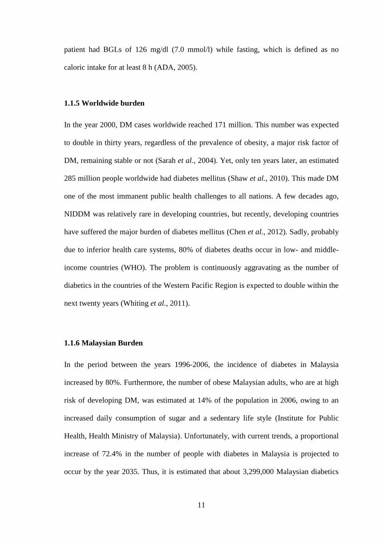
1.1.5 Worldwide burden
In the year 2000, DM cases worldwide reached 171 million. This number was expected
to double in thirty years, regardless of the prevalence of obesity, a major risk factor of
DM, remaining stable or not (Sarah et al., 2004). Yet, only ten years later, an estimated
285 million people worldwide had diabetes mellitus (Shaw et al., 2010). This made DM
one of the most immanent public health challenges to all nations. A few decades ago,
NIDDM was relatively rare in developing countries, but recently, developing countries
have suffered the major burden of diabetes mellitus (Chen et al., 2012). Sadly, probably
due to inferior health care systems, 80% of diabetes deaths occur in low- and middle-
income countries (WHO). The problem is continuously aggravating as the number of
diabetics in the countries of the Western Pacific Region is expected to double within the
next twenty years (Whiting et al., 2011).
1.1.6 Malaysian Burden
In the period between the years 1996-2006, the incidence of diabetes in Malaysia
increased by 80%. Furthermore, the number of obese Malaysian adults, who are at high
risk of developing DM, was estimated at 14% of the population in 2006, owing to an
increased daily consumption of sugar and a sedentary life style (Institute for Public
Health, Health Ministry of Malaysia). Unfortunately, with current trends, a proportional
increase of 72.4% in the number of people with diabetes in Malaysia is projected to
occur by the year 2035. Thus, it is estimated that about 3,299,000 Malaysian diabetics

12
will be in need of life-time medical care in just two decades (Guariguata et al., 2014)—
which, to say the least, would be crippling for all governmental health service sectors.
1.2 Clarifying ambiguous terminology: antidiabetic vs. antihyperglycemic and
hypoglycemic
Drugs used in diabetic care are usually described as antidiabetic, antihyperglycemic,
and/or hypoglycemic. These terms seem to be often used by researchers
interchangeably. However, subtle differences between these words may render such
practice, on occasions, incorrect. Hence, it seems only warranted that a brief description
be provided herein for accuracy purposes.
As defined in Farlex Partner Medical Dictionary, the term “anti-diabetic” denotes an
agent that reduces blood sugar. Though this might be the case for most antidiabetic
drugs, it is not necessarily true for all. A more accurate definition may be found in
Mosby's Medical Dictionary, in which an antidiabetic drug is rather defined as “any
agent that prevents or relieves the symptoms of diabetes”, whereas an
antihyperglycemic agent is described as “a substance or therapy that counteracts high
levels of glucose in the blood”. Lastly, the term “hypoglycemic”, according to the
definition of The American Heritage® Medical Dictionary, may apply to any agent that
lowers the concentration of glucose in the blood. However, a drug might be labeled as
“hypoglycemic” to indicate that it can induce a state of hypoglycemia, whereby BGLs
fall below the normal range (2015).
1.3 Trend to herbal therapy
Several studies carried out in the field of diabetes research have shown that traditional
medicines could provide better glycemic control than currently used conventional drugs

13
(Rates, 2001; Roja and Rao, 2000). Hence, plants possessing hypoglycemic activity
may provide a useful source of new oral antidiabetic compounds for the development of
pharmaceutical entities or as simple dietary adjuncts to existing therapies (Ogundipe et
al., 2003). However, despite a lack of knowledge about most of the interactions between
herbs and drugs, nearly 20% of patients taking prescription medication also take herbal
and other dietary supplements (Kaufman et al., 2002; Gardiner et al., 2006). Though
there are various approaches to reducing the ill effects of diabetes and its secondary
complications, herbal medicines are still preferred due to less side effects and lower
costs (Srivastava et al., 2012). The use of herbs from various corners of the world is
being increasingly popularized in the modern world as adjuncts to conventional
treatments for NIDDM, mainly by immigrants (Bailey et al., 1986).
1.4 Gongronema latifolium Overview
Gongronema latifolium Benth. (Apocyanaceae) (GL) is commonly grown in gardens in
Calabar, Cross River State, Nigeria. Owing to its extensive presence in the tropical and
the subtropical regions of Africa, GL has long been an integral part of the African
traditional medicine. It has been used for a variety of medical conditions, such as
hypertension, diabetes mellitus, malaria, mental, intestinal disorders (Ekong et al., 2014;
Ugochukwu et al., 2003), intestinal worms, cough, dysentery, and dyspepsia (Iwu,
2014). GL extracts have anti-inflammatory (Morebise et al., 2002), antifungal (Nwosu
and Okafor, 1995), and moderate anti-laxative effects (Gamaniel and Akah, 1996).
Overall, GL’s beneficial properties have been well documented, with a focus on it
antidiabetic use (Eze and Nwanguma, 2012). It has also been commercially utilized as a
substitute for commercial hops in large-scale beer production operations (Adenuga et
al., 2010). Recently in the United States, it has been integrated into a DM Tea blend

14
claimed to maintain healthy BGLs (Akpaso et al., 2011) by Neimeth Pharmaceuticals
(Iwu, 2014). Early reports justified GL antidiabetic use based on its antioxidant
potential (Ugochukwu and Babady, 2002) and glucose lowering effect (Ugochukwu and
Babady, 2003). However, over a decade after these preliminary reports, GL use in the
treatment of DM, its mechanisms of action and potential long-term toxicity have
remained vaguely understood.
1.4.1 Morphological description
GL is a perennial edible herbaceous shrub, with milky or less often, clear latex and soft
flexible stems (Akpaso et al., 2011). Its flowers are mainly yellow (Edet et al., 2009)
and, as seen in (Plate 1.1), the leaves are simple, smooth, opposite or occasionally
whorled, very rarely alternate, usually without obvious stipules; and the margins are
nearly always entire (Bingtao et al., 1977). It is a climbing plant with a soft and fibrous
stem (Iwu, 2014).
1.4.2 Taxonomy
GL belongs to the kingdom Plantae and the Magnoliophyta phylum. It is of the
Magnoliopsida class and the order of Gentianales. The family of GL is Apocyanaceae
and it is of the Gongronema genus. The species is called Gongronema latifolium, and is
commonly referred to as Bush Buck and Tafel Boom. Traditionally, GL has been called
Utasi (Efiks, Ibibios and Quas tribes), Utazi (Igbos), Arokeke (Yorubas in Nigeria), as
well as Aborode, akam, nsurogya (Ghana).

15
Plate 1.1: Gongronema latifolium Benth. (Apocyanaceae) courtesy of West African
Plants.
1.4.3 Chemical composition
(Eneji et al., 2011) have reported that GL extracts contained a variety of phytochemical
compounds, including alkaloids, saponins, tannins, flavonoids, and glycosides.
Furthermore, different part of GL contain some phytochemicals like B-sistosterol,
lupenyl esters, pregnane ester, and essential oils (Ekundayo, 1980; Morebise et al.,
2002). It is believed that these phytochemicals in GL can influence cellular proteins
with enzymatic activities (Eze and Nwanguma, 2012).

16
1.4.4 GL use in DM treatment
GL leaves, which are distinctly bitter (Eze and Nwanguma, 2012), were cooked as a
vegetable soup and eaten as such for diabetes (Ogundipe et al., 2003). In some African
cultures it is utilized as a spice for pancreas-related ailments (Okafor, 1999). A handful
of studies have shown that GL possesses a considerable antihyperglycemic effect in
animal diabetic models and in human subjects (Ejike et al., 2013). Yet, like most of the
medicinal plants which have been identified in the recent decades, there is a lack of data
on GL antidiabetic use, chronic and subchronic toxicity, dosing, and mechanisms of
action. (Ugochukwu and Babady, 2003) argued that GL had an insulin-like activity.
(Adebajo et al., 2012) reported that it acted by increasing insulin release. Meanwhile,
(Ogbu et al., 2013) concluded that it attenuated blood glucose excursions partly by
delaying gastric emptying.
1.5 Problem statement
The prevalence of DM is on the rise in South East Asia, particularly in Malaysia.
Available DM drug classes vary in efficacy and method of administration. They often
cause a spectrum of side effects, including weight gain, hypoglycemia and increased
risk of mortality. Most are costly and have poor safety profiles. Natural alternatives like
antidiabetic herbs can have similar capabilities and less adverse effects and/or cost.
Several reports on GL have confirmed the validity of its traditional use in Africa in the
treatment of DM. This suggests that GL might be beneficial and may make for a source
of antidiabetic medication, or that it may work to commemorate the effects of
antidiabetic drugs as an adjunct or a supplement. Hence, due to existing problems with
currently available antidiabetic drugs, as well as a global trend towards herbal medicine,
it was decided that it was of interest to look into GL further for potential therapeutic

17
agents. Studies have, in the recent years, managed to demonstrate its antioxidant
potential and glucose lowering effect. However, GL use in the treatment of DM, its
mechanisms of action and long-term toxicity remain vaguely understood. Hence, this
study assessed GL blood glucose lowering activity, anti-diabetic mechanisms of action
and sub-chronic toxicity using in vitro techniques and rat models.
1.6 Tested hypothesis
A soxhlet ethanolic extract of GL, GLES, retains a significant antihyperglycemic
activity and produces structural recovery on the level of the pancreas. It can delay the
transport of glucose via the intestinal wall and act as an insulin sensitizer. Furthermore,
it has a good safety profile and does not cause systemic toxicity.
1.7 Study objectives
Main objective of the study:
To investigate the antihyperglycemic/antidiabetic activity of Nigerian Gongronema
latifolium ethanolic extract in normal and chemically-induced (by STZ) diabetic
rats.
Secondary objectives:
1) To assess the effects of Gongronema latfolium ethanolic extract on pancreatic
Langerhans islets.
3) To investigate the mechanism(s) of action of Gongronema latifolium extract by
examining its effect on intestinal glucose absorption and glucose uptake by
isolated body tissues.
4) To determine the safety of oral sub-chronic (90 days) exposure to Gongronema
latifolium extract in Sprauge-Dawely rats of both sexes.

18
5) To make recommendations based on in vivo/in vitro antidiabetic and toxicity
results to provide researchers and health care professionals with the safe and
effective levels of the doses of GL ethanolic extract.

19
CHAPTER TWO: GENERAL METHODOLOGY
2.1 Plant material
Gongronema latifolium Benth (Apocynaceae) (GL), locally known as Utazi or Arokeke,
was collected as whole plant from Yakkur, Cross River State, Nigeria at the following
GPS coordinates (6ₒ 08’17.35”N 8
ₒ 41’15.54”E
elev 420 ft) under the supervision of the
Department of Biochemistry at the University of Calabar. Authentication was carried on
by Pastor Frank, a botanist in the Department of Botany, and voucher specimen
(ERU/2011/718) was deposited at the same department. The leaves were plucked of the
twigs, washed with tap water and dried in the shade. The dried leaves were ground into
powder. The powder was placed into a properly packaged courier to arrive at the
Department of Pharmacology, Universiti Sains Malaysia via courier within 7 days.
Upon receipt of the powder, it was placed at 4 ⁰C for further use. The plant name was
verified by checking with www.theplantlist.org (website accessed on 16th
of September
2015).
2.2 Preparation of plant extract
Upon receipt, 400 g of the powdered dried GL were extracted in ethanol using Soxhlet
apparatus at 40-60 °C in ratio of 1:5 to 1:10 of material : solvent (w/v) for three days.
Anti-pumping silicon granules were added each day before the apparatus was turned on.
The extract was filtered and concentrated to about 1/10 of its original volume in a rotary
evaporator at 40 °C. Thereafter, the concentrate was freeze-dried to obtain dried extract.
The totally dry extract, referred to as GLES, amounted to a yield of ~ 12% and was
stored at 4 ºC until further use.

20
2.3 Experimental animals
Male and female Sprague-Dawley (SD) rats were used over the course of the present
research. The animals weighed initially between 180 g - 220 g (6-7 weeks old) and were
obtained from the Animal Research and Service Centre, Main Campus, Universiti Sains
Malaysia (USM), Penang. The rats were housed pair-wise under standard environmental
conditions (temperature, 25 ± 5 ºC; relative humidity, 50 ± 5% and 12 h light/dark
cycle) throughout the period of the experiments. Prior to experimentation, the animals
were acclimatized to laboratory conditions for one week in the Animal Transit Room at
the School of Pharmaceutical Sciences, USM. The animals were allowed free access to
standard rat pellets (Gold Coin Feedmills, Butterworth, Penang, Malaysia) and tap water
ad libitum. The toxicity study was carried out according to OECD guidline 408
(Murbach et al., 2014; OECD, 1998) and US FDA Redbook 2000, IV.C.4.a (90-day
study) (FDA, 2003). Care and handling of study animals were performed according to
the guidelines set by the WHO (World Health Organization, Geneva, Switzerland) with
consideration to the principles of the Hungarian Act 2011 CLVIII (modification of
Hungarian Act 1998 XXVIII) regulating animal protection. The institutional Animal
Ethics Committee at Universiti Sains Malaysia approved the research [Approval
number: USM/Animal Ethics Approval/2013/(90)(509)].
2.4 Induction of diabetes
Diabetes was induced by a single intraperitoneal (IP) injection of streptozotocin (STZ)
(Sigma-Aldrich, St. Louis, MO, USA) at a dose of (55 mg/kg) to rats fasted for 12
hours. The injection solution was freshly prepared by reconstituting STZ in cold normal
saline. The diabetic condition of the animals was confirmed 72 hours after the STZ

21
injection through measurement of the fasting blood glucose level (FBG) using the One
Touch Glucometer (Accu-Chek Performa, Roche Diagnostics, Mannheim, Germany)
via a single puncture of the tail vein. Only diabetic rats with FBG within 15.0-20.0
mMol/L (270-360 mg/dl) were included in the study.
2.5 Preparation of metformin for oral dosing
Metformin 500 mg tablet (Glucophage®, Bristol-Myers Squibb, New York, USA) was
grinded in a suitable amount of distilled water using a mortar and a pestle followed by
ultra-sonication (UC-10 Ultrasonic Cleaner, Jeiotech, Seoul, Korea) for 15 min at 37 ⁰C.
The amount of water used and the desired concentration of the solution were calculated
for an administration volume of 10 ml/kg b.w. in all of the treated rat groups.
2.6 Statistical analysis
Data obtained from the male and female treatment groups were compared separately.
The statistical comparison aimed at determining whether the differences observed
between the treatment groups and the control resulted from GL consumption. Results
were expressed as the mean ± SEM. Graphs were drawn and data was typed into
Microsoft Excel 2007 (Microsoft Corp., Redmond, WA). Statistical analysis was
performed using version 21 of the IBM-SPSS statistical program (IBM Corp., Armonk,
NY). Normal distribution of the data was confirmed using the relevant tool in the same
software program. One-way ANOVA was used followed by Dunnett’s Test as a Post
Hoc Test for parametric multiple comparisons between the control and the treatment
groups. Pre-treatment and post-treatment comparisons were performed using the paired
t-test. Differences were considered significant when the P value was less than 0.05.

22
2.6 Flow chart of study protocols

23
CHAPTER THREE: ANTIDIABETIC STUDY OF GONGRONEMA
LATIFOLIUM
3.1 Background
Diabetes Mellitus (DM) refers to a medical condition marked by hyperglycemia
due to loss of control over blood glucose homeostasis. Diabetes Mellitus is a major
cause of death worldwide. As projected in 2013 by the World Health Organization
(WHO), DM is expected to become the 7th leading cause of death in 2030. There are
two types of Diabetes: Type I, which is more severe, describes a condition in which
insulin secretion is completely absent due to immune destruction of pancreatic β-cells
(i.e., Insulin-Dependent Diabetes Mellitus: IDDM), whereas Type II describes a
condition in which bodily cells (other than neurons) become irresponsive to insulin (i.e.,
Non-Insulin-Dependent Diabetes Mellitus: NIDDM). According to the Malaysian
Ministry of Health, there were more than 3 million Malaysians with DM in 2011 (Amal
et al., 2011). It is very alarming considering the fact that 80% of diabetes deaths occur
in low- and middle-income countries, and that the number of diabetics in the Western
Pacific Region is expected to double within twenty years from now (WHO). Treatment
for NIDDM, a multifactorial disease, includes many different, expensive combinations
of drugs whose use is restricted by their pharmacokinetic properties, secondary failure
rates, and many accompanying side effects (Hammouda and Amer, 1966), which
include weight gain, increased risk of hypoglycemia, and increased risk of mortality
(Rang and Dale, 1999). Thus, there is a grave need for natural drugs that have similar
efficacy but do not have such adverse effects or cost.
Gongronema latifolium Benth (Apocynaceae) is a perennial edible shrub with a
soft stem. Its leaves are usually simple, opposite or occasionally whorled with no clear

24
stipules (Bingtao et al., 1977). It is widely used in the West African sub region for a
number of medicinal and nutritional purposes (Akpaso et al., 2011) and to treat a variety
of ailments, such as hypertension, diabetes mellitus, malaria, mental and intestinal
disorders (Ekong et al., 2014; Ugochukwu et al., 2003). Traditionally, the leaves of GL
were cooked as a vegetable soup and eaten as such for diabetes (Ogundipe et al., 2003)
and in some African cultures it is utilized as a spice to support the pancreas (Okafor,
1999). Recently in the United States, it has been integrated into a DM Tea blend which
is claimed to maintain healthy glycemia (Akpaso et al., 2011). Previous reports
(Ugochukwu et al., 2003; Ugochukwu and Cobourne, 2003), justified its traditional use
for DM based on its effect in ameliorating the oxidative stress which accompanies DM
and which underlies many of the complications of diabetes. Although more recent
reports (Akpaso et al., 2011; Ejike et al., 2013; Nnodim et al., 2012) have confirmed
that GL possesses antihyperglycemic activity, its use in the treatment of DM, as well as
its mechanisms of action, remain vaguely understood. While (Ugochukwu and Babady,
2003) attributed an insulin-like activity to GL ethanolic extract, (Adebajo et al., 2012)
reported that the extract acted by increasing insulin release.
This study evaluates the effect of GL leaf-extract on BGLs upon acute and sub-
chronic administration in diabetic rats. Furthermore, it examines the effect of sub-
chronic administration of GL extract on serum lipid profile, kidney function indices and
the insulin-secreting cells (Langerhans islets). It explores for the first time, the effect of
GL extract on in vitro insulin and glucose transduction in isolated rat abdominal muscle
and jejunum. Lastly, a profiling of the chemical composition of the extract is done with
the use of Gas Chromatography-Mass Spectroscopy (GC-MS).