Embed Size (px)
Citation preview
Dr JAMILA EL MEDANY
Dr SAEED VOHRA
&


•Interosseous membrane:
A thin & strong membrane, that binds the interosseous borders of tibia & fibula. It binds the two bones and provides attachment for muscles.
•The deep fascia surrounds the leg and attached to Anterior & Medial borders of Tibia.
•Two Intermuscular Septa
Pass from the deep aspect of this fascia to be attached to :
Anterior border of fibula (Anterior fascial septum)
Posterior border of fibula (Posterior fascial septum)

Together with the interosseus membrane, the two septa divide the leg into
(3) Compartments : 1-Anterior
2-Lateral (peroneal)
3-Posterior
Each one has its own Muscles (with specific action), Blood vessels and Nerves.

A thickening of deep fascia that keep the long tendons around ankle joint in position
Superior Extensor retinaculum :
Attached to anterior borders of tibia & fibula above ankle
Inferior Extensor retinaculum:
Y-shaped band located inferior to ankle

From medial to lateral:1.Tom2.Has3.Very (vessels)4.Nice ( nerve)5.Dog &6.Pigion

Muscles of Anterior Compartment
TibialisAnterior
Extensor HalluciusLongus
Extensor DigitorumLongus
Peroneus Tertius

Criteria of Anterior CompartmentOrigin:All muscles arise from the
fibula EXCEPT Tibialis Anterior .
Nerve supply:DeepPeroneal.Blood Supply:Anterior tibial.Action: Dorsiflexion of the
ankle joint & Extension of the toes & (Inversion).

Tibialis Anterior & Extensor Digitorum Longus

Muscle Origin Insertion
Action
Peroneus tertius
Anterior surface of shaft of fibula
Base of 5th metatarsal bone
Dorsi flex foot at ankle joint; Everts foot at subtalar and transverse tarsal joints
Peroneus Tertius, & Extensor Hallucis Longus

MUSCLES NERVE BLODDSUPPLY
PERONEUSLONGUS
Superficial Peroneal
Peroneal A
PERONEUSBREVIS

Peroneus Longus
Origin Insertion Action
Lateral surface of shaft of fibula
Base of first metatarsal and the medial cuneiform
Plantar flexes foot at ankle joint;
Everts foot at subtalar and transverse tarsal joints;
Supports Lateral longitudinal & Transverse arches

Peroneus Brevis
Origin Insertion ActionLateral
surface of shaft of fibula
Base of fifth metatarsal bone
Plantar flexes foot at ankle joint; Everts foot at subtalar and transverse tarsal joint; Supports Lateral longitudinal arch of foot

Tendons of peronei are surrounded by a single common tubular synovial sheath,
deep to inferior peroneal retinaculum, they have separate sheaths
Superior peroneal retinaculum
Connects the lateral malleolus to calcaneum & holds the tendons of peroneus longus & brevis
Inferior peroneal retinaculum

It is very thin, but just distal to ankle joint, it is thickened to form Inferior extensor retinaculum

MUSCLE BLODDD VESSEL
NERVE
Extensor Digitorum Brevis
Dorsalis Pedis
DEEP & SuperficialPeroneal
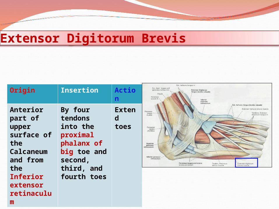
Extensor Digitorum Brevis
Origin Insertion Action
Anterior part of upper surface of the Calcaneum and from the Inferior extensor retinaculum
By four tendons into the proximal phalanx of big toe and second, third, and fourth toes
Extend toes

Insertion of Long Extensor Tendons (Extensor Expansion)
The tendons of Extensor digitorum longus pass to the lateral four toes.
Each tendon to the 2nd , 3rd & 4th toes is joined on its lateral side by a tendon of Extensor digitorum brevis.
The extensor tendons form a Fascial Expansion (Extensor
Expansion) on the dorsum of each toe. The expansion divides into (3) parts. Central part: inserted into the Base
of Middle ph. Two Lateral parts: inserted into the
Base of Distal ph. The (Extensor Expansion) receives
insertion of : Interossei & Lumbrical muscles.
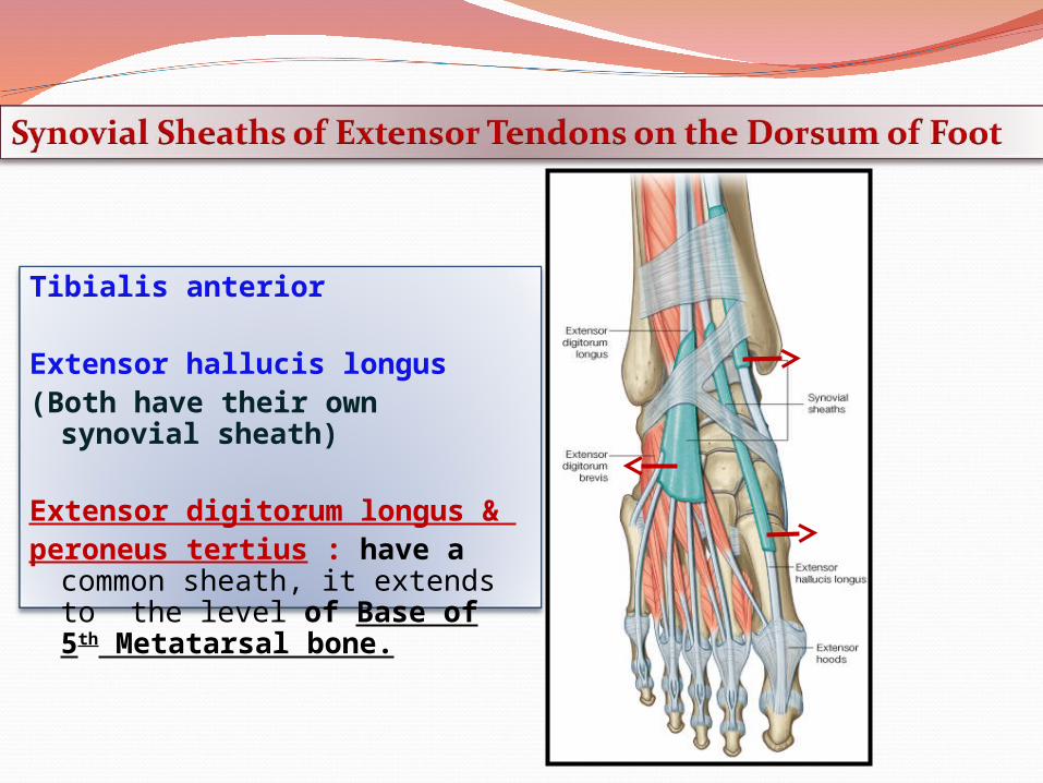
Tibialis anterior
Extensor hallucis longus(Both have their own synovial sheath)
Extensor digitorum longus & peroneus tertius : have a common
sheath, it extends to the level of Base of 5th Metatarsal bone.

THANK YOU & BEST WISHES