Embed Size (px)
DESCRIPTION
Animal Reproduction and Development. Reproduction Song. Animals reproductive by asexual and sexual methods: Asexual is the production of offspring with genes all from one individual, without the fusion of gametes. Sexual involves formation of gametes and fertilization, genetic variation. - PowerPoint PPT Presentation
Citation preview

Animal Reproduction and Development
Reproduction Song

Animals reproductive by asexual and sexual methods:•Asexual is the production of offspring with genes all from one individual, without the fusion of gametes.•Sexual involves formation of gametes and fertilization, genetic variation
Asexual Methods Include:
Gemmules in Sponges
Budding in Hydra Starfish Regeneration

Parthenogenesis: an unfertilized egg develops
Honey bees: drones are males and are produced parthenogeneticly and female workers and queens form from fertilized eggs.
Queen Worker (female) Drone (male)

Sexual Reproduction Hermaphroditism: individual has both male and female reproductive systems
Earthworm

Fertilization can occur externally or internallyAquatic animals tend to be externalTerrestrial animals tend to be internal
Development can be external in the water, external on land, or internal.
Oviparous: lay eggs, Amniotic eggs are terrestrial eggsOvoviparous: live birth from eggs (some sharks and
snakes)Viviparous: live placental birth

What are the adaptive values of each style off sexual animal reproduction?
Number of Eggs: ?Parental Care: ?Habitat: ?
Introduction to Reproduction System

Vertebrates Fertilization Development Parental Care
Fish
Amphibian
Reptile
Bird
Mammal
Complete the type of sexual reproduction typical of each group
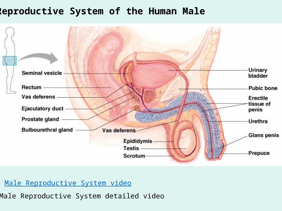
Reproductive System of the Human Male
Male Reproductive System video
Male Reproductive System detailed video

The male reproductive system produces sperm cells and provides a mechanism for delivering them to the female's body.
Identify the role of:
Testes
Epididymis
Vas deferens
Scrotum

Outline the functions of the male reproductive organs
Vasectomy Surgery video

Hormonal control of the Testes


Development of Eggs and Sperm
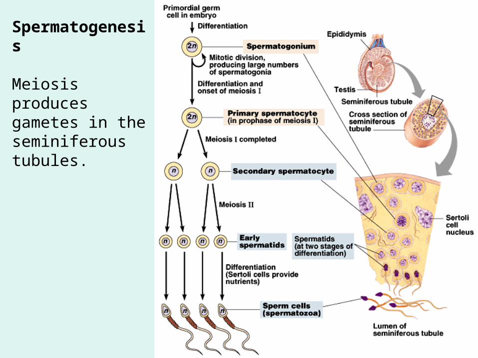
Spermatogenesis
Meiosis produces gametes in the seminiferous tubules.
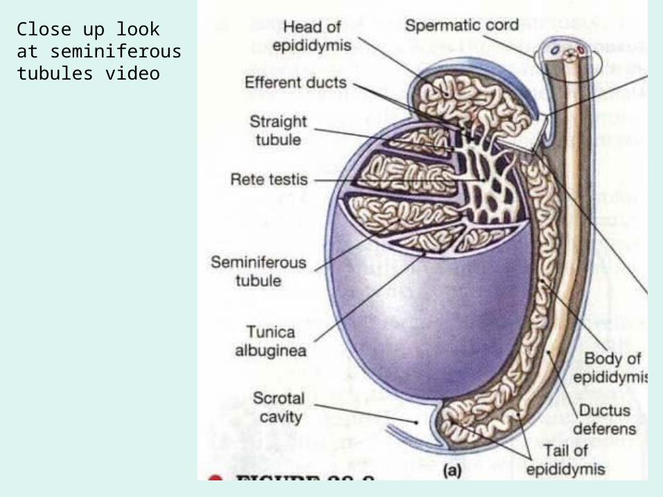
Close up look at seminiferous tubules video

Seminiferous Tubules synthesize sperm Sperm cells
Sperm Structures

Female Reproductive System

Female Reproductive System
Female Reproductive System video

Ovary Follicle

Oogenesis
This is the state of the egg when fertilized
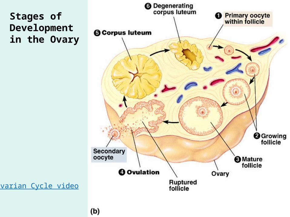
Stages of Development in the Ovary
Ovarian Cycle video


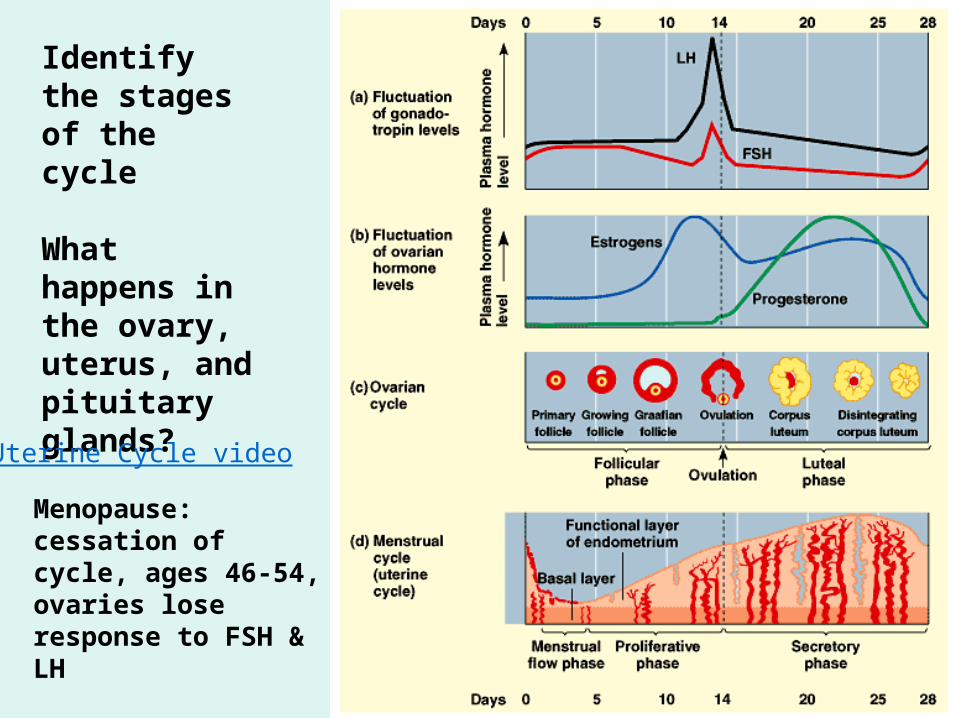
Identify the stages of the cycle
What happens in the ovary, uterus, and pituitary glands?
Uterine Cycle video
Menopause: cessation of cycle, ages 46-54, ovaries lose response to FSH & LH

Formation of the zygote and early post-fertilization events

Cleavage occurs over several days following fertilization. The zygote divides as it travels through the oviduct. By the time the cilia of the oviduct deliver the embryo to the uterus, the embryo is a ball of cells called a blastocyst. The blastocyst implants in the endometrium.

A blastocyst forms nearly a week after fertilization
Four membranes protect and nourish the embryo, which consists of three tissue layers.

Nutrients and waste products are exchanged between the fetus and the mother within the placenta. The umbilical vein (red) carries oxygen-rich blood and nutrients to the fetus. The umbilical arteries are blue, indicating that they carry oxygen-depleted blood and waste products away from the fetus.

Placental circulation: materials are exchanged by diffusion, active transport, and selective absorption. Not by direct blood contact.
Structure and function of the placenta video
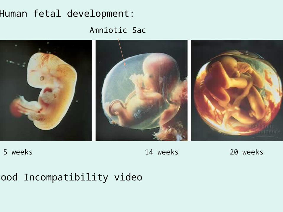
5 weeks 14 weeks 20 weeks
Human fetal development:
Amniotic Sac
Blood Incompatibility video

Hormonal induction of labor

Name the structures and its role in reproduction and metabolism

Name the structures and its role in reproduction and metabolism

Name the phases and state of the organs.
Name the hormones:Black:Red:Blue:Green:

Animal Development
Preformation: shows infant in sperm from 1694 engraving “homunculus”
Epigenesis: animal emerges gradually from the egg

Fertilization in Mammals
Zona pellucida
Changes that occur in the egg after fertilization video

Cleavage partitions the zygote into many smaller cellsRapid cell division without growth produces cells called blastomeres.

• In both sea urchins and frogs first two cleavages are vertical.
• The third division is horizontal.• The result is an eight-celled embryo with two tiers of four
cells.

• Continued cleavage produces the morula.

• A blastocoel forms within the morula blastula
View the sea urchin development video on the CD

Gastrulation rearranges the blastula to form a three-layered embryo with a primitive gut
Development after fertilization video

(triploblastic)

Organogenesis forms the organs from the three embryonic germ layers
Organogenesis in a frog embryo
View frog development video on CD

Amniote embryos develop in a fluid-filled sac within a shell or uterusThe amniote embryo is an adaptation for reproduction in the terrestrial environment.
Shelled eggs of reptiles and birds.Uterus of placental mammals.

Extraembryonic membranes in a chickIdentify the membranes that provide supporting functions
Protection from mechanical shock
Gas exchange
Disposal of uric acid
Nutrient source

Extraembryonic membranes in a chick
How extraembryonic membranes support chick video

• The four extraembryonic membranes are the yolk sac, amnion, chorion, and allantois.– Cells of the yolk sac digest yolk providing nutrients to the
embryo.– The amnion encloses the embryo in a fluid-filled amniotic
sac which protects the embryo from drying out.– The chorion cushions the embryo against mechanical
shocks.– The allantois functions as a disposal sac for uric acid.

Chick embryo 54 hours old
Most major organs have formed
Formation of structures in embryo video

Early development of a human embryo and its extraembryonic membranes

Embryonic membranes – homologous with those of shelled eggs. Chorion: completely surrounds the embryo and other
embryonic membranes. Amnion: encloses the embryo in a fluid-filled amniotic
cavity. Yolk sac: found below the developing embryo.
Develops from the hypoblast. Site of early formation of blood cells which later
migrate to the embryo. Allantois: develops as an outpocketing of the
embryo’s rudimentary gut. Incorporated into the umbilical cord, where it forms
blood vessels.• Organogenesis begins with the formation of the neural
tube, notochord, and somites.

Embryonic membranes – homologous with those of shelled eggs.
Impact of Drugs and Chemicals on Fetal Development video

During labor, hormones stimulate the uterus to contract. The contractions push the baby out of the mother's body.

The Process of Birth


![Ocw [animal reproduction]](https://img.dokumen.tips/doc/110x75/58835b371a28ab42678b649f/ocw-animal-reproduction.jpg)