Embed Size (px)
Citation preview

Androgen Receptor Deregulation Drives Bromodomain-MediatedChromatin Alterations in Prostate Cancer
Urbanucci, A., Barfeld, S. J., Kytölä, V., Itkonen, H. M., Coleman, I. M., Vodák, D., ... Mills, I. G. (2017).Androgen Receptor Deregulation Drives Bromodomain-Mediated Chromatin Alterations in Prostate Cancer. CellReports, 19(10), 2045-2059. https://doi.org/10.1016/j.celrep.2017.05.049
Published in:Cell Reports
Document Version:Publisher's PDF, also known as Version of record
Queen's University Belfast - Research Portal:Link to publication record in Queen's University Belfast Research Portal
Publisher rights© 2017 The Authors.This is an open access article published under a Creative Commons Attribution-NonCommercial-NoDerivs License(https://creativecommons.org/licenses/by-nc-nd/4.0/), which permits distribution and reproduction for non-commercial purposes, provided theauthor and source are cited.
General rightsCopyright for the publications made accessible via the Queen's University Belfast Research Portal is retained by the author(s) and / or othercopyright owners and it is a condition of accessing these publications that users recognise and abide by the legal requirements associatedwith these rights.
Take down policyThe Research Portal is Queen's institutional repository that provides access to Queen's research output. Every effort has been made toensure that content in the Research Portal does not infringe any person's rights, or applicable UK laws. If you discover content in theResearch Portal that you believe breaches copyright or violates any law, please contact [email protected].
Download date:05. Mar. 2020

Article
Androgen Receptor Dereg
ulation DrivesBromodomain-Mediated Chromatin Alterations inProstate CancerGraphical Abstract
Highlights
d Androgen receptor (AR) overexpression drives genome-wide
chromatin relaxation
d Bromodomain (BRD) inhibitors reduce local chromatin
opening at AR target genes
d AR overexpression upregulates BRD proteins ATAD2, BRD2,
and BRD4
d The BROMO-10 gene signature stratifies patients for therapy
with BRD inhibitors
Urbanucci et al., 2017, Cell Reports 19, 2045–2059June 6, 2017 ª 2017 The Author(s).http://dx.doi.org/10.1016/j.celrep.2017.05.049
Authors
Alfonso Urbanucci, Stefan J. Barfeld,
Ville Kytola, ..., Matti Nykter,
Tapio Visakorpi, Ian G. Mills
[email protected] (A.U.),[email protected] (I.G.M.)
In Brief
Urbanucci et al. report how upregulated
androgen receptor (AR) and AR-targeted
bromodomain proteins contribute to
genome-wide chromatin relaxation and
increased gene transcription in advanced
prostate cancer. They show that ATAD2 is
a strong tissue biomarker and propose a
BROMO-10 gene signature to stratify
patients for combination therapies with
bromodomain inhibitors.
Accession Numbers
GSE73989

Cell Reports
Article
Androgen Receptor DeregulationDrives Bromodomain-MediatedChromatin Alterations in Prostate CancerAlfonsoUrbanucci,1,2,* Stefan J. Barfeld,1 Ville Kytola,3 Harri M. Itkonen,1 IlsaM. Coleman,4 Daniel Vodak,5 Liisa Sjoblom,6
Xia Sheng,7 Teemu Tolonen,8 Sarah Minner,9 Christoph Burdelski,10 Kati K. Kivinummi,3 Annika Kohvakka,6
Steven Kregel,11,12 Mandeep Takhar,13 Mohammed Alshalalfa,13 Elai Davicioni,13 Nicholas Erho,13 Paul Lloyd,14,15
R. Jeffrey Karnes,16 Ashley E. Ross,17 EdwardM. Schaeffer,18 Donald J. Vander Griend,11 Stefan Knapp,19,20 Eva Corey,21
Felix Y. Feng,14,15,22 Peter S. Nelson,4,21,23 Fahri Saatcioglu,7,24 Karen E. Knudsen,25 Teuvo L.J. Tammela,26
Guido Sauter,9 Thorsten Schlomm,27 Matti Nykter,3 Tapio Visakorpi,6 and Ian G. Mills1,2,28,29,*1Centre for Molecular Medicine Norway, Nordic European Molecular Biology Laboratory Partnership, Forskningsparken, University of Oslo,
21 0349 Oslo, Norway2Department of Molecular Oncology, Institute for Cancer Research, Oslo University Hospital, 0424 Oslo, Norway3Prostate Cancer Research Center, Institute of Biosciences and Medical Technology (BioMediTech), University of Tampere and Tampere
University of Technology, 33520 Tampere, Finland4Division of Human Biology, Fred Hutchinson Cancer Research Center, Seattle, WA 98109, USA5Department of Tumor Biology, Institute for Cancer Research, The Norwegian Radium Hospital, Oslo University Hospital, 0424 Oslo, Norway6Prostate Cancer Research Center, Institute of Biosciences and Medical Technology (BioMediTech), University of Tampere and Fimlab
Laboratories, Tampere University Hospital, 33520 Tampere, Finland7Department of Biosciences, University of Oslo, 0316 Oslo, Norway8Department of Pathology, Fimlab Laboratories, Tampere University Hospital, 33520 Tampere, Finland9University Medical Center Hamburg-Eppendorf, 20251 Hamburg, Germany10General, Visceral and Thoracic Surgery Department and Clinic, University Medical Center Hamburg-Eppendorf, 20246 Hamburg, Germany11Department of Surgery - Section of Urology, University of Chicago, Chicago, IL 60637, USA12Michigan Center for Translational Pathology, University of Michigan, Ann Arbor, MI 48109-0940, USA13Research and Development, GenomeDx Biosciences, Vancouver, BC V6B 1B8, Canada14Department of Medicine, University of California at San Francisco, San Francisco, CA 94143-0410, USA15Helen Diller Comprehensive Cancer Center, University of California, San Francisco, CA 94143-0981, USA16Department of Urology, Mayo Clinic, Rochester, MN 55902, USA17Brady Urological Institute, Johns Hopkins Medical Institute, Baltimore, MD 21287, USA18Department of Urology, Northwestern University, Feinberg School of Medicine, 303 East Chicago Avenue, Tarry 16-703, Chicago,
IL 60611-3008, USA19Nuffield Department of Clinical Medicine, University of Oxford, Old Road Campus, Roosevelt Drive, Oxford OX3 7DQ, UK20Institute for Pharmaceutical Chemistry, Goethe-University Frankfurt, CampusRiedberg,Max-von Laue Strasse 9, 60438 Frankfurt amMain,
Germany21Department of Urology, University of Washington, Seattle, WA 98195, USA22Department of Radiation Oncology, University of California, San Francisco, San Francisco, CA 94115, USA23Department of Pathology, University of Washington, Seattle, WA 98195, USA24Institute for Cancer Genetics and Informatics, Oslo University Hospital, 0424 Oslo, Norway25Sidney Kimmel Cancer Center, Thomas Jefferson University, Philadelphia, PA 19107, USA26Prostate Cancer Research Center and Department of Urology, University of Tampere and Tampere University Hospital, 33014 Tampere,
Finland27Martini-Clinic, Prostate Cancer Center, University Medical Center Hamburg-Eppendorf, Hamburg 20095, Germany28PCUK Movember Centre of Excellence, CCRCB, Queen’s University, Belfast BT7 1NN, Northern Ireland, UK29Lead Contact
*Correspondence: [email protected] (A.U.), [email protected] (I.G.M.)
http://dx.doi.org/10.1016/j.celrep.2017.05.049
SUMMARY
Global changes in chromatin accessibility may drivecancer progression by reprogramming transcriptionfactor (TF) binding. In addition, histone acetylationreaders such as bromodomain-containing protein 4(BRD4) have been shown to associate with theseTFs and contribute to aggressive cancers includingprostate cancer (PC). Here, we show that chromatin
CelThis is an open access article under the CC BY-N
accessibility defines castration-resistant prostatecancer (CRPC). We show that the deregulation ofandrogen receptor (AR) expression is a driver of chro-matin relaxationand thatAR/androgen-regulatedbro-modomain-containing proteins (BRDs) mediate thiseffect. We also report that BRDs are overexpressedin CRPCs and that ATAD2 and BRD2 have prog-nostic value. Finally, we developed gene stratification
l Reports 19, 2045–2059, June 6, 2017 ª 2017 The Author(s). 2045C-ND license (http://creativecommons.org/licenses/by-nc-nd/4.0/).

signature (BROMO-10) for bromodomain responseand PC prognostication, to inform current and futuretrials with drugs targeting these processes. Our find-ings provide a compelling rational for combinationtherapy targeting bromodomains in selected patientsin which BRD-mediated TF binding is enhanced ormodified as cancer progresses.
INTRODUCTION
Prostate cancer (PC) is the most common male cancer in the
United States and Europe (Center et al., 2012). Androgen recep-
tor (AR) signaling is required for the development of the prostate
gland and is maintained in PC including at the stage of progres-
sion to castration-resistant prostate cancer (CRPC) (Zhang et al.,
2013).
CRPC is characterized by copy number gain at the AR locus
occurring in around 30% of advanced cases. Consequently AR
is overexpressed in these tumors. However, AR deregulation is
also a frequent feature (>90%) of advanced castrate-resistant
cases (Waltering et al., 2012), which persist after resistance to
antiandrogens such as enzalutamide and abiraterone (Buttigliero
et al., 2015). We have previously shown that AR deregulation
is associated with local chromatin landscape changes, which
are able to reinforce the binding of AR to chromatin even in
low androgen environments (Urbanucci et al., 2012a, 2012b).
This mimics the conditions occurring in CRPC (Xu et al., 2006).
Phenotypically, AR deregulation results in increased growth
rates even under conditions of androgen deprivation (Waltering
et al., 2009). Moreover, genome-wide AR recruitment to chro-
matin is detectable in such conditions (Andreu-Vieyra et al.,
2011), suggesting that the chromatin is open and in some way
primed by pre-docked AR even before the cells are treated
with hormones. Androgen treatment then enhances AR recruit-
ment (Urbanucci et al., 2012a). This suggests that nucleosome
positioning is predetermined in PC cells, a hypothesis that has
been confirmed by a recent study (Chen et al., 2015).
Clinical epigenetics, defined as functionally relevant changes
to the genome that do not involve a change in the nucleotide
sequence and impact on disease phenotypes, is becoming
extremely important for cancer detection and treatment. One
indirect assessment of epigenetic alteration is the accessibility
of DNA, determined by DNase hypersensitivity analysis or by
formaldehyde-assisted isolation of regulatory elements (FAIREs)
(Song et al., 2011).
Proof of a de-regulated epigenome in CRPC includes altered
patterns of DNA methylation, histone modifications (Perry
et al., 2010), and increased and altered AR binding to chromatin
(Pomerantz et al., 2015; Sharma et al., 2013).
In addition, epigenetic readers such as bromodomain-con-
taining protein 4 (BRD4) have been shown to associate with tran-
scription factors (TFs) such as AR (Asangani et al., 2014; Nagar-
ajan et al., 2014; Shi and Vakoc, 2014) and contribute to
aggressive cancers of many types (Delmore et al., 2011; Shi
and Vakoc, 2014) including PC (Asangani et al., 2014; Wyce
et al., 2013). Nevertheless, the underlying mechanisms of action
of bromodomain inhibitors have not yet been completely eluci-
2046 Cell Reports 19, 2045–2059, June 6, 2017
dated (Shi and Vakoc, 2014). Therefore, we set out to investigate
the underlying global changes in chromatin accessibility as a
driver of cancer progression (Lever and Sheer, 2010; Timp and
Feinberg, 2013).
Here, we report that DNA accessibility alone is able to
discriminate advanced prostate tumors from earlier disease
states and benign tissue. The chromatin of these tumors
is more accessible due to indirect mechanisms in which the
AR plays a role. We show that BRDs, such as BRD4 and
androgen-regulated BRD2 and ATAD2, are the mediators
of such increased accessibility and are prognostic tissue
markers overexpressed in CRPC. Finally we provide a ten-
gene signature, BROMO-10, that can be used to stratify patients
with poorer outcome and guide PC patient selection for combi-
natorial trials of bromodomain and extra-terminal (BET)-targeted
therapies with other agents.
RESULTS
Deregulation of the AR Enhances Bromodomain-Mediated Chromatin Opening in Advanced TumorsTo understand whether progression to CRPC is associated with
global changes in chromatin accessibility, we assessed chro-
matin opening and DNA regions with regulatory activity in three
benign prostate hyperplasia (BPH), three primary untreated
PC, and three locally recurrent CRPC samples using FAIRE
sequencing (FAIRE-seq) (Giresi and Lieb, 2009).
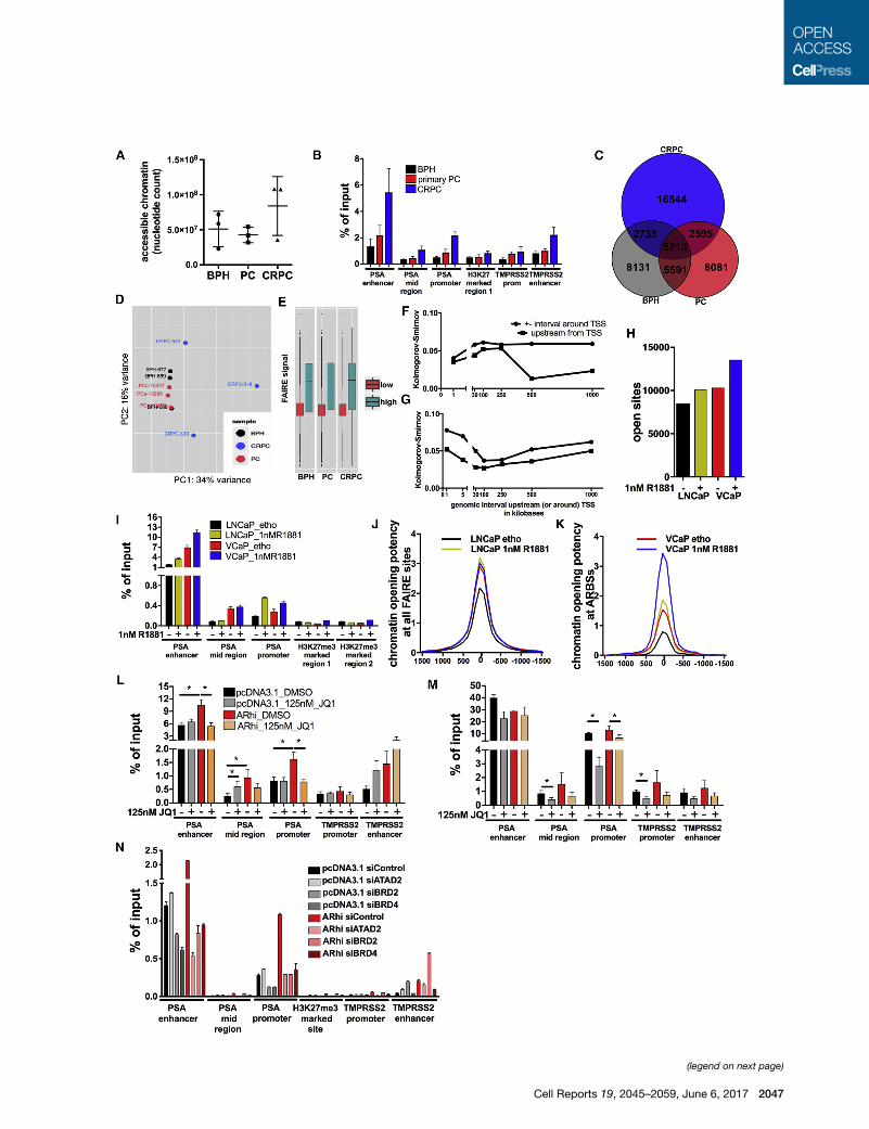
One-third of the genome consisted of open regions of chro-
matin in two CRPC samples, whereas only about one-sixth of
the genome comprised open chromatin in the other samples
(Figure 1A and Table S1A). We validated open chromatin re-
gions via FAIRE-qPCR at prostate specific antigen (PSA) and
TMPRSS2 gene loci, showing enhanced chromatin opening in
CRPC (Figure 1B).
We built disease-stage-specific high-confidence consensus
open chromatin maps. On average, only 15% of the FAIRE-seq
sites overlapped between the samples (Table S1A) highlighting
epigenetic heterogeneity. We found that the majority of open
sites were unique to CRPC samples (Figure 1C) and were larger
than in PC or BPH (Figure S1A). To classify disease stage, we
used principal component analysis of the FAIRE-seq data
and found that in terms of chromatin state, CRPC chromatin
appeared more diverse when compared with BPH or PC (Fig-
ure 1D), indicating that extensive chromatin remodeling is a
late event in PC progression.
RNA sequencing (RNA-seq) of the same clinical samples
showed a positive correlation between upregulated genes and
an open chromatin state (Figure 1E) up to 250 kb upstream the
transcriptional start sites (TSSs) (Figure 1F). In agreement with
published studies (Cedar and Bergman, 2012), DNA methylation
profiles obtained for the same clinical samples showed a nega-
tive correlation with gene expression within 1 and 5 kb around
the genes’ TSS (Figure 1G). Importantly, such correlations
were independent of sample type, indicating conserved mecha-
nisms across all disease stages.
Next, to investigate the effect of the AR deregulation on
chromatin opening, we performed FAIRE-seq in CRPC cells
such as lymph node carcinoma of the prostrate (LNCaP) and
(legend on next page)
Cell Reports 19, 2045–2059, June 6, 2017 2047

vertebral-cancer of the prostate (VCaP) cultured in the presence
and absence of androgens. VCaP cells overexpress the AR
compared to LNCaP due to AR gene locus amplification (Urban-
ucci et al., 2013). More FAIRE-seq sites were found in VCaP than
in LNCaP cells (Figure 1H and Table S1A), and VCaP cells also
showed a greater increase in the number of FAIRE-seq sites in
the presence of androgens (Figure 1H).
One-third of the FAIRE-seq sites were present in both cell
lines (Figure 2A). Androgen treatment reprogrammed chromatin
accessibility affecting 20%–50% of sites (Figure 2B).
80% of common FAIRE-seq sites were conserved between
cell lines and tissue samples (Table S1B). Chromatin opening
measured via FAIRE-qPCR at PSA (Figure 1I) and TMPRSS2
(Figure 2C) loci or measured in silico genome-wide (Figures 1J
and 2D–2F) was greater in VCaP cells than in LNCaP cells. An
average of 21% of clinically relevant AR binding sites (ARBSs)
in CRPC tissue (Sharma et al., 2013) and 45% E26 transforma-
tion-specific or E-twenty-six family (ETS)-related gene (ERG)
binding sites (ERGBSs) in VCaP cells (from Yu et al., 2010) over-
lapped with FAIRE sites (Table S1B).
Androgens enhanced chromatin opening at FAIRE sites over-
lapping with CRPCs’ ARBSs and ERGBSs only in LNCaP cells
(Figures 2D and 2E), while they had no effect in VCaP cells. In
VCaP cells, androgens enhanced chromatin opening at matched
cell lines ARBSs (Figure 1K) and at ARBSs present in cell lines
and tumors (Figure 2F) but not at ERGBSs (Figure 2E).
To validate the role of AR in chromatin opening, we used an
LNCaP-based model expressing endogenous and increased
AR levels (Waltering et al., 2009) (Figure 2G) and confirmed
enhanced chromatin opening in AR-overexpressing cells at the
PSA and TMPRSS2 loci. These data suggest that both ARdereg-
ulation and androgens affect chromatin opening in CRPC cells at
ARBSs, but not at ERGBSs.
Distribution of FAIRE-seq sites in clinical samples and cell
lines showed increased opening at intronic/intergenic regions
(Figures S1B–S1I), which is in agreement with previous findings
Figure 1. Deregulation of AR Favors Bromodomain-Mediated Chroma
(A) Number of nucleotides located within open chromatin peaks identified via fo
(FAIRE-seq) in three benign prostate hyperplasia (BPH), three primary prostate
(CRPC) tissue specimens.
(B) FAIRE-qPCR validation of local chromatin opening at the PSA and TMPRSS2 l
control (closed region).
(C) Overlap of open chromatin regions commonly found in BPH, PC, or CRPC sa
(D) Principal component analysis of three benign and six cancer (three primary a
(E) Association between local chromatin opening, up to 1 kb upstream the gen
according to RNA-seq.
(F and G) Analysis of Kolmogorov-Smirnov statistical test values describing the
chromatin accessibility (F) or local DNA methylation (G) from a random set of ass
accessibility and DNA methylation were measured at the indicated intervals and
tissue (see Figure S4 for details).
(H) Number of open chromatin regions found by FAIRE-seq analysis of LNCaP a
(I) FAIRE-qPCR validation of local chromatin opening at the PSA locus in LNCaP
(J and K) Chromatin opening potency assessed by FAIRE-seq reads distribution
sites (ARBSs) (K) in LNCaP and VCaP cells treated as above.
(L–N) FAIRE-qPCR analysis of local chromatin opening at the PSA and TMPRSS2
FAIRE-qPCR in LNCaP-pcDNA3.1 and -ARhi cells following 4 days of hormone st
vehicle (DMSO) (�) (*p < 0.05 according to t test). (N) FAIRE-qPCR upon transfec
BRD4 (siBRD4) (two biological repeats with three technical replicates each).
Error bars represent SEM. See also Figure S1 and Tables S1A–S1D.
2048 Cell Reports 19, 2045–2059, June 6, 2017
showing increased AR chromatin binding at these regions
(Sharma et al., 2013; Urbanucci et al., 2012b).
Given that chromatin remodeling is not exclusively associated
with ARBSs, we tested whether enhanced chromatin opening
was associated with the presence of different motifs in CRPC
by performing motif analysis on FAIRE site maps of tissue
samples and cell lines (Table S1C). Consistent with its role in
maintaining chromatin compaction (Tark-Dame et al., 2014),
CCCTC-binding factor (CTCF)-like motifs were among the top
enriched motifs in both clinical specimens and cell lines, fol-
lowed by ETS-like motifs. CTCF and ETS motifs were enriched
in all clinical specimens, including common sites, while c-MYC
motifs were exclusively present in open regions found in CRPC
samples. Nuclear transcription factor Y subunit (NFY) and SP1
motifs were highly enriched in both treatment conditions in
both cell lines. Although they were not enriched at FAIRE
sites in tumors, NFY and SP1 have been shown to be involved
in chromatin regulation and to have a potential role in cancer
progression (Dolfini and Mantovani, 2013; Tewari et al., 2012).
Interestingly, forkhead box (FOX)-like motifs were significantly
enriched only in the LNCaP FAIRE sites (Table S1C). This sug-
gests that only a subset of FAIRE sites, those that may overlap
with ARBSs, are regulated by FOXA1, and other chromatin re-
modelers may play a role.
Therefore, we sought to understand whether enhanced chro-
matin opening in the context of AR deregulation favors chromatin
binding of different proteins such as nuclear transcription factor
Y subunit alpha (NFYA), the regulatory subunit of the NFY com-
plex (Dolfini and Mantovani, 2013), and c-Myc.
We retrieved publicly available consensus binding data from
ENCODE. Overlap of FAIRE-seq data with ENCODE data on
chromatin binding of CTCF, MYC, and NFYA showed that, on
average, 44% of the ENCODE NFYA sites, 18% of MYC sites,
and only 12% of CTCF sites lay within the open chromatin sites
(Table S1D). However, no significant increase or decrease of
overlapping sites was observed in CRPCs, which may be due
tin Opening in Castration-Resistant Prostate Cancer
rmaldehyde-assisted isolation of regulatory elements followed by sequencing
cancer (PC), and three locally recurrent castration-resistant prostate cancer
oci in the clinical samples. An H3K27me3 marked region was used as negative
mples according to the FAIRE-seq analysis.
nd three CRPC) prostate tissue samples according to chromatin shape.
es transcription start site, and gene expression in matched tissue samples,
deviation of correlative events between matched gene expression and local
ociative events of the same type, in all nine clinical samples. Local chromatin
correlated in a gene-wise manner to respective gene expression in the same
nd VCaP cells treated with 1 nM R1881 or vehicle.
and VCaP cells treated with 1 nM R1881 or vehicle.
around all FAIRE-seq sites (J) or around matched androgen receptor binding
loci in a LNCaP-based AR overexpression model (*p < 0.05 according to t test).
arvation (L) or without starvation (full serum) (M), treated with 125 nM JQ1 (+) or
tion with siRNA control or siRNA against ATAD2 (siATAD2), BRD2 (siBRD2), or
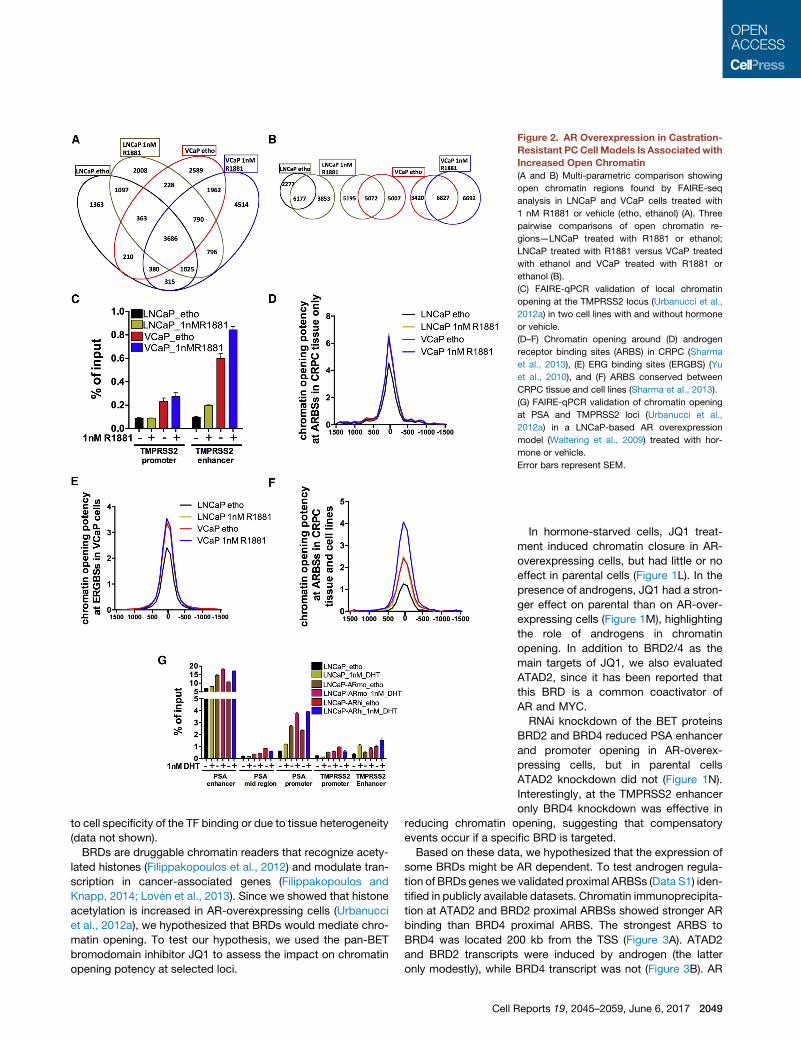
Figure 2. AR Overexpression in Castration-
Resistant PCCell Models Is Associatedwith
Increased Open Chromatin
(A and B) Multi-parametric comparison showing
open chromatin regions found by FAIRE-seq
analysis in LNCaP and VCaP cells treated with
1 nM R1881 or vehicle (etho, ethanol) (A). Three
pairwise comparisons of open chromatin re-
gions—LNCaP treated with R1881 or ethanol;
LNCaP treated with R1881 versus VCaP treated
with ethanol and VCaP treated with R1881 or
ethanol (B).
(C) FAIRE-qPCR validation of local chromatin
opening at the TMPRSS2 locus (Urbanucci et al.,
2012a) in two cell lines with and without hormone
or vehicle.
(D–F) Chromatin opening around (D) androgen
receptor binding sites (ARBS) in CRPC (Sharma
et al., 2013), (E) ERG binding sites (ERGBS) (Yu
et al., 2010), and (F) ARBS conserved between
CRPC tissue and cell lines (Sharma et al., 2013).
(G) FAIRE-qPCR validation of chromatin opening
at PSA and TMPRSS2 loci (Urbanucci et al.,
2012a) in a LNCaP-based AR overexpression
model (Waltering et al., 2009) treated with hor-
mone or vehicle.
Error bars represent SEM.
to cell specificity of the TF binding or due to tissue heterogeneity
(data not shown).
BRDs are druggable chromatin readers that recognize acety-
lated histones (Filippakopoulos et al., 2012) and modulate tran-
scription in cancer-associated genes (Filippakopoulos and
Knapp, 2014; Loven et al., 2013). Since we showed that histone
acetylation is increased in AR-overexpressing cells (Urbanucci
et al., 2012a), we hypothesized that BRDs would mediate chro-
matin opening. To test our hypothesis, we used the pan-BET
bromodomain inhibitor JQ1 to assess the impact on chromatin
opening potency at selected loci.
Cell R
In hormone-starved cells, JQ1 treat-
ment induced chromatin closure in AR-
overexpressing cells, but had little or no
effect in parental cells (Figure 1L). In the
presence of androgens, JQ1 had a stron-
ger effect on parental than on AR-over-
expressing cells (Figure 1M), highlighting
the role of androgens in chromatin
opening. In addition to BRD2/4 as the
main targets of JQ1, we also evaluated
ATAD2, since it has been reported that
this BRD is a common coactivator of
AR and MYC.
RNAi knockdown of the BET proteins
BRD2 and BRD4 reduced PSA enhancer
and promoter opening in AR-overex-
pressing cells, but in parental cells
ATAD2 knockdown did not (Figure 1N).
Interestingly, at the TMPRSS2 enhancer
only BRD4 knockdown was effective in
reducing chromatin opening, suggesting that compensatory
events occur if a specific BRD is targeted.
Based on these data, we hypothesized that the expression of
some BRDs might be AR dependent. To test androgen regula-
tion of BRDs genes we validated proximal ARBSs (Data S1) iden-
tified in publicly available datasets. Chromatin immunoprecipita-
tion at ATAD2 and BRD2 proximal ARBSs showed stronger AR
binding than BRD4 proximal ARBS. The strongest ARBS to
BRD4 was located 200 kb from the TSS (Figure 3A). ATAD2
and BRD2 transcripts were induced by androgen (the latter
only modestly), while BRD4 transcript was not (Figure 3B). AR
eports 19, 2045–2059, June 6, 2017 2049

Figure 3. Androgens and AR Regulate BRDs Expression
(A) Androgen receptor binding sites (ARBSs) close to BRD4, BRD2, and ATAD2 genes according to publicly available datasets (see Data S1) were validated by
chromatin immunoprecipitation (ChIP)-qPCR analysis. PSA mid-region served as ARBS negative control.
(B) Indicated transcript levels measured by qRT-PCR after hormone treatment.
(C) ATAD2 gene expression measured by qRT-PCR in LNCaP-pcDNA3.1, -ARmo, and -ARhi treated with hormone. The mean and SEM of ATAD2 against TATA-
binding protein (TBP) values normalized measure with no treatment (0M).
(D) Western blot analysis of indicated proteins levels in cells treated with hormone.
See Figure S2.
overexpression sensitized ATAD2 transcription to lower concen-
trations of androgens (Figure 3C). In contrast to BRD4, ATAD2
and BRD2 protein levels were increased in AR-overexpressing
cells and further increased by androgen stimulation (Figures
3D and S2A). AR knockdown in LNCaP cells during a time course
of androgen treatment reduced ATAD2 transcript and protein
levels within 24 hr (Figures S2B and S2C). In contrast, BRD2
protein levels were downregulated only after 48 hr treatment
with AR-targeted small interfering RNA (siRNA) (Figure S2C).
BRDs Are Tissue Biomarkers Overexpressed inCastration-Resistant PCsTo establish the predictive clinical value of BRD2/4 and ATAD2,
we performed qRT-PCR (Figures 4A–4D) and immunohisto-
chemical (IHC) analyses (Figures 4E–4H) using benign prostate
tissue and PC specimens and found that all transcripts were
overexpressed in cancer compared to BPH. The long form of
BRD4 (BRD4-L) (p > 0.05) andBRD2 (p < 0.0001) nuclear staining
was increased in CRPC (Figures 4I and 4J). BRD4 (Figure S3A)
and BRD2 (Figure 4K) protein levels determined by IHC were
not prognostic for biochemical recurrence, although BRD2 stain-
ing separated patients with poor prognosis and was significantly
2050 Cell Reports 19, 2045–2059, June 6, 2017
(p = 0.0154) associated with mortality (Figure 4L). Strong nuclear
(Figure 4M) and cytoplasmic (Figure S3B) staining for ATAD2
was significantly increased in CRPC cases (p < 0.0001). More-
over, strong nuclear (Figure 4N) but not cytoplasmic (Figure S3C)
staining was associated with poor outcome (Figure S3D). We
confirmed the significant (p < 0.001) prognostic relevance of
ATAD2 as a tissue biomarker for biochemical recurrence in an
independent cohort of 12,427 patients’ samples (Figures 4O
and S3). Positive staining for ATAD2 was associated with Ki67
staining, tumor stage, and AR protein expression (Figure S3).
Androgen-Receptor-Overexpressing Cells Are MoreSensitive to Bromodomain InhibitorsBRD inhibitors have been reported to reduce the viability of PC
cells (Asangani et al., 2014). To determine whether these effects
are dependent on AR expression levels, we performed knock-
downs of BRDs in AR-overexpression cell models in the pres-
ence of androgens (Figures S4A and S4B). Silencing BRD4
decreased MYC levels in both LNCaP and VCaP cells, while up-
regulating slightly AR, and PSA only in VCaP cells (Figure S4B).
Knockdown of BRD4 decreased viability in all the AR-positive
cell lines tested (LNCaP-pcDNA3.1, LNCaP-ARhi, and VCaP),

Figure 4. BRDs Are Tissue Biomarkers Overexpressed in Castration-Resistant PC
(A–D) Expression of BRD4-long (BRD4-L) (A), BRD4-short (BRD4-S) transcript form (B), BRD2 (C), and ATAD2 (D) gene transcripts relative to TBP levels in BPH
(n = 15), primary untreated PC (n = 27), and CRPC (n = 15) specimens according to qRT-PCR. Kruskal-Wallis with Dunn post-test results are shown (***p < 0.0001;
**p < 0.001; *p = 0.01–0.05; ns, not significant).
(E and F) CRPC immunohistochemical (IHC) strong stainings (score = 3) for BRD4 long isoform (E) and BRD2 (F).
(G and H) CRPC IHC stainings showing examples of low (0%) (G) and high (<5%) (H) staining of nuclear ATAD2. Images are 83 magnification.
(I and J) Proportions of tumors according to BRD4 long isoform (I) and BRD2 (J) staining intensity in PC (n = 159 for BRD4 and n = 90 for BRD2) and CRPC (n = 128
for BRD4 long isoform and n = 34 for BRD2).
(K) Kaplan-Meier analysis of biochemical progression-free survival in 90 prostatectomy-treated patients according to BRD2 stainings (p = ns calculated with
Mantel-Cox test).
(L) Kaplan-Meier analysis showing shorter time to death in 37 men that died of PC out of the 90 patients for which material was stained for BRD2 (p = 0.015
calculated with Mantel-Cox test).
(M) Proportions of tumors according to percentage of ATAD2 positive nuclei in PC (n = 258) and CRPC (n = 121) specimens (p < 0.0001 according to X2 test).
(N) Kaplan-Meier analysis of biochemical progression-free survival in prostatectomy-treated patients according to the percentage of ATAD2 positive nuclei. Six
patients with high frequency of ATAD2-positive nuclei had very short progression-free time (p = 0.0354 calculated with Mantel-Cox test).
(O) Kaplan-Meier analysis of biochemical progression-free survival in a validation cohort of 8,541 prostatectomy-treated patients according to ATAD2 staining
(p < 0.001). 26% (n = 2,216) of the stainings were positive for ATAD2, of which strong ATAD2 staining accounted for 81% (n = 1,789).
Error bars represent SEM. See Figure S3.
Cell Reports 19, 2045–2059, June 6, 2017 2051

but knockdown of BRD2 or ATAD2 alone had no effect on
viability (Figure 5A). ATAD2 inhibition via a small molecular probe
was shown to have limited effect on viability of LNCaP cells
(Bamborough et al., 2016). Silencing ATAD2 upregulated AR
specifically in LNCaP and MYC in VCaP cells, while silencing
BRD2 slightly upregulated MYC in both LNCaP and VCaP cells
(Figure S4B). Therefore, our data suggest that compensatory
mechanisms such as enhanced AR/MYC signaling may take
place and promote cell survival when single BRDs are targeted.
In fact, co-targeting both ATAD2 and BRD2 in LNCaP cells via a
combinatorial knockdown had no additive effect on decreasing
cell viability compared to targeting BRD2 alone (Figure 5B) and
highlighted that BRD4 is important but not the only contributor
to cell viability.
To test whether AR deregulation enhances sensitivity to bro-
modomain inhibitors, we treated a panel of AR-positive PC cell
lines with JQ1 in the presence of androgens (Figures 5C, S4C,
and S4D). AR levels in 22RV1 and LNCaP cells are similar (Erzur-
umlu and Ballar, 2017), while LNCaP-ARhi and VCaP cells
overexpress AR compared to LNCaP (or LNCaP-pcDNA3.1)
with VCaP showing the highest levels of AR (Urbanucci et al.,
2012b; Waltering et al., 2009). VCaP cells were indeed the
most sensitive cells to JQ1 treatment. LNCaP-ARhi and parental
LNCaP cells were equally responsive to JQ1 treatment (Fig-
ure S4D). However, when such cells were grown in androgen
deprivation conditions (castrate conditions), AR-overexpressing
cells were more sensitive to JQ1 (Figure 5D). Also, combined
treatment with enzalutamide and JQ1 was more effective in
AR-overexpressing cells (Figures 5E and 5F), suggesting that
AR deregulation is implicated in response to BET inhibition.
Combined treatment with JQ1 and enzalutamide triggered
apoptosis in VCaP cells in full media (Figure 5G), but not in
LNCaP cells, even when deprived of androgens (Figures S4E
and S4F). This indicates that AR activity/level defines whether
this drug combination has a cytostatic (low-level AR activation)
or cytotoxic (high-level AR activation) effect.
When cells are treated with anti-androgens, resistance can
emerge in which AR activity is maintained (Buttigliero et al.,
2015). Using an enzalutamide-resistant LNCaP model (Kregel
et al., 2013), we found that the inhibitory effect of JQ1 was
retained (Figure 5H).
Gene Expression Analysis of Bromodomain Inhibitor-Treated Cells and Six Independent PC Cohorts Reveal aTen-Gene Signature for Patient StratificationTo identify patients that could potentially benefit from BET-tar-
geted therapies, we sought to identify a gene signature able to
stratify CRPC responders. We performed a time-course treat-
ment with JQ1 in LNCaP (Figure S5A) and VCaP (Figure S5B)
cells to identify affected genes. Upregulated genes (Table S2A)
showed significant overrepresentation of histone genes (Figures
S5C and S5D) and overrepresentation of GO terms for chromatin
compaction (p < 10�6) (Table S3), corroborating the tendency for
chromatin closure upon JQ1 treatment, while downregulated
transcripts (Table S2B) included AR targets found in tissues of
CRPC patients (Sharma et al., 2013) (Figure 6A). We validated
decreased protein levels of the AR targets PSA and CAMKK2
upon JQ1 treatment (Figure S2A). CRPC-associated genes
2052 Cell Reports 19, 2045–2059, June 6, 2017
such as UBE2C, HOXB13, AURKA, and CAMKK2 (Data S1)
that were downregulated by JQ1 treatment (Figures S5E and
S5F) and affected by BRD4 knockdown (Figure S5G) also
showed bromodomain-dependent local chromatin opening (Fig-
ure S5H). AR deregulation affected chromatin opening especially
at ARBSs, when present (Figures S5I and S5J).
Finally, we used published clinical expression array data
(Taylor et al., 2010) and RNA-seq of clinical specimens (Ylipaa
et al., 2015) to identify a clinical gene signature of overex-
pressed genes in CRPC (Tables S4A and S4B). We compared
lists of CRPC-overexpressed genes with those that displayed
increased proximal chromatin opening in CRPC (Figure 6B and
Table S5) and genes downregulated by JQ1 treatment in cell
lines and obtained a set of 15 genes (Figures 6C and 6D).
A generalized linear model with elastic net regularization (Erho
et al., 2013) was used on the Mayo Clinic I (MCI) PC cohort (Erho
et al., 2013) as the discovery dataset to assess the association of
the 15 genes to biochemical recurrence, PC specific mortality,
and metastatic recurrence. Ten of the 15 genes contributed
significantly to the model.
We called the resulting ten-gene signature ‘‘BROMO-10.’’
To evaluate the prognostic significance of BROMO-10, first we
used RNA-seq data from an independent cohort of PC from
the Fred Hutchinson Cancer Research Center (Kumar et al.,
2016; Roudier et al., 2016) (Figures 6E and 6F). Genes
comprising the signature were deregulated in this cohort with
seven out of ten genes being differentially expressed when
comparing CRPCs to primary PCs.
Next, we assessed the independent prognostic value of each
gene according to various endpoints in two additional validation
cohorts (Karnes et al., 2013; Ross et al., 2016) (Table S6).
According to univariable and multivariable analysis, BROMO-
10 contributed independent prognostic information over the clin-
icopathological variables. In the Johns Hopkins Medical Institu-
tions-Radical Prostatectomy (JHMI-RP) cohort of high-risk men
treated with radical prostatectomy without adjuvant or salvage
therapy prior to metastatic onset, BROMO-10 discriminated for
the biochemical recurrence endpoint (Figures 7A, S6A, and
S6B) and the PC-specific mortality endpoint in the MCII cohort
of high-risk men (Figures 7B, S6C, and S6D).
We further assessed the prognostic value of BROMO-10 in
predicting the onset of CRPC. Fifty-five patients that developed
metastasis after radical prostatectomy without any adjuvant or
salvage therapy (‘‘natural history cohort’’) treated at Johns Hop-
kins were evaluated using Kaplan-Meier analysis considering
time to CRPC from metastatic onset. Upon metastatic onset,
all patients received androgen deprivation therapy (ADT). Higher
BROMO-10 scores were associated with an increased rate of
CRPC after ADT (Figure 7C). The upper quartile of BROMO-10
signature scores had a median time to CRPC of 12 months
compared to the lower quartile of 84 months (p = 0.01).
Finally, to assess whether BROMO-10 is able to predict
responsiveness of PC to bromodomain inhibitors, we used
RNA-seq profiles of panels of PC cell lines and expression array
profiles of patient-derived xenografts (PDXs) (Nguyen et al.,
2017) to generate BROMO-10 scores (Figures 7D and 7E). We
then correlated these scores with publicly available IC50 data
for JQ1 (Asangani et al., 2016) (Figure 7F), ZEN-3694 (Attwell

Figure 5. Impact of Bromodomain Inhibition on PC Cell Viability Is Enhanced by AR Deregulation
(A) Viability of LNCaP-pcDNA3.1, LNCaP-ARhi, and VCaP cells 3 days after transfection with siControl (siCont), siBRD2, siATAD2, or siBRD4, relative to control
siRNA values.
(B) Viability of parental LNCaP cells 3 days after transfection with siRNA as indicated. *p < 0.05 according to t test versus the siCont column. n = 6 for each
replicate, for each condition. Each experiment was repeated three times.
(C) Relative viability of 22RV1, LNCaP-pcDNA3.1, LNCaP-ARhi, and VCaP cells cultured in full serum and treated with DMSO or JQ1(*p < 0.05 according to t test
between the indicated conditions).
(D) LNCaP-pcDNA3.1 and -ARhi cells were treated with 1 nM DHT as well as JQ1 or vehicle (DMSO).
(E and F) Solo or combinatorial treatment of LNCaP-pcDNA3.1 and LNCaP-ARhi cells (E) or VCaP cells (F) with MDV3100 and JQ1. Viability compared to DMSO
was assessed after 3 days treatment (*p < 0.05 according to t test versus JQ1 treatment alone).
(G) Caspase activation assay upon treatment of VCaP cells with JQ1, MDV3100, or a combination; hydroxyl-urea was used as a positive control.
(H) Viability of MDV3100-resistant LNCaP cells treated with JQ1(*p < 0.05 according to t test versus DMSO).
Refer also to Figure S4.
Cell Reports 19, 2045–2059, June 6, 2017 2053

Figure 6. Bromodomain Inhibition Targets Clinically Relevant Transcriptional Program Useful for Selecting Patients Responsive to
Bromodomain-Targeted Therapies
(A) GSEA of AR target gene signature (150 core genes identified in CRPC tissue) (Sharma et al., 2013) in expression analysis of VCaP cells treated with JQ1.
(B) Heatmap of genes displaying differential chromatin opening (varying distances upstream of TSSs) in CRPC versus primary PC.
(C) Overlaps between overexpressed genes in CRPC in two clinical microarray datasets (Tables S4A and S4B), the genes associated with open chromatin sites in
CRPC (Table S5) and the consensus genes downregulated by JQ1 treatment of two cell lines (Table S2B).
(D) 15 genes associated with open chromatin, overexpressed in CRPC and downregulated by JQ1 treatment.
(legend continued on next page)
2054 Cell Reports 19, 2045–2059, June 6, 2017

Figure 7. Prognostic and Predictive Value of the Ten Genes Signature BROMO-10
(A–C) Kaplan-Meier curves indicating the prognostic separation achieved by high and low BROMO-10 scores versus (A) biochemical recurrence in the JHMI-RP
validation cohort (Ross et al., 2016) (p = 0.008) (B) prostate cancer-specificmortality (PCSM)-free survival in theMayo Clinic validation cohort (Karnes et al., 2013)
(p = 0.0089) and (C) CRPC-free survival of patients post-ADT treatment of metastatic patients in the JHMI cohort as expression quartiles (n = 55, p value 0.01).
(D and E) Heatmaps of BROMO-10 score and individual gene expression in cell lines (D) and also in PDX models (E).
(F–I) Two-sample t test evaluation of the significance of growth inhibition or reduction in tumor volume with IC50 dose administration of BET bromodomain
inhibitors JQ1 (F), ZEN-3694 (G), and I-BET762 (H) to cell lines or of ZEN-3694 to PDX xenografts (I).
See Figure S6 and Table S6.
et al., 2016) (Figure 7G), I-BET762 (Figure 7H), and responsive-
ness to I-BET762 treatment measured as a significant reduction
in tumor volume (Wyce et al., 2013) (Figure 7I). A high BROMO-
10 score was significantly (p < 0.001) associated with respon-
siveness of PC cell lines to JQ1 and I-BET762 and reduction
of tumor volume upon treatment of PC PDX models with
(E and F) Underlined genes were also significant as assessed in Fred Hutchinson
(PrCa; Roudier et al., 2016) and castration-resistant PrCa (Kumar et al., 2016) (E
t test (F).
Refer also to Figure S5 and Tables S2, S3, S4, and S5.
I-BET762 (p < 0.01). However, the comparison between groups
was not significant for responsiveness to ZEN-364 because
PC3 cells, notably an AR negative PC cell line, had a low
BROMO-10 score and was nevertheless sensitive to ZEN-364.
Taken together, these data confirm the ability of BROMO-10 to
predict responsiveness to BET inhibitors.
Cancer Research Center (FHCRC) cohort comprising primary prostate cancers
) with indicated fold-change values and p values according to a two-sample
Cell Reports 19, 2045–2059, June 6, 2017 2055

DISCUSSION
Here, we show that chromatin accessibility increases during PC
cancer progression due tomechanisms that involve the AR over-
expression and the activity of BRDs. Importantly, we found that
the chromatin structure in CRPC is able to classify disease stage
demonstrating that genome-wide chromatin structure is reprog-
rammed as disease progresses, and it shows distinct features
compared to primary tumors or benign tissue. Increased chro-
matin accessibility in PC was inferred in a recent study, although
the low number of peaks found in the healthy tissue dominated
the results (Stelloo et al., 2015). For the present study, we
developed an advanced analysis pipeline to exclude possible
confounding factors such as variations in ploidy from sample
to sample (see Supplemental Information). This was essential
for improved analyses because copy number variation has pre-
viously been shown to be strongly associated with poor prog-
nosis in advance disease (Taylor et al., 2010).
Interestingly, androgens were able to enhance chromatin
opening especially at ARBSs. This suggests a positive feedback
loop in which the AR is able to bind more tightly to the genome
due to increased opening at ARBSs. These data are concordant
with our previous results showing stronger AR binding to chro-
matin in AR-overexpressing cells (Massie et al., 2011; Urbanucci
et al., 2012b) and to other reports showing that chromatin acces-
sibility is pre-docked prior AR binding at ARBSs (Andreu-Vieyra
et al., 2011; He et al., 2010).
BRDs have gained extensive attention due to their tissue-spe-
cific capacity to modulate key transcriptional events during can-
cer progression (Fu et al., 2015) also in CRPC where they are
therapeutically relevant (Asangani et al., 2014). We found that
selected key BRDs such as ATAD2, BRD2, and BRD4 have
a locus-specific effect on chromatin opening, suggesting that
compensatory mechanismsmay take place if BRDs are inhibited
with single agents. For instance, we show that upregulation of
the AR or MYC proteins occurs while inhibiting ATAD2 or
BRD2 and possibly explains the limited effect of their inhibition
on cell viability.
We also show that ATAD2 and BRD2 are androgen regulated.
ATAD2 was previously reported to be androgen regulated (Zou
et al., 2009). However, here we report that ATAD2 expression is
enhanced in AR-overexpressing cells at low concentrations of
androgens, and BRD4 long isoform, BRD2, and ATAD2 are all
overexpressed in CRPC tissues. These results support the pres-
ence of an AR deregulation-mediated positive feedback loop
that boosts the expression of BRDs in order to increase AR
chromatin accessibility. Moreover, ATAD2 had strong prog-
nostic value on a cohort of 10,000 patients. Our data also
suggest that, while ATAD2 is an optimal tissue biomarker in
identifying PC tissues where active transcription due to heavy
cell-cycle turnover is ongoing, it may not be a good target for
PC therapy, as also shown recently (Bamborough et al.,
2016), if not targeted in combination with other agents such
as antiandrogens.
The role of ATAD2 as a regulator of chromatin dynamics is well
known in yeast (Cattaneo et al., 2014). A recent study showed
that ATAD2 is highly expressed in replicating PC cells and
ATAD2 expression correlates with expression of cell-cycle and
2056 Cell Reports 19, 2045–2059, June 6, 2017
DNA replication genes that have overlapping function in meiosis
and tumor progression (Koo et al., 2016). Moreover, in highly
proliferating embryonic stem cells, ATAD2 was reported to sus-
tain specific gene expression programs via regulating chromatin
opening guided by histone acetylation (Morozumi et al., 2016),
which is in agreement with our data. These findings corroborate
our data and are supportive of ATAD2 being a possible contrib-
utor to increased transcription plasticity in CRPC.
Our study also suggests that theremight be a subpopulation of
tumors that are more dependent on BRDs activity than others.
Therefore, we built a ten-gene signature, BROMO-10, which
is able to discriminate patients with poorer outcome, which
takes into account chromatin structure and additionally incorpo-
rates key PC-specific downstream targets of BRDs. Interest-
ingly, these targets include FEN1, which we have previously
described to be important for PC progression and proposed as
a tissue biomarker for biochemical recurrence (Urbanucci
et al., 2012b), EEF1A2, which has been proposed as a marker
of prostate cell transformation (Scaggiante et al., 2012; Sun
et al., 2014), KAT2A, which encodes a histone acetyl-transferase
controlling the PI3/AKT pathwaywith therapeutic potential in leu-
kemia (Sun et al., 2015), andHSPH1, which enhances MYC tran-
scription and drives B cell non-Hodgkin lymphoma (Zappasodi
et al., 2015).
Mechanisms of resistance to BET inhibition have been re-
ported. Therefore, it is extremely important to define patients
that will respond to BET-inhibition therapies in combination
with standard therapies to avoid resistance. We show that
BROMO-10 is able to predict response to BET-inhibition thera-
pies, but the use of this signature should be limited to tumors
with intact AR signaling, and further studies are needed to refine
the signature for different compounds.
In conclusion, we propose AR deregulation-driven chromatin
structure as a key determinant of tumor progression. We
describe the effect of BET inhibition on chromatin accessibility
as an additional mechanism by which it is able to repress
cell growth in a cell-specific manner. Moreover, we propose
BROMO-10 signature to be used to select patients more likely
to benefit from BET-targeted therapies and avoid recurrence.
The selection of PC patients into future trials evaluating the effi-
cacy should be based on the assessment of AR status, key BRDs
expression, such as ATAD2, and the gene signature that reflects
the chromatin status of these tumors.
EXPERIMENTAL PROCEDURES
Clinical Samples
Allworkonclinical sampleshasbeencarriedout in compliancewith theHelsinki
Declaration and with the approval of ethics boards at collaborating institutions
as outlined below. The tissue microarray from the Department of Urology and
the Martini Clinics at the University Medical Centre Hamburg-Eppendorf
consisted of archived diagnostic leftover tissues. Manufacture and analysis
was approved by the local ethics committee (Ethics commission Hamburg,
WF-049/09 and PV3652). According to local laws (HmbKHG, x12,1), informed
consent was not required for this study. Patient records and information were
anonymized and de-identified prior to analysis.
Three BPH, six primary PC, and three CRPCs were used for FAIRE-seq as-
says and FAIRE-qPCR assays. RNA-seq data from transcriptomes of 12 BPH,
28 untreated PCs, and 13 CRPCs, including the samples used for FAIRE-seq,
were publicly available (Ylipaa et al., 2015). These samples and the tissue

microarray described below were provided by Tampere University Hospital.
The use of these samples for FAIRE-seq and of the tissue microarray was
approved by the ethical committee of Tampere University Hospital and the Na-
tional Authority forMedicolegal Affairs. Written informed consent was obtained
from the subjects for sequencing the samples.
The Tampere patients’ cohort of tissue microarrays (TMAs) contained 258
formalin-fixed paraffin-embedded prostatectomy and 121 CRPC specimens.
A subset of the cohort was used to immunostain for ATAD2, BRD2, and
BRD4. The Hamburg patients’ TMA cohort contained 9,467 prostatectomy tis-
sue specimens. Radical prostatectomy specimens were available from 12,427
patients. PSA values were measured following surgery, and PSA recurrence
was defined as the time point when postoperative PSA was increasing from
at least 0.2 ng/mL.
FAIREs
Two replicates were processed for each cell line and condition for subsequent
sequencing analysis. Three to five replicates were processed for qPCR anal-
ysis. Four million cells were plated and hormone deprived for 4 days. Cells
were then treated with R1881 or DHT for 4 hr. To perform tissue FAIRE from
clinical material, 3 mL of PBS containing 2 3 protease inhibitor (Roche) was
added to 40 3 20-mm sections of freshly frozen tissue specimens. Down-
stream fixation and processing of both sample types are as described in the
Supplemental Information.
FAIRE-Seq Analysis
Peak detection for FAIRE-seq was performed using model-based analysis of
ChIP-seq (MACS) (Zhang et al., 2008) with default parameters using inputs
of each of the FAIRE samples as controls and with F-Seq (Boyle et al.,
2008). Refer to Supplemental Information for more detail.
Evaluation of the 15-Gene Signature Prognostic Value
Microarray data from the Decipher GRID were extracted for three radical
prostatectomy cohorts from previously described (Erho et al., 2013; Karnes
et al., 2013; Ross et al., 2016) validation studies. A classifier to distinguish
between metastatic versus non-metastatic cancers was developed. The
classifier was constructed from 15 genes (Figure 5C) using a generalized
linear model with elastic net regularization as previously described (Erho
et al., 2013). The model was generated using the MCI cohort (GSE46691)
as training data. In the final model, ten of the 15 genes contributed to the
model score with six positively associated and four negatively associated
genes as determined by the regularized coefficients (see also Table S6).
Scores were then generated for samples from the Mayo Clinic II (MCII) and
JHMI-RP validation cohorts, and performance in each cohort was assessed
using survival analysis.
BROMO-10 scores in cell lines and patients derived xenografts were calcu-
lated using GSVA Bioconductor package (https://www.bioconductor.org/
packages/release/bioc/html/GSVA.html).
Statistics
Statistical analyses were performed using GraphPad Prism, MATLAB, and
Microsoft Excel. All statistical tests were two-tailed with testing level thresh-
olds of a = 0.05.
ACCESSION NUMBERS
The accession number for the gene expression and FAIRE-seq data reported
in this paper is GEO: GSE73989. The data analysis script referred to in the
Supplementary Experimental Procedures has also been deposited and is
accessible at https://github.com/dvbcfo/depth_track_window.
SUPPLEMENTAL INFORMATION
Supplemental Information includes six figures, six tables, and one data file and
can be found with this article online at http://dx.doi.org/10.1016/j.celrep.2017.
05.049.
AUTHOR CONTRIBUTIONS
A.U. wrote the manuscript. I.G.M. and A.U. designed the study and analyzed
the data. A.U., T.T., S.M., C.B., K.K.K., X.S., and S.J.B. performed experi-
ments and edited the manuscript. A.U., V.K., D.V., M.T., M.A., E.D., I.M.C.,
P.L., and N.E. performed the bioinformatic analyses and edited the manu-
script. H.M.I., A.K., and F.S. assisted in the preparation of the revised manu-
script. D.J.V.G., S. Knapp, S. Kregel, R.J.K., A.E.R., P.S.N., E.C., F.Y.F.,
E.M.S., K.E.K., and M.N. contributed reagents or data and edited the manu-
script. G.S., T.S., and T.L.J.T. provided clinical material and edited the manu-
script. I.M., T.V., and L.S. contributed reagents and edited the manuscript.
ACKNOWLEDGMENTS
We thank Ms. Paivi Martikainen for the skillful technical assistance. We
acknowledge the last scientific contribution of our belovedMs.Mariitta Vakkuri
to this work. A.U. is supported by the South-East Norway Health Authorities
(Helse Sor-Ost grant ID 2014040) at the Oslo University Hospital and the
Norwegian Centre for Molecular Medicine. I.M. has been funded by the
MLS (390000 and 143295), Helse Sor-Ost (2014040), Norwegian Research
Council (230559), and the Faculty of Medicine at the Oslo University Hospital.
P.S.N., E.C., and I.M.C. are supported by NIH grants P50CA097186 and
P01CA163227, and the Prostate Cancer Foundation. H.M.I. is supported by
the Norwegian Cancer Society (711072, 102032, and 4521627). S. Knapp is
grateful for support by the SGC, a registered charity (number 1097737) that re-
ceives funds from AbbVie, Bayer Pharma AG, Boehringer Ingelheim, Canada
Foundation for Innovation, Eshelman Institute for Innovation, Genome Can-
ada, Innovative Medicines Initiative (EU/EFPIA), Janssen, Merck & Co., Novar-
tis Pharma AG, Ontario Ministry of Economic Development and Innovation,
Pfizer, S~ao Paulo Research Foundation-FAPESP, Takeda, and the Wellcome
Trust. The funders had no role in the study design, data collection and analysis,
the decision to publish, or preparation of the article.
Received: September 11, 2016
Revised: April 1, 2017
Accepted: May 12, 2017
Published: June 6, 2017
REFERENCES
Andreu-Vieyra, C., Lai, J., Berman, B.P., Frenkel, B., Jia, L., Jones, P.A., and
Coetzee, G.A. (2011). Dynamic nucleosome-depleted regions at androgen
receptor enhancers in the absence of ligand in prostate cancer cells. Mol.
Cell. Biol. 31, 4648–4662.
Asangani, I.A., Dommeti, V.L., Wang, X., Malik, R., Cieslik, M., Yang, R.,
Escara-Wilke, J., Wilder-Romans, K., Dhanireddy, S., Engelke, C., et al.
(2014). Therapeutic targeting of BET bromodomain proteins in castration-
resistant prostate cancer. Nature 510, 278–282.
Asangani, I.A., Wilder-Romans, K., Dommeti, V.L., Krishnamurthy, P.M., Apel,
I.J., Escara-Wilke, J., Plymate, S.R., Navone, N.M., Wang, S., Feng, F.Y., and
Chinnaiyan, A.M. (2016). BET bromodomain inhibitors enhance efficacy and
disrupt resistance to AR antagonists in the treatment of prostate cancer.
Mol. Cancer Res. 14, 324–331.
Attwell, S., Jahagirdar, R., Norek, K., Calosing, C., Tsujikawa, L., Kharenko,
O.A., Patel, R.G., Gesner, E.M., Corey, E., Nguyen, H.M., et al. (2016). Abstract
LB-207: Preclinical characterization of ZEN-3694, a novel BET bromodomain
inhibitor entering phase I studies for metastatic castration-resistant prostate
cancer (mCRPC). Cancer Res. 76, LB-207–LB-207.
Bamborough, P., Chung, C.W., Demont, E.H., Furze, R.C., Bannister, A.J.,
Che, K.H., Diallo, H., Douault, C., Grandi, P., Kouzarides, T., et al. (2016).
A chemical probe for the ATAD2 bromodomain. Angew. Chem. Int. Ed. Engl.
55, 11382–11386.
Boyle, A.P., Guinney, J., Crawford, G.E., and Furey, T.S. (2008). F-Seq:
A feature density estimator for high-throughput sequence tags. Bioinformatics
24, 2537–2538.
Cell Reports 19, 2045–2059, June 6, 2017 2057

Buttigliero, C., Tucci, M., Bertaglia, V., Vignani, F., Bironzo, P., Di Maio, M., and
Scagliotti, G.V. (2015). Understanding and overcoming the mechanisms of pri-
mary and acquired resistance to abiraterone and enzalutamide in castration
resistant prostate cancer. Cancer Treat. Rev. 41, 884–892.
Cattaneo, M., Morozumi, Y., Perazza, D., Boussouar, F., Jamshidikia, M.,
Rousseaux, S., Verdel, A., and Khochbin, S. (2014). Lessons from yeast on
emerging roles of the ATAD2 protein family in gene regulation and genome
organization. Mol. Cells 37, 851–856.
Cedar, H., andBergman, Y. (2012). Programming of DNAmethylation patterns.
Annu. Rev. Biochem. 81, 97–117.
Center, M.M., Jemal, A., Lortet-Tieulent, J., Ward, E., Ferlay, J., Brawley, O.,
and Bray, F. (2012). International variation in prostate cancer incidence and
mortality rates. Eur. Urol. 61, 1079–1092.
Chen, Z., Lan, X., Thomas-Ahner, J.M., Wu, D., Liu, X., Ye, Z., Wang, L.,
Sunkel, B., Grenade, C., Chen, J., et al. (2015). Agonist and antagonist switch
DNA motifs recognized by human androgen receptor in prostate cancer.
EMBO J. 34, 502–516.
Delmore, J.E., Issa, G.C., Lemieux, M.E., Rahl, P.B., Shi, J., Jacobs, H.M.,
Kastritis, E., Gilpatrick, T., Paranal, R.M., Qi, J., et al. (2011). BET bromodo-
main inhibition as a therapeutic strategy to target c-Myc. Cell 146, 904–917.
Dolfini, D., and Mantovani, R. (2013). Targeting the Y/CCAAT box in cancer:
YB-1 (YBX1) or NF-Y? Cell Death Differ. 20, 676–685.
Erho, N., Crisan, A., Vergara, I.A., Mitra, A.P., Ghadessi, M., Buerki, C., Berg-
stralh, E.J., Kollmeyer, T., Fink, S., Haddad, Z., et al. (2013). Discovery and vali-
dation of a prostate cancer genomic classifier that predicts early metastasis
following radical prostatectomy. PLoS ONE 8, e66855.
Erzurumlu, Y., and Ballar, P. (2017). Androgen mediated regulation of endo-
plasmic reticulum-associated degradation and its effects on prostate cancer.
Sci. Rep. 7, 40719.
Filippakopoulos, P., and Knapp, S. (2014). Targeting bromodomains: Epige-
netic readers of lysine acetylation. Nat. Rev. Drug Discov. 13, 337–356.
Filippakopoulos, P., Picaud, S., Mangos, M., Keates, T., Lambert, J.P.,
Barsyte-Lovejoy, D., Felletar, I., Volkmer, R., M€uller, S., Pawson, T., et al.
(2012). Histone recognition and large-scale structural analysis of the human
bromodomain family. Cell 149, 214–231.
Fu, L.L., Tian, M., Li, X., Li, J.J., Huang, J., Ouyang, L., Zhang, Y., and Liu, B.
(2015). Inhibition of BET bromodomains as a therapeutic strategy for cancer
drug discovery. Oncotarget 6, 5501–5516.
Giresi, P.G., and Lieb, J.D. (2009). Isolation of active regulatory elements from
eukaryotic chromatin using FAIRE (formaldehyde assisted isolation of regula-
tory elements). Methods 48, 233–239.
He, H.H., Meyer, C.A., Shin, H., Bailey, S.T., Wei, G., Wang, Q., Zhang, Y., Xu,
K., Ni, M., Lupien, M., et al. (2010). Nucleosome dynamics define transcrip-
tional enhancers. Nat. Genet. 42, 343–347.
Karnes, R.J., Bergstralh, E.J., Davicioni, E., Ghadessi, M., Buerki, C., Mitra,
A.P., Crisan, A., Erho, N., Vergara, I.A., Lam, L.L., et al. (2013). Validation of
a genomic classifier that predicts metastasis following radical prostatectomy
in an at risk patient population. J. Urol. 190, 2047–2053.
Koo, S.J., Fernandez-Montalvan, A.E., Badock, V., Ott, C.J., Holton, S.J., von
Ahsen, O., Toedling, J., Vittori, S., Bradner, J.E., and Gorjanacz, M. (2016).
ATAD2 is an epigenetic reader of newly synthesized histone marks during
DNA replication. Oncotarget 7, 70323–70335.
Kregel, S., Kiriluk, K.J., Rosen, A.M., Cai, Y., Reyes, E.E., Otto, K.B., Tom, W.,
Paner, G.P., Szmulewitz, R.Z., and Vander Griend, D.J. (2013). Sox2 is an
androgen receptor-repressed gene that promotes castration-resistant pros-
tate cancer. PLoS ONE 8, e53701.
Kumar, A., Coleman, I., Morrissey, C., Zhang, X., True, L.D., Gulati, R., Etzioni,
R., Bolouri, H., Montgomery, B., White, T., et al. (2016). Substantial interindi-
vidual and limited intraindividual genomic diversity among tumors from men
with metastatic prostate cancer. Nat. Med. 22, 369–378.
Lever, E., and Sheer, D. (2010). The role of nuclear organization in cancer.
J. Pathol. 220, 114–125.
2058 Cell Reports 19, 2045–2059, June 6, 2017
Loven, J., Hoke, H.A., Lin, C.Y., Lau, A., Orlando, D.A., Vakoc, C.R., Bradner,
J.E., Lee, T.I., and Young, R.A. (2013). Selective inhibition of tumor oncogenes
by disruption of super-enhancers. Cell 153, 320–334.
Massie, C.E., Lynch, A., Ramos-Montoya, A., Boren, J., Stark, R., Fazli, L.,
Warren, A., Scott, H., Madhu, B., Sharma, N., et al. (2011). The androgen
receptor fuels prostate cancer by regulating central metabolism and biosyn-
thesis. EMBO J. 30, 2719–2733.
Morozumi, Y., Boussouar, F., Tan, M., Chaikuad, A., Jamshidikia, M., Colak,
G., He, H., Nie, L., Petosa, C., de Dieuleveult, M., et al. (2016). Atad2 is a gener-
alist facilitator of chromatin dynamics in embryonic stem cells. J. Mol. Cell Biol.
8, 349–362.
Nagarajan, S., Hossan, T., Alawi, M., Najafova, Z., Indenbirken, D., Bedi, U.,
Taipaleenmaki, H., Ben-Batalla, I., Scheller, M., Loges, S., et al. (2014). Bromo-
domain protein BRD4 is required for estrogen receptor-dependent enhancer
activation and gene transcription. Cell Rep. 8, 460–469.
Nguyen, H.M., Vessella, R.L., Morrissey, C., Brown, L.G., Coleman, I.M.,
Higano, C.S., Mostaghel, E.A., Zhang, X., True, L.D., Lam, H.M., et al.
(2017). LuCaP prostate cancer patient-derived xenografts reflect the molecu-
lar heterogeneity of advanced disease and serve asmodels for evaluating can-
cer therapeutics. Prostate 77, 654–671.
Perry, A.S., Watson, R.W., Lawler, M., and Hollywood, D. (2010). The epige-
nome as a therapeutic target in prostate cancer. Nat. Rev. Urol. 7, 668–680.
Pomerantz, M.M., Li, F., Takeda, D.Y., Lenci, R., Chonkar, A., Chabot, M.,
Cejas, P., Vazquez, F., Cook, J., Shivdasani, R.A., et al. (2015). The androgen
receptor cistrome is extensively reprogrammed in human prostate tumorigen-
esis. Nat. Genet. 47, 1346–1351.
Ross, A.E., Johnson, M.H., Yousefi, K., Davicioni, E., Netto, G.J., Marchionni,
L., Fedor, H.L., Glavaris, S., Choeurng, V., Buerki, C., et al. (2016). Tissue-
based genomics augments post-prostatectomy risk stratification in a natural
history cohort of intermediate- and high-risk men. Eur. Urol.
Roudier, M.P., Winters, B.R., Coleman, I., Lam, H.M., Zhang, X., Coleman, R.,
Chery, L., True, L.D., Higano, C.S., Montgomery, B., et al. (2016). Character-
izing the molecular features of ERG-positive tumors in primary and castration
resistant prostate cancer. Prostate 76, 810–822.
Scaggiante, B., Dapas, B., Bonin, S., Grassi, M., Zennaro, C., Farra, R., Cris-
tiano, L., Siracusano, S., Zanconati, F., Giansante, C., and Grassi, G. (2012).
Dissecting the expression of EEF1A1/2 genes in human prostate cancer cells:
The potential of EEF1A2 as a hallmark for prostate transformation and progres-
sion. Br. J. Cancer 106, 166–173.
Sharma, N.L., Massie, C.E., Ramos-Montoya, A., Zecchini, V., Scott, H.E.,
Lamb, A.D., MacArthur, S., Stark, R., Warren, A.Y., Mills, I.G., and Neal, D.E.
(2013). The androgen receptor induces a distinct transcriptional program in
castration-resistant prostate cancer in man. Cancer Cell 23, 35–47.
Shi, J., and Vakoc, C.R. (2014). The mechanisms behind the therapeutic activ-
ity of BET bromodomain inhibition. Mol. Cell 54, 728–736.
Song, L., Zhang, Z., Grasfeder, L.L., Boyle, A.P., Giresi, P.G., Lee, B.K., Shef-
field, N.C., Graf, S., Huss, M., Keefe, D., et al. (2011). Open chromatin defined
by DNaseI and FAIRE identifies regulatory elements that shape cell-type iden-
tity. Genome Res. 21, 1757–1767.
Stelloo, S., Nevedomskaya, E., van der Poel, H.G., de Jong, J., van Leenders,
G.J., Jenster, G., Wessels, L.F., Bergman, A.M., and Zwart, W. (2015).
Androgen receptor profiling predicts prostate cancer outcome. EMBO Mol.
Med. 7, 1450–1464.
Sun, Y., Du, C., Wang, B., Zhang, Y., Liu, X., and Ren, G. (2014). Up-regulation
of eEF1A2 promotes proliferation and inhibits apoptosis in prostate cancer.
Biochem. Biophys. Res. Commun. 450, 1–6.
Sun, X.J., Man, N., Tan, Y., Nimer, S.D., and Wang, L. (2015). The role of
histone acetyltransferases in normal and malignant hematopoiesis. Front.
Oncol. 5, 108.
Tark-Dame,M., Jerabek, H.,Manders, E.M., van derWateren, I.M., Heermann,
D.W., and van Driel, R. (2014). Depletion of the chromatin looping proteins
CTCF and cohesin causes chromatin compaction: Insight into chromatin
folding by polymer modelling. PLoS Comput. Biol. 10, e1003877.

Taylor, B.S., Schultz, N., Hieronymus, H., Gopalan, A., Xiao, Y., Carver, B.S.,
Arora, V.K., Kaushik, P., Cerami, E., Reva, B., et al. (2010). Integrative genomic
profiling of human prostate cancer. Cancer Cell 18, 11–22.
Tewari, A.K., Yardimci, G.G., Shibata, Y., Sheffield, N.C., Song, L., Taylor,
B.S., Georgiev, S.G., Coetzee, G.A., Ohler, U., Furey, T.S., et al. (2012).
Chromatin accessibility reveals insights into androgen receptor activation
and transcriptional specificity. Genome Biol. 13, R88.
Timp, W., and Feinberg, A.P. (2013). Cancer as a dysregulated epigenome
allowing cellular growth advantage at the expense of the host. Nat. Rev. Can-
cer 13, 497–510.
Urbanucci, A., Marttila, S., Janne, O.A., and Visakorpi, T. (2012a). Androgen
receptor overexpression alters binding dynamics of the receptor to chromatin
and chromatin structure. Prostate.
Urbanucci, A., Sahu, B., Seppala, J., Larjo, A., Latonen, L.M., Waltering, K.K.,
Tammela, T.L., Vessella, R.L., Lahdesmaki, H., Janne, O.A., and Visakorpi, T.
(2012b). Overexpression of androgen receptor enhances the binding of the
receptor to the chromatin in prostate cancer. Oncogene 31, 2153–2163.
Urbanucci, A., Waltering, K., Mills, I., and Visakorpi, T. (2013). The effect of
AR overexpression on androgen signaling in prostate cancer. In Androgen-
Responsive Genes in Prostate Cancer, Z. Wang, ed. (Springer), pp. 187–200.
Waltering, K.K., Helenius, M.A., Sahu, B., Manni, V., Linja, M.J., Janne, O.A.,
and Visakorpi, T. (2009). Increased expression of androgen receptor sensitizes
prostate cancer cells to low levels of androgens. Cancer Res. 69, 8141–8149.
Waltering, K.K., Urbanucci, A., and Visakorpi, T. (2012). Androgen receptor
(AR) aberrations in castration-resistant prostate cancer. Mol. Cell. Endocrinol.
360, 38–43.
Wyce, A., Degenhardt, Y., Bai, Y., Le, B., Korenchuk, S., Crouthamel, M.-C.,
McHugh, C., Vessella, R., Creasy, C., Tummino, P., et al. (2013). Inhibition of
BET bromodomain proteins as a therapeutic approach in prostate cancer.
Oncotarget 4, 2419–2429.
Xu, Y., Dalrymple, S.L., Becker, R.E., Denmeade, S.R., and Isaacs, J.T. (2006).
Pharmacologic basis for the enhanced efficacy of dutasteride against pros-
tatic cancers. Clin. Cancer Res. 12, 4072–4079.
Ylipaa, A., Kivinummi, K., Kohvakka, A., Annala, M., Latonen, L., Scaravilli, M.,
Kartasalo, K., Leppanen, S.P., Karakurt, S., Seppala, J., et al. (2015). Tran-
scriptome sequencing reveals PCAT5 as a novel ERG-regulated long non-
coding RNA in prostate cancer. Cancer Res. 75, 4026–4031.
Yu, J., Yu, J., Mani, R.S., Cao, Q., Brenner, C.J., Cao, X., Wang, X., Wu, L., Li,
J., Hu, M., et al. (2010). An integrated network of androgen receptor, poly-
comb, and TMPRSS2-ERG gene fusions in prostate cancer progression. Can-
cer Cell 17, 443–454.
Zappasodi, R., Ruggiero, G., Guarnotta, C., Tortoreto, M., Tringali, C., Cavane,
A., Cabras, A.D., Castagnoli, L., Venerando, B., Zaffaroni, N., et al. (2015).
HSPH1 inhibition downregulates Bcl-6 and c-Myc and hampers the growth
of human aggressive B-cell non-Hodgkin lymphoma. Blood 125, 1768–1771.
Zhang, Y., Liu, T., Meyer, C.A., Eeckhoute, J., Johnson, D.S., Bernstein, B.E.,
Nusbaum, C., Myers, R.M., Brown, M., Li, W., and Liu, X.S. (2008). Model-
based analysis of ChIP-Seq (MACS). Genome Biol. 9, R137.
Zhang, T.Y., Agarwal, N., Sonpavde, G., DiLorenzo, G., Bellmunt, J., and Vo-
gelzang, N.J. (2013). Management of castrate resistant prostate cancer-recent
advances and optimal sequence of treatments. Curr. Urol. Rep. 14, 174–183.
Zou, J.X., Guo, L., Revenko, A.S., Tepper, C.G., Gemo, A.T., Kung, H.J., and
Chen, H.W. (2009). Androgen-induced coactivator ANCCA mediates specific
androgen receptor signaling in prostate cancer. Cancer Res. 69, 3339–3346.
Cell Reports 19, 2045–2059, June 6, 2017 2059


![Changes in Androgen Receptor Nongenotropic …...[CANCER RESEARCH 64, 7156–7168, October 1, 2004] Changes in Androgen Receptor Nongenotropic Signaling Correlate with Transition of](https://img.dokumen.tips/doc/110x75/5fbb5d2af676fe7f7f6d7a0b/changes-in-androgen-receptor-nongenotropic-cancer-research-64-7156a7168.jpg)