Embed Size (px)
DESCRIPTION
ANATOMY OF THE FEMALE BONY PELVIS and FETAL SKULL. Quoted with modification from Dr. Salwa Neyazi King Saud University. THE BONY PELVIS. WHICH BONES COMPOSE THE BONY PELVIS? Ilium, Ischium, Pubis Sacrum Coccyx. THE BONY PELVIS. WHAT IS THE PELVIC BRIM? - PowerPoint PPT Presentation
Citation preview
ANATOMY OF THE FEMALE BONY ANATOMY OF THE FEMALE BONY PELVIS and FETAL SKULLPELVIS and FETAL SKULL
Quoted with modification from Dr. Salwa Neyazi Quoted with modification from Dr. Salwa Neyazi
King Saud UniversityKing Saud University
THE BONY PELVISTHE BONY PELVIS
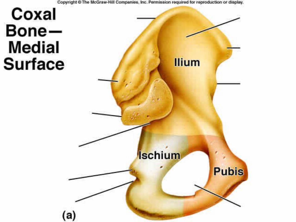
WHICH BONES COMPOSE THE BONY PELVIS?
Ilium, Ischium, Pubis
Sacrum
Coccyx
THE BONY PELVISTHE BONY PELVIS
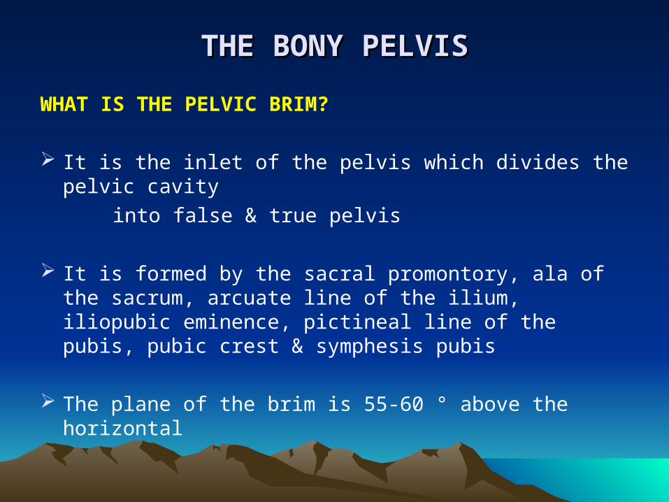
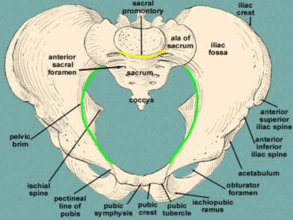
WHAT IS THE PELVIC BRIM?
It is the inlet of the pelvis which divides the pelvic cavity
into false & true pelvis
It is formed by the sacral promontory, ala of the sacrum, arcuate line of the ilium, iliopubic eminence, pictineal line of the pubis, pubic crest & symphesis pubis
The plane of the brim is 55-60 ° above the horizontal
THE BONY PELVISTHE BONY PELVIS
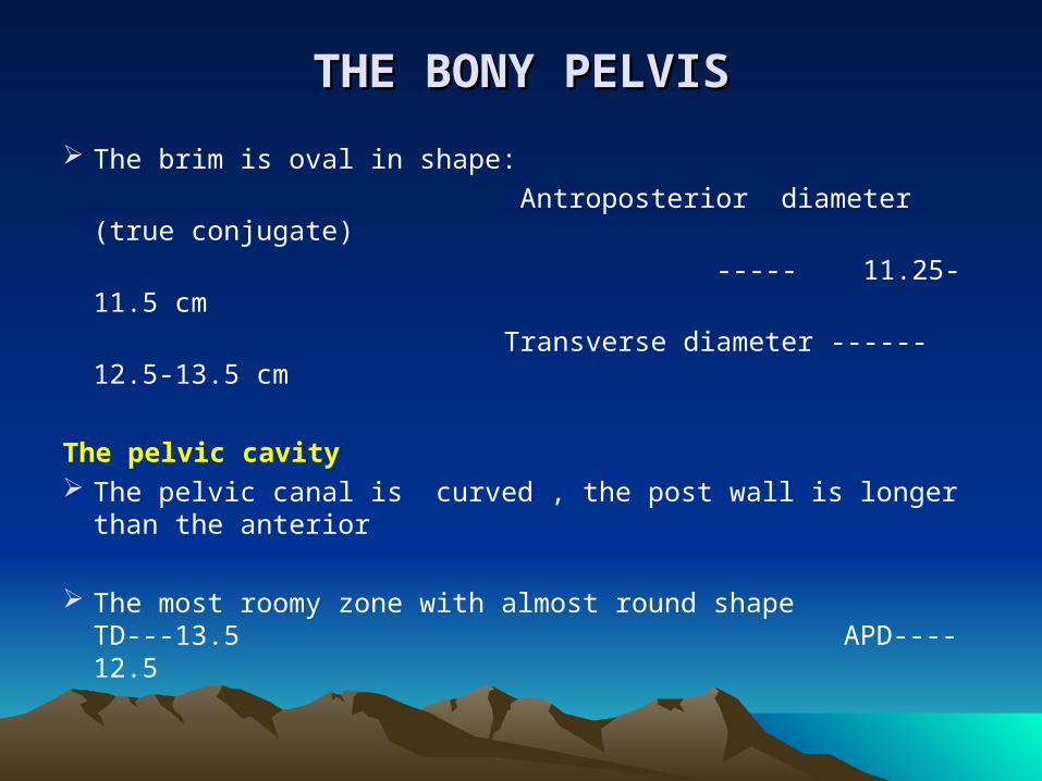
The brim is oval in shape:
Antroposterior diameter (true conjugate)
----- 11.25- 11.5 cm
Transverse diameter ------ 12.5-13.5 cm
The pelvic cavity The pelvic canal is curved , the post wall is longer than
the anterior
The most roomy zone with almost round shape TD---13.5 APD----12.5
THE BONY PELVISTHE BONY PELVIS
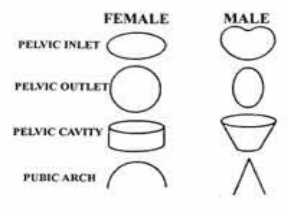
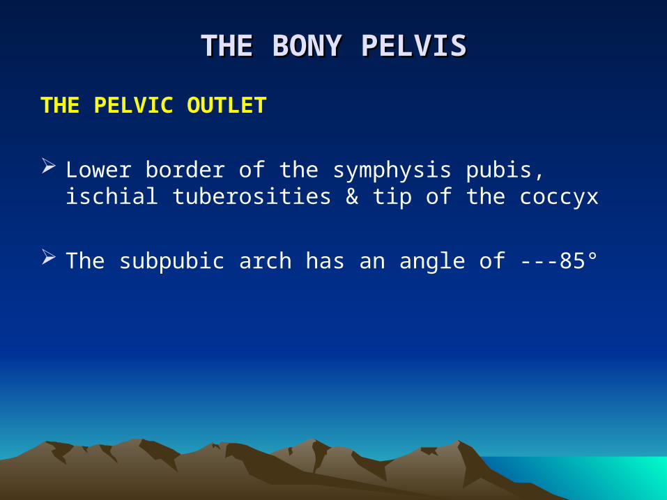
THE PELVIC OUTLET
Lower border of the symphysis pubis, ischial tuberosities & tip of the coccyx
The subpubic arch has an angle of ---85°
THE BONY PELVISTHE BONY PELVIS
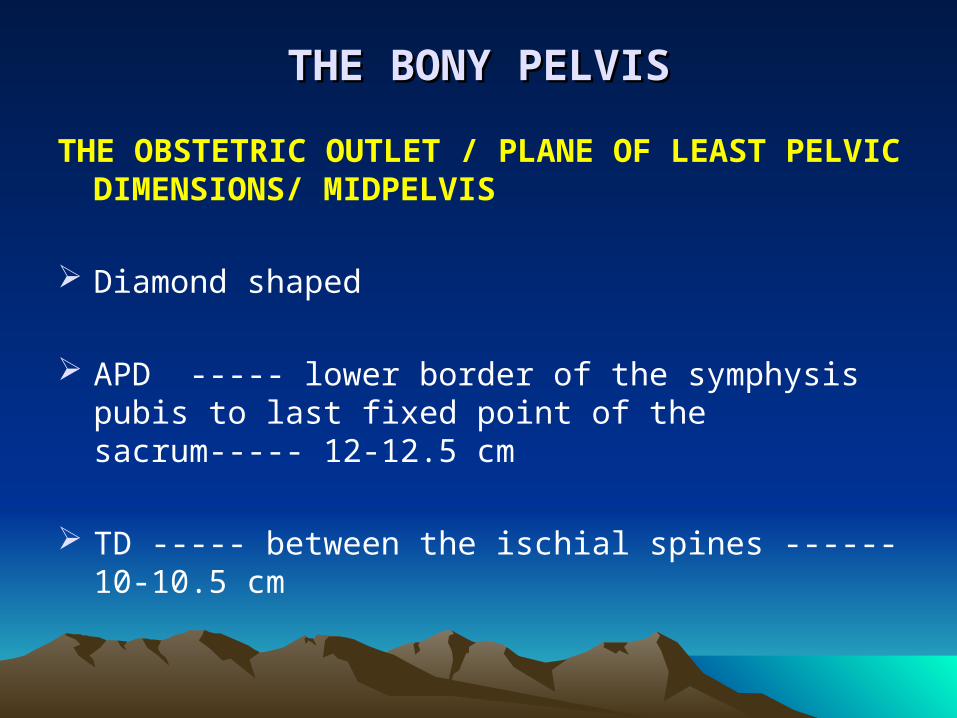
THE OBSTETRIC OUTLET / PLANE OF LEAST PELVIC DIMENSIONS/ MIDPELVIS
Diamond shaped
APD ----- lower border of the symphysis pubis to last fixed point of the sacrum----- 12-12.5 cm
TD ----- between the ischial spines ------ 10-10.5 cm
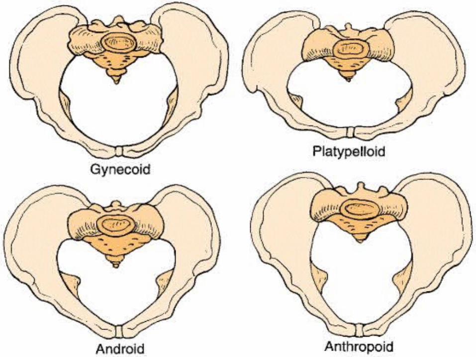
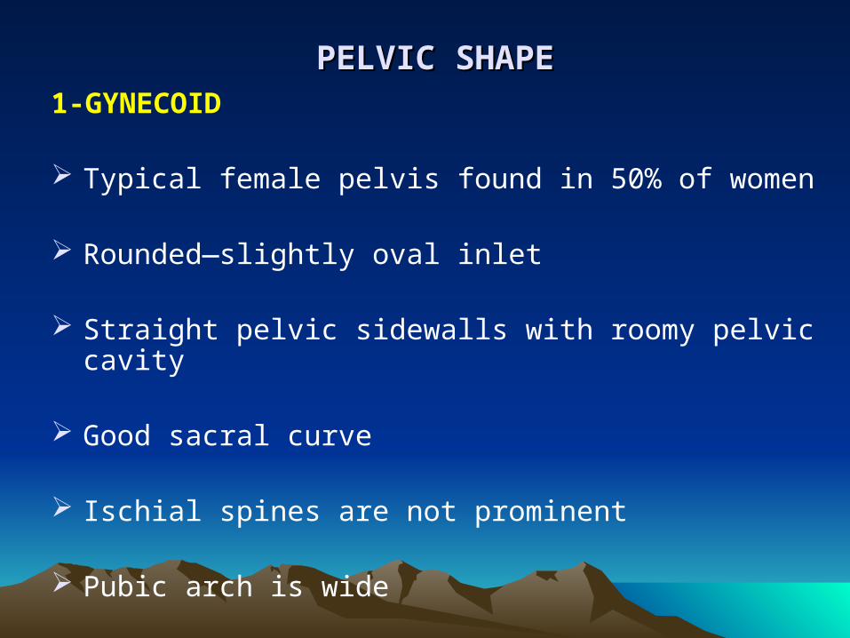
PELVIC SHAPEPELVIC SHAPE 1-GYNECOID
Typical female pelvis found in 50% of women
Rounded—slightly oval inlet
Straight pelvic sidewalls with roomy pelvic cavity
Good sacral curve
Ischial spines are not prominent
Pubic arch is wide
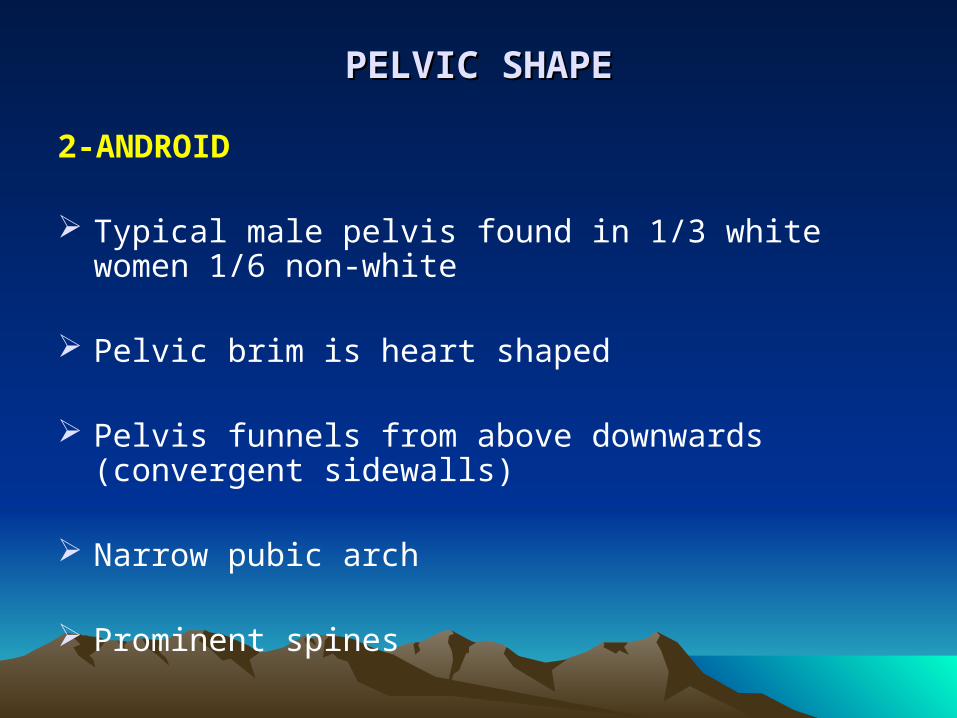
PELVIC SHAPEPELVIC SHAPE
2-ANDROID
Typical male pelvis found in 1/3 white women 1/6 non-white
Pelvic brim is heart shaped
Pelvis funnels from above downwards (convergent sidewalls)
Narrow pubic arch
Prominent spines
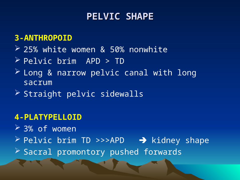
PELVIC SHAPEPELVIC SHAPE
3-ANTHROPOID 25% white women & 50% nonwhite Pelvic brim APD > TD Long & narrow pelvic canal with long sacrum Straight pelvic sidewalls
4-PLATYPELLOID 3% of women Pelvic brim TD >>>APD kidney shape Sacral promontory pushed forwards
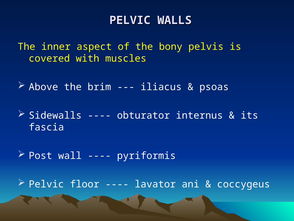
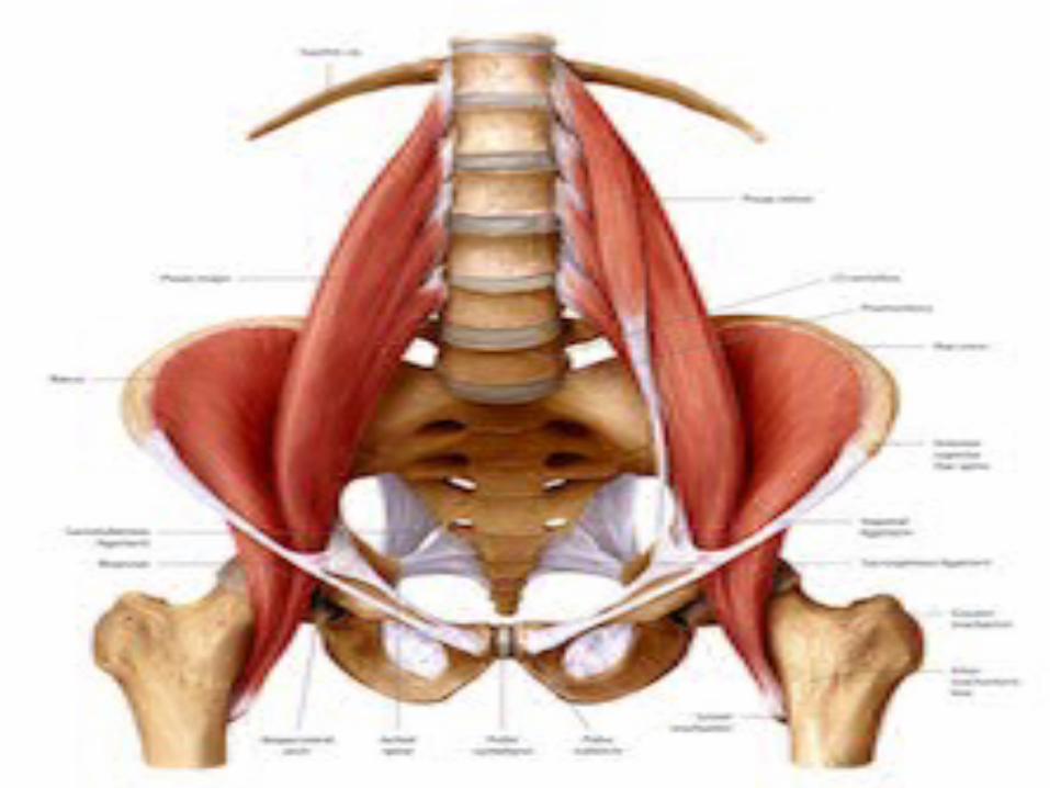
PELVIC WALLSPELVIC WALLS
The inner aspect of the bony pelvis is covered with muscles
Above the brim --- iliacus & psoas
Sidewalls ---- obturator internus & its fascia
Post wall ---- pyriformis
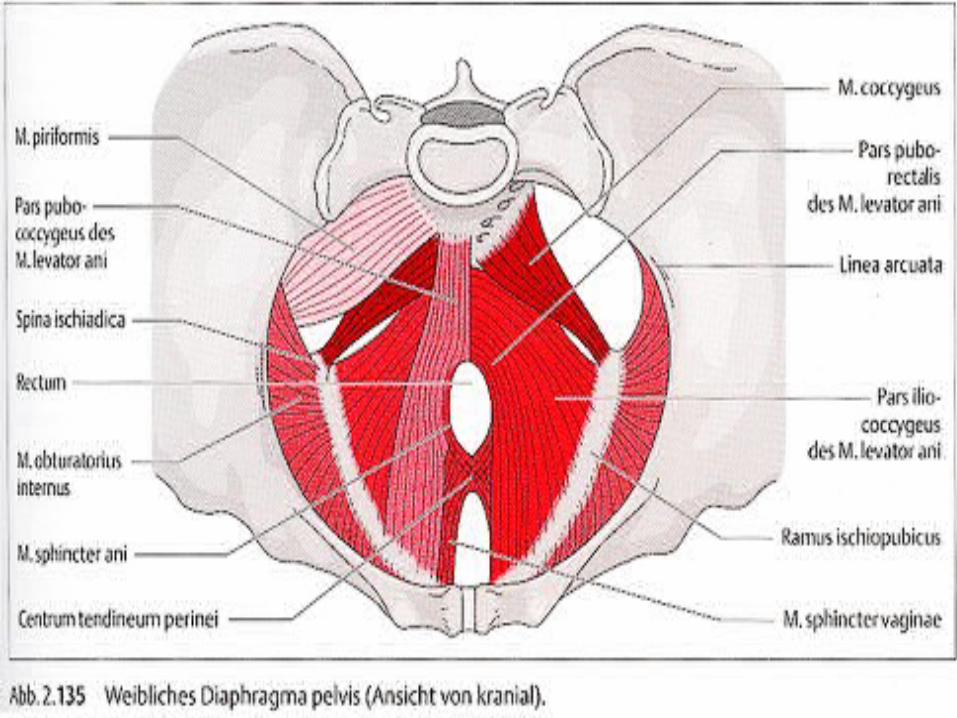
Pelvic floor ---- lavator ani & coccygeus
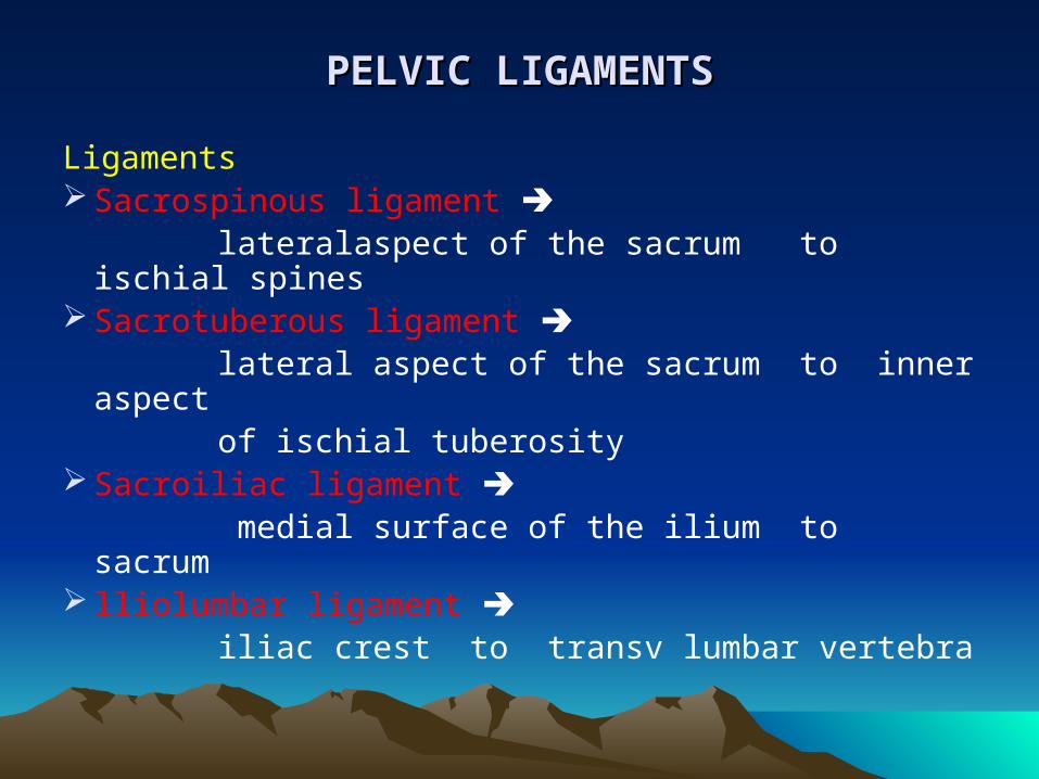
PELVIC LIGAMENTSPELVIC LIGAMENTS
Ligaments Sacrospinous ligament lateralaspect of the sacrum to ischial spines Sacrotuberous ligament lateral aspect of the sacrum to inner aspect of ischial tuberosity Sacroiliac ligament medial surface of the ilium to sacrum lliolumbar ligament iliac crest to transv lumbar vertebra
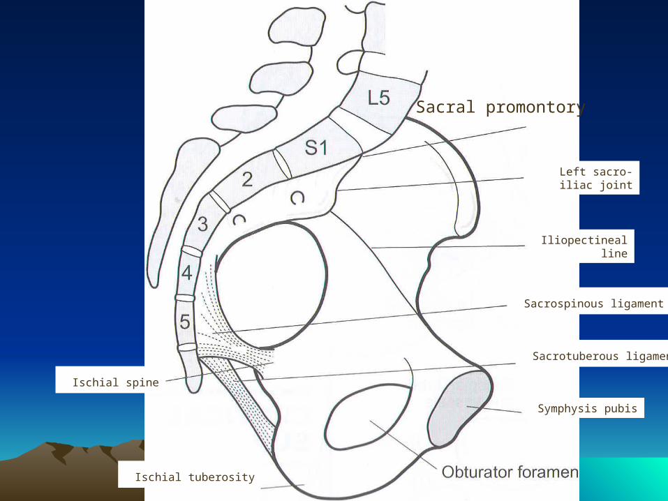
Sacral promontory
Left sacro-iliac joint
Iliopectineal line
Sacrospinous ligament
Sacrotuberous ligament
Symphysis pubis
Ischial tuberosity
Ischial spine
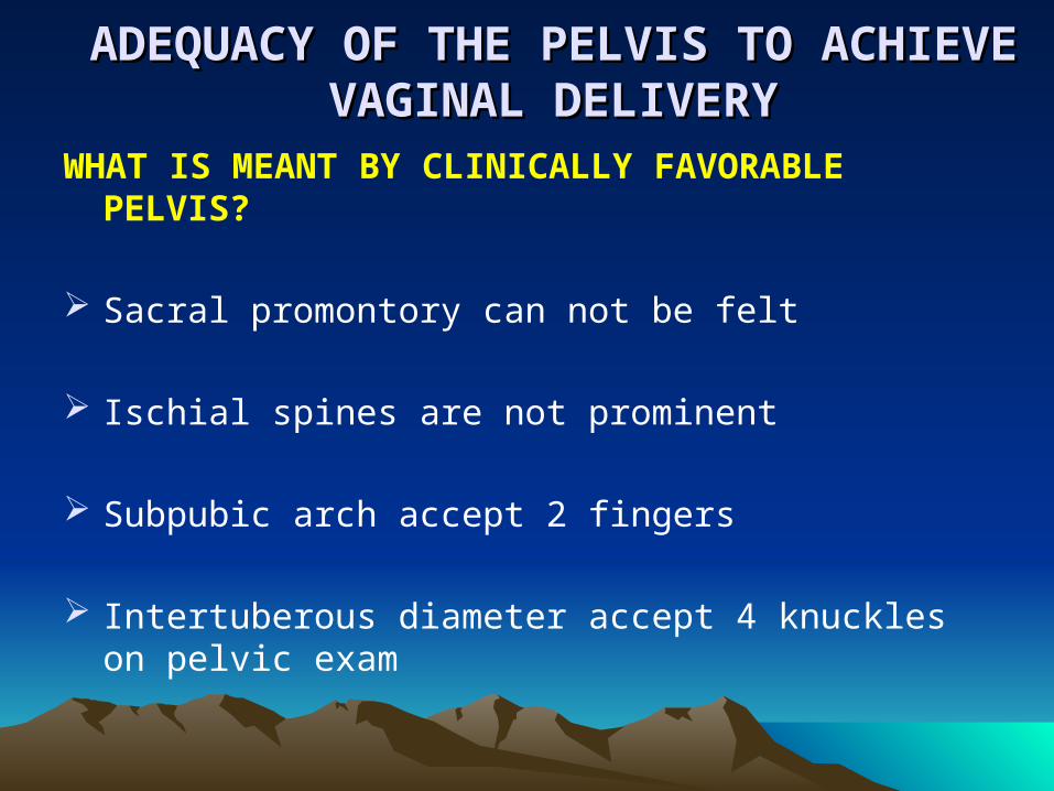
ADEQUACY OF THE PELVIS TO ACHIEVE ADEQUACY OF THE PELVIS TO ACHIEVE VAGINAL DELIVERYVAGINAL DELIVERY
WHAT IS MEANT BY CLINICALLY FAVORABLE PELVIS?
Sacral promontory can not be felt
Ischial spines are not prominent
Subpubic arch accept 2 fingers
Intertuberous diameter accept 4 knuckles on pelvic exam
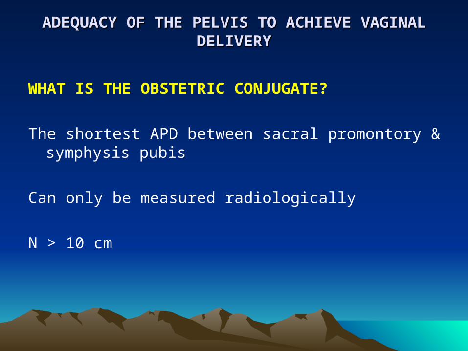
ADEQUACY OF THE PELVIS TO ACHIEVE VAGINAL ADEQUACY OF THE PELVIS TO ACHIEVE VAGINAL DELIVERYDELIVERY
WHAT IS THE OBSTETRIC CONJUGATE?
The shortest APD between sacral promontory & symphysis pubis
Can only be measured radiologically
N > 10 cm
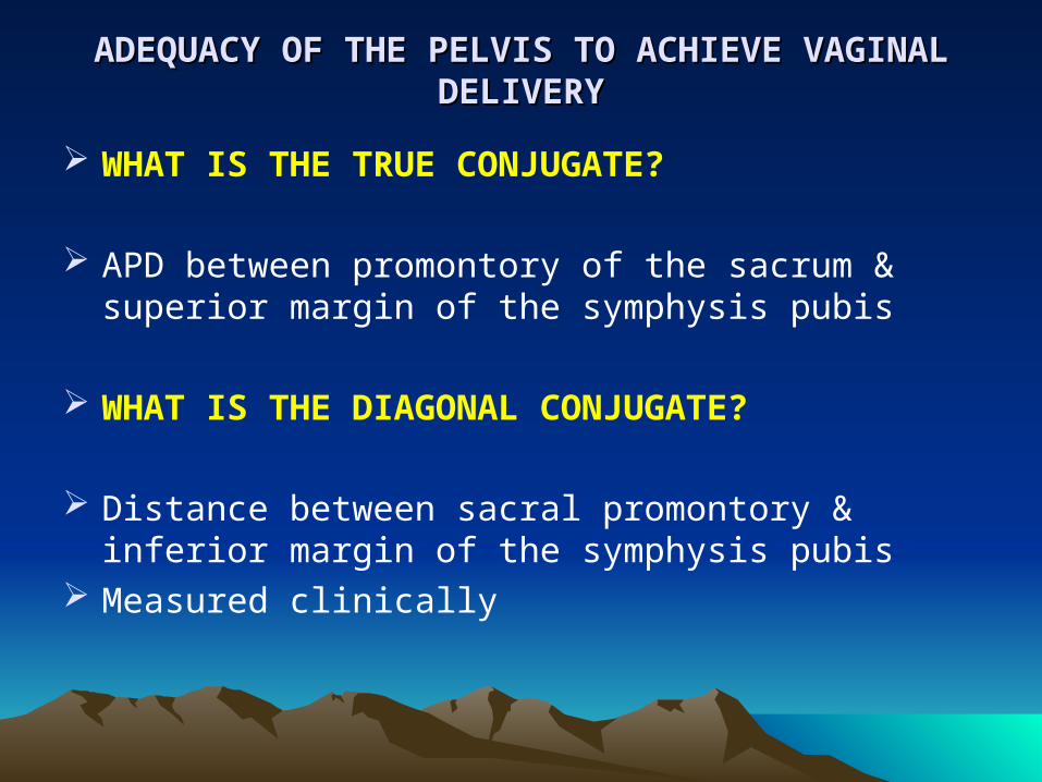
ADEQUACY OF THE PELVIS TO ACHIEVE VAGINAL ADEQUACY OF THE PELVIS TO ACHIEVE VAGINAL DELIVERYDELIVERY
WHAT IS THE TRUE CONJUGATE?
APD between promontory of the sacrum & superior
margin of the symphysis pubis
WHAT IS THE DIAGONAL CONJUGATE?
Distance between sacral promontory & inferior margin of the symphysis pubis
Measured clinically

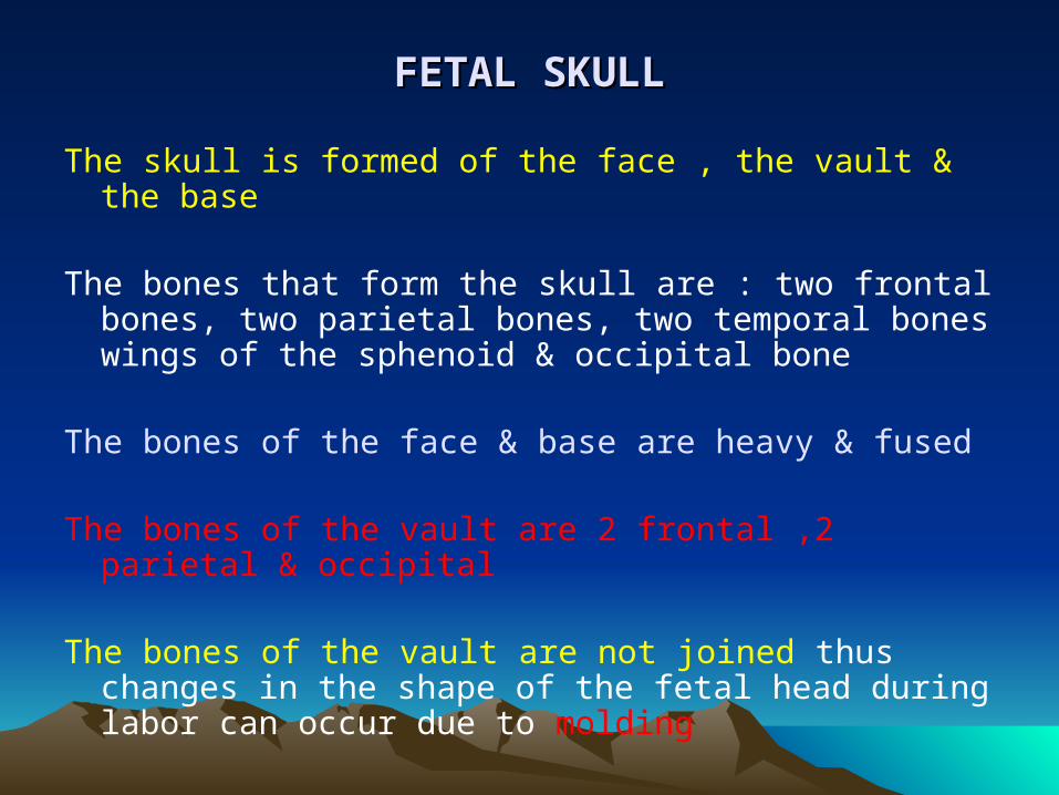
FETAL SKULLFETAL SKULL
The skull is formed of the face , the vault & the base The bones that form the skull are : two frontal bones, two
parietal bones, two temporal bones wings of the sphenoid & occipital bone
The bones of the face & base are heavy & fused
The bones of the vault are 2 frontal ,2 parietal & occipital
The bones of the vault are not joined thus changes in the shape of the fetal head during labor can occur due to molding
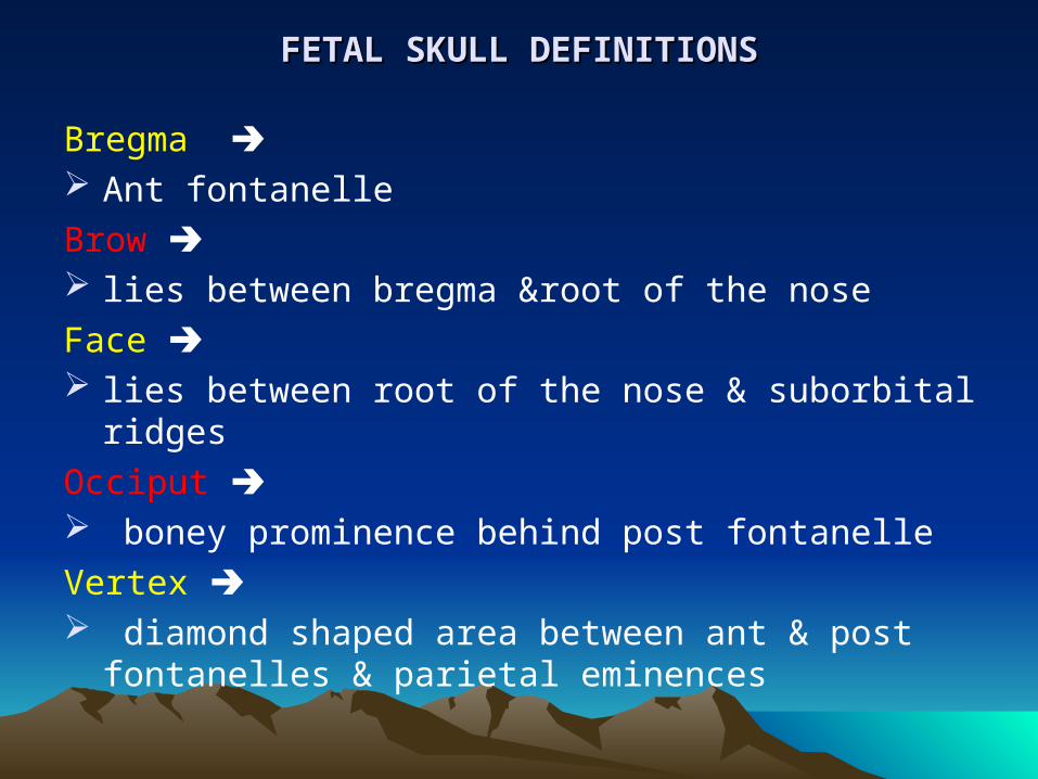
FETAL SKULL DEFINITIONSFETAL SKULL DEFINITIONS
Bregma Ant fontanelle
Brow lies between bregma &root of the nose
Face lies between root of the nose & suborbital ridges
Occiput boney prominence behind post fontanelle
Vertex diamond shaped area between ant & post fontanelles &
parietal eminences
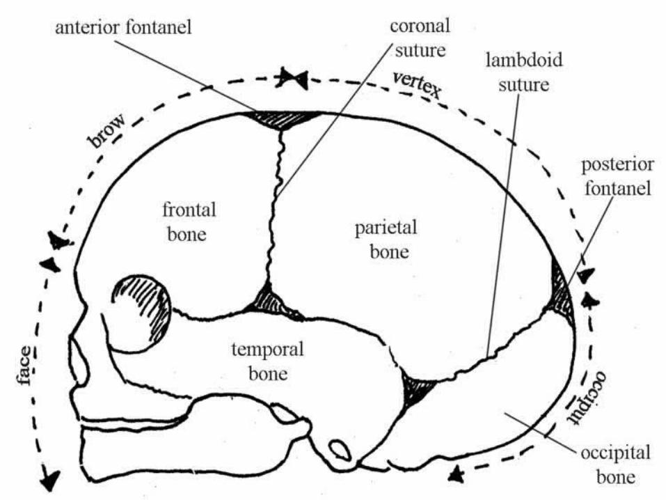
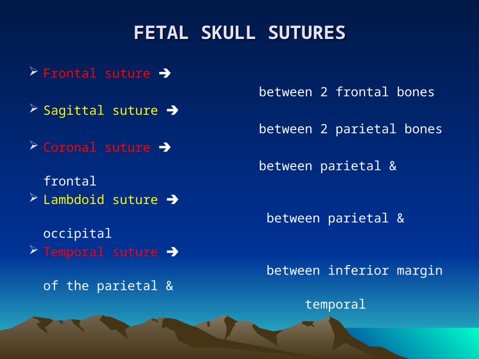
FETAL SKULL SUTURESFETAL SKULL SUTURES
Frontal suture between 2 frontal bones Sagittal suture between 2 parietal bones Coronal suture
between parietal & frontal Lambdoid suture
between parietal & occipital Temporal suture between inferior margin of the parietal &
temporal
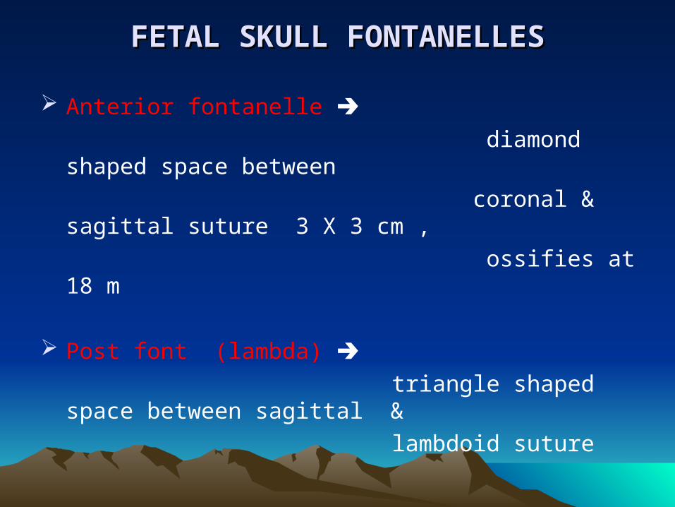
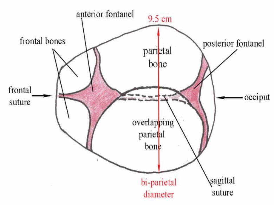
FETAL SKULL FONTANELLESFETAL SKULL FONTANELLES
Anterior fontanelle diamond shaped space between
coronal & sagittal suture 3 X 3 cm ,
ossifies at 18 m
Post font (lambda) triangle shaped space between sagittal &
lambdoid suture
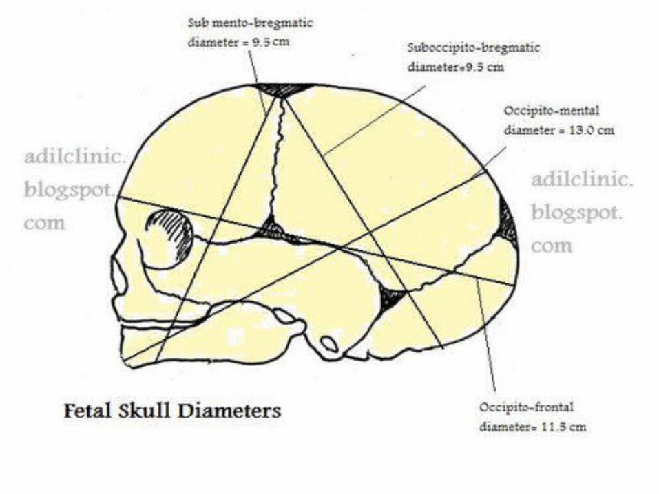
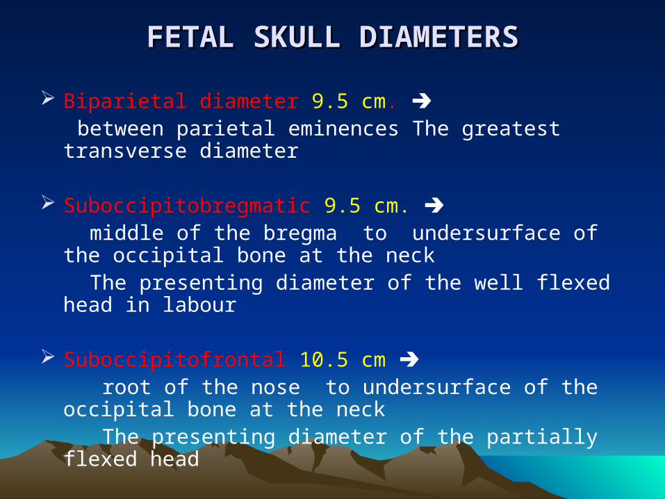
FETAL SKULL DIAMETERSFETAL SKULL DIAMETERS
Biparietal diameter 9.5 cm. between parietal eminences The greatest transverse
diameter
Suboccipitobregmatic 9.5 cm. middle of the bregma to undersurface of the occipital
bone at the neck The presenting diameter of the well flexed head in labour
Suboccipitofrontal 10.5 cm root of the nose to undersurface of the occipital bone at
the neck The presenting diameter of the partially flexed head

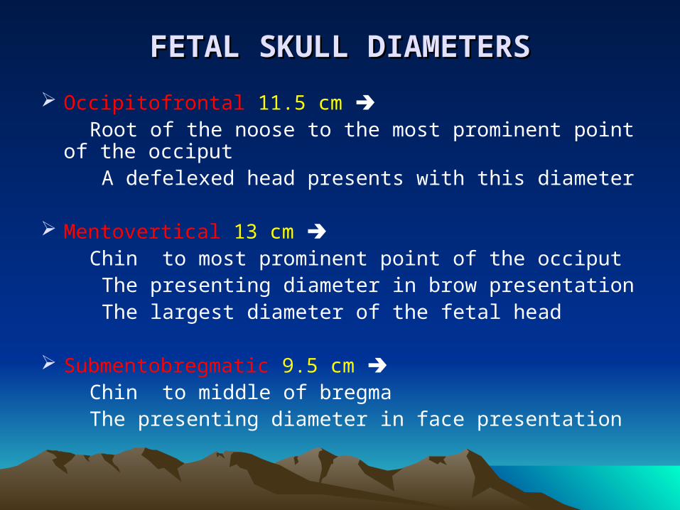
FETAL SKULL DIAMETERSFETAL SKULL DIAMETERS
Occipitofrontal 11.5 cm Root of the noose to the most prominent point of the
occiput A defelexed head presents with this diameter
Mentovertical 13 cm Chin to most prominent point of the occiput The presenting diameter in brow presentation The largest diameter of the fetal head
Submentobregmatic 9.5 cm Chin to middle of bregma The presenting diameter in face presentation
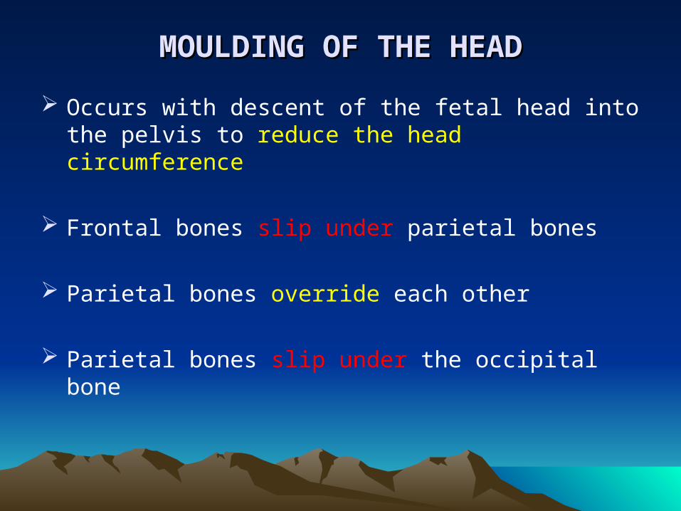
MOULDING OF THE HEADMOULDING OF THE HEAD
Occurs with descent of the fetal head into the pelvis to reduce the head circumference
Frontal bones slip under parietal bones
Parietal bones override each other
Parietal bones slip under the occipital bone
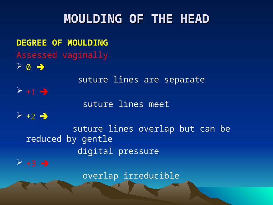
MOULDING OF THE HEADMOULDING OF THE HEAD
DEGREE OF MOULDING
Assessed vaginally 0
suture lines are separate + suture lines meet + suture lines overlap but can be reduced by gentle
digital pressure +3 overlap irreducible