Embed Size (px)
Citation preview

INFECTION AND IMMUNITY, Dec. 2003, p. 7002–7013 Vol. 71, No. 120019-9567/03/$08.00�0 DOI: 10.1128/IAI.71.12.7002–7013.2003Copyright © 2003, American Society for Microbiology. All Rights Reserved.
Analysis of Virulence and Inflammatory Potential of Shigella flexneriPurine Biosynthesis Mutants
Antonella Cersini,1 Maria Celeste Martino,1 Irene Martini,1 Giacomo Rossi,2 andMaria Lina Bernardini1*
Dipartimento di Biologia Cellulare e dello Sviluppo, Sezione di Scienze Microbiologiche, Universita La Sapienza,00185 Rome,1 and Facolta di Medicina Veterinaria, Universita di Camerino, 62032 Matelica,2 Italy
Received 24 March 2003/Returned for modification 24 June 2003/Accepted 8 September 2003
Several Shigella flexneri mutants with defects in aromatic amino acid and/or purine biosynthesis have beenevaluated as vaccines in humans or in animal models. To be suitable as a vaccine, a mutant has to showvirulence attenuation, minimal reactogenicity, and a good immunogenic potential in animal models. With thisaim, we have constructed five S. flexneri 5 (wild-type strain M90T) mutants with inactivation of one or two ofthe loci purEK, purHD, and guaBA, governing early or late steps of purine biosynthesis. The mutants have beenanalyzed in vitro in cell cultures and in vivo in the Sereny test and in the murine pulmonary model ofshigellosis. M90T guaBA, M90T guaBA purEK, M90T guaBA purHD, and M90T purHD purEK gave a negativeresult in the Sereny test. In contrast, in the murine pulmonary model all of the strains had the same 50% lethaldose as the wild type, except M90T guaBA purHD, which did not result in death of the animals. Nevertheless,bacterial counts in infected lungs, immunohistochemistry, and reverse transcription-PCR analysis of mRNAsfor tumor necrosis factor alpha (TNF-�), gamma interferon (IFN-�), interleukin-1� (IL-1�), IL-6, IL-12, andinducible nitric oxide synthase (iNOS) revealed significant differences among the strains. At 72 h postinfection,M90T guaBA purHD still induced proinflammatory cytokines and factors such as IL-1�, IL-6, TNF-�, andiNOS, along with cytokines such as IL-12 and IFN-�. Moreover, in the absence of evident lesions in murinetissues, this mutant highly stimulated major histocompatibility complex class II expression, showing a signif-icant ability to activate the innate immunity of the host.
Shigellosis is a severe inflammatory diarrhea of humanscaused by bacteria of the genus Shigella, of which S. flexneri isthe predominant species responsible for endemic shigellosis.There are about 160 millions cases of this disease per year,resulting in more than one million deaths (19). Shigella spp. areinvasive bacteria that are able to penetrate and proliferatewithin human colonic mucosa. In epithelial cell cultures bac-terial entry is mediated by the invasion plasmid antigens (IpaB,-C, and -D) (23), which are directly translocated within thecytosol via a type III secretion apparatus (16). Soon afterinternalization, shigellae lyse the membrane surrounding thephagocytic vacuole, and therefore they can exploit the nutri-ents present in the cytoplasm of the host cell (36). This leads tothe intracellular proliferation of the microorganisms, whichmigrate from cell to cell through an actin-based motility mech-anism (7). Upon bacterial entry, epithelial cells activate NF-�B(31), which in turn induces interleukin-8 (IL-8) production andsecretion. In animal models of shigellosis, IL-8 plays a relevantrole (34), contributing toward the stimulation of a massiveinflammatory response characteristic of natural infections. In-flammation is supported mainly by a polymorphonuclear leu-kocyte (PMN) influx that destroys intercellular junctions andallows bacteria to access the basolateral pole of epithelial cells,eventually facilitating colonization. However, PMNs are ableto kill shigellae (22), in this way limiting bacterial spread to
deeper tissues. Following infection with shigellae, macro-phages undergo caspase-1-mediated apoptosis, accompaniedby IL-1� and IL-18 release (35, 42, 43) that further contributesto the inflammatory reaction (32). In conclusion, shigellae havedeveloped several mechanisms to provoke inflammation in hu-man intestinal tissues. Taking into account this notion, in de-signing attenuated Shigella mutants to be used as vaccine can-didates, the inflammatory potential of the strains should becarefully defined in order to swing the balance between inflam-mation and immunogenicity toward immunogenicity. In thelast few years encouraging progress has been made in thisregard, and a number of Shigella vaccine candidates have beenconstructed and evaluated based on rational attenuation ofvirulence. These candidates encompass mutants harboring mu-tations in metabolic pathways (1, 6, 20, 40) and/or mutations invirulence genes (10, 18, 28, 33, 41). The rationale underlyingthese constructions is to reduce the multiplication of shigellaewithin the host cells and tissues along with their ability tospread or to induce specific damages. However, despite knowl-edge of the protective immunity provided by vaccine candi-dates, current understanding about their virulence phenotypes,in terms of inflammatory potential and ability to stimulatenatural immunity, is limited. The integration of this knowledgein a scheme encompassing the virulence profile of a mutant isa critical aspect of the design and improvement of a newgeneration of live vaccine candidates. With this aim, in thisstudy we have analyzed how the inactivation of different stepsof purine biosynthesis alters the virulence phenotype of Shi-gella. In 1971 a study performed by Formal and coworkers (14)reported that purine-requiring Shigella strains obtained by mu-
* Corresponding author. Mailing address: Dipartimento di BiologiaCellulare e dello Sviluppo, Sezione di Scienze Microbiologiche, Uni-versita ‘La Sapienza,’ Via dei Sardi 70, 00185 Rome, Italy. Phone: 3906 49917579/49917850. Fax: 39 06 49917594. E-mail: [email protected].
7002
on March 30, 2020 by guest
http://iai.asm.org/
Dow
nloaded from

tagenesis with N-methyl-N�-nitro-N-nitrosoguanidine retainedvirulence in spite of their purine dependence for growth. Like-wise, it has been shown that adenine auxotrophy slightly re-duces Shigella virulence in vivo (9), whereas guanine auxotro-phy strongly attenuates virulence in vivo and in vitro (27).
Here we have created five mutants having different levels ofattenuation and harboring inactivation of one or two of thefollowing loci: purEK and purHD, whose products control threeearly steps leading to both IMP and thiamine synthesis, andguaBA, which governs two late steps of GMP synthesis. Thevirulence of these mutants has been analyzed in vitro in cellculture assays and in vivo in both the Sereny test, which mea-sures the ability of shigellae to induce keratoconjunctivitis inguinea pigs, and the murine pulmonary model of shigellosis.To design a virulence profile of each strain, inflammation-related parameters, including expression and production ofcytokines and major histocompatibility complex (MHC) classII (MHC-II) in infected tissues, have been evaluated.
MATERIALS AND METHODS
Bacterial strains and growth conditions. The bacterial strains used in thisstudy are listed in Table 1. Bacteria were routinely cultured in Trypticase soybroth (BBL, Becton Dickinson and Co., Cockeysville, Md.) or on Trypticase soyagar (TSA). M9 salts (24) were used to prepare minimal medium. Glucose wasadded at a final concentration of 0.2%, and the medium was supplemented withnicotinic acid (10 �g/ml) to support the growth of shigellae. M9 was also sup-plemented with hypoxanthine (100 �g/ml), adenine (100 �g/ml), or guanine (100�g/ml) and B1 (10 �g/ml), required by the strains harboring purHD and/orguaBA mutations. The ability of bacteria to bind the pigment Congo red (Crbphenotype) was assessed by using Trypticase soy agar plates containing 0.01%Congo red dye. When necessary, kanamycin, ampicillin, streptomycin, tetracy-cline, and chloramphenicol were added to cultures at 50, 100, 100, 12, and 20�g/ml, respectively.
Genetic procedures. Conjugation was performed as described by Miller (24).P1 transduction experiments were carried out as previously described (9) accord-ing to Miller’s procedure (24). Transductants were first selected on the basis ofantibiotic resistance. They were analyzed at the molecular level by PCR withprimers external to the mutation introduced and in minimal medium for theacquired auxotrophies.
Recombinant DNA techniques. Genomic and plasmid DNAs were preparedwith commercial kits (Qiagen GmbH, Hilden, Germany). Enzymes and buffers
for recombinant DNA procedures were obtained from Boehringer (Indianapolis,Ind.). DNA electroporation was conducted with a Bio-Rad (Hercules, Calif.)Gene Pulser. PCR products were cloned by using a Sure Clone ligation kit(Amersham Pharmacia Biotech) or a Perfectly Blunt cloning kit (Novagen,Madison, Wis.).
(i) Construction of M90T �purHD. A 539-bp fragment, corresponding to the5� terminus of the purHD locus, was amplified by using the forward primerAGATCTGTCGTCCAGTCC and the reverse primer GTCGACGCAGTGTGTTCGAAGG. The fragment was digested with BglII and SalI (sites are under-lined) and cloned into the suicide vector pGP704 to create pZB213. A 502-bpsegment was then amplified from the 3� terminus of the purHD locus with theforward primer GTCGACGGCAACACCTACA and the reverse primer TCTAGATAGTTCTGCTCGC. The fragment was digested with SalI and XbaI (sitesare underlined) and cloned into pZB213. The resulting plasmid, pZB214, wascleaved with SalI between the two fragments and ligated with a 1.2-kb SalIcassette encoding kanamycin resistance from pUC4K to yield pZB215. pZB215was maintained in DH5� �pir, introduced into Sm10 �pir, and transferred intoM90T Smr by conjugation. Transconjugants were selected on kanamycin agarplates and screened for ampicillin sensitivity. They were expected to be producedthrough double allelic exchange between the 5� and 3� fragments cloned intopZB215 and the corresponding regions on the M90T chromosome. Transconju-gants were also checked for the acquired hypoxanthine and B1 auxotrophies onM9 agar plates. Kmr, Aps, and Crb� mutants and thiamine and hypoxanthineauxotrophs were subjected to PCR and sequence analysis.
(ii) Construction of M90T �guaBA. Two DNA fragments, of 646 bp (fragment1) and 781 bp (fragment 2), of the S. flexneri 5a guaB and guaA genes, respec-tively, were amplified by PCR with the following primers: guaBF (5�-GCTCTAGAACCGTTCTGCCGAATACTGCTGAC-3�) and guaBR (5�-TGCACTGCAGCGCGTCAACACGCTCTTCGTTACC-3�) for fragment 1 and guaAF (5�-TGCACTGCAGCGTGCGTGAGCTGGTGTTTACTG-3�) and guaAR (5�-CGGAATTCCAATGTTAAGACCAAAGTGATCGCC-3�) for fragment 2. Thefragments were digested with XbaI and PstI (fragment 1) and PstI and EcoRI(fragment 2) (sites are underlined), ligated with an 829-bp PstI cassette encodingchloramphenicol resistance from pGEM-CAT (a gift from A. Covacci, ChironVaccines, Siena, Italy), and cloned into the XbaI-EcoRI sites of the suicide vectorpGP704 to create pZB5101. Plasmid pZB5101 was transferred into M90T Smr byconjugation, and transconjugants were selected on chloramphenicol agar plates,screened for ampicillin sensitivity as described above, and checked for the ac-quired guanine auxotrophy on M9 agar plates. Cmr, Aps, and Crb� mutants andguanine auxotrophs were subjected to PCR and sequence analysis.
(iii) Cloning of Shigella purHD and guaBA loci. To clone the purHD operon, afragment of 2.8 kb containing the complete sequences of purH and purD wasamplified from M90T genomic DNA with the forward primer AGATCTGTCGTCCAGTCC and the reverse primer TCTAGATAGTTCTGCTCGC. The am-
TABLE 1. Strains and plasmids
Strain or plasmid Relevant characteristics Growth requirement(s) Reference orsource
S. flexneri 5M90T Smr Spontaneous streptomycin-resistant derivative of wild-type
strain M90T harboring pWR100Nicotinic acid 2
ZB2209 M90T Smr �purHD Kmr Hypoxanthine, nicotinic acid, B1 This studyZB501 M90T Smr �guaBA Cmr Nicotinic acid, guanine This studyZB502 M90T Smr �guaBA Cmr �purHD Kmr Nicotinic acid, Guanine, Adenine
(Hypoxanthine), B1
This study
ZB503 M90T �purHD Kmr �purEK Nicotinic acid, hypoxanthine(adenine), B1
This study
ZB504 M90T �guaBA Cmr �purEK Nicotinic acid, guanine, adenine This study
E. coliDH5� �pir DH5� (�pir) tet::Mu recA 8SM10 �pir thi thr leu tonA lacY supE recA::RP4-2Tc::Mu (Kmr) �pir 38
PlasmidspZB216 pACYC184 containing the full 2.8-kb BamHI-XbaI purHD
operonThis study
pZB5102 pSTBlue-1 carrying a 3-kb fragment containing the guaBAoperon
This study
VOL. 71, 2003 CONTRIBUTION OF PURINE BIOSYNTHESIS TO SHIGELLA VIRULENCE 7003
on March 30, 2020 by guest
http://iai.asm.org/
Dow
nloaded from

plified DNA was digested with BglII and XbaI (sites are underlined) and ligatedinto the BamHI-XbaI sites of pACYC184 to create pZB216.
To clone the guaBA locus, a fragment of 3.0 kb containing the completesequences of guaB and guaA was amplified from the M90T genomic DNA byusing the forward primer 5�-ATGCTACGTATCGCTAAAG-3� and the reverseprimer 5�-TCATTCCCACTCAATGGTAG-3�. The amplified DNA was clonedinto pSTBlue-1 blunt vector (Perfectly Blunt cloning kit; Novagen) to createpZB5102. The cloning of the purHD and guaBA operons was verified (i) at themolecular level through PCR analysis and (ii) at the phenotypic level throughtranscomplementation of the purHD deletion in M90T �purHD and of theguaBA deletion in M90T �guaBA in unsupplemented M9 minimal medium.
Virulence assays. (i) HeLa cell culture conditions. Cells were maintained inminimal essential medium (GIBCO-BRL) supplemented with fetal calf serum(HyClone Laboratories, Inc., Logan, Utah) at a concentration of 10%.
(ii) Intracellular multiplication. Multiplication of bacteria was assayed atmultiplicities of infection (MOIs) of 5, 10, and 100 on nonconfluent monolayersof HeLa cells (105/ml) on 35-mm-diameter dishes as previously described (9).
(iii) Plaque assay. The plaque assay was carried out as described previously (9)at MOIs of 1, 10, and 100.
(iv) Sereny test. The keratoconjunctivitis assay in guinea pigs was performedas described previously (15) with two challenges, 108 and 109 CFU. The degreeof keratoconjunctivitis was ranked on the basis of time of development, severity,and (when possible) rate of clearance of symptoms, with the following scores: 0,no disease; 1, mild conjunctivitis; 2, keratoconjunctivitis with no purulence; and3, fully developed keratoconjunctivitis with purulence.
(v) Intranasal infection of mice. Five-week-old BALB/c female mice (CharlesRiver, Calco, Italy) were anesthetized intramuscularly with 50 �l of a solutioncontaining Zoletil (1 mg) (Virbac, Carros, France) and Xilor (2%) (BIO 985, SanLazzaro, Italy) and inoculated intranasally with 20 �l of 0.9% NaCl suspensionscontaining 108 CFU of each strain (21). After 72 h, mice were euthanatized bycervical dislocation and lungs were removed and processed for histopathologicalstudies, bacterial counts, and reverse transcription-PCR (RT-PCR) analysis. Forbacterial counts, 5 ml of saline buffer was added to samples that were immedi-ately stored in ice. Samples were then ground with an Ultraturrax apparatus(Janke and Kunkel, GmbH, and Co., Staufen, Germany). Serial dilutions of theresulting solutions were plated on Congo red agar plates supplemented with theappropriate antibiotic(s). Lungs processed for RT-PCR were removed afterhaving been extensively washed with 10 ml of saline solution introduced throughthe right atrium of the heart and then were immediately frozen in liquid N2. Tenmice were used per group, and experiments were repeated twice.
Histology and immunohistochemistry. For routine histology and immunohis-tochemistry, lung samples were removed and fixed in 10% buffered formalin for48 h, processed, and embedded in paraffin. Sections, 3 �m thick, were stainedwith hematoxylin-eosin or processed for immunohistochemistry. For the immu-nohistochemical study, lung sections were allowed to adhere to pretreated slides(Bioptica S.p.A., Milan, Italy) and then deparaffinized and rehydrated. Endog-enous peroxidase activity was removed by incubation with 0.5% hydrogen per-oxide in distilled water for 1 h at room temperature. The primary monoclonalantibodies (MAbs) employed were the following: a rat anti-mouse MHC-II MAb(Serotec, Oxford, United Kingdom), a rat anti-mouse tumor necrosis factoralpha (TNF-�) MAb (Serotec), and a murine anti-S. flexneri 5a lipopolysaccha-ride (LPS) MAb (6 mg/ml) (30). Tissue sections were incubated overnight in amoist chamber at 4°C with different primary antibodies diluted 1:100 in Tris-buffered saline (TBS) containing 0.1% crystalline bovine serum albumin. A1:200-diluted biotinylated rabbit anti-rat immunoglobulin G (Vector Laborato-ries, Inc., Burlingame, Calif.) and a 1:200-diluted biotinylated goat anti-mouseimmunoglobulin G (AO433; DAKO, Glostrup, Denmark) were applied for 45min at room temperature as secondary antibodies. After two 5-min rinses withTBS, tissue sections which had been incubated with anti-S. flexneri LPS or withanti-TNF-� MAb were incubated with the avidin-biotin-peroxidase complex(Vector Laboratories) diluted 1:50 for 45 min at room temperature. Sectionstreated with anti-MHC-II primary MAb were incubated with the avidin-biotin-alkaline phosphatase complex (Vector Laboratories) under the same conditions.The immunoreactions were then revealed by using either the 3,3�-diaminoben-zidine tetrahydrochloride (Sigma Chemicals Co.) or Vector red (Vector Labo-ratories) as chromogens for the sections incubated with avidin-biotin-peroxidasecomplex and for those incubated with avidin-biotin-alkaline phosphatase com-plex, respectively. Sections were counterstained with Mayer hematoxylin, dehy-drated, and mounted. Specific primary antibodies replaced with TBS or nonim-mune sera were used as negative controls in immunohistochemical techniques.
Histological examination included assessment of inflammation by scoring thenumber of inflammatory cells (mononuclear cells, such as macrophages, lympho-cytes, and plasma cells, and neutrophils) at a magnification of 400. The number
of inflammatory cells was evaluated by using a visual analogue scale modified formurine pulmonary specimens, and results are reported as the mean for the entirespecimen. When considerable variation of intensity of infiltration was evident inthe same specimen, the mean for several areas was determined and the specimenwas scored accordingly. Neutrophils and mononuclear cells were classified asabsent (score of 0) when there were no or fewer than 5 cells per high-power field(HPF) (at a magnification of 400), mild (score of 1) for 5 to 19 per HPF,moderate (score of 2) for 20 to 49 cells per HPF, marked (score of 3) for 50 to99 cells per HPF, and severe (score of 4) for 100 to 200 cells or more per HPF.
Histological criteria for normal pulmonary characteristics included detectionof no or only a few mononuclear cells per HPF and no or only a few scatteredneutrophils in bronchioli and alveoli without tissue changes (no interstitial thick-ening or bronchiolar-associated lymphoid tissue [BALT] activation and airwaysfree from exudate).
RNA extraction and RT-PCR analysis. Total RNA from homogenized lungswas extracted by using Trizol solution (Invitrogen, S. Giuliano Monzese, Italy)according to the manufacturer’s instructions. RNase-free DNase (BoehringerMannheim) was used to remove genomic DNA as described by Dilworth andMcCarrey (13). RT of total RNA (1 �g) and cDNA PCR were performed byusing the SuperScript one-step RT-PCR with Platinum Taq (Invitrogen) inaccordance with the manufacturer’s manual. PCR with primers for �-actin wasperformed on each individual sample as an internal positive-control standard. Asa negative control, PCR without cDNA (with water as the substitute) was runconcurrently. Cytokine mRNAs were quantitated by using Quantity-One soft-ware (Bio-Rad Laboratories), and results were normalized to the amount of�-actin mRNA. The median value from three runs was used to estimate mRNAlevels for individual mice.
RESULTS
Construction of ZB2209 (M90T �purHD), ZB501 (M90T�guaBA), ZB502 (M90T �guaBA �purHD), ZB503 (M90T�purHD �purEK), and ZB504 (M90T �guaBA �purEK). Toobtain a Shigella mutant with a deletion in the purHD locus,encoding 5�-phosphoribosylglycinamide synthetase and 5�-phos-phoribosyl-5-aminoimidazole-4-carboaxamide transformylase-IMP-cyclohydrolase, the purHD operon was cloned, mu-tagenized in vitro, and reintroduced into the M90T genome.As expected, the resulting strain, ZB2209 (M90T �purHDKm), did not grow on minimal medium in the absence ofhypoxanthine and B1. Likewise ZB501 (M90T �guaBA Cm),which is unable to synthesize IMP dehydrogenase andguanosine synthetase, was constructed through double allelicexchange between the wild-type guaBA locus on the M90Tchromosome and a guaBA copy mutagenized in vitro. ZB501did not grow on minimal medium without guanine. Trans-complementation of ZB2209 (M90T �purHD) with the profi-cient purHD operon cloned into pZB216 and of ZB501 (M90T�guaBA) with the functional guaBA operon cloned intopZB5102 restored the ability of both mutants to grow on un-supplemented minimal medium.
To stress purine auxotrophy in M90T we created three othermutants with defects in purine biosynthesis in addition toZB501 and ZB2209. A strain was obtained by transducing thepurHD Km mutation from ZB2209 into ZB501. This mutant,ZB502, carried a deletion of both operons guaBA and purHD.We had previously constructed an M90T strain, ZB2106, har-boring a deletion of the purEK locus encoding phosphoribo-sylaminoimidazole carboxylase (9). This strain showed minimalattenuation in vivo. Therefore, the purHD Km deletion wastransferred by P1 transduction from ZB2209 to ZB2106, givingZB503 (M90T �purHD �purEK). Likewise, the guaBA Cmmutation was transduced into ZB2106 (M90T �purEK), result-ing in ZB504 (M90T �guaBA �purEK). The mutants are listedin Table 1, and Fig. 1 summarizes the relevant steps of purine
7004 CERSINI ET AL. INFECT. IMMUN.
on March 30, 2020 by guest
http://iai.asm.org/
Dow
nloaded from

biosynthesis and the relative positions of enzymatic steps per-formed by the enzymes deleted in the mutants constructed inthis study.
Invasion ability, intracellular multiplication, and dissemi-nation of ZB2209 (M90T �purHD), ZB501 (M90T �guaBA),ZB502 (M90T �guaBA �purHD), ZB503 (M90T �purHD�purEK), and ZB504 (M90T �guaBA �purEK). The intracel-
lular multiplication kinetics of ZB2209 (M90T �purHD),ZB501 (M90T �guaBA), ZB502 (M90T �guaBA �purHD),ZB503 (M90T �purHD �purEK), and ZB504 (M90T �guaBA�purEK) were analyzed within HeLa cell monolayers. Thestrains were assessed for intracellular proliferation within 6 hof infection. The results are shown in Fig. 2. At an MOI of 100,by evaluating the number of bacteria per monolayer the mu-
FIG. 1. Purine de novo biosynthesis pathway and essential steps of purine salvage and interconversion. The individual enzymes are identifiedby their gene symbols. Solid lines indicate de novo biosynthesis; dotted lines represent purine salvage and interconversion. The arrowheadrepresents pathways in which the individual steps are not shown. Asterisks indicate mutations introduced in the S. flexneri 5 wild-type strain M90T.Abbreviations: PRA, 5�-phosphoribosylamine; GAR, 5�-phosphoribosylglycinamide; AIR, 5�-phosphoribosyl-5-aminoimidazole; NCAIR, 5�-phos-phoribosyl-5-carboxyaminoimidazole; CAIR, 5�-phosphoribosyl-5-aminoimidazole-4-carboxylic acid; AICAR, 5�-phosphoribosyl-4-carboxamide-5-aminoimidazole; FAICAR, 5�-phosphoribosyl-4-carboxamide-5-formamidoimidazole; Hx, hypoxanthine; R and dR, ribonucleosides and de-oxyribonucleosides, respectively.
FIG. 2. Intracellular growth kinetics of M90T, ZB2209 (M90T �purHD), ZB501 (M90T �guaBA), ZB502 (M90T �guaBA �purHD), ZB503(M90T �purHD �purEK), and ZB504 (M90T �guaBA �purEK) during incubation for 6 h p.i. (A) Means ( standard deviations) of the numbersof bacteria calculated in five separate invasion assays. (B) Data from one representative experiment. Similar results were obtained in three identicalinvasion assays. Standard deviations were within 20% of the values presented.
VOL. 71, 2003 CONTRIBUTION OF PURINE BIOSYNTHESIS TO SHIGELLA VIRULENCE 7005
on March 30, 2020 by guest
http://iai.asm.org/
Dow
nloaded from

tants appeared to be impaired in intracellular proliferation tovarious extents depending on the mutation(s) introduced (Fig.2A). This difference became more evident when the MOI waslowered to 10 and the multiplication kinetics per infected cellrather than per monolayer were analyzed (Fig. 2B).
Although intracellular proliferation of ZB2209 (M90T�purHD) was reduced compared with that of M90T, the asso-ciation of purHD and purEK made ZB503 (M90T �purEK�purHD) unable to proliferate intracellularly. Mutants harbor-ing the guaBA inactivation, i.e., ZB501 (M90T �guaBA),ZB502 (M90T �guaBA �purHD), and ZB504 (M90T �guaBA�purEK) all showed the same kinetics, characterized by (i) areduced ability to penetrate HeLa cells and (ii) a significantdecrease in the ability to proliferate intracellularly.
The proportion of infected cells after 1 h of incubationpostinfection (p.i.) was 59.3% 21.1% for M90T and 15.0% 4.2% for ZB501 (M90T �guaBA), obtained in four differentexperiments with a minimum of 500 cells counted per sample.With ZB502 (M90T �guaBA �purHD) and ZB504 (M90T�guaBA �purEK), we obtained percentages of infected cellssimilar to those for the parent strain ZB501 (M90T �guaBA),i.e., 19.2%. 5.7% and 17.9% 2.9%, respectively. In con-trast, ZB2209 (M90T �purHD) and ZB503 (M90T �purHD�purEK) behaved like the wild-type strain, with values of51.9% 11.1% and 48.7% 15.3%, respectively. During thefirst 4 h of invasion, the number of intracellular M90T organ-isms roughly increased 10-fold, whereas that of the mutantsharboring the guaBA mutation hardly increased 3-fold. Find-ings concerning the mutants carrying the guaBA deletion areconsistent with those previously reported by Noriega and co-workers (27) for an S. flexneri 2a guaBA mutant and point toboth an impaired ability to invade and an altered ability togrow as the major effects of this mutation in Shigella.
The ability of ZB2209 (M90T �purHD), ZB501 (M90T�guaBA), ZB502 (M90T �guaBA �purHD), ZB503 (M90T�purHD �purEK), and ZB504 (M90T �guaBA �purEK) tospread intra- or intercellularly was determined from the fol-lowing data: (i) actin tails were produced within the infectedcells as assessed through nitrobenzoxadiazole-phalloidin label-ing of polymerized actin (data not shown), and (ii) an increas-ing number of HeLa cells were infected during 6 h of cellinvasion in the presence of gentamicin, which is assumed toprevent cell reinfection by extracellular bacteria.
The mutants were also assessed by the plaque assay, whichcovers at least three parameters: the invasion ability, the in-tracellular multiplication, and the intra- and intercellularmovement and cytotoxicity, resulting in a cytopathic effect ob-servable as areas of cell death on the confluent monolayer.ZB2209 (M90T �purHD), ZB501 (M90T �guaBA), ZB502(M90T �guaBA �purHD), ZB503 (M90T �purHD �purEK),and ZB504 (M90T �guaBA �purEK) were assessed in theplaque assay at MOIs of 100, 10, and 1. Two mutants, ZB2209(M90T �purHD) and ZB501 (M90T �guaBA), producedplaques smaller than those of M90T. In particular, plaquesproduced by ZB2209 (M90T �purHD) were roughly 50%, andthose by ZB501 (M90T �guaBA) were about 10%, of the sizeof plaques produced by M90T, albeit for the latter only at anMOI of 100. ZB503 (M90T �purHD �purEK), ZB502 (M90T�guaBA �purHD), and ZB504 (M90T �guaBA �purEK) didnot produce plaques at all. ZB2209 (M90T �purHD)(pZB216)
had its ability to produce plaques of a size comparable to thoseof M90T restored, and intracellular proliferation reverted con-sistently to the wild-type values. Likewise, transcomplementa-tion of ZB501 (M90T �guaBA) with pZB5102 reestablishedthe ability of this strain to proliferate within HeLa cell mono-layers and to provoke a positive plaque assay at MOI 1. ZB502(M90T �guaBA purHD) was restored to full virulence only bythe concomitant presence of the proficient purHD and guaBAoperons cloned into pZB216 and pZB5102, respectively. Like-wise, ZB503 (M90T �purHD �purEK)(pZB216) and ZB504(M90T �guaBA �purEK)(pZB5102) entered and proliferatedwithin HeLa cells and gave the same positive result in theplaque assay as the wild-type strain.
Sereny test. ZB2209 (M90T �purHD), ZB501 (M90T�guaBA), ZB502 (M90T �guaBA �purHD), ZB503 (M90T�purHD �purEK), and ZB504 (M90T �guaBA �purEK) wereanalyzed in the Sereny test with two challenges of 108 and 109
CFU. With ZB2209 (M90T �purHD) at 108 CFU, the reactionwas weaker than that produced by M90T, and symptoms weredelayed by 24 h. At the higher inoculum, however, the intensityof keratoconjunctivitis was similar to that with M90T eventhough the appearance of symptoms was delayed by about24 h. ZB2209 (M90T �purHD)(pZB216) induced reactions ofthe same intensity as M90T. Timing of symptom appearancewas also similar to that for wild-type organisms. ZB501 (M90T�guaBA), ZB502 (M90T �guaBA �purHD), ZB503 (M90T�purHD �purEK), and ZB504 (M90T �guaBA �purEK) gave anegative result in this test at any challenge dose. The resultsare shown in Table 2. ZB501(pZB5102), ZB502(pZB216,pZB5102), ZB503(pZB216), and ZB504(pZB5102) were Ser-eny positive at both challenge doses. ZB502(pZB216) was Ser-eny negative whereas ZB502(pZB5102) behaved like ZB2209(data not shown).
Intranasal infection of mice. The mutants were analyzed inthe murine pulmonary model of shigellosis (21). Twenty micewere infected with 108 microorganisms per strain and in-spected daily. At 72 h postchallenge, the 50% lethal dose(LD50) was reached with M90T. At this time point, survivingmice were sacrificed and lungs were removed for bacterialcounts, RT-PCR, and histopathological analyses. Mortalityand bacterial counts were recorded (Table 2). After 72 h ofinfection 50 to 60% of the animals infected with ZB2209(M90T �purHD), ZB501 (M90T �guaBA), ZB503 (M90T�purHD �purEK), and ZB504 (M90T �guaBA �purEK) died.Lungs removed from the survivors contained from 102 to 104
bacteria, depending on the strain. Surprisingly, no animal diedupon the infection with ZB502 (M90T �guaBA purHD), andno bacteria (�50 bacteria/organ) were found in lungs after72 h. When either ZB502 (M90T �guaBA �purHD)(pZB216),ZB502(pZB5102), or ZB502(pZB216, pZB5102) was used toinfect mice, the LD50 was reached after 72 h as for M90T andthe other mutants. This analysis clearly revealed that thepurHD and the guaBA deletions together drastically attenuateM90T virulence and differentiate the corresponding mutant,ZB502 (M90T �guaBA purHD), from the others used in thisstudy.
Expression of cytokines in lungs. To obtain major insightinto the inflammatory potential of these strains, the expressionof relevant cytokines in lungs of mice infected with M90T andthe mutants was investigated by RT-PCR. We analyzed the
7006 CERSINI ET AL. INFECT. IMMUN.
on March 30, 2020 by guest
http://iai.asm.org/
Dow
nloaded from
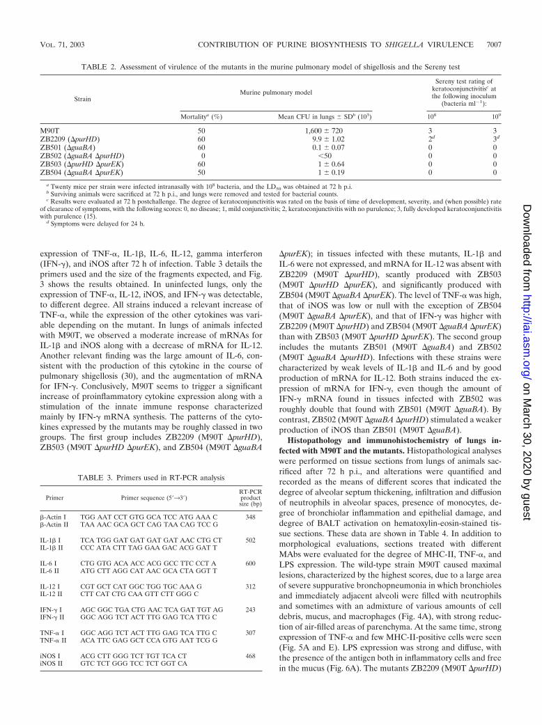
expression of TNF-�, IL-1�, IL-6, IL-12, gamma interferon(IFN-�), and iNOS after 72 h of infection. Table 3 details theprimers used and the size of the fragments expected, and Fig.3 shows the results obtained. In uninfected lungs, only theexpression of TNF-�, IL-12, iNOS, and IFN-� was detectable,to different degree. All strains induced a relevant increase ofTNF-�, while the expression of the other cytokines was vari-able depending on the mutant. In lungs of animals infectedwith M90T, we observed a moderate increase of mRNAs forIL-1� and iNOS along with a decrease of mRNA for IL-12.Another relevant finding was the large amount of IL-6, con-sistent with the production of this cytokine in the course ofpulmonary shigellosis (30), and the augmentation of mRNAfor IFN-�. Conclusively, M90T seems to trigger a significantincrease of proinflammatory cytokine expression along with astimulation of the innate immune response characterizedmainly by IFN-� mRNA synthesis. The patterns of the cyto-kines expressed by the mutants may be roughly classed in twogroups. The first group includes ZB2209 (M90T �purHD),ZB503 (M90T �purHD �purEK), and ZB504 (M90T �guaBA
�purEK); in tissues infected with these mutants, IL-1� andIL-6 were not expressed, and mRNA for IL-12 was absent withZB2209 (M90T �purHD), scantly produced with ZB503(M90T �purHD �purEK), and significantly produced withZB504 (M90T �guaBA �purEK). The level of TNF-� was high,that of iNOS was low or null with the exception of ZB504(M90T �guaBA �purEK), and that of IFN-� was higher withZB2209 (M90T �purHD) and ZB504 (M90T �guaBA �purEK)than with ZB503 (M90T �purHD �purEK). The second groupincludes the mutants ZB501 (M90T �guaBA) and ZB502(M90T �guaBA �purHD). Infections with these strains werecharacterized by weak levels of IL-1� and IL-6 and by goodproduction of mRNA for IL-12. Both strains induced the ex-pression of mRNA for IFN-�, even though the amount ofIFN-� mRNA found in tissues infected with ZB502 wasroughly double that found with ZB501 (M90T �guaBA). Bycontrast, ZB502 (M90T �guaBA �purHD) stimulated a weakerproduction of iNOS than ZB501 (M90T �guaBA).
Histopathology and immunohistochemistry of lungs in-fected with M90T and the mutants. Histopathological analyseswere performed on tissue sections from lungs of animals sac-rificed after 72 h p.i., and alterations were quantified andrecorded as the means of different scores that indicated thedegree of alveolar septum thickening, infiltration and diffusionof neutrophils in alveolar spaces, presence of monocytes, de-gree of bronchiolar inflammation and epithelial damage, anddegree of BALT activation on hematoxylin-eosin-stained tis-sue sections. These data are shown in Table 4. In addition tomorphological evaluations, sections treated with differentMAbs were evaluated for the degree of MHC-II, TNF-�, andLPS expression. The wild-type strain M90T caused maximallesions, characterized by the highest scores, due to a large areaof severe suppurative bronchopneumonia in which bronchiolesand immediately adjacent alveoli were filled with neutrophilsand sometimes with an admixture of various amounts of celldebris, mucus, and macrophages (Fig. 4A), with strong reduc-tion of air-filled areas of parenchyma. At the same time, strongexpression of TNF-� and few MHC-II-positive cells were seen(Fig. 5A and E). LPS expression was strong and diffuse, withthe presence of the antigen both in inflammatory cells and freein the mucus (Fig. 6A). The mutants ZB2209 (M90T �purHD)
TABLE 2. Assessment of virulence of the mutants in the murine pulmonary model of shigellosis and the Sereny test
StrainMurine pulmonary model
Sereny test rating ofkeratoconjunctivitisc atthe following inoculum
(bacteria ml 1):
Mortalitya (%) Mean CFU in lungs SDb (103) 108 109
M90T 50 1,600 720 3 3ZB2209 (�purHD) 60 9.9 1.02 2d 3d
ZB501 (�guaBA) 60 0.1 0.07 0 0ZB502 (�guaBA �purHD) 0 �50 0 0ZB503 (�purHD �purEK) 60 1 0.64 0 0ZB504 (�guaBA �purEK) 50 1 0.19 0 0
a Twenty mice per strain were infected intranasally with 108 bacteria, and the LD50 was obtained at 72 h p.i.b Surviving animals were sacrificed at 72 h p.i., and lungs were removed and tested for bacterial counts.c Results were evaluated at 72 h postchallenge. The degree of keratoconjunctivitis was rated on the basis of time of development, severity, and (when possible) rate
of clearance of symptoms, with the following scores: 0, no disease; 1, mild conjunctivitis; 2, keratoconjunctivitis with no purulence; 3, fully developed keratoconjunctivitiswith purulence (15).
d Symptoms were delayed for 24 h.
TABLE 3. Primers used in RT-PCR analysis
Primer Primer sequence (5�33�)RT-PCRproductsize (bp)
�-Actin I TGG AAT CCT GTG GCA TCC ATG AAA C 348�-Actin II TAA AAC GCA GCT CAG TAA CAG TCC G
IL-1� I TCA TGG GAT GAT GAT GAT AAC CTG CT 502IL-1� II CCC ATA CTT TAG GAA GAC ACG GAT T
IL-6 I CTG GTG ACA ACC ACG GCC TTC CCT A 600IL-6 II ATG CTT AGG CAT AAC GCA CTA GGT T
IL-12 I CGT GCT CAT GGC TGG TGC AAA G 312IL-12 II CTT CAT CTG CAA GTT CTT GGG C
IFN-� I AGC GGC TGA CTG AAC TCA GAT TGT AG 243IFN-� II GGC AGG TCT ACT TTG GAG TCA TTG C
TNF-� I GGC AGG TCT ACT TTG GAG TCA TTG C 307TNF-� II ACA TTC GAG GCT CCA GTG AAT TCG G
iNOS I ACG CTT GGG TCT TGT TCA CT 468iNOS II GTC TCT GGG TCC TCT GGT CA
VOL. 71, 2003 CONTRIBUTION OF PURINE BIOSYNTHESIS TO SHIGELLA VIRULENCE 7007
on March 30, 2020 by guest
http://iai.asm.org/
Dow
nloaded from

and ZB503 (M90T �purHD �purEK) showed a similar patternof inflammation and expression of the different antigens em-ployed, although minor lesions were observed in mice infectedwith ZB503 (M90T �purHD �purEK) (Fig. 4B and 5B and F).With the latter mutant, LPS was abundant, and its localizationwas maximal inside mononuclear cells (Fig. 6B). Intermediatevalues of phlogosis were observed in mice infected with themutant ZB501 (M90T �guaBA), with which a different patternof inflammatory reaction was observed, characterized by a lowlevel of neutrophils in air spaces and moderate BALT activa-tion associated with severe thickening of alveolar septa (Fig.
4C). In ZB501 (M90T �guaBA)-infected lungs, weak TNF-�expression was observed (Fig. 5C), in contrast to the highnumber of the MHC-II-positive cells (Fig. 5G). In the samemanner, low levels of LPS were observed, and the expressionof the antigen was restricted to the cytoplasm of mononuclearcells constituting the BALT aggregates. Finally, the mutantsZB502 (M90T �guaBA �purHD) and ZB504 (M90T �guaBA�purEK) showed a dramatic decrease in the pattern of pathol-ogy. In particular, in lungs infected with the ZB502 (M90T�guaBA �purHD) mutant, the alveolar septa and airways ingeneral remained similar to those observed in control, unin-
FIG. 3. RT-PCR and densitometry of products of RNAs extracted from lungs of three mice infected with either M90T or mutants, usingprimers specific for �-actin, IL-1�, IL-6, IL-12, IFN-�, TNF-�, and iNOS at 72 h p.i. (A) Control, uninfected control lung; purHD, ZB2209 (M90T�purHD); guaBA purEK, ZB504 (M90T �guaBA �purEK); purHD purEK, ZB503 (M90T �purHD �purEK). (B) Control, uninfected control lung;guaBA, ZB501 (M90T �guaBA); guaBA purHD, ZB502 (M90T �guaBA �purHD). Similar results were obtained in three identical experiments.Standard deviations for three experiments were within 10% of the values of the arbitrary units (a.u.).
7008 CERSINI ET AL. INFECT. IMMUN.
on March 30, 2020 by guest
http://iai.asm.org/
Dow
nloaded from

fected organs. An interesting observation was the very strongreaction of BALT and, only in lungs infected with this strain, thedevelopment of characteristic perivascular cuffing (Fig. 4D). Inthe same samples, immunostaining to MHC-II revealed the high-est number of positive cells, compared to a weak positivity forTNF-� antigen (Fig. 5D and H). As observed in lungs infectedwith ZB501 (M90T �guaBA), with ZB502 (M90T �guaBA�purHD) LPS expression was essentially localized in macro-phages, resident or recruited as monocytes, constituting vascularcuffing and BALT aggregates (Fig. 6C).
DISCUSSION
Auxotrophic mutants may be defective in those virulencephenotypes that are influenced by the growth impairmentwhenever the required metabolites are present in insufficientamounts in the host compartment where the bacteria reside.As a consequence, the virulence and immunogenicity of thesemutants are difficult to predict a priori. Starting from thesepremises, in this study we have tried to analyze how the ab-sence of some enzymatic functions, governing key steps inpurine biosynthesis, alters the interplay between Shigella andthe host’s cells and tissues.
In Escherichia coli the requirement for IMP, a key interme-diate in adenine and guanine biosynthesis, may be satisfied byhypoxanthine, which, through the purine salvage pathway andinterconversion, is transformed into both AMP and GMP (29)(Fig. 1). In contrast, the absence of enzymes participating insteps beyond IMP, such as those governed by the products ofguaBA locus, could impair the virulence of pathogenic bacteriathat have to scavenge preformed purines to fulfill their require-ments. In agreement with this theory, it has been reported thatan S. flexneri 2a �guaBA mutant was highly attenuated in vitroas well as in vivo (27). The data reported in this study indicatethat deletion of the purHD locus, leading to IMP synthesis,moderately attenuates Shigella virulence in that this strain ex-hibits a lower ability to proliferate intracellularly and probablyin host tissues. In contrast, the deletion of the guaBA locusrenders the S. flexneri 5 guaBA mutant, M90T guaBA, severelyattenuated. In this case, attenuation is based on two alteredbehaviors: first, impairment in growth, and second, as previ-ously reported (27) for the S. flexneri 2 guaBA mutant, theunexpected reduced ability to invade epithelial cells. There-fore, the relevant attenuation of M90T guaBA and its ability tostimulate the immune potential results from a synergistic effect
of these two defects. The ability of M90T guaBA to invade cellculture monolayers and virulence were reestablished by theintroduction of a functional copy of this locus, demonstratingthat attenuation of this mutant directly relies on the absence ofthe guaBA products.
In the pulmonary model of shigellosis, M90T purHD, M90TguaBA, M90T purHD purEK, and M90T guaBA purEK, haveroughly the same LD50 as the wild-type strain, while M90TpurHD guaBA did not cause death of the animals. Neverthe-less, major differences were observed in the inflammatory po-tential of these mutants and in their persistence within lungs.TNF-�, IL-1�, and iNOS are considered the main mediators ofinflammation following exposure of host tissues to LPS. Inparticular, TNF-� is assumed to be one of the principal mol-ecules responsible for lesions in tissues infected either natu-rally or experimentally with Shigella (12), and high concentra-tions of TNF-� have been found in the stools of patients withshigellosis (11) and in intestinal fluid of ligated ileal loops ofexperimentally infected rabbits (12). In agreement with thesedata, all of the strains induced strong TNF-� mRNA synthesis;however, M90T and to a lesser extent M90T purHD induced amassive, widely diffused TNF-� production, whereas mutantsharboring the guaBA inactivation, particularly M90T guaBApurHD, provoked a localized distribution of this molecule inthe areas of BALT aggregates. At the time point analyzed, i.e.,after 72 h p.i., only M90T, M90T purHD guaBA, and M90TguaBA still induced a small amount of IL-1� mRNA and IL-6mRNA. Moreover, iNOS production, assessed through iNOSmRNA quantification, which is assumed to have a beneficialeffect in host defense mechanisms against various pathogenicbacteria (26), was present in lungs infected with M90T andmutants with guaBA inactivation. However, despite the resid-ual expression of these mediators associated with inflamma-tion, the lesions observed in tissues infected with the strainsharboring guaBA inactivation and M90T were completely dif-ferent. As already described (30, 39), M90T induces wide areasof necrosis characterized by the massive presence of PMNs,whereas in lungs infected with either M90T guaBA purEK,M90T guaBA purHD, or, to a lesser extent, M90T guaBA, thearchitecture of the tissue remained essentially unaffected, con-sistent with the small amount of neutrophils present in alveolarspaces. Interestingly, in tissues of animals that received M90TguaBA purEK or M90T guaBA purHD, a significant presence ofmonocytes was observed along with BALT reaction. BALTactivation might be responsible for the high levels of IFN-� and
TABLE 4. Histological examination of murine lungs infected with S. flexneri wild-type strain M90T and its mutants
Strain Interstitiuma Intralveolardesquamationb
Intrabronchialmaterialc
Polymorphonuclearcellsd
Mononuclearcellsd BALTe
M90T 3 4 2 4 2 2ZB2209 (�purHD) 3 3 1 4 2 2ZB501 (�guaBA) 2 2 0 2 2 2ZB502 (�guaBA �purHD) 1 1 0 0 4 4ZB503 (�purHD �purEK) 2 3 1 2 2 2ZB504 (�guaBA �purEK) 1 2 1 1 3 3
a Degree of thickening of interalveolar septa due to inflammatory edema.b Degree of broncho- and bronchiolar epithelium desquamation and necrosis.c Degree of mucopurulent exudate, i.e., cellular debris, polymorphonuclear cells, and proteinaceous material observed in airways.d Scored as cells per HPF at a magnification of 400: 0, fewer than 5 cells; 1, 5 to 19 cells; 2, 20 to 49 cells; 3, 50 to 99 cells; 4, more than 100 cells.e Degree of activation of BALT, i.e. presence and size of clear center and follicular structuration of BALT aggregates.
VOL. 71, 2003 CONTRIBUTION OF PURINE BIOSYNTHESIS TO SHIGELLA VIRULENCE 7009
on March 30, 2020 by guest
http://iai.asm.org/
Dow
nloaded from

IL-12 found with the guaBA mutants and particularly withM90T guaBA purHD. These data taken together indicate thatthe association of these two mutations, guaBA and purHD,might stimulate a Th1 response. This would be consistent withthe cytokine pattern obtained in humans during immunizationwith a S. flexneri 2 vaccine with virG, sen, set, and guaBAinactivation (18), which is characteristic of a Th1 response.
In addition to the presence of cytokines and costimulatorysignals, Th1/Th2 dichotomy may be influenced by the densityof peptide-MHC complexes on antigen-presenting cells (25). Awide diffuse presence of MHC-II was detected in tissues ofanimals infected with M90T guaBA purHD or M90T guaBApurEK and not in those of animals that had received M90T,despite the high number of bacteria found in the lungs. Con-clusively, our results point out the immune potential of thesetwo strains and support the hypothesis that infections withthese mutants might influence the Th1/Th2 outcome.
Shigella subverts host defenses by inhibiting macrophagepresentation of antigens and eventually inducing apoptosis ofthis cell population (37, 42). We found that in the absence oflive bacteria (�50 bacteria) in lungs of animals that had re-ceived M90T guaBA purHD, the presence of LPS was scatteredbut intense and was mainly associated with macrophages ormonocytes located in BALT or in vascular cuffings. In contrast,the LPS distribution in lungs infected with M90T was diffuseand organized in large patches consistent with the high bacte-rial counts. These results suggest that bacterial material fromM90T guaBA purHD may persist in BALT, acting as an antigenstimulating antigen-presenting cells and lymphocytes andthereby favoring the switch from innate to adaptive immunity.
Vaccines harboring the inactivation of the guaBA operonhave provided encouraging results in terms of protection andimmunogenicity as Shigella vaccine candidates (4, 18) and asShigella-based vaccine vectors (3, 4, 5, 17). Our results indicatethat the association of this mutation with purEK or, better, withpurHD in M90T guaBA purEK and M90T guaBA purHDstrengthens the virulence attenuation, along with the immuno-potential of the double mutants with respect to M90T guaBA.Therefore, the combination of these mutations might improvethe immunogenicity provided by the introduction of the guaBAmutation in Shigella.
To switch to a new generation of Shigella vaccines, it might bebeneficial to investigate on the qualitative differences in hostresponses elicited by the individual genetic defects. This might
FIG. 4. Hematoxylin-eosin staining of tissue sections of lungs ofmice infected with M90T (A), ZB503 (M90T �purHD �purEK) (B),ZB501 (M90T �guaBA) (C), and ZB502 (M90T �guaBA �purHD)(D) at 72 h p.i. and of uninfected control mice (E). (A) The lungsection shows severe suppurative bronchopneumonia characterized bycellular exudate filling alveolar spaces and airways in the absence ofBALT activation. Arrows point to alveolar spaces filled with PMNs.(B) Arrows point to some alveolar spaces with moderate degrees ofPMN infiltration. (C) Mild phlogosis is observable; arrows indicateareas of moderate BALT activation. (D) Alveolar spaces and airwaysare free from inflammatory cells; the arrow indicates an area of strongBALT reaction, and arrowheads point to a characteristic perivascularcuff. (E) The uninfected control shows a normal lung section; note theabsence of inflammatory infiltrates, the thin aspect of alveolar septa,and the absence of BALT activation or perivascular cuffs. Bars, 50 �m.
7010 CERSINI ET AL. INFECT. IMMUN.
on March 30, 2020 by guest
http://iai.asm.org/
Dow
nloaded from

FIG. 5. Avidin-biotin immunoperoxidase labeling of TNF-� (A, B, C, and D) and of MHC-II complexes (E, F, G, and H) in tissue sections of lungsof mice infected with M90T (A and E), ZB503 (M90T �purHD �purEK) (B and F), ZB501 (M90T �guaBA) (C and G), and ZB502 (M90T �guaBA�purHD) (D and H) at 72 h p.i. In panel A arrows point to high expression of TNF-� in neutrophil aggregates, in panels E and F arrows indicate a fewMHC-II-expressing cells, in panel G arrows point to some MHC-II-expressing bronchiolar cells and arrowheads point to BALT MHC-II-positive cells,and in panel H some bronchiolar mucosal and mononuclear MHC-II-expressing cells are indicated by arrows and arrowheads, respectively. Bars, 25 �m(A, B, C, and D) and 50 �m (E, F, G, and H).
7011
on March 30, 2020 by guest
http://iai.asm.org/
Dow
nloaded from

constitute the basis for a list of mutations whose characterizationwould allow their rational association in vaccine candidates usefulfor various medical purposes in which a targeted immunomodu-lation is desired.
ACKNOWLEDGMENTS
We gratefully acknowledge G. Cohen and M. Mavris for carefulreading of the manuscript and useful comments.
This work was supported by a grant from the European Union(QLK2-1999]00938).
REFERENCES
1. Ahmed, Z. U., R. S. Mahfuzur, and D. A. Sack. 1990. Protection of adultrabbits and monkeys against shigellosis by oral immunization with a thymine-requiring and temperature-sensitive mutant of Shigella flexneri Y. Vaccine8:153–158.
2. Allaoui, A., J. Mounier, M.-C. Prevost, P. J. Sansonetti, and C. Parsot. 1992.icsB: a Shigella flexneri virulence gene necessary for the lysis of protrusionsduring intercellular spread. Mol. Microbiol. 6:1605–1616.
3. Altboum, Z., E. M. Barry, G. Losonsky, J. E. Galen, and M. M. Levine. 2001.Attenuated Shigella flexneri 2a �guaBA strain CVD 1204 expressing entero-toxigenic Escherichia coli (ETEC) CS2 and CS3 fimbriae as a live mucosalvaccine against Shigella and ETEC infection. Infect. Immun. 69:3150–3158.
4. Anderson, R. J., M. F. Pasetti, M. B. Sztein, M. M. Levine, and F. R. Noriega.2000. �guaBA attenuated Shigella flexneri 2a strain CVD 1204 as a Shigellavaccine and as a live mucosal delivery system for fragment C of tetanus toxin.Vaccine 18:2193–2202.
5. Barry, E. M., Z. Altboum, G. Losonsky, and M. M. Levine. 2003. Immuneresponses elicited against multiple enterotoxigenic Escherichia coli fimbriaeand mutant LT expressed in attenuated Shigella vaccine strains. Vaccine21:333–340.
6. Bernardini, M. L., J. Arondel, I. Martini, A. Aidara, and P. J. Sansonetti.2001. Parameters underlying successful protection with live attenuated mu-tants in experimental shigellosis. Infect. Immun. 69:1072–1083.
7. Bernardini, M. L., J. Mounier, H. d’Hauteville, M. Coquis-Rondon, and P. J.Sansonetti. 1989. Identification of icsA, a plasmid locus of Shigella flexnerithat governs intra and intercellular spread through interaction with F-actin.Proc. Natl. Acad. Sci. USA 86:3867–3871.
8. Bullock, W. O., J. M. Fernandez, and J. M. Short. 1987. XL1-Blue: a highefficiency plasmid transforming recA Escherichia coli strain with beta-galac-tosidase selection. BioTechniques 5:376–378.
9. Cersini A., A. M. Salvia, and M. L. Bernardini. 1998. Intracellular multipli-cation and virulence of Shigella flexneri auxotrophic mutants. Infect. Immun.66:549–557.
10. Coster, T. S., C. W. Hoge, L. L. Van De Verg, A. B. Hartman, E. V. Oaks,M. M. Venkatesan, D. Cohen, G. Robin, A. Fontaine-Thompson, P. J. San-sonetti, and T. L. Hale. 1999. Vaccination against shigellosis with attenuatedShigella flexneri 2a strain SC602. Infect. Immun. 67:3437–3443.
11. de Silva, D. G., L. N. Mendis, N. Sheron, G. J. M. Alexander, D. C. A. Candy,H. Chart, and B. Rowe. 1993. Concentrations of interleukin 6 and tumornecrosis factor in serum and stools of children with Shigella dysenteriae Iinfection. Gut 34:194–198.
12. d’Hauteville, H., S. Khan, D. J. Maskell, A. Kussak, A. Weintraub, J. Ma-thison, R. J. Ulevitch, N. Wuscher, C. Parsot, and P. J. Sansonetti. 2002.Two msbB genes encoding maximal acylation of lipid A are required forinvasive Shigella flexneri to mediate inflammatory rupture and destruction ofthe intestinal epithelium. J. Immunol. 168:5240–5251.
13. Dilworth, D. D., and J. R. McCarrey. 1992. Single step elimination of con-taminating DNA prior to reverse transcriptase PCR. PCR Methods Appl.1:279–282.
14. Formal, S. B., P. Gemsky, Jr., L. S. Baron, and E. H. LaBrec. 1971. Achromosomal locus which controls the ability of Shigella flexneri to evokekeratoconjunctivitis. Infect. Immun. 3:73–79.
15. Hartman, A. B., C. Powell, C. L. Schultz, E. V. Oaks, and K. H. Eckels. 1991.Small animal model to measure efficacy and immunogenicity of Shigellavaccine strains. Infect. Immun. 59:4075–4083.
16. Hueck, C. J. 1998. Type III protein secretion systems in bacterial pathogensof animals and plants. Microbiol. Mol. Biol. Rev. 62:379–433.
17. Koprowski, H., II, M. M. Levine, R. J. Anderson, G. Losonsky, M. Pizza, andE. M. Barry. 2000. Attenuated Shigella flexneri 2a vaccine strain CVD 1204expressing colonization factor antigen I and mutant heat-labile enterotoxinof enterotoxigenic Escherichia coli. Infect. Immun. 68:4884–4892.
18. Kotloff, K. L., F. R. Noriega, T. Samandari, M. B. Sztein, G. A. Losonsky,J. P. Nataro, W. D. Picking, E. M. Barry, and M. M. Levine. 2000. Shigellaflexneri 2a strain CVD 1207, with specific deletions in virG, sen, set, andguaBA, is highly attenuated in humans. Infect. Immun. 68:1034–1039.
19. Kotloff, K. L., J. P. Winickoff, B. Ivanoff, J. D. Clemens, D. L. Swerdlow, P. J.Sansonetti, G. K. Adak, and M. M. Levine. 1999. Global burden of Shigellainfections: implications for vaccine development and implementation. Bull.W. H. O. 77:651–656.
20. Lindberg, A. A., A. Karnell, B. A. D. Stocker, S. Katakura, H. Sweiha, and F.Reinholt. 1988. Development of an auxotrophic oral live Shigella flexnerivaccine. Vaccine 6:146–150.
21. Mallett, C. P., L. L. Van De Verg, H. H. Collins, and T. L. Hale. 1993.Evaluation of Shigella vaccine safety and efficacy in an intranasal challengedmouse model. Vaccine 11:190–196.
22. Mandic-Mulec, I., J. Weiss, and A. Zychlinsky. 1997. Shigella flexneri istrapped in polymorphonuclear leukocyte vacuoles and efficiently killed. In-fect. Immun. 65:110–115.
23. Menard, R., P. J. Sansonetti, and C. Parsot. 1993. Nonpolar mutagenesis of
FIG. 6. Avidin-biotin immunoperoxidase labeling of serotype 5 so-matic antigen by anti-LPS monoclonal immunoglobulin A of tissuesections of lungs of mice infected with M90T (A), ZB503 (M90T�purHD �purEK) (B), and ZB502 (M90T �guaBA �purHD) (C) at72 h p.i. In panel A arrows point to some of the several areas showinga diffuse LPS positivity, in panel B LPS is mainly concentrated insidemononuclear cells, and in panel C arrows indicate the rare but strongand macrophage-localized presence of LPS. Bars, 25 �m.
7012 CERSINI ET AL. INFECT. IMMUN.
on March 30, 2020 by guest
http://iai.asm.org/
Dow
nloaded from

the ipa genes defines IpaB, IpaC, and IpaD as effectors of Shigella flexnerientry into epithelial cells. J. Bacteriol. 175:5899–5906.
24. Miller, J. H. 1992. A short course in bacterial genetics. A laboratory manualand handbook for Escherichia coli and related bacteria. Cold Spring HarborLaboratory Press, Cold Spring Harbor, N.Y.
25. Murray, J. S., J. P. Kasselman, and T. Schountz. 1995. High-density pre-sentation of an immunodominant minimal peptide on B cells is MHC-linkedto Th1-like immunity. Cell. Immunol. 166:9–15.
26. Nathan, C., and M. U. Shiloh. 2000. Reactive oxygen and nitrogen interme-diates in the relationship between mammalian hosts and microbial patho-gens. Proc. Natl. Acad. Sci. USA 97:8841–8848.
27. Noriega, F. R., G. Losonsky, C. Lauderbaugh, F. M. Liao, J. Y. Wang, andM. M. Levine. 1996. Engineered �guaB-A �virG Shigella flexneri 2a strainCVD 1205: construction, safety, immunogenicity, and potential efficacy as amucosal vaccine. Infect. Immun. 64:3055–3061.
28. Noriega, F. R., G. Losonsky, J. Y. Wang, S. B. Formal, and M. M. Levine.1996. Further characterization of �aroA �virG Shigella flexneri 2A strainCVD 1203 as a mucosal Shigella vaccine and as a live-vector vaccine fordelivering antigens of enterotoxigenic Escherichia coli. Infect. Immun. 64:23–27.
29. Nygaard, Y. 1983. Utilization of preformed purine bases and nucleosides, p.27–93. In A. Munch-Petersen (ed.), Metabolism of nucleotides, nucleosides,and nucleobases in microorganisms. Academic Press, Inc., London, UnitedKingdom.
30. Phalipon, A., M. Kaufmann, P. Michetti, J. M. Cavaillon, M. Huerre, P. J.Sansonetti, and J. P. Kraehenbuhl. 1995. Monoclonal immunoglobulin Aantibody directed against serotype-specific epitope of Shigella flexneri lipo-polysaccharide protects against murine experimental shigellosis. J. Exp.Med. 182:769–778.
31. Philpott, D. J., S. Yamaoka, A. Israel, and P. J. Sansonetti. 2000. InvasiveShigella flexneri activates NF-�B through a lipopolysaccharide-dependentinnate intracellular response and leads to IL-8 expression in epithelial cells.J. Immunol. 165:903–914.
32. Sansonetti, P. J., J. Arondel, J. M. Cavaillon, and M. Huerre. 1995. Role ofinterleukin-1 in the pathogenesis of experimental shigellosis. J. Clin. Inves-tig. 96:884–892.
33. Sansonetti, P. J., J. Arondel, A. Fontaine, H. d’Hauteville, and M. L. Ber-
nardini. 1991. ompB (osmo-regulation) and icsA (cell to cell spread) mutantsof Shigella flexneri: vaccine candidates and probes to study the pathogenesisof shigellosis. Vaccine 9:416–422.
34. Sansonetti, P. J., J. Arondel, M. Huerre, A. Harada, and K. Matsushima.1999. Interleukin-8 controls bacterial transepithelial translocation at the costof epithelial destruction in experimental shigellosis. Infect. Immun. 67:1471–1480.
35. Sansonetti, P. J., A. Phalipon, J. Arondel, K. Thirumalai, S. Banerjee, S.Akira, K. Takeda, and A. Zychlinsky. 2000. Caspase-1 activation of IL-1betaand IL-18 are essential for Shigella flexneri-induced inflammation. Immunity12:581–590.
36. Sansonetti, P. J., A. Ryter, P. Clerc, A. T. Maurelli, and J. Mounier. 1986.Multiplication of Shigella flexneri within HeLa cells: lysis of the phagocyticvacuole and plasmid-mediated contact hemolysis. Infect. Immun. 51:461–469.
37. Schwan, W. R., and D. J. Kopecko. 1997. Uptake of pathogenic intracellularbacteria into human and murine macrophages downregulates the eukaryotic26S protease complex ATPase gene. Infect. Immun. 65:4754–4760.
38. Simon, R., U. Priefer, and A. Puhler. 1983. A broad host range mobilizationsystem for in vivo genetic engineering: transposon mutagenesis in Gramnegative bacteria. Bio/Technology 1:784–791.
39. van de Verg, L. L., C. P. Mallett, H. H. Collins, T. Larsen, C. Hammack, andT. L. Hale. 1995. Antibody and cytokine responses in a mouse pulmonarymodel of Shigella flexneri serotype 2a infection. Infect. Immun. 63:1947–1954.
40. Verma, N. K., and A. A. Lindberg. 1991. Construction of aromatic dependentShigella flexneri 2a live vaccine candidate strains: deletion mutations in thearoA and the aroD genes. Vaccine 9:6–9.
41. Yoshikawa, M., C. Sasakawa, N. Okada, M. Takasaka, M. Nakayama, Y.Yoshikawa, A. Kohno, H. Danbara, H. Nariuchi, H. Shimada, and M. To-riumi. 1995. Construction and evaluation of a virG thyA double mutant ofShigella flexneri 2a as candidate live-attenuated oral vaccine. Vaccine 13:1436–1440.
42. Zychlinsky, A., K. Thirumalai, J. Arondel, J. R. Cantey, A. O. Aliprantis, andP. J. Sansonetti. 1996. In vivo apoptosis in Shigella flexneri infections. Infect.Immun. 64:5357–5365.
43. Zychlinsky, A., M. C. Prevost, and P. J. Sansonetti. 1992. Shigella flexneriinduces apoptosis in infected macrophages. Nature 358:167–169.
Editor: A. D. O’Brien
VOL. 71, 2003 CONTRIBUTION OF PURINE BIOSYNTHESIS TO SHIGELLA VIRULENCE 7013
on March 30, 2020 by guest
http://iai.asm.org/
Dow
nloaded from