Embed Size (px)
Citation preview

HAL Id: tel-00333225https://tel.archives-ouvertes.fr/tel-00333225
Submitted on 22 Oct 2008
HAL is a multi-disciplinary open accessarchive for the deposit and dissemination of sci-entific research documents, whether they are pub-lished or not. The documents may come fromteaching and research institutions in France orabroad, or from public or private research centers.
L’archive ouverte pluridisciplinaire HAL, estdestinée au dépôt et à la diffusion de documentsscientifiques de niveau recherche, publiés ou non,émanant des établissements d’enseignement et derecherche français ou étrangers, des laboratoirespublics ou privés.
Analyse des génomes à la recherche de répétitions entandem polymorphes : outils d?épidémiologiebactérienne et locus hypermutables humains
France Denoeud
To cite this version:France Denoeud. Analyse des génomes à la recherche de répétitions en tandem polymorphes : outilsd?épidémiologie bactérienne et locus hypermutables humains. Sciences du Vivant [q-bio]. UniversitéParis Sud - Paris XI, 2003. Français. <tel-00333225>

UNIVERSITÉ PARIS XI
UFR SCIENTIFIQUE D’ORSAY
THÈSE
Présentée pour obtenir
Le GRADE de DOCTEUR EN SCIENCES
DE L’UNIVERSITÉ PARIS XI ORSAY
PAR
France DENOEUD
Sujet :
Analyse des génomes à la recherche de répétitions en tandem polymorphes : outils d’épidémiologie bactérienne et locus hypermutables humains
Soutenue le 1er décembre 2003 devant la commission d’examen :
Me Marie-France Sagot Rapporteur
M Jean Weissenbach Rapporteur
M Patrick Forterre Président du jury
M Gilles Vergnaud Examinateur
M Alain Nicolas Directeur de thèse

Le chemin le plus court d'un point à un autre c'est de ne pas y aller.
Le Chat (Philippe Geluck).

Je remercie Madame Marie-France Sagot et Monsieur Jean Weissenbach, mes
rapporteurs, ainsi que Monsieur Patrick Forterre, président du jury, de m’avoir fait
l’honneur de juger ce travail.
Je remercie Alain Nicolas, mon directeur de thèse, de m’avoir permis d’effectuer cette
thèse dans les meilleures conditions.
Je tiens à remercier Gilles Vergnaud de m’avoir accueillie dans son laboratoire et
encadrée pendant ces quatre années. Merci surtout pour la confiance qu’il m’a
accordée.
J’adresse mes remerciements à Gary Benson, qui a été un collaborateur disponible et
attentif.
Je remercie l’équipe GPMS pour son accueil, et en particulier les utilisateurs de la
base de données : rien n’est plus valorisant que de développer un outil « qui sert
vraiment ». Merci de m’avoir permis de contribuer à tous ces projets, et d’avoir veillé
avec moi à la maintenance et à l’amélioration du site web.
Merci à tous ceux que j’ai côtoyés à la cantine et à la cafet’ : Lucie, Philippe, Sophie,
et les autres. En effet, « c’est bien d’avoir des collègues », mais surtout pour parler
d’autre chose que de travail ! Vous allez me manquer.
Enfin, en dehors du labo, mais jamais vraiment loin grâce à la magie de l’e-mail, je
remercie mes amis (Hélène, Charlotte, Etienne et « la bande »…) et ma famille (Jean-
Maxime, Lucile, Lise…) pour avoir grandement contribué à ma bonne humeur au
quotidien.
Et encore un grand merci pour votre aide précieuse lors de la relecture et de
l’impression du manuscrit comme de la préparation de la soutenance : Lucie, Hélène,
Lise, JM… et à Lucile pour le pot de thèse !
1
Table des matières
1 Introduction...................................................................................................6
1.1 Le séquençage des génomes ....................................................................7
1.1.1 Les génomes procaryotes........................................................................................7
1.1.2 Les génomes eucaryotes .......................................................................................12
1.2 Les répétitions en tandem ......................................................................19
1.2.1 Définitions ............................................................................................................19
1.2.2 Intérêts des répétitions en tandem chez les bactéries ...........................................19
1.2.3 Intérêts des répétitions en tandem chez l’Homme................................................26
1.2.4 Mécanismes de mutation des répétitions en tandem ............................................42
1.2.5 Origine des répétitions en tandem ........................................................................56
1.3 Identification des répétitions en tandem dans les génomes...................58
1.3.1 La bioinformatique ...............................................................................................58
1.3.2 Logiciels d’identification de répétitions en tandem ...............................................59
1.3.3 Identification des répétitions en tandem polymorphes .....................................................66
2 Résultats.......................................................................................................68
2.1 Présentation de la base de données des répétitions en tandem..............69
2.1.1 Élaboration de la base de données ........................................................................70
2.1.2 Fonctionnalités de la base de données..................................................................75
2.2 Utilisation de la base de données pour l’épidémiologie bactérienne ....77
2.2.1 Application au génotypage de Yersinia pestis et Bacillus anthracis....................77
2.2.2 Application à l’identification de souches du complexe Mycobacterium
tuberculosis.......................................................................................................................79

2
2.2.3 Conclusions ..........................................................................................................81
2.3 Utilisation de la base de données pour l’étude des minisatellites humains ............................................................................................................83
2.3.1 Etude de la répartition des minisatellites dans des chromosomes eucaryotes entièrement séquencés ......................................................................................................83
2.3.2 Prédiction du polymorphisme de minisatellites humains .....................................85
2.3.3 Recherche de minisatellites potentiellement polymorphes dans les séquences codantes 87
3 Discussion et perspectives ........................................................................101
3.1 La base de données des répétitions en tandem ....................................102
3.2 Répétitions en tandem et phylogénie...................................................103
3.2.1 Intérêt phylogénique du typage de répétitions en tandem bactériennes .............103
3.2.2 Analyse des séquences de répétitions en tandem ...............................................104
3.3 Prédiction du polymorphisme..............................................................106
3.3.1 Critères de séquence corrélés au polymorphisme...............................................106
3.3.2 Mécanismes de mutation ....................................................................................107
Bibliographie ....................................................................................................109
Annexes.............................................................................................................135

3
Liste des abréviations
ADN : acide désoxyribonucléique
AFLP : amplified fragment length polymorphism
ARN : acide ribonucléique
ARNm : ARN messager
ASP: active server pages
BAC: bacterial artificial chromosome
BLAST: basic local alignment search tool
CDS : coding sequence
CEPH : centre d’étude du polymorphisme humain
CRISPR : clustered regularly interspaced short palindromic repeat
EST : expressed sequence tag
GOLD : genomes online database
HGP : human genome project
HS : hierarchical shotgun
HSP : high scoring segment pair
IP : Internet protocol
IS : insertion sequence
kb : kilobase
LINE : long interspersed nucleotide element
LPS : lipopolysaccharide
LTR : long terminal repeat
Mb : mégabase
MIRUs: mycobacterial interspersed repetitive units
MLST : multilocus sequence typing
MLVA: multilocus VNTR analysis
MMR : mismatch repair
MPTR : multi-period tandem repeat
MSI : microsatellite instability
MSP: maximal segment pair
MVR-PCR : minisatellite variant repeat -
polymerase chain reaction
NCBI : national center for biotechnology
information
ORF : open reading frame
pb : paire de bases
PCR : polymerase chain reaction
PIC : polymorphism information content
RAPD : random amplified polymorphic
DNA
RFLP : restriction fragment length
polymorphism
SINE : short interspersed nucleotide
element
SNP : single nucleotide polymorphism
SSM : slipped-strand mispairing
SSR : short sequence repeat
STR : short tandem repeat
TIGR : the institute for genomic research
TRDB: tandem repeats database
TRF : tandem repeats finder
UTR : untranslated region
VLTR : variable length tandem repeat
VNTR : variable number of tandem repeats
WGS : whole genome shotgun

4
Avant-propos
Le séquençage des génomes, qui a connu un essor très important depuis quelques années, est à
l’origine d’une nouvelle discipline : la génomique. Le séquençage du génome humain, dont
l’achèvement a été annoncé dans les médias au printemps 2001 (même s’il ne s’agissait que
de la séquence brouillon), ouvre la voie à de nouvelles stratégies pour la recherche bio-
médicale. Cependant, ce « décryptage » n’est pas suffisant pour comprendre le
fonctionnement des cellules humaines : d’une part, 40% des gènes prédits ont une fonction
inconnue, et d’autre part, même si la fonction de tous les gènes était inférée, il faudrait encore
être en mesure de prévoir leur expression différentielle dans les tissus et dans le temps. Moins
médiatisé, le séquençage de génomes bactériens (en particulier des pathogènes humains) a
énormément bénéficié des progrès technologiques induits par le projet « génome humain ».
Actuellement, plus de 120 génomes bactériens sont disponibles et près de 400 sont en cours
de séquençage. Chez ces organismes, la comparaison avec les banques de séquences permet
d'inférer une fonction pour seulement 50% des gènes prédits. Afin d’attribuer une fonction
aux gènes inconnus, on peut faire appel à la génomique fonctionnelle. En effet, chez les
organismes unicellulaires, on peut espérer induire un phénotype mutant après l’inactivation
ciblée d’un gène, ce qui est beaucoup plus délicat pour les métazoaires. Des projets de
génomique fonctionnelle à grande échelle, impliquant la collaboration internationale entre de
nombreux laboratoires, ont été initiés depuis quelques années, par exemple pour la bactérie
modèle Bacillus subtilis.
Les séquences codantes, qui représentent une proportion très variable des génomes (moins de
5% pour les eucaryotes à plus de 95% pour certains procaryotes), pouvaient déjà être étudiées
avant l’avènement des projets de séquençage systématique. Par exemple, des banques d’EST
(« Expressed Sequence Tags » : séquences d’ADNs complémentaires, correspondant à des
gènes exprimés) avaient été constituées pour de nombreux organismes. Le séquençage de
génomes complets permet dorénavant d’accéder à d’autres éléments essentiels, comme les
locus polymorphes, points chauds d’évolution. Les répétitions en tandem sont l’une des
sources les plus importantes de variabilité dans les génomes. Ces séquences particulières,
constituées de répétitions successives de motifs nucléiques, sont présentes dans l’ensemble du
monde vivant, et peuvent être situées dans les gènes comme dans les régions intergéniques.
Même si leur fonction biologique reste à élucider, les répétitions en tandem ont, grâce à leur
polymorphisme, des applications dans de nombreux domaines. Tout d’abord chez les
bactéries, les répétitions en tandem polymorphes, dont le nombre d’unités varie d’une souche
à l’autre, se révèlent un outil puissant pour le génotypage de souches à des fins
épidémiologiques. D’autre part, certaines répétitions en tandem humaines, notamment les
minisatellites (classe de taille particulière), ont la propriété d’être extrêmement instables

5
d’une génération à l’autre : les minisatellites hypermutables sont les éléments les plus
instables du génome humain. Ils peuvent être utilisés comme biomarqueurs d’exposition à des
agents potentiellement mutagènes tels que les radiations ionisantes. Par ailleurs, d’un point de
vue plus fondamental, ils sont un modèle d’étude de certains mécanismes d’instabilité des
génomes.
A partir des nombreuses séquences génomiques disponibles, il faut être en mesure d’identifier
les répétitions en tandem d’intérêt : marqueurs polymorphes chez les bactéries, ou
minisatellites hypermutables chez l’homme. Cette tâche peut être accomplie grâce à une
discipline fortement associée à la génomique : la bioinformatique. En effet, il est impensable
d’espérer traiter les énormes quantités de données de séquençage générées quotidiennement
sans outils informatiques efficaces. Ainsi, des programmes performants permettent
l’identification des répétitions en tandem dans les séquences d’ADN. Cependant, lorsque j’ai
commencé cette thèse, aucun utilitaire ne facilitait l’accès aux données sur les répétitions en
tandem de génomes entiers. C’est pourquoi nous avons décidé de développer une base de
données des répétitions en tandem, d’accès public (http://minisatellites.u-psud.fr), mise à jour
régulièrement afin de mettre à disposition les répétitions en tandem de tous les génomes
séquencés. Je présenterai dans cette thèse l’élaboration de la base de données, ainsi que
différentes applications que nous en avons faites au laboratoire « Génomes Polymorphisme et
Minisatellites », pour le génotypage de souches de bactéries pathogènes, comme pour
l’identification de minisatellites hypermutables humains. Nous traiterons notamment d’un
point qui n’avait encore jamais été abordé : la prédiction du polymorphisme des répétitions en
tandem, qui constituerait une avancée majeure, tant à des fins pratiques (économies de
typages) que théoriques (meilleure compréhension des mécanismes de mutation).

6
1 Introduction

7
1.1 Le séquençage des génomes
1.1.1 Les génomes procaryotes
1.1.1.1 Génomes de bactéries : la situation actuelle
La publication de la première séquence complète d’un génome bactérien, celui
d’Haemophilus influenzae, en 1995 (Fleischmann 1995), a démontré l’efficacité de l’approche
« WGS » (whole genome shotgun) pour le séquençage de génomes complets (voir Figure 4),
et les progrès spectaculaires qui ont été faits au niveau des techniques de séquençage, des
stratégies d’assemblage et de finition, et des méthodes d’annotation. La microbiologie est
certainement parmi les premiers bénéficiaires de cette évolution. A l’heure actuelle, plus de
100 génomes bactériens ont été entièrement séquencés et trois fois plus sont en cours de
séquençage (au 5 août 2003, d’après le site GOLD, « Genome Online Database »
[http://ergo.integratedgenomics.com/GOLD/] (Bernal 2001), 114 génomes bactériens étaient
achevés et 355 en cours). La Figure 1 montre l’évolution du nombre de génomes procaryotes
séquencés chaque année depuis 1995, ce qui témoigne des progrès effectués ces dernières
années pour le séquençage systématique des génomes.
Nombre de procaryotes séquencés chaque année
0
5
10
15
20
25
30
35
40
19
95
19
96
19
97
19
98
19
99
20
00
20
01
20
02
20
03
(=>
juill
et)
archées
bactéries
Figure 1 : Evolution du nombre de génomes séquencés chaque année depuis 1995.
Actuellement, le séquençage complet d’un génome bactérien peut être achevé en quelques
mois, avec un taux d’erreur de l’ordre de 1/100000 seulement, et à un coût de 7 à 8 centimes
d’euros par paire de bases (Fraser 2002), ce qui correspond, pour un génome de quelques
mégabases à un coût de quelques centaines de milliers d’euros.

8
La Figure 2 décrit les propriétés des différentes bactéries séquencées. On peut les classer dans
trois grandes catégories, correspondant aux motivations de leur séquençage :
- Les bactéries d’intérêt médical : pathogènes humains (Yersinia pestis : peste, deux
souches ; Mycobacterium tuberculosis : tuberculose, 3 souches ; Staphylococcus aureus :
infections nosocomiales, 6 souches) et bactéries présentant un intérêt pharmaceutique
(Streptomyces coelicolor : production d’antibiotiques).
- Les bactéries d’intérêt économique : pathogènes agroalimentaires (Xylella fastidiosa :
phytopathogène), bactéries présentant un intérêt pour l’industrie agroalimentaire
(Lactococcus lactis), bactéries utilisées pour la synthèse d’acides aminés
(Corynebacterium efficiens) ou de solvants (Clostridium acetobutylicum), ou encore pour
la dépollution (Pseudomonas putida, Shewanella oneidensis).
- Les bactéries d’intérêt pour la recherche en microbiologie : organismes modèles
(Escherichia coli, Bacillus subtilis) ou ayant des propriétés biologiques intéressantes
(résistance à des conditions de vie en milieux « extrêmes » : par exemple, Deinococcus
radiodurans qui survit dans des milieux fortement irradiés, ou Thermotoga maritima,
organisme thermophile).
Figure 2 : Motivations du séquençage de génomes bactériens.
On constate que la grande majorité des bactéries séquencées à ce jour est d’intérêt médical :
entre 1995 et 2000, les organismes séquencés étaient essentiellement des bactéries pathogènes
pour l’homme auxquelles s’ajoutaient quelques organismes d’intérêt plus fondamental
(organismes modèles). Depuis 2001, un nombre croissant de projets de séquençage implique

9
des bactéries d’intérêt économique (Nelson 2000). Le biais en faveur des bactéries d’intérêt
médical devrait donc s’atténuer dans les prochaines années.
Le règne bactérien est d’une grande hétérogénéité, du point de vue du pourcentage en GC (de
22% pour Wigglesworthia glossinidia à 72% pour Streptomyces coelicolor) comme de la
taille du génome (de 580 kb pour Mycoplasma genitalium, pathogène intracellulaire
obligatoire, à 9105 kb pour Bradyrhizobium japonicum) ou du nombre de gènes. La Figure 3
représente le nombre de gènes en fonction de la taille du génome pour les 114 bactéries
séquencées au 5 août 2003. La densité en gènes des bactéries est relativement constante et
voisine de 1 gène par kilobase (elle varie entre 0,49 pour Mycobacterium leprae,
particulièrement peu dense : cette espèce présente une fraction importante d'ADN non codant
et de pseudogènes –non transcrits ou non traduits- (Cole 2001), et 1,29 pour Escherichia coli
O157:H7 EDL933). La fraction codante est généralement de l’ordre de 90%. Près de la moitié
des ORFs (pour « Open Reading Frames », ou phases ouvertes de lecture) de chaque espèce
est de fonction inconnue. De plus, environ un quart des ORFs n’a aucune homologie avec des
protéines existantes dans les bases de données de séquences. Ce pourcentage devrait diminuer
avec le séquençage de davantage de génomes bactériens, mais il témoigne de la grande
diversité biologique au sein des organismes procaryotes (Fraser 2000).
Figure 3 : Nombre de séquences codantes en fonction de la taille des génomes bactériens.

10
1.1.1.2 Applications du séquençage des génomes bactériens
Le séquençage de pathogènes humains devrait permettre des avancées dans le domaine
médical, pour le diagnostic (développement de tests plus rapides) ainsi que l’élaboration de
nouveaux vaccins et de nouveaux agents anti-microbiens, dont le besoin est grandissant
compte-tenu de la propagation des résistances aux antibiotiques (Fraser 2000). Par exemple,
Pizza et al. ont identifié de nouveaux candidats de vaccins contre les souches de Neisseria
meningitidis du sérogroupe B, par une approche basée sur la génomique (Pizza 2000). Les
protéines putatives de la surface cellulaire ou sécrétées ont été identifiées à partir de la
séquence complète d’une souche du sérogroupe B (MC58). La majorité a pu être exprimée
dans E. coli et utilisée pour immuniser des souris : parmi 7 protéines à l’origine d’une liaison
des anticorps à la surface des méningocoques et d’une activité bactéricide, deux se sont
révélées très bien conservées entre différentes souches de N. meningitidis. Ces deux protéines
sont des vaccins potentiels. Cette étude est la première à avoir tiré parti du séquençage des
génomes afin d’accélérer le développement de vaccins contre des organismes pathogènes.
D’autres applications de l’analyse des génomes pour l’identification de vaccins sont
présentées dans la revue (Zagursky 2003).
D’autre part, le séquençage de génomes entiers de pathogènes a révélé l’existence de
mécanismes permettant de générer une variation antigénique au niveau des protéines de la
surface cellulaire (Fraser 2000). Ces mécanismes sont de trois types :
- Glissement de la réplication au niveau de répétitions en tandem situées au voisinage de
gènes ou dans les régions codantes : ce phénomène a été décrit pour H. influenzae
(Fleischmann 1995), H. pylori (Tomb 1997) et M. tuberculosis (Cole 1998). Il sera évoqué
plus en détail au paragraphe 1.2.2.1.
- Recombinaison entre des gènes homologues codant pour des protéines de surface : ce
phénomène a été décrit chez les mycoplasmes M. genitalium et M. pneumoniae (Fraser
1995 ; Himmelreich 1996) et chez Treponema pallidum (Fraser 1998).
- Variabilité clonale au niveau des protéines de surface, semblable à celle qui est observée
chez le parasite responsable de la malaria, Plasmodium falciparum (Gardner 1998) et qui
semble également s’appliquer au spirochète Borrelia burgdorferi (Fraser 1997).
Enfin, le séquençage de génomes est l’approche la plus puissante pour identifier une
variabilité génomique au sein d’espèces bactériennes, jusqu’au niveau de la souche ou même
de l’isolat. A la suite de l’attaque bioterroriste d’octobre 2001 qui a disséminé par voie postale
une poudre de Bacille du charbon (Bacillus anthracis), la séquence de la souche des
enveloppes (dite « Floride ») a été comparée à la souche d’origine (dite « Ames ») afin de
mettre en évidence les locus polymorphes entre ces souches et de tenter d’identifier le
laboratoire d’où provient la souche « Floride » (Read 2002). De telles comparaisons de
génomes sont grandement facilitées lorsque au moins l’un des génomes est entièrement
séquencé (et non-pas sous forme de fragments), ce qui est le cas pour un nombre croissant de
11
bactéries d’intérêt médical ou économique. Par exemple, 6 souches de Staphylococcus aureus
sont actuellement disponibles. Par ailleurs, la comparaison de génomes d’espèces proches
mais causant des maladies très différentes, telles que M. leprae et M. tuberculosis, ou N.
meningitidis et N. gonorrhoeae, devrait aider à l’identification des gènes responsables de tel
ou tel effet pathogène.
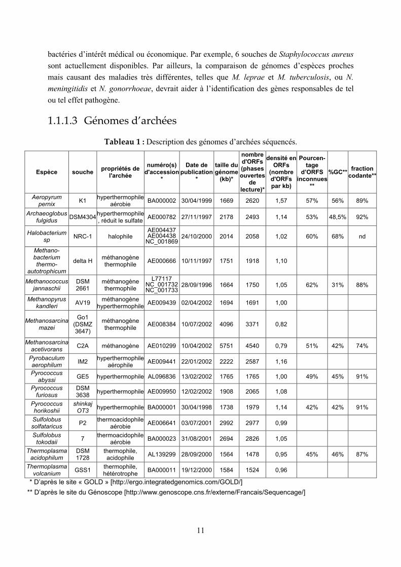
1.1.1.3 Génomes d’archées
Tableau 1 : Description des génomes d’archées séquencés.
Espèce souche propriétés de
l'archée
numéro(s) d'accession
*
Date de publication
*
taille du génome
(kb)*
nombre d'ORFs (phases ouvertes
de lecture)*
densité en ORFs
(nombre d'ORFs par kb)
Pourcen-tage
d’ORFS inconnues
**
%GC**fraction
codante**
Aeropyrum pernix
K1 hyperthermophile
aérobie BA000002 30/04/1999 1669 2620 1,57 57% 56% 89%
Archaeoglobus fulgidus
DSM4304 hyperthermophile, réduit le sulfate
AE000782 27/11/1997 2178 2493 1,14 53% 48,5% 92%
Halobacterium sp
NRC-1 halophile AE004437 AE004438
NC_00186924/10/2000 2014 2058 1,02 60% 68% nd
Methano- bacterium thermo-
autotrophicum
delta H méthanogène thermophile
AE000666 10/11/1997 1751 1918 1,10
Methanococcus jannaschii
DSM 2661
méthanogène thermophile
L77117 NC_001732 NC_001733
28/09/1996 1664 1750 1,05 62% 31% 88%
Methanopyrus kandleri
AV19 méthanogène
hyperthermophile AE009439 02/04/2002 1694 1691 1,00
Methanosarcina mazei
Go1 (DSMZ 3647)
méthanogène thermophile
AE008384 10/07/2002 4096 3371 0,82
Methanosarcina acetivorans
C2A méthanogène AE010299 10/04/2002 5751 4540 0,79 51% 42% 74%
Pyrobaculum aerophilum
IM2 hyperthermophile
aérophile AE009441 22/01/2002 2222 2587 1,16
Pyrococcus abyssi
GE5 hyperthermophile AL096836 13/02/2002 1765 1765 1,00 49% 45% 91%
Pyrococcus furiosus
DSM 3638
hyperthermophile AE009950 12/02/2002 1908 2065 1,08
Pyrococcus horikoshii
shinkaj OT3
hyperthermophile BA000001 30/04/1998 1738 1979 1,14 42% 42% 91%
Sulfolobus solfataricus
P2 thermoacidophile
aérobie AE006641 03/07/2001 2992 2977 0,99
Sulfolobus tokodaii
7 thermoacidophile
aérobie BA000023 31/08/2001 2694 2826 1,05
Thermoplasma acidophilum
DSM 1728
thermophile, acidophile
AL139299 28/09/2000 1564 1478 0,95 45% 46% 87%
Thermoplasma volcanium
GSS1 thermophile, hétérotrophe
BA000011 19/12/2000 1584 1524 0,96
* D’après le site « GOLD » [http://ergo.integratedgenomics.com/GOLD/]
** D’après le site du Génoscope [http://www.genoscope.cns.fr/externe/Francais/Sequencage/]

12
Les 16 génomes d’archées dont le séquençage était achevé au 5 août 2003 sont présentés dans
le Tableau 1. Comme les bactéries, les archées sont hétérogènes (en ce qui concerne le
pourcentage en GC, la taille des génomes, le nombre de gènes…). Plus de 50% des ORFs
n’ont pas de fonction connue, ce qui peut refléter le fait que « seulement » 16 archées ont été
séquencées à ce jour. Certaines espèces proches ont été séquencées, par exemple les
Pyrococcus : la comparaison de ces trois génomes a mis en évidence des mécanismes
d’instabilité chromosomique (Zivanovic 2002).
1.1.2 Les génomes eucaryotes
1.1.2.1 Le génome humain
1.1.2.1.1 Le Projet Génome Humain
Le Projet Génome Humain (HGP : « Human Genome Project ») est né de deux idées qui ont
émergé au début des années 80 : d’une part, qu’une vue d’ensemble sur les génomes pourrait
accélérer grandement la recherche biomédicale en permettant aux chercheurs d’appréhender
les problèmes de façon plus éclairée et plus efficace, et d’autre part, que les données produites
devraient être mises à disposition de l’humanité entière et que leur dissémination ne devrait
pas être limitée par des intérêts privés ou nationaux. Ces deux notions ont eu comme
corollaire la prise de conscience que l’obtention de ces données devrait se faire au travers
d’efforts collaboratifs internationaux, sans précédent dans le domaine de la biologie. Cet état
d’esprit d’une communauté consciente de l’ampleur de la tâche que peut représenter l’étude
du génome humain avait d’ailleurs déjà conduit à des initiatives, dont l’une des plus connues
est probablement le CEPH (Centre d’Etudes du Polymorphisme Humain), fondé par le
Français Jean Dausset. Le CEPH a joué un rôle fondamental dans l’organisation du travail de
cartographie génétique humaine de plusieurs dizaines de laboratoires, souvent concurrents, au
cours des années 80. L’idée de séquencer le génome humain a été évoquée pour la première
fois dans des congrès organisés par le Département à l’énergie américain entre 1984 et 1986.
Le Projet Génome Humain (en l’occurrence la phase de séquençage proprement dite) a été
lancé en 1990, sur l’initiative de 4 pays (USA, Grande-Bretagne, Japon et France), qui ont
chacun fondé un centre de séquençage. D’autres pays se sont ensuite joints au projet, comme
l’Allemagne et la Chine : au final, plus de 20 laboratoires s’y sont impliqués. Jusqu’à 1995, le
projet a progressé sur deux voies principales : la construction des cartes génétique et physique
chez l’homme et la souris d’une part, et le séquençage de la levure S. cerevisiae et du
nématode C. elegans ainsi que de certaines régions de génomes mammifères d’autre part.
Cette première phase a prouvé que le séquençage à grande échelle était envisageable, par
l’approche en deux étapes consistant tout d’abord à générer les séquences de fragments
couvrant le génome de façon fortement redondante (8 à 10 fois), puis à effectuer l’assemblage
et la finition c’est-à-dire à boucher les trous (« gaps ») et résoudre les ambiguïtés. Cette phase

13
du projet a également permis de montrer que les séquences complètes fournissent des
informations sur les gènes, les régions régulatrices, et la structure des chromosomes, qui ne
sont pas accessibles par l’étude exclusive des ADNs complémentaires. En 1995, l’idée de
produire un brouillon du génome humain (« draft genome sequence ») a été proposée, mais
elle était prématurée car l’efficacité du séquençage à grande échelle d’un génome complexe et
riche en répétitions tel que le génome humain n’avait pas encore été prouvée. Des projets
pilotes ont donc été lancés, afin de démontrer la faisabilité d’un tel projet, avec échéance en
mars 1999 : ils ont produit avec succès des séquences de bonne qualité (99.99%) et sans
« gaps », représentant 15% du génome humain tout en permettant de mettre au point des
stratégies cohérentes pour le séquençage. En mars 1999, le projet de séquençage à grande
échelle du génome humain a donc débuté, avec pour but initial de produire un « brouillon » de
la séquence génome humain couvrant cette fois la majeure partie du génome, avant juin 2000.
L’article paru en février 2001 (Lander 2001) décrit cette première version de la séquence du
génome humain. Depuis, la phase de finition a été amorcée, en particulier avec le séquençage
de clones permettant l’élimination des gaps. L’accomplissement de cette tâche a été annoncé
en avril 2003, pour le cinquantenaire de la découverte de l’ADN (voir Science du 11 avril
2003 et Nature du 24 avril 2003). Tout au long du projet, les séquences ont été régulièrement
mises à jour et ont toujours été d’accès libre. De plus, l’analyse des séquences de
chromosomes complets a été publiée au fur et à mesure de leur achèvement. Ainsi, les articles
concernant les chromosomes 22 (Dunham 1999) et 21 (Hattori 2000) sont parus avant la
publication de la séquence brouillon. Depuis, les séquences des chromosomes 20 (Deloukas
2001), 14 (Heilig 2003) et 7 (Scherer 2003 ; Hillier 2003) ont été publiées. La France, qui est
l’un des quatre pays à l’initiative du Projet Génome Humain, s’est impliquée tout
particulièrement dans le séquençage du chromosome 14, par l’intermédiaire de son Centre
National de Séquençage, le Génoscope (Heilig 2003).
L’analyse de la séquence « brouillon » du génome humain (22 paires d’autosomes et une paire
de chromosomes sexuels), premier génome de vertébré séquencé, de taille équivalant à 8 fois
l’ensemble des génomes séquencés jusque là, soit environ 3000 mégabases (Mb) permet de
préciser un certain nombre d’observations antérieures (Lander 2001) :
- Le génome est constitué de zones hétérogènes à divers points de vue : densité en gènes, en
éléments transposables, pourcentage en GC, îlots CpG, taux de recombinaison.
- Les séquences codantes représentent moins de 5% du génome humain, tandis que les
séquences répétées en constituent plus de 50% dont : (1) des séquences dérivées de
transposons qui ne sont plus actifs, appelées répétitions dispersées, dont les LINEs (« long
interspersed elements » ; 20% du génome), les SINEs (« short interspersed
elements » ; 13%), les transposons LTR (8%), et les transposons à ADN (3%) ;
(2) des copies inactives (partiellement) de gènes provenant de rétropositions, appelées
pseudogènes ; (3) des séquences répétées en tandem en majorité des répétitions de
dinucléotides, (« SSR : simple sequence repeat ») qui représentent environ 3% du
génome ; (4) des duplications de segments de 10 à 300 kb, copiés d’une région du génome
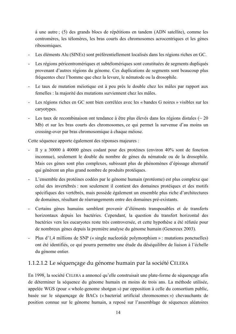
14
à une autre ; (5) des grands blocs de répétitions en tandem (ADN satellite), comme les
centromères, les télomères, les bras courts des chromosomes acrocentriques et les gènes
ribosomiques.
- Les éléments Alu (SINEs) sont préférentiellement localisés dans les régions riches en GC.
- Les régions péricentromériques et subtélomériques sont constituées de segments dupliqués
provenant d’autres régions du génome. Ces duplications de segments sont beaucoup plus
fréquentes chez l’homme que chez la levure, le nématode ou la drosophile.
- Le taux de mutation méiotique est à peu près le double chez les mâles par rapport aux
femelles : la majorité des mutations surviennent chez les mâles.
- Les régions riches en GC sont bien corrélées avec les « bandes G noires » visibles sur les
caryotypes.
- Les taux de recombinaison ont tendance à être plus élevés dans les régions distales (~ 20
Mb) et sur les bras courts des chromosomes, ce qui permet la survenue d’au moins un
crossing-over par bras chromosomique à chaque méiose.
Cette séquence apporte également des réponses majeures :
- Il y a 30000 à 40000 gènes codant pour des protéines (environ 40% sont de fonction
inconnue), seulement le double du nombre de gènes du nématode ou de la drosophile.
Mais ces gènes sont plus complexes, subissant plus de phénomènes d’épissage alternatif
qui génèrent un plus grand nombre de produits protéiques.
- L’ensemble des protéines codées par le génome humain (protéome) est plus complexe que
celui des invertébrés : non seulement il contient des domaines protéiques et des motifs
spécifiques des vertébrés, mais possède également un ensemble plus riche d’architectures
de domaines, résultant de réarrangements entre des domaines pré-existants.
- Certains gènes humains semblent provenir d’éléments transposables et de transferts
horizontaux depuis les bactéries. Cependant, la question du transfert horizontal des
bactéries vers les eucaryotes reste très controversée, et cette hypothèse a été réfutée pour
de nombreux gènes depuis la première analyse du génome humain (Genereux 2003).
- Plus d’1,4 millions de SNP (« single nucleotide polymorphism » : mutations ponctuelles)
ont été identifiés, ce qui pourra permettre une étude du déséquilibre de liaison à l’échelle
du génome entier.
1.1.2.1.2 Le séquençage du génome humain par la société CELERA
En 1998, la société CELERA a annoncé qu’elle construisait une plate-forme de séquençage afin
de déterminer la séquence du génome humain en moins de trois ans. La méthode utilisée,
appelée WGS (pour « whole-genome shotgun ») par opposition à celle du consortium public,
basée sur le séquençage de BACs (« bacterial artificial chromosomes ») chevauchants de
position connue sur le génome humain, a reposé sur l’assemblage de séquences aléatoires

15
provenant de banques plasmidiques générées à partir du génome entier de 5 individus (Venter
2001). La Figure 4 illustre ces deux stratégies de séquençage (Waterston 2002).
Figure 4 : Comparaison entre les stratégies de séquençage « hierarchical shotgun » et
« whole genome shotgun », d’après (Waterston 2002).
L’approche WGS a été inventée et appliquée initialement pour le séquençage des génomes
bactériens (voir chapitre 1.1.1). Afin de gagner du temps, la société CELERA a incorporé les
données publiquement accessibles provenant du Projet Genome Humain, aux données
produites par l’approche WGS afin de générer son propre assemblage. Le résultat de ce
deuxième séquençage est paru le 16 février 2001 (Venter 2001), en même temps que celui du
consortium public (15 février 2001 : Lander 2001), et a conduit à des conclusions similaires.
Le Tableau 2 présente quelques caractéristiques du génome humain issues de la version
CELERA (Venter 2001).
Le séquençage du génome humain par la société CELERA, dont le résultat n’est pas librement
accessible, a déclenché de nombreuses polémiques. Par exemple, dans un article paru en mars
2002, trois membres du consortium du Projet Génome Humain affirment que la stratégie de
séquençage « whole-genome shotgun » pour un génome aussi complexe et riche en répétitions
que le génome humain n’a pas été prouvée par l’article de Venter et al., car leur assemblage a
largement profité de l’assemblage effectué par le consortium public (Waterston 2002).
L’équipe CELERA a aussitôt répondu (Myers 2002) que ces assertions n’étaient pas fondées :
les données provenant du Projet Genome Humain utilisées par CELERA auraient été
suffisamment morcelées pour ne pas permettre de rétablir l’assemblage HGP. En tout état de
cause, les séquences produites par ces deux projets ne sont pas indépendantes, ce qui n’a

16
jamais été nié par l’équipe CELERA. Nous en verrons une illustration dans l’article décrit au
chapitre 2.3.2 (Denoeud 2003).
Tableau 2 : Quelques caractéristiques de la version du génome humain produite par CELERA, d’après (Venter 2001).
taille du génome (y compris les gaps) 2,91 Gb
taille du génome (excluant les gaps) 2,66 Gb
%AT 54
%GC 38
% bases indéterminées 9
fenêtre de 50 kb la plus riche en GC chr 2 (65%)
fenêtre de 50 kb la moins riche en GC chr X (25%)
% de la séquence correspondant à des répétitions 35
Nombre de gènes annotés 26383
Pourcentage de gènes annotés de fonction inconnue 42
Nombre de gènes hypothétiques et annotés 39114
Pourcentage de gènes hypothétiques et annotés de fonction inconnue 59
Gène contenant le plus d'exons Titin (234 exons)
Taille moyenne des gènes 27 kb
Chromosome le plus riche en gènes chr 19 (23/Mb)
Chromosome le moins riche en gènes chr Y, chr 13 (5/Mb)
Longueur totale des déserts (>500 kb sans gènes annotés) 605 Mb
% pb dans des gènes (annotés / annotés + hypothétiques) 25,5 / 37,8
% pb dans des exons (gènes annotés / annotés + hypothétiques) 1,1 / 1,4
% pb dans des introns (gènes annotés / annotés + hypothétiques) 24,4 / 36,4
% pb intergéniques (gènes annotés / annotés + hypothétiques) 74,5 / 63,6
Chromosome avec la plus grande proportion d'ADN dans des exons annotés chr 19 (9,33)
Chromosome avec la plus faible proportion d'ADN dans des exons annotés chr Y (0,36)
Plus longue région intergénique chr 13 (~3 Mb)
Taux de mutations ponctuelles (SNPs) 1/1250 pb
1.1.2.1.3 Comparaison entre les deux séquences produites (HGP et CELERA)
Les observations globales (nombre de gènes, pourcentage en GC, éléments répétés, grandes
duplications…) issues des deux séquences « brouillons » du génome humain sont similaires.
Cependant, même si ces deux études ont identifié environ 30000 gènes codant pour des
protéines, il y a peu de recouvrement entre les gènes prédits de novo (Hogenesch 2001). Il
apparaît que cette incohérence provient des séquences utilisées pour la prédiction de ces gènes
et non pas de la méthode de prédiction : il y a des différences majeures entre ces deux
assemblages du génome humain. En effet, la distribution et la longueur des contigs diffèrent
(Aach 2001 ; Semple 2002), et les deux assemblages possèdent un certain nombre,
comparable, de séquences uniques (Aach 2001). La Figure 5 présente une comparaison des
assemblages CELERA et HGP pour les chromosomes 21 et 22, obtenue en positionnant des
minisatellites identifiés au cours de cette thèse (Denoeud 2003) sur les scaffolds CELERA et
les contigs HGP : on peut mettre en évidence des inversions entre les deux assemblages
proposés.

17
Figure 5 : Comparaison des assemblages proposés par le consortium public et la société CELERA pour les chromosomes 21 et 22.
Une comparaison des différents assemblages d’une portion du chromosome 4 suggère que le
séquençage de répétitions en tandem est particulièrement délicat par l’approche « WGS »
(Semple 2002). Nous présenterons au chapitre 2.3.2 une comparaison de la qualité du
séquençage des répétitions en tandem dans les versions HGP et CELERA des chromosomes 21
et 22 (Denoeud 2003).
1.1.2.1.4 Applications du séquençage du génome humain
La fin du séquençage ne marque évidemment que le début de l’étude du génome humain. La
première application du séquençage du génome humain porte sur les gènes responsables de
pathologies : le fait de disposer de cette séquence a facilité l’identification de gènes de
fonction biochimique inconnue par le clonage positionnel (Wooster 1995), ainsi que l’étude
de liaisons génétiques en fournissant des candidats de marqueurs microsatellites. Cette
séquence a également conduit à la mise en évidence des délétions chromosomiques à l’origine
de syndromes : de telles délétions sont causées dans certains cas par des phénomènes de
recombinaison entre deux régions provenant d’une duplication intrachromosomique (Shaikh
2000). De plus, la séquence du génome humain permet d’identifier des paralogues de gènes
responsables de maladies, ce qui peut avoir deux intérêts : d’une part, de mettre en évidence

18
d’autres gènes causant des maladies génétiques apparentées (Kohl 2000), et d’autre part
d’identifier de nouvelles voies thérapeutiques (Olivieri 1998).
Une seconde application porte sur l’identification de cibles pour les médicaments.
L’identification de paralogues à des cibles connues de médicaments est prometteuse pour
découvrir de nouvelles cibles (Davies 1999 ; Heise 2000).
Enfin, le séquençage du génome humain devrait également apporter des informations plus
fondamentales sur la biologie et la physiologie humaines (Matsunami 2000).
1.1.2.2 Autres génomes eucaryotes
Chez les eucaryotes, 18 génomes (ou chromosomes) étaient séquencés en plus du génome
humain au 5 août 2003 (voir GOLD [http://ergo.integratedgenomics.com/GOLD]), parmi
lesquels ceux décrits dans le Tableau 3. La proportion de gènes de fonction inconnue varie
entre 40 et 60% selon le génome eucaryote considéré.
Tableau 3 : Description de quelques génomes d’eucaryotes séquencés.
Organisme Site Web dédié à la
séquence Nombre de
chromosomesTaille du génome
Nombre d’ORFs (phases ouvertes
de lecture)
Densité en ORFs (nb
d’ORFs/kb)
Saccharomyces cerevisiae
(levure)
http://www.yeastgenome.org/
16 12069 kb 6294 0,52
Plasmodium falciparum
(parasite causant la malaria)
http://www.plasmodb.org/
14 22900 kb 5268 0,23
Caenorhabditis elegans (nématode)
http://www.sanger.ac.uk/Projects/C_el
egans/ 6 97000 kb 19099 0,20
Arabidopsis. Thaliana (plante)
http://arabidopsis.org/home.html
5 115428 kb 25498 0,22
Drosophila melanogaster
(drosophile)
http://flybase.bio.indiana.edu/
6 137000 kb 14100 0,10
Mus musculus (souris) http://www.ncbi.nlm.nih.gov/genome/g
uide/mouse/ 20 ~2500 Mb ~30000 ~ 0,01

19
1.2 Les répétitions en tandem
1.2.1 Définitions
Une répétition en tandem est une succession de motifs d’ADN répétés les uns derrière les
autres, par opposition aux répétitions dispersées dont les unités répétées sont éparpillées dans
le génome (comme décrit au paragraphe 1.1.2.1). Les différentes unités formant la répétition
en tandem ne sont pas nécessairement identiques entre elles : le degré de conservation au sein
d’une répétition en tandem est très variable. Les répétitions en tandem ont été en premier lieu
étudiées chez les mammifères, où trois catégories ont été distinguées : les satellites, les
minisatellites, et les microsatellites. Cette distinction correspond à différentes plages de taille
(pour la longueur totale) et a été faite de façon plus ou moins arbitraire : l’ADN « satellite » a
tout d’abord été observé et isolé par centrifugation sur gradient de densité, où il constituait
une fraction particulière (dite « satellite ») (Britten 1968 ; Meneveri 1984). Ensuite, des
répétitions en tandem de taille inférieure, pouvant être analysées grâce à la technique de
Southern Blot, ont été caractérisées (Wyman 1980) puis appelées « minisatellites » (Jeffreys
1985a). Enfin, les répétitions en tandem de taille encore inférieure ont été nommées
« microsatellites » lorsque l’avènement de la technique de PCR (« polymerase chain
reaction ») en a fait des outils courants de génétique moléculaire. Avec le recul, les études
menées, principalement chez les eucaryotes, sur les mécanismes de création et de mutation de
ces structures souvent polymorphes laissent penser que cette distinction recouvre une réalité
biologique, comme nous le verrons dans les paragraphes suivants.
Les répétitions en tandem sont présentes, dans des proportions variables, chez tous les
organismes : eucaryotes, procaryotes, et même virus. Dans ces deux derniers groupes, les
mécanismes sous-jacents n’ont quasiment pas été étudiés, et la distinction entre
microsatellites et minisatellites est rarement faite. D’autres termes sont couramment
employés, chez les procaryotes, mais également parfois chez les eucaryotes, pour désigner les
répétitions en tandem : les SSR (« simple sequence repeat ») et STR (« short tandem repeat »)
désignent des répétitions en tandem « simples » : elles correspondent aux microsatellites et
aux minisatellites de petite taille (quelques centaines de paires de bases). Les VNTR
(« variable number of tandem repeats ») désignent les répétitions en tandem polymorphes, qui
peuvent appartenir à la classe des microsatellites ou des minisatellites.
1.2.2 Intérêts des répétitions en tandem chez les
bactéries
Les répétitions sont une composante importante des génomes bactériens : elles peuvent
constituer jusqu’à 10% de ces petits génomes, denses en gènes, et on trouve souvent dans la

20
littérature des allusions, peu précises, au fait que les répétitions en tandem bactériennes se
trouveraient « fréquemment » dans des gènes. Même si ces observations sont fondées pour un
certain nombre d’espèces bactériennes, elles restent impossibles à généraliser : la base de
données des répétitions en tandem que je présente dans cette thèse (voir chapitre 2.1) devrait
permettre de répondre à ces questions d’une façon plus rigoureuse.
1.2.2.1 Rôles dans l’adaptation et la virulence des bactéries :
régulation de l’expression de gènes
Dans un certain nombre de pathogènes, des répétitions en tandem (SSR pour « simple
sequence repeats ») présentes en amont ou dans les séquences codantes de protéines de
surface sont polymorphes et contribuent à l’adaptation de la bactérie aux changements de
conditions survenant au cours de l’infection de l’hôte. Ce phénomène, appelé variation de
phase (revue : Henderson 1999), « allume »/« éteint » la synthèse protéique ou fait varier la
structure de la protéine. Les locus à fort taux de mutation qui y sont impliqués sont appelés
locus de contingence (Moxon 1994). Par exemple, chez la bactérie Haemophilus influenzae,
qui colonise les voies respiratoires et peut causer des pneumonies et des méningites, le rôle
des répétitions en tandem dans la modulation de la virulence est bien documenté. Tout
d’abord, la variabilité des répétitions a été associée expérimentalement à la modulation des
gènes impliqués dans la synthèse des pilus et du lipopolysaccharide (LPS) (Weiser 1989).
Une répétition de dinucléotides dans un promoteur de gènes codant des sous-unités de pilus
est un facteur régulateur majeur de leur expression : selon le nombre d’unités répétées,
l’espacement entre les boîtes -35 et -10 est soit favorable soit défavorable à la reconnaissance
de ce site par l’ARN polymérase (van Ham 1993). Les répétitions en tandem peuvent aussi
moduler l’expression génique en étant à l’origine de blocages de la réplication (Krasilnikova
1998), ou en tant que terminateurs de transcription (Guerin 1998).
D’autres répétitions en tandem ont un effet au niveau de la traduction. Chez H. influenzae,
différents gènes codant pour des enzymes de synthèse du LPS contiennent des répétitions de
tétranucléotides, localisées dans les séquences codantes, ce qui est à l’origine de décalages du
cadre de lecture (Weiser 1990). Un tétranucléotide, situé dans un gène homologue à une
méthyltransférase de type III, est tellement instable qu’il génère même du mosaïcisme dans
les cultures bactériennes (De Bolle 2000). Chez Neisseria meningitidis, une répétition en
tandem de 7 pb dans la phase codante du gène PilQ affecte la biosynthèse des pilus de façon
quantitative (Tonjum 1998).
L’implication des SSR dans la modulation de l’expression de gènes a été mise en évidence
dans une grande variété d’autres bactéries (pour revue : voir van Belkum 1999b) dont
Escherichia coli (Foster 1994), Neisseria meningitidis (van der Ende 1995), Bacillus
anthracis (Jackson 1997), ou Mycoplasma gallisepticum (Glew 1998). Le séquençage des
génomes bactériens a ouvert la voie à une analyse plus systématique des gènes
potentiellement impliqués dans la variation de phase. Lorsque la séquence complète du
21
génome de H. influenzae a été connue, un catalogue des répétitions en tandem de type
microsatellites a pu être établi (Fleischmann 1995) : la plupart de ces répétitions sont
associées à des gènes potentiellement impliqués dans la virulence (molécules d’adhésion,
enzymes de synthèse du LPS…) (Hood 1996). De la même façon, le séquençage du
pathogène Helicobacter pylori (Tomb 1997) a permis de mettre en évidence une trentaine de
gènes associés à des SSR, c’est-à-dire potentiellement impliqués dans la variation de phase
(Saunders 1998). Ces gènes codent pour des enzymes de biosynthèse du LPS, des protéines de
surface, et des enzymes de restriction. Le séquençage d’une seconde souche de cette bactérie
(Alm 1999) a rendu possible l’identification de SSR polymorphes entre les deux souches
considérées. La plupart de ces candidats subissent bel et bien une variation d’expression selon
le nombre de répétitions.
Par ailleurs, des répétitions en tandem appartenant à des phases ouvertes de lecture et dont le
motif est multiple de 3 génèrent des polymorphismes au niveau des protéines, ce qui peut être
à l’origine d’une variation antigénique :
- Chez Staphylococcus aureus, des protéines de surface impliquées dans la reconnaissance
des molécules d’adhésion de la matrice extracellulaire de l’hôte contiennent de nombreuses
répétitions en tandem, dont le nombre de répétitions influe sur l’accessibilité du domaine actif
(voir Tableau 4).
- Chez Bacillus anthracis, une répétition en tandem dans le gène de l’exosporium est à
l’origine de variations de la longueur des filaments de la surface des spores (Sylvestre 2003).
- Chez les streptocoques du groupe A, la protéine M, protéine de surface et facteur de
virulence est soumise à une grande variabilité antigénique causée vraisemblablement par des
événements de recombinaison homologue (Hollingshead 1987).
- Chez les streptocoques du groupe B, la protéine alpha C, antigène de surface, contient
une répétition en tandem polymorphe qui lorsqu’elle est délétée permet d’échapper à la
réponse immunitaire de l’hôte (Madoff 1996 ; Gravekamp 1998).
- Chez le mycoplasme Mycoplasma hyorhinis, les protéines de surface du système VIp
confèrent aux bactéries, par leur variation de taille, une résistance contre les anticorps produits
par l’hôte (porc) (Citti 1997).
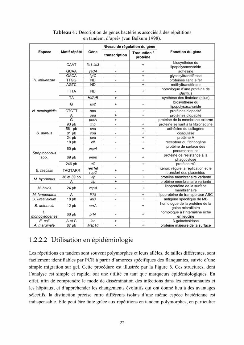
Le Tableau 4 liste des répétitions en tandem associées à des gènes de fonction connue chez
différentes bactéries (van Belkum 1998) : selon la taille de leurs motifs répétés, certaines
appartiennent à la classe des microsatellites et d’autres à la classe des minisatellites.
22
Tableau 4 : Description de gènes bactériens associés à des répétitions en tandem, d’après (van Belkum 1998).
Niveau de régulation du gène
Espèce Motif répété Gène transcription
Traduction / protéine
Fonction du gène
CAAT lic1-lic3 - + biosynthèse du
lipopolysaccharide
GCAA yadA - + adhésine
GACA lgtC - + glycosyltransférase
TTGG ND - + protéines liant le fer
AGTC ND - + méthyltransférase
TTTA ND - + homologue d’une protéine de
Bacillus
H. influenzae
TA HifA/B + - synthèse des fimbriae (pilus)
G lsi2 + - biosynthèse du
lipopolysaccharide
CTCTT opa - + protéines d’opacité
A opa + - protéines d’opacité
N. meningitidis
G porA + - protéine de la membrane externe
93 pb fnb - + protéine se liant à la fibronectine
561 pb cna - + adhésine du collagène
81 pb coa - + coagulase
24 pb spa - + protéine A
S. aureus
18 pb clf - + récepteur du fibrinogène
60 pb pspA - + protéine de surface des
pneumocoques
69 pb emm - + protéine de résistance à la
phagocytose
Streptococcus spp.
246 pb αC - + protéine αC
E. faecalis TAGTARR rep1et rep2
+ - itéron: régule la réplication et le
transfert des plasmides
36 et 39 pb vlp - + protéine membranaire variante M. hyorhinus
A vlp + - protéine membranaire variante
M. bovis 24 pb vspA - + lipoprotéine de la surface
membranaire
M. fermentans A P78 - + lipoprotéine de transporteur ABC
U. urealyticum 18 pb MB - + antigène spécifique de MB
B. anthracis 12 pb vvrA - + homologue de la protéine de la
gaine microfilaire
L. monocytogenes
66 pb prfA - + homologue à l’internaline riche
en leucine
E. coli A et C lac + - β-galactosidase
A. marginale 87 pb Msp1α - + protéine majeure de la surface
1.2.2.2 Utilisation en épidémiologie
Les répétitions en tandem sont souvent polymorphes et leurs allèles, de tailles différentes, sont
facilement identifiables par PCR à partir d’amorces spécifiques des flanquantes, suivie d’une
simple migration sur gel. Cette procédure est illustrée par la Figure 6. Ces structures, dont
l’analyse est simple et rapide, ont une utilité en tant que marqueurs épidémiologiques. En
effet, afin de comprendre le mode de dissémination des infections dans les communautés et
les hôpitaux, et d’appréhender les changements évolutifs qui ont donné lieu à des avantages
sélectifs, la distinction précise entre différents isolats d’une même espèce bactérienne est
indispensable. Elle peut être faite grâce aux répétitions en tandem polymorphes, en particulier

23
dans des espèces d’émergence récente, comme Yersinia pestis (Achtman 1999) et Bacillus
anthracis, pour lesquelles elles constituent une source majeure de polymorphisme.
Figure 6 : Utilisation d’une répétition en tandem polymorphe afin de distinguer des souches bactériennes.
Quelques précautions doivent cependant être observées quant à l’utilisation des répétitions en
tandem pour l’épidémiologie bactérienne (van Belkum 1999a). D’une part, certaines
répétitions ne sont pas neutres du point de vue évolutif, c’est-à-dire que certains allèles
peuvent conférer un avantage sélectif à certains isolats. Elles peuvent donc conduire à des
conclusions erronées sur la proximité des souches, qui peuvent avoir le même allèle car elles
ont été confrontées au même hôte et non pas parce qu’elles sont génétiquement proches. Cela
dit, de nombreuses répétitions en tandem non neutres sont utilisées de façon efficace en
épidémiologie : par exemple, celle de la coagulase de Staphylococcus aureus (Shopsin 2000).

24
D’autre part, les locus de contingence, subissant la variation de phase (voir paragraphe
1.2.2.1) n’ont aucune valeur épidémiologique et doivent par conséquent être évités. Ces locus
sont toutefois rares : dans l’ensemble, les répétitions en tandem ne subissent pas d’altérations
au cours de leur manipulation en laboratoire (van Belkum 1997 ; Stothard 1998).
Les répétitions en tandem ont été validées en tant que marqueurs épidémiologiques dans
différentes espèces bactériennes, dont Mycobacterium tuberculosis (Frothingham 1998 ;
Supply 2000), Bacillus anthracis (Jackson 1997 ; Keim 2000), et Haemophilus influenzae
(van Belkum 1997). L’un des avantages majeurs de la technique de typage de répétitions en
tandem est qu’elle est relativement simple et peu coûteuse par rapport aux autres techniques
de génotypage communément utilisées (van Belkum 2001 ; Olive 1999). En outre, cette
méthode est d’analyse facile, et reproductible, contrairement à celles qui génèrent des profils
mutlibandes, comme :
- l’électrophorèse en champ pulsé (Tenover 1995)
- la RAPD (Random Amplified Polymorphic DNA) (Williams 1990)
- l’AFLP (Amplified fragment Length Polymorphism) (Vos 1995)
- la RFLP (Restriction Fragment Length Polymorphism), nommée ribotypage lorsqu’elle
est appliquée à l’ADN ribosomique 16s-23s (Bingen 1994).
La technique MLST (multilocus sequence typing) (Maiden 1998), qui consiste à séquencer
différents gènes de ménage, est très reproductible mais beaucoup plus coûteuse que le typage
de répétitions en tandem ou MLVA (multilocus VNTR analysis). Le séquençage d’un nombre
croissant de génomes bactériens va faciliter grandement l’identification de ce type de
séquences. Dans la suite de cette thèse, je présenterai les différents outils informatiques que
j’ai développés en tirant profit de la disponibilité de séquences, parfois de plusieurs souches
d’une même espèce, afin d’identifier des marqueurs polymorphes pour le génotypage. Ces
outils ont été appliqués avec succès à diverses espèces bactériennes, ce qui a fait l’objet de
plusieurs publications (Le Flèche 2001; Le Flèche 2002 ; Pourcel 2003 ; Onteniente 2003).
1.2.2.3 Les familles de répétitions en tandem bactériennes
La notion de famille de répétitions en tandem a été introduite pour décrire des locus ayant des
motifs répétés similaires et qui sont trouvés en plusieurs localisations. Souvent, il s’agit d’une
simple coïncidence, d’une similitude de séquence due à la faible complexité du motif
élémentaire et dans ce cas le terme de famille est abusif. Parfois, les « familles » existent bel
et bien : deux cas se distinguent alors. Le plus simple est le cas de la famille dans laquelle la
flanquante est également conservée. Par exemple, si un élément transposable contient une
répétition en tandem, la répétition en tandem est alors un simple passager. Le cas de familles
de répétitions en tandem sans flanquantes communes est plus curieux, et encore
inexpliqué (Supply 1997). De façon encore plus intéressante, une famille de répétitions en
tandem présente dans de nombreux génomes procaryotes (bactéries et archées) a été mise en
25
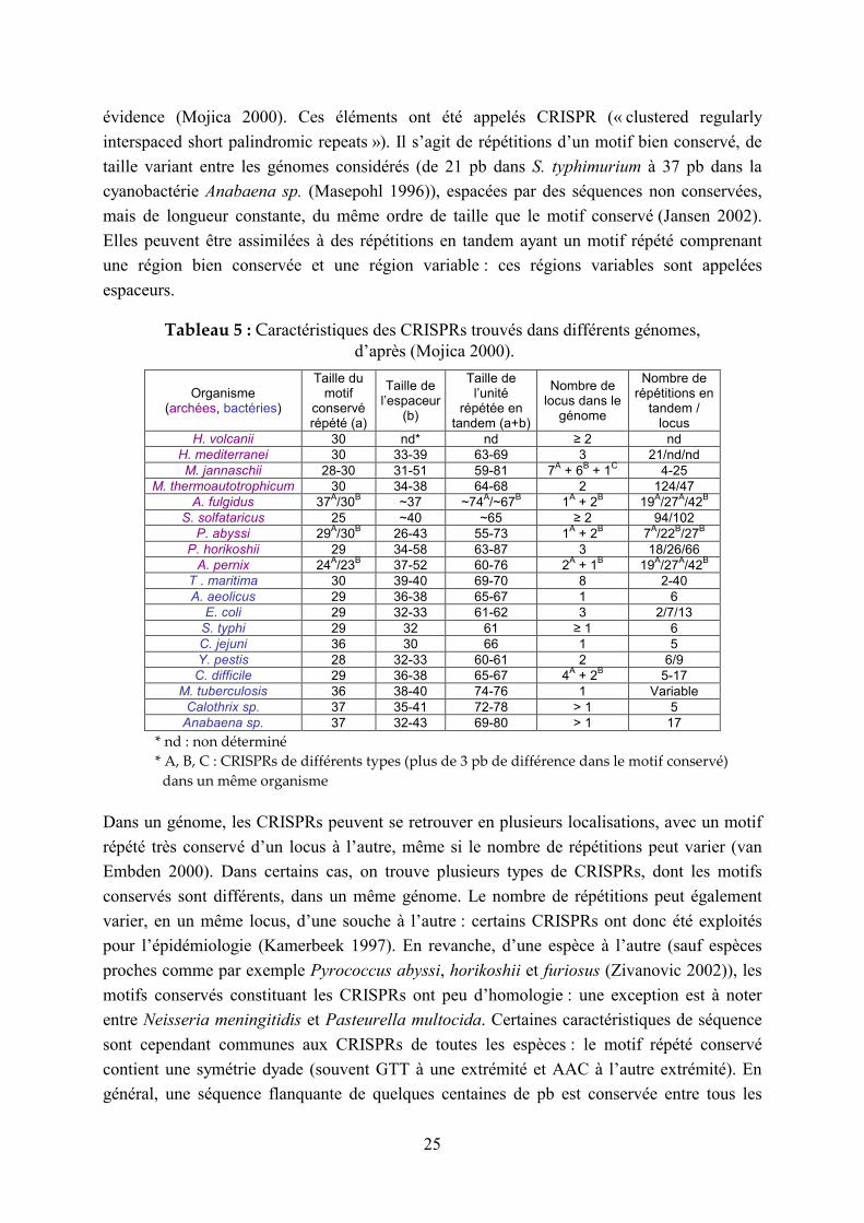
évidence (Mojica 2000). Ces éléments ont été appelés CRISPR (« clustered regularly
interspaced short palindromic repeats »). Il s’agit de répétitions d’un motif bien conservé, de
taille variant entre les génomes considérés (de 21 pb dans S. typhimurium à 37 pb dans la
cyanobactérie Anabaena sp. (Masepohl 1996)), espacées par des séquences non conservées,
mais de longueur constante, du même ordre de taille que le motif conservé (Jansen 2002).
Elles peuvent être assimilées à des répétitions en tandem ayant un motif répété comprenant
une région bien conservée et une région variable : ces régions variables sont appelées
espaceurs.
Tableau 5 : Caractéristiques des CRISPRs trouvés dans différents génomes, d’après (Mojica 2000).
Organisme (archées, bactéries)
Taille du motif
conservé répété (a)
Taille de l’espaceur
(b)
Taille de l’unité
répétée en tandem (a+b)
Nombre de locus dans le
génome
Nombre de répétitions en
tandem / locus
H. volcanii 30 nd* nd ≥ 2 nd
H. mediterranei 30 33-39 63-69 3 21/nd/nd
M. jannaschii 28-30 31-51 59-81 7A + 6
B + 1
C 4-25
M. thermoautotrophicum 30 34-38 64-68 2 124/47
A. fulgidus 37A/30
B ~37 ~74
A/~67
B 1
A + 2
B 19
A/27
A/42
B
S. solfataricus 25 ~40 ~65 ≥ 2 94/102
P. abyssi 29A/30
B 26-43 55-73 1
A + 2
B 7
A/22
B/27
B
P. horikoshii 29 34-58 63-87 3 18/26/66
A. pernix 24A/23
B 37-52 60-76 2
A + 1
B 19
A/27
A/42
B
T . maritima 30 39-40 69-70 8 2-40
A. aeolicus 29 36-38 65-67 1 6
E. coli 29 32-33 61-62 3 2/7/13
S. typhi 29 32 61 ≥ 1 6
C. jejuni 36 30 66 1 5
Y. pestis 28 32-33 60-61 2 6/9
C. difficile 29 36-38 65-67 4A + 2
B 5-17
M. tuberculosis 36 38-40 74-76 1 Variable
Calothrix sp. 37 35-41 72-78 > 1 5
Anabaena sp. 37 32-43 69-80 > 1 17
* nd : non déterminé
* A, B, C : CRISPRs de différents types (plus de 3 pb de différence dans le motif conservé)
dans un même organisme
Dans un génome, les CRISPRs peuvent se retrouver en plusieurs localisations, avec un motif
répété très conservé d’un locus à l’autre, même si le nombre de répétitions peut varier (van
Embden 2000). Dans certains cas, on trouve plusieurs types de CRISPRs, dont les motifs
conservés sont différents, dans un même génome. Le nombre de répétitions peut également
varier, en un même locus, d’une souche à l’autre : certains CRISPRs ont donc été exploités
pour l’épidémiologie (Kamerbeek 1997). En revanche, d’une espèce à l’autre (sauf espèces
proches comme par exemple Pyrococcus abyssi, horikoshii et furiosus (Zivanovic 2002)), les
motifs conservés constituant les CRISPRs ont peu d’homologie : une exception est à noter
entre Neisseria meningitidis et Pasteurella multocida. Certaines caractéristiques de séquence
sont cependant communes aux CRISPRs de toutes les espèces : le motif répété conservé
contient une symétrie dyade (souvent GTT à une extrémité et AAC à l’autre extrémité). En
général, une séquence flanquante de quelques centaines de pb est conservée entre tous les

26
locus CRISPRs d’un génome donné, qui n’a pas d’homologie avec les flanquantes des
CRISPRs d’autres génomes. Enfin, tous les génomes bactériens ne contiennent pas de
CRISPRs et la présence de ces CRISPRs est strictement associée à des gènes appelés cas pour
« CRISPR associated genes », qui pourraient correspondre à des protéines se liant à l’ADN
(Jansen 2002). Les mécanismes de création et de dispersion dans les génomes, ainsi que la
fonction biologique des CRISPRs, sont encore inconnus et méritent d’être étudiés. Le
Tableau 5 présente les caractéristiques des CRISPRs trouvés dans différents génomes (Mojica
2000). Il s’agit cependant là d’une classe très particulière de répétitions en tandem, qu’il ne
faut pas confondre avec les répétitions en tandem de type mini- ou microsatellites.
L’existence de répétitions de type CRISPR dans les génomes eucaryotes n’a pas été
documentée jusqu’à présent. Il serait toutefois intéressant de s’assurer de l’absence de tels
éléments en explorant de façon systématique les génomes eucaryotes dont la séquence est
achevée (une recherche effectuée sur les génomes eucaryotes accessibles dans la base de
données des répétitions en tandem n’a pour l’instant pas mis en évidence de locus CRISPR).
1.2.3 Intérêts des répétitions en tandem chez l’Homme
D’après l’article analysant la séquence issue du Projet Génome Humain, les répétitions en
tandem représentent environ 3% du génome humain, la majorité (en nombre de locus) étant
des répétitions de dinucléotides (0.5% de l’ensemble du génome). Il y a environ 1 répétition
en tandem tous les 2 kilobases, soit 437 répétitions en tandem par mégabase.
1.2.3.1 L’ADN satellite
L’ADN satellite rencontré dans les grands génomes correspond à des structures dont la
longueur totale atteint plusieurs mégabases (Mb). L’ADN satellite est localisé dans les régions
péricentromériques ou d’hétérochromatine télomérique des eucaryotes (Charlesworth 1994).
Les satellites semblent jouer des rôles importants dans la structuration des génomes : ils sont
les constituants majeurs des centromères fonctionnels, ce qui a été montré chez l’homme
(Schueler 2001) et chez la drosophile (Sun 1997), où ils sont nécessaires pour le bon
déroulement de la mitose et de la méiose (Csink 1998). Les séquences des satellites
centromériques diffèrent même entre des organismes proches, ce qui est associé à des
changements dans les histones correspondantes (Henikoff 2001). L’ADN satellite
centromérique évolue rapidement (Ugarkovic 2002), ce qui pourrait, en provoquant
l’évolution adaptative des histones centromériques, être à l’origine du processus de spéciation
(Malik 2001).
1.2.3.2 Intérêts des microsatellites humains
Les microsatellites ont une unité répétée de 1 à 10 paires de bases (pb) environ, et une
longueur totale de quelques dizaines à quelques centaines de paires de bases. Ils sont répartis

27
de façon relativement homogène le long des chromosomes eucaryotes, même si leur
abondance dans différentes régions du génome varie selon le type de répétition
(mononucléotides, dinucléotides…) (Toth 2000) et que dans certaines espèces, ils sont moins
abondants aux environs des centromères, par exemple chez A. thaliana (Lin 1999) ou ont
tendance à s’organiser en clusters, par exemple chez la drosophile (Bachtrog 1999). En
particulier, les régions codantes sont moins riches en microsatellites que les régions non-
codantes sauf en ce qui concerne les motifs de 3 ou 6 pb, ce qui peut correspondre à une
contre-sélection des mutations de décalage du cadre de lecture (Metzgar 2000 ; Morgante
2002 ; Li 2002). Certaines de ces observations restent approximatives et mériteront d’être ré-
examinées, puisque, comme nous le montrons dans cette thèse, l’étude des répétitions en
tandem à l’échelle de génomes entiers est dorénavant possible.
1.2.3.2.1 Utilisation comme marqueurs génétiques
Les microsatellites, qui sont distribués de façon homogène sur tout le génome humain, et sont
fréquemment polymorphes, représentent la source la plus abondante de marqueurs génétiques
chez l’Homme. Ils sont à la base de l’élaboration de la carte du génome humain par le
Généthon, en particulier les microsatellites de type (CA/GT) (Gyapay 1994 ; Dib 1996). Les
répétitions de mononucléotides poly (A/T), de dinucléotides (CA/GT) et de trinucléotides
(AAT) sont les plus communes (Beckmann 1992 ; Hancock 1998 ; Lander 2001). Le
Tableau 6 présente le nombre de répétitions en tandem de longueur comprise entre 1 et 10 sur
le génome humain (Lander 2001).
Tableau 6 : Description des répétitions en tandem trouvées dans la séquence du génome humain produite par le consortium public, d’après (Lander 2001).
Taille du motif répété (pb)
Nombre moyen de bases appartenant à une répétition en tandem, par Mb
Nombre moyen de répétitions en tandem par Mb
1 1660 36,7
2 5046
(dont AC: 50%, AT: 35%, AG: 15%, GC: 0,1%) 43,1
3 1013
(dont AAT: 33%, AAC: 21%, ACC: 4%, AGC: 2,2%, ACT: 1,4%, ACG: 0,1%)
11,8
4 3383 32,5
5 2686 17,6
6 1376 15,2
7 906 8,4
8 1139 11,1
9 900 8,6
10 1576 8,6
1.2.3.2.2 Différents rôles biologiques attribués aux microsatellites
Les microsatellites sont associés à un certain nombre de processus cellulaires, chez l’homme
comme chez d’autres organismes :

28
- Structure de l’ADN :
Dans différentes espèces, l’abondance relative de répétitions dinucléotidiques semble être
associée à différentes caractéristiques structurales à grande échelle de l’ADN, comme la
courbure ou le surenroulement (Karlin 1998 ; Baldi 2000).
- Structure des télomères :
Les télomères humains sont constitués d’une répétition en tandem d’un motif de 6 paires
de bases, de type microsatellite : TTAGGG. L’ADN des mammifères étant linéaire, deux
problèmes majeurs se posent : comment répliquer les molécules d’ADN sans en raccourcir
les extrémités, et comment les extrémités de l’ADN sont-elles protégées de l’activité des
exonucléases ? Chez la plupart des eucaryotes, les extrémités des chromosomes
(télomères) sont répliquées par une polymérase particulière, appelée télomérase. Cette
ribonucléoprotéine qui a une activité de transcriptase inverse utilise son propre ARN
comme matrice pour la synthèse d’ADN. L’activité de la télomérase est élevée dans les
cellules embryonnaires et devient inexistante dans les cellules différenciées. De plus, son
activité varie lorsque les cellules entrent ou sortent du cycle cellulaire. Ces observations
ont conduit à formuler l'hypothèse suivante : le raccourcissement progressif des télomères
dans les cellules somatiques conduirait à l'arrêt de la division cellulaire et à la sénescence ;
les télomères seraient ainsi impliqués dans le processus de vieillissement cellulaire et de
cancérogénèse (Autexier 1996). A l’inverse, en conservant une longueur constante de
leurs télomères, les cellules germinales (télomères courts) et cancéreuses (télomères
longs) continueraient de se diviser indéfiniment. La séquence répétée constituant les
télomères humains (TTAGGG)n est riche en G, ce qui confère à ces séquences simple-
brin la capacité de se replier sous forme de structures 4-brins ou tétraplexes (Henderson
1987), stables dans les conditions physiologiques et insensibles aux endo- et exonucléases.
- Effet sur la recombinaison :
Les microsatellites pourraient correspondre à des points chauds de recombinaison. En
effet, une répétition (GT)30 insérée dans un chromosome de levure, augmente la fréquence
de recombinaison méiotique et de conversion génique dans les régions adjacentes (Treco
1986). De même, dans des cellules humaines en culture, cette répétition (GT)30 augmente
la recombinaison entre vecteurs plasmidiques (Wahls 1990). De plus, les répétitions
dinucléotidiques ont une forte affinité pour les enzymes de recombinaison (Biet 1999), qui
augmente avec le nombre de copies (Dutreix 1997). Il a également été proposé que les
microsatellites influencent directement la recombinaison par l’intermédiaire de leurs effets
sur la structure de l’ADN (conformation Z) (Biet 1999). Enfin, sur le chromosome 22
humain, la distribution des répétitions de type (GT/CA) est corrélée aux points chauds de
recombinaison (Majewski 2000).
- Effet sur la réplication de l’ADN et le cycle cellulaire :
Les microsatellites pourraient affecter la réplication. Par exemple, dans des cellules de
mammifères, des phénomènes d’amplification sont co-sélectionnables avec un
29
microsatellite instable (Caligo 1999). De plus, certaines enzymes du contrôle du cycle
cellulaire sont codées par des gènes contenant des répétitions en tandem, qui si elles sont
mutées, par exemple dans un contexte d’instabilité causé par des mutations du système de
réparation des mésappariements, peuvent contribuer à la transformation cancéreuse
(Johannsdottir 2000).
- Association avec les enzymes de réparation des mésappariements (« MMR ») :
Les enzymes de réparation des mésappariements corrigent les erreurs de réplication et
inhibent la recombinaison entre des séquences divergées, ce qui contrôle le taux de
mutation et l’adaptation évolutive. Les gènes MMR mineurs contiennent des répétitions
de A chez une grande variété d’eucaryotes : Homo sapiens, Mus musculus,
Saccharomyces cerevisiae, Schizosaccharomyces pombe, Drosophila melanogaster,
Arabidopsis thaliana ainsi que chez le procaryote Escherichia coli. Ces microsatellites,
vulnérables aux insertions et délétions spontanées, peuvent introduire des décalages du
cadre de lecture dans les protéines correspondantes. La perte d’activité d’un de ces gènes
mineurs de réparation des mésappariements provoquerait alors des phénotypes mutateurs
modérés (par rapport à des mutations dans les gènes MMR majeurs), ce qui permettrait de
moduler le taux de mutations adaptatives nécessaires à l’évolution (Chang 2001).
- Régulation de gènes :
Les répétitions en tandem présentes dans les promoteurs semblent pouvoir affecter
l’activité des gènes en agissant au niveau transcriptionnel : l’augmentation du nombre de
répétitions peut faire diminuer le taux de transcription (Lanz 1995 ; Gebhardt 2000), ou le
faire augmenter (Okladnova 1998 ; Xu 1998). Des microsatellites présents dans des
introns semblent aussi affecter la transcription, soit en agissant comme des régulateurs
transcriptionnels (Meloni 1998), soit en modifiant la courbure de l’ADN pour rapprocher
le promoteur de régions régulatrices (Gebhardt 1999). Par ailleurs, chez S. Cerevisiae, les
répétitions trinucléotidiques sont plus fréquentes dans les gènes régulateurs de la
transcription et de la transduction du signal, et sont sous-représentées dans les gènes de
protéines de structure, ce qui suggère également une influence de ces éléments sur la
transcription (Young 2000).
Certaines séquences microsatellites ont également la capacité de lier des protéines
(éléments régulateurs). Par exemple, des protéines se liant aux poly(GA) et poly(GT) ont
été identifiées dans des fibroblastes humains (Aharoni 1993), et des séquences (GT)n ou
(GT)n(GA)n présentes dans les introns de gènes du système immunitaire se lient à des
protéines nucléaires (Epplen 1993). Le nombre de répétitions peut influer sur ces liaisons,
la protéine ne se liant qu’à des répétitions en tandem d’une certaine taille (Winter 1989).
Enfin, différentes études ont montré que les microsatellites avaient un effet sur la
traduction, par exemple chez la bactérie Escherichia coli (Martin-Farmer 1999), et chez
l’homme (Sandberg 1997).
30
De plus, le polymorphisme des microsatellites codants peut avoir des conséquences sur
des pathologies humaines, comme nous le décrirons dans le paragraphe suivant.
1.2.3.2.3 Pathologies associées aux microsatellites
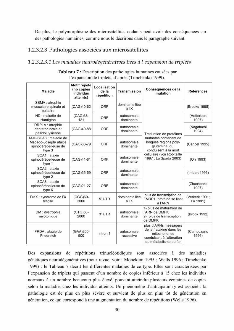
1.2.3.2.3.1 Les maladies neurodégénératives liées à l’expansion de triplets
Tableau 7 : Description des pathologies humaines causées par l’expansion de triplets, d’après (Timchenko 1999).
Maladie
Motif répété (nb copies individus atteints)
Localisation de la
répétition Transmission
Conséquences de la mutation
Références
SBMA : atrophie musculaire spinale et
bulbaire (CAG)40-62 ORF
dominante liée à l’X
(Brooks 1995)
HD : maladie de Huntigton
(CAG)36-121
ORF autosomale dominante
(Hofferbert 1997)
DRPLA : atrophie dentatorubrale et pallidoluysienne
(CAG)49-88 ORF autosomale dominante
(Nagafuchi 1994)
MJD/SCA3 : maladie de Macado-Joseph/ ataxie spinocérébelleuse de
type 3
(CAG)68-79 ORF autosomale dominante
(Cancel 1995)
SCA1 : ataxie spinocérébelleuse de
type 1 (CAG)41-81 ORF
autosomale dominante
(Orr 1993)
SCA2 : ataxie spinocérébelleuse de
type 2 (CAG)35-59 ORF
autosomale dominante
(Imbert 1996)
SCA6 : ataxie spinocérébelleuse de
type 6 (CAG)21-27 ORF
autosomale dominante
Traduction de protéines mutantes contenant de longues régions poly-
glutamine, qui conduisent à la mort
cellulaire (voir Robitaille 1997 ; La Spada 2003)
(Zhuchenko 1997)
FraX : syndrome de l’X fragile
(CGG)60-2000
5’ UTR dominante liée
à l’X
plus de transcription de FMRP1, protéine se liant
à l’ARN
(Verkerk 1991; Fu 1991)
DM : dystrophie myotonique
(CTG)50-2000
3’ UTR autosomale dominante
1- plus de maturation de l’ARN de DMPK 2- plus de transcription de DMPK
(Brook 1992)
FRDA : ataxie de Friedreich
(GAA)200-900
intron 1 autosomale récessive
plus d’ARNs messagers de la frataxine dans les
mitochondries conduisant à l’altération du métabolisme du fer
(Campuzano 1996)
Des expansions de répétitions trinucléotidiques sont associées à des maladies
génétiques neurodégénératives (pour revue, voir : Monckton 1995 ; Wells 1996 ; Timchenko
1999) : le Tableau 7 décrit les différentes maladies de ce type. Elles sont caractérisées par
l’expansion de triplets qui passent d’un nombre de copies inférieur à 15 chez les individus
normaux à un nombre beaucoup plus élevé, pouvant atteindre plusieurs centaines de copies
selon la maladie, chez les individus atteints. Un phénomène d’anticipation y est associé : la
pathologie est de plus en plus sévère et survient de plus en plus tôt de génération en
génération, ce qui correspond à une augmentation du nombre de répétitions (Wells 1996).
31
D’autres maladies sont associées à des microsatellites dont l’unité n’est pas un triplet. C’est le
cas pour la rétinopathie diabétique, associée à une répétition (CA)n en 5’ de l’aldose réductase
(Fujisawa 1999), ainsi qu’à une répétition (CCTTT)n dans le promoteur de NOS2A (Warpeha
1999) et pour la fibrose du poumon après greffe, associée à une répétition (CA)n dans le
premier intron de l’interféron け (Awad 1999).
1.2.3.2.3.2 Microsatellites et cancer
Environ 15% des cancers sporadiques colorectaux (Liu 1995), gastriques (Leung 2000) et
endométriaux (Basil 2000) sont associés à une instabilité des microsatellites ou MSI
(« microsatellite instability ») (revue : Atkin 2001). On distingue deux types de variations des
microsatellites associées aux cancers humains : le type A correspond à des petites variations
de taille, de moins de 6 paires de bases, et le type B à des variations plus importantes,
supérieures à 8 pb. L’instabilité de type A a été associée à des défauts du système de
réparation des mésappariements. Une telle corrélation n’est toujours pas claire pour
l’instabilité de type B, qui semble par ailleurs être associée à des prédispositions familiales
(revue : Li 2002). Lorsque le système de réparation des mésappariements n’est plus efficace,
le taux de mutation des microsatellites augmente 100 fois et les protéines contenant ce type
de séquences risquent donc d’être altérées. Des mutations de décalage du cadre de lecture ont
effectivement été trouvées, avec des fréquences variables, dans des gènes codant pour des
protéines impliquées dans la transduction du signal, l’apoptose et les mécanismes
inflammatoires, la régulation de la transcription, et la réparation de l’ADN (Jacob 2002).
Cependant, il est difficile de savoir si ces gènes sont les véritables cibles du processus
d’inactivation par décalage du cadre de lecture et sont donc impliqués dans l’initiation des
tumeurs ou leur progression, ou si leur mutation n’est qu’une conséquence sans pertinence de
l’altération du système de réparation des mésappariements (Woerner 2003). Différentes
cibles, suppresseurs de tumeurs, contenant des répétitions mutant à forte fréquence dans le
contexte « MSI » ont déjà été identifiées, par exemple, TGFく-RII (récepteur d’hormone de
croissance) (Markowitz 1995), et APC (Laken 1997).
1.2.3.3 Intérêts des minisatellites humains
Les minisatellites sont constitués d’unités d’une quinzaine à plusieurs centaines de paires de
bases répétées sur une centaine à quelques milliers de paires de bases. Le premier minisatellite
humain a été mis en évidence en 1980 par Wyman et al. (Wyman 1980), et le premier
minisatellite de levure en 1984 par Horowitz et Haber (Horowitz 1984). Le nom de
« minisatellites » leur a été donné par Jeffreys en 1985, qui a découvert la grande utilité de ces
structures extrêmement polymorphes pour réaliser les premières « empreintes génétiques »
(Jeffreys 1985a ; Jeffreys 1985b). Des minisatellites ont été identifiés et caractérisés dans
divers génomes (pour revue, voir Bois 2003). Chez l’homme, 90% des minisatellites sont
localisés dans les régions subtélomériques (Royle 1988), ce qui n’est pas le cas pour d’autres
eucaryotes comme la souris, le rat ou le porc (Amarger 1998).

32
1.2.3.3.1 Utilisation pour les empreintes génétiques et en tant que
marqueurs génétiques
Les minisatellites ont été largement utilisés pour établir des empreintes génétiques (Jeffreys
1985b). Il s’agit d’hybrider des sondes contenant des séquences riches en GC sur des
Southern Blots. La technique de Southern Blot consiste à digérer l’ADN génomique d’un
individu par des enzymes de restriction. Les fragments d’ADN sont ensuite séparés par
électrophorèse puis transférés sur une membrane. La sonde s’hybridant à de nombreuses
répétitions en tandem de taille variable, des profils complexes, uniques pour chaque individu,
sont générés. Cette technique a été très utilisée dans les débuts de la médecine légale et pour
les recherches de paternité mais a été depuis remplacée par le typage de microsatellites : elle
avait en effet l’inconvénient d’être difficile à interpréter à cause de la multiplicité des bandes
(revue : Bennett 2000). Par ailleurs, même si les microsatellites sont, grâce à leur distribution
homogène, un outil de choix pour la cartographie génétique, les minisatellites restent une
source de marqueurs génétiques pour des investigations concernant les extrémités
télomériques où ils sont plus abondants. Par exemple, le retard mental est souvent associé à
des délétions d’extrémités chromosomiques, et certaines ont pu être mises en évidences grâce
à des marqueurs minisatellites (Giraudeau 1997 ; Giraudeau 2001).
1.2.3.3.2 Rôles biologiques attribués aux minisatellites
La plupart des minisatellites sont localisés dans des régions intergéniques. Ils peuvent
cependant avoir des fonctions. Elles sont évoquées dans la revue (Vergnaud & Denoeud
2000) qui sera présentée au chapitre 2.3.1:
- Certains sont soupçonnés de jouer un rôle dans les phénomènes d’empreinte parentale
(Chaillet 1995 ; Neumann 1995).
- D’autres constituent des sites fragiles (revue : Handt 2000), cassures chromosomiques
survenant lors de la métaphase à des localisations caractéristiques et induites par différents
types de traitements des cellules en culture (Sutherland 1998). Par exemple, le site fragile
FRA10B est un minisatellite d’unité répétée de 42 pb (Hewett 1998) et FRA16B est un
minisatellite de motif de 33 pb riche en AT (Yamauchi 2000). Le minisatellite situé dans
le locus IGH (Tableau 8) joue peut-être également un rôle dans les événements de
recombinaison qui ont lieu à ce locus (Brusco 1999).
- Enfin, de façon plus hypothétique, les minisatellites pourraient jouer un rôle dans
l’initiation de l’appariement des chromosomes (Ashley 1994 ; Sybenga 1999), ce qui peut
être lié à leurs propriétés recombinogènes supposées (Boan 1998 ; Wahls 1998).
- Des minisatellites associés à des gènes, dans les séquences régulatrices, les introns, ou les
séquences codantes, peuvent avoir un effet sur certaines pathologies humaines : nous en
dressons une liste dans le paragraphe suivant.
33
1.2.3.3.3 Minisatellites associés à des gènes
La revue que nous présenterons au chapitre 2.3.1 (Vergnaud & Denoeud 2000) ne détaille pas
les minisatellites associés à des gènes. Je présente donc ici une synthèse bibliographique des
minisatellites présents dans des introns, exons, ou au voisinage de gènes, et dont le
polymorphisme a été étudié : certains sont impliqués dans des pathologies.
1.2.3.3.3.1 Minisatellites à proximité de gènes ou dans des introns (non codants)
Le Tableau 8 liste les minisatellites situés dans des introns, des régions flanquantes en 3’ ou
en 5’ de gènes, ou au niveau de jonctions introns/exons, et dont le polymorphisme a été
étudié. D’autres répétitions en tandem polymorphes, ou « VNTRs » (pour « Variable Number
of Tandem Repeats »), associées à des gènes ont été étudiées, mais elles n’appartiennent pas à
la classe des minisatellites, et ne sont donc pas listées ici. Cette recherche bibliographique a
permis de dénombrer (par des requêtes dans PubMed [http://www.ncbi.nlm.nih.gov/PubMed/]
du type « minisatellite+gene+polymorphism» qui génère plus de 600 résultats) : 23
minisatellites polymorphes localisés dans des introns, dont 3 au niveau de sites d’épissage, et
20 localisés en amont ou en aval de gènes. Pour la plupart de ces minisatellites, une
association entre certains allèles et des pathologies humaines a été décelée, soit indirectement,
l’association reflétant alors un déséquilibre de liaison dû à la proximité entre le minisatellite et
la mutation responsable du phénotype, soit directement, le minisatellite lui-même étant
impliqué dans la pathologie par l’une de ces deux voies majeures:
- soit en modulant l’expression du gène situé à proximité (pour revue, voir : Nakamura
1998) : c’est par exemple le cas pour les minisatellites en amont des gènes l’insuline
(Bennett 1995) et de la monoamine oxydase A (Sabol 1998).
- soit, pour les minisatellites localisés dans des introns, en jouant un rôle sur l’épissage
(Turri 1995) : par exemple, le minisatellite de l’intron 7 du facteur de coagulation VII est
le site d’épissages anormaux conduisant à une pathologie (Borensztajn 2003).
Parmi les 43 minisatellites décrits dans le Tableau 8, 6 sont fortement polymorphes (ils ont 20
allèles ou plus). Par exemple, le minisatellite en amont du gène HRAS compte 4 allèles dit
« communs » (30, 46, 68 et 84 copies) et plus d’une trentaine d’allèles rares, qui sont associés
à la cancérogenèse (Krontiris 1993). Certains de ces minisatellites très polymorphes sont
instables. C’est le cas des allèles « prémutateurs » du minisatellite du gène CSTB impliqué
dans l’épilepsie, comptant 12 à 17 copies d’un dodécamère, qui mutent à une fréquence
élevée pour générer des allèles « mutants » comptant plus de 45 copies, responsables de la
pathologie via une diminution du nombre de transcrits (Lalioti 1997).

34
Tableau 8 : Minisatellites polymorphes au voisinage de gènes ou dans des introns.
Nom du gène (nom du
minisatellite) fonction(s) de la protéine
pathologie(s) associée(s) au gène
association du polymorphisme
du ms avec la(les)
pathologie(s)
localisation du
minisatellite dans le gène
taille du motif répété
plage de nombre de
copies
nombre d'allèles identifiés
numéro d'accession (position du
minisatellite dans la séquence)
Locus ID*
(Locus Link)
Chromo some
références
ABO glycosyltransférase qui génère les antigènes spécifiques des
groupes sanguins A ou B
? (fixation de facteurs de
transcription) 5' UTR 43 pb 1-4 2
AF014105 (108-280)
28 9q34.1-q34.2
(Kominato 1997 ; Irshaid 1999)
APOB apolipoprotéine B: rôle dans l'homéostasie du cholestérol
diverses pathologies dues à l'hypercholestérolémie
- région
hypervariable en 3'
15 pb 15-55 > 25 J05157 338 2p24-p23(Huang 1987 ; Destro-
Bisol 2000)
CBS cystathionine beta-synthase:
métabolisme de l'homocystéine
hyperhomocystéinémie légère: facteur de risque pour les
maladies cardiovasculaires +
frontière exon 13-intron 13
31 pb 16-21 5 AF042836 (22990-
23516) 875 21q22.3
(Yang 2000 ; Lievers 2001)
COL2A collagène de type II, chaîne
alpha 1
collagénoses : achondroplasie (défaut de formation du
cartilage), ostéoarthrose… +
région flanquante en
3'
34 et 31 pb
8-17 >20 L10160 1280 12q13.11
-q13.2 (Berg 1993 ; Katsuyama 1998; Meulenbelt 1999)
COL3A1 collagène de type III, chaîne
alpha 1 syndrome coronarien aigu + intron 25 15 pb 3-13 11
AY016295 (57618-57692)
1281 2q31 (Mays 1992 ; Muckian
2002)
CSTB (EPM1 minisatellite)
cystatine B: inhibiteur de protéase à cystéine
épilepsie myoclonique progressive du type de Unverricht-Lundborg
+ : pathologie due à l'expansion du dodécamère (fait baisser le taux d'ARNm)
région flanquante en
5' 12 pb 2-70 > 30
AF208234 (3255-3294)
1476 21q22.3(Lalioti 1997 ; Larson
1999)
DAT transporteur de la dopamine troubles psychiatriques, maladie
de Parkinson, hyperactivité…
+ (agit sur le niveau de
transcription) 3' UTR 40 pb 3-11 > 8
NM_001044 (2741-3144)
6531 5p15.3 (Vandenbergh 1992 ;
Doucette-Stamm 1995 ; Mill 2002 ; Lin 2003)
F7/ IVS7 repeated element
facteur de coagulation VII: dépendant de la vitamine K
hémorragie sévère + (effet sur l'épissage)
Frontière exon7-intron 7
37 pb 4-8 5? J02933 (10234-
10461) 2155 13q34
(O'Hara 1988 ; Borensztajn 2003)
FSTL3 « follistatin-like 3 »
(glycoprotéine sécrétée) rôle dans l’apparition de
leucémies ? ? 3' UTR 24 pb ?-13-? >2
U76702 (1213-1524)
10272 19p13 (Hayette 1998)
G1P3 protéine inductible par
l’interféron alpha : fonction inconnue
site d'épissage
alternatif frontière exon
2 - intron 2 12 pb 1-3 3
NM_022873 (170-206)
2537 1p35 (Turri 1995)
HRAS1 proto-oncogène : impliqué dans
la transduction du signal mitogène et la différenciation
cancer
+ (effet sur la transcription ; allèles rares =
risques de cancer)
1 kb en aval (3’)
28 pb 30-100
> 30 (4communs: 30, 46, 68, 84 copies)
a1:AF105318 a2:AF105319 a3:AF105320 a4:AF105321
3265 11p15.5(Krontiris 1993 ; Weitzel 2000 ; Langdon 2003)
hTERT (hTERT-VNTR 2-1)
sous-unité catalytique de la télomérase
cancer : télomérase=oncogène? (fixation de facteurs de
transcription) intron 2 42 pb 40-111 6
AY007685 (27650-29650)
7015 5p15.33 (Leem 2002)
hTERT (hTERT-VNTR 2-2)
sous-unité catalytique de la télomérase
cancer : télomérase=oncogène? (fixation de facteurs de
transcription) intron 2 61 pb 40-44 4
AY007685 (34111-35453)
7015 5p15.33 (Leem 2002)
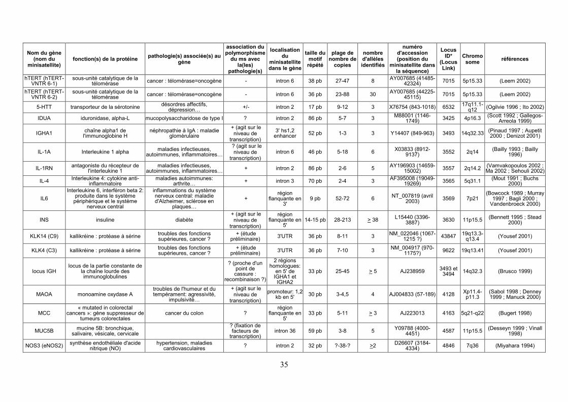
35
Nom du gène (nom du
minisatellite) fonction(s) de la protéine
pathologie(s) associée(s) au gène
association du polymorphisme
du ms avec la(les)
pathologie(s)
localisation du
minisatellite dans le gène
taille du motif répété
plage de nombre de
copies
nombre d'allèles identifiés
numéro d'accession (position du
minisatellite dans la séquence)
Locus ID*
(Locus Link)
Chromo some
références
hTERT (hTERT-VNTR 6-1)
sous-unité catalytique de la télomérase
cancer : télomérase=oncogène - intron 6 38 pb 27-47 8 AY007685 (41485-
42324) 7015 5p15.33 (Leem 2002)
hTERT (hTERT-VNTR 6-2)
sous-unité catalytique de la télomérase
cancer : télomérase=oncogène - intron 6 36 pb 23-88 30 AY007685 (44225-
45115) 7015 5p15.33 (Leem 2002)
5-HTT transporteur de la sérotonine désordres affectifs,
dépression… +/- intron 2 17 pb 9-12 3 X76754 (843-1018) 6532
17q11.1-q12
(Ogilvie 1996 ; Ito 2002)
IDUA iduronidase, alpha-L mucopolysaccharidose de type I ? intron 2 86 pb 5-7 3 M88001 (1146-
1749) 3425 4p16.3
(Scott 1992 ; Gallegos-Arreola 1999)
IGHA1 chaîne alpha1 de
l'immunoglobine H néphropathie à IgA : maladie
glomérulaire
+ (agit sur le niveau de
transcription)
3' hs1,2 enhancer
52 pb 1-3 3 Y14407 (849-963) 3493 14q32.33(Pinaud 1997 ; Aupetit 2000 ; Denizot 2001)
IL-1A Interleukine 1 alpha maladies infectieuses,
autoimmunes, inflammatoires…
? (agit sur le niveau de
transcription) intron 6 46 pb 5-18 6
X03833 (8912-9137)
3552 2q14 (Bailly 1993 ; Bailly
1996)
IL-1RN antagoniste du récepteur de
l'interleukine 1 maladies infectieuses,
autoimmunes, inflammatoires… + intron 2 86 pb 2-6 5
AY196903 (14659-15002)
3557 2q14.2 (Vamvakopoulos 2002 ; Ma 2002 ; Sehouli 2002)
IL-4 Interleukine 4: cytokine anti-
inflammatoire maladies autoimmunes:
arthrite… + intron 3 70 pb 2-4 3
AF395008 (19049-19269)
3565 5q31.1 (Mout 1991 ; Buchs
2000)
IL6
Interleukine 6, interféron beta 2: produite dans le système
périphérique et le système nerveux central
inflammations du système nerveux central: maladie d'Alzheimer, sclérose en
plaques…
+ région
flanquante en 3'
9 pb 52-72 6 NT_007819 (avril
2003) 3569 7p21
(Bowcock 1989 ; Murray 1997 ; Bagli 2000 ;
Vandenbroeck 2000)
INS insuline diabète + (agit sur le
niveau de transcription)
région flanquante en
5' 14-15 pb 28-213 > 38
L15440 (3396-3887)
3630 11p15.5(Bennett 1995 ; Stead
2000)
KLK14 (C9) kallikréine : protéase à sérine troubles des fonctions supérieures, cancer ?
+ (étude préliminaire)
3'UTR 36 pb 8-11 3 NM_022046 (1067-
1215 ?) 43847
19q13.3-q13.4
(Yousef 2001)
KLK4 (C3) kallikréine : protéase à sérine troubles des fonctions supérieures, cancer ?
+ (étude préliminaire)
3'UTR 36 pb 7-10 3 NM_004917 (970-
1175?) 9622 19q13.41 (Yousef 2001)
locus IGH locus de la partie constante de
la chaîne lourde des immunoglobulines
? (proche d'un point de cassure :
recombinaison ?)
2 régions homologues:
en 5' de IGHA1 et IGHA2
33 pb 25-45 > 5 AJ238959 3493 et
3494 14q32.3 (Brusco 1999)
MAOA monoamine oxydase A troubles de l'humeur et du tempérament: agressivité,
impulsivité…
+ (agit sur le niveau de
transcription)
promoteur: 1,2 kb en 5'
30 pb 3-4,5 4 AJ004833 (57-189) 4128 Xp11.4-p11.3
(Sabol 1998 ; Denney 1999 ; Manuck 2000)
MCC « mutated in colorectal
cancers »: gène suppresseur de tumeurs colorectales
cancer du colon ? région
flanquante en 5'
33 pb 5-11 > 3 AJ223013 4163 5q21-q22 (Bugert 1998)
MUC5B mucine 5B: bronchique,
salivaire, vésicale, cervicale
? (fixation de facteurs de
transcription) intron 36 59 pb 3-8 5
Y09788 (4000-4451)
4587 11p15.5(Desseyn 1999 ; Vinall
1998)
NOS3 (eNOS2) synthèse endothéliale d'acide
nitrique (NO) hypertension, maladies
cardiovasculaires ? intron 2 32 pb ?-38-? >2
D26607 (3184-4334)
4846 7q36 (Miyahara 1994)

36
Nom du gène (nom du
minisatellite) fonction(s) de la protéine
pathologie(s) associée(s) au gène
association du polymorphisme
du ms avec la(les)
pathologie(s)
localisation du
minisatellite dans le gène
taille du motif répété
plage de nombre de
copies
nombre d'allèles identifiés
numéro d'accession (position du
minisatellite dans la séquence)
Locus ID*
(Locus Link)
Chromo some
références
NOS3 (eNOS4) synthèse endothéliale d'acide
nitrique (NO) hypertension, maladies
cardiovasculaires + intron 4 27 pb 4-6 3
D26607 (5132-5284)
4846 7q36 (Miyahara 1994 ; Song
2003)
PAH phénylalanine hydroxylase phénylcétonurie + 3 kb en aval
(3') 30 pb 6-9 4 S41936 5053
12q22-q24.2
(Goltsov 1992)
PDGFA (IVS3 minisatellite)
facteur de croissance « platelet-derived », chaine A : mitogène
? intron 3 44 pb 4-12 >6 AJ238420 (7500-
7602) 5154 7p22 (Bonthron 1999)
PKD1
protéine membranaire impliquée dans l'interaction avec la matrice
extracellulaire et dans la transduction du signal
polykystose rénale autosomique dominante
? intron 1 26-27 pb 2-15 >2 L39891 (10976-
11382) 5310 16p13.3
(Burn 1995 ; De Fonzo 1998)
PLA2G4C phospholipase cytosolique A2-
gamma dans la région du suppresseur
de gliome ? 5' UTR 27 pb 1-3 3
NM_003706 (1 copie?)
8605 19q13.3 (Hartmann 2002)
POMT1 protéine-O-mannosyltransferase syndrome de Walker-Warburg (trouble sévère de la migration
neuronale) ? intron 13 17-19 pb 36-56 8
AF095146 (150-1127)
10585 9q34.1 (Jurado 1999)
PTHRP « parathyroid hormone-related
peptide » développement : squelettogénèse
? intron 6 ? 10 à 24
pb 3-17 8 L07553 5744
12p12.1-p11.2
(Pausova 1993)
RB1 rétinoblastome-1 : suppresseur
de tumeur rétinoblastome, ostéosarcome,
cancer de la vessie ? intron 16 53 pb ~10-~35 11
L11910 (123912-125501)
5925 13q14.2 (Scharf 1992)
RECQL4 (IVS12 minisatellite)
« RecQ protein-like 4 »: hélicase syndrome de Rothmund-
Thomson ? intron 12 30 pb 17-20 >2
NT_037704 (màj avril 2003)
9401 8q24.3 (Roversi 2003)
SEC14L1 « SEC14 S. cerevisiae-like 1 » : rôle possible dans un système
de transport intracellulaire ? 3' UTR 13 pb 20-60 >3
NM_003003 (4139-4789)
6397 17q25.1-17q25.2
(Chinen 1996 ; Kalikin 2001)
SNAPC1
« small nuclear RNA activating complex »: active la transcription
des gènes de petits ARNs nucléaires
? 5' UTR 17 pb 2-5 4 NT_026437 (màj
avril 2003) 6617 14q22 (Maeng 1998)
TMPT thiopurine méthyltransférase sensibilité à la 6-mercaptopurine
+ : effet sur le taux de
transcription et sur le niveau d'activité de
l'enzyme
région flanquante en 5' : promoteur
17 ou 18 pb
3-9 7 AF060074 7172 6p22.3 (Spire-Vayron de la
Moureyre 1999 ; Yan 2000)
TPO
thyroide péroxydase: glycoprotéine liée à la
membrane, jouant un rôle central dans la fonction de la
glande thyroïde
maladies en rapport à la synthèse d'hormones
thyroïdiennes: hypothyroïdie congénitale, goitre congénital,
déficience de synthèse de l’hormone thyroïdienne, IIA
? (pas d'effet sur l'épissage alternatif)
intron 10 50 pb 4-34 >20 M68651 (1918-
2370) 7173 2p25 (Bikker 1992)
*Locuslink : http://www.ncbi.nlm.nih.gov/LocusLink/
37
1.2.3.3.3.2 Minisatellites appartenant à des séquences codantes
Le Tableau 9 liste les minisatellites polymorphes déjà étudiés appartenant à des séquences
codantes, c’est-à-dire générant des répétitions en tandem dans la séquence en acides aminés
de la protéine correspondante. La grande majorité des gènes décrits codent pour des
glycoprotéines, généralement des protéines sécrétées, constituants de la matrice
extracellulaire : aggrécane (cartilage), involucrine (kératocytes), mucines (muqueuses). Le
collagène est également une protéine de la matrice extracellulaire contenant des répétitions en
tandem : cette famille de protéines ne figure pas dans le Tableau 9 car le motif répété qui la
caractérise, glycine-X-Y, est de la classe des microsatellites. Les répétitions en tandem
d’acides aminés, qui occupent en général la majeure partie de la longueur de ces macro-
protéines, jouent un rôle important dans leur structure fonctionnelle, en étant le site de
glycosylations (attachement des glucosaminoglycanes pour l’aggrécane, O-glycosylations
pour les mucines). Les mucines, protéines exprimées par les cellules épithéliales, sécrétées ou
membranaires, entrent dans la composition des mucus. Elles constituent une famille de
protéines contenant des répétitions en tandem polymorphes d’acides aminés riches en sérine
et en thréonine, qui sont le site d’O-glycosylations (Perez-Vilar 1999 ; Kinarsky 2003). Les
répétitions en tandem couvrent 50% ou plus de la protéine et sont plus ou moins polymorphes,
pouvant faire varier du simple au double la taille de la protéine (Vinall 1998 ; Debailleul
1998). Ces protéines ont un intérêt médical car elles sont souvent surexprimées par les
cellules cancéreuses ou lors de réactions inflammatoires. De nombreuses autres protéines
contiennent des domaines d’O-glycosylation, appelés « mucin-like domains » : par exemple,
GP1BA et PSGL-1 pour lesquelles un polymorphisme au niveau des répétitions en tandem a
également été caractérisé (Tableau 9).
Un autre intérêt des minisatellites codants réside dans le fait que certaines répétitions en tandem d’acides aminés ont la particularité d’être immunogènes (Mollick 2003) : des épitopes répétitifs stimulent la production d’immunoglobulines.

38
Tableau 9 : Minisatellites polymorphes appartenant à des séquences codantes.
Nom du gène
fonction(s) de la protéine pathologie(s) associée(s)
au gène
association du
polymorphisme du ms
avec la(les) pathologie(s)
localisation du
minisatellite
dans le gène
taille du
motif répété
Plage de nombre
de copies
nombre d'allèles identifiés
numéro d'accession (position du minisatellite
dans la séquence)
Locus ID* (LocusLink)
Chromo some
références répétition d'acides aminés
AGC1 aggrécane : protéoglycane,
constituant majeur du cartilage
scoliose - exon G3 57 pb 13-33 13 M55172
(2866-4495) 176 15q26.1
(Doege 1997 ; Zorkol'tseva 2002)
19aa:PGVEDISGLPSGEVLETAA
CEL
carboxyl ester lipase: rôle dans l'absorption intestinale
du cholestérol et des vitamines liposolubles
athérosclérose ? exon 11 33pb 13-18 6 NM_001807 (1693-2218)
1056 9q34.3 (Higuchi 2002) 11aa: PVPPTGDSGAP
DRD4 récepteur de la dopamine
troubles de la personnalité, hyperactivité; recherche de
nouveauté (dépendance aux drogues)…
+/- exon 3 48 pb 1-10 9 L12398 (744-
1080) 1815 11p15.5
(Lichter 1993 ; Swanson 2000 ; Li
1997) 16aa: LPQDPCGPDCAPPAPG
GP1BA glycoprotéine Ib alpha des plaquettes (« mucin like »)
thrombose artérielle + exon 2 39 pb 1-3 3 S34439 2811 17pter-
p12
(Lopez 1992 ; Gonzalez-Conejero
1998 ; Jilma-Stohlawetz 2003)
13aa: EPTSEPAPSPTTP
IVL involucrine: précurseur de
l'enveloppe cornée des kératocytes différenciés
? exon 3 30 pb ?-39-? >8 NM_005547 (398-1567)
3713 1q21 (Simon 1991 ; Urquhart 1993)
10aa: ELPEQQEGQL
MUC1
mucine 1: protéine membranaire (face apicale
des cellules épithéliales sécrétoires), antigène associé
au cancer du sein
carcinogenèse + exon 2 60 pb 20-120 >30 M61170
(3821-3881) 4582 1q21
(Gendler 1988 ; Engelmann 2001 ;
Silva 2001 ; Kinarsky 2003)
20aa: PDTRPAPGSTAPPAHGVTSA
MUC2 mucine 2: mucine intestinale,
trachéale et bronchique asthme atopique + exon 69 pb 51-115 >20
NM_002457(5713-12612)
4583 11p15.5(Gum 1989 ;
Toribara 1991 ; Vinall 2000)
23aa: PTTTPITTTTTVTPTPTPTGTQT
MUC3 mucine 3: mucine intestinale colite ulcérante + exon 51 pb? (9,38-18kb)
>30 AF113616 (2-
1025) 4584 7q22
(Gum 1990 ; Van Klinken 1997)
17aa: HSTPSFTSSITTTETTS
MUC4 mucine 4: trachéale et
bronchique, associée à la membrane
surexprimée dans les tumeurs pancréatiques
? exon 2 48 pb ~145-395 > 30 NM_018406
(3393-3485: 2 copies)
4585 3q29 (Nollet 1998 ;
Debailleul 1998 ; Vinall 2000)
16aa: ATPLPVTSTSSASTGH

39
Nom du gène
fonction(s) de la protéine pathologie(s) associée(s)
au gène
association du
polymorphisme du ms
avec la(les) pathologie(s)
localisation du
minisatellite
dans le gène
taille du
motif répété
Plage de nombre
de copies
nombre d'allèles identifiés
numéro d'accession (position du minisatellite
dans la séquence)
Locus ID* (LocusLink)
Chromo some
références répétition d'acides aminés
MUC5AC
mucine 5AC: bronchique, gastrique; exprimée
anormalement dans les adénomes colorectaux, cancers pancréatiques
désordres respiratoires liés à l’hypersécrétion
? exon 24 pb? (? +/- 0,5kb)
4 AJ298318 (1-
4047) 4586 11p15.5
(Escande 2001 ; Vinall 1998 ; Vinall
2000) 8aa: TTSTTSAP
MUC5B mucine 5B: bronchique,
salivaire, vésicale, cervicale ? exon 87 pb
?(15,5kb-19,5kb)
3 Z72496 (2188-
10584) 4587 11p15.5
(Vinall 1998 ; Desseyn 1997)
29aa: SSTPGTTWILTELTTAATTTAGTGPTATP
MUC6
mucine 6, mucine gastrique : protection du tractus gastro-
intestinal contre acides, protéases, microorganismes pathogènes, traumatismes
mécaniques…
prédisposition au développement de
carcinomes gastriques + exon 507 pb 8-13,5kb >12
U97698 (1-507: 1 copie)
4588 11p15.5(Toribara 1993 ; Garcia 1997 ; Vinall 1998)
169aa: SPFSSTGPMTATSFQTTTTYPTPSHPQTTLPTHVPPFSTSLVTPSTGTVITPTHAQMATSASIHSTPTGTIPPPTTLKATGSTHTAPPMTPTTSGTSQAHSSFSTAKTSTSLHSHTSSTHHPEVTPTSTTTITPNPTSTGTSTPVAHTTSATSSRLPTPFTTHSPPTGS
MUC7 mucine 7: mucine salivaire se lie avec les bactéries
orales, rôle dans l'asthme + exon 3 69 pb 5-8 3
NM_152291 (534-1009)
4589 4q13-q21
(Bobek 1993 ; Kirkbride 2001)
23aa: TTAAPPTPPATTPAPPSSSAPPE
OGFR
récepteur de facteur de croissance opioïde: module la
prolifération cellulaire et l'organisation des tissus lors du développement. (« mucin
like »)
rôles dans le cancer, le renouvellement cellulaire, la cicatrisation, et le rejet
de tumeurs dirigé par l'antigène de l'angiogénèse
? exon 60 pb 0-8 5 NM_007346 (1576-1996)
11054 20q13.3 (Zagon 2000) 20aa: SPSETPGPRPAGPAGDEPAE
PRNP prion (glycoprotéine
membranaire ayant tendance à former des agrégats)
maladie de Creutzfeldt-Jakob héréditaire
+ exon 24 pb 4-13 ≥ 11 NM_000311(2
60--372) 5621 20p12
(Campbell 1996 ; Laplanche 1995)
8 aa: GGGWGQPH
PSGL-1
« P-selectin glycoprotein ligand 1 (mucin like) »: rôle dans la liaison plaquettes-neutrophilles (liaison à la
sélectine sur l'endothélium des vaisseaux sanguins)
accidents vasculaires cérébraux ischémiques
+ exon 2 30 pb 14-16 3 U25956 (590-
1063) 6404 12q24
Afshar-Kharghan, 2001 ; Lozano,
2001 10aa: ATEAQTTPPA
*Locuslink : http://www.ncbi.nlm.nih.gov/LocusLink/

40
1.2.3.3.3.3 Localisation chromosomique des gènes associés à des minisatellites
La Figure 7 présente la localisation des 59 gènes associés à des minisatellites polymorphes :
dans leur voisinage, leurs introns (Tableau 8) ou leurs exons (Tableau 9). Cette répartition est
biaisée vers les extrémités des chromosomes. En particulier, l’extrémité 11p15.5 est riche en
gènes associés à des minisatellites : DRD4, HRAS, INS, mucines MUC2, MUC5AC,
MUC5B, MUC6. Il a été montré que ce cluster de mucines dérive probablement d’une série
de duplications (Desseyn 1998), et que cette région du chromosome 11 est riche en
recombinaisons méiotiques (Pigny 1996), ce qui peut expliquer la présence de nombreux
minisatellites fortement polymorphes. En effet, comme nous le verrons au paragraphe
1.2.4.2., l’instabilité des minisatellites (à l’origine de leur polymorphisme) peut provenir
d’événements de recombinaison méiotique, impliquant des cassures double-brin.
Figure 7 : Localisation chromosomique des gènes associés à des minisatellites polymorphes étudiés.
1.2.3.4 Intérêts des minisatellites hypermutables
Les minisatellites hypermutables humains, décrits en détail dans la revue présentée au
chapitre 2.3.1 (Vergnaud & Denoeud 2000), sont des structures très instables dont le taux de
mutation dans la lignée germinale excède 0,5% : cette définition correspond au seuil de
détection imposé par les études de pedigrees. Ces répétitions en tandem ont été beaucoup
étudiées, afin de comprendre les mécanismes qui provoquent leur instabilité. Comme nous le

41
verrons au paragraphe 1.2.4.2.1, ces minisatellites mutent par des mécanismes complexes
probablement initiés par des points chauds de cassures double-brin. Ils ont donc un intérêt
fondamental pour l’étude de ces points chauds, mal connus chez l’homme, ainsi que des
mécanismes de réparation des cassures double-brin.
Par ailleurs, les minisatellites hypermutables s’avèrent être des outils de choix pour l’étude de
l’effet d’agents génotoxiques, en particulier les faibles doses de radiations ionisantes, car leur
fort taux de mutation spontanée permet de détecter des changements de taux de mutation dans
des échantillons de population relativement petits. Chez l’homme, ces minisatellites ont
permis de détecter une augmentation du taux de mutation dans des populations vivant dans
des zones contaminées comme la région de Tchernobyl, et donc soumises à une exposition
chronique à des faibles doses de radiations (Dubrova 1996 ; Dubrova 1997 ; Dubrova 2002 ;
Dubrova 2002). Ces observations sur l’effet des radiations ionisantes ont été étendues aux
souris (Dubrova 1993 ; Dubrova 1998), oiseaux (Ellegren 1997), et plantes (Kovalchuk 2000).
D’autres agents cytotoxiques induisent également une augmentation de l’instabilité des
minisatellites, comme montré par des études menées chez le goéland (Yauk 1996) et la souris
(Hedenskog 1997). Cependant, de façon contradictoire, aucune augmentation substantielle du
taux de mutation des minisatellites n’a pu être corrélée chez l’homme avec des hautes doses
de radiations ionisantes, reçues de façon ponctuelle (exposition aiguë). Cette observation
provient d’études menées sur les descendants de personnes ayant subi une radiothérapie
testiculaire (Armour 1999 ; May 2000) ou ayant survécu à une exposition aiguë, lors de
l’explosion de bombes atomiques à Hiroshima et Nagasaki (Kodaira 1995), comme du
nettoyage de la centrale nucléaire de Tchernobyl après l’accident de 1986 (Livshits 2001). Ce
phénomène pourrait s’expliquer par un rôle du temps d’exposition aux radiations. Les
dommages causés par une irradiation ponctuelle pourraient être réparés (les minisatellites ne
seraient alors pas instables), tandis que les dommages provenant d’une exposition chronique à
des faibles doses de rayonnements ne seraient pas réparés (ce qui générerait une instabilité des
minisatellites) (May 2000). Par ailleurs, les radiations ionisantes pourraient avoir un effet sur
les cellules germinales en cours de maturation, par exemple lors de la méiose, mais pas sur les
cellules souches de la lignée germinale. Il n’y aurait donc pas d’effet de l’exposition aiguë sur
des descendants conçus plusieurs années plus tard : la mise en évidence de mutations
nécessiterait alors l’étude de grands échantillons d’enfants conçus juste après l’exposition
(Livshits 2001).
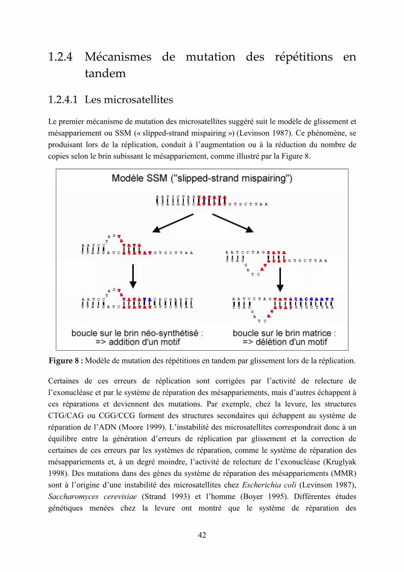
42
1.2.4 Mécanismes de mutation des répétitions en
tandem
1.2.4.1 Les microsatellites
Le premier mécanisme de mutation des microsatellites suggéré suit le modèle de glissement et
mésappariement ou SSM (« slipped-strand mispairing ») (Levinson 1987). Ce phénomène, se
produisant lors de la réplication, conduit à l’augmentation ou à la réduction du nombre de
copies selon le brin subissant le mésappariement, comme illustré par la Figure 8.
Figure 8 : Modèle de mutation des répétitions en tandem par glissement lors de la réplication.
Certaines de ces erreurs de réplication sont corrigées par l’activité de relecture de
l’exonucléase et par le système de réparation des mésappariements, mais d’autres échappent à
ces réparations et deviennent des mutations. Par exemple, chez la levure, les structures
CTG/CAG ou CGG/CCG forment des structures secondaires qui échappent au système de
réparation de l’ADN (Moore 1999). L’instabilité des microsatellites correspondrait donc à un
équilibre entre la génération d’erreurs de réplication par glissement et la correction de
certaines de ces erreurs par les systèmes de réparation, comme le système de réparation des
mésappariements et, à un degré moindre, l’activité de relecture de l’exonucléase (Kruglyak
1998). Des mutations dans des gènes du système de réparation des mésappariements (MMR)
sont à l’origine d’une instabilité des microsatellites chez Escherichia coli (Levinson 1987),
Saccharomyces cerevisiae (Strand 1993) et l’homme (Boyer 1995). Différentes études
génétiques menées chez la levure ont montré que le système de réparation des

43
mésappariements ne corrige efficacement que les boucles de 1 à 14 nucléotides mais pas
celles de plus de 16 nucléotides (Tran 1996 ; Sia 1997), ce qui démontre que les mécanismes
de mutation des répétitions en tandem ne peuvent se réduire au « SSM », du moins pour les
unités répétées de taille supérieure à 16 pb (minisatellites).
Des études menées sur différentes espèces ont montré que le taux de mutation des
microsatellites augmente avec leur nombre de répétitions, entre locus de longueur moyenne
différente (Weber 1990 ; Brinkmann 1998), mais également entre différents allèles du même
locus (Wierdl 1997 ; Schlotterer 1998 ; Ellegren 2000b). Pour expliquer cette observation, on
peut émettre l’hypothèse que le glissement lors de la réplication soit susceptible de survenir
plus fréquemment si le microsatellite compte davantage d’unités répétées. D’autre part,
lorsque des mutations ponctuelles surviennent dans les microsatellites, conduisant à une
répétition imparfaite, les locus sont moins variables (Petes 1997), ce qui correspondrait à un
taux de mutation par glissement de la réplication inférieur dans les répétitions en tandem
interrompues (Jin 1996). En effet, il semble que la présence de variations internes stabilise ces
structures car elle favorise la reconnaissance par le système de réparation des
mésappariements des régions où un glissement a eu lieu lors de la réplication (Strand 1993).
Le taux de mutation des microsatellites dépendrait donc de la taille de la plus grande
répétition non interrompue, et non pas du nombre de répétitions total, dégénérées ou non.
Des études structurales ont montré que les microsatellites (en particulier les triplets CNG)
simple-brin peuvent former des structures secondaires en épingle à cheveu (Mitas 1997 ;
McMurray 1999). En outre, lors de la réplication de l’ADN, des régions d’ADN simple-brin
ont plus de chances de se former lors de la synthèse du brin retardé que lors de la synthèse du
brin direct, ce qui peut générer des structures secondaires à l’origine d’erreurs de réplication.
En effet, l’ADN double-brin étant antiparallèle, l’un des deux brins (brin direct) est synthétisé
de façon continue au fur et à mesure que la fourche de réplication avance, mais l’autre
s’allonge dans le sens opposé à la progression de la fourche (brin retardé). Sur ce brin, des
petits fragments d’ADN, appelés fragments d’Okazaki, sont synthétisés à partir d’amorces
d’ARN, qui sont ensuite éliminées par la RNAse H au fur et à mesure de la réplication, puis
les fragments d’ADN sont reliés pour achever la synthèse. Pendant la réplication du brin
retardé, l’extension de l’amorce peut continuer alors que l’ARN initiateur est en cours de
destruction. Le déplacement d’une amorce en aval peut alors résulter en la formation d’une
structure « flap » (extrémité 5’ battante). Gordenin et collègues ont suggéré que la présence
d’une répétition en tandem formant une structure secondaire dans le flap pourrait conduire à
une expansion (Gordenin 1997). L’enzyme FEN1 chez l’homme (Li 1995 ; Bambara 1997),
homologue de Rad27 chez la levure, est impliquée dans la résolution de ces structures. Il
s’agit d’une exonucléase 5’-3’, qui est également nécessaire à l’élimination du dernier
ribonucléotide en 5’ des fragments d’Okazaki. Chez la levure, la fréquence
d’insertion/délétion dans les microsatellites augmente dans un contexte mutant pour Rad27
(Schweitzer 1998 ; Freudenreich 1998 ; Kokoska 1998).
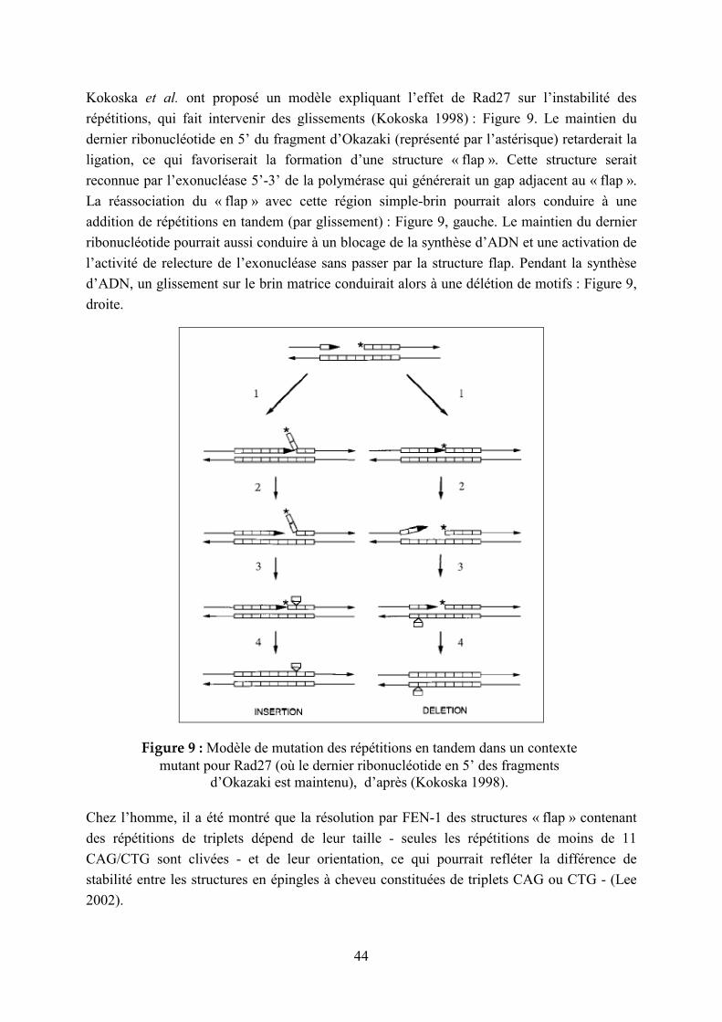
44
Kokoska et al. ont proposé un modèle expliquant l’effet de Rad27 sur l’instabilité des
répétitions, qui fait intervenir des glissements (Kokoska 1998) : Figure 9. Le maintien du
dernier ribonucléotide en 5’ du fragment d’Okazaki (représenté par l’astérisque) retarderait la
ligation, ce qui favoriserait la formation d’une structure « flap ». Cette structure serait
reconnue par l’exonucléase 5’-3’ de la polymérase qui générerait un gap adjacent au « flap ».
La réassociation du « flap » avec cette région simple-brin pourrait alors conduire à une
addition de répétitions en tandem (par glissement) : Figure 9, gauche. Le maintien du dernier
ribonucléotide pourrait aussi conduire à un blocage de la synthèse d’ADN et une activation de
l’activité de relecture de l’exonucléase sans passer par la structure flap. Pendant la synthèse
d’ADN, un glissement sur le brin matrice conduirait alors à une délétion de motifs : Figure 9,
droite.
Figure 9 : Modèle de mutation des répétitions en tandem dans un contexte mutant pour Rad27 (où le dernier ribonucléotide en 5’ des fragments
d’Okazaki est maintenu), d’après (Kokoska 1998).
Chez l’homme, il a été montré que la résolution par FEN-1 des structures « flap » contenant
des répétitions de triplets dépend de leur taille - seules les répétitions de moins de 11
CAG/CTG sont clivées - et de leur orientation, ce qui pourrait refléter la différence de
stabilité entre les structures en épingles à cheveu constituées de triplets CAG ou CTG - (Lee
2002).
45
Des mécanismes impliquant la recombinaison ont aussi été proposés pour la mutation des
microsatellites : des phénomènes de crossing-over inégal ou de conversion génique pourraient
faire varier la taille de ces structures (Brohede 1999 ; Jakupciak 2000 ; Parniewski 2002).
Cependant (d’après Ellegren 2000a), de plus en plus de preuves penchent en faveur du
mécanisme de glissement de la réplication par rapport aux phénomènes de recombinaison :
- Les allèles mutants sont en général non-recombinants au niveau des marqueurs
flanquants, ce qui correspondrait à des événements de mutation intra-alléliques (Morral
1991 ; Mahtani 1993).
- Le taux de mutation des microsatellites ne diffère pas entre les chromosomes sexuels et
les autosomes, ce qui suggère que les événements de mutation ne nécessitent pas le
contact entre chromosomes homologues (Kayser 2000 ; Heyer 1997).
- Le type de mutations rencontrées est cohérent avec le mécanisme de glissement de la
réplication : augmentation du taux de mutation avec la taille et l’homogénéité. De plus,
des études in vitro montrent que les microsatellites ont la capacité intrinsèque à subir ce
glissement (Schlotterer 1992).
- Chez la levure, la stabilité des microsatellites n’est pas affectée par des mutations dans les
gènes de recombinaison (Henderson 1992) mais elle est fortement réduite par des
mutations dans les gènes affectant le système de réparation des mésappariements (Strand
1993), ce qui est également le cas chez E. coli (Levinson 1987).
Des mécanismes de mutation des microsatellites impliquant simultanément le glissement et la
recombinaison sont également envisageables. En effet, dans certains cas, une instabilité plus
importante des microsatellites a été observée dans la lignée germinale (Cohen 1999 ; Seznec
2000). Lors de la recombinaison entre deux chromosomes homologues, la jonction de
Holiday, structure 4-brins, contient des régions d’ADN hétéroduplex (avec des
mésappariements). Ces régions subissent une correction dépendante de la réplication, qui fait
intervenir des cassures double-brin, et au cours de laquelle des phénomènes de glissement
peuvent survenir et générer de nouveaux allèles (Gendrel 2000 ; Richard 2000 ; Jankowski
2000).
Aux locus microsatellites, les insertions/délétions d’une seule unité sont les plus fréquentes
(Henderson 1992), mais pas les seules. La fréquence d’insertion/délétion de plus d’une unité,
en général de 2 à 5, varie selon les espèces ; elle est de 4 à 14% pour l’homme (Kayser 2000 ;
Brinkmann 1998 ; Xu 2000). De plus, les mutations survenant dans les microsatellites sont
biaisées vers une augmentation de taille (Amos 1996 ; Yamada 2002) : ce phénomène est
appelé « évolution directionnelle » (Amos 1996). Cependant, les microsatellites excèdent
rarement quelques dizaines de répétitions, ce qui laisse supposer l’existence de facteurs
empêchant leur croissance indéfinie:

46
- Plusieurs études ont montré que les délétions d’unités répétées étaient plus fréquentes ou
de plus grande taille plus le nombre de copies était grand (Wierdl 1997 ; Parniewski 2002
; Ellegren 2000b ; Harr 2000). La taille des microsatellites pourrait donc correspondre à
un équilibre où les taux d’expansion et de contraction seraient égaux (Xu 2000). Ces
observations nécessitent encore d’être généralisées aux différents types de répétitions et
espèces (Ellegren 2000a).
- Une autre hypothèse expliquant la limitation de l’accroissement de taille des microsatellites serait un équilibre entre le processus de mutation biaisée en faveur de l’accroissement de taille et les mutations ponctuelles qui tendent à faire diminuer cette taille en ralentissant les expansions ultérieures (Kruglyak 1998). Ce modèle pourrait expliquer les différences de taille des microsatellites entre organismes, qui dépendraient du taux de mutations ponctuelles et du taux de glissement réplicatif. On prédirait alors des répétitions plus courtes dans les génomes avec de faibles taux de glissement, ce qui est le cas pour Drosophila melanogaster (Schlotterer 1998). De façon intéressante, le taux de glissement ne serait pas faible parce que les répétitions sont courtes mais les répétitions seraient courtes à cause du faible taux de glissement (Ellegren 2000a).
- Enfin, la dernière hypothèse pourrait être une action de la sélection naturelle sur la taille des microsatellites. Les grands allèles pourraient être contre-sélectionnés, ce qui introduirait un plafond pour la longueur des répétitions (Garza 1995). La sélection naturelle pourrait aussi introduire des limites de taille supérieure et inférieure (Li 2000). Des pressions de sélection très différentes semblent par ailleurs agir sur les régions non-codantes en 5’ et en 3’ de gènes et les régions codantes, comme suggéré par l’étude de Morgante et al. (Morgante 2002) sur le génome d’A. thaliana.
1.2.4.2 Les minisatellites
Plusieurs catégories de minisatellites méritent d’être distinguées, en tout cas chez l’homme,
où ont été découverts des minisatellites dits hypermutables. Ces derniers ont la particularité
d’être extrêmement instables. Leur taux de mutation en lignée germinale dépasse 0,5%, seuil
de détection imposé par l’étude de pedigrees (pour revue, voir : Jeffreys 1999 ; Vergnaud
2000), et peut même atteindre jusqu’à 20%, pour le minisatellite CEB1 (Vergnaud 1991) en
méiose mâle (Buard 1998). Les minisatellites hypermutables n’ont pas été découverts dans
d’autres espèces à ce jour (Bois 2003), mais les minisatellites hypermutables humains ont été
largement étudiés, y compris dans des organismes modèles (c’est-à-dire la souris, et surtout la
levure Saccharomyces cerevisiae (Appelgren 1997 ; Debrauwère 1999; Paques 2001; Lopes
2002)), afin d’appréhender leurs mécanismes de mutation. Ils seront détaillés dans le
paragraphe suivant.
1.2.4.2.1 Minisatellites hypermutables
La première caractéristique des mutations survenant au niveau des minisatellites
hypermutables est qu’elles n’ont pas la même nature ni la même fréquence entre les lignées

47
somatique et germinale. Ces mutations sont beaucoup plus fréquentes dans les cellules
germinales que dans les cellules somatiques, comme montré par des études menées sur les
minisatellites MS31A, MS32 (Jeffreys 1994 ; Jeffreys 1997), MS205 (May 1996), CEB1
(Buard 2000) et B6-7 (Tamaki 1999). De plus, les mutations survenant dans les cellules
somatiques correspondent à des événements intra-alléliques relativement simples par
comparaison aux réarrangements complexes survenant dans les cellules germinales (Jeffreys
1997 ; Buard 2000). Ces observations suggèrent que la déstabilisation de ces minisatellites
s’opère par des voies différentes dans les lignées somatique et germinale. Il faut cependant
noter qu’un même mécanisme, comme la réparation d’une cassure double brin se produisant
entre chromatides sœurs, dans les cellules somatiques, ou entre chromosomes homologues,
dans les cellules germinales, pourrait parfaitement expliquer la création de ces deux types
d’allèles mutants.
1.2.4.2.1.1 Historique : les deux premiers modèles de mutation proposés
Figure 10 : Principe de la technique MVR-PCR.
En 1994, deux études majeures ont été menées sur les minisatellites humains CEB1 (Buard
1994) et MS32 (Jeffreys 1994), en mettant à profit la technique appelée « MVR-PCR »
(« minisatellite variant repeat -PCR» : voir Figure 10). Cette technique, développée par
Jeffreys et collègues (Jeffreys 1991), permet de typer les positions variantes dans les allèles de
minisatellites en utilisant des amorces spécifiques. En effet, tous les minisatellites
hypermutables caractérisés jusqu’à présent (leur liste figure dans la revue présentée au
chapitre 2.3.1 (Vergnaud 2000)) possèdent des variants internes, ce qui permet de caractériser
les mutations survenues.
Ces études d’allèles mutants dans les cellules germinales ont mis en évidence des événements
de recombinaison complexes, inter-alléliques (de type conversion génique) et intra-alléliques,

48
et ont conduit à l’hypothèse que ces mutations pourraient être initiées par des cassures double-
brin et impliquer un intermédiaire d’ADN hétéroduplex, suite à l’invasion d’un brin du
chromosome homologue : Figure 11.
Figure 11 : Deux mécanismes de mutation méiotique des minisatellites hypermutables proposés en 1994 par (Buard 1994) et (Jeffreys 1994).
La différence majeure entre ces deux modèles réside dans la localisation du point chaud de
cassure double-brin. Selon Jeffreys et collègues (Jeffreys 1994), il se situerait dans la
flanquante. Ils ont en effet observé, pour les minisatellites MS32, MS31A et MS205, que les
mutations surviennent plus fréquemment vers une extrémité de la répétition que vers l’autre,
ce qui laisse présager d’un effet de la flanquante sur l’instabilité des minisatellites. De plus,
une mutation ponctuelle dans la flanquante du minisatellite MS32 est associée à une forte
réduction de l’instabilité du minisatellite (Monckton 1994). Selon le modèle proposé par
Vergnaud et collègues (Buard 1994), la cassure double-brin surviendrait dans la répétition en
tandem elle-même, avec la possibilité d’un décalage entre les cassures des deux brins. Ce
détail était nécessaire pour rendre compte de la mise en évidence au locus CEB1
d’événements de conversion génique associés à la duplication de motifs de part et d’autre de
l’insertion (produit 3 dans la Figure 11). Ces deux modèles, même s’ils sont similaires du
point de vue des mécanismes (cassure double-brin, intermédiaire hétéroduplex),
correspondent à deux conceptions des minisatellites hypermutables : dans l’un des cas, la
séquence du minisatellite n’aurait pas de rôle sur l’instabilité, qui serait générée par la
séquence flanquante ou les caractéristiques de la chromatine environnante, et dans l’autre cas,
l’hypermutabilité serait liée à la structure du minisatellite.

49
1.2.4.2.1.2 Présentation du mécanisme de mutation proposé dans la revue
(Vergnaud & Denoeud, 2000)
Pendant mon DEA, j’ai participé à la rédaction de la revue intitulée « Minisatellites :
mutability and genome architecture » (Minisatellites : mutabilité et architecture des génomes)
(Vergnaud & Denoeud 2000), qui sera présentée plus en détail au chapitre 2.3.1, car elle
propose, outre une vue d’ensemble sur les minisatellites hypermutables humains, de nouveaux
résultats concernant la distribution des répétitions en tandem dans les génomes eucaryotes.
Figure 12 : Mécanisme de mutation méiotique des minisatellites hypermutables proposé en 2000, dans la revue (Vergnaud & Denoeud 2000).
Cette revue propose un modèle de mutation des minisatellites hypermutables qui prend en
compte toutes les observations faites jusque là. L’avancée essentielle par rapport à 1994
provient d’une étude menée sur le minisatellite humain CEB1 inséré dans l’organisme modèle

50
Saccharomyces cerevisiae. Cette étude a montré que l’instabilité méiotique dépendait du site
d’insertion (Debrauwère 1999) : le minisatellite est beaucoup plus instable lorsqu’il est inséré
à proximité d’un point chaud de cassures double-brin, et cette instabilité dépend des cassures
double-brin induites par l’enzyme Spo11. Le déplacement d’un minisatellite de levure (non
instable) à proximité d’un point chaud de cassures double-brin permet également de générer
une instabilité méiotique (Bishop 2000). Ces observations ont conduit au modèle dans lequel
une cassure double-brin générée dans la flanquante du minisatellite, comme initialement
proposé par Jeffreys et collègues, s’étend dans la répétition en tandem. Ensuite, l’invasion par
le chromosome homologue peut conduire à des phénomènes de conversion génique,
impliquant un intermédiaire hétéroduplex, comme cela a été mis en évidence dans certains
allèles mutants lors de l’étude de Debrauwère et al. (Debrauwère 1999). Ce modèle, présenté
sur la Figure 12, propose deux épisodes successifs d’invasion suite à la cassure double-brin :
par la chromatide sœur puis par le chromosome homologue. Plusieurs étapes d’invasion
pourraient expliquer les réorganisations extrêmement complexes observées fréquemment pour
le minisatellite B6.7, et qui ne pouvaient pas être expliquées par les modèles proposés en 1994
(Tamaki 1999). Le mécanisme faisant intervenir l’invasion par la chromatide sœur peut en
outre être invoqué en situation haploïde, par exemple pour le minisatellite MSY1, situé sur le
chromosome Y humain. L’instabilité de MSY1 est estimée à 5% dans les cellules germinales
(mâles, exclusivement, étant donné sa localisation sur le chromosome Y) (Vogt 1997). La
comparaison des allèles de ce minisatellite suggère un mécanisme de mutation provoquant la
diffusion des variants de façon linéaire, probablement par glissement de la réplication dans les
groupes de répétitions homogènes (Jobling 1998). On observe en outre un processus
d’homogénéisation des répétitions (une substitution dans un motif va se répercuter tout le long
de la répétition). Il pourrait être causé par la réparation biaisée des mésappariements dans de
l’ADN hétéroduplex généré par des mécanismes de glissement ou d’échange inégal entre
chromatides sœurs (Bouzekri 1998). La séquence de ce minisatellite, riche en AT, a tendance
à former des structures en épingle à cheveux et pourrait donc être impliquée directement dans
les phénomènes de mutation en cause.
Le modèle présenté sur la Figure 12 réconcilie les deux modèles initiaux de 1994 : il permet
de créer les événements de conversion génique avec duplications de motifs de part et d’autre,
tout en initiant la cassure dans la flanquante du minisatellite.
1.2.4.2.1.3 Evolutions récentes de la compréhension des mécanismes de mutation
Le modèle proposé dans la revue (Vergnaud & Denoeud 2000), présenté au paragraphe
précédent, reste toujours de mise pour expliquer les mécanismes de mutation méiotique des
minisatellites hypermutables humains. Cependant, d’autres avancées ont été faites depuis, en
particulier en ce qui concerne les mutations somatiques.
Il s’avère que des mutations dans les protéines Rad27 (enzyme impliquée dans la résolution
des structures « flap » générées sur le brin retardé lors de la synthèse des fragments
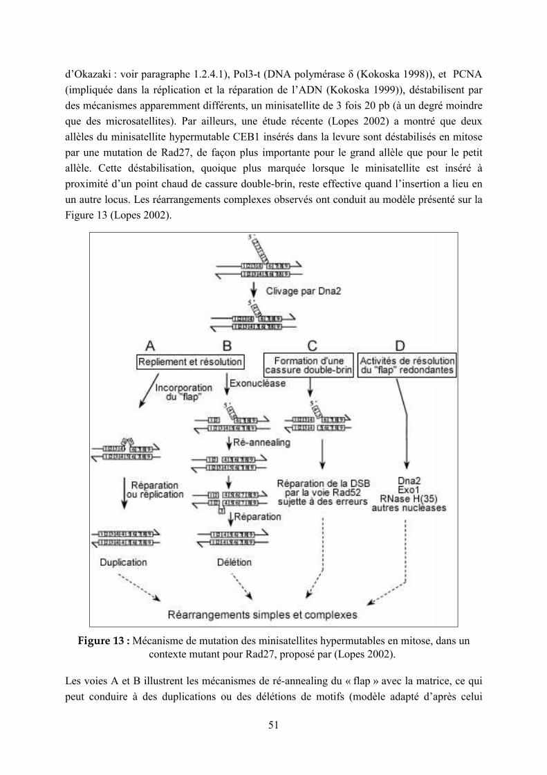
51
d’Okazaki : voir paragraphe 1.2.4.1), Pol3-t (DNA polymérase δ (Kokoska 1998)), et PCNA
(impliquée dans la réplication et la réparation de l’ADN (Kokoska 1999)), déstabilisent par
des mécanismes apparemment différents, un minisatellite de 3 fois 20 pb (à un degré moindre
que des microsatellites). Par ailleurs, une étude récente (Lopes 2002) a montré que deux
allèles du minisatellite hypermutable CEB1 insérés dans la levure sont déstabilisés en mitose
par une mutation de Rad27, de façon plus importante pour le grand allèle que pour le petit
allèle. Cette déstabilisation, quoique plus marquée lorsque le minisatellite est inséré à
proximité d’un point chaud de cassure double-brin, reste effective quand l’insertion a lieu en
un autre locus. Les réarrangements complexes observés ont conduit au modèle présenté sur la
Figure 13 (Lopes 2002).
Figure 13 : Mécanisme de mutation des minisatellites hypermutables en mitose, dans un contexte mutant pour Rad27, proposé par (Lopes 2002).
Les voies A et B illustrent les mécanismes de ré-annealing du « flap » avec la matrice, ce qui
peut conduire à des duplications ou des délétions de motifs (modèle adapté d’après celui
52
proposé par Kokoska et al. (Kokoska 1998) : voir Figure 9). La voie C montre une résolution
des structures flap conduisant à la formation d’une cassure double-brin mal réparée par les
mécanismes de réparation par recombinaison, comme proposé par Tishkoff et al. (Tishkoff
1997). La voie D correspond à une résolution de la structure « flap » par une des exonucléases
Dna2, Exo1 ou RNase H(35).
Ce modèle de mutation somatique est proche de celui de 1994 proposant une cassure dans le
minisatellite (Buard 1994). La séquence du minisatellite pourrait alors jouer un rôle direct sur
ce mécanisme de mutation : par exemple, la tendance à former des structures secondaires
pourrait influer sur la probabilité de suivre telle ou telle voie pour la réparation de la structure
« flap ». Un rôle de la séquence des différents allèles pourrait alors expliquer les résultats
disparates qui ont été observés aux locus de minisatellites humains, concernant la relation
entre la taille des allèles et leur instabilité : pour CEB1 (Buard 1998) et B6-7 (Tamaki 1999),
l’instabilité augmente avec la taille de la répétition en tandem, mais elle varie toutefois entre
différents allèles de même taille, tandis que pour MS32 et MS205, le taux de mutation est
indépendant de la taille des allèles (Jeffreys 1994) ; (May 1996). L’instabilité peut également
être influencée par la divergence entre les deux allèles du minisatellite : le taux de mutations
germinales du minisatellite MS205 intégré dans la levure est deux fois plus élevé chez un
hétérozygote (31-51 répétitions) que chez un homozygote pour le plus grand allèle (51-51).
Une hypothèse pour expliquer ce phénomène serait que le système de réparation des
mésappariements, détectant l’ADN hétéroduplex, induirait des cassures double-brin
secondaires, qui seraient alors réparées par une étape de recombinaison supplémentaire (He
2002).
Il apparaît donc que les minisatellites hypermutables insérés dans le génome de levure mutent par deux mécanismes, l’un en méiose, faisant intervenir l’initiation de la recombinaison homologue à proximité d’un minisatellite (Debrauwère 1999), l’autre en mitose, faisant intervenir une mauvaise maturation des fragments d’Okazaki au cours de la réplication (Lopes 2002) : étant donné que les mutations survenant en mitose chez la levure délétée pour Rad27 ont beaucoup de similitudes avec celles observées en méiose chez les minisatellites CEB1 et B6-7 chez l’homme (Buard 1998 ; Tamaki 1999), on peut imaginer qu’en plus des événements de recombinaison se produisant en prophase méiotique, des défauts spontanés de réplication pourraient aussi se produire lors de la gamétogène chez l’homme (Lopes 2002).
Les mécanismes de mutation des minisatellites chez les bactéries n’ont jusqu’à présent jamais été étudiés. En revanche, le mécanisme de mutation par glissement lors de la réplication s’applique aux répétitions en tandem bactériennes de la classe de taille des microsatellites : voir chapitre 1.2.4.1.
1.2.4.2.2 Minisatellites non hypermutables
Les minisatellites non-hypermutables, parfois polymorphes mais ayant un degré d’instabilité
faible (inférieur au seuil de détection imposé par les études de familles, de 0.5% de mutation),

53
ont été beaucoup moins étudiés du point de vue de leurs mécanismes de mutation. Pour la
plupart des minisatellites de ce type, on observe une distribution des longueurs totales
bimodale (minisatellites de l’insuline (Bell 1984) et D2S44 (Holmlund 1998)) ou tri-modale
(minisatellites D19S20 (Nakamura 1988) et MS51(D11S97) (Armour 1989)). Ce type de
distributions résulte probablement d’une fixation de certains allèles dans la population par la
dérive génétique, les mutations survenant dans ces allèles ne faisant ensuite varier leur taille
que de quelques unités. On peut également imaginer des processus de mutation biaisés vers
certaines tailles d’allèles ou même des phénomènes de sélection (Stead 2000).
Une étude menée par Stead et Jeffreys (Stead 2000) sur le minisatellite de l’insuline a permis
de distinguer deux modes de mutation dans les lignées germinales : le premier, se produisant à
un taux de 10-3, implique des insertions et délétions de 1-2 unités, survenant en majorité dans
des régions homogènes, ce qui suggère un mécanisme de glissement de la polymérase, comme
pour les microsatellites. Les auteurs émettent l’hypothèse que de tels événements surviennent
en mitose, même si on ne peut pas distinguer ces mutations d’événements de recombinaison
méiotique intra-alléliques. Le deuxième mode de mutation, qui survient à un taux de 2.10-5
environ, correspond à des réarrangements complexes, de type conversion génique, semblables
à ceux observés –de façon beaucoup plus fréquente- chez les minisatellites hypermutables. La
diversité des allèles du minisatellite de HRAS1 suggère que des événements de conversion
surviennent à ce locus relativement stable, mais à une fréquence inconnue (Ding 1999). Dans
ce minisatellite, les mutations d’allèles associés au cancer surviennent préférentiellement dans
trois régions : événements complexes de recombinaison inter-allélique dans une région,
simples événements de duplication/délétion dans une autre, et événements de différents types
dans la troisième région.
Il semble donc que chez ces minisatellites, comme chez les minisatellites hypermutables,
deux mécanismes de mutation soient impliqués. Le premier, survenant en mitose (dans la
lignée germinale ou dans la lignée somatique) impliquerait des glissements lors de la
réplication, et des phénomènes de maturation erronée des fragments d’Okazaki, à des taux
semblables entre les deux types de locus (Jeffreys 1997 ; Stead 2000). Le second, survenant
en méiose, impliquerait des réarrangements complexes de type conversion génique, initiés par
des cassures double-brin . Ce deuxième type d’événement serait rare chez les minisatellites
« ordinaires » mais très fréquent chez les minisatellites hypermutables. Dans la revue que
nous présenterons au chapitre 2.3.1, nous émettons donc l’hypothèse que les minisatellites
seraient hypermutables lorsqu’ils se trouvent à proximité d’un point chaud de cassures
double-brin (Vergnaud & Denoeud 2000). Ensuite, si une mutation survient dans la
flanquante et affecte les propriétés de recombinaison à ce locus (une transversion G->C en
amont de MS32 a en effet été associée à une diminution de l’instabilité de ce minisatellite
(Monckton 1994)), la dérive génétique (Jeffreys 2002) peut conduire à l’extinction du
minisatellite hypermutable, qui redeviendra alors un minisatellite ordinaire. Le site de cassure
double-brin pourrait également provenir de l’arrangement de la chromatine et non de la
54
séquence elle-même : l’extinction de l’hypermutabilité proviendrait alors de mécanismes de
réarrangement du génome à plus grande échelle. Pour les minisatellites non-hypermutables,
les rares mutations survenant dans la lignée germinale pourraient résulter de cassures double-
brin aléatoires survenant en méiose ou d’événements de type somatique, survenant lors des
divisions mitotiques préalables.
1.2.4.3 Distinction entre microsatellites et minisatellites
Les études menées sur les mécanismes de mutation des répétitions en tandem montrent que
les microsatellites comme les minisatellites sont déstabilisés en mitose, et en méiose (à une
fréquence très importante pour les minisatellites hypermutables). Les mécanismes en cause
font intervenir le glissement de la polymérase et la résolution des structures « flaps » générées
par les fragments d’Okazaki sur le brin retardé lors de la réplication et/ou la réparation de
cassures double-brin survenues dans ou à proximité de la répétition en tandem. La distinction
entre leurs modes de mutation est plus quantitative (fréquences relatives des différents
événements) que qualitative. Elle sera rendue plus aisée lorsque certains de ces mécanismes,
encore très énigmatiques, pourront être élucidés.
Des distinctions majeures peuvent toutefois être déduites de l’analyse des mutations survenant
dans les microsatellites et les minisatellites (les répétitions en tandem présentant l’intérêt de
conserver parfois la trace d’anomalies du métabolisme de l’ADN, et de reconstituer a
posteriori la séquence des événements, dans des systèmes biologiques peu propices à des
investigations directes) :
- L’homogénéité interne des microsatellites est supérieure à celle des minisatellites, ce qui
est cohérent avec un mécanisme de mutation impliquant le glissement de la polymérase
pour les premiers, et un mécanisme compatible avec l’hétérogénéité des motifs, comme la
réparation de cassures double-brins, pour les deuxièmes.
- Des mutations dans le système de réparation des mésappariements déstabilisent les
microsatellites mais pas les minisatellites (sauf MS1, minisatellite d’unité répétée de
« type » microsatellite : 9 pb, décrit plus bas (Berg 2003)). Cette observation peut
s’expliquer par le fait que les boucles générées lors de glissements ne sont corrigées par ce
système que lorsque leur taille est inférieure à 16 paires de bases (Sia 1997).
- Des mutations dans les enzymes permettant la résolution des structures « flap » générées
par les fragments d’Okazaki déstabilisent les microsatellites et les minisatellites, mais à
des degrés très différents (Kokoska 1998) : les minisatellites y sont beaucoup moins
sensibles, ce qui pourrait être lié à la taille des fragments d’Okazaki (100 à 150 paires de
bases (MacNeill 2001)). Le fait que plusieurs unités répétées soient contenues sur un
fragment d’Okazaki pourrait en effet augmenter la probabilité d’événements de mutation
(Monckton 1995).

55
Les caractéristiques structurales des répétitions en tandem semblent donc moduler
l’importance relative des différents mécanismes de mutation à l’œuvre, et distinguent donc la
classe des microsatellites de celle des minisatellites. Cependant, certaines répétitions en
tandem se situent à la « frontière » entre ces deux classes, ce qui rend difficile l’établissement
d’une définition stricte. Deux exemples en sont présentés ci-après :
- Le minisatellite hypermutable EPM1, situé en 5’ du gène de la cystatine B, et dont
l’amplification est responsable de l’épilepsie myoclonique progressive de type 1 (voir
Tableau 8), a un motif répété de 12 paires de bases. Ce locus cause la maladie par un
phénomène d’amplification semblable à ceux qui surviennent pour les maladies
neurodégénératives causées par des expansions de triplets (voir Tableau 7). Les
phénomènes de mutations correspondent en majorité à des expansions/contractions d’une
seule unité (Lalioti 1997). Larson et al. ont émis l’hypothèse que l’instabilité,
apparemment germinale, extrême des allèles amplifiés (taux de mutation de 0.47) pourrait
être causée par des erreurs de réplication dans une région de la répétition contenant 100%
de GC sur une longueur considérable (600-800 pb). Le taux de mutation légèrement plus
faible des allèles pré-mutateurs résulterait alors de leur longueur plus faible (Larson
1999).
- Le minisatellite hypermutable MS1 (D1S7) a une unité répétée de 9 paires de bases
seulement : son nombre de copies varie de 60 à plus de 1000, avec un taux
d’hétérozygotie (relatif au polymorphisme de taille des allèles) supérieur à 99% (Wong
1987 ; Royle 1988). Son taux de mutation en lignée germinale a été estimé à 5.2%
(Jeffreys 1988). Le grand nombre d’unités répétées et l’instabilité extrême de cette
répétition en tandem la classent dans la catégorie des minisatellites plutôt que des
microsatellites, même si son motif répété est court. Cependant, il a certaines particularités
qui le rapprochent plus des microsatellites que des minisatellites. En particulier, c’est le
seul minisatellite connu qui soit déstabilisé dans des cellules de cancer du colon montrant
une instabilité des microsatellites (Berg 2003), ce qui suggère que la réplication ou les
erreurs de réparation peuvent contribuer à son instabilité. De plus, inséré dans la levure, ce
minisatellite mute à haute fréquence en méiose comme en mitose, ce qui n’est pas le cas
d’autres minisatellites étudiés dans le même contexte, pour lesquels un fort taux de
mutation n’est observé qu’en méiose (Appelgren 1997 ; He 1999). En mitose, l’instabilité
de MS1 est dépendante de la taille des allèles (seuil d’instabilité à 750 pb) (Maleki 1997),
et de la structure interne des allèles. En méiose, l’instabilité correspond principalement à
des événements de type conversion génique, intra- ou inter-allélique, comme pour les
autres minisatellites hypermutables (Berg 2000). Récemment, des études ont été menées
non plus dans le modèle levure mais chez l’homme (Berg 2003). Les événements
méiotiques intra-alléliques y sont plus fréquents que les événements inter-alléliques, et un
autre phénomène de mutation a lieu : il s’agit de grandes délétions qui surviennent dans de
longues régions homogènes, constituées d’au moins 12 unités du type « C » (108 pb), ce
qui est similaire au seuil d’instabilité des triplets CGG du microsatellite associé au
56
syndrome de l’X fragile (Eichler 1994) (34-38 répétitions soit 102-114 pb). De plus, on
observe une stabilisation de la répétition lorsque l’homogénéité est interrompue, ce qui
rappelle également les mécanismes de mutation de microsatellites. Ce phénomène n’ayant
pas été mis en évidence dans les lignées somatiques, il semble peu probable qu’il soit dû à
un glissement survenant lors de la réplication mais plutôt lors de la recombinaison
méiotique (Berg 2003). Certains mécanismes interprétés comme des glissements
réplicatifs pourraient en fait correspondre à des recombinaisons entre chromatides sœurs :
il est possible que jusqu’à présent, le phénomène de glissement lors de la réplication ait
été surestimé au détriment de mécanismes plus complexes. Cependant, ces événements
complexes se produisent bel et bien à certains locus, et ils mériteraient donc d’être
invoqués également dans les cas « simples ».
Enfin, en plus des différences entre microsatellites et minisatellites inhérentes à leurs
mécanismes de mutation, ces deux types de répétitions en tandem peuvent être distingués par
leur distribution chromosomique, du moins dans le génome humain, comme nous le montrons
dans la revue présentée au chapitre 2.3.1 (Vergnaud & Denoeud 2000). En effet, parmi
l’ensemble des répétitions en tandem du chromosome 22 humain, il apparaît que la transition
entre une répartition homogène et une répartition biaisée vers les télomères correspond à une
unité répétée de 17 pb, taille des boucles non réparables par le système de réparation des
mésappariements (Sia 1997), ou à une longueur totale de 120-140 pb, seuil similaire à celui
décrit plus haut pour l’instabilité des triplets. Ainsi, il semble que les mécanismes de création
et/ou de maintien (pression de sélection…) des microsatellites et des minisatellites dans le
génome humain soient distincts, comme le reflète leur différence de distribution
chromosomique, mais ces phénomènes restent pour l’instant énigmatiques.
Les minisatellites étaient définis usuellement comme des structures de longueur totale de
l’ordre du kilobase, ce seuil étant imposé par les contraintes expérimentales qui ont
accompagné leur première caractérisation (Southern Blot). Nous proposons d’élargir cette
définition (nous le verrons dans la revue présentée au paragraphe 2.3.1 (Vergnaud & Denoeud
2000)) à des répétitions en tandem de longueur totale supérieure à 140 pb environ et/ou
d’unité répétée supérieure à 15 pb environ. Des structures d’unité répété plus petite (entre 6 et
15 pb) pourront être considérées comme des minisatellites si elles sont très étendues : c’est le
cas des minisatellites MS1 et EPM1, mais nous avons vu que certaines de leurs
caractéristiques restent similaires à celles de microsatellites.
1.2.5 Origine des répétitions en tandem
Etant donné que les répétitions en tandem sont retrouvées chez toutes les espèces, eucaryotes
comme procaryotes, analysées jusqu’à présent, et qu’il n’existe pas d’homologie entre les
répétitions en tandem de différentes espèces, sauf très proches (Taylor 1999), la formation de

57
nouvelles répétitions en tandem doit être un événement survenant fréquemment dans les
génomes.
Le premier modèle de génération des répétitions en tandem, proposé par Levinson et Gutman
(Levinson 1987), implique la formation de petites répétitions en tandem survenant par hasard
suite à des mutations ponctuelles, qui seraient ensuite amplifiées par glissement lors de la
réplication. Cependant, même si ce modèle est plausible pour de petites répétitions comme les
microsatellites (par exemple une étude chez les primates a montré que deux répétitions en
tandem de motif de 2pb et 4pb avaient pu être générées par des mutations aléatoires (Messier
1996)), il n’est pas envisageable pour des répétitions en tandem de motifs plus grands
(minisatellites). De plus, une distinction entre les mécanismes générant les microsatellites et
les minisatellites pourrait expliquer les différentes distributions chromosomiques observées
pour ces deux types de répétitions en tandem.
Pour certains minisatellites, on observe que la région répétée est flanquée de part et d’autre
par quelques (5 à 10) nucléotides identiques, c’est-à-dire que la répétition se termine par un
motif incomplet de quelques paires de bases : un exemple en est présenté sur la Figure 14A.
Ce type de structure a été décrit chez la levure S. cerevisiae (Haber 1998) ainsi que d’autres
champignons (Giraud 1998), l’homme (Haber 1998 ; Murray 1999), les oiseaux (Gyllensten
1989), le saumon (Goodier 1998) et les mitochondries de divers organismes (Taylor 2000).
Ces observations ont conduit à l’hypothèse selon laquelle des répétitions non-contiguës
seraient à l’origine d’une duplication de la région comprise entre ces deux répétitions, par
glissement lors de la réplication (un modèle est proposé par Taylor et al. : Taylor 2000), ou
par crossing-over inégal (Haber 1998). Cette répétition pourrait ensuite être amplifiée par les
mécanismes susceptibles de faire évoluer les répétitions en tandem que nous avons vus
précédemment, tels que le glissement réplicatif ou la conversion génique (Haber 1998 ;
Taylor 2000) : voir Figure 14B. Selon le temps écoulé depuis la création du minisatellite,
l’homologie entre les petites séquences flanquant la répétition en tandem pourra avoir été
perdue ou conservée (Haber 1998). Ce modèle nécessite la présence fortuite de répétitions
non adjacentes, qui ont plus de chances de survenir dans des génomes de faible complexité, à
fort biais de composition (Achaz 2002).
Cependant, de manière plus remarquable, de nombreux minisatellites comptent un nombre
entier de motifs. Le modèle proposé dans la Figure 14 n’explique pas la formation de telles
répétitions. L’hypothèse proposant que de courtes répétitions non adjacentes, survenant par
hasard, seraient à l’origine de la formation des minisatellites reste plausible. Mais pour tenir
compte des minisatellites « parfaits » il faudrait un modèle selon lequel ces petites répétitions
seraient ensuite « avalées » dans la répétition en tandem. Jusqu’à présent, peu d’attention a été
portée au problème de l’origine des minisatellites, et aucun modèle satisfaisant n’a pu être
proposé : les mécanismes en cause restent encore très énigmatiques.

58
Figure 14 : Une hypothèse sur l’origine des minisatellites.
1.3 Identification des répétitions en tandem dans les génomes
1.3.1 La bioinformatique
La bioinformatique est née lorsque, suite à l’essor des techniques de séquençage, deux
nécessités sont apparues pour la recherche en biologie : le stockage de séquences dans des
bases de données informatiques, et l’analyse de ces séquences (nucléiques ou protéiques). La
bioinformatique correspond à un domaine interdisciplinaire et peut être définie comme
« l’application de l’informatique pour l’acquisition, la gestion, et l’analyse de données
biologiques » (Brown 2003). La plupart des algorithmes qui sont à la base des outils
bioinformatiques actuels sont nés dans les années 70-80, et proviennent en fait de recherches
théoriques :
- 1970 : algorithme de Needleman et Wunsch pour la comparaison de séquences
(alignement global) (Needleman 1970).
- 1978 : algorithme de Chou et Fasman pour la prédiction de structures secondaires de
protéines à partir de leur séquence d’acides aminés (Chou 1978).
- 1979 : package Staden pour l’assemblage de fragments d’ADN chevauchants provenant
de projets de séquençage (Staden 1979 ; Staden 1980).
- 1981 : algorithme de Smith et Waterman pour l’alignement de séquences (alignement
local) (Smith 1981).

59
- 1982 : profils d’hydrophobicité des protéines par Kyte et Doolittle (Kyte 1982).
- 1987 : alignements multiples de séquences par Feng et Doolittle (Feng 1987).
- 1987 : méthode du « neighbor joining » pour la reconstruction phylogénétique par Saitou
et Nei (Saitou 1987).
- 1988 : Pearson et Lipman créent FASTA, logiciel de recherche de similitudes entre une
séquence et une base de séquences (Pearson 1988).
- 1990 : Altschul et collègues implémentent BLAST au NCBI, autre méthode de
comparaison entre une séquence et une base de séquences (Altschul 1990 ; Altschul
1994). Cette méthode sera détaillée au paragraphe 2.1.1.3.
Le terme « bioinformatics » a été indexé dans les mots-clés ou titres de PubMed/Medline
[http://www.ncbi.nlm.nih.gov/PubMed/] à partir de 1993. Depuis, le nombre d’occurrences de
ce terme comme du terme « computational biology » n’a cessé de croître (Brown 2003).
Ces dernières années, le nombre de génomes séquencés a augmenté de façon spectaculaire, en
particulier chez les bactéries (voir chapitre 1.1.1.1), ce qui a été accompagné de progrès dans
les domaines de la génomique, de la protéomique mais aussi de la bioinformatique :
raffinements aux algorithmes de recherche d’homologie dans les bases de données, prédiction
de structures protéiques secondaires et tertiaires, logiciels de recherche de motifs protéiques
ou nucléiques, analyse des séquences répétées (Heringa 1998). En particulier, comme nous le
détaillons au paragraphe suivant, différents outils permettant l’identification de répétitions en
tandem dans les séquences ont été élaborés.
1.3.2 Logiciels d’identification de répétitions en tandem
Jusqu’à ces dernières années, les répétitions en tandem étaient identifiées de façon
expérimentale (hybridation de sondes sur des Southern Blots…) mais dorénavant la
disponibilité de données de séquence permet leur identification systématique, in silico, dans
les génomes. Lorsque mon DEA a commencé, en 1999, les outils disponibles étaient peu
nombreux : la plupart permettaient la recherche de répétitions dans les séquence protéiques,
de façon plus ou moins efficace (Heringa 1998 ; Pellegrini 1999). Les programmes détectant
les répétitions en tandem nucléiques avaient certaines lacunes : certains, comme le
programme « REPuter » (Kurtz 1999), ne détectaient que les répétitions en tandem
strictement conservées. Ce type d’outils a une utilité pour identifier les microsatellites, mais
devient inapproprié pour les minisatellites, très rarement strictement conservés. D’autres
permettaient la détection de répétitions en tandem dégénérées mais seulement dans la plage de
taille des microsatellites (par exemple sputnik : http://espressosoftware.com:8080/
esd/pages/sputnik.jsp, créé par Abajian en 1994), ou nécessitaient de préciser certains
paramètres tels que la longueur du motif répété recherché et son degré de conservation
(Korotkov 1997 ; Sagot 1998), et/ou requéraient des temps de calcul très longs, ce qui rendait
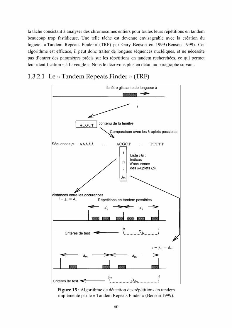
60
la tâche consistant à analyser des chromosomes entiers pour toutes leurs répétitions en tandem
beaucoup trop fastidieuse. Une telle tâche est devenue envisageable avec la création du
logiciel « Tandem Repeats Finder » (TRF) par Gary Benson en 1999 (Benson 1999). Cet
algorithme est efficace, il peut donc traiter de longues séquences nucléiques, et ne nécessite
pas d’entrer des paramètres précis sur les répétitions en tandem recherchées, ce qui permet
leur identification « à l’aveugle ». Nous le décrivons plus en détail au paragraphe suivant.
1.3.2.1 Le « Tandem Repeats Finder » (TRF)
Figure 15 : Algorithme de détection des répétitions en tandem
implémenté par le « Tandem Repeats Finder » (Benson 1999).

61
L’algorithme utilisé est de type probabiliste, heuristique, c’est-à-dire qu’il n’est pas garanti de
trouver toutes les répétitions en tandem existantes, mais il a l’avantage, par rapport aux
algorithmes exacts, de ne pas nécessiter un temps de calcul trop long ou de rentrer des
paramètres trop précis sur les répétitions en tandem recherchées. La recherche repose sur le
fait que dans une répétition en tandem, une même base, ou un groupe de k bases successives
(k-uplet), sera retrouvé à intervalles réguliers (distance d), sauf mésappariements. La Figure
15 présente les différentes étapes pour la détection des répétitions en tandem. Tout d’abord, la
séquence est parcourue pour détecter les positions i de tous les k-uplets (où k =5 par exemple)
possibles (4k) : cette liste sera notée Hp (ensemble des positions où se trouve le k-uplet p).
Lorsqu’une position i est ajoutée à la liste Hp des occurrences d’un k-uplet donné (p), toutes
les occurrences de p déjà identifiées sont recherchées : si une occurrence a eu lieu à la
position j, la distance d = i-j est la taille possible d’un motif répété en tandem. Dans ce cas,
d’autres k-uplets seront trouvés au voisinage, éloignés de la même distance d. La liste Dd
contient l’ensemble des distances d survenant entre des k-uplets dont l’une des occurrences se
situe entre les positions j et i. Ces listes de distances sont alors testées selon un critère
statistique, basé sur la loi de Bernoulli, qui dépend de la taille du motif, de la probabilité de
match, de la probabilité d’insertion/délétion, et de la taille du n-uplet. Si le test est favorable,
un motif candidat allant des positions j+1 à i est alors aligné avec la séquence environnante :
si au moins deux copies du motif s’alignent avec la séquence, la répétition en tandem est
mentionnée. Un motif consensus est ensuite généré, correspondant au motif s’alignant le
mieux avec toutes les unités répétées, et l’alignement des unités avec ce motif consensus est
proposé en sortie. La taille de la période (« period size ») correspond à la distance entre les
caractères matchant et peut être différente de la taille du consensus.
Ce logiciel présente l’inconvénient de générer des sorties redondantes (pourtant limitées à
trois par répétition en tandem détectée) : par exemple, une même plage peut être proposée
avec un motif répété de 24 et un motif de 48 pb. D’autre part, certaines plages de répétitions
en tandem se chevauchent, ou certaines répétitions en tandem peuvent être segmentées si elles
contiennent une portion mal conservée. Cet inconvénient ne constitue cependant pas une vraie
limite, étant donné que le choix de la « meilleure » répétition en tandem n’est pas évident, et
nécessite donc dans la plupart des cas l’intervention de l’utilisateur (en particulier en ce qui
concerne la détermination exacte des bornes des répétitions en tandem). De plus, comme nous
le verrons dans le paragraphe suivant, les différents autres logiciels proposés depuis le TRF ne
s’avèrent pas meilleurs. Le TRF, logiciel d’utilisation conviviale, accessible à l’adresse
[http://tandem.biomath.mssm.edu/trf/trf.html] reste donc un outil de référence pour la
détection de répétitions en tandem dans les génomes (Yeramian 1999) : par exemple, il a été
utilisé lors de l’analyse de la séquence brouillon du génome humain par le consortium public
(Lander 2001).

62
1.3.2.2 Autres outils de détection des répétitions en tandem
Depuis 1999, d’autres outils de recherche et de manipulation des répétitions en tandem ont été
élaborés et des outils existants ont été améliorés:
- Le programme REPuter, basé sur des arbres de suffixes, a été amélioré pour permettre la
détection de répétitions approximatives. Cependant, il ne fournit qu’une vue globale des
répétitions mais pas leurs séquences détaillées (Kurtz 2000) [http://www.genomes.de].
- Le package EMBOSS (Rice 2000) propose des logiciels de détection des répétitions en
tandem : etandem et equicktandem, qui ont l’intérêt de pouvoir traiter des séquences
nucléiques comme protéiques. L’interface est toutefois peu conviviale, et nécessite
d’entrer un seuil de score peu intuitif. De plus, la sortie par défaut ne propose pas la
séquence des répétitions [http://www.hgmp.mrc.ac.uk/Software/EMBOSS/Apps/
equicktandem.html].
- Une méthode basée sur des codes couleurs attribués aux différentes bases permet une
identification visuelle des répétitions en tandem (Yoshida 2000).
- Un algorithme, accessible via une interface web [http://csweb.cs.haifa.ac.il/library/
appro_try1.html], permet de rechercher des répétitions en tandem approximatives (Landau
2001). Ce programme présente l’inconvénient de nécessiter que l’utilisateur entre le
nombre de mésappariements maximal dans les répétitions : pour des taux de
mésappariement égaux, ce nombre variera en même temps que la taille du motif répété.
Pour faire des recherches exhaustives, il sera donc nécessaire de lancer plusieurs
exécutions avec différentes valeurs du paramètre (l’augmentation de cette valeur
ralentissant notablement l’exécution).
- Un programme de détection et de classification des répétitions (dont les répétitions en
tandem) dans les génomes, basé sur les arbres de suffixes a été mis au point par Volfovsky
et al. (Volfovsky 2001), mais il ne dispose pas d’une interface web.
- Le logiciel TROLL identifie de façon très efficace les petites répétitions en tandem
correspondant à un « dictionnaire » de répétitions connues (Castelo 2002).
- En 2002, Hauth et al. ont proposé un algorithme basé sur l’« extension de graines » pour
détecter les répétitions en tandem « classiques » mais aussi de deux autres types, les
« VLTRs » (variable length tandem repeats) et les « MPTRs » (multi-period tandem
repeats) : voir Figure 16. Ces répétitions en tandem étaient également identifiées par le
TRF par exemple, lors de sorties redondantes, mais elles n’étaient pas spécifiées comme
telles (Hauth 2002).

63
Figure 16 : Description de deux « nouveaux » types de répétitions en tandem,
les VLTR et les MPTR (Hauth 2002).
- Le programme « MREPS », accessible à l’adresse [http://bioweb.pasteur.fr/seqanal/
interfaces/mreps.html], possède une interface conviviale et permet une identification
efficace des répétitions en tandem (Kolpakov 2003). Les résultats obtenus ne sont
toutefois pas meilleurs que ceux provenant du TRF, comme nous le verrons dans le
paragraphe suivant. De plus, ce logiciel requiert que l’utilisateur saisisse un paramètre de
« résolution » peu intuitif.
- Le logiciel TRAP améliore l’assemblage des répétitions en tandem (Tammi 2003), une
des principales sources d’erreurs lors du séquençage des génomes.
1.3.2.3 Comparaison entre les logiciels de détection des répétitions en tandem disponibles sur le web
Pour différentes répétitions en tandem, les sorties des logiciels accessibles sur Internet : TRF
(Benson 1999) [http://tandem.biomath.mssm.edu/trf/trf.html], MREPS (Kolpakov 2003)
[http://bioweb.pasteur.fr/seqanal/interfaces/mreps.html], et « A Program for Approximate
Tandem Repeat » (Landau 2001) [http://csweb.cs.haifa.ac.il/library/appro_try1.html], ont été
comparées. Les résultats sont présentés sur la Figure 17.
Le microsatellite, D21S11 a une structure répétée irrégulière : les logiciels TRF et MREPS
ont pu la mettre en évidence (31 x 4 pb dégénérées). Toutefois, le logiciel MREPS propose de
nombreuses autres répétitions en tandem dans cette plage, alors que seul ce microsatellite est
proposé par le TRF. On note en outre que pour le logiciel TRF, cette répétition en tandem a
été détectée avec tous les paramètres proposés, tandis que pour MREPS, le paramètre par
défaut, ne permettant pas de détecter des répétitions en tandem dégénérées, ne l’a évidemment
pas détectée : il faut utiliser un paramètre de « résolution » de 5 ou plus. Le logiciel « A

64
Program for Approximate Tandem Repeats » ne détecte pas la séquence entière du
microsatellite : elle est fragmentée en zones bien conservées. Même une augmentation du
nombre de mismatchs autorisés n’améliore pas sa détection. En outre, ce microsatellite n’est
même pas partiellement détecté par les paramètres par défaut, qui n’autorisent la détection
que d’unités répétées de 6 paires de bases ou plus.
Ces différents logiciels ont également été testés sur le minisatellite CEB268 du chromosome
21 humain, qui est relativement mal conservé (74% de conservation moyenne entre les unités
répétées et le motif consensus) (Denoeud 2003). Le TRF détecte des motifs redondants,
différents selon les paramètres choisis : la répétition en tandem considérée comme la plus
représentative de la région (10,2 x 54 pb) n’est détectée dans sa totalité qu’avec les
paramètres les moins restrictifs (2,3,5 : match,mismatch,indel). Le logiciel MREPS ne détecte
cette répétition, de façon un peu décalée par rapport au TRF (les bornes des répétitions en
tandem sont en effet souvent difficiles à définir), qu’avec un paramètre de résolution
maximal : 140 (la valeur de 145 est qualifiée de trop élevée pour la séquence à traiter). Le
logiciel « A Program for Approximate Tandem Repeats » produit une erreur : le site
mentionne qu’il accepte les séquences jusqu’à 1024 pb, mais il semble que ce minisatellite
soit trop complexe, ce qui génère un temps de calcul trop long. Le logiciel « A Program for
Approximate Tandem Repeats » n’est clairement pas adapté pour traiter des longues
séquences : les seuls outils vraiment efficaces sont TRF et MREPS.
Un avantage du TRF est que les paramètres sont pré-définis, et seuls 4 choix sont possibles.
En revanche, pour MREPS (comme pour le troisième logiciel), le paramètre permettant de
jouer sur l’homogénéité des répétitions, ou paramètre de « résolution », est libre, et
l’utilisateur ne peut pas savoir quelles valeurs pertinentes lui donner (en particulier en ce qui
concerne le seuil maximal). Pour le TRF, la restriction du nombre de paramètres accessibles
pourrait être déplorée car elle reflète des choix faits par l’algorithme lui-même, que
l’utilisateur ne peut pas contrôler et difficilement comprendre. Cependant, pour l’utilisation
que nous aurons du TRF (identification rapide de répétitions en tandem dans des génomes
complets), il est essentiel de pouvoir effectuer une seule exécution du programme par
séquence à traiter, même si l’identification systématique des répétitions en tandem n’est pas
garantie. Du point de vue de la facilité d’utilisation, qui ne requiert pas trop de mises au point
pour définir les meilleurs paramètres, comme de la présentation des résultats, qui propose
l’alignement des répétitions avec le consensus, le TRF reste le meilleur outil disponible à
l’heure actuelle.

65
Figure 17 : Comparaison de programmes de détection de répétitions en tandem accessibles sur Internet.
66
1.3.3 Identification des répétitions en tandem polymorphes
Nous avons vu que des logiciels permettaient la détection de répétitions en tandem dans les
séquences. Cependant, les répétitions en tandem d’intérêt sont les répétitions polymorphes,
soit pour l’épidémiologie bactérienne, soit pour l’étude de pathologies humaines associées à
ces polymorphismes, ou encore si on recherche des minisatellites hypermutables, en premier
lieu extrêmement polymorphes. Pour l’instant, il n’existe pas d’outil « miracle » permettant de
détecter les répétitions en tandem polymorphes. Cependant, quelques pistes peuvent être
suivies, en particulier pour les microsatellites.
1.3.3.1 Microsatellites
Comme nous l’avons vu au paragraphe 1.2.4.1, le mode de mutation des microsatellites par
glissement de la réplication dépend de la longueur et de la conservation de ces répétitions en
tandem (Weber 1990). Il est donc probable que les microsatellites les plus polymorphes seront
les mieux conservés, et les plus longs. Une étude menée par Wren et al. a en effet montré
qu’une requête sélectionnant des microsatellites très conservés fournissait une grande
proportion de locus polymorphes (Wren 2000).
Cependant, d’une part, le glissement de la réplication n’est pas le seul mécanisme en cause
dans la mutation des microsatellites, et d’autre part, si des microsatellites autrefois instables
car bien conservés sont devenus plus stables, par exemple suite à des mutations ponctuelles
interrompant leur homogénéité, plusieurs allèles pourront subsister dans la population : ces
locus « inactifs » mais pourtant polymorphes, risquent de ne pas être détectés par ces critères
de longueur et de conservation, ce qui, selon le but recherché, peut être problématique. En
effet, si on cherche des locus instables pour étudier les modes de mutation, ce type de locus ne
sera pas intéressant. En revanche, si on recherche des locus polymorphes pour l’identification
de souches bactériennes et la phylogénie, les bons marqueurs épidémiologiques devront au
contraire être stables de génération en génération, et refléter le plus possible de « vrais
événements » évolutifs, auquel cas les locus inactifs seront privilégiés.
1.3.3.2 Minisatellites
Pour les minisatellites, la tâche visant à déterminer le polymorphisme à partir de la séquence
est encore plus ardue. En effet, les mécanismes de mutation semblent peu influencés par les
caractéristiques de séquence (Buard 1998), et aucune caractéristique commune aux séquences
de minisatellites hypermutables n’a pu être mise en évidence (Murray 1999). Cependant,
jusqu’à présent, l’analyse des séquences de répétitions en tandem ne s’est intéressée qu’à des
critères simples (longueur, composition…) et pas à l’organisation des mutations dans les
motifs répétés. Ce type d’information mériterait toutefois d’être utilisé : par exemple, une
répétition « active » aura probablement une plus grande proportion de mutations communes à
67
plusieurs motifs qu’une répétition inactive, dans laquelle des mutations ponctuelles auront pu
survenir sans être distribuées dans l’ensemble de la répétition (mutations uniques, spécifiques
d’une unité particulière). Par ailleurs, la séquence des allèles de minisatellites reflète les
mécanismes de mutation ayant encore cours, mais également ceux qui sont révolus : pour les
minisatellites anciennement hypermutables qui sont devenus stables, des allèles extrêmement
variables, en ce qui concerne la taille comme la complexité de l’arrangement des motifs,
seront observés dans la population. Si l’extinction du point chaud de cassures double-brin à
l’origine de leur instabilité a eu lieu depuis longtemps, l’analyse des séquences alléliques sera
encore compliquée par des mutations ponctuelles. Dans cette thèse, nous présenterons un
article traitant de la prédiction du polymorphisme des minisatellites humains, à partir de
caractéristiques de séquence simples et complexes (arrangement des motifs) (Denoeud 2003) :
voir chapitre 2.3.2.
1.3.3.3 But de la thèse
Le but de cette thèse est de mettre à profit les données de séquençage des génomes afin
d’identifier des répétitions en tandem polymorphes. L’étape visant à identifier les répétitions
en tandem dans les génomes a été menée à bien par l’élaboration d’une base de données des
répétitions en tandem [http://minisatellites.u-psud.fr], grâce au logiciel TRF : cette base de
données sera présentée dans le chapitre 2.1. La seconde étape, visant à identifier parmi le
grand nombre de répétitions en tandem, celles qui sont polymorphes, a été traitée chez les
bactéries pour lesquelles les séquences de plusieurs souches étaient disponibles, par la
comparaison entre ces séquences. Nous présenterons dans le chapitre 2.2 l’utilisation de la
base de données pour l’identification de marqueurs polymorphes utilisables en épidémiologie
bactérienne. Enfin, une étude menée sur les minisatellites humains a permis de définir des
critères prédictifs de la séquence d’un seul allèle de minisatellite sur le polymorphisme (les
séquences CELERA et HGP n’étant pas indépendantes, leur comparaison s’est avérée
décevante), et a en outre identifié un nouveau minisatellite hypermutable situé dans une
séquence codante putative. Ces résultats seront exposés dans le chapitre 2.3, ainsi qu’une
application à la détection de minisatellites associés à des séquences codantes, et donc
potentiellement impliqués dans des processus biologiques d’intérêt.

68
2 Résultats

69
2.1 Présentation de la base de données des répétitions en tandem
Cette base de donnée peut être définie comme un outil pour l’identification, la caractérisation,
et la capitalisation des connaissances concernant les répétitions en tandem.
De multiples ressources « bioinformatiques » accessibles via Internet fleurissent presque
quotidiennement. Certaines sont des logiciels d’analyse couplés à une interface permettant à
un utilisateur une analyse commode de ses données. En France, Infobiogen
[http://www.infobiogen.fr/] est probablement le fournisseur le plus connu de ce type de
service. D’autres rendent accessibles des données qu’elles hébergent. Le site le plus consulté
est le site NCBI aux Etats-Unis [http://www.ncbi.nlm.nih.gov/]. En plus de ces sites majeurs,
il existe une multitude d’autres sites, souvent d’utilité restreinte et de ce fait éphémères. Il est
donc raisonnable, avant de développer un nouvel outil de ce type, de se poser la question du
bien-fondé de la démarche. Le site que j’ai développé a avant tout cherché à répondre à des
besoins immédiats du laboratoire, et son premier utilisateur et évaluateur en est donc le
laboratoire. Le site a été rendu accessible sur Internet, dans l’espoir que cette ressource sera
également utile à d’autres utilisateurs, par exemple pour la recherche en épidémiologie
bactérienne.
L’outil comporte plusieurs volets qui se sont ajoutés progressivement :
- Une base de données proprement dite, qui contient, pour des génomes ou des chromosomes
entièrement séquencés (virus, archées, bactéries, eucaryotes, plasmides et organelles),
l’ensemble des répétitions en tandem détectées par le logiciel Tandem Repeats Finder (TRF)
(Benson 1999). La base permet d’effectuer des requêtes sur cette collection de répétitions en
tandem, visant par exemple à sélectionner des candidats dont on testera le polymorphisme.
Elle contient également des informations sur les répétitions en tandem déjà étudiées. Ce
dernier aspect nécessite un travail éditorial manuel (étude de la littérature) et les données
incluses ne sont donc pas exhaustives.
- Une base de données similaire, mais permettant la comparaison de génomes très proches
(par exemple, lorsque deux souches bactériennes de la même espèce ont été séquencées), ce
qui permet d’identifier les répétitions en tandem polymorphes.
- Des fonctionnalités permettant la recherche d’un locus particulier, soit par mots-clés, soit par
similitude de séquence, en utilisant le logiciel « BLAST ».
- Une page permettant l’identification de souches après génotypage, par comparaison avec des
données que nous avons produites, et rendues accessibles dans une base de génotypes.
J’ai effectué les premiers développements de cette ressource à l’occasion de mon travail de
DEA. Ces développements apparaissent dans la revue (Vergnaud & Denoeud 2000) présentée
au chapitre 2.3.1. Les développements ultérieurs m’ont permis d’être associée à plusieurs

70
articles d’épidémiologie moléculaire du laboratoire (chapitre 2.2). La version actuelle du site
fait l’objet de deux publications (présentation au congrès ECCB 2003 et article paru dans
BMC Bioinformatics (Denoeud 2004)), présentées au chapitre 2.1.2. Dans la section suivante,
je vais décrire plus en détail la mise en place des différentes fonctionnalités de la base de
données, accessibles sur le web à l’adresse [http://minisatellites.u-psud.fr].
2.1.1 Élaboration de la base de données
2.1.1.1 Traitement des séquences et import dans la base de données
La base de données tire parti des données de séquences rendues publiques, qui sont pré-
traitées afin d’identifier les répétitions en tandem. Lorsqu’une requête est lancée dans la base,
il n’y a donc pas de nouvelle analyse. Les séquences de chromosomes complets ont été
rapatriées à partir du serveur ftp du NCBI [ftp://ftp.ncbi.nih.gov/genomes/]. Elles ont ensuite
été soumises au logiciel Tandem Repeats Finder (TRF) (Benson 1999)
[http://tandem.bu.edu/trf/trf.html] (voir paragraphe 1.3.2.1) avec les paramètres : « match,
mismatch, indels » : (2,3,5) ; « Minimum Alignment Score To Report Repeat » : 50 ;
« Maximum Period Size » : 500. Afin de générer les fichiers à importer dans la base de
données, j’ai développé une procédure, implémentée en Perl. Son rôle principal est d’éliminer
la redondance présente dans les sorties du TRF. En effet, une répétition en tandem peut assez
souvent être vue de différentes façons, en terme de taille de motif, et de point de départ et de
terminaison, ce qui génère des ambiguïtés : par exemple 10 fois 20 pb et 5 fois 40 pb.
Cependant, pour des raisons de simplicité évidentes, il a paru préférable que chaque répétition
en tandem n’apparaisse (au moins en première analyse) que sous une seule forme. Cette
élimination de la redondance a donc fait l’objet de choix. Nous avons décidé de conserver la
répétition en tandem ayant le plus court motif, parmi les répétitions en tandem les plus
étendues et ayant la meilleure conservation interne, comme expliqué dans (Denoeud 2003).
L’Annexe 1 présente les fonctions relatives au traitement de la redondance extraites du
programme Perl générant les fichiers à importer dans la base de données. Nous utilisons une
base de données Access, sous Windows 2000 Server.
2.1.1.2 Interrogation de la base de données
La base de données est interrogeable sur le web, à l’adresse [http://minisatellites.u-psud.fr].
La procédure d’interrogation utilise la technologie ASP (« Active Server Pages »), avec
VBscript et Perlscript.

71
Figure 18 : Une requête dans la base de données des répétitions en tandem.

72
La Figure 18 illustre le résultat d’une requête via la page de recherche de répétitions en
tandem déjà étudiées, appliquée à Mycobacterium tuberculosis. Cette requête fournit les
caractéristiques des répétitions en tandem, les conditions de typage (amorces PCRs,
température d’hybridation) et le polymorphisme. Elle comporte également des liens vers les
articles correspondants, les informations sur les gènes associés, et, quand plusieurs souches
sont présentes dans la base, les informations sur la(les) répétition(s) en tandem
correspondante(s) dans la(les) autre(s) souche(s). Les autres requêtes pouvant être effectuées
dans la base de données seront détaillées dans les articles présentés dans les sections suivantes
(Vergnaud 2000 ; Le Flèche 2001 ; Le Flèche 2002 ; Denoeud 2003 ; Denoeud 2004).
2.1.1.3 BLAST dans la base de données
BLAST (« basic local alignment search tool ») (Altschul 1990 ; Altschul 1994 ; Altschul 1997)
est le logiciel de recherche d’homologie et d’alignement de séquences le plus connu et le plus
utilisé. Il fonctionne en trois grandes étapes :
- le pré-traitement de la séquence requête (où tous les mots (w) de longueur donnée de la
séquence requête sont comparés, et les mots similaires -selon un seuil T- sont identifiés)
- la génération des « hits » (couples de mots similaires entre les deux séquences)
- l’extension des « hits » (afin d’obtenir des segments de similitude de score supérieur à un
seuil S, ou HSP : « high scoring segment pairs »).
Lors de la comparaison de deux séquences, le HSP obtenant le meilleur score est appelé MSP
(« Maximal Segment Pair ») : on peut lui associer une P-value, soit la probabilité d'obtenir
par hasard un score au moins supérieur ou égal à celui du MSP.
La page de BLAST dans la base de données fonctionne via un script Perl, qui fait appel au
programme de BLAST téléchargé sur le site ftp du NCBI [ftp://ftp.ncbi.nih.gov/blast/
executables/]. Les requêtes BLAST peuvent être effectuées dans les répétitions en tandem et
leurs flanquantes, ce qui permet d’identifier des « familles » de répétitions en tandem. Ces
familles peuvent avoir un motif similaire mais des flanquantes différentes : c’est par exemple
le cas des « MIRUs » (mycobacterial interspersed repetitive units) observés chez les
mycobactéries (Supply 1997). Elles peuvent aussi être constituées de régions plus vastes telles
que des éléments IS contenant des répétitions en tandem, pour lesquels le motif répété et les
flanquantes seront conservés.
Afin de faciliter la recherche d’amorces PCRs, une page dédiée au BLAST de couples
d’amorces a également été créée [http://minisatellites.u-psud.fr/Blast]. Elle fournit la taille des
produits PCRs attendus dans les différents souches où les amorces matchent. Un extrait du
script Perl correspondant est présenté en Annexe 2.

73
2.1.1.4 Comparaison de souches
Lorsque les séquences de plusieurs souches d’une même espèce bactérienne sont disponibles,
on peut effectuer la comparaison de ces souches. Il s’agit de trouver les correspondances entre
les répétitions en tandem des différents génomes, ce qui repose sur la comparaison des
souches deux à deux. Si plus de deux souches sont disponibles, on effectuera alors une
synthèse de plusieurs comparaisons deux à deux pour générer les correspondances entre les
répétitions en tandem.
Figure 19 : Méthode de comparaison de souches bactériennes,
basée sur le Blast des flanquantes des répétitions en tandem.
La Figure 19 présente la méthode utilisée pour comparer deux souches. Elle repose sur la
recherche par BLAST (Altschul 1997) des flanquantes des répétitions en tandem d’une souche
dans le génome complet de l’autre souche, et réciproquement. En effet, d’une part, il n’est pas
possible d’utiliser la répétition elle-même pour trouver le locus homologue dans la deuxième
souche avec un outil tel que BLAST : certains motifs répétés peuvent avoir des similitudes
suffisantes pour générer des matchs entre des répétitions en tandem non-apparentées. Ceci
avait été illustré par l’hybridation d’une sonde synthétique constituée d’un motif élémentaire
aléatoire répété en tandem sur des Southern Blots du génome humain (démarche qui peut
s’apparenter au BLAST). De telles sondes identifient plusieurs locus polymorphes
indépendants (Vergnaud 1989). D’autre part les BLASTs doivent être effectués de façon
réciproque afin de ne pas passer à côté des répétitions en tandem qui n’auraient été détectées
74
(avec les seuils utilisés lors du traitement par le TRF) que dans une des deux souches, l’autre
contenant une répétition trop courte ou insuffisamment conservée. Cette méthode nécessite
une homologie suffisante entre les flanquantes, c’est-à-dire que les souches comparées soient
peu divergées. Cette contrainte n’en est pas une en pratique, étant donné que le typage par
PCR du polymorphisme de répétitions en tandem n’a en général de sens qu’à l’intérieur d’une
espèce, pour distinguer des souches bactériennes ou des individus. Les programmes de
comparaison ont été implémentés en Perl. Les résultats des différentes comparaisons ont été
importés dans la base de données qui peut être interrogée via la page de comparaison de
souches, accessible à l’adresse [http://minisatellites.u-psud.fr/comparison], et permet entre-
autres d’identifier les répétitions en tandem polymorphes, c’est-à-dire de longueur variable
entre les souches comparées (Denoeud 2004). Dans les cas où plus de deux souches sont
disponibles, une étape supplémentaire permet d’obtenir les correspondances entre les
répétitions en tandem de toutes les souches. Il s’agit d’effectuer une synthèse entre plusieurs
comparaisons de deux souches : par exemple, pour comparer A, B, C et D, les comparaisons
AB, AC et AD sont réalisées puis synthétisées en utilisant les positions sur le génome A qui
sert ici de référence.
2.1.1.5 Page de génotypage de bactéries
La page de génotypage, dédiée à l’identification de souches, repose en grande partie sur le
logiciel Bionumerics (Applied Maths), qui gère une base de données de génotypes (les
données brutes étant les images de gels, qui sont analysées par Bionumerics). Ce logiciel
permet entre-autres de classifier les souches suivant différents « caractères » : répétitions en
tandem polymorphes dans notre cas. Le logiciel Bionumerics fournit également des outils
pour interroger la base de données de génotypes depuis Internet. Pour cela, l’utilisateur saisit
un génotype, et le serveur Bionumerics retourne la liste des souches ayant le génotype le plus
proche du génotype saisi parmi ceux présents dans la base de données hébergée au
laboratoire. Cette procédure est effectuée par des scripts propres au logiciel Bionumerics.
Cependant, certaines améliorations d’interface méritaient d’y être apportées. En effet, la page
de saisie utilisée par Bionumerics contient une zone de texte dans laquelle copier-coller les
données de génotypage, ce qui présente les inconvénients d’être peu convivial, et surtout, de
laisser trop de « liberté » à l’utilisateur. Les programmes de classification utilisés considèrent
comme différents des allèles n’ayant qu’une paire de bases d’écart, il est donc prudent de
discrétiser les données qui leur sont transmises. Nous aurions pu choisir de traiter les allèles
entrés afin de les faire correspondre à l’allèle le plus proche dans notre base de génotypes,
mais nous préférons laisser ce soin à l’utilisateur, afin de lui permettre d’analyser de
nouveaux allèles, absents de notre base. J’ai donc créé une page de saisie dans laquelle une
liste d’allèles possibles est proposée, mais qui permet également de saisir de nouveaux allèles.
La plupart des données de génotypage interrogeables par la page d’identification
correspondent à des allèles exprimés en nombres de copies et non en longueur totale. La page
d’identification permet une saisie en taille (pb) ou en nombre de copies, et effectue la
75
conversion requise pour permettre la comparaison avec les génotypes de la base. La page
d’identification de souches utilise la technologie ASP pour générer la page de requête utilisée
par Bionumerics. Le fonctionnement de la page de génotypage de bactéries est décrit et
illustré dans les articles (Denoeud 2004) et (Le Flèche 2002) présentés plus loin (chapitres
2.1.2 et 2.2.2).
2.1.2 Fonctionnalités de la base de données
L’article suivant, intitulé « Resources for Bacterial Strain Identification
Using Polymorphic Tandem Repeats » (ressources pour l’identification de souches
bactériennes grâce aux répétitions en tandem polymorphes) a fait l’objet d’une présentation
orale au congrès ECCB2003 : « European Conference on Computational Biology » (27-30
septembre 2003, Paris). Cet article court présente les différentes fonctionnalités de la base de
données des répétitions en tandem, pour une application à l’épidémiologie bactérienne.
Résumé :
Chez les bactéries pathogènes, pour lesquelles les études épidémiologiques requièrent une
identification précise des souches, les répétitions en tandem se sont avérées être une source de
marqueurs très informatifs. Nous avons développé des ressources Internet qui mettent à profit
la disponibilité des séquences de génomes bactériens afin de faciliter l’identification de
souches basée sur les répétitions en tandem. La première fonctionnalité, la base de données
des répétitions en tandem elle-même, permet l’identification de répétitions en tandem au sein
de génomes entiers. La deuxième fonctionnalité, la page de comparaison de souches, permet
l’identification directe de répétitions en tandem polymorphes pour les espèces dont plusieurs
souches ont déjà été séquencées. Afin de faciliter le développement de nouveaux marqueurs,
de rechercher des locus similaires, ou d’identifier des locus déjà caractérisés, il est également
possible de lancer des requêtes BLAST dans la base de données. Enfin, lorsque qu’une
procédure de typage de répétitions en tandem a été développée, les génotypes peuvent être
rendus accessibles, comme nous l’avons fait au laboratoire avec la page de génotypage
bactérien, un service Internet d’identification de souches.

Resources for Bacterial Strain Identification Using Polymorphic Tandem Repeats
France Denœud (1) and Gilles Vergnaud (1,2)
(1) Laboratoire GPMS, Institut de Génétique et Microbiologie, Bat 400, Université Paris-Sud, 91405 Orsay cedex, France
(2) Centre d'Etudes du Bouchet, BP3, 91710 Vert le Petit, France
Keywords. VNTR, tandem repeat, polymorphism, bacteria, strain identification, genotyping, database, epidemiology
Abstract
In pathogenic bacteria, where precise identification at the strain level is essential for epidemiological purposes,
tandem repeats have been shown to provide a source of very informative markers. We have developed an
internet-based resource which takes advantage of available genome sequences to help develop and run tandem
repeats based strain identifications. The first utility, the Tandem Repeats Database per se, enables the
identification of tandem repeats across entire genomes. The second utility, the Strain Comparison Page, makes
direct identification of polymorphic tandem repeats possible for species in which already more than one strain
has been sequenced. In order to facilitate the development of new markers, to search for related loci across
different genomes, or to identify independently characterised loci, the possibility to run Blast searches in the
tandem repeats database has been implemented. Finally, once a tandem repeat typing assay has been developed,
genotyping data can be made accessible and directly queried as illustrated for instances developed in our
laboratory by the Bacterial Genotyping Page, a web-based service for strain identification.
Introduction
Epidemiological analyses of infections caused by pathogenic bacteria depend on the accurate identification of
strains. Tandem repeats have been shown to provide a source of very informative markers in various bacterial
species [1], [2]. The tandem repeats database (accessible at http://minisatellites.u-psud.fr) enables the
identification of tandem repeats across entire genomes [3], [4], so that all that is required to obtain informative
markers is experimental testing for polymorphism (at present at least, the polymorphism associated with a
bacterial tandem repeat still cannot be easily inferred from its primary sequence [4][5][6]). In some instances,
this still represents a significant task. The Strain Comparison Page takes advantage of the availability of genome
sequences from more than one strain from a growing number of species, and directly identifies tandem repeats
differing in size between the sequenced strains. The Blast in the Tandem Repeat Database page facilitates the
search for a known tandem repeat, the prediction of PCR amplification products, the verification of primer
specificity, and more generally the validation of amplification primers choices. Once the informative tandem
repeats have been typed on a relevant set of strains, it is relatively easy to set-up databases of genotypes for
localuse, or to be queried across the internet. The Bacterial Genotyping Page illustrates a freely accessible, fast,
and easy to use, internet-based service for strain identification.
The Strain Comparison Page (http://minisatellites.u-psud.fr/comparison)
The comparison of two strains is based on a pre-computed BLASTing [7] of the flanking sequences of tandem
repeats from each strain against the other. The search must run in both ways to avoid missing some tandem
repeats that are absent in the tandem repeats database for one strain because they were not detected by the
Tandem Repeats Finder [8]. This occurs for instance when there is only one copy of the repeated unit in one
strain, or even a deletion, or more generally because in one strain the locus is not identified by TRF as a
significant tandem repeat. The database set-up allows queries in the tandem repeats database according to the
length difference between the two strains, as well as other tandem repeats characteristics (unit length, copy
number, etc.). It is possible to further select tandem repeats with length difference a multiple of the repeat unit
length. The query “length difference ≥ 9 bp and unit length ≥ 9 bp” applied to Mycobacterium tuberculosis
strains H37Rv and CDC1551 identifies 54 tandem repeats, 31 of which are already known as informative
markers [2], meaning that detected length differences between the strains really correspond to variable number
of tandem repeats, and not to sequencing errors in one of the strains.
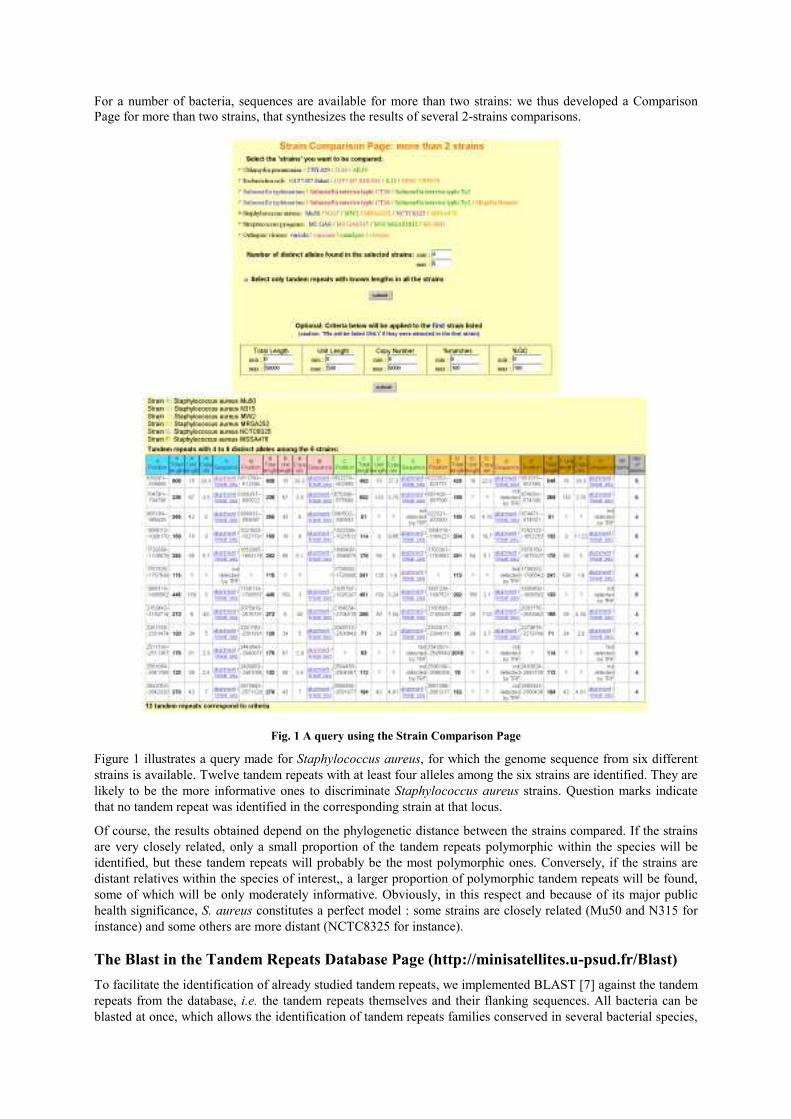
For a number of bacteria, sequences are available for more than two strains: we thus developed a Comparison
Page for more than two strains, that synthesizes the results of several 2-strains comparisons.
Fig. 1 A query using the Strain Comparison Page
Figure 1 illustrates a query made for Staphylococcus aureus, for which the genome sequence from six different
strains is available. Twelve tandem repeats with at least four alleles among the six strains are identified. They are
likely to be the more informative ones to discriminate Staphylococcus aureus strains. Question marks indicate
that no tandem repeat was identified in the corresponding strain at that locus.
Of course, the results obtained depend on the phylogenetic distance between the strains compared. If the strains
are very closely related, only a small proportion of the tandem repeats polymorphic within the species will be
identified, but these tandem repeats will probably be the most polymorphic ones. Conversely, if the strains are
distant relatives within the species of interest,, a larger proportion of polymorphic tandem repeats will be found,
some of which will be only moderately informative. Obviously, in this respect and because of its major public
health significance, S. aureus constitutes a perfect model : some strains are closely related (Mu50 and N315 for
instance) and some others are more distant (NCTC8325 for instance).
The Blast in the Tandem Repeats Database Page (http://minisatellites.u-psud.fr/Blast)
To facilitate the identification of already studied tandem repeats, we implemented BLAST [7] against the tandem
repeats from the database, i.e. the tandem repeats themselves and their flanking sequences. All bacteria can be
blasted at once, which allows the identification of tandem repeats families conserved in several bacterial species,
even when they share a similar consensus motif but different flanking sequences: insertions/deletions due to
tandem repeats polymorphism generally do not affect the recognition. Another page is dedicated to the Blast of
PCR primers: it provides the size of the PCR products in all the species/strains where the primers match, and
thus facilitates the primer picking step.
The Bacterial Genotyping Page (http://bacterial-genotyping.igmors.u-psud.fr)
Once the tandem repeats identified via the Tandem Repeats Database or the Strain Comparison Page have been
typed by PCR, and genotypes have been assigned to a sufficient number of strains, the next step is to build a
strain classification to evaluate the proximity of the strains. But, often, such studies remain internal to a
laboratory, despite the high portability of tandem repeats typing assay. The Bacterial Genotyping Page aims to
remedy this lack : it allows external users to query a number of genotyping data (Bacillus anthracis, Yersinia
pestis, Mycobacterium tuberculosis for the moment) in order to identify bacterial strains [2]. For each locus,
allele sizes can be selected among a list of possibilities (observed sizes). The results of the query indicate a
similarity score and include links to the complete data for each strain listed. If the tandem repeats typing
approach is considered to be of use by the community interested in a particular species, and given that the
associated data is highly portable, it should then be relatively easy, through collaborative efforts, to significantly
expand the available data. It is hoped that this data will constitute an easy-to-use high-resolution classification
resource which will then help address medical and epidemiological issues.
References
[1] A. van Belkum, S. Scherer, W. van Leeuwen, D. Willemse, L. van Alphen and H. Verbrugh, Variable
number of tandem repeats in clinical strains of Haemophilus influenzae. Infect Immun 65:5017-27, 1997.
[2] P. Le Flèche, M. Fabre, F. Denœud, J.L. Koeck and G. Vergnaud, High resolution, on-line identification of
strains from the Mycobacterium tuberculosis complex based on tandem repeat typing. BMC Microbiol 2:37,
2002.
[3] G. Vergnaud and F. Denœud, Minisatellites: Mutability and Genome Architecture. Genome Res 10:899-907,
2000.
[4] P. Le Flèche, Y. Hauck, L. Onteniente, A. Prieur, F. Denœud, V.Ramisse, P. Sylvestre, G. Benson, F.
Ramisse and G. Vergnaud, A tandem repeats database for bacterial genomes: application to the genotyping of
Yersinia pestis and Bacillus anthracis. BMC Microbiol 1:2, 2001.
[5] F. Denoeud, G. Vergnaud and G. Benson, Predicting human minisatellite polymorphism. Genome Res
13(5):856-67, 2003.
[6] C. Pourcel, Y. Vidgop, F. Ramisse, G. Vergnaud, and C. Tram, Characterization of a tandem repeat
polymorphism in Legionella pneumophila and its use for genotyping. J Clin Microbiol. 41(5):1819-26, 2003.
[7] S.F. Altschul, T.L Madden, A.A Schaffer, J. Zhang, Z. Zhang, W. Miller and D.J. Lipman, Gapped BLAST
and PSI-BLAST: a new generation of protein database search programs. Nucleic Acids Res 25:3389-402, 1997.
[8] G. Benson, Tandem repeats finder: a program to analyze DNA sequences. Nucleic Acids Res 27:573-580,
1999.

76
L’article suivant (Denoeud 2004), intitulé « Identification of polymorphic tandem repeats by
direct comparison of genome sequence from different bacterial strains : a Web-based
ressource » (identification de répétitions en tandem polymorphes par la comparaison directe
des séquences génomiques de différentes souches bactériennes : une ressource web) présente
les fonctionnalités de la base de données utiles à l’épidémiologie bactérienne. Il insiste plus
particulièrement sur la page de comparaison de souches, qui identifie directement les
répétitions en tandem polymorphes entre plusieurs souches séquencées. Cet article aborde
également le problème récurrent de la prédiction du polymorphisme d’une répétition en
tandem à partir de sa séquence. Nous y montrons que certaines caractéristiques des séquences
de répétitions en tandem bactériennes (conservation interne, nombre de copies) sont corrélées
au polymorphisme mais ont une valeur prédictive très variable d’une espèce à l’autre. La
meilleure approche pour identifier des répétitions en tandem polymorphes chez les bactéries
reste donc la comparaison de souches, quand celle-ci est rendue possible par la disponibilité
des séquences.
Résumé :
Contexte : Les répétitions en tandem polymorphes sont utilisées pour le typage de souches
chez un nombre grandissant de bactéries pathogènes pour l’Homme, dont Bacillus anthracis,
Mycobacterium tuberculosis, Yersinia pestis, Pseudomonas aeruginosa, Legionella pneumophila,
Salmonella typhi, Brucella, Francisella tularensis et Staphylococcus aureus. Les répétitions en
tandem peuvent facilement être identifiées à partir des nombreuses séquences de génomes
bactériens actuellement disponibles. La mise en place d’une procédure de génotypage ne
nécessite donc plus que l’évaluation de leur polymorphisme, ce qui est fait en général par des
tests systématiques. Ces tests peuvent toutefois représenter une tâche laborieuse. Pour de
nombreux pathogènes d’importance, tels que S. aureus, plus d’une souche a été séquencée, ce
qui permet dorénavant d’identifier in silico les répétitions en tandem polymorphes parmi
différentes souches.
Résultats : En supplément de la base de données des répétitions en tandem déjà décrite, nous
avons développé une page d’identification automatique des répétitions en tandem de longueur
différente dans les génomes de plus de deux souches bactériennes proches. Les comparaisons
de génomes sont effectuées puis importées dans une base de données, qui peut être interrogée
par Internet selon des critères d’intérêt tels que la longueur du motif, la différence de taille
prédite, etc. Les comparaisons sont disponibles pour 16 espèces bactériennes et les virus du
groupe orthopox, comprenant le virus de la variole et trois de ses voisins proches.
Conclusions : Nous présentons une ressource Internet qui facilite le développement de
méthodes de typage de souches bactériennes à partir des répétitions en tandem. Elle comprend
actuellement quatre parties, accessibles à partir de l’adresse http://minisatellites.u-psud.fr. La
base de données des répétitions en tandem permet d’identifier les répétitions en tandem dans
des génomes entiers. La page de comparaison de souches sélectionne les répétitions en
tandem différentes entre plusieurs génomes d’une même espèce. La page de Blast dans la base
de données facilite la recherche de répétitions en tandem connues et la validation des couples
d’amorces PCR. La page de génotypage permet l’identification de souches en ligne.
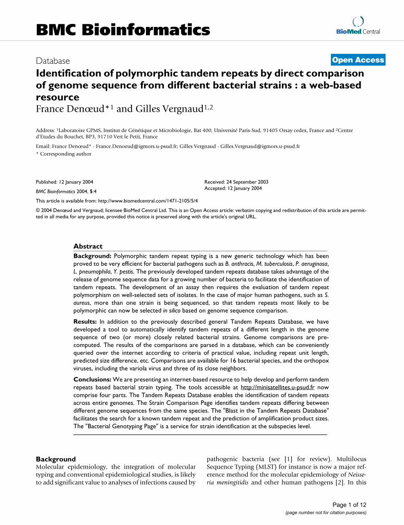
BioMed Central
Page 1 of 12
(page number not for citation purposes)
BMC Bioinformatics
Open AccessDatabase
Identification of polymorphic tandem repeats by direct comparison of genome sequence from different bacterial strains : a web-based resourceFrance Denœud*1 and Gilles Vergnaud1,2
Address: 1Laboratoire GPMS, Institut de Génétique et Microbiologie, Bat 400, Université Paris-Sud, 91405 Orsay cedex, France and 2Centre d'Etudes du Bouchet, BP3, 91710 Vert le Petit, France
Email: France Denœud* - [email protected]; Gilles Vergnaud - [email protected]
* Corresponding author
Abstract
Background: Polymorphic tandem repeat typing is a new generic technology which has been
proved to be very efficient for bacterial pathogens such as B. anthracis, M. tuberculosis, P. aeruginosa,
L. pneumophila, Y. pestis. The previously developed tandem repeats database takes advantage of the
release of genome sequence data for a growing number of bacteria to facilitate the identification of
tandem repeats. The development of an assay then requires the evaluation of tandem repeat
polymorphism on well-selected sets of isolates. In the case of major human pathogens, such as S.
aureus, more than one strain is being sequenced, so that tandem repeats most likely to be
polymorphic can now be selected in silico based on genome sequence comparison.
Results: In addition to the previously described general Tandem Repeats Database, we have
developed a tool to automatically identify tandem repeats of a different length in the genome
sequence of two (or more) closely related bacterial strains. Genome comparisons are pre-
computed. The results of the comparisons are parsed in a database, which can be conveniently
queried over the internet according to criteria of practical value, including repeat unit length,
predicted size difference, etc. Comparisons are available for 16 bacterial species, and the orthopox
viruses, including the variola virus and three of its close neighbors.
Conclusions: We are presenting an internet-based resource to help develop and perform tandem
repeats based bacterial strain typing. The tools accessible at http://minisatellites.u-psud.fr now
comprise four parts. The Tandem Repeats Database enables the identification of tandem repeats
across entire genomes. The Strain Comparison Page identifies tandem repeats differing between
different genome sequences from the same species. The "Blast in the Tandem Repeats Database"
facilitates the search for a known tandem repeat and the prediction of amplification product sizes.
The "Bacterial Genotyping Page" is a service for strain identification at the subspecies level.
BackgroundMolecular epidemiology, the integration of moleculartyping and conventional epidemiological studies, is likelyto add significant value to analyses of infections caused by
pathogenic bacteria (see [1] for review). MultilocusSequence Typing (MLST) for instance is now a major ref-erence method for the molecular epidemiology of Neisse-ria meningitidis and other human pathogens [2]. In this
Published: 12 January 2004
BMC Bioinformatics 2004, 5:4
Received: 24 September 2003Accepted: 12 January 2004
This article is available from: http://www.biomedcentral.com/1471-2105/5/4
© 2004 Denœud and Vergnaud; licensee BioMed Central Ltd. This is an Open Access article: verbatim copying and redistribution of this article are permit-ted in all media for any purpose, provided this notice is preserved along with the article's original URL.
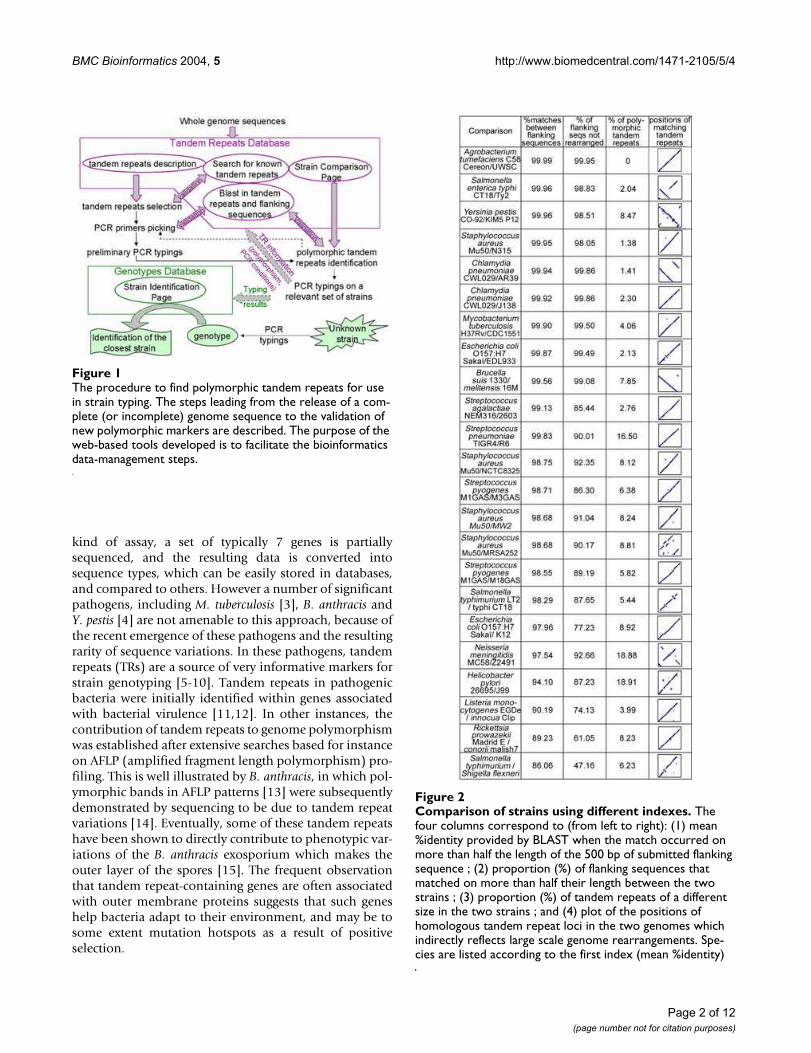
BMC Bioinformatics 2004, 5 http://www.biomedcentral.com/1471-2105/5/4
Page 2 of 12
(page number not for citation purposes)
kind of assay, a set of typically 7 genes is partiallysequenced, and the resulting data is converted intosequence types, which can be easily stored in databases,and compared to others. However a number of significantpathogens, including M. tuberculosis [3], B. anthracis andY. pestis [4] are not amenable to this approach, because ofthe recent emergence of these pathogens and the resultingrarity of sequence variations. In these pathogens, tandemrepeats (TRs) are a source of very informative markers forstrain genotyping [5-10]. Tandem repeats in pathogenicbacteria were initially identified within genes associatedwith bacterial virulence [11,12]. In other instances, thecontribution of tandem repeats to genome polymorphismwas established after extensive searches based for instanceon AFLP (amplified fragment length polymorphism) pro-filing. This is well illustrated by B. anthracis, in which pol-ymorphic bands in AFLP patterns [13] were subsequentlydemonstrated by sequencing to be due to tandem repeatvariations [14]. Eventually, some of these tandem repeatshave been shown to directly contribute to phenotypic var-iations of the B. anthracis exosporium which makes theouter layer of the spores [15]. The frequent observationthat tandem repeat-containing genes are often associatedwith outer membrane proteins suggests that such geneshelp bacteria adapt to their environment, and may be tosome extent mutation hotspots as a result of positiveselection.
The procedure to find polymorphic tandem repeats for use in strain typingFigure 1The procedure to find polymorphic tandem repeats for use in strain typing. The steps leading from the release of a com-plete (or incomplete) genome sequence to the validation of new polymorphic markers are described. The purpose of the web-based tools developed is to facilitate the bioinformatics data-management steps.
Comparison of strains using different indexesFigure 2Comparison of strains using different indexes. The four columns correspond to (from left to right): (1) mean %identity provided by BLAST when the match occurred on more than half the length of the 500 bp of submitted flanking sequence ; (2) proportion (%) of flanking sequences that matched on more than half their length between the two strains ; (3) proportion (%) of tandem repeats of a different size in the two strains ; and (4) plot of the positions of homologous tandem repeat loci in the two genomes which indirectly reflects large scale genome rearrangements. Spe-cies are listed according to the first index (mean %identity)
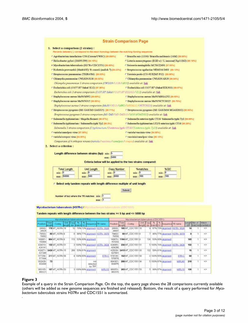
BMC Bioinformatics 2004, 5 http://www.biomedcentral.com/1471-2105/5/4
Page 3 of 12
(page number not for citation purposes)
Example of a query in the Strain Comparison PageFigure 3Example of a query in the Strain Comparison Page. On the top, the query page shows the 28 comparisons currently available (others will be added as new genome sequences are finished and released). Bottom, the result of a query performed for Myco-bacterium tuberculosis strains H37Rv and CDC1551 is summarized.
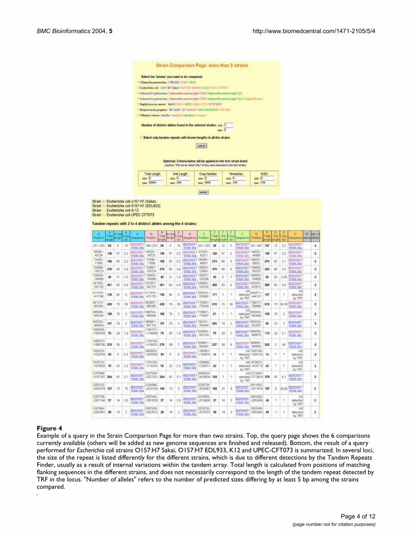
BMC Bioinformatics 2004, 5 http://www.biomedcentral.com/1471-2105/5/4
Page 4 of 12
(page number not for citation purposes)
Example of a query in the Strain Comparison Page for more than two strainsFigure 4Example of a query in the Strain Comparison Page for more than two strains. Top, the query page shows the 6 comparisons currently available (others will be added as new genome sequences are finished and released). Bottom, the result of a query performed for Escherichia coli strains O157:H7 Sakaï, O157:H7 EDL933, K12 and UPEC-CFT073 is summarized. In several loci, the size of the repeat is listed differently for the different strains, which is due to different detections by the Tandem Repeats Finder, usually as a result of internal variations within the tandem array. Total length is calculated from positions of matching flanking sequences in the different strains, and does not necessarily correspond to the length of the tandem repeat detected by TRF in the locus. "Number of alleles" refers to the number of predicted sizes differing by at least 5 bp among the strains compared.

BMC Bioinformatics 2004, 5 http://www.biomedcentral.com/1471-2105/5/4
Page 5 of 12
(page number not for citation purposes)
Polymorphic tandem repeats (VNTRs, for VariableNumber of Tandem Repeats), once identified, provideconvenient tools requiring ordinary molecular biologyequipment and the data can be easily exchanged and com-pared. The resulting assay, called MLVA (for multiplelocus VNTR analysis) can even be automated [16]. Wehave developed tools which facilitate the bioinformaticsstep of genome analysis required to start a project. A pre-viously described Tandem Repeats Database enables theidentification of tandem repeats across entire genomes[9,10,17-19]. It has been constantly updated, with nowmore than a hundred bacterial genomes available, com-pared to 35 at the onset of the database. We present herea new and major development of this resource whichtakes advantage of the fact that more than two differentstrains from the same species have now been sequenced atleast for a number of major human pathogens. As a result,the tools accessible over the Internet at http://minisatellites.u-psud.fr now comprise four complementary parts.The newly added resource, the Strain Comparison Page,takes advantage of the availability of genome sequencesfrom more than one strain from a growing number of spe-cies to directly identify tandem repeats differing betweenthe sequenced strains. This is of interest because the vastmajority of tandem repeats is often not polymorphic [19].The "Blast in the Tandem Repeats Database" page facili-tates the search for a known tandem repeat, the predictionof PCR amplification products size, and the verification ofprimer specificity. Once an MLVA assay has been set up,and carefully validated by typing collections of isolates, itis relatively easy to construct databases of genotypes to beused locally or which can be queried across the Internet.The "Bacterial Genotyping Page" illustrates a freely acces-sible, fast and easy to use internet-based service for straincomparisons, in which a user can compare a genotypeproduced for one of his isolates to the existing data.
Construction and contentThe Tandem Repeats Database main page
Tandem repeats were identified from finished microbialgenome sequences (as listed by the Genome OnLineDatabase [20]) using the tandem repeats finder (TRF)software [21,22] with the following options: alignmentparameters, "2,3,5" (these parameters are the less strin-gent ones), minimum alignment score to report repeat, 50(this score allows to detect short structures), maximumperiod size, 500 base-pairs. When the program reportedredundant (overlapping) repeats, the redundancy waseliminated as described in [23], before import in the data-base. The database uses Microsoft Access 2000 and thequerying process uses Active Server Pages (ASP, Microsoft)with Perlscripts or VBscripts. Perl was obtained from theActiveState Programmer Network [24]. The database ishosted on a server running under Windows 2000 server
(Microsoft). The tandem repeats database main page isdescribed in more detail in [9].
The Strain Comparison page
Sequence comparisons used BLAST [25]. The BLAST soft-ware was obtained from the NCBI FTP site [26]. The flank-ing sequences of TRs from one strain were compared tothe whole sequence of the other strain (and reciprocally,to avoid missing some tandem repeats that would notappear in the tandem repeats database for one strainbecause they were not detected by the Tandem RepeatsFinder [21] -for instance because there is only one copy ofthe repeated unit in the considered strain). The resultinglist of matching tandem repeats was then imported in thedatabase, where it can be queried. The comparison ofmore than two strains was made possible through a sup-plemental step before import in the database: the synthe-sis of several 2-strains comparisons, of the same"reference" strain against each of the others (matchingbetween TRs of the different strains was deduced from thepositions on the reference strain).
The Blast page
The Blast Page allows users to run BLAST [25] in the tan-dem repeats and flanking sequences from the database viaPerlscripts. The Blast outputs are linked to the database, inorder to easily obtain the description of identified tandemrepeats.
The Bacterial Genotyping page
The web-page site performing identifications was devel-oped using the BNserver application (version 3.0,Applied-Maths, Belgium) and ASP (Microsoft) using Per-lscript. The typing results (gel images and resulting data)were managed using the Bionumerics software package asdescribed in [10]. The output of a query is a list of strainsand genotypes from the database together with similarityscores.
UtilityThe procedure to find polymorphic tandem repeats (TRs)
for use in strain typing
Figure 1 shows the steps leading from a genome sequenceto the exploitation of polymorphic tandem repeats forbacterial strain genotyping. Although Tandem Repeats areeasily identified using the Tandem Repeats Database, TRpolymorphism must be evaluated by typing across a set ofrelevant strains. If the sequences of several strains of thespecies of interest are available, the Strain ComparisonPage can be used to directly identify tandem repeats pre-dicted to be polymorphic in size between the two (ormore) sequenced strains. However, it is important to keepin mind that the tandem repeats predicted as being poly-morphic will depend on the sequenced strains and well-planned surveys of isolates will still be necessary. The

BMC Bioinformatics 2004, 5 http://www.biomedcentral.com/1471-2105/5/4
Page 6 of 12
(page number not for citation purposes)
available tools do not replace this validation step, as thevalue of each marker must be carefully established on anappropriate set of isolates. The definition of an appropri-ate set of isolates depends upon the question which isbeing addressed, i.e. large scale or local epidemiology. TheBlast Page has been implemented in the tandem repeatsdatabase in order to easily determine the size of theexpected PCR amplification products. The database is alsomanually updated to contain PCR conditions as well aspolymorphism index, and links to the original reports[27] (input from users is welcome). Eventually, when anMLVA assay has been fully developed and validated, typ-ing data can be made accessible so that individual queriescan be run. The Bacterial Genotyping Page illustrates howthis could work. The genotyping data for a strain can beentered and submitted via this page. The output is thedescription of the closest strains. The data which has beensubmitted is not incorporated in the database itself, sincethis would require stringent data validation steps. In thefollowing sections, we are presenting the web-basedresources associated with this procedure.
The "Strain Comparison" pages
The strain comparison pages are available via [28]. Thecomparison of two strains is based on a pre-computedBLAST [25] analysis of the flanking sequences of tandemrepeats from one strain against the other, and vice-versa.Figure 2 summarizes the results of this first step for 23comparisons. Three indexes are scored (see figure legend):(1) the "mean %identity" between the flanking sequencesis a measure of single nucleotide polymorphism (SNPs)frequency (not insertions-deletions), (2) the proportion(%) of flanking sequences that matched the flankingsequence of its homologue in the other strain on morethan half of the 500 bp assayed here – i.e. that were notrearranged, by insertion of mobile elements for instance -, (3) the proportion (%) of tandem repeats that werefound to be of a different length between the two strainsbeing compared. In addition, the positions of matchingtandem repeats in the two genomes is plotted to reveallarge-scale genome rearrangements. A number of situa-tions are observed: for instance Yersinia pestis orientalisstrain CO-92 [29], and medievalis strain KIM5 P12 [30]show a very high "mean %identity" (99.96 %), in agree-ment with the recent emergence of Yersinia pestis [4]. Inspite of this, the two strains differ by a high number oflarge rearrangements (as seen on the plot), which reflectsthe high genome plasticity observed in this species [31],together with a relatively high rate of polymorphic tan-dem repeats (8.47%). In contrast, Listeria monocytogenesstrain EGD-e and Listeria innocua strain Clip 11262 have alower homology (90.19%) and only 3.99% of polymor-phic tandem repeats in spite of the evolutionary distance(see Figure 2).
The strain comparison page allows queries in the tandemrepeats database according to the tandem repeat lengthdifference between the two strains compared, and also toother tandem repeats characteristics (unit length, copynumber, etc...). Figure 3 illustrates a query done for Myco-bacterium tuberculosis strains H37Rv and CDC1551 [32]:the query "length difference ≥ 5 bp" identifies 58 tandemrepeats (8 are shown on Figure 3). This prediction hasbeen tested for the 30 loci amenable to PCR analysis andpolymorphism has been confirmed in all cases [10].
When more than two strains have been sequenced, a syn-thesis of the results of several 2-strains comparisons is alsoavailable. Figure 4 illustrates a query made for Escherichiacoli strains O157:H7 Sakaï, O157:H7 EDL933, K12, andUPEC-CFT073 [33-35]: 87 tandem repeats were foundwith 2 to 4 alleles among the 4 strains (18 of which arelisted in Figure 4).
The "Blast in the Tandem Repeats Database" page
To facilitate the identification of already studied tandemrepeats, we implemented BLAST [25] against the tandemrepeats from the database, i.e. the tandem repeats them-selves and their flanking sequences. The Blast page isavailable at [36]. All bacteria can be queried at once,which allows the identification of tandem repeats fami-lies, conserved in several bacterial species. Another page isdedicated to the Blast of PCR primers and provides thesize of the PCR products in all the species/strains wherethe primers match. Figure 5 shows the results of searchingthe PCR primer pair from tandem repeat H37Rv_0024_18bp [10] in all bacteria: as expected, the PCR primer pairmatches Mycobacterium tuberculosis strains H37Rv andCDC1551, providing different PCR product lengths.
The Bacterial Genotyping page
The Bacterial Genotyping page [37] provides one illustra-tion on how tandem repeat typing data can be made avail-able via internet to allow external users to querygenotyping data (Bacillus anthracis, Yersinia pestis, Mycobac-terium tuberculosis, Pseudomonas aeruginosa for themoment) and compare a new strain to existing data aspreviously described in [10]. For each locus, allele sizescan be selected among a list of possibilities (observedsizes). The results of the query indicate a similarity scoreand include links to the complete data recorded for eachstrain listed. This page is just meant as an illustration andprototype. MLVA reference data could also be made avail-able for downloading as tabular data files, or can be cop-ied from published datasets, which can then becomplemented by in-house data, and analyzed by theappropriate clustering software.

BMC Bioinformatics 2004, 5 http://www.biomedcentral.com/1471-2105/5/4
Page 7 of 12
(page number not for citation purposes)
DiscussionBacterial genomes evolution
As shown by the indexes from Figure 2, there are differentways to represent the divergence/similarity between twostrains. They are not correlated, suggesting independentevolution processes. First, the "mean %identity" betweentwo genomes reflects point mutations, and is an indicatorof the time passed since the two strains diverged. For
instance, Yersinia pestis is known to be of recent emergence[4] and shows a high "mean %identity" between strainsCO-92 (orientalis) and KIM5 P12 (medievalis). In con-trast, and as shown by the dot plot, large genome rear-rangements occurred in this genome, which isrepresentative of a high genome plasticity [31]. The index"% of flanking sequences not rearranged" is an indicatorof small-scale genome rearrangements, such as the inser-
Example of a query in the "Blast of PCR primers" page, providing the length of the PCR products in the strains/species where the primer pair matches, and links to the corresponding tandem repeats descriptionsFigure 5Example of a query in the "Blast of PCR primers" page, providing the length of the PCR products in the strains/species where the primer pair matches, and links to the corresponding tandem repeats descriptions.

BMC Bioinformatics 2004, 5 http://www.biomedcentral.com/1471-2105/5/4
Page 8 of 12
(page number not for citation purposes)
tions of mobile elements. This index is low for genomesrich in mobile elements, like Streptococcus agalactiae, inwhich such elements significantly contribute to straindiversity [38]. Finally, the index "% of polymorphic tan-dem repeats" between two strains represents the tandemrepeats evolution rate. For the moment, the mechanismsof bacterial VNTRs mutations have not been preciselyinvestigated, but it seems likely to be independent of theother processes mentioned, as there are no correlationsbetween the indexes. Figure 2 provides clues to assesswhich typing method(s) will be efficient in the differentspecies. For instance, the two bacterial species Salmonellatyphimurium strain LT2 [39] and Shigella flexneri strain2a301 [40] share only 86.06% of sequence identity,clearly making the identification of matching tandemrepeats between the two species difficult and of low signif-icance. MLVA analysis appears to be of highest interest forthe subspecies typing of highly monomorphic speciesincluding Yersinia pestis, Bacillus anthracis, Mycobacteriumtuberculosis and Brucella [9,10,41].
Strain comparison efficiency
The sequencing of more than one strain for some bacterialspecies allows direct identification of polymorphic tan-dem repeats, assuming that no sequencing errorsoccurred. Earlier investigations provide good reasons tobelieve that tandem repeats in the size range consideredhere (a few hundred base-pairs) are correctly sequenced,and consequently, that the strain comparison data is reli-able. As a negative control, the comparison of two inde-pendent sequences from the same strain of Agrobacteriumtumefaciens strain (C58), one from Cereon genomics [42]and the other from Washington University [43], showsthat no length polymorphism is detected among tandemrepeats (Figure 2) between the two independentsequences. As a positive control, the tandem repeats pre-dicted to be polymorphic by genome sequence compari-son between the two strains of M. tuberculosis have indeedbeen proved polymorphic by PCR typing of isolates [10].
Selection based on comparison of sequence data from twostrains will miss some polymorphic loci. Indeed, theresults provided by the approach rely upon the phyloge-netic distance between the two strains being compared. Ifthe strains are very closely related, only a few TRs will befound different between them, but these tandem repeatswill probably be the most polymorphic ones. Conversely,if the strains are distant in the phylogenetic tree, a largernumber of polymorphic TRs will be found, some of themwill be only moderately polymorphic. Obviously, when afew well-selected strains have been sequenced, it is likelythat very few polymorphic tandem repeats are undetectedin the Strain Comparison pages.
It is of course still going to be very important to determinethe TR allele frequency for isolates carefully selected to berepresentative of the global diversity of a given pathogenbefore suggesting the configuration of an MLVA assay touse in subsequent studies. In addition, those TR markersthat are highly polymorphic in diverse test panels of iso-lates may be monomorphic when applied to isolatesresponsible for local outbreaks. The configuration of TRmarkers used to make up an assay needs to be determinedempirically with representative local isolates and tailoredto the study population and study questions.
Polymorphic tandem repeats selection for species with
only one sequenced strain
The identification of simple criteria able to predict tan-dem repeat polymorphism when genome sequence data isavailable for only one strain would indeed greatly facili-tate the development of MLVA assays. It would seem rea-sonable for instance to expect that the number of copiesand the internal homogeneity of tandem arrays are strongpredictors [23]. We take advantage here of the many straincomparisons which are made available via the strain com-parison pages to evaluate such criteria.
We have analyzed bacteria with at least three sequencedgenomes (Staphylococcus aureus: 6 strains, Escherichia coli: 4strains, Streptococcus pyogenes: 4 strains and Salmonellatyphi and typhimurium: 3 strains). We assume that in suchcases, only a few polymorphic tandem repeats are missedin the comparisons. We compared the distribution of tan-dem repeats sequence characteristics among the group of"polymorphic" loci (differing in at least two of the strainscompared, excluding length differences between strainsthat resulted from microdeletions in the flankingsequences) and the others. Comparisons were performedfor the following sequence characteristics: unit length,copy number, total length, %GC, GC bias (=|%G-%C|/(%G+%C)), %matches, and HistoryR (a score derivedfrom tandem repeat history reconstruction algorithm [44]as described in [23]). None of the variables were normallydistributed, as tested with Kolmogorov-Smirnov test, so anon-parametric Wilcoxon test was used to compare thedistributions, which were judged significantly different atthe .05 level of the statistic (2 tailed). Distributions weresignificantly different for all 4 species studied for%matches, total length and copy number. As shown onFigure 6, polymorphic TRs have a higher internal conser-vation and total length than monomorphic ones. Copynumber, which is correlated with total length, is alsohigher among polymorphic TRs.
Selecting the longest and most conserved tandem repeatsshould thus improve polymorphic TRs identification.Table 1 illustrates the query "total length ≥ 80 bp and%matches ≥ 80%" applied to the four species used to find

BMC Bioinformatics 2004, 5 http://www.biomedcentral.com/1471-2105/5/4
Page 9 of 12
(page number not for citation purposes)
Proportion of predicted polymorphic (pink) and monomorphic (grey) tandem repeats according to different parameters (inter-nal homogeneity of the repeat array (%matches) or total length)Figure 6Proportion of predicted polymorphic (pink) and monomorphic (grey) tandem repeats according to different parameters (inter-nal homogeneity of the repeat array (%matches) or total length). P-values obtained for the non-parametric Wilcoxon tests appear below each histogram.
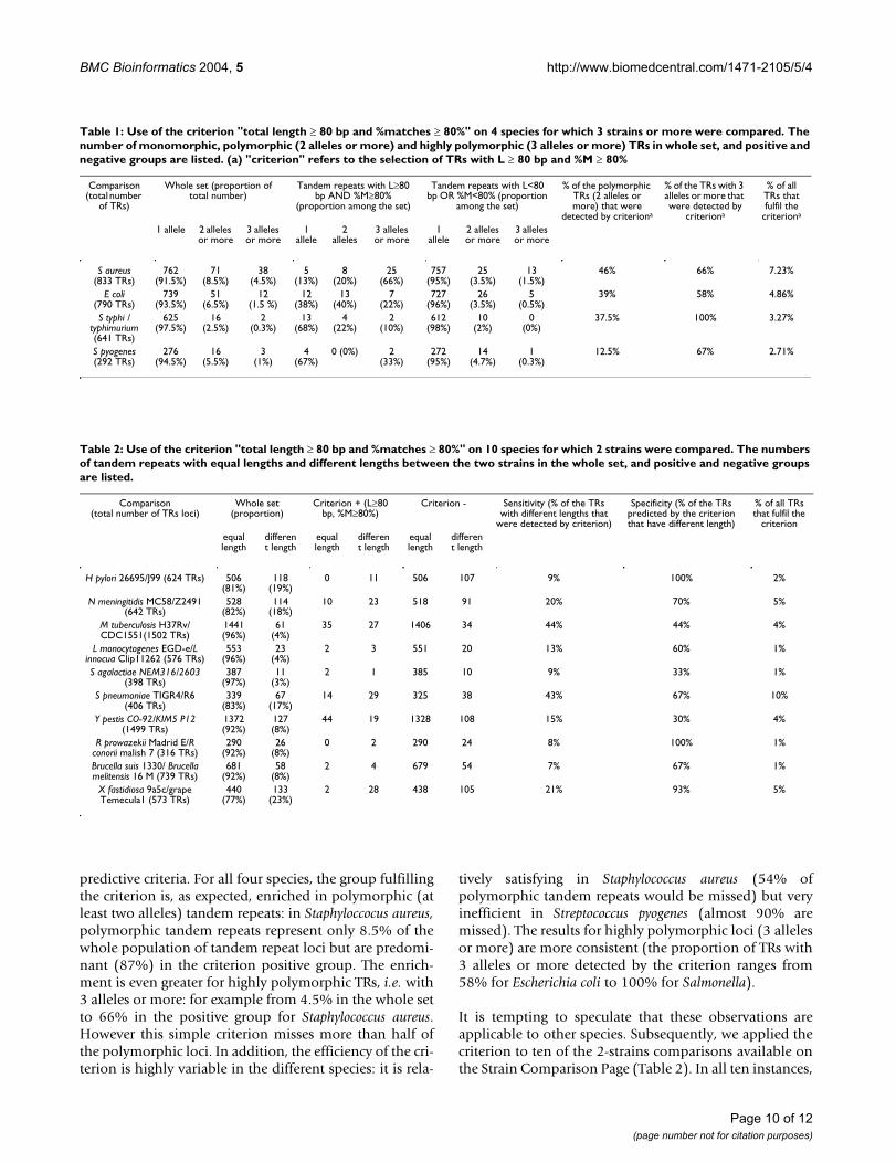
BMC Bioinformatics 2004, 5 http://www.biomedcentral.com/1471-2105/5/4
Page 10 of 12
(page number not for citation purposes)
predictive criteria. For all four species, the group fulfillingthe criterion is, as expected, enriched in polymorphic (atleast two alleles) tandem repeats: in Staphyloccocus aureus,polymorphic tandem repeats represent only 8.5% of thewhole population of tandem repeat loci but are predomi-nant (87%) in the criterion positive group. The enrich-ment is even greater for highly polymorphic TRs, i.e. with3 alleles or more: for example from 4.5% in the whole setto 66% in the positive group for Staphylococcus aureus.However this simple criterion misses more than half ofthe polymorphic loci. In addition, the efficiency of the cri-terion is highly variable in the different species: it is rela-
tively satisfying in Staphylococcus aureus (54% ofpolymorphic tandem repeats would be missed) but veryinefficient in Streptococcus pyogenes (almost 90% aremissed). The results for highly polymorphic loci (3 allelesor more) are more consistent (the proportion of TRs with3 alleles or more detected by the criterion ranges from58% for Escherichia coli to 100% for Salmonella).
It is tempting to speculate that these observations areapplicable to other species. Subsequently, we applied thecriterion to ten of the 2-strains comparisons available onthe Strain Comparison Page (Table 2). In all ten instances,
Table 1: Use of the criterion "total length ≥ 80 bp and %matches ≥ 80%" on 4 species for which 3 strains or more were compared. The
number of monomorphic, polymorphic (2 alleles or more) and highly polymorphic (3 alleles or more) TRs in whole set, and positive and
negative groups are listed. (a) "criterion" refers to the selection of TRs with L ≥ 80 bp and %M ≥ 80%
Comparison (total number
of TRs)
Whole set (proportion of total number)
Tandem repeats with L≥80 bp AND %M≥80%
(proportion among the set)
Tandem repeats with L<80 bp OR %M<80% (proportion
among the set)
% of the polymorphic TRs (2 alleles or more) that were
detected by criteriona
% of the TRs with 3 alleles or more that were detected by
criteriona
% of all TRs that fulfil the
criteriona
1 allele 2 alleles or more
3 alleles or more
1 allele
2 alleles
3 alleles or more
1 allele
2 alleles or more
3 alleles or more
S aureus (833 TRs)
762 (91.5%)
71 (8.5%)
38 (4.5%)
5 (13%)
8 (20%)
25 (66%)
757 (95%)
25 (3.5%)
13 (1.5%)
46% 66% 7.23%
E coli (790 TRs)
739 (93.5%)
51 (6.5%)
12 (1.5 %)
12 (38%)
13 (40%)
7 (22%)
727 (96%)
26 (3.5%)
5 (0.5%)
39% 58% 4.86%
S typhi / typhimurium (641 TRs)
625 (97.5%)
16 (2.5%)
2 (0.3%)
13 (68%)
4 (22%)
2 (10%)
612 (98%)
10 (2%)
0 (0%)
37.5% 100% 3.27%
S pyogenes (292 TRs)
276 (94.5%)
16 (5.5%)
3 (1%)
4 (67%)
0 (0%) 2 (33%)
272 (95%)
14 (4.7%)
1 (0.3%)
12.5% 67% 2.71%
Table 2: Use of the criterion "total length ≥ 80 bp and %matches ≥ 80%" on 10 species for which 2 strains were compared. The numbers
of tandem repeats with equal lengths and different lengths between the two strains in the whole set, and positive and negative groups
are listed.
Comparison (total number of TRs loci)
Whole set (proportion)
Criterion + (L≥80 bp, %M≥80%)
Criterion - Sensitivity (% of the TRs with different lengths that
were detected by criterion)
Specificity (% of the TRs predicted by the criterion that have different length)
% of all TRs that fulfil the
criterion
equal length
different length
equal length
different length
equal length
different length
H pylori 26695/J99 (624 TRs) 506 (81%)
118 (19%)
0 11 506 107 9% 100% 2%
N meningitidis MC58/Z2491 (642 TRs)
528 (82%)
114 (18%)
10 23 518 91 20% 70% 5%
M tuberculosis H37Rv/CDC1551(1502 TRs)
1441 (96%)
61 (4%)
35 27 1406 34 44% 44% 4%
L monocytogenes EGD-e/L innocua Clip11262 (576 TRs)
553 (96%)
23 (4%)
2 3 551 20 13% 60% 1%
S agalactiae NEM316/2603 (398 TRs)
387 (97%)
11 (3%)
2 1 385 10 9% 33% 1%
S pneumoniae TIGR4/R6 (406 TRs)
339 (83%)
67 (17%)
14 29 325 38 43% 67% 10%
Y pestis CO-92/KIM5 P12 (1499 TRs)
1372 (92%)
127 (8%)
44 19 1328 108 15% 30% 4%
R prowazekii Madrid E/R conorii malish 7 (316 TRs)
290 (92%)
26 (8%)
0 2 290 24 8% 100% 1%
Brucella suis 1330/ Brucella melitensis 16 M (739 TRs)
681 (92%)
58 (8%)
2 4 679 54 7% 67% 1%
X fastidiosa 9a5c/grape Temecula1 (573 TRs)
440 (77%)
133 (23%)
2 28 438 105 21% 93% 5%

BMC Bioinformatics 2004, 5 http://www.biomedcentral.com/1471-2105/5/4
Page 11 of 12
(page number not for citation purposes)
the criterion positive group is enriched in TRs with differ-ent lengths between the two strains, compared to thewhole set. This proportion varies from less than 3% inStreptococcus agalactiae to more than 20% in Xylella fastidi-osa in the whole set. It is increased to 33% and 93%respectively among the set of loci which satisfy the crite-rion (these percentages correspond to the predictor's spe-cificity), but the vast majority of polymorphic loci will bemissed (90% and 80% respectively). Sensitivity, that is %of the TRs with different lengths that were detected by cri-terion varies from 6.90% for Brucella to 44.26% for Myco-bacterium tuberculosis.
The finding that polymorphic tandem repeats have, onaverage, a higher internal conservation, total length, andcopy number than monomorphic ones is in agreementwith previous observations that TR polymorphism is cor-related with conservation in Yersinia pestis and with totallength in Bacillus anthracis [9]. It is also reminiscent of thebehavior of microsatellites (also called short sequencerepeats: SSR, see [45] for review), which are stabilized byinternal variations [46] and by reduction of the number ofrepeats [47]. Unfortunately, we show here that such sim-ple prediction criteria may miss a very large proportion ofpolymorphic tandem repeats, and provide highly variableresults in different species. This indicates that, in theabsence of sequence data from two strains or more, thesystematic testing of tandem repeats polymorphismacross a set of relevant strains remains the most appropri-ate way to develop an MLVA assay. Consequently, theStrain Comparison page is of great use when two strainsor more have been sequenced.
ConclusionsBacterial strain typing at the subspecies level is essentialfor epidemiological issues in the context of disease con-trol. This can be used to determine if an S. aureus or P. aer-uginosa infection for instance has been acquired in anhospital environment or not. On a larger scale, it can beused to trace the emergence of new, more virulent or drugresistant M. tuberculosis strains. It is also of interest in thefield of bioterrorism and bioweapons control, as wasshown by the investigations following the 2001 B.anthracis attacks. Tandem repeats typing has recentlyemerged as one way to address this issue. Indeed, in thecase of a number of highly monomorphic bacterial spe-cies, including B. anthracis and Y. pestis, tandem repeatstyping is the method of choice for subspecies typing. Inaddition to the fact that these loci represent an importantfraction of the existing polymorphism, it offers a numberof practical advantages, including the ease of typing, andof data exchanges among different countries. It is hopedthat the tools which are described here will help evaluatethe potential of tandem repeats typing assays for a largerrange of pathogens.
AvailabilityAll the tools presented are freely available from http://minisatellites.u-psud.fr.
List of abbreviations usedASP: active server pages
MLVA: multiple locus VNTR analysis
PCR: polymerase chain reaction
TR: tandem repeat
TRF: tandem repeats finder
Authors contributionsFD is the developer of the database and web site, and thecurator of the database. GV participated in the develop-ment of the initial procedure for the tandem repeat sizecomparisons between two genomes. The two authors con-tributed equally to the writing.
AcknowledgmentsThis work was funded by grants from Délégation Générale de l'Armement
(DGA, France) aimed at facilitating the typing of dangerous pathogens.
References1. van Belkum A: High-throughput epidemiologic typing in clini-
cal microbiology. Clin Microbiol Infect 2003, 9:86-100.2. Enright MC, Spratt BG: Multilocus sequence typing. Trends
Microbiol 1999, 7:482-7.3. Gutacker MM, Smoot JC, Migliaccio CA, Ricklefs SM, Hua S, Cousins
DV, Graviss EA, Shashkina E, Kreiswirth BN, Musser JM: Genome-wide analysis of synonymous single nucleotide polymor-phisms in Mycobacterium tuberculosis complex organisms:resolution of genetic relationships among closely relatedmicrobial strains. Genetics 2002, 162:1533-43.
4. Achtman M, Zurth K, Morelli G, Torrea G, Guiyoule A, Carniel E:Yersinia pestis, the cause of plague, is a recently emergedclone of Yersinia pseudotuberculosis. Proc Natl Acad Sci U S A 1999,96:14043-8.
5. van Belkum A, Scherer S, van Leeuwen W, Willemse D, van AlphenL, Verbrugh H: Variable number of tandem repeats in clinicalstrains of Haemophilus influenzae. Infect Immun 1997,65:5017-27.
6. Frothingham R, Meeker-O'Connell WA: Genetic diversity in theMycobacterium tuberculosis complex based on variable num-bers of tandem DNA repeats. Microbiology 1998, 144:1189-1196.
7. Supply P, Mazars E, Lesjean S, Vincent V, Gicquel B, Locht C: Varia-ble human minisatellite-like regions in the Mycobacteriumtuberculosis genome. Mol Microbiol 2000, 36:762-771.
8. Adair DM, Worsham PL, Hill KK, Klevytska AM, Jackson PJ, Fried-lander AM, Keim P: Diversity in a variable-number tandemrepeat from Yersinia pestis. J Clin Microbiol 2000, 38:1516-9.
9. Le Flèche P, Hauck Y, Onteniente L, Prieur A, Denoeud F, Ramisse V,Sylvestre P, Benson G, Ramisse F, Vergnaud G: A tandem repeatsdatabase for bacterial genomes: application to the genotyp-ing of Yersinia pestis and Bacillus anthracis. BMC Microbiol 2001,1:2.
10. Le Flèche P, Fabre M, Denoeud F, Koeck JL, Vergnaud G: High res-olution, on-line identification of strains from the Mycobacte-rium tuberculosis complex based on tandem repeat typing.BMC Microbiol 2002, 2:37.
11. Spanier JG, Jones SJ, Cleary P: Small DNA deletions creatingavirulence in Streptococcus pyogenes. Science 1984, 225:935-8.
12. Hollingshead SK, Fischetti VA, Scott JR: Size variation in group Astreptococcal M protein is generated by homologous recom-

BMC Bioinformatics 2004, 5 http://www.biomedcentral.com/1471-2105/5/4
Page 12 of 12
(page number not for citation purposes)
bination between intragenic repeats. Mol Gen Genet 1987,207:196-203.
13. Keim P, Kalif A, Schupp J, Hill K, Travis SE, Richmond K, Adair DM,Hugh-Jones M, Kuske CR, Jackson P: Molecular evolution anddiversity in Bacillus anthracis as detected by amplified frag-ment length polymorphism markers. J Bacteriol 1997,179:818-24.
14. Keim P, Price LB, Klevytska AM, Smith KL, Schupp JM, Okinaka R,Jackson PJ, Hugh-Jones ME: Multiple-Locus Variable-NumberTandem Repeat Analysis Reveals Genetic Relationshipswithin Bacillus anthracis. J Bacteriol 2000, 182:2928-2936.
15. Sylvestre P, Couture-Tosi E, Mock M: Polymorphism in the colla-gen-like region of the Bacillus anthracis BclA protein leads tovariation in exosporium filament length. J Bacteriol 2003,185:1555-63.
16. Supply P, Lesjean S, Savine E, Kremer K, van Soolingen D, Locht C:Automated high-throughput genotyping for study of globalepidemiology of Mycobacterium tuberculosis based on myco-bacterial interspersed repetitive units. J Clin Microbiol 2001,39:3563-3571.
17. Vergnaud G, Denoeud F: Minisatellites: Mutability and GenomeArchitecture. Genome Res 2000, 10:899-907.
18. Pourcel C, Vidgop Y, Ramisse F, Vergnaud G, Tram C: Characteri-zation of a Tandem Repeat Polymorphism in Legionellapneumophila and Its Use for Genotyping. J Clin Microbiol 2003,41:1819-1826.
19. Onteniente L, Brisse S, Tassios PT, Vergnaud G: Evaluation of thepolymorphisms associated with tandem repeats for Pseu-domonas aeruginosa strain typing. J Clin Microbiol 2003,41:4991-7.
20. GOLD Genomes OnLine Database [http://ergo.integratedgenomics.com/GOLD/]
21. Benson G: Tandem repeats finder: a program to analyze DNAsequences. Nucleic Acids Res 1999, 27:573-580.
22. The Tandem Repeats Finder [http://tandem.bu.edu/trf/trf.html]23. Denoeud F, Vergnaud G, Benson G: Predicting Human Minisatel-
lite Polymorphism. Genome Res 2003, 13:856-867.24. The ActiveState Programmer Network (ASPN) ActivePerl
download page [http://www.activestate.com/products/ActivePerl]25. Altschul SF, Madden TL, Schaffer AA, Zhang J, Zhang Z, Miller W, Lip-
man DJ: Gapped BLAST and PSI-BLAST: a new generation ofprotein database search programs. Nucleic Acids Res 1997,25:3389-402.
26. The NCBI BLAST ftp site [ftp://ftp.ncbi.nih.gov/blast/]27. The tandem repeats database [http://minisatellites.u-psud.fr]28. The Strain Comparison Page [http://minisatellites.u-psud.fr/
comparison]29. Parkhill J, Wren BW, Thomson NR, Titball RW, Holden MT, Prentice
MB, Sebaihia M, James KD, Churcher C, Mungall KL, Baker S, BashamD, Bentley SD, Brooks K, Cerdeno-Tarraga AM, Chillingworth T,Cronin A, Davies RM, Davis P, Dougan G, Feltwell T, Hamlin N, Hol-royd S, Jagels K, Karlyshev AV, Leather S, Moule S, Oyston PC, QuailM, Rutherford K, Simmonds M, Skelton J, Stevens K, Whitehead S,Barrell BG: Genome sequence of Yersinia pestis, the causativeagent of plague. Nature 2001, 413:523-7.
30. Deng W, Burland V, Plunkett G 3rd, Boutin A, Mayhew GF, Liss P,Perna NT, Rose DJ, Mau B, Zhou S, Schwartz DC, Fetherston JD, Lin-dler LE, Brubaker RR, Plano GV, Straley SC, McDonough KA, NillesML, Matson JS, Blattner FR, Perry RD: Genome sequence of Yers-inia pestis KIM. J Bacteriol 2002, 184:4601-11.
31. Radnedge L, Agron PG, Worsham PL, Andersen GL: Genome plas-ticity in Yersinia pestis. Microbiology 2002, 148:1687-98.
32. Fleischmann RD, Alland D, Eisen JA, Carpenter L, White O, PetersonJ, DeBoy R, Dodson R, Gwinn M, Haft D, Hickey E, Kolonay JF, Nel-son WC, Umayam LA, Ermolaeva M, Salzberg SL, Delcher A, Utter-back T, Weidman J, Khouri H, Gill J, Mikula A, Bishai W, Jacobs WRJr, Venter JC, Fraser CM: Whole-Genome Comparison of Myco-bacterium tuberculosis Clinical and Laboratory Strains. JBacteriol 2002, 184:5479-5490.
33. Hayashi T, Makino K, Ohnishi M, Kurokawa K, Ishii K, Yokoyama K,Han CG, Ohtsubo E, Nakayama K, Murata T, Tanaka M, Tobe T, IidaT, Takami H, Honda T, Sasakawa C, Ogasawara N, Yasunaga T,Kuhara S, Shiba T, Hattori M, Shinagawa H: Complete genomesequence of enterohemorrhagic Escherichia coli O157:H7and genomic comparison with a laboratory strain K-12. DNARes 2001, 8:11-22.
34. Perna NT, Plunkett G 3rd, Burland V, Mau B, Glasner JD, Rose DJ,Mayhew GF, Evans PS, Gregor J, Kirkpatrick HA, Posfai G, Hackett J,Klink S, Boutin A, Shao Y, Miller L, Grotbeck EJ, Davis NW, Lim A,Dimalanta ET, Potamousis KD, Apodaca J, Anantharaman TS, Lin J,Yen G, Schwartz DC, Welch RA, Blattner FR: Genome sequenceof enterohaemorrhagic Escherichia coli O157:H7. Nature 2001,409:529-33.
35. Welch RA, Burland V, Plunkett G 3rd, Redford P, Roesch P, Rasko D,Buckles EL, Liou SR, Boutin A, Hackett J, Stroud D, Mayhew GF, RoseDJ, Zhou S, Schwartz DC, Perna NT, Mobley HL, Donnenberg MS,Blattner FR: Extensive mosaic structure revealed by the com-plete genome sequence of uropathogenic Escherichia coli.Proc Natl Acad Sci U S A 2002, 99:17020-4.
36. The Blast in the tandem repeats database page [http://minisatellites.u-psud.fr/Blast]
37. The Bacterial Genotyping Page [http://bacterial-genotyping.igmors.u-psud.fr]
38. Tettelin H, Masignani V, Cieslewicz MJ, Eisen JA, Peterson S, WesselsMR, Paulsen IT, Nelson KE, Margarit I, Read TD, Madoff LC, Wolf AM,Beanan MJ, Brinkac LM, Daugherty SC, DeBoy RT, Durkin AS, Kolo-nay JF, Madupu R, Lewis MR, Radune D, Fedorova NB, Scanlan D,Khouri H, Mulligan S, Carty HA, Cline RT, Van Aken SE, Gill J, ScarselliM, Mora M, Iacobini ET, Brettoni C, Galli G, Mariani M, Vegni F,Maione D, Rinaudo D, Rappuoli R, Telford JL, Kasper DL, Grandi G,Fraser CM: Complete genome sequence and comparativegenomic analysis of an emerging human pathogen, serotypeV Streptococcus agalactiae. Proc Natl Acad Sci U S A 2002,99:12391-6.
39. McClelland M, Sanderson KE, Spieth J, Clifton SW, Latreille P, Court-ney L, Porwollik S, Ali J, Dante M, Du F, Hou S, Layman D, Leonard S,Nguyen C, Scott K, Holmes A, Grewal N, Mulvaney E, Ryan E, Sun H,Florea L, Miller W, Stoneking T, Nhan M, Waterston R, Wilson RK:Complete genome sequence of Salmonella enterica serovarTyphimurium LT2. Nature 2001, 413:852-6.
40. Jin Q, Yuan Z, Xu J, Wang Y, Shen Y, Lu W, Wang J, Liu H, Yang J,Yang F, Zhang X, Zhang J, Yang G, Wu H, Qu D, Dong J, Sun L, XueY, Zhao A, Gao Y, Zhu J, Kan B, Ding K, Chen S, Cheng H, Yao Z, HeB, Chen R, Ma D, Qiang B, Wen Y, Hou Y, Yu J: Genome sequenceof Shigella flexneri 2a: insights into pathogenicity throughcomparison with genomes of Escherichia coli K12 and O157.Nucleic Acids Res 2002, 30:4432-41.
41. Bricker BJ, Ewalt DR, Halling SM: Brucella 'Hoof-Prints': straintyping by multi-locus analysis of variable number tandemrepeats (VNTRs). BMC Microbiol 2003, 3:15.
42. Goodner B, Hinkle G, Gattung S, Miller N, Blanchard M, Qurollo B,Goldman BS, Cao Y, Askenazi M, Halling C, Mullin L, Houmiel K, Gor-don J, Vaudin M, Iartchouk O, Epp A, Liu F, Wollam C, Allinger M,Doughty D, Scott C, Lappas C, Markelz B, Flanagan C, Crowell C,Gurson J, Lomo C, Sear C, Strub G, Cielo C, Slater S: Genomesequence of the plant pathogen and biotechnology agentAgrobacterium tumefaciens C58. Science 2001, 294:2323-8.
43. Wood DW, Setubal JC, Kaul R, Monks DE, Kitajima JP, Okura VK,Zhou Y, Chen L, Wood GE, Almeida NF Jr, Woo L, Chen Y, PaulsenIT, Eisen JA, Karp PD, Bovee D Sr, Chapman P, Clendenning J, Dea-therage G, Gillet W, Grant C, Kutyavin T, Levy R, Li MJ, McClellandE, Palmieri A, Raymond C, Rouse G, Saenphimmachak C, Wu Z,Romero P, Gordon D, Zhang S, Yoo H, Tao Y, Biddle P, Jung M, Kre-span W, Perry M, Gordon-Kamm B, Liao L, Kim S, Hendrick C, ZhaoZY, Dolan M, Chumley F, Tingey SV, Tomb JF, Gordon MP, Olson MV,Nester EW: The genome of the natural genetic engineer Agro-bacterium tumefaciens C58. Science 2001, 294:2317-23.
44. Benson G, Dong L: Reconstructing the duplication history of atandem repeat. Proc Int Conf Intell Syst Mol Biol 1999:44-53.
45. van Belkum A, Scherer S, van Alphen L, Verbrugh H: Short-sequence DNA repeats in prokaryotic genomes. Microbiol MolBiol Rev 1998, 62:275-93.
46. Schumacher S, Fuchs RP, Bichara M: Two distinct models accountfor short and long deletions within sequence repeats inEscherichia coli. J Bacteriol 1997, 179:6512-7.
47. De Bolle X, Bayliss CD, Field D, van de Ven T, Saunders NJ, HoodDW, Moxon ER: The length of a tetranucleotide repeat tractin Haemophilus influenzae determines the phase variationrate of a gene with homology to type III DNAmethyltransferases. Mol Microbiol 2000, 35:211-22.
77
2.2 Utilisation de la base de données pour l’épidémiologie bactérienne
2.2.1 Application au génotypage de Yersinia pestis et Bacillus anthracis
L’article suivant (Le Flèche 2001) intitulé « A tandem repeats database for bacterial
genomes : application to the genotyping of Yersinia pestis and Bacillus anthracis » (une base
de données des répétitions en tandem des génomes bactériens : application au génotypage de
Yersinia pestis et Bacillus anthracis), a constitué la première application de la base de
données en épidémiologie moléculaire. Ma contribution a été la constitution de la première
version de la base, avec à l’époque 35 génomes analysés. J’ai également effectué une
comparaison de différentes espèces bactériennes entièrement séquencées, selon les
caractéristiques de leurs répétitions en tandem (densité en répétitions en tandem, proportion
de répétitions en tandem avec une unité multiple de 3…), qui révèle une grande hétérogénéité
du règne bactérien. Enfin, j’ai constitué des fiches descriptives des marqueurs analysés. Le
travail est orienté sur des bactéries considérées comme étant des menaces « bioterroristes ».
Quelques mois plus tard d’ailleurs, aux Etats-Unis, des enveloppes contenant des spores de
Bacillus anthracis ont été envoyées par courrier (Jernigan 2002). L’enquête qui a suivi a
recherché l’origine des spores utilisées : les différentes enveloppes contenaient-elles la même
souche, et quelle souche ? L’approche utilisée est identique à celle décrite ici, c’est-à-dire le
typage de répétitions en tandem, et elle a fourni l’essentiel des réponses attendues en quelques
jours. Le laboratoire qui a réalisé le travail aux Etats-Unis a comparé le génotype obtenu à un
fichier préalablement constitué grâce à l’analyse de souches provenant du monde entier.
Résumé :
Contexte : Certaines espèces de bactéries pathogènes sont génétiquement très homogènes, ce
qui rend difficile la distinction entre les souches. Ces dernières années, les répétitions en
tandem ont été mises en avant comme des marqueurs de choix pour le génotypage d’un
certain nombre de pathogènes. L’évolution rapide de ces structures semble contribuer à la
flexibilité phénotypique des bactéries pathogènes. La disponibilité de séquences de génomes
entiers a ouvert la voie à l’évaluation systématique de la diversité des répétitions en tandem et
à leur utilisation pour des études épidémiologiques.
Résultats : Cet article présente une base de données (http://minisatellites.u-psud.fr) des
répétitions en tandem de génomes bactériens, d’accès public, qui facilite l’identification et la
sélection des répétitions en tandem. Nous illustrons son utilisation par la caractérisation de
minisatellites de deux importants pathogènes humains, Yersinia pestis et Bacillus anthracis.

78
Afin d’éviter les locus de contingence, qui sont probablement de faible valeur en tant que
marqueurs épidémiologiques, et de proposer des outils de génotypage exploitables par
électrophorèse sur des gels d’agarose classiques, seules les répétitions en tandem d’unité
répétée d’au moins 9 pb ont été évaluées. Yersinia pestis contient 64 minisatellites de ce type,
dans lesquels l’unité est répétée au moins 7 fois. Un lot de 12 locus supplémentaires,
contenant 6 unités répétées et ayant une forte conservation interne a également été testé.
Parmi 5 souches de Yersinia, 49 locus sont polymorphes (25 parmi 3 souches de Yersinia
pestis). Bacillus anthracis contient 30 structures comparables, dans lesquelles l’unité est
répétée au moins 10 fois. La moitié de ces répétitions en tandem est polymorphe parmi les
souches testées.
Conclusions: L’analyse des séquences des génomes bactériens actuellement disponibles
montre que Bacillus anthracis et Yersinia pestis ont une densité intermédiaire en répétitions
en tandem de plus de 100 pb (environ 30 par Mb) par rapport aux autres génomes bactériens
analysés jusqu’à présent. Dans les deux cas, tester le polymorphisme d’une fraction seulement
de ces séquences a suffi pour développer rapidement un lot de plus de 15 marqueurs
informatifs, certains montrant un très fort degré de polymorphisme. Par exemple, pour le
marqueur BAMS7 de Bacillus anthracis, l’indice de polymorphisme (PIC) atteint 0.82, avec
des allèles couvrant une large plage de tailles (600 à 1950 pb), et 9 allèles sont distingués
parmi le nombre restreint de souches typées dans cette étude.
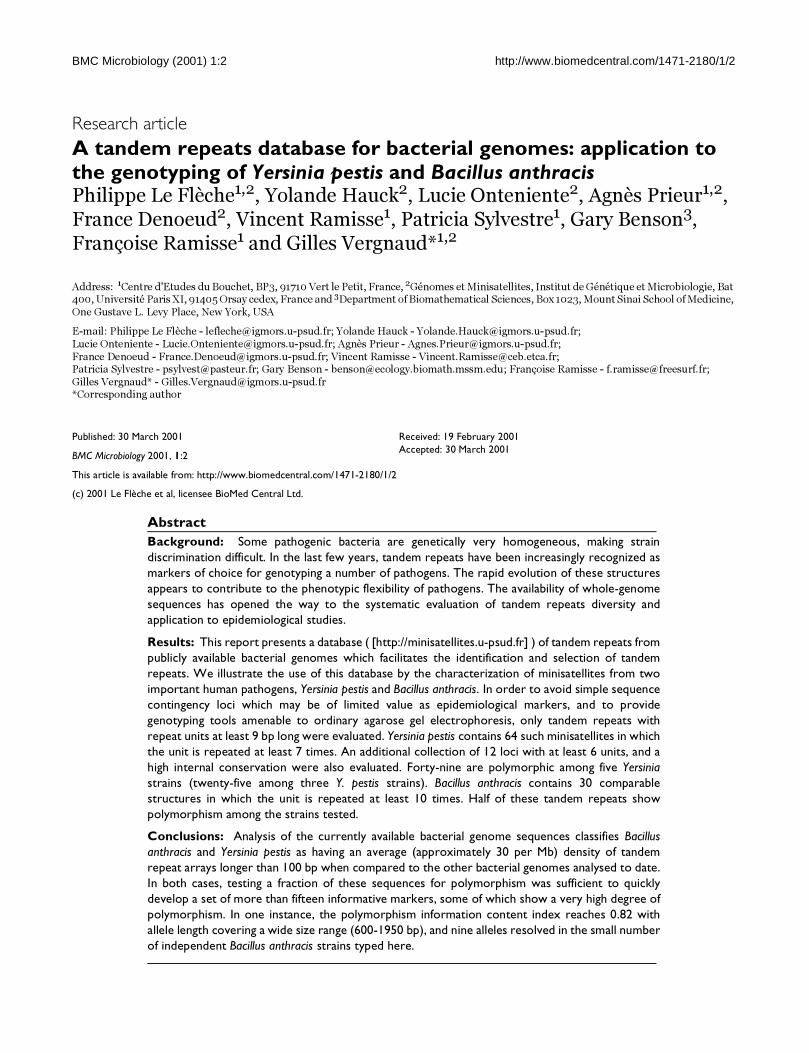
BMC Microbiology (2001) 1:2 http://www.biomedcentral.com/1471-2180/1/2
BMC Microbiology (2001) 1:2Research article
A tandem repeats database for bacterial genomes: application to the genotyping of Yersinia pestis and Bacillus anthracisPhilippe Le Flèche1,2, Yolande Hauck2, Lucie Onteniente2, Agnès Prieur1,2,
France Denoeud2, Vincent Ramisse1, Patricia Sylvestre1, Gary Benson3,
Françoise Ramisse1 and Gilles Vergnaud*1,2
Address: 1Centre d'Etudes du Bouchet, BP3, 91710 Vert le Petit, France, 2Génomes et Minisatellites, Institut de Génétique et Microbiologie, Bat 400, Université Paris XI, 91405 Orsay cedex, France and 3Department of Biomathematical Sciences, Box 1023, Mount Sinai School of Medicine,
One Gustave L. Levy Place, New York, USA
E-mail: Philippe Le Flèche - [email protected]; Yolande Hauck - [email protected];
Lucie Onteniente - [email protected]; Agnès Prieur - [email protected];
France Denoeud - [email protected]; Vincent Ramisse - [email protected]; Patricia Sylvestre - [email protected]; Gary Benson - [email protected]; Françoise Ramisse - [email protected];
Gilles Vergnaud* - [email protected]*Corresponding author
Abstract
Background: Some pathogenic bacteria are genetically very homogeneous, making strain
discrimination difficult. In the last few years, tandem repeats have been increasingly recognized as
markers of choice for genotyping a number of pathogens. The rapid evolution of these structures
appears to contribute to the phenotypic flexibility of pathogens. The availability of whole-genome
sequences has opened the way to the systematic evaluation of tandem repeats diversity and
application to epidemiological studies.
Results: This report presents a database ( [http://minisatellites.u-psud.fr] ) of tandem repeats from
publicly available bacterial genomes which facilitates the identification and selection of tandem
repeats. We illustrate the use of this database by the characterization of minisatellites from two
important human pathogens, Yersinia pestis and Bacillus anthracis. In order to avoid simple sequence
contingency loci which may be of limited value as epidemiological markers, and to provide
genotyping tools amenable to ordinary agarose gel electrophoresis, only tandem repeats with
repeat units at least 9 bp long were evaluated. Yersinia pestis contains 64 such minisatellites in which
the unit is repeated at least 7 times. An additional collection of 12 loci with at least 6 units, and a
high internal conservation were also evaluated. Forty-nine are polymorphic among five Yersinia
strains (twenty-five among three Y. pestis strains). Bacillus anthracis contains 30 comparable
structures in which the unit is repeated at least 10 times. Half of these tandem repeats show
polymorphism among the strains tested.
Conclusions: Analysis of the currently available bacterial genome sequences classifies Bacillus
anthracis and Yersinia pestis as having an average (approximately 30 per Mb) density of tandem
repeat arrays longer than 100 bp when compared to the other bacterial genomes analysed to date.
In both cases, testing a fraction of these sequences for polymorphism was sufficient to quickly
develop a set of more than fifteen informative markers, some of which show a very high degree of
polymorphism. In one instance, the polymorphism information content index reaches 0.82 with
allele length covering a wide size range (600-1950 bp), and nine alleles resolved in the small number
of independent Bacillus anthracis strains typed here.
Published: 30 March 2001
BMC Microbiology 2001, 1:2
This article is available from: http://www.biomedcentral.com/1471-2180/1/2
(c) 2001 Le Flèche et al, licensee BioMed Central Ltd.
Received: 19 February 2001
Accepted: 30 March 2001

BMC Microbiology (2001) 1:2 http://www.biomedcentral.com/1471-2180/1/2
BackgroundThe polymorphism associated with tandem repeats has
been instrumental in mammalian genetics for the con-
struction of genetic maps and still is the basis of DNA fin-gerprinting in forensic applications. Tandem repeats are
usually classified among satellites (spanning megabases
of DNA, associated with heterochromatin), minisatel-
lites (repeat units in the range 6-100 bp, spanning hun-
dreds of base-pairs) and microsatellites (repeat units in
the range 1-5 bp, spanning a few tens of nucleotides).
More recently, a number of studies have supported the
notion that tandem repeats reminiscent of mini and mi-
crosatellites are likely to be a highly significant source of
very informative markers for the identification of patho-
genic bacteria even when these pathogens are recently
emerged, highly monomorphic species [1–5]. This prob-
ably reflects the important contribution of tandem re-
peats to the adaptation of the pathogen to its host.
Tandem repeats appear to contribute to phenotypic var-
iation in bacteria in at least two ways. Tandem repeats lo-
cated within the regulatory region of a gene can
constitute an on/off switch of gene expression at the
transcriptional level [6,7]. Similarly, tandem repeats
within coding regions with repeat units length not a mul-
tiple of three can induce a reversible premature end of
translation when a mutation changes the number of re-
peats (reviewed in [8–10]). In other instances, the re-
peated unit length is a multiple of three, and the tandemrepeat contributes to a coding region. In such cases, var-
iations in the number of copies modify the gene product
itself [11].
Mutation mechanisms of micro and minisatellites have
been studied in some detail in eukaryotes, essentially hu-
man and yeast (reviewed in [12]). In brief, the data ob-
tained so far suggest that microsatellites mutate by
replication slippage processes; mutation rates depend
upon the efficiency of mismatch repair mechanisms and
an internal heterogeneity within the array strongly stabi-
lizes the tandem repeat. In contrast, minisatellites mu-
tate predominantly as the result of the repair of a double
strand break initiated within, or very close to, the tan-
dem repeat. In eukaryotes at least, these events can be of
replicative origin [13], or can be genetically controlled,
and specifically induced, during meiosis, at double
strand breaks hot-spots. Minisatellite mutation rate in
eukaryotes appears to be insensitive to mismatch repair
efficiency, and internal heterogeneity is compatible with
a high mutation rate [12, 14].
In bacteria, loci containing a tandem repeat from the mi-
crosatellite class (repeat unit sizes of 1-8 bp) have been
called simple sequence contingency loci [8]. Alterednumber of repeats allows for reversible on and off states
of expression for the corresponding gene. The mutation
rate of a tetranucleotide (microsatellite) tract in Haemo-
philus influenzae is higher than 10-4 and contributes to
the adaptation of the pathogen to its hosts as the infec-tion progresses [15]. In such an extreme situation, the
microsatellite is of limited value for strain identification,
epidemiological and phylogenetic studies. The tandem
repeat array is composed of perfect copies of the elemen-
tary unit, and different alleles are observed in a single
culture. In contrast, the phylogenetic identity of minisat-
ellite alleles of identical size can usually be further
checked by DNA sequencing, since the repeated units are
often not perfect [16]. The pattern of variants along the
array provides an additional level of allele identification
and phylogenetic information. In addition, tandem re-
peats with longer repeat unit length can be relatively eas-
ily typed in the size range of a few hundred base-pairs
using ordinary horizontal gel electrophoresis.
In this report, we will first describe the use of a tandem
repeats database for bacterial genomes ( [http://minis-
atellites.u-psud.fr] ) and briefly compare the general
characteristics of tandem repeats in a number of bacteri-
al genomes for which the sequence has been determined
and made publicly available. We will then show how this
tool can easily be applied to the rapid characterization of
new highly polymorphic markers in two pathogens, Y.
pestis and B. anthracis.
Both Y. pestis (causative agent of plague) and B. anthra-
cis (causative agent of anthrax) are recently emerged
clones of respectively Y. pseudotuberculosis [17] and B.
cereus [18]. In the case of Y. pestis, a high resolution typ-
ing tool based on RFLP (Restriction Fragment Length
Polymorphism) analysis of IS100 locations has already
been developed [17]. However this technology is more
demanding than PCR typing, which justifies the develop-
ment of such an assay. In the case of B. anthracis, poly-
morphisms were initially identified essentially using
AFLP (Amplified Fragment Length Polymorphism) typ-
ing [19]. Subsequent analyses demonstrated that the
most informative fragments in AFLP patterns resulted
from tandem repeat array length variations (five minis-
atellite loci were characterized in this way [2]).
Results and discussionUse of the tandem repeats database
To date, 36 bacterial genome sequences from 32 species
have been released in the public domain and are includ-
ed in the database (Figure 1A; the nine archaebacteria
genomes sequenced to date are presented in an other
page, which can be accessed from [http://minisatel-
lites.u-psud.fr/] ). As many other sequencing projects
are under way ( [http://www.ncbi.nlm.nih.gov/PMGifs/Genomes/bact.html] ; [http://www.tigr.org/tdb/mdb/

BMC Microbiology (2001) 1:2 http://www.biomedcentral.com/1471-2180/1/2
Figure 1Querying the tandem repeats database 1A: bacterial tandem repeats main page Bacteria species are listed in alphabeticalorder. The name of the strain used for sequencing is indicated after the species name and before the genome size (expressedin megabase). The rightmost figure indicates the density (per Mb) of tandem repeat arrays longer than 100 bp. The search fortandem repeats can be restricted according to a combination of criteria, including total array length (L), repeat unit length (U),number of repeats (N), internal conservation of the repeats (V), position (expressed in kilobase) on the genome (Pos), GCcontent of the array (%GC), strand bias (B). Three different biases can be evaluated, GC bias, AT bias and Purine-Pyrimidinebias. The bias reflects strand asymmetry of the repeat sequence. The search output can either present a list of characteristicsof the tandem repeats fulfilling criteria, ordered according to their position on the genome, or classify the tandem repeatsaccording to a selected structural parameter. 1B: examples of queries in three genomes All tandem repeat arrays spanningmore than 100 base-pairs are classified according to repeat unit length. The query was run on Buchnera sp. (left panel), Yersiniapestis (middle panel) and Pseudomonas aeruginosa (right panel).

BMC Microbiology (2001) 1:2 http://www.biomedcentral.com/1471-2180/1/2
mdbinprogress.html] ; [http://www.sanger.ac.uk/
Projects/Microbes/] ), the database will be regularly up-
dated. The collection of tandem repeats present in a giv-
en genome can be queried according to a combination ofcriteria, total tandem repeat array length (L), repeat unit
length (U), number of repeats (N), percentage of conser-
vation of the repeats along the array (V), position on the
genome (Pos), average GC percent of the repeats (%GC),
strand bias in nucleotide composition (B) (these values
have been precomputed using the Tandem Repeats Find-
er software described in [20]). The results shown on Fig-
ure 1B use the "Tandem Repeats Distribution according
to repeat unit length" option (Figure 1A). Three genomes
were searched for tandem repeat arrays longer than 100
base-pairs (L ≥ 100). The genomes selected illustrate
three different behaviors. On the right panel, Pseu-
domonas aeruginosa shows a very striking bias towards
minisatellites with a motif length multiple of three. On
the left and middle panels of Figure 1B, Buchnera sp and
Y. pestis, show no such bias. The overall density of tan-
dem repeat arrays longer than 100 base-pairs varies in
the different genomes. Buchnera sp. contains 103 such
loci, for a total genome size of 641 kb, which corresponds
to a density per megabase of 161. Pseudomonas aerugi-
nosa, with a total genome length of 6.3 Mb, has a density
of 48. Y. pestis has an intermediate value of 30. Figure 2
summarizes the values observed in the 32 species. Ten
non pathogenic species are presented in the upper part,
22 pathogenic species on the lower part. The species areordered from top to bottom according to increasing ge-
nome size. The dark bars indicate for each genome the
density per megabase of tandem repeat arrays longer
than 100 bp. The clear bars reflect the excess of tandem
repeats with unit length a multiple of three. A wide range
of situations is observed, with a remarkable excess of
tandem repeats multiples of three in Mycobacterium tu-
berculosis and Pseudomonas aeruginosa, presumably
reflecting a significant contribution of tandem repeats to
coding regions in these two bacteria.
As a quick illustration of the use of this database to facil-
itate the development of genotyping tools for bacterial
genomes, we have evaluated the polymorphism associat-
ed with tandem repeats from Y. pestis on one hand and
B. anthracis on the other (in this second instance, the ge-
nome sequence has not been completed yet and does not
appear on the publicly accessible Tandem Repeats Data-
base page, Figure 1A).
Application to Y. pestis
Figure 3A presents the result of a query run on Y. pestis,
to identify tandem repeats with repeat units longer than
9 base-pairs repeated at least 7 times in the strain which
has been sequenced (CO-92 biovar Orientalis).Sixty-fourtandem repeats fulfill these criteria (an additional group
of forty-nine have 6 copies of the motif; the twelve loci
with the highest internal conservation were also included
in this study). The output includes links to individual
alignment files, as produced by the Tandem Repeat
Finder software [20]. The alignment file also includes
200 base-pairs of flanking sequence from each side of
the tandem repeat, from which primers can be selected
for PCR amplification. Figure 3B shows an annotated ex-
tract of one alignment file. The positions of the primers
selected for subsequent PCR amplification are under-
lined. Three Y. pestis (representing the Antiqua, Medie-
valis, and Orientalis biovars [17]) and two Y.
pseudotuberculosis strains were used for the initial iden-
tification of minisatellites sufficiently polymorphic to be
of interest for further studies. Table 1 summarizes the
PCR conditions used for each polymorphic locus and the
results obtained. A total of 76 tandem repeats were test-
ed. PCR amplification failed in 6 cases. Twenty one loci
are monomorphic in the five Yersinia strains typed here.
Forty-nine of the loci are polymorphic (Table 1). Twenty-five of these are polymorphic among the Y. pestis strains.
Figure 2Relative frequency of tandem repeats within bacte-rial genomes The ten non-pathogen species are listed ontop. Within each category, species are ordered according togenome size (smallest genome on top). The density of tan-dem repeat arrays longer than 100 bp is plotted for each spe-
cies (dark bars). The clear bars reflect the excess (χ2 values)of tandem repeats with a repeat unit length multiple of three.
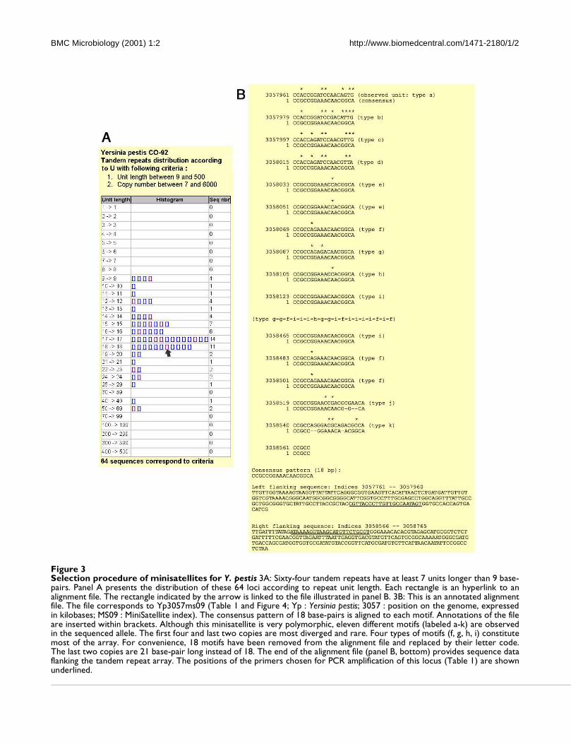
BMC Microbiology (2001) 1:2 http://www.biomedcentral.com/1471-2180/1/2
Figure 3Selection procedure of minisatellites for Y. pestis 3A: Sixty-four tandem repeats have at least 7 units longer than 9 base-pairs. Panel A presents the distribution of these 64 loci according to repeat unit length. Each rectangle is an hyperlink to analignment file. The rectangle indicated by the arrow is linked to the file illustrated in panel B. 3B: This is an annotated alignmentfile. The file corresponds to Yp3057ms09 (Table 1 and Figure 4; Yp : Yersinia pestis; 3057 : position on the genome, expressedin kilobases; MS09 : MiniSatellite index). The consensus pattern of 18 base-pairs is aligned to each motif. Annotations of the fileare inserted within brackets. Although this minisatellite is very polymorphic, eleven different motifs (labeled a-k) are observedin the sequenced allele. The first four and last two copies are most diverged and rare. Four types of motifs (f, g, h, i) constitutemost of the array. For convenience, 18 motifs have been removed from the alignment file and replaced by their letter code.The last two copies are 21 base-pair long instead of 18. The end of the alignment file (panel B, bottom) provides sequence dataflanking the tandem repeat array. The positions of the primers chosen for PCR amplification of this locus (Table 1) are shownunderlined.

BMC Microbiology (2001) 1:2 http://www.biomedcentral.com/1471-2180/1/2
Seven present a different allele in each of the five Yersin-
ia strains, thirteen have a different allele in each of the
three Y. pestis strains. Gel images for the 25 loci poly-
morphic among Y. pestis are shown in Figure 4. As can beseen, the repeat unit size and the overall length of the
PCR products are such that tandem repeats differing by
a single repeat unit can be distinguished by simple agar-
ose gel electrophoresis.
Application to B. anthracis
Given the relatively low overall size of most bacterial tan-
dem repeats, tandem repeat search can be run even on
unfinished sequences. Tandem Repeats Finder was ap-
plied to B. anthracis sequence obtained from The Insti-
tute for Genomic Research through the website at
[http://www.tigr.org] . The sequence was recovered as
approximately 1000 contigs, for a total amount of slight-
ly more than 5 Mb. Thirty tandem repeats have at least 10
copies of a repeat unit longer than 9 base-pairs. Fourteen
of them are polymorphic among the 31 B. anthracis
strains typed here (Table 2). Twenty-seven different gen-
otypes are identified. Polymorphism information con-
tent (PIC) indexes based on the 27 genotypes vary from
0.07 to 0.82. Nine PIC values are above 0.5. Eight alleles
are identified for CEB-Bams30, in a size range 270-900
base-pairs (Figure 5). In this case, the resolution of the
largest alleles would probably be improved by using an
automated DNA sequencer, and more alleles might be
resolved. There are clear gaps in the size range coverageshown in Figure 5, and it is likely that the typing of addi-
tional strains would uncover new alleles. The genotyping
data obtained was used to construct a phylogenetic tree
based upon the Neighbor-Joining method ( [http://
www.infobiogen.fr] ). In order to be able to correlate the
tree obtained here with earlier studies [2], 5 minisatel-
lites and one microsatellite reported previously were also
typed. Figure 6 presents the data obtained and the re-
sulting tree, using the nomenclature previously pro-
posed [2]. Six Bacillus cereus strains have also been
included and used as an outgroup in the analysis. Occa-
sionally B. cereus strains will not amplify (scored as 0 in
Figure 6) or will give weak amplification signals (Figure
5, last six lanes on the right). The proposed tree is in good
agreement with earlier results. In particular, the A and B
clusters are well defined. We have apparently no repre-
sentatives for the A1b and A3a group, whereas strains
9533 and 9502 to 9505 appear to define a new branch.
The correspondence between allele numbering and allele
size is indicated in Table 3.
Correlations between polymorphism and structural char-
acteristics of minisatellites
We have looked for correlations between on one hand
the number of alleles and polymorphism of the minisat-ellites, and on the other, simple structural characteristics
of the tandem repeats in the sequenced strain : motif
size, number of motifs, total length, conservation of the
motifs along the array (percent identity), GC content,
strand bias. In the case of B. anthracis, a highly signifi-cant correlation (0.01 level) is observed between poly-
morphism and both total length and GC content. This is
not true for Y. pestis in which a strong correlation is seen
between the number of alleles and the conservation of
the motifs (Figure 7).
ConclusionsWe limited here our investigation of tandem repeats to
minisatellites, i.e. repeat units longer than 9 base-pairs,
so as to avoid simple sequence contingency loci [8] of
limited epidemiological value, and to facilitate the typing
of alleles with agarose gel electrophoresis. However, sim-
ple sequence contingency loci are also represented in the
database and are of great interest for molecular patho-
genicity studies [6–8]. The use of the tandem repeats da-
tabase was demonstrated here on two of the most
genetically homogeneous human pathogenes, Y. pestis
and B. anthracis. There is consequently a possibility that
a common database format for identification and epide-
miological analyses of pathogens amenable to minisatel-
lite typing be developed. As more data becomes available
on polymorphism associated with tandem repeats, it will
be added to the database presented here in order to avoid
duplication of work and nomenclature.
Bacterial species differ very significantly in the density of
tandem repeats within their genome, and also in their
use of tandem repeats. Some species have a very strong
excess of tandem repeats with repeat units length which
are multiple of three, the most striking examples being
M. tuberculosis and P. aeruginosa. Polymorphism in
such tandem repeats is likely to modulate the protein
structure rather than gene activity. In M. tuberculosis, all
tandem repeats with total length (L) higher than 100 bp
and 9 or 15 base-pairs long units are located with ORFs
[21]. An important proportion of these tandem repeats
correspond to the so-called PE and PPE multigene fami-
lies [21].
In the two species studied here, tandem repeat polymor-
phism is strongly correlated with one or more of the se-
quenced allele characteristics, as illustrated in Figure 7.
In Yersinia pestis a strong correlation is observed be-
tween number of alleles observed and homogeneity of
the tandem array. In Bacillus anthracis, the strongest
correlations are with total array length and GC content.
It appears that the correlations are not the same in the
two species, so that at present at least, the polymorphism
associated with a tandem repeat cannot be inferred from
its primary sequence. In particular, and in contrast towhat is known for microsatellites (1-5 bp repeat units),
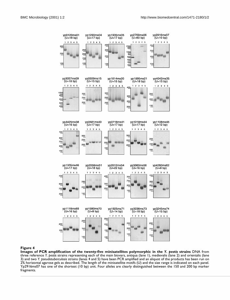
BMC Microbiology (2001) 1:2 http://www.biomedcentral.com/1471-2180/1/2
Figure 4Images of PCR amplification of the twenty-five minisatellites polymorphic in the Y. pestis strains DNA fromthree reference Y. pestis strains representing each of the main biovars, antiqua (lane 1), medievalis (lane 2) and orientalis (lane3) and two Y. pseudotuberculosis strains (lanes 4 and 5) have been PCR amplified and an aliquot of the products has been run on2% horizontal agarose gels as described. The length of the minisatellite motifs (U) and the size range is indicated on each panel.Yp2916ms07 has one of the shortest (10 bp) unit. Four alleles are clearly distinguished between the 150 and 200 bp markerfragments.
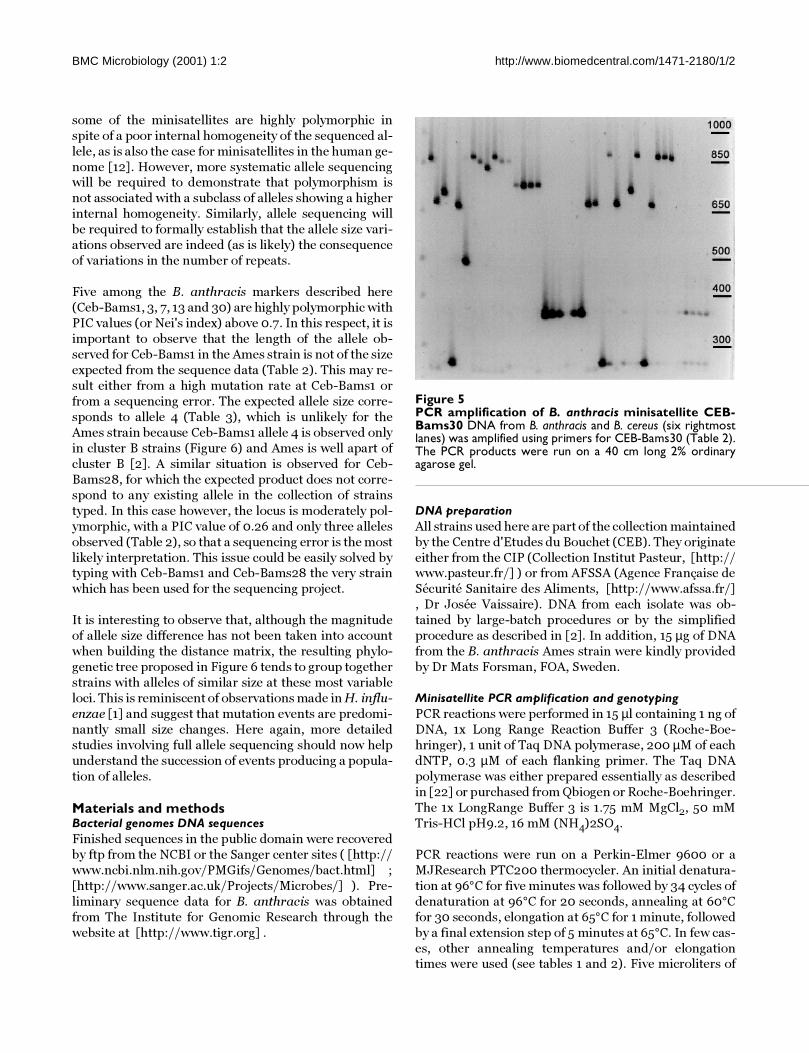
BMC Microbiology (2001) 1:2 http://www.biomedcentral.com/1471-2180/1/2
some of the minisatellites are highly polymorphic in
spite of a poor internal homogeneity of the sequenced al-
lele, as is also the case for minisatellites in the human ge-
nome [12]. However, more systematic allele sequencingwill be required to demonstrate that polymorphism is
not associated with a subclass of alleles showing a higher
internal homogeneity. Similarly, allele sequencing will
be required to formally establish that the allele size vari-
ations observed are indeed (as is likely) the consequence
of variations in the number of repeats.
Five among the B. anthracis markers described here
(Ceb-Bams1, 3, 7, 13 and 30) are highly polymorphic with
PIC values (or Nei's index) above 0.7. In this respect, it is
important to observe that the length of the allele ob-
served for Ceb-Bams1 in the Ames strain is not of the size
expected from the sequence data (Table 2). This may re-
sult either from a high mutation rate at Ceb-Bams1 or
from a sequencing error. The expected allele size corre-
sponds to allele 4 (Table 3), which is unlikely for the
Ames strain because Ceb-Bams1 allele 4 is observed only
in cluster B strains (Figure 6) and Ames is well apart of
cluster B [2]. A similar situation is observed for Ceb-
Bams28, for which the expected product does not corre-
spond to any existing allele in the collection of strains
typed. In this case however, the locus is moderately pol-
ymorphic, with a PIC value of 0.26 and only three alleles
observed (Table 2), so that a sequencing error is the most
likely interpretation. This issue could be easily solved bytyping with Ceb-Bams1 and Ceb-Bams28 the very strain
which has been used for the sequencing project.
It is interesting to observe that, although the magnitude
of allele size difference has not been taken into account
when building the distance matrix, the resulting phylo-
genetic tree proposed in Figure 6 tends to group together
strains with alleles of similar size at these most variable
loci. This is reminiscent of observations made in H. influ-
enzae [1] and suggest that mutation events are predomi-
nantly small size changes. Here again, more detailed
studies involving full allele sequencing should now help
understand the succession of events producing a popula-
tion of alleles.
Materials and methodsBacterial genomes DNA sequences
Finished sequences in the public domain were recovered
by ftp from the NCBI or the Sanger center sites ( [http://
www.ncbi.nlm.nih.gov/PMGifs/Genomes/bact.html] ;
[http://www.sanger.ac.uk/Projects/Microbes/] ). Pre-
liminary sequence data for B. anthracis was obtained
from The Institute for Genomic Research through the
website at [http://www.tigr.org] .
DNA preparation
All strains used here are part of the collection maintained
by the Centre d'Etudes du Bouchet (CEB). They originate
either from the CIP (Collection Institut Pasteur, [http://www.pasteur.fr/] ) or from AFSSA (Agence Française de
Sécurité Sanitaire des Aliments, [http://www.afssa.fr/]
, Dr Josée Vaissaire). DNA from each isolate was ob-
tained by large-batch procedures or by the simplified
procedure as described in [2]. In addition, 15 µg of DNA
from the B. anthracis Ames strain were kindly provided
by Dr Mats Forsman, FOA, Sweden.
Minisatellite PCR amplification and genotyping
PCR reactions were performed in 15 µl containing 1 ng of
DNA, 1x Long Range Reaction Buffer 3 (Roche-Boe-
hringer), 1 unit of Taq DNA polymerase, 200 µM of each
dNTP, 0.3 µM of each flanking primer. The Taq DNA
polymerase was either prepared essentially as described
in [22] or purchased from Qbiogen or Roche-Boehringer.
The 1x LongRange Buffer 3 is 1.75 mM MgCl2, 50 mM
Tris-HCl pH9.2, 16 mM (NH4)2SO4.
PCR reactions were run on a Perkin-Elmer 9600 or a
MJResearch PTC200 thermocycler. An initial denatura-
tion at 96°C for five minutes was followed by 34 cycles of
denaturation at 96°C for 20 seconds, annealing at 60°C
for 30 seconds, elongation at 65°C for 1 minute, followed
by a final extension step of 5 minutes at 65°C. In few cas-
es, other annealing temperatures and/or elongationtimes were used (see tables 1 and 2). Five microliters of
Figure 5PCR amplification of B. anthracis minisatellite CEB-Bams30 DNA from B. anthracis and B. cereus (six rightmostlanes) was amplified using primers for CEB-Bams30 (Table 2).The PCR products were run on a 40 cm long 2% ordinaryagarose gel.

BMC Microbiology (2001) 1:2 http://www.biomedcentral.com/1471-2180/1/2
Figure 6Bacillus anthracis phylogenetic tree The genotype of each strain for the polymorphic minisatellites is given (size estimatesfor each allele are given in Table 3). "0" indicates a failure of the PCR amplification. This is most often associated with B. cereusstrains, and probably reflects in these cases sequence divergence in the flanking sequence. The phylogenetic tree was producedusing the Neighbor-Joining method as available on-line at [http://www.infobiogen.fr.]

BMC Microbiology (2001) 1:2 http://www.biomedcentral.com/1471-2180/1/2
the PCR products where run on standard 1% or 2% agar-
ose gel (Qbiogen) in 0.5 x TBE buffer at a voltage of 10 V/
cm as indicated in Tables 1 and 2. Gel length of 10 to 40
cm were used according to PCR product size and motiflength. Gels were stained with ethidium bromide and
visualized under UV light. Allele sizes were estimated us-
ing as size markers the 1 kb ladder plus (Gibco-BRL
which also includes a 100 bp ladder between 100 bp and
500 bp, plus 650, 850 and 1000 bp bands) or the 50 bp
ladder (Euromedex) which provides a 50 bp ladder be-
tween 50 and 300 bp and a 100 bp ladder from 300 bp to
1000 bp.
Data analysis
Tandem Repeats Finder analysis:
Sequences were processed using the Tandem Repeats
Finder software ( [http://c3.biomath.mssm.edu/
trf.html] ). The output was processed to eliminate dupli-
cates before being imported in a database (running un-
der Access2000, Microsoft Corp.) as described
previously [12]. The B. anthracis preliminary sequence
data file uses FASTA type of headers (i.e. >sequenceId)
to separate the independent contigs. The headers were
replaced by runs of 10 Ns before running Tandem Re-
peats Finder.
Blast queries against the M. tuberculosis genome:
The identifications of the open reading frames contain-
ing a given tandem repeat from M. tuberculosis weredone by running a BLAST search on the dedicated web
page at [http://www.sanger.ac.uk/Projects/
M_tuberculosis/blast_server.shtml] .
Estimation of the excess of tandem repeats with motif length multi-
ple of three:
A χ2 test was calculated for the difference between the
observed number of tandem repeats with motif length
multiple of 3 and the expected number of tandem repeats
with motif length multiple of 3 (expected value in the ab-
sence of bias being the total number of tandem repeats
divided by 3). The χ2 values vary from 0.01 to 253.5.
There is a significant excess (χ2 > 3.841) for all species
but 6 (Buchnera sp, T. maritima, H. influenzae, M. gen-
italium, R. prowazekii, Y. pestis).
Polymorphism index:
Polymorphism Information Index (PIC) or Nei's diversi-
ty index is calculated as 1 - Σ (allele frequency)2 based
upon the unique genotypes.
Phylogenetic reconstruction:
A phenetic approach, based on a distance matrix was
used. Distance matrix between strains was obtained by
counting the number of differences between the corre-sponding genotypes. Then, Neighbor Joining cluster
Figure 7Significant correlation between number of alleles andminisatellites structural characteristics The number ofalleles is plotted as a function of Total length and %GC forBacillus anthracis, and %matches for Yersinia pestis (the corre-lations are highly significant at the 0.01 level). Number of alle-les for each locus is the total number detected (i.e. Bacillusanthracis and B. cereus; Yersinia pestis and Y. pseudotuberculo-sis).
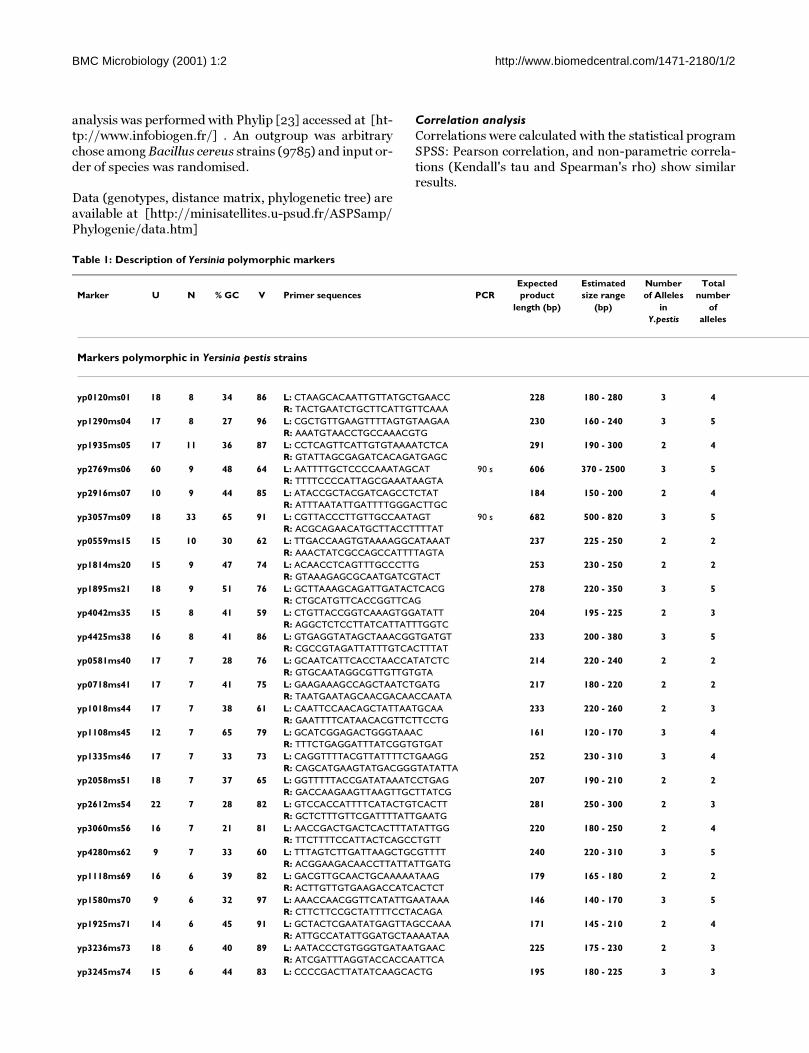
BMC Microbiology (2001) 1:2 http://www.biomedcentral.com/1471-2180/1/2
analysis was performed with Phylip [23] accessed at [ht-
tp://www.infobiogen.fr/] . An outgroup was arbitrary
chose among Bacillus cereus strains (9785) and input or-
der of species was randomised.
Data (genotypes, distance matrix, phylogenetic tree) are
available at [http://minisatellites.u-psud.fr/ASPSamp/
Phylogenie/data.htm]
Correlation analysis
Correlations were calculated with the statistical program
SPSS: Pearson correlation, and non-parametric correla-
tions (Kendall's tau and Spearman's rho) show similarresults.
Table 1: Description of Yersinia polymorphic markers
Expected Estimated Number Total
Marker U N % GC V Primer sequences PCR product size range of Alleles number
length (bp) (bp) in of
Y.pestis alleles
Markers polymorphic in Yersinia pestis strains
yp0120ms01 18 8 34 86 L: CTAAGCACAATTGTTATGCTGAACC 228 180 - 280 3 4
R: TACTGAATCTGCTTCATTGTTCAAA
yp1290ms04 17 8 27 96 L: CGCTGTTGAAGTTTTAGTGTAAGAA 230 160 - 240 3 5
R: AAATGTAACCTGCCAAACGTG
yp1935ms05 17 11 36 87 L: CCTCAGTTCATTGTGTAAAATCTCA 291 190 - 300 2 4
R: GTATTAGCGAGATCACAGATGAGC
yp2769ms06 60 9 48 64 L: AATTTTGCTCCCCAAATAGCAT 90 s 606 370 - 2500 3 5
R: TTTTCCCCATTAGCGAAATAAGTA
yp2916ms07 10 9 44 85 L: ATACCGCTACGATCAGCCTCTAT 184 150 - 200 2 4
R: ATTTAATATTGATTTTGGGACTTGC
yp3057ms09 18 33 65 91 L: CGTTACCCTTGTTGCCAATAGT 90 s 682 500 - 820 3 5
R: ACGCAGAACATGCTTACCTTTTAT
yp0559ms15 15 10 30 62 L: TTGACCAAGTGTAAAAGGCATAAAT 237 225 - 250 2 2
R: AAACTATCGCCAGCCATTTTAGTA
yp1814ms20 15 9 47 74 L: ACAACCTCAGTTTGCCCTTG 253 230 - 250 2 2
R: GTAAAGAGCGCAATGATCGTACT
yp1895ms21 18 9 51 76 L: GCTTAAAGCAGATTGATACTCACG 278 220 - 350 3 5
R: CTGCATGTTCACCGGTTCAG
yp4042ms35 15 8 41 59 L: CTGTTACCGGTCAAAGTGGATATT 204 195 - 225 2 3
R: AGGCTCTCCTTATCATTATTTGGTC
yp4425ms38 16 8 41 86 L: GTGAGGTATAGCTAAACGGTGATGT 233 200 - 380 3 5
R: CGCCGTAGATTATTTGTCACTTTAT
yp0581ms40 17 7 28 76 L: GCAATCATTCACCTAACCATATCTC 214 220 - 240 2 2
R: GTGCAATAGGCGTTGTTGTGTA
yp0718ms41 17 7 41 75 L: GAAGAAAGCCAGCTAATCTGATG 217 180 - 220 2 2
R: TAATGAATAGCAACGACAACCAATA
yp1018ms44 17 7 38 61 L: CAATTCCAACAGCTATTAATGCAA 233 220 - 260 2 3
R: GAATTTTCATAACACGTTCTTCCTG
yp1108ms45 12 7 65 79 L: GCATCGGAGACTGGGTAAAC 161 120 - 170 3 4
R: TTTCTGAGGATTTATCGGTGTGAT
yp1335ms46 17 7 33 73 L: CAGGTTTTACGTTATTTTCTGAAGG 252 230 - 310 3 4
R: CAGCATGAAGTATGACGGGTATATTA
yp2058ms51 18 7 37 65 L: GGTTTTTACCGATATAAATCCTGAG 207 190 - 210 2 2
R: GACCAAGAAGTTAAGTTGCTTATCG
yp2612ms54 22 7 28 82 L: GTCCACCATTTTCATACTGTCACTT 281 250 - 300 2 3
R: GCTCTTTGTTCGATTTTATTGAATG
yp3060ms56 16 7 21 81 L: AACCGACTGACTCACTTTATATTGG 220 180 - 250 2 4
R: TTCTTTTCCATTACTCAGCCTGTT
yp4280ms62 9 7 33 60 L: TTTAGTCTTGATTAAGCTGCGTTTT 240 220 - 310 3 5
R: ACGGAAGACAACCTTATTATTGATG
yp1118ms69 16 6 39 82 L: GACGTTGCAACTGCAAAAATAAG 179 165 - 180 2 2
R: ACTTGTTGTGAAGACCATCACTCT
yp1580ms70 9 6 32 97 L: AAACCAACGGTTCATATTGAATAAA 146 140 - 170 3 5
R: CTTCTTCCGCTATTTTCCTACAGA
yp1925ms71 14 6 45 91 L: GCTACTCGAATATGAGTTAGCCAAA 171 145 - 210 2 4
R: ATTGCCATATTGGATGCTAAAATAA
yp3236ms73 18 6 40 89 L: AATACCCTGTGGGTGATAATGAAC 225 175 - 230 2 3
R: ATCGATTTAGGTACCACCAATTCA
yp3245ms74 15 6 44 83 L: CCCCGACTTATATCAAGCACTG 195 180 - 225 3 3

BMC Microbiology (2001) 1:2 http://www.biomedcentral.com/1471-2180/1/2
R: AACTGACGATCTTTTTCACTGAGTT
Markers polymorphic in 5 Yersinia strains (monomorphic in pestis)
yp0802ms02 18 12 49 86 L: CTGACACAAAACGAGAGCCTATTT 53°C 314 240 - 315 1 2
R: AGCGTGAGTGGGCTATCAATAC 1 min
yp2925ms08 15 12 39 63 L: AGCCTTTTTGTTGATTATCAGTCAT 270 270 - 290 1 2
R: CGATAATAATACTGAATTACCGGATG
yp4411ms10 14 8 32 69 L: ATCATGCTTTTGCCTCAATATAATC 191 190 - 210 1 2
R: GAAACGCAGTCCCTGTTGTAG
yp0813ms16 17 8 39 64 L: GTTGGTTATCCGACAGTCTTCAATA 235 230 - 270 1 2
R: GCAATTCGTTATGGCTTAGTAAAAA
yp1269ms18 27 9 54 55 L: GCAAAAGCTGAAGCAGATAAAATAG 303 220 - 250 1 2
R: AAACCACCAAACAAATCATCAAC
yp2196ms22 20 8 12 55 L: AAACCAACAAGAAAACTGTAACCAC 90 s 265 270 - 1500 1 2
R: CATTCACCATTGATGTCCTTAGAC
yp2324ms24 19 8 34 65 L: TTCACCGGGTTACCTTAATTACATA 255 215 - 255 1 2
R: CTACCTTGCTGTCAACACTCGAC
yp2331ms25 17 9 36 76 L: AACGCGTTAATAAAACAATAAAGTG 181 190 - 230 1 3
R: CAATATCCTTTTACTCAGCCGATG
yp2679ms27 16 8 20 76 L: ATGATTACTGGCAAGAGCACTATGT 217 200 - 220 1 2
R: AACAAGATCACCTGGTCGTTAAA
yp2908ms28 18 8 40 69 L: GCAGAAATAATCTTCAGGAGAAACA 242 190 - 290 1 2
R: AGATCGTCGTTAGTCCATGTCAG
yp2958ms29 16 8 23 61 L: AAAATAGTCTGTGTTTCAGCAAAGC 215 215 - 245 1 2
R: CCTTAAAAACCCTAAGTGGGTAAAA
yp3225ms30 54 11 51 52 L: CAATAATACCATCGTGCGTGATAC 683 680 - 900 1 2
R: TATTAATGGTGGTGTTAGTCGCTGT
yp3532ms31 14 8 30 67 L: GTTATTTATTTTTGCCCCAACTTGT 217 215- 245 1 2
R: TTAGCCTGTTTGTTCTTCAAATAGC
yp3787ms32 18 8 49 65 L: CGATAACGTTAATGCCATCAGTAG 218 190 - 240 1 3
R: GCGCCGGTAAGTTTTGTTTATTA
yp3795ms33 15 8 43 67 L: CCCTTCTTTTTATGCTTGAAGATACT 210 210 - 225 1 2
R: GTTGAACCACAGGCTGTTGAG
yp4371ms37 18 8 35 82 L: TACTTAGGCATTGTCTCTTCACTCC 235 235 - 255 1 2
R: CTGAAATTATCAGTAGGTGGTTCGT
yp0999ms43 17 7 38 80 L: ATTCCACCACCAACAATTATCAC 211 220 - 300 1 3
R: GGTATTCGTATTGAAGATGACATTG
yp1962ms50 18 7 34 71 L: TACCGAGGTATTCCCTGGTCTAAT 225 225 - 240 1 2
R: AGTTGACTCCCAGTCACTTTTCC
yp3734ms59 16 7 36 69 L: ATTATCATGACCCTTCCAGTGCTAT 218 200 - 220 1 2
R: CATCAAAATGCCAGGAGAGTAAC
yp4338ms63 17 7 38 72 L: ATTAACGATTTCTTGTCGCTCAGT 194 190 - 275 1 3
R: AATCAGTAACGGCATGTGTCAGTA
yp0549ms66 18 6 41 83 L: TAAAAGCGTCAACAAAGTAGGTCAT 212 200 - 220 1 2
R: GTTCCTGTTGTTGAAAATGCTG
yp0782ms67 18 6 40 90 L: TTCCAGGCTAAAGATATTGACTTTG 248 250 - 270 1 2
R: CTCGGCTTGTTCTACGTTTAATG
yp1053ms68 18 6 32 82 L: CCGTTATCTGGTGAAAGTGAACAG 182 175 - 205 1 3
R: GTCCGGTAGCCTGATTGTTTATT
yp3634ms75 15 6 36 80 L: ATGTGAGCTTGATTGCTGAGTAGT 210 180 - 210 1 3
R: TCATATTTTAGGTGTTTTGCCTTTG
Some structural characteristics of the tandem repeats are presented : U (unit length), N (number of repeats), %GC, V (% of conservation). PCR and electrophoresis conditions are as described in the material and methods section : annealing temperature is 60°C, elongation time is 60 seconds and gels are 2% agarose except when indicated otherwise. Total number of alleles means number of alleles in 3 Y. pestis and 2 Y. pseudotuberculosis strains.
Table 1: Description of Yersinia polymorphic markers

BMC Microbiology (2001) 1:2 http://www.biomedcentral.com/1471-2180/1/2
Table 2: Description of Bacillus anthracis polymorphic markers
Expected Number
product Estimated of Total PIC
Marker U N % V Primer sequences PCR length in size alleles number index
GC bp range in B. of
(observed) (bp) anthracis alleles
Ceb-Bams 1 21 16 44 88 L: GTTGAGCATGAGAGGTACCTTGTCCTTTTT 485 410-520 5 5 0.72
R: AGTTCAAGCGCCAGAAGGTTATGAGTTATC (520)
Ceb-Bams 3 15 25 59 73 L: GCAGCAACAGAAAACTTCTCTCCAATAACA 544 480-860 6 9 0.75
R: TCCTCCCTGAGAACTGCTATCACCTTTAAC 1%
Ceb-Bams 5 39 10 49 92 L: GCAGGAAGAACAAAAGAAACTAGAAGAGCA 53°C 307 305-385 3 3 0.56
R:ATTATTAGCAGGGGCCTCTCCTGCATTACC 60s
Ceb-Bams 7 18 49 55 69 L: GAATATTCGTGCCACCTAACAAAACAGAAA 65°C 1017 600-1950 9 9 0.82
R: TGTCAGATCTAGTTGGCCCTACTTTTCCTC 1%
Ceb-Bams 13 9 70 60 79 L: AATTGAGAAATTGCTGTACCAAACT 120s 814 330-850 8 11 0.79
R: CTAGTGCATTTGACCCTAATCTTGT 1%
Ceb-Bams 15 18 12 57 77 L: GTATTTCCCCCAGATACAGTAATCC 409 410-610 5 5 0.59
R: GTGTACATGTTGATTCATGCTGTTT
Ceb-Bams 21 45 11 43 75 L: TGTAGTGCCAGATTTGTCTTCTGTA 676 540-680 3 3 0.14
R: CAAATTTTGAGATGGGAGTTTTACT
Ceb-Bams 22 36 15 39 81 L: ATCAAAAATTCTTGGCAGACTGA 735 590-950 4 6 0.51
R: ACCGTTAATTCACGTTTAGCAGA
Ceb-Bams 23 42 11 37 85 L: CGGTCTGTCTCTATTATTCAGTGGT 653 570-820 3 4 0.49
R: CCTGTTGCTCCTAGTGATTTCTTAC
Ceb-Bams 24 42 11 44 80 L: CTTCTACTTCCGTACTTGAAATTGG 630 335-670 3 6 0.2
R: CGTCACGTACCATTTAATGTTGTTA
Ceb-Bams 25 15 14 45 60 L: CCGAATACGTAAGAAATAAATCCAC 391 375-390 2 2 0.07
R: TGAAAGATCTTGAAAAACAAGCATT
Ceb-Bams 28 24 14 36 70 L: CTCTGTTGTAACAAAATTTCCGTCT 493 300-400 3 3 0.26
R: TATTAAACCAGGCGTTACTTACAGC (400)
Ceb-Bams 30 27 16 58 78 L: AGCTAATCACCTACAACACCTGGTA 120s 200-890 11 11 0.77
R: CAGAAAATATTGGACCTACCTTCC 1%
Ceb-Bams 31 9 64 58 57 L: GCTGTATTTATCGAGCTTCAAAATCT 772 300-850 4 4 0.32
R: GGAGTACTGTTTGTTGAATGTTGTTT 1%
Some structural characteristics of the tandem repeats are presented : U (unit length), N (number of repeats), %GC, V (% of conservation). PCR and electrophoresis conditions are as described in the material and methods section : annealing temperature is 60°C, elongation time is 60 seconds and gels are 2% agarose except when indicated otherwise. The expected product length is deduced from the sequencing data corresponding to the Ames strain. When the Ames strains typing does not fit with the expected value, the observed value is indicated between (). Only one side of the Ceb-Bams30 minisatellite can be identified in the available Ames sequence. The other side was identified in the course of the independent, partial sequencing of B. anthracis strains (Vergnaud and col., unpublished data). Total number of alleles includes alleles observed in the B. cereus strains. Polymorphism Information Index (PIC) or Nei's diversity index is calculated as 1 - Σ (allele frequency)2.
Table 3: Correspondence between B. anthracis allele sizes and allele numbering
allele nb
marker name 1 2 3 4 5 6 7 8 9 10
Ceb-Bams1 ~ 410 ~ 430 ~ 450 ~ 480 ~ 520
Ceb-Bams3 484 514 544 559 574 589 704 734 857
Ceb-Bams5 307 346 385
Ceb-Bams7 603 1017 1305 1503 1557 1647 1809 1899 1953
Ceb-Bams13 328 382 454 481 490 652 742 787 814 850
Ceb-Bams15 409 535 571 589 607
Ceb-Bams21 541 631 676
Ceb-Bams22 591 627 699 735 ~ 900 ~ 950
Ceb-Bams23 569 611 653 821
Ceb-Bams24 336 420 462 504 630 672
Ceb-Bams25 376 391
Ceb-Bams28 ~ 300 ~ 375 ~ 400
Ceb-Bams30 266 375 500 660 695 730 760 850 to
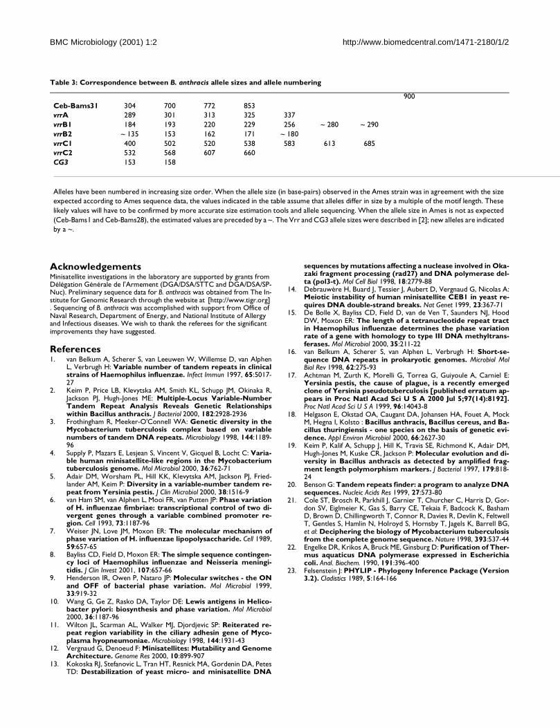
BMC Microbiology (2001) 1:2 http://www.biomedcentral.com/1471-2180/1/2
AcknowledgementsMinisatellite investigations in the laboratory are supported by grants from Délégation Générale de l'Armement (DGA/DSA/STTC and DGA/DSA/SP-Nuc). Preliminary sequence data for B. anthracis was obtained from The In-stitute for Genomic Research through the website at [http://www.tigr.org] . Sequencing of B. anthracis was accomplished with support from Office of Naval Research, Department of Energy, and National Institute of Allergy and Infectious diseases. We wish to thank the referees for the significant improvements they have suggested.
References1. van Belkum A, Scherer S, van Leeuwen W, Willemse D, van Alphen
L, Verbrugh H: Variable number of tandem repeats in clinicalstrains of Haemophilus influenzae. Infect Immun 1997, 65:5017-27
2. Keim P, Price LB, Klevytska AM, Smith KL, Schupp JM, Okinaka R,Jackson PJ, Hugh-Jones ME: Multiple-Locus Variable-NumberTandem Repeat Analysis Reveals Genetic Relationshipswithin Bacillus anthracis. J Bacteriol 2000, 182:2928-2936
3. Frothingham R, Meeker-O'Connell WA: Genetic diversity in theMycobacterium tuberculosis complex based on variablenumbers of tandem DNA repeats. Microbiology 1998, 144:1189-96
4. Supply P, Mazars E, Lesjean S, Vincent V, Gicquel B, Locht C: Varia-ble human minisatellite-like regions in the Mycobacteriumtuberculosis genome. Mol Microbiol 2000, 36:762-71
5. Adair DM, Worsham PL, Hill KK, Klevytska AM, Jackson PJ, Fried-lander AM, Keim P: Diversity in a variable-number tandem re-peat from Yersinia pestis. J Clin Microbiol 2000, 38:1516-9
6. van Ham SM, van Alphen L, Mooi FR, van Putten JP: Phase variationof H. influenzae fimbriae: transcriptional control of two di-vergent genes through a variable combined promoter re-gion. Cell 1993, 73:1187-96
7. Weiser JN, Love JM, Moxon ER: The molecular mechanism ofphase variation of H. influenzae lipopolysaccharide. Cell 1989,59:657-65
8. Bayliss CD, Field D, Moxon ER: The simple sequence contingen-cy loci of Haemophilus influenzae and Neisseria meningi-tidis. J Clin Invest 2001, 107:657-66
9. Henderson IR, Owen P, Nataro JP: Molecular switches - the ONand OFF of bacterial phase variation. Mol Microbiol 1999,33:919-32
10. Wang G, Ge Z, Rasko DA, Taylor DE: Lewis antigens in Helico-bacter pylori: biosynthesis and phase variation. Mol Microbiol2000, 36:1187-96
11. Wilton JL, Scarman AL, Walker MJ, Djordjevic SP: Reiterated re-peat region variability in the ciliary adhesin gene of Myco-plasma hyopneumoniae. Microbiology 1998, 144:1931-43
12. Vergnaud G, Denoeud F: Minisatellites: Mutability and GenomeArchitecture. Genome Res 2000, 10:899-907
13. Kokoska RJ, Stefanovic L, Tran HT, Resnick MA, Gordenin DA, PetesTD: Destabilization of yeast micro- and minisatellite DNA
sequences by mutations affecting a nuclease involved in Oka-zaki fragment processing (rad27) and DNA polymerase del-ta (pol3-t). Mol Cell Biol 1998, 18:2779-88
14. Debrauwère H, Buard J, Tessier J, Aubert D, Vergnaud G, Nicolas A:Meiotic instability of human minisatellite CEB1 in yeast re-quires DNA double-strand breaks. Nat Genet 1999, 23:367-71
15. De Bolle X, Bayliss CD, Field D, van de Ven T, Saunders NJ, HoodDW, Moxon ER: The length of a tetranucleotide repeat tractin Haemophilus influenzae determines the phase variationrate of a gene with homology to type III DNA methyltrans-ferases. Mol Microbiol 2000, 35:211-22
16. van Belkum A, Scherer S, van Alphen L, Verbrugh H: Short-se-quence DNA repeats in prokaryotic genomes. Microbiol MolBiol Rev 1998, 62:275-93
17. Achtman M, Zurth K, Morelli G, Torrea G, Guiyoule A, Carniel E:Yersinia pestis, the cause of plague, is a recently emergedclone of Yersinia pseudotuberculosis [published erratum ap-pears in Proc Natl Acad Sci U S A 2000 Jul 5;97(14):8192].Proc Natl Acad Sci U S A 1999, 96:14043-8
18. Helgason E, Okstad OA, Caugant DA, Johansen HA, Fouet A, MockM, Hegna I, Kolsto : Bacillus anthracis, Bacillus cereus, and Ba-cillus thuringiensis - one species on the basis of genetic evi-dence. Appl Environ Microbiol 2000, 66:2627-30
19. Keim P, Kalif A, Schupp J, Hill K, Travis SE, Richmond K, Adair DM,Hugh-Jones M, Kuske CR, Jackson P: Molecular evolution and di-versity in Bacillus anthracis as detected by amplified frag-ment length polymorphism markers. J Bacteriol 1997, 179:818-24
20. Benson G: Tandem repeats finder: a program to analyze DNAsequences. Nucleic Acids Res 1999, 27:573-80
21. Cole ST, Brosch R, Parkhill J, Garnier T, Churcher C, Harris D, Gor-don SV, Eiglmeier K, Gas S, Barry CE, Tekaia F, Badcock K, BashamD, Brown D, Chillingworth T, Connor R, Davies R, Devlin K, FeltwellT, Gentles S, Hamlin N, Holroyd S, Hornsby T, Jagels K, Barrell BG,et al: Deciphering the biology of Mycobacterium tuberculosisfrom the complete genome sequence. Nature 1998, 393:537-44
22. Engelke DR, Krikos A, Bruck ME, Ginsburg D: Purification of Ther-mus aquaticus DNA polymerase expressed in Escherichiacoli. Anal. Biochem. 1990, 191:396-400
23. Felsenstein J: PHYLIP - Phylogeny Inference Package (Version3.2). Cladistics 1989, 5:164-166
900
Ceb-Bams31 304 700 772 853
vrrA 289 301 313 325 337
vrrB1 184 193 220 229 256 ~ 280 ~ 290
vrrB2 ~ 135 153 162 171 ~ 180
vrrC1 400 502 520 538 583 613 685
vrrC2 532 568 607 660
CG3 153 158
Alleles have been numbered in increasing size order. When the allele size (in base-pairs) observed in the Ames strain was in agreement with the size
expected according to Ames sequence data, the values indicated in the table assume that alleles differ in size by a multiple of the motif length. These
likely values will have to be confirmed by more accurate size estimation tools and allele sequencing. When the allele size in Ames is not as expected
(Ceb-Bams1 and Ceb-Bams28), the estimated values are preceded by a ~. The Vrr and CG3 allele sizes were described in [2]; new alleles are indicated
by a ~.
Table 3: Correspondence between B. anthracis allele sizes and allele numbering

79
2.2.2 Application à l’identification de souches du
complexe Mycobacterium tuberculosis
L’article suivant (Le Flèche 2002), intitulé « High resolution, on-line identification of strains
from the Mycobacterium tuberculosis complex based on tandem repeat typing »
(identification en ligne, à haute résolution, de souches du complexe Mycobacterium
tuberculosis basée sur le typage de répétitions en tandem) présente l’application de la base de
données des répétitions en tandem à l’identification de marqueurs polymorphes chez
Mycobacterium tuberculosis. D’autres études avaient démontré la validité du typage par les
répétitions en tandem dans le complexe tuberculosis. Cet article effectue une synthèse des
travaux antérieurs, et étudie la plupart des répétitions en tandem non encore explorées. La
collection de souches utilisées est particulièrement riche en souches du type Africanum. Cette
étude est donc relativement unique, à la fois pour le grand nombre de marqueurs utilisés, et
pour la représentation du complexe tuberculosis. Elle montre que la classification obtenue par
le typage des répétitions en tandem est pertinente d’un point de vue phylogénétique. En outre,
un service d’identification de souches en ligne (http://bacterial-genotyping.igmors.u-psud.fr) a
été élaboré : l’utilisateur saisit un génotype, et obtient la liste des souches les plus proches,
parmi celles qui figurent dans la base de données de génotypes hébergée au laboratoire. Cet
article marque une nouvelle évolution de la base. Ma contribution a été la comparaison des
trois souches du complexe tuberculosis pour lesquelles la séquence complète avait été
déterminée. Bien que dans ce cas la conservation entre les souches soit telle que la
comparaison des répétitions en tandem puisse se faire aisément avec la base de données
existante, ce projet a été l’occasion du développement de l’outil automatique permettant la
comparaison de souches. Cet outil a été décrit plus en détail dans la paragraphe 2.1.1.4. J’ai
également participé à l’élaboration de la page d’identification de souches, décrite dans le
paragraphe 2.1.1.5.
Résumé :
Contexte : Les méthodes de référence actuellement disponibles pour l’épidémiologie
moléculaire du complexe Mycobacterium tuberculosis manquent de sensibilité ou sont encore
trop lentes et fastidieuses pour être applicables en routine. Le typage de répétitions en tandem
est récemment apparu comme une alternative potentielle. Cet article contribue au
développement du typage de répétitions en tandem chez M. tuberculosis : une synthèse des
données existantes a été effectuée, de nouveaux marqueurs polymorphes ont été développés,
et un service Internet gratuit, rapide, et facile d’utilisation a été élaboré pour permettre
l’identification de souches.
Résultats: Un lot de 21 VNTRs comprenant 13 locus déjà décrits et 8 nouveaux marqueurs a
été utilisé pour génotyper 90 souches du complexe M. tuberculosis (M. tuberculosis : 64
souches ; M. bovis : 9 souches dont 4 BCG ; M. africanum : 17 souches). 84 génotypes

80
différents ont été définis. Une analyse de classification montre que les souches de M.
africanum tombent dans trois grands groupes, l’un étant plus proche des souches de M.
tuberculosis, et un autre plus proche des souches de M. bovis. Les résultats sont publiquement
accessibles sur Internet [http://bacterial-genotyping.igmors.u-psud.fr/bnserver] pour permettre
des requêtes d’identification de souches.
Conclusions: Le typage de répétitions en tandem, basé sur la technique de PCR, pourrait se
révéler être un puissant complément aux outils épidémiologiques existants pour le complexe
M. tuberculosis. Le nombre de marqueurs à typer dépend de la précision d’identification
requise : l’identification peut être effectuée rapidement et à moindre coût en termes de
consommables, d’expertise scientifique, et d’équipement.
BioMed Central
Page 1 of 12
(page number not for citation purposes)
BMC Microbiology
Open AccessBMC Microbiology 2002, 2 xResearch article
High resolution, on-line identification of strains from the Mycobacterium tuberculosis complex based on tandem repeat typingPhilippe Le Flèche1,2, Michel Fabre3, France Denoeud2, Jean-Louis Koeck4 and Gilles Vergnaud*1,2
Address: 1Centre d'Etudes du Bouchet BP3, 91710 Vert le Petit, France, 2GPMS, Bât. 400, Institut de Génétique et Microbiologie, Université Paris Sud, 91405 Orsay cedex, France, 3Laboratoire de Biologie Clinique, HIA Percy, 92141 Clamart, France and 4Département de biologie médicale, HIA Val-de-Grâce, 75230 Paris, France
E-mail: Philippe Le Flèche - [email protected]; Michel Fabre - [email protected]; France Denoeud - [email protected]; Jean-Louis Koeck - [email protected]; Gilles Vergnaud* - [email protected]
*Corresponding author
Abstract
Background: Currently available reference methods for the molecular epidemiology of the
Mycobacterium tuberculosis complex either lack sensitivity or are still too tedious and slow for
routine application. Recently, tandem repeat typing has emerged as a potential alternative. This
report contributes to the development of tandem repeat typing for M. tuberculosis by summarising
the existing data, developing additional markers, and setting up a freely accessible, fast, and easy to
use, internet-based service for strain identification.
Results: A collection of 21 VNTRs incorporating 13 previously described loci and 8 newly
evaluated markers was used to genotype 90 strains from the M. tuberculosis complex (M. tuberculosis
(64 strains), M. bovis (9 strains including 4 BCG representatives), M. africanum (17 strains)). Eighty-
four different genotypes are defined. Clustering analysis shows that the M. africanum strains fall into
three main groups, one of which is closer to the M. tuberculosis strains, and an other one is closer
to the M. bovis strains. The resulting data has been made freely accessible over the internet [http:/
/bacterial-genotyping.igmors.u-psud.fr/bnserver] to allow direct strain identification queries.
Conclusions: Tandem-repeat typing is a PCR-based assay which may prove to be a powerful
complement to the existing epidemiological tools for the M. tuberculosis complex. The number of
markers to type depends on the identification precision which is required, so that identification can
be achieved quickly at low cost in terms of consumables, technical expertise and equipment.
BackgroundThe precise identification of bacterial pathogens at thestrain level is essential for epidemiological purposes. Con-sequently, constant efforts are undertaken to develop easyto use, low cost and standardized methods which caneventually be applied routinely in a clinical laboratory.
Newer developments are usually genetic methods basedon PCR (Polymerase Chain Reaction) to type variationsdirectly at the DNA level. The development of polymor-phic markers is now further facilitated by the availabilityof whole genome sequences for bacterial genomes. Re-cently, it has been shown that tandem repeat (usually
Published: 27 November 2002
BMC Microbiology 2002, 2:37
Received: 17 September 2002Accepted: 27 November 2002
This article is available from: http://www.biomedcentral.com/1471-2180/2/37
© 2002 Le Flèche et al; licensee BioMed Central Ltd. This is an Open Access article: verbatim copying and redistribution of this article are permitted in all media for any purpose, provided this notice is preserved along with the article's original URL.

BMC Microbiology 2002, 2 http://www.biomedcentral.com/1471-2180/2/37
Page 2 of 12
(page number not for citation purposes)
called minisatellites or VNTRs for Variable Number ofTandem Repeats) loci provide a source of very informativemarkers not only in humans where some are still in usefor identification purposes (paternity analyses, forensics)but also in bacteria. Tandem repeats are easily identifiedfrom genome sequence data, the typing of tandem repeatlength is relatively straight forward, and the resulting datacan be easily coded and exchanged between laboratoriesindependently of the technology used to measure PCRfragment sizes. Furthermore, the resolution of tandem re-peats typing is cumulative, i.e. the inclusion of moremarkers in the typing assay can, when necessary, increasethe identification resolution. However, the density of tan-dem repeats in bacterial genomes varies from species tospecies, and not all tandem repeats are polymorphic [1].In addition, some tandem repeats are so unstable thatthey have no or little long-term epidemiological value [2].This indicates that for each species under consideration,tandem repeats must be evaluated using representativecollections of strains before they can be used. Tandem re-peats for bacterial identification have already proved theirutility for the typing of the highly monomorphic patho-gens Bacillus anthracis, Yersinia pestis, [1] and M. tuberculo-sis. In this last case, the value of tandem repeat basedidentification was recognised very early [3]. The so-calledDR (direct repeat) locus is a relatively large tandem repeatlocus of unknown biological significance. The motif is 72bp long, one half is highly conserved, whereas the otherhalf (called the spacer element) is highly diverged. Thespoligotyping method [4] takes advantage of these inter-nal variations to distinguish the hundreds of different al-leles at this locus, which have been reported in the M.tuberculosis complex among the thousands of strains typedso far [5]. Although it is quite powerful, with many advan-tages, spoligotyping suffers from a lack of resolution com-pared to the current gold-standard in M. tuberculosisgenetic identification, IS6110 typing [6]. IS6110 typing isan RFLP (Restriction Fragment Length Polymorphism)method using the mobile element IS6110 as a probe.Strains with a low-copy number of IS6110 elements (suchas most M. bovis strains) are poorly resolved by this meth-od. The so-called PGRS (polymorphic GC-rich sequence)method is an other RFLP approach in which the probeused is a GC-rich tandem repeat. The polymorphismswhich are scored at multiple loci simultaneously on theSouthern blot are variations in the tandem repeats length(and not internal variations at a single locus as assayed byspoligotyping). The profiles generated are very informa-tive, but in comparison with IS6110 typing, PGRS resultsare more difficult to score, because the intensity of thebands are highly variable (alleles with a small tandem ar-ray yield a lower hybridisation signal) [6]. Both PGRS andIS6110 typing are hindered by the requirement for rela-tively large amounts of high quality DNA which is an issuefor slow-growing mycobacteria.
More recently, and owing to the release of genome se-quence data, the allele-length polymorphism of tandemrepeat loci has been evaluated by PCR. Essentially threecomplementary sets of markers have been developed [7–9]. In the first report, exact tandem repeats (ETRs) wereidentified by searching the existing literature as well asearly versions of the M. tuberculosis genome sequence data[7]. The resolution provided by this first set of five loci islower than both IS6110 RFLP typing and spoligotypingaccording to a comparative study [6]. In the second report,a family of tandem repeats characterized by similar repeatunits was identified by sequence similarity search in thegenome sequence data. A set of 12 loci was selected (in-cluding two of the five ETR loci) and the resulting panelhas a resolution close to IS6110 typing according to [10].In the third report tandem repeats with highly conserved(>95%) motifs longer than 50 bp identified in the M. tu-berculosis genome sequence have been investigated. Alto-gether, the currently available collection of polymorphictandem repeats for the typing of M. tuberculosis comprises27 loci (taking into account duplicates) (Table 1). Fifteenhave a polymorphism index above 0.5.
This collection of markers should already provide a typingresolution comparable to the current reference methods.Given that not all tandem repeats present in M. tuberculosishave been evaluated for polymorphism, it is likely that thetyping resolution of minisatellites could further be im-proved. Eventually, normalisation work will have to bedone in order to promote the use of tandem repeats. Anumber of the loci analysed are known under differentnames in different studies, (for instance, ETRD [7] is alsoknown as MIRU4 in [10]; and VNTR 0580 in [11]) and thecoding (number of motifs in an allele) of alleles can alsobe different in different studies, for reasons explained in[11]. This is due in part to the fact that the number of re-peats is not necessarily an integer value (Table 1). Further-more, because the repeats in an array are not necessarilyexact repeats, there can be ambiguities in the definition ofthe first and last base pair of the array. Finally, in additionto length variations due to the addition or deletion of anexact number of units, microdeletions or insertions with-in some repeat units are sometimes observed (MIRU4 isone such instance [12]).
One purpose of the present report is to contribute to thedevelopment of Multiple Loci VNTR Analysis (MVLA)through the evaluation of new markers and the setting upof an on-line identification tool for the M. tuberculosiscomplex which can be queried very easily with the user'spersonal data. In the present report, we first take advan-tage of the availability of genome sequence from two M.tuberculosis strains to complement the current collection ofpolymorphic tandem repeat markers. We identified in sil-ico tandem repeats showing a different length in the two

BM
C M
icro
bio
logy 2
002
, 2
http
://w
ww
.bio
me
dcentr
al.com
/147
1-2
18
0/2
/37
Pa
ge 3
of 1
2
(pa
ge
num
be
r n
ot fo
r cita
tio
n p
urp
ose
s)Table 1: Polymorphic minisatellite markers for the M. tuberculosis complex
Locus name "MIRU" alias [8]
"ETR" alias [7]
"QUB" alias [9,11]
Other alias
Reference TR location
on H37Rv genome
Expected length in H37Rv (copy number)
Expected length in CDC1551 (copy number)
Expected length in M bovis AF2122 (copy number)
N° ofstrains
Size range observed (copy number)
N° ofalleles
observed
Polymor-phismindex
H37Rv_0024_18 bp Mtub01 This report
24648 328 (10) 310 (9) 310 (9) 92 274–328 bp (7–10) 4 0.48
H37Rv_0079_9 bp Mtub02 This report
79503 230 (6) 239 (7) 239 (7) 92 221–275 bp (5–11) 7 0.76
H37Rv_0154_53 bp MIRU2 [8] 154111 508 (2) 508 (2) 508 (2) 92 455–561 bp (1–3) 3 0.09
H37Rv_0424_51 bp Mtub04 This report
424010 269 (2.6) 371 (4.6) 269 (2.6) 28 218 – 320 (1.6–3.6) 3 0.52
H37Rv_0531_15 bp MPTR-A [7] 531430 328 (16) 328 (16) 328 (16) 48 (15–17) 3 0.23
H37Rv_0577_58 bp ETR-C [8] 577172 346 (4) 288 (3) 404 (5) 92 230–404 bp (2–5) 4 0.63
H37Rv_0580_77 bp MIRU4 ETR-D [7] 580546 353 (3.3) 330 (3) 483 (5) 92 253–715 bp (2–8) 7 0.35
H37Rv_0802_54 bp MIRU40 [8] 802194 199 (1) 415 (5) 253 (2) 92 199–469 bp (1–6) 5 0.71
H37Rv_0959_53 bp MIRU10 [8] 959868 643 (3) 750 (5) 590 (2) 92 537–1014 bp (1–10) 9 0.76
H37Rv_1121_15 bp Mtub12 This report
1121658 215 (4) 230 (5) 215 (4) 92 200–230 bp (3–5) 3 0.19
H37Rv_1443_56 bp Mtub16 This report
1443417 291 (1) 347 (2) 347 (2) 11 291–515 (1–5) 3 0.56
H37Rv_1451_57 bp QUB-1451c [9] 1451778 305 (3.8) 305 (3.8) 305 (3.8) 56 (2–4) (bovis) 2 0.12
H37Rv_1612_21 bp QUB-23 [11] 1612529 141 (5) 162 (6) 162 (6) 20 141–203 (5–8) 3 0.18
H37Rv_1644_53 bp MIRU16 [8] 1644026 671 (2) 724 (3) 671 (2) 92 618–777 bp (1–4) 4 0.59
H37Rv_1895_57 bp QUB-1895 [9] 1895344 319 (4) 205 (2) 319 (4) 56 (2–4) (bovis) 3 0.35
H37Rv_1955_57 bp Mtub21 This report
1955580 206 (2) 263 (3) 263 (3) 92 149–491 bp (1–7) 7 0.76
H37Rv_1982_78 bp QUB-18 [11] 1982873 621 (5) 777 (7) 465 (3) 24 387–1167 (2–12) 9 0.74
H37Rv_2059_77 bp MIRU20 [8] 2059429 591 (2) 591 (2) 591 (2) 53 (1–2) 2 0.29
H37Rv_2074_56 bp* Mtub24 This report
2074431 805 (3.6) 693 (1.6) 693 (1.6) 44 637–749 (0.6–2.6) 3 0.52
H37Rv_2163_a_69 bp QUB-11a pUCD1 [11] 2163607 305 (3) 581 (7) 788 (10) 92 305–1832 bp (3–26) 15 0.88
H37Rv_2163_b_69 bp QUB-11b pUCD1 [11] 2163729 412 (5) 274 (3) 343 (4) 52 136–826 (1–11) 8 0.82
H37Rv_2165_75 pb ETR-A [7] 2165223 397 (3) 322 (2) 847 (9) 92 322–847 bp (2–9) 8 0.73
H37Rv_2347_57 bp Mtub29 This report
2347393 350 (4) 292 (3) 293 (3) 92 236–350 bp (2–4) 3 0.55
H37Rv_2401_58 bp Mtub30 This report
2401815 319 (2) 435 (4) 435 (4) 92 261–435 bp (1–4) 3 0.55
H37Rv_2461_57 bp ETR-B [7] 2461279 292 (3) 235 (2) 406 (5) 92 178–406 bp (1–5) 6 0.51
H37Rv_2531_53 bp MIRU23 [8] 2531560 873 (6) 820 (5) 767 (4) 92 608–979 bp (1–8) 7 0.60
H37RV_2387_54 bp MIRU24 [8] 2684427 447 (1) 447 (1) 447 (1) 53 (1–2) 2 0.24
H37Rv_2990_55 bp Mtub31 This report
2990582 257 (2) 312 (3) 312 (3) 49 202–312 bp (1–3) 3 0.15
H37Rv_2996_51 bp MIRU26 [8] 2996002 614 (3) 716 (5) 716 (5) 57 563–818 (2–7) 5 0.61
H37Rv_3006_53 bp MIRU27 QUB-5 [8] 3006875 657 (3) 657 (3) 657 (3) 92 551–710 bp (1–4) 4 0.25

BM
C M
icro
bio
logy 2
002
, 2
http
://w
ww
.bio
me
dcentr
al.com
/147
1-2
18
0/2
/37
Pa
ge 4
of 1
2
(pa
ge
num
be
r n
ot fo
r cita
tio
n p
urp
ose
s)
H37Rv_3171_54 bp Mtub34 This report
3171465 279 (3) 225 (2) 279 (3) 11 171–225 (1–2) 2 0.3
H37Rv_3192_53 bp MIRU31 ETR-E [7] 3192168 651 (3) 651 (3) 651 (3) 92 545–810 bp (1–6) 6 0.67
H37Rv_3232_56 bp QUB-3232 [9] 3232649 591 (3) 760 (6) 703 (5) 56 (4–22) (bovis) 10 0.65
H37Rv_3239_79 bp ETR-F [7] 3239469 476 (2.8) 476 (2.8) 421 (2.1) 48 (1–3) 3 0.49
H37Rv_3336_59 bp QUB-3336 [9] 3336499 407 (5) 466 (6) 289 (3) 56 (3–21) (bovis) 8 0.55
H37Rv_3663_63 bp** Mtub38 This report
3663751 373 (2.7) 310 (1.7) 310 (1.7) 92 247–400 bp (0.7–3.1) 5 0.35
H37Rv_3690_58 bp* Mtub39 This report
3690947 341 (2.6)* 397 (3.6) 341 (2.6) 92 247–1349 bp (1–20)* 11 0.64
H37Rv_4052_111 bp QUB-26 [11] 4052969 708 (5) 819 (6) 597 (4) 100 (4–14) (bovis) 5 0.41
H37Rv_4156_59 bp QUB-4156c [9] 4156797 224 (2) 283 (3) 165 (1) 52 106–283 (0–3) 4 0.69
H37Rv_4348_53 bp MIRU39 [8] 4348401 646 (2) 646 (2) 646 (2) 92 593–699 bp (1–3) 3 0.31
The markers are listed according to their position in the H37Rv genome. The proposed reference name includes the size of the repeat unit. The twenty-one markers used in the present report are italicised and underlined. Alias names identified in the literature are indicated. QUB11a, QUB11b, and ETR-A (position 2163–2165) are located within the gene PPE34 [19]. The expected length assumes that the primers listed in Table 2 were used. * : the observed size (Table 3) is not the expected size. ** : the repeat unit is not easily defined, size variations do not correspond to a multiple of 63 base-pairs. Polymorphism index is calculated as 1 - ∑ (allele frequency)2 among the 86 distinct genotypes. The values are deduced from the original report in nine cases (indicated by the absence of size range in the "size range" column). In some instances [9,11], the population of strains used is biased (M. bovis strains).
Table 1: Polymorphic minisatellite markers for the M. tuberculosis complex (Continued)

BMC Microbiology 2002, 2 http://www.biomedcentral.com/1471-2180/2/37
Page 5 of 12
(page number not for citation purposes)
strains using the previously described tandem repeat data-base [http://minisatellites.u-psud.fr][1]. Thirteen lociwith a different predicted length in the two genomes andwhich have not been previously investigated have beentested for polymorphism and ease of typing.
Eight among the 13 polymorphic loci were used togetherwith 13 among the previously described markers to geno-
type a collection of different M. tuberculosis complexstrains. The data produced clusters the strains as suggestedby morphological observations and biochemical analyses.The resulting data can be queried from a dedicated webpage [http://bacterial-genotyping.igmors.u-psud.fr/bn-server].
Table 2: Set of primers for MLVA analysis
Locus name forward primer reverse primer
H37Rv_0024_18 bp GAGAAACAGGAGGGCGTTG TATTACGACGACCGCTATGC
H37Rv_0079_9 bp CGTGCACAGTTGGGTGTTTA TTCGTTCAGGAACTCCAAGG
H37Rv_0154_53 bp TGGACTTGCAGCAATGGACCAACT TACTCGGACGCCGGCTCAAAAT
H37Rv_0424_51 bp GTCCAGGTTGCAAGAGATGG GGCATCCTCAACAACGGTAG
H37Rv_0531_15 bp GGTTACCACTTCGATGCGTCTGCG AGCCGCCGAAACCCATC
H37Rv_0577_58 bp* GACTTCAATGCGTTGTTGGA* GTCTTGACCTCCACGAGTGC*
H37Rv_0580_77 bp CAGGTCACAACGAGAGGAAGAGC GCGGATCGGCCAGCGACTCCTC
H37Rv_0802_54 bp* AAGCGCAAGAGCACCAAG* GTGGGCTTGTACTTGCGAAT*
H37Rv_0959_53 bp GTTCTTGACCAACTGCAGTCGTCC GCCACCTTGGTGATCAGCTACCT
H37Rv_1121_15 bp CTCCCACACCCAGGACAC CGGCCTACCCAACATTCC
H37Rv_1443_56 bp GGTAATCCTGGTCGCTTGTC ACCCAAATTGCCCTGGTC
H37Rv_1451_57 bp GGTAGCCGTCGTCGAGAAGC CGCCACCACCGCACTGGC
H37Rv_1612_21 bp GCTGCACCGGTGCCCATC CACCGGAGCCGGAACGGC
H37Rv_1644_53 bp TCGGTGATCGGGTCCAGTCCAAGTA CCCGTCGTGCAGCCCTGGTAC
H37Rv_1895_57 bp GGTGCACGGCCTCGGCTCC AAGCCCCGCCGCCAATCAA
H37Rv_1955_57 bp AGATCCCAGTTGTCGTCGTC CAACATCGCCTGGTTCTGTA
H37Rv_1982_78 bp* ATCGTCAGCTGCGGAATAGT* AATACCGGGGATATCGGTTC*
H37Rv_2059_77 bp TCGGAGAGATGCCCTTCGAGTTAG GGAGACCGCGACCAGGTACTTGTA
H37Rv_2074_56 bp AAATTCAAAGAGTTTCTCGACAGTG GATCTTGAGAACCAAGATGTCCTT
H37Rv_2163_a_69 bp CCCATCCCGCTTAGCACATTCGTA TTCAGGGGGGATCCGGGA
H37Rv_2163_b_69 bp CGTAAGGGGGATGCGGGAAATAGG CGAAGTGAATGGTGGCAT
H37Rv_2165_75pb* ATTTCGATCGGGATGTTGAT* TCGGTCCCATCACCTTCTTA*
H37Rv_2347_57 bp AACCCATGTCAGCCAGGTTA ATGATGGCACACCGAAGAAC
H37Rv_2401_58 bp AGTCACCTTTCCTACCACTCGTAAC ATTAGTAGGGCACTAGCACCTCAAG
H37Rv_2461_57 bp GCGAACACCAGGACAGCATCATG GGCATGCCGGTGATCGAGTGG
H37Rv_2531_53 bp CAGCGAAACGAACTGTGCTATCAC CGTGTCCGAGCAGAAAAGGGTAT
H37RV_2387_54 bp CGACCAAGATGTGCAGGAATACAT GGGCGAGTTGAGCTCACAGAA
H37Rv_2990_55 bp GTGACGTTTACCGTGCTCTATTTC GTCGTCGGACAGTTCTAGCTTT
H37Rv_2996_51 bp CCCGCCTTCGAAACGTCGCT TGGACATAGGCGACCAGGCGAATA
H37Rv_3006_53 bp TCGAAAGCCTCTGCGTGCCAGTAA GCGATGTGAGCGTGCCACTCAA
H37Rv_3171_54 bp GCAGATAACCCGCAGGAATA GGAGAGGATACGTGGATTTGAG
H37Rv_3192_53 bp* ACTGATTGGCTTCATACGGCTTTA* GTGCCGACGTGGTCTTGAT*
H37Rv_3232_56 bp CAGACCCGGCGTCATCAAC CCAAGGGCGGCATTGTGTT
H37Rv_3239_79 bp CTCGGTGATGGTCCGGCCGGTCAC GGAAGTGCTCGACAACGCCATGCC
H37Rv_3336_59 bp ATCCCCGCGGTACCCATC GCCAGCGGTGTCGACTATCC
H37Rv_3663_63 bp GCCCAAAAAGCATGGGAACGTGCCCCT GGTTGTCCCCGCAGTATCTC
H37Rv_3690_58 bp AATCACGGTAACTTGGGTTGTTT GATGCATGTTCGACCCGTAG
H37Rv_4052_111 bp AACGCTCAGCTGTCGGAT GGCCAGGTCCTTCCCGAT
H37Rv_4156_59 bp* TGGTCGCTACGCATCGTGTCGGCCCGT* TACCACCCGGGCAGTTTAC*
H37Rv_4348_53 bp CGCATCGACAAACTGGAGCCAAAC CGGAAACGTCTACGCCCCACACAT
* : the primers indicated are not the primers used in the princeps publication, but were designed for the present study, usually in order to reduce the size of the PCR product and consequently to improve allele size identification.

BMC Microbiology 2002, 2 http://www.biomedcentral.com/1471-2180/2/37
Page 6 of 12
(page number not for citation purposes)
ResultsTandem repeats predicted to be of a different size in
H37Rv and CDC1551
The size of tandem repeats in the two M. tuberculosisstrains sequenced to date, H37Rv and CDC1551, wascompared using the tandem repeat database [http://min-isatellites.u-psud.fr]. Fifty-one of the tandem repeats iden-tified in CDC1551 have repeat units longer than 9 base-pairs and a predicted overall size which differs from theH37Rv homolog estimate by at least 9 base-pairs. Seven-teen have an expected product size above one kilobase.They include the DR locus and members of the family ofPGRS sequences [13] and were not investigated further.Eighteen have been analyzed in previous investigations[7–9,11]. Three produced multiband patterns or incon-sistent results. The results obtained for the remaining 13loci together with the description of the 18 previously de-scribed loci are summarized in Table 1. In addition, Table1 includes nine markers which are not polymorphic be-tween H37Rv and CDC1551 but have already been quot-ed in the literature. Each locus is designated by its position(expressed in kilobases) on the H37Rv genome and by therepeat unit length as defined by the Tandem Repeat Findersoftware and indicated in the Tandem Repeat Database[http://minisatellites.u-psud.fr]. All thirteen newly evalu-ated loci are polymorphic as predicted. In two cases (Table1) the expected product size is not the observed size. Theexpected size has not been observed in the collection ofstrains used here, which suggests that the incorrect predic-tion is due to an artifact along the sequencing process.Eight loci among the thirteen have polymorphism indexesabove 0.50 (two are above 0.7). The vast majority of therepeats units are more than 50 bp long (Table 1) whichmakes them easy to assay by ordinary agarose gel electro-phoresis when using the primer pairs indicated in Table 2.In one instance however (H37Rv_3663_63 bp) the PCRsize products clearly do not differ by a perfect number of(63 bp) repeat units (Table 1).
Typing of strains and clustering analysis
The forty loci listed in Table 1 were used to genotype a col-lection of 90 strains from the M. tuberculosis complex, us-ing the primers listed in Table 2. In our hands, some of themarkers did not prove to be sufficiently robust for easyand reproducible typing in the conditions used here. Onthis basis, we have selected a collection of 21 markers(comprising thirteen previously described markers andeight among the new loci evaluated). The 21 markers usedare italicised and underlined in Table 1 and 2. After anal-ysis of the images using Bionumerics 3.0, and conversionof allele sizes in copy numbers of motifs in the tandem ar-rays, clustering analysis was done using the categoricaland Ward parameters. The results of the clustering analy-sis are shown in Figure 1. The genotyping data fromstrains M. tuberculosis CDC1551 and M. bovis AF2122/
97 was deduced (Table 1) from the sequence data and in-cluded in the analysis. Six major groups are defined (Fig-ure 1). Group I contains the M. bovis strains and 5 of theM. africanum strains. Group II is composed of nine M. af-ricanum strains. The third group includes three M. africa-num strains and seven M. tuberculosis strains.Interestingly, five of these strains have been independent-ly identified as representing the Beijing type [14] (the lasttwo have not been tested). The last three groups comprisethe vast majority of the M. tuberculosis strains. M. africa-num strains which are negative for nitrate reduction (Afri-canum I type [15]) are among the first two groups, closerto the M. bovis strains as previously observed [16,17]. Incontrast, the three M. africanum strains which are positivefor nitrate reduction are in the third group, closer to M. tu-berculosis strains. In order to facilitate the comparisonwith earlier investigations [16,17], Figure 1 displays thegenotypes for the five ETR markers, extracted from the fulldata presented in Table 3. Group I in Figure 1 is reminis-cent of group A in [17] and group A1 in [18]. Group II inFigure 1 is reminiscent of group B in [17] and group A2 in[18] which are both characterized by the 42432 ETR pat-tern.
The ETR panel alone discriminates 44 genotypes (insteadof 84 with the panel of 21 loci; 86 genotypes when includ-ing the CDC1551 and AF2122/97 data, Figure 1) and isnot sufficient to clearly separate the M. africanum strainsfrom the M. tuberculosis strains (analysis not shown) ascan be achieved using the 21 loci.
Internet-based identifications
The genotyping data presented in Table 3 can be querieddirectly via an internet service [http://bacterial-genotyp-ing.igmors.u-psud.fr/bnserver/]. Figure 2 provides a briefdescription of the current M. tuberculosis query page (likelyto evolve as updates are made). For each locus, allele sizescan be selected among a list of possibilities (observed siz-es). Alternatively, more experienced users will go directlyto a "copy-paste" page using the appropriate format. Theresults of the query indicate a similarity score and includelinks to the complete data for each strain listed. Help filesare available, including a link to updated versions of Fig-ure 1.
Testing the reproducibility of the approach
In order to test the reproducibility of the approach, tenblinded-coded control samples were typed. Figure 3shows the typing of two markers, H37Rv_0802_54 bp(left, 54 bp unit; H37Rv allele : 1 unit, 199 bp PCR prod-uct) and H37Rv_1955_57 bp (right, 57 bp unit; H37Rv al-lele : 2 units, 206 bp PCR product). The number of unitsin each allele can be unambiguously deduced by compar-ison with the H37Rv control lanes and the 100 base-pairs

BM
C M
icro
bio
log
y 2
002, 2
http://w
ww
.bio
med
ce
ntr
al.com
/1471
-2180
/2/3
7
Pag
e 7
of
12
(pa
ge
nu
mb
er
not
for
cita
tio
n p
urp
ose
s)
lad
der
siz
e m
ark
er. A
ll t
en u
nk
no
wn
str
ain
s w
ere
corr
ectl
yid
enti
fied
usi
ng
the
inte
rnet
bas
e se
rvic
e d
escr
ibed
ab
ove
.
Dis
cu
ssio
nT
he
list
of
40
mar
ker
s gi
ven
in
Tab
le 1
is
clo
se t
o r
epre
-se
nti
ng
the
com
ple
te c
oll
ecti
on
of
tan
dem
rep
eats
of
in-
tere
st fo
r M
LV
A t
ypin
g in
M. tu
berc
ulo
sis.
It in
clu
des
all
lo
ciw
ith
a d
iffe
ren
t p
red
icte
d s
ize
in H
37
Rv
and
CD
C1
55
1an
d w
hic
h a
re a
men
able
to
ro
uti
ne
PC
R t
ypin
g. N
ine
ad-
dit
ion
al lo
ci w
hic
h h
ave
bee
n q
uo
ted
in
pu
bli
shed
rep
ort
sar
e al
so i
ncl
ud
ed e
ven
if
they
do
no
t fu
lfil
l th
is c
rite
ria.
Clu
ster
ing
anal
ysis
(Fig
ure
1)
sho
ws
that
th
e tw
o s
trai
ns
CD
C1
55
1 an
d H
37
Rv
(Fig
ure
1
) ar
e re
lati
vely
d
ista
nt
wit
hin
th
e M
. tu
berc
ulo
sis
spec
ies.
Th
is w
ou
ld p
red
ict
that
tan
dem
rep
eats
of
iden
tica
l si
ze i
n t
he
two
str
ain
s ar
e li
ke-
ly t
o b
e p
oo
rly
info
rmat
ive
acro
ss t
he
com
ple
x. H
ow
ever
,th
is a
pp
ears
no
t to
be
abso
lute
ly t
rue,
sin
ce f
or
inst
ance
,E
TR
-E (
H3
7R
v_3
19
2_5
3 b
p)
hap
pen
s to
hav
e th
e sa
me
size
in
H3
7R
v, C
DC
15
51
an
d e
ven
AF2
12
2/9
7 (
Tab
le 1
)in
sp
ite
of
its
very
hig
h p
oly
mo
rph
ism
in
dex
(0
.69
, T
able
1).
Co
nse
qu
entl
y, t
he
few
ad
dit
ion
al l
oci
, n
ot
exp
lore
dh
ere,
wh
ich
are
of
equ
al s
ize
in H
37
Rv
and
CD
C1
55
1, b
ut
dif
fer
wit
h t
he
pre
dic
ted
siz
e fo
r M
. bo
vis
stra
in A
F2
12
2/9
7m
igh
t al
so p
rove
to
be
of
inte
rest
.
As
can
be
seen
in
Tab
le 1
, mo
st r
epea
t u
nit
s ar
e m
ore
th
an5
0 b
p l
on
g an
d a
llel
e si
zes
rare
ly e
xcee
d 1
00
0 b
p. A
s a
re-
sult
, th
e p
reci
sio
n w
hic
h c
an b
e ac
hie
ved
by
ord
inar
y ag
a-ro
se
gel
elec
tro
ph
ore
sis
is
suff
icie
nt
to
esti
mat
e th
en
um
ber
of
un
its
in a
n a
llel
e. T
he
sele
ctio
n o
f 2
1 m
ark
ers
pro
po
sed
her
e w
as t
este
d s
pec
ific
ally
in
ord
er t
o b
e ea
sily
assa
yed
usi
ng
this
lo
w-c
ost
tec
hn
olo
gica
l ap
pro
ach
. A
l-th
ou
gh a
dat
abas
e sy
stem
is
nec
essa
ry t
o e
ffic
ien
tly
man
-ag
e a
gen
oty
pin
g p
roje
ct w
ith
a h
igh
nu
mb
er o
f m
ark
ers
and
str
ain
s, th
e id
enti
fica
tio
n o
f u
p to
a few
str
ain
s p
er d
ayin
a c
lin
ical
set
tin
g fo
r in
stan
ce r
equ
ires
no
so
ph
isti
cate
deq
uip
men
t n
or
cost
ly co
nsu
mab
les.
G
eno
typ
es ca
n b
esc
ore
d b
y vi
sual
an
alys
is o
f th
e ge
l im
ages
, an
d a
su
bse
t o
fth
e co
llec
tio
n o
f av
aila
ble
mar
ker
s ca
n b
e ch
ose
n f
or
rou
-ti
ne
iden
tifi
cati
on
pu
rpo
ses.
Th
e d
ata
can
th
en b
e an
a-ly
sed
usi
ng
the
site
des
crib
ed i
n F
igu
re 2
.
Th
e ro
le o
f ta
nd
em r
epea
ts i
n t
he
M.
tube
rculo
sis
gen
om
eis
lar
gely
un
kn
ow
n. T
wen
ty-o
ne
of
the
loci
lis
ted
in
Tab
le1
hav
e re
pea
t u
nit
s w
hic
h a
re a
mu
ltip
le o
f th
ree
bas
e-p
airs
. Th
e m
ajo
rity
(fi
ftee
n)
fall
s w
ith
in p
uta
tive
gen
es, o
f-te
n o
f u
nk
no
wn
fu
nct
ion
, su
ch a
s th
e P
PE
fam
ily
of
gen
es[1
9].
Th
e m
ost
rem
ark
able
in
stan
ce i
s p
rob
ably
PP
E3
4 a
tp
osi
tio
n 2
16
3–
21
65
of
the
gen
om
e (R
v19
17
c in
[h
ttp
://
Fig
ure
1D
en
dro
gra
m d
ed
uced
fro
m t
he c
lust
eri
ng a
naly
sis
of
the 9
2 s
train
s (i
nclu
din
g C
DC
1551 a
nd
AF
2122/9
7)
. T
he
first
colu
mn fro
m t
he left
identify
the s
trai
ns.
The s
eco
nd c
olu
mn indic
ates
the s
peci
es
(Red : M
. bo
vis
stra
ins;
gre
en : M
. afr
ica-
num
str
ains;
yel
low
: M
. tu
berc
ulos
is s
trai
ns
know
n t
o b
e of th
e B
eijin
g ty
pe
and indic
ated
"bei
jing
tuber
culo
sis"
; blu
e : oth
er M
.tu
berc
ulos
is s
trai
ns)
. T
he
thir
d c
olu
mn indic
ates
the
geogr
aphic
ori
gin o
f th
e s
trai
n. T
he f
ourt
h c
olu
mn indic
ates
the E
TR
pat
-te
rn (
ET
R-A
to E
TR
-E)
extr
acte
d fro
m t
he full
dat
a pre
sente
d in T
able
3. T
he las
t fo
ur
colu
mns
indic
ate,
fro
m lef
t to
rig
ht, t
he
resu
lt o
f th
e n
iaci
ne p
roduct
ion, nitra
te r
educt
ase, T
CH
susc
eptibili
ty a
nd L
ebek t
est
s (0
, nega
tive
; 1
, posi
tive
) w
hen a
vaila
ble
.
Categorical
MLVA
10
0
80
60
40
20
0-20
-40
-60
percy196
percy197
percy142
percy62
percy43a
percy53
percy184
percy55
AF2122/97
percy54
percy61
percy119
percy57
cipt950052
percy18
cipt960340
percy59
percy16
percy17
percy99a
percy13
percy58
percy56
percy60
percy27
percy7
percy91
percy122
cipt20001272
cipt991053
cipt990590
cipt971135
percy248
percy164
percy170
percy211
percy189a
percy128
percy210
percy172
percy231
CDC1551
percy155
percy44
percy216
percy6b
percy239
percy208
percy232
percy222
percy237
percy42
percy27b
percy28b
percy169
percy228
percy219
percy84
percy256
percy259
percy11
percy43b
percy31
percy18b
percy201
percy35
percy39
percy250
percy40
percy33
percy20
percy16b
percy234
percy37
percy249
percy7b
percy41
percy165
percy230
percy245
percy220
percy215
percy33b
percy238
percy217
percy29b
percy240
H37Rv
percy221
percy241
percy236
percy244
bcg
bcg
bcg
bcg
bovis
bovis
bovis
bovis
bovis
bovis
africanum
africanum
africanum
africanum
africanum
africanum
africanum
africanum
africanum
africanum
africanum
africanum
africanum
africanum
africanum
africanum
africanum
beijing tuberculosis
beijing tuberculosis
beijing tuberculosis
beijing tuberculosis
beijing tuberculosis
tuberculosis
tuberculosis
tuberculosis
tuberculosis
tuberculosis
tuberculosis
tuberculosis
tuberculosis
tuberculosis
tuberculosis
tuberculosis
tuberculosis
tuberculosis
tuberculosis
tuberculosis
tuberculosis
tuberculosis
tuberculosis
tuberculosis
tuberculosis
tuberculosis
tuberculosis
tuberculosis
tuberculosis
tuberculosis
tuberculosis
tuberculosis
tuberculosis
tuberculosis
tuberculosis
tuberculosis
tuberculosis
tuberculosis
tuberculosis
tuberculosis
tuberculosis
tuberculosis
tuberculosis
tuberculosis
tuberculosis
tuberculosis
tuberculosis
tuberculosis
tuberculosis
tuberculosis
tuberculosis
tuberculosis
tuberculosis
tuberculosis
tuberculosis
tuberculosis
tuberculosis
tuberculosis
tuberculosis
tuberculosis
tuberculosis
tuberculosis
tuberculosis
tuberculosis
tuberculosis
France
France
France
France
France
France
Afrique
France
France
Afrique
Djibouti
Afrique
Afrique
Niger
Afrique
Afrique
Cameroon
Niger
Afrique
Niger
Afrique
Cameroon
Cameroon
Djibouti
Djibouti
Djibouti
Djibouti
Pasteur
Pasteur
Pasteur
Pasteur
France
France
France
Afrique
Djibouti
France
France
France
France
France
Cameroon
France
Niger
France
France
Niger
France
France
Cameroon
Niger
Niger
France
France
France
France
France
Djibouti
Niger
Cameroon
Niger
Niger
Niger
Cameroon
Cameroon
Cameroon
Cameroon
France
Niger
Niger
Niger
Cameroon
Niger
Niger
Cameroon
France
France
France
France
France
Niger
France
France
Niger
France
France
France
France
France
France
1998
1999
1997
1995
1992
1995
1985
1993
1996
1997
1997
1999
1994
2001
2001
1998
1999
2001
2001
2001
1999
2001
1999
1997
1999
2001
1997
1998
1998
1997
1996
1996
2001
1999
1997
2000
1998
1999
1997
1997
1997
1996
1997
1997
1997
1997
2001
1997
1996
1997
1997
1993
1997
1997
2001
1994
1996
1999
1997
1999
1999
1997
1997
1999
1998
1998
1994
5553.33
5553.33
5553.33
5553.33
55543
52545
64341
64543
85553
95543
44545
25536
75535
64535
64535
22432
32432
42432
42432
42232
44432
42432
42432
42432
42235
42235
42234
42433
42435
42435
42425
42432
42435
42435
74485
74435
34445
61446
71464
32334
32333
22333
32333
32333
32333
32333
32433
32333
32333
32333
22333
32333
32333
32334
32333
32333
32333
32333
33453
33432
44433
42433
42433
42433
42433
42433
42433
42433
42433
42435
42442
31.7333
31.7333
32533
42433
32433
33433
32433
32433
32233
22433
22333
32332
32334
32434
32431
22433
3343.33
21434
22433
22432
22432
0
0
0
0
0
0
0
0
0
0
1
1
1
0
1
1
1
1
1
1
1
1
1
1
1
1
1
1
1
1
1
1
1
1
1
1
1
1
1
1
1
1
1
1
1
1
1
1
1
1
1
1
1
1
1
1
1
1
1
1
1
1
1
1
1
1
1
1
1
1
1
1
1
1
1
1
1
1
1
1
1
1
1
1
0
0
0
0
0
0
0
0
0
0
0
0
0
0
0
0
0
0
0
0
0
1
1
1
1
1
1
1
0
1
1
1
1
1
1
1
1
1
1
1
1
1
1
1
1
1
1
1
1
0
1
1
1
1
1
1
1
1
1
1
1
1
1
1
1
1
1
1
1
1
1
1
1
1
1
1
1
1
1
1
1
1
1
1
0
0
0
0
0
0
0
0
0
0
0
0
0
1
0
0
0
0
1
1
1
0
1
1
1
1
1
1
1
1
1
1
1
1
1
1
1
1
1
1
1
1
1
1
1
1
1
1
0
1
1
1
1
1
1
1
1
1
1
1
1
1
1
1
1
1
1
1
1
1
1
1
1
1
1
1
1
1
1
1
1
0
1
1
1
1
1
1
1
1
1
1
1
1
1
1
1
1
1
1
0
1
0
0
0
strain id. species origin year ETRs nia. nit. tch Leb
Group I
Group II
Group III
Group IV
Group V
Group VI

BMC Microbiology 2002, 2 http://www.biomedcentral.com/1471-2180/2/37
Page 8 of 12
(page number not for citation purposes)
genolist.pasteur.fr/TubercuList/]) which contains threeminisatellites [20] (Table 1, Qub11a, Qub11b, ETR-A).
The present study includes 17 M. africanum strains. Allstrains have been identified as such independently, based
on morphological features of the colonies grown on Lo-wenstein-Jensen medium, and biochemical analyses. M.africanum has long since been recognized as showing anextensive phenotypic heterogeneity [21], suggesting thatM. africanum could display a phenotypic continuum be-tween M. tuberculosis and M. bovis. This was recently sup-ported by the study of deletion events distinguishing theH37Rv M. tuberculosis strain and the BCG M. bovis strain[22] and suggesting that M. bovis is the most recent mem-ber of the M. tuberculosis complex. The analysis of deletionevents in the M. africanum strains investigated showedthat West African strains fall into two groups, clearly dis-tinguished from the M. tuberculosis strains. In contrast, nodeletion event distinguished East African M. africanumstrains from M. tuberculosis strains. The present study in-cludes three Africanum type II strains (positive nitrate re-ductase test). All three originate from East Africa(Djibouti). Although the MLVA analysis presented heredoes confirm that they are very close to M. tuberculosisstrains, they are clearly distinct, at least within the collec-tion of strains evaluated. Interestingly, they appear to beclosest to the Beijing type of M. tuberculosis strains (Figure1, Group III, strains percy7, percy27 and percy91).
ConclusionsIn its present form, the database should be considered aspreliminary. More strains must be typed in order to pro-vide a continuous and robust coverage of the M. tuberculo-sis complex, and the clustering analysis presented inFigure 1 should be considered as provisional. If the MLVAapproach is considered to be of use by the community,and given that the associated data is highly portable, thenit should be relatively easy, through collaborative efforts,to significantly expand the available data. It is hoped thatthis data will constitute an easy-to-use high-resolutionclassification resource which will then help address med-ical and epidemiological issues regarding the M. tuberculo-sis complex.
MethodsStrains and DNA preparation
Identification of mycobacteria used conventional mor-phological and biochemical tests as previously described[23]. In particular, M. tuberculosis, M. africanum and M. bo-vis were distinguished according to their morphology onLowenstein-Jensen plates. M. tuberculosis strains areeugonic. The dysgonic M. africanum strains colonies arerough and flat. The dysgonic M. bovis colonies are smooth,hemispheric and white. Biochemical analyses included ni-acin production, nitrate reduction, TCH (thiophene-2-carboxylic acid hydrazide) sensitivity tests and growthcharacteristics on Lebek medium. DNA for PCR analysiswas prepared using a simple thermolysis procedure. Brief-ly, a few colonies were resuspended in 1 ml water, and in-
Figure 2Internet database interrogation page . The query pagecan be accessed via [http://bacterial-genotyping.igmors.u-psud.fr/bnserver]. The home page (not shown) includes a linkto help files (and data updates information), and links to indi-vidual species query pages. Currently, identification pages areavailable for Y. pestis, B. anthracis (based on the data publishedin [1] and some additional unpublished data) and M. tuberculo-sis. Figure 2 shows the current M. tuberculosis query page. Foreach marker, allele sizes can be selected among the list ofobserved sizes. Allele sizes are indicated either as number ofmotifs, or as fragment sizes, assuming that the primers usedare the primers listed in Table 2. The allele size listed ingreen corresponds to the H37RV control strain allele. Moreexperienced users can go directly to a page on which data(expressed in base-pairs or in repeat unit number) can bedirectly pasted using the appropriate format.
The Orsay Bacterial Genotyping Page
Mycobacterium tuberculosis complex
Go to submission page directly (copy-paste data)
Please select the alleles you obtained for your strain (in green: H37Rv allele) :
N = copy number, corresponding size is between "( )" . If you obtained "other" (not listed) alleles, they can be entered either as copy numbers (default) or as sizes in bp (then select "size (bp)" below). Help file
Other alleles entered in: Copy number (N) Size (bp)
H37Rv_0024 (=Mtub01), unit length = 18 bp: N=7 (274 bp) N=8 (292 bp) N=9 (310 bp) N=10 (328 bp) not typed
other:
H37Rv_0079 (=Mtub02), unit length = 9 bp: N=5 (221bp) N=6 (230 bp) N=7 (239 bp) N=8 (248 bp) N=9 (257 bp)
N=10 (266 bp) N=11 (275 bp) not typed other:
H37Rv_0154: Miru2, unit length = 53 bp:
N=1 (455 bp) N=2 (508 bp) N=3 (561 bp) not typed other:
H37Rv_0577: ETR_C,unit length = 58 bp: N=2 (230 bp) N=3 (288 bp) N=4 (346 bp) N=5 (404 bp) N=6 (462 bp)
N=10 (684 bp) not typed other:
H37Rv_0580: Miru4=ETR_D, unit length = 77 bp: N=2 (253 bp) N=2.3 (276 bp) N=3 (330 bp) N=3.3 (353 bp) N=4 (407 bp)
N=4.3 (430 bp) N=5 (484 bp) N=6 (561 bp) N=7 (638 bp) N=8 (715 bp)
N=9 (792 bp) N=10 (869 bp) not typed other:
H37Rv_0802: Miru40, unit length = 54 bp: N=1 (199 bp) N=2 (253 bp) N=3 (307 bp) N=4 (361 bp) N=5 (415 bp)
N=6 (469 bp) N=7 (523 bp) N=8 (577 bp) not typed other:
H37Rv_0959: Miru10, unit length = 53 bp: N=1 (537 bp) N=2 (590 bp) N=3 (643 bp) N=4 (696 bp) N=5 (749 bp)
Orsay Bacterial Genotyping Page
Submit Reset
������ �����
����� ����� ����� ����� ������ �����
����� ����� ����� ����� ����� �����
����� ������ �����
����� ����� ����� ������ �����
����� ����� ����� ����� ����� �����
������ �����
����� ����� ����� ����� �����
����� ����� ����� ����� ����� �����
����� ������ �����
����� ����� ����� ����� ����� �����
����� ����� ������ �����
����� ����� ����� ����� ����� �����

BM
C M
icro
bio
logy 2
002
, 2
http
://w
ww
.bio
me
dcentr
al.com
/147
1-2
18
0/2
/37
Pa
ge 9
of 1
2
(pa
ge
num
be
r n
ot fo
r cita
tio
n p
urp
ose
s)Table 3: Genotype data for 21 loci and 92 strains (including CDC1551 and AF2122/97)
strain id. species 24 79 154 577 580 802 959 1121 1644 1955 2163 2165 2347 2401 2461 2531 3006 3192 3663 3690 4348
percy196 bcg 9 11 2 5 3.3 2 2 4 3 1 9 5 2 2 5 4 3 3 1.7 2 2percy197 bcg 9 11 2 5 3.3 2 2 4 3 1 6 5 2 2 5 4 3 3 1.7 2 2percy142 bcg 9 11 2 5 3.3 2 2 4 3 1 11 5 2 2 5 4 3 3 1.7 2 2percy62 bcg 9 11 2 5 3.3 2 2 4 3 1 11 5 2 2 5 4 3 3 1.7 2 2percy43a bovis 9 10 2 5 4 2 2 4 3 1 10 5 3 4 5 4 1 3 1.7 2 2percy53 bovis 9 9 2 5 4 2 2 4 3 1 11 5 3 4 2 4 2 5 1.7 2 2percy184 bovis 9 8 2 3 4 2 2 4 2 3 6 6 3 4 4 4 3 1 1.7 3 2percy55 bovis 9 8 2 5 4 2 2 4 3 3 6 6 3 4 4 4 3 3 1.7 3 2AF2122 bovis 9 7 2 5 5 2 2 4 2 3 10 8 3 4 5 4 3 3 1.7 2percy54 bovis 9 8 2 5 4 1 2 4 2 3 10 9 3 4 5 4 3 3 1.7 2 2percy61 africanum 9 7 2 5 4 2 5 4 2 3 10 4 3 4 4 4 3 5 1.7 4 2percy119 africanum 9 8 2 5 3 1 7 4 4 3 10 2 3 4 5 4 3 6 1.7 1 2percy57 africanum 9 6 2 5 3 1 10 4 2 4 10 7 3 4 5 4 3 5 1.1 5 2cipt950052 africanum 9 7 2 5 3 2 4 4 4 4 10 6 2 4 4 4 3 5 1.7 3 2percy18 africanum 9 8 2 5 3 2 5 4 4 4 10 6 3 4 4 4 3 5 1.7 6 2cipt960340 africanum 9 5 2 4 3 1 4 4 4 2 9 2 3 4 2 4 4 2 1.7 4 2percy59 africanum 9 6 2 4 3 1 4 4 4 2 9 3 3 4 2 4 4 2 1.7 3 2percy16 africanum 9 5 2 4 3 1 4 4 3 2 9 4 3 4 2 4 4 2 1.7 3 2percy17 africanum 9 5 2 4 3 1 4 4 4 2 8 4 3 4 2 4 4 2 1.7 3 2percy99a africanum 9 6 2 2 3 1 4 4 4 2 9 4 3 2 2 4 4 2 1.7 1 2percy13 africanum 9 5 2 4 3 1 4 4 3 2 9 4 3 4 4 4 3 2 1.7 2 2percy58 africanum 9 5 2 4 3 1 4 4 4 2 9 4 3 2 2 4 3 2 1.7 4 2percy56 africanum 9 5 2 4 3 1 4 3 4 2 9 4 3 4 2 2 4 2 1.7 3 2percy60 africanum 9 5 2 4 3 1 4 3 4 2 9 4 3 4 2 2 4 2 1.7 3 2percy27 africanum 9 9 2 2 3 3 4 3 1 4 10 4 4 2 2 5 3 5 1.7 3 3percy7 africanum 9 9 2 2 3 3 4 3 3 4 10 4 4 2 2 5 3 5 1.7 3 3percy91 africanum 9 9 2 2 3 3 4 3 3 4 10 4 4 2 2 5 3 4 1.7 3 3percy122 beijing tuberculosis 9 6 2 4 3 1 3 4 3 6 11 4 4 4 2 5 1 3 0.7 3 2cipt20001272 beijing tuberculosis 9 11 2 4 3 3 3 4 4 5 6 4 4 4 2 5 3 5 0.7 3 2cipt991053 beijing tuberculosis 9 11 2 4 3 3 2 4 3 5 9 4 4 4 2 5 3 5 0.7 3 2cipt990590 beijing tuberculosis 9 11 2 4 2 3 2 4 3 5 9 4 4 4 2 3 3 5 0.7 3 3cipt971135 beijing tuberculosis 9 11 2 4 3 3 3 4 3 4 9 4 4 4 2 5 3 2 0.7 3 3percy248 tuberculosis 9 11 2 4 3 3 3 3 3 1 14 4 4 4 2 5 3 5 0.7 3 3percy164 tuberculosis 9 6 2 4 3 3 3 4 3 5 10 4 4 4 2 5 3 5 0.7 3 3percy170 tuberculosis 9 8 2 4 8 2 6 3 2 5 10 7 3 2 4 5 3 5 1.7 4 3percy211 tuberculosis 9 8 2 4 3 3 3 4 2 5 11 7 3 2 4 5 3 5 1.7 4 3percy189a tuberculosis 9 9 2 4 4 3 4 4 3 6 5 3 3 2 4 4 3 5 1.7 7 3percy128 tuberculosis 9 8 2 4 4 4 3 4 3 6 6 6 3 1 1 8 3 6 1.7 4 3percy210 tuberculosis 9 8 2 4 6 4 4 4 3 7 24 7 3 1 1 6 3 4 1.7 4 3percy172 tuberculosis 9 10 2 3 3 3 4 5 3 3 10 3 4 4 2 5 3 4 1.7 3 2percy231 tuberculosis 9 10 2 3 3 2 4 4 3 3 11 3 4 4 2 5 3 3 1.7 3 2CDC1551 tuberculosis 9 7 2 3 3 5 5 5 3 3 7 2 3 4 2 5 3 3 1.7 2percy155 tuberculosis 9 8 2 3 3 3 5 4 3 2 10 3 4 4 2 5 3 3 1.7 3 2percy44 tuberculosis 9 8 2 3 3 3 5 4 3 2 8 3 4 4 2 5 3 3 1.7 3 2percy216 tuberculosis 8 8 2 3 3 3 5 4 3 2 7 3 4 4 2 5 3 3 1.7 3 2percy6b tuberculosis 9 8 2 3 3 4 5 4 3 4 7 3 4 4 2 5 3 3 1.7 3 2percy239 tuberculosis 9 8 2 4 3 2 5 5 3 2 24 3 4 4 2 5 3 3 1.7 3 2

BM
C M
icro
bio
logy 2
002
, 2
http
://w
ww
.bio
me
dcentr
al.com
/147
1-2
18
0/2
/37
Pag
e 1
0 o
f 1
2
(pa
ge
num
be
r n
ot fo
r cita
tio
n p
urp
ose
s)
percy208 tuberculosis 9 8 2 3 3 3 5 4 3 3 11 3 4 4 2 5 3 3 1.7 5 2percy232 tuberculosis 9 8 2 3 3 3 5 4 3 3 11 3 4 4 2 5 3 3 1.7 3 2percy222 tuberculosis 9 8 2 3 3 3 5 4 2 3 10 3 4 4 2 5 4 3 1.7 3 2percy237 tuberculosis 9 8 2 3 3 3 5 4 3 3 6 2 2 4 2 5 3 3 1.7 3 2percy42 tuberculosis 9 8 2 3 3 2 5 4 3 3 4 3 3 4 2 5 3 3 1.7 3 2percy27b tuberculosis 9 8 2 3 3 6 3 4 3 4 7 3 4 4 2 5 3 3 1.7 3 3percy28b tuberculosis 9 8 2 3 3 6 3 4 3 4 7 3 4 4 2 5 3 4 1.7 3 3percy169 tuberculosis 9 8 2 3 3 3 5 4 3 2 5 3 2 2 2 3 3 3 1.7 3 2percy228 tuberculosis 9 8 2 3 3 3 5 4 3 3 24 3 2 4 2 3 3 3 1.7 3 2percy219 tuberculosis 9 9 2 3 3 3 5 4 3 2 9 3 2 4 2 3 3 3 1.7 6 2percy84 tuberculosis 9 8 2 3 3 3 4 4 2 2 11 3 2 4 2 3 3 3 1.7 3 2percy256 tuberculosis 9 6 1 4 5 3 7 4 4 4 12 3 4 2 3 4 3 3 1.7 1 2percy259 tuberculosis 10 6 2 4 3 2 1 5 3 1 26 3 2 2 3 5 1 2 1.7 3 1percy11 tuberculosis 10 6 2 4 3 3 3 4 3 3 6 4 4 2 4 5 3 3 1.7 2 2percy43b tuberculosis 10 6 2 4 3 1 3 4 3 3 6 4 4 2 2 5 3 3 1.7 20 2percy31 tuberculosis 10 6 2 4 3 3 3 4 2 3 6 4 4 2 2 5 3 3 1.7 17 2percy18b tuberculosis 10 6 2 4 3 3 3 4 3 3 6 4 4 2 2 5 3 3 1.7 3 2percy201 tuberculosis 10 6 2 4 3 3 3 4 3 3 6 4 4 2 2 5 3 3 1.7 3 2percy35 tuberculosis 10 6 2 4 3 3 3 4 3 3 6 4 4 2 2 5 3 3 1.7 3 2percy39 tuberculosis 10 6 2 4 3 3 3 4 3 3 6 4 4 2 2 5 3 3 1.7 3 2percy250 tuberculosis 10 6 2 4 3 3 3 4 3 3 6 4 2 2 2 5 3 3 1.7 3 2percy40 tuberculosis 10 6 2 4 3 3 3 4 3 3 6 4 4 2 2 5 3 3 1.7 2percy33 tuberculosis 10 6 2 4 3 3 3 4 3 2 25 4 4 2 2 5 3 5 1.7 4 2percy20 tuberculosis 10 6 3 4 4 2 3 4 3 1 4 4 4 2 2 5 3 2 1.7 3 2percy16b tuberculosis 10 6 2 3 3 3 3 4 3 3 24 3 4 2 1.7 5 3 3 1.7 3 2percy234 tuberculosis 10 6 2 3 3 3 3 4 3 3 25 3 4 2 1.7 5 3 3 1.7 2 2percy37 tuberculosis 10 6 2 5 3 3 3 4 4 3 24 3 4 2 2 5 3 3 1.7 4 2percy249 tuberculosis 10 6 4 3 3 3 4 2 3 25 4 4 2 2 5 3 3 1.7 2 2percy7b tuberculosis 10 6 2 4 3 3 3 4 2 3 25 3 4 2 2 5 3 3 1.7 9 2percy41 tuberculosis 10 6 2 4 3 4 3 4 2 3 25 3 4 2 3 5 3 3 1.7 10 2percy165 tuberculosis 10 6 2 4 3 3 3 4 2 2 10 3 4 2 2 5 3 3 1.7 3 2percy230 tuberculosis 10 6 2 4 3 4 2 4 2 2 6 3 4 2 2 5 3 3 1.7 3 2percy245 tuberculosis 10 6 2 2 3 2 2 4 2 2 11 3 4 2 2 5 3 3 1.7 3 2percy220 tuberculosis 8 5 2 4 3 2 2 4 2 2 24 2 4 2 2 5 3 3 2.7 3 2percy215 tuberculosis 10 6 2 3 3 2 3 4 2 2 5 2 4 2 2 5 3 3 2.7 3 2percy33b tuberculosis 10 6 2 3 3 2 3 4 2 3 6 3 4 2 2 5 3 2 1.7 3 2percy238 tuberculosis 10 6 2 3 3 2 3 4 1 2 6 3 4 2 2 1 3 4 3.1 3 2percy217 tuberculosis 10 6 2 4 3 4 3 4 3 2 24 3 4 2 2 6 3 4 2.7 3 2percy29b tuberculosis 10 6 2 4 3 4 3 4 3 1 25 3 4 2 2 5 3 1 2.7 3 2percy240 tuberculosis 10 6 2 4 3 4 3 4 1 2 4 2 4 2 2 5 3 3 2.7 5 2H37Rv tuberculosis 10 6 2 4 3.3 1 3 4 2 2 3 3 4 2 3 6 3 3 2.7 5 2percy221 tuberculosis 10 5 2 4 3 1 4 4 2 3 8 2 4 1 1 6 1 4 1.1 2 2percy241 tuberculosis 10 5 2 4 3 3 4 4 2 3 6 2 4 2 2 6 3 3 1.7 1 2percy236 tuberculosis 7 5 1 4 3 6 3 4 3 3 23 2 4 1 2 6 3 2 1.7 1 2percy244 tuberculosis 10 5 1 4 3 4 2 4 3 3 8 2 4 1 2 6 3 2 1.7 3 2
Allele sizes were converted to number of repeats according to the correspondence indicated in Table 1. In some instances, decimal values are used, reflecting the existence of alleles with inter-mediate size. The markers are named and listed according to their position on the genome (Table 1). The strains are listed according to their position in the clustering analysis (Figure 1). M. tuber-culosis CDC1551 and M. bovis AF2122/97 are included based on the predicted allele sizes (Table 1) with the exception of locus H37Rv_3690 (disagreement between observed and expected size for H37Rv at this locus).
Table 3: Genotype data for 21 loci and 92 strains (including CDC1551 and AF2122/97) (Continued)

BMC Microbiology 2002, 2 http://www.biomedcentral.com/1471-2180/2/37
Page 11 of 12
(page number not for citation purposes)
cubated at 95°C for 30 minutes. The tube was thencentrifuged and the supernatant was recovered.
Identification of tandem repeats
The tandem repeats database described in [1] and accessi-ble at [http://minisatellites.u-psud.fr] was used to identi-fy tandem repeats with a predicted size which differsbetween the two strains H37Rv [24] and CDC1551 [19].The database uses the Tandem Repeat Finder software[25] [http://tandem.biomath.mssm.edu/trf.html] to iden-tify tandem repeats in bacterial genomes. Predicted PCRproducts size in M. bovis AF2122/97 was deduced usingthe M. bovis blast server at [http://www.sanger.ac.uk/Projects/M_bovis/blast_server.shtml].
Minisatellite PCR amplification and genotyping
PCR reactions were performed in 15 µl containing approx-imately 1 ng of DNA (2 µl of the thermolysate), 1× PCRbuffer, 1 unit of Taq DNA polymerase, 200 µM of eachdNTP, 0.3 µM of each flanking primer. The Taq DNApolymerase was obtained from Qbiogen and used as rec-ommended by the manufacturer.
PCR reactions were run on a MJResearch PTC200 thermo-cycler. An initial denaturation at 94°C for five minutes
was followed by 40 cycles of denaturation at 94°C for 1minute, annealing at 62°C for one minute (except forH37Rv_0079 and H37Rv_2387 : annealing temperature55°C), elongation at 72°C for 90 seconds, followed by afinal extension step of 10 minutes at 72°C. Five microlit-ers of the PCR products were run on standard 2% agarosegel (Qbiogen) in 0.5 × TBE buffer at a voltage of 10 V/cm(10× TBE is 890 mM Tris base, 890 mM boric acid, 20 mMEDTA, pH 8.3). Samples were manipulated and dispensed(including gel loading) with multi-channel electronic pi-pettes (Biohit) in order to reduce the risk of errors. Gellength of 20 cm were used. Gels were stained with ethid-ium bromide, visualized under UV light, and photo-graphed.
Allele sizes were estimated using a 100 bp ladder (MBIFermentas or Biorad) as size marker. Each 50 wells gelcontained 8 regularly spaced size-marker lanes. In addi-tion, strain H37Rv was included as a control for size as-signments (one H37Rv control for each set of five DNAsamples; see Figure 3). Gel images and resulting data weremanaged using the Bionumerics software package (ver-sion 3.0, Applied-Maths, Belgium).
Data analysis and on-line access
Band size estimates were exported from Bionumerics andconverted to number of units. The resulting data was im-ported in Bionumerics as an opened character data set.Clustering analysis of genotyping data was performed us-ing the Bionumerics package (categorical and Ward). Theuse of the categorical coefficient implies that the characterstates are considered as unordered. The same weight is giv-en to a large vs. a small number of differences in thenumber of repeats at a locus. Among the many possibili-ties available for clustering analysis, the categorical andWard combination were empirically selected for theirability to cluster the strains in almost perfect agreementwith the microbiological analysis (Figure 1).
The web-page site running identifications was developedusing the BNserver application (version 3.0, Applied-Maths, Belgium).
Authors' contributionsPLF has compiled and evaluated previously describedmarkers, evaluated new markers, and genotyped thestrains. FD has analyzed the H37Rv, CDC1551 andAF2122/97 sequence data to identify tandem repeats, andis the curator of the tandem repeat database [http://min-isatellites.u-psud.fr] in which known data on individualmarkers is available. FD and GV have designed and set-upthe internet strain identification service. GV conceived thestudy and participated in its design and coordination. MFand JLK have isolated and characterized the strains at thebiochemical level, and also prepared PCR-quality DNA.
Figure 3Set-up of the genotyping on agarose gels . The figureillustrates the usual setup for the running of pcr products onagarose gels. Twelve DNA samples (including two "H37Rv"control lanes) are typed at two loci. A 100 bp ladder sizemarker lane (L) flanks both sides of each group of 6 PCRproducts. The experiment shown is part of a reproducibilitytest. The ten blinded-coded samples are numbered from oneto ten (percy59, percy55, percy40, percy189a, percy122,percy33, percy28b, percy33b, percy31, percy53). Thenumber of units is easily deduced from the pattern observed,the largest alleles contain six copies of the repeat unit.
L L L L L
100
H37Rv_0802_54bp_1U_199bp(MIRU40) H37Rv_1955_57bp_2U_206bp(Mtub21)
H3
7R
v
1 2 3 4 5 6 7 8 9 10
200
300
400
500
600
(bp)
1 2 3 4 5 6 7 8 9 10
H3
7R
v
H3
7R
v
H3
7R
v

BMC Microbiology 2002, 2 http://www.biomedcentral.com/1471-2180/2/37
Page 12 of 12
(page number not for citation purposes)
Publish with BioMed Central and every scientist can read your work free of charge
"BioMedcentral will be the most significant development for
disseminating the results of biomedical research in our lifetime."
Paul Nurse, Director-General, Imperial Cancer Research Fund
Publish with BMC and your research papers will be:
available free of charge to the entire biomedical community
peer reviewed and published immediately upon acceptance
cited in PubMed and archived on PubMed Central
yours - you keep the copyright
Submit your manuscript here:
http://www.biomedcentral.com/manuscript/
BioMedcentral.com
All authors contributed to the writing of the paper and ap-proved the final manuscript.
AcknowledgementsWe thank Drs V. Hervé (HIA Percy) and R. Teyssou (HIA Val de Grâce) for their support to this project. The setting up of a database for the iden-tification of human pathogens is supported by grants from the Délégation Générale de l'Armement (DGA/DSA/SP-Num). The sequence data for M. bovis AF2122/97 was produced by the M. bovis Sequencing Group at the Sanger Institute and can be obtained from [ftp://ftp.sanger.ac.uk/pub/path-ogens/mb]. We thank Dr V. Vincent, Institut Pasteur, Paris, for the provi-sion of two M. africanum strains and four M. tuberculosis strains of the Beijing type.
References1. Le Fleche P, Hauck Y, Onteniente L, Prieur A, Denoeud F, Ramisse V,
Sylvestre P, Benson G, Ramisse F, Vergnaud G: A tandem repeatsdatabase for bacterial genomes: application to the genotyp-ing of Yersinia pestis and Bacillus anthracis. BMC Microbiol 2001,1:2
2. Bayliss CD, Field D, Moxon ER: The simple sequence contingen-cy loci of Haemophilus influenzae and Neisseria meningitidis. JClin Invest 2001, 107:657-666
3. Hermans PW, van Soolingen D, Bik EM, de Haas PE, Dale JW, van Em-bden JD: Insertion element IS987 from Mycobacterium bovisBCG is located in a hot-spot integration region for insertionelements in Mycobacterium tuberculosis complex strains. In-fect Immun 1991, 59:2695-2705
4. van Embden JD, van Gorkom T, Kremer K, Jansen R, van Der ZeijstBA, Schouls LM: Genetic variation and evolutionary origin ofthe direct repeat locus of Mycobacterium tuberculosis com-plex bacteria. J Bacteriol 2000, 182:2393-2401
5. Sola C, Filliol I, Gutierrez MC, Mokrousov I, Vincent V, Rastogi N:Spoligotype database of Mycobacterium tuberculosis: biogeo-graphic distribution of shared types and epidemiologic andphylogenetic perspectives. Emerg Infect Dis 2001, 7:390-396
6. Kremer K, van Soolingen D, Frothingham R, Haas WH, Hermans PW,Martin C, Palittapongarnpim P, Plikaytis BB, Riley LW, Yakrus MA, etal: Comparison of methods based on different molecular ep-idemiological markers for typing of Mycobacterium tubercu-losis complex strains: interlaboratory study of discriminatorypower and reproducibility. J Clin Microbiol 1999, 37:2607-2618
7. Frothingham R, Meeker-O'Connell WA: Genetic diversity in theMycobacterium tuberculosis complex based on variable num-bers of tandem DNA repeats. Microbiology 1998, 144:1189-1196
8. Supply P, Mazars E, Lesjean S, Vincent V, Gicquel B, Locht C: Varia-ble human minisatellite-like regions in the Mycobacterium tu-berculosis genome. Mol Microbiol 2000, 36:762-771
9. Roring S, Scott A, Brittain D, Walker I, Hewinson G, Neill S, Skuce R:Development of variable-number tandem repeat typing ofMycobacterium bovis: comparison of results with those ob-tained by using existing exact tandem repeats and spoligo-typing. J Clin Microbiol 2002, 40:2126-2133
10. Mazars E, Lesjean S, Banuls AL, Gilbert M, Vincent VV, Gicquel B, Ti-bayrenc M, Locht C, Supply P: High-resolution minisatellite-based typing as a portable approach to global analysis of My-cobacterium tuberculosis molecular epidemiology. Proc NatlAcad Sci U S A 2001, 98:1901-1906
11. Skuce RA, McCorry TP, McCarroll JF, Roring SM, Scott AN, BrittainD, Hughes SL, Hewinson RG, Neill SD: Discrimination of Myco-bacterium tuberculosis complex bacteria using novel VNTR-PCR targets. Microbiology 2002, 148:519-528
12. Supply P, Lesjean S, Savine E, Kremer K, van Soolingen D, Locht C:Automated high-throughput genotyping for study of globalepidemiology of Mycobacterium tuberculosis based on myco-bacterial interspersed repetitive units. J Clin Microbiol 2001,39:3563-3571
13. Ross BC, Raios K, Jackson K, Dwyer B: Molecular cloning of ahighly repeated DNA element from Mycobacterium tubercu-losis and its use as an epidemiological tool. J Clin Microbiol 1992,30:942-946
14. van Soolingen D, Qian L, de Haas PE, Douglas JT, Traore H, PortaelsF, Qing HZ, Enkhsaikan D, Nymadawa P, van Embden JD: Predomi-
nance of a single genotype of Mycobacterium tuberculosis incountries of east Asia. J Clin Microbiol 1995, 33:3234-3238
15. Collins CH, Yates MD, Grange JM: Subdivision of Mycobacteriumtuberculosis into five variants for epidemiological purposes:methods and nomenclature. J Hyg (Lond) 1982, 89:235-242
16. Haas WH, Bretzel G, Amthor B, Schilke K, Krommes G, Rusch-Ger-des S, Sticht-Groh V, Bremer HJ: Comparison of DNA finger-print patterns of isolates of Mycobacterium africanum fromeast and west Africa. J Clin Microbiol 1997, 35:663-666
17. Frothingham R, Strickland PL, Bretzel G, Ramaswamy S, Musser JM,Williams DL: Phenotypic and genotypic characterization ofMycobacterium africanum isolates from West Africa. J ClinMicrobiol 1999, 37:1921-1926
18. Viana-Niero C, Gutierrez C, Sola C, Filliol I, Boulahbal F, Vincent V,Rastogi N: Genetic diversity of Mycobacterium africanum clin-ical isolates based on IS6110-restriction fragment length pol-ymorphism analysis, spoligotyping, and variable number oftandem DNA repeats. J Clin Microbiol 2001, 39:57-65
19. Fleischmann RD, Alland D, Eisen JA, Carpenter L, White O, PetersonJ, DeBoy R, Dodson R, Gwinn M, Haft D, et al: Whole-GenomeComparison of Mycobacterium tuberculosis Clinical and Lab-oratory Strains. J Bacteriol 2002, 184:5479-5490
20. Sampson SL, Lukey P, Warren RM, van Helden PD, Richardson M, Ev-erett MJ: Expression, characterization and subcellular locali-zation of the Mycobacterium tuberculosis PPE gene Rv1917c.Tuberculosis (Edinb) 2001, 81:305-317
21. David HL, Jahan MT, Jumin A, Grandry J, Lehmann EH: Numericaltaxonomy of Mycobacterium africanum. Int J Syst Bacteriol 1978,28:467-472
22. Brosch R, Gordon SV, Marmiesse M, Brodin P, Buchrieser C, Eiglmei-er K, Garnier T, Gutierrez C, Hewinson G, Kremer K, et al: A newevolutionary scenario for the Mycobacterium tuberculosiscomplex. Proc Natl Acad Sci U S A 2002, 99:3684-3689
23. Levy-Frebault VV, Portaels F: Proposed minimal standards forthe genus Mycobacterium and for description of new slowlygrowing Mycobacterium species. Int J Syst Bacteriol 1992, 42:315-323
24. Cole ST, Brosch R, Parkhill J, Garnier T, Churcher C, Harris D, Gor-don SV, Eiglmeier K, Gas S, Barry CE, et al: Deciphering the biolo-gy of Mycobacterium tuberculosis from the complete genomesequence. Nature 1998, 393:537-544
25. Benson G: Tandem repeats finder: a program to analyze DNAsequences. Nucleic Acids Res 1999, 27:573-580

81
2.2.3 Conclusions
La base de données des répétitions en tandem et les fonctionnalités associées (comparaison de
souches, identification de souches…) sont un ensemble d’outils utiles à l’épidémiologie
bactérienne. Dans le laboratoire, ces outils ont été (ou sont actuellement) mis à profit afin
d’identifier des marqueurs polymorphes dans un nombre d’espèces bactériennes croissant.
Outre Yersinia pestis, Bacillus anthracis, et Mycobacterium tuberculosis, le typage de
répétitions en tandem a été achevé ou est encore en cours pour les organismes suivants:
- Pseudomonas aeruginosa (Onteniente 2003) : cette espèce a la particularité d’être
extrêmement riche en répétitions en tandem mais très peu se sont révélées polymorphes.
- Legionella pneumophila (Pourcel 2003) : peu de marqueurs polymorphes ont pu être
exploités dans cette espèce car les souches sont très divergées (difficulté à développer des
amorces PCR « universelles » dans l’espèce).
- Staphylococcus aureus (article en préparation) : pour cette espère bactérienne 6 souches
sont séquencées, ce qui fait de la page de comparaison un outil très efficace pour la
sélection de répétitions en tandem polymorphes.
- Burkholderia mallei/pseudomallei (en cours)
- Neisseria meningitidis (en cours)
- Brucella melitensis/suis (en cours)
- Coxiella burnetii (en cours)
Par ailleurs, une vérification régulière des connexions à la base de données révèle de
nombreuses utilisations de cette base par des laboratoires extérieurs, français ou étrangers, sur
différentes espèces. En particulier, nous avons comptabilisé le nombre de fichiers
d’alignements consultés entre janvier et août 2003 : ce chiffre s’élève à 5884. Le chargement
d’un fichier d’alignement nécessite au préalable d’effectuer une requête dans la base de
données puis de cliquer sur le lien vers l’alignement : nous ne prenons donc en compte que les
démarches « actives », c’est-à-dire potentiellement intéressées, des internautes. L’histogramme
présenté sur la Figure 20 montre la distribution du nombre de fichiers consultés par utilisateur
distant. Les 5884 fichiers ont été consultés par 505 utilisateurs. Plus de 400 utilisateurs ont
consulté moins de 5 fichiers (ce qui concerne 675 fichiers d’alignements) : on peut considérer
ces requêtes comme anecdotiques. Une centaine d’utilisateurs ont effectué des requêtes plus
soutenues (ce qui concerne 5209 fichiers d’alignements). Ils ont chargé plus de 5 fichiers
d’alignements, en une ou plusieurs fois entre janvier et août 2003 (les utilisateurs ont été
considérés comme identiques lorsque leurs adresses IP étaient identiques ou ne différaient que
par le numéro de machine). Un tiers environ de ces utilisateurs ont effectué leurs requêtes
dans des génomes bactériens. Ces requêtes ont concerné plus d’une vingtaine d’espèces : une
vingtaine de projets MLVA (multilocus VNTR analysis) sont donc susceptibles de voir le jour
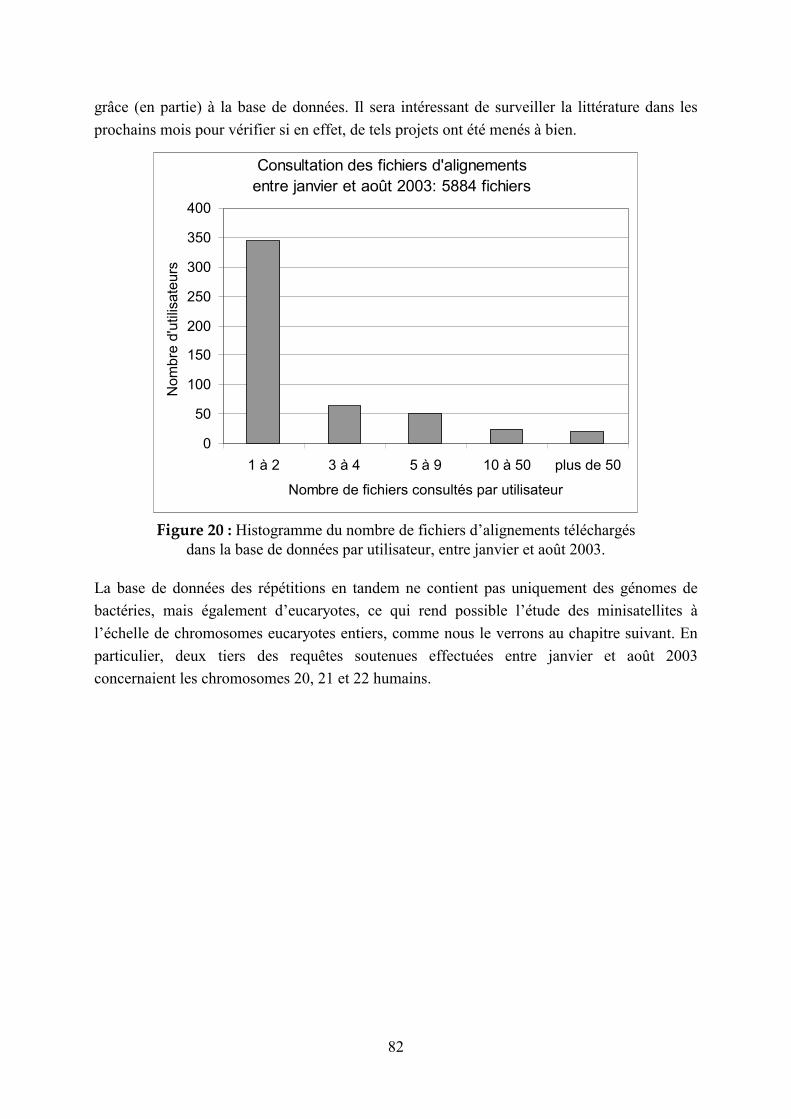
82
grâce (en partie) à la base de données. Il sera intéressant de surveiller la littérature dans les
prochains mois pour vérifier si en effet, de tels projets ont été menés à bien.
Consultation des fichiers d'alignements
entre janvier et août 2003: 5884 fichiers
0
50
100
150
200
250
300
350
400
1 à 2 3 à 4 5 à 9 10 à 50 plus de 50
Nombre de fichiers consultés par utilisateur
Nom
bre
d'u
tilis
ate
urs
Figure 20 : Histogramme du nombre de fichiers d’alignements téléchargés
dans la base de données par utilisateur, entre janvier et août 2003.
La base de données des répétitions en tandem ne contient pas uniquement des génomes de
bactéries, mais également d’eucaryotes, ce qui rend possible l’étude des minisatellites à
l’échelle de chromosomes eucaryotes entiers, comme nous le verrons au chapitre suivant. En
particulier, deux tiers des requêtes soutenues effectuées entre janvier et août 2003
concernaient les chromosomes 20, 21 et 22 humains.

83
2.3 Utilisation de la base de données pour l’étude des minisatellites humains
2.3.1 Etude de la répartition des minisatellites dans des chromosomes eucaryotes entièrement séquencés
La revue suivante (Vergnaud & Denoeud 2000), intitulée « Minisatellites : mutability and
genome architecture » (Minisatellites : mutabilité et architecture des génomes) présente une
synthèse sur les minisatellites humains, en particulier hypermutables, ainsi qu’une étude de la
répartition des minisatellites au sein de différents chromosomes eucaryotes, mettant à profit la
base de données des répétitions en tandem. En effet, jusqu’alors, les minisatellites étaient
identifiés par des techniques expérimentales telles que l’hybridation de sondes contenant des
motifs répétés (Vergnaud 1989). Notre étude est la première à tirer parti du séquençage des
génomes afin d’identifier de façon exhaustive les répétitions en tandem de chromosomes
entiers.
En outre, cette étude met le doigt sur la difficulté de définir avec précision les minisatellites
au sein de l’ensemble des répétitions en tandem. Nous y proposons une définition plus vaste
que celle précédemment imposée par les contraintes expérimentales (Southern Blot) qui
n’identifiaient que des minisatellites de longueur totale de l’ordre du kilobase, ou supérieure.
Ces travaux antérieurs ont montré que cette catégorie de répétitions en tandem présente, chez
l’homme, un biais de distribution télomérique très marqué (Amarger 1998). Il s’avère que des
répétitions en tandem de quelques centaines de paires de bases tombent également dans la
catégorie des répétitions en tandem plus souvent associées aux extrémités télomériques. Le
nombre de minisatellites du génome humain peut alors être estimé à plusieurs dizaines de
milliers, soit dix fois plus qu’initialement supposé.
Ainsi, il semble peu réaliste de mesurer le polymorphisme (par typages PCR, ou Southern
Blot) de tous les minisatellites du génome humain, dans le but, par exemple, d’identifier des
minisatellites hypermutables qui sont en premier lieu très polymorphes. Cependant, les
données de séquences pourraient être mises à profit afin de trouver des critères prédictifs sur
le polymorphisme, qui permettraient alors de se cantonner à un nombre raisonnable de
candidats à typer. Un article (Denoeud 2003) présentant une étude sur la prédiction du
polymorphisme des minisatellites, menée sur les chromosomes 21 et 22 humains, sera
présenté au chapitre suivant.
84
Résumé :
Il a été montré que les minisatellites sont associés à des aspects importants de la biologie du
génome humain tels que la régulation de gènes, les sites fragiles ou les phénomènes
d’empreinte parentale. Notre connaissance de la biologie des minisatellites a fortement
augmenté ces dix dernières années, grâce à l’identification et l’analyse détaillée des
minisatellites hypermutables humains, aux modèles expérimentaux dans la levure, et aux
études in vitro menées récemment sur les propriétés de recombinaison des minisatellites. En
parallèle, les minisatellites ont été mis en avant comme des biomarqueurs potentiels pour
mesurer l’effet d’agents génotoxiques comme les radiations ionisantes. Dans cette revue, nous
synthétisons et discutons des observations récentes sur les minisatellites. De plus, nous
mettons à profit la parution récente de séquences de chromosomes entiers pour proposer une
vue unificatrice qui devrait faciliter l’annotation des répétitions en tandem.

Minisatellites: Mutability and GenomeArchitecture
Gilles Vergnaud 1,2,3 and France Denoeud1
1Institut de Genetique et Microbiologie, Universite Paris Sud 91405 Orsay, France; 2Centre d’Etudes du Bouchet, 91710 Vert
le Petit, France
Minisatellites have been found in association with important features of human genome biology such as gene
regulation, chromosomal fragile sites, and imprinting. Our knowledge of minisatellite biology has greatly
increased in the past 10 years owing to the identification and careful analysis of human hypermutable
minisatellites, experimental models in yeast, and recent in vitro studies of minisatellite recombination properties.
In parallel, minisatellites have been put forward as potential biomarkers for the monitoring of genotoxic agents
such as ionizing radiation. We summarize and discuss recent observations on minisatellites. In addition we take
advantage of recent whole chromosome sequence data releases to provide a unifying view which may facilitate
the annotation of tandem repeat sequences.
Classic Definition and Early Applications
of Minisatellites
Minisatellites are usually defined as the repetition in
tandem of a short (6- to 100-bp) motif spanning 0.5 kb
to several kilobases. Although the first examples de-
scribed 20 years ago were of human origin, (Wyman
and White 1980), similar DNA structures have been
found in many organisms including bacteria. Com-
parisons of the repeat units in classical minisatellites
led early on to the notion of consensus or core se-
quences, which exhibit some similarities with the �
sequence of � phage (GCTGTGG). In general, the ma-
jority of classical minisatellites are GC rich, with a
strong strand asymmetry.
Because of their length polymorphism, which re-
sults from variations in the number of repeats, and the
ability of some of these arrays to cross-hybridize with
tens of other similar loci throughout the genome,
minisatellites have opened the way to DNA finger-
printing for individual identification (Jeffreys et al.
1985). Minisatellites also provided the first highly
polymorphic, multiallelic markers for linkage studies
(Nakamura et al. 1987). The usefulness of polymorphic
minisatellites (also called VNTRs for variable number
of tandem repeats) in the early stages of human ge-
nome mapping is reflected in the Centre d’Etude du
Polymorphisme Humain/National Institutes of Health
consortium linkage maps (National Institutes of
Health/Centre d’Etude du Polymorphisme Humain
collaborative mapping group 1992).
In parallel, tandem repeats belonging to the mini-
satellite class were found to be associated with many
interesting features of human genome biology and
evolution, usually revealed by pathologies of genetic
origin. In brief, minisatellites are thought to contribute
to genome function in one of three ways: (1) Some are
part of an open reading frame, which may or may not
display polymorphism in the human population (for
review, see Bois and Jeffreys 1999). (2) Some bind pro-
teins with a variety of functional consequences,
strongly suspected or still very hypothetical. Minisat-
ellites located in the 5� region of genes participate in
the regulation of transcription (Kennedy et al. 1995).
Others located within introns interfere with splicing
(Turri et al. 1995). Minisatellites at imprinted loci are
thought to play a role in the imprint control (Chaillet
et al. 1995; Neumann et al. 1995). More speculatively,
minisatellites have been proposed as intermediates in
chromosome pairing initiation in some eukaryote ge-
nomes (Ashley 1994; Sybenga 1999), which might be
related to their proposed recombinogenic properties
(Boan et al. 1998; Wahls and Moore 1998). (3) Finally,
minisatellites may constitute chromosome fragile sites
(for review, see Sutherland et al. 1998) and have been
found in the vicinity of a number of recurrent translo-
cation breakpoints and in the switch recombination
site in immunoglobulin heavy chain genes (Brusco et
al. 1999). These aspects of minisatellite biology have
been reviewed elsewhere and will not be further dis-
cussed in this article.
Novel Insights and Applications
in Minisatellite Biology
Although the high degree of length polymorphism
among minisatellites indicates that they are fast-
evolving sequences, most of them are in fact quite
stable, and neomutated alleles have been observed
only at a few loci. Recent research has focused on iden-
tifying these rare hypermutable loci in human and
3Corresponding author.E-MAIL [email protected]; FAX 33-1-69-15-66-78.
Review
10:899–907 ©2000 by Cold Spring Harbor Laboratory Press ISSN 1088-9051/00 $5.00; www.genome.org Genome Research 899www.genome.org

other genomes because they seem the most appropri-
ate models to illustrate the mechanisms of minisatel-
lite variability. Newly mutated alleles at human hyper-
mutable minisatellites have been characterized in de-
tail, leading to the current model of minisatellite
mutation initiation by double-strand breaks (DSBs),
and a number of attempts have been made to transfer
human minisatellite instability into a more tractable
system. We will present and discuss the work done on
the subclass of minisatellites that are hypermutable in
meiosis.
We will also show how investigations on the sen-
sitivity of minisatellites to some genotoxic agents
might provide new insight on minisatellite mutation
processes. This work may lead to new applications for
minisatellite sequences, such as the development of
genotoxicity assays to specifically detect agents inter-
fering with DNA recombination or replication.
Finally, the release of whole genome sequence
data allows new approaches to minisatellite character-
ization. In spite of the fact that our understanding of
minisatellite biology has improved very significantly
in the last 10 years, minisatellites are usually not dis-
cussed or even annotated in releases of new sequence
data. This is likely due to the lack of a clear and satis-
fying definition of these structures. We will briefly re-
view the history of minisatellite characterization and
chromosomal localization and compare the picture
that these earlier investigations produced to the global
view provided now by the sequencing of the human
genome.
Insights from the Study of Mutant Alleles at Human
Hypermutable Minisatellites
For practical reasons linked to the size of available
pedigrees, a minisatellite will usually be classified as
hypermutable if its average mutation rate in the germ-
line is higher than 0.5% (the ratio of mutation events
in the male and female germline is variable; it can be
highly skewed toward paternal events as in CEB1, or
equal as in MS1, see Table 1). As a rough estimate,
approximately 300 human minisatellites have been
typed across families (Armarger et al. 1998; Armour et
al. 1990; Nakamura et al. 1987) and less than ten of
these qualify as hypermutable (Table 1). The structural
features of hypermutable minisatellites described in
Table 1 are not specific for this subclass of tandem
repeats, and the proportion of telomeric versus inter-
stitial loci (MS32 and MS1 being interstitial) in this
collection fits with the proportion of telomeric and
interstitial loci among human minisatellites in general
(see below).
All hypermutable minisatellites characterized so
far possess internal variants, which have provided one
way to undertake mutant allele analysis (Table 1). Jef-
freys and colleagues developed a polymerase chain re-
action-based assay (Jeffreys et al. 1991) which has
proved very efficient at typing the position of variants
along alleles. These internal maps can be used to iden-
tify the origin of additional repeats in mutant alleles as
compared to their progenitors. An important part of
our current knowledge of hypermutable minisatellite
biology comes from this technology. Two reports in
which neomutated alleles at the CEB1 and MS32 hy-
permutable minisatellites were typed (Buard and Verg-
naud 1994; Jeffreys et al. 1994) pointed to DSBs as ini-
tiating events of the meiotic mutations. Both interal-
lelic (gene conversion-like) and intra-allelic exchanges
were observed, with a different proportion of the two
classes of events at the two loci. The detailed typing
achieved by the CEB1 study provided data showing
that some of the interallelic insertions are flanked by
duplicated motifs from the recipient allele. Figure 1
illustrates a model which fits with our current knowl-
edge of meiotic DSBs within hotspots (i.e., in yeast
they occur outside the tandem array (Debrauwere et al.
1999) and are almost blunt) while being compatible
with observations on CEB1 in the human context.
Subsequent studies have investigated the role of
the flanking sequence in the mutation process. This
interest in flanking sequences was prompted by the
observation that a point mutation very close to one
end of the MS32 array was associated with a strongly
reduced mutation rate of the corresponding allele
(Monckton et al. 1994). In addition, meiotic mutation
events in MS32 strongly clustered toward one end of
the array (Jeffreys et al. 1994). In contrast, somatic mu-
tations at MS32 (Jeffreys and Neumann 1997) do not
show clustering toward one end; they occur at a much
lower frequency and are simple intra-allelic events,
predominantly deletions.
Experimental Models
of Minisatellite Mutation
The development of experimental models to study
minisatellite mutation processes has been necessary in
order to analyze more precisely the timing of the mu-
tation processes, the underlying genetics, and test the
predictions made by the current models.
Attempts to develop animal models based on the
identification of naturally occurring hypermutable
minisatellites failed. Two hypermutable tandem re-
peats characterized in mice are the amplification of
short (respectively, 4- and 5-bp) units, which do not
fully qualify as minisatellites and are not amenable to
variant typing (for review, see Bois and Jeffreys, 1999).
For this reason, Jeffreys and colleagues developed a
transgenic mouse model. The inserts injected were ei-
ther an MS32 tandem array with only a few hundred
base pairs of flanking sequence or a complete cosmid
insert from the MS32 or CEB1 locus. Interestingly, al-
though the mitotic instability was transferred, no mei-
Vergnaud and Denoeud
900 Genome Researchwww.genome.org

Table 1. Known Human Hypermutable Minisatellites
Minisatellitename
Locus orD-number Location
Accessionnumber
Consensuslength
Nb ofvariant
positions
Percentmatch(%M)
GC%(bias)
Purine %(bias) Instability
Proportion ofpaternal events
CEB1 D2S90 2q37.3 (T) AF048727 39 8 90 72 (0.53) 68 (0.38) 6.7% 97%CEB15 D1S172 1p36.33 (T) AL096805 18 8 91 68 (0.33) 77 (0.56) 1.5% 100%CEB25 D10S180 10q26.3 (T) AL096806 52 >20 82 53 (0.73) 67 (0.36) 2.5% 65%CEB36 D10S473 10q26.3 (T) AL096810 42 18 93 64 (0.65) 66 (0.32) 1.8% 50%CEB42 D8S358 8q24.3 (T) AL096807 41 4 95 63 (0.11) 53 (0.08) 0.5% 100%CEB72 D17S888 17q25 (T) AL096808 21 0 100 71 (0.60) 80 (0.62) 1.8% 65%MS1 D1S7 1p33–35 (I) X14856 9 3 85 58 (0.93) 79 (0.60) 5% 50%MS32 D1S8 1q42 (I) AF048729 29 3 92 61 (0.37) 67 (0.35) 0.4% 50%B6.7 20q13.3 (T) AF081787 34 6 91 60 (0.5) 68 (0.37) 5.5% 64%CSTB EPM1 21q22.3 (T) U46692 12 1 96 100 (0.5) 100 (0.5) 47% 76%
Minisatellites showing instability higher than 1% in the male or female germline are listed (with the exception of MS32, with a lower mutation rate, but which is a referenceminisatellite in many investigations). The instability values indicated are most often average values, as measured usually in the large Centre d’Etudes du Polymorphisme Humainfamilies (http://www.cephb.fr) with the exception of the cystatin B (CSTB) gene minisatellite. In this last case, the values given were measured in pathogenic, expanded alleles(Larson et al. 1999). Similarly, the mutation rate at CEB1 alleles has been shown to vary between <0.02% (at smaller alleles) and >20% (Buard et al. 1998). Percent match (%M)is the average similarity of motifs with the consensus motif. GC bias is the absolute value of (G% � C%)/(G% + C%). Purine bias is the absolute value of (pur% � pyr%).
Min
isate
llites
Genom
eR
esearch
901
ww
w.gen
om
e.org

otic instability was observed (Bois et al. 1997; Buard et
al. 2000). However, in these investigations the integra-
tion site was random, and no attempts were made to
target potentially more active loci of the mouse ge-
nome.
Alternative approaches have used yeast. The work
in yeast was pioneered by Rannug, Cederberg, and col-
leagues, who showed meiotic induction of human
minisatellite MS32 instability. The minisatellite was in-
serted in the vicinity of the LEU2 yeast hotspot for
recombination initiation, where DSBs frequently form
(Appelgren et al. 1997). Tetrad analysis demonstrated
that interallelic mutants, which might look like bona
fide crossover events (exchange of flanking markers,
no complex secondary rearrangements), are in fact
conversion events (Appelgren et al. 1999), which is of
some importance when interpreting similar human
data (Jeffreys et al. 1998). In a more recent investiga-
tion, the similar meiotic instability of a CEB1 allele
introduced in yeast was shown to be dependent on the
integration site (Debrauwere et al. 1999). Integration in
a cold spot for recombination initiation resulted in a
very low meiotic instability compared to integration
adjacent to the ARG4 recombination hotspot. At this
site, the tandem array did not modify the DSB hotspot:
DSBs remained detectable on both sides of the array at
a frequency comparable to the wild-type situation.
Suppression of the DSBs, either by a failure in activat-
ing the site, as obtained in a rad50 deficient strain, or
by the absence of the topoisomerase (Spo11) respon-
sible for the DSBs (Bergerat et al. 1997), reduced the
meiotic instability of the minisatellite to the mitotic
level. Finally, taking advantage of mismatch repair-
deficient strains, the predicted heteroduplex interme-
diates (Fig. 1) have been observed in some (but not all)
mutant alleles.
These observations, combined with the attempts
to develop a mouse model and the data in humans,
very strongly suggest that the production of an experi-
mental model in which the minisatellite shows meiotic
Figure 1 Revised model for meiotic mutation events demon-strating the formation of interallelic events with duplicationsflanking the converted motifs. In order to explain the observedduplication flanking meiotic interallelic events, the model initiallyproposed by Buard and Vergnaud (1994) and subsequentlyadopted by others (Bois and Jeffreys 1999) invoked DSBs initiatedwithin the array by staggered single-strand breaks separated by80 nucleotides or more. This would require a strong associatedhelicase activity and does not fit with the view now provided bythe yeast work (Debrauwere et al. 1999). Alternatively, the pre-sented model adapted from Debrauwere et al. (1999) shows howan almost blunt DSB, produced in a flanking DSB hotspot outsidethe minisatellite (step 1), can produce interallelic exchanges witha duplication flanking the converted motifs, as well as most, if notall, minisatellite rearrangements observed in man or yeast. After5�-3� resection (step 2), the repair is initiated by invading thesister chromatid and priming DNA synthesis on one or both (assuggested here, step 3) strands. After DNA synthesis, the newlysynthesized strands independently unwind (step 4) and are freeto engage in other DNA–DNA interactions (here, both strands areshown invading the homolog, step 5). Eventually, the newly syn-thesized strands reanneal together with properly aligned flankingsequences. A loop may form on one (as shown here, step 6) orboth (Debrauwere et al. 1999) strands. This loop can be con-verted into the corrected allele (Debrauwere 2000) via a singlestrand cut on the opposing DNA strand (step 7), or removed.Depending on which strand is used to correct the heteroduplex,a direct duplication of repeats flanking the converted patch isproduced (step 8, bottom). All models proposed so far predictthe existence of patches of heteroduplex intermediates producedby the reannealing of similar, but different, minisatellite motifs(here, step 6). This last prediction was successfully tested in De-brauwere et al. (1999). An interesting aspect of this model is thatthe lower strand may extend in the flanking sequence at steps 2and 4. This will produce a heteroduplex region in the flankingsequence (steps 6 and 7; left unrepaired here in step 8) which,once repaired, may introduce a conversion patch in the finalproduct flanking sequence.
Vergnaud and Denoeud
902 Genome Researchwww.genome.org
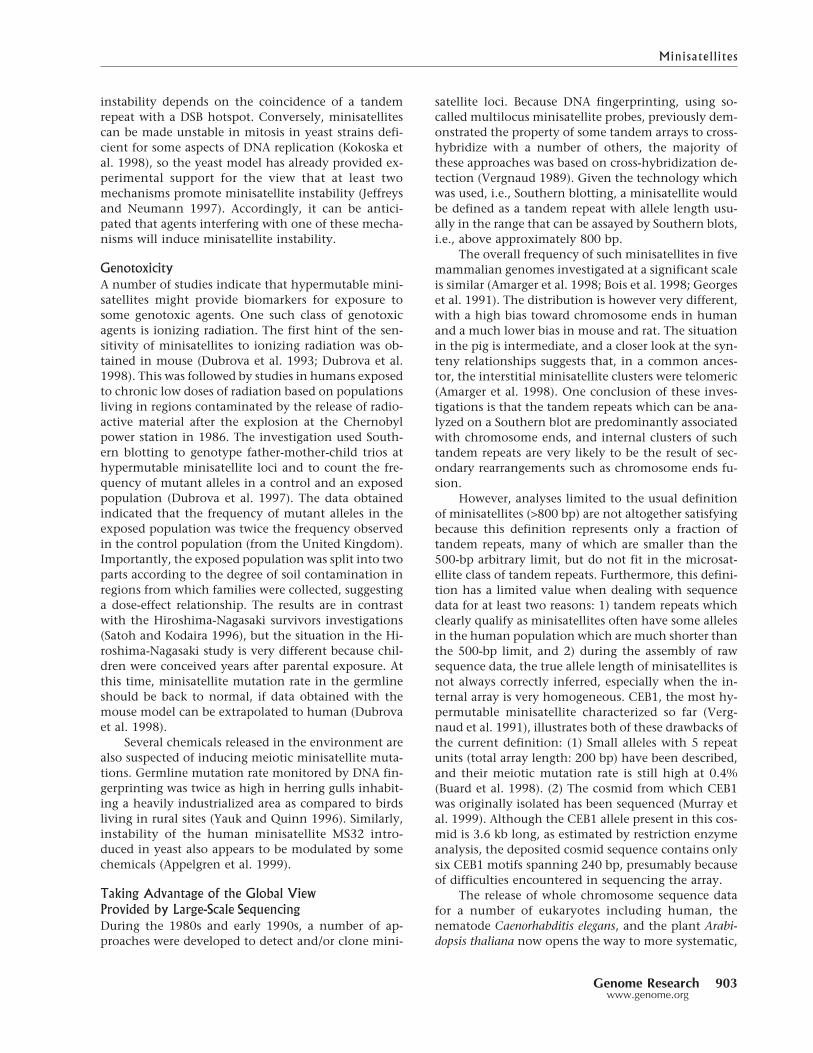
instability depends on the coincidence of a tandem
repeat with a DSB hotspot. Conversely, minisatellites
can be made unstable in mitosis in yeast strains defi-
cient for some aspects of DNA replication (Kokoska et
al. 1998), so the yeast model has already provided ex-
perimental support for the view that at least two
mechanisms promote minisatellite instability (Jeffreys
and Neumann 1997). Accordingly, it can be antici-
pated that agents interfering with one of these mecha-
nisms will induce minisatellite instability.
Genotoxicity
A number of studies indicate that hypermutable mini-
satellites might provide biomarkers for exposure to
some genotoxic agents. One such class of genotoxic
agents is ionizing radiation. The first hint of the sen-
sitivity of minisatellites to ionizing radiation was ob-
tained in mouse (Dubrova et al. 1993; Dubrova et al.
1998). This was followed by studies in humans exposed
to chronic low doses of radiation based on populations
living in regions contaminated by the release of radio-
active material after the explosion at the Chernobyl
power station in 1986. The investigation used South-
ern blotting to genotype father-mother-child trios at
hypermutable minisatellite loci and to count the fre-
quency of mutant alleles in a control and an exposed
population (Dubrova et al. 1997). The data obtained
indicated that the frequency of mutant alleles in the
exposed population was twice the frequency observed
in the control population (from the United Kingdom).
Importantly, the exposed population was split into two
parts according to the degree of soil contamination in
regions from which families were collected, suggesting
a dose-effect relationship. The results are in contrast
with the Hiroshima-Nagasaki survivors investigations
(Satoh and Kodaira 1996), but the situation in the Hi-
roshima-Nagasaki study is very different because chil-
dren were conceived years after parental exposure. At
this time, minisatellite mutation rate in the germline
should be back to normal, if data obtained with the
mouse model can be extrapolated to human (Dubrova
et al. 1998).
Several chemicals released in the environment are
also suspected of inducing meiotic minisatellite muta-
tions. Germline mutation rate monitored by DNA fin-
gerprinting was twice as high in herring gulls inhabit-
ing a heavily industrialized area as compared to birds
living in rural sites (Yauk and Quinn 1996). Similarly,
instability of the human minisatellite MS32 intro-
duced in yeast also appears to be modulated by some
chemicals (Appelgren et al. 1999).
Taking Advantage of the Global View
Provided by Large-Scale Sequencing
During the 1980s and early 1990s, a number of ap-
proaches were developed to detect and/or clone mini-
satellite loci. Because DNA fingerprinting, using so-
called multilocus minisatellite probes, previously dem-
onstrated the property of some tandem arrays to cross-
hybridize with a number of others, the majority of
these approaches was based on cross-hybridization de-
tection (Vergnaud 1989). Given the technology which
was used, i.e., Southern blotting, a minisatellite would
be defined as a tandem repeat with allele length usu-
ally in the range that can be assayed by Southern blots,
i.e., above approximately 800 bp.
The overall frequency of such minisatellites in five
mammalian genomes investigated at a significant scale
is similar (Amarger et al. 1998; Bois et al. 1998; Georges
et al. 1991). The distribution is however very different,
with a high bias toward chromosome ends in human
and a much lower bias in mouse and rat. The situation
in the pig is intermediate, and a closer look at the syn-
teny relationships suggests that, in a common ances-
tor, the interstitial minisatellite clusters were telomeric
(Amarger et al. 1998). One conclusion of these inves-
tigations is that the tandem repeats which can be ana-
lyzed on a Southern blot are predominantly associated
with chromosome ends, and internal clusters of such
tandem repeats are very likely to be the result of sec-
ondary rearrangements such as chromosome ends fu-
sion.
However, analyses limited to the usual definition
of minisatellites (>800 bp) are not altogether satisfying
because this definition represents only a fraction of
tandem repeats, many of which are smaller than the
500-bp arbitrary limit, but do not fit in the microsat-
ellite class of tandem repeats. Furthermore, this defini-
tion has a limited value when dealing with sequence
data for at least two reasons: 1) tandem repeats which
clearly qualify as minisatellites often have some alleles
in the human population which are much shorter than
the 500-bp limit, and 2) during the assembly of raw
sequence data, the true allele length of minisatellites is
not always correctly inferred, especially when the in-
ternal array is very homogeneous. CEB1, the most hy-
permutable minisatellite characterized so far (Verg-
naud et al. 1991), illustrates both of these drawbacks of
the current definition: (1) Small alleles with 5 repeat
units (total array length: 200 bp) have been described,
and their meiotic mutation rate is still high at 0.4%
(Buard et al. 1998). (2) The cosmid from which CEB1
was originally isolated has been sequenced (Murray et
al. 1999). Although the CEB1 allele present in this cos-
mid is 3.6 kb long, as estimated by restriction enzyme
analysis, the deposited cosmid sequence contains only
six CEB1 motifs spanning 240 bp, presumably because
of difficulties encountered in sequencing the array.
The release of whole chromosome sequence data
for a number of eukaryotes including human, the
nematode Caenorhabditis elegans, and the plant Arabi-
dopsis thaliana now opens the way to more systematic,
Minisatell ites
Genome Research 903www.genome.org

sequence-based investigations. For this purpose, we
have constructed a prototype tandem repeat database
(http://minisatellites.u-psud.fr) using the Tandem Re-
peats Finder software (Benson 1999) to identify the re-
peats. The database contains more than 14,000 tandem
repeats for the acrocentric chr22 (34.6 Mb) (Dunham
et al. 1999) and can be queried according to a number
of features (see legend, Fig. 2). One-third of human
chr22 tandem repeats (Fig. 2A) satisfies an enlarged
definition of minisatellites, as used in this review (at
least three units, repeat unit longer than 6 bp). Among
them, minisatellites with repeat units longer than 16
bp and total length greater than 100 bp display a dis-
tribution strongly biased toward the chr22 long-arm
telomere (Fig. 2B).
Figure 2C shows the result of a query mimicking
characteristics of classical minisatellites, i.e., query “B”
plus a high GC content, strong strand bias, and strong
internal homogeneity (see legend, Fig. 2, for details).
Half of the 62 minisatellites fitting this query are lo-
cated within the terminal 10% of chr22. Such simple
queries demonstrate that a fraction of minisatellites,
comprising hundreds of loci on chromosome 22 alone,
do behave as initially suggested by the subset of clas-
sical minisatellites, i.e., are present at a much higher
frequency in the terminal R band of human chromo-
somes (Amarger et al. 1998).
Chromosome ends appear to be relatively poor in
recombination nodules during human male meiosis,
which is surprising given the very high male recombi-
nation rates observed toward chromosome ends. Sub-
telomeric minisatellites are one class of sequences that
have been put forward as candidates to help explain
this paradox (Ashley 1994; Sybenga 1999). Specific
mechanisms would be activated in male meiosis, and
minisatellites would be involved in chromosome pair-
ing, either directly or via interactions with pairing pro-
teins. This predicts that minisatellites should not dis-
play subtelomeric clustering in plants, where no such
discrepancy between recombination nodules and rates
is observed. Figure 3 presents comparisons of the three
species using C.elegans chromosome 1 (12.75 Mb) and
A. thaliana chromosome 4 (17.8 Mb) (The C. elegans
Sequencing Consortium 1998; Mayer et al. 1999). The
total number of tandem repeats found with Tandem
Repeats Finder in the three species is not proportional
to chromosome length (Fig. 3). It is significantly higher
in the nematode (637 Mb) when compared to man and
A. thaliana (415 Mb and 445 Mb, respectively).
The result of a representative query is shown in
Figure 3, bottom row. The number of positive minisat-
ellites is similar in the three species, taking into ac-
count chromosome size difference. A strong telomeric
bias is observed for C. elegans chr1, (right panel) remi-
niscent of the situation in human chr22. In contrast,
the distribution of minisatellites in A. thaliana (middle)
is strikingly different from that of the two other ge-
nomes: tandem repeats are mainly located around the
centromere. Figure 4A plots, for each species, the ratio
of telomeric versus nontelomeric tandem repeats ac-
cording to repeat unit length. C. elegans chr1 demon-
strates telomeric bias for both short units (in particular,
6- and 12-bp units, due to the presence of many
(TTAGGC)n telomere-like tandem arrays (The C. el-
egans Sequencing Consortium 1998) and longer units
(above approximately 18 bp). Human chr22 demon-
strates telomeric bias for repeat units above 17 bp. It
may be worth noting that in yeast, 16 bp is the thresh-
old above which mismatch repair mechanisms are un-
able to correct DNA loops (Sia et al. 1997). Figure 4B
plots the same measure of telomeric bias according to
the overall array length. In contrast with C. elegans, the
telomeric bias for human chr22 appears only for arrays
longer than 120–140 bp. This threshold is reminiscent
Figure 2 Distribution of tandem repeats corresponding to dif-ferent queries along human chromosome 22. Tandem repeatshave been identified within the human chromosome 22 se-quence using the Tandem Repeats Finder (TRF) software with thefollowing options: alignment parameters = (2,3,5), minimumalignment score to report repeat = 50, maximum periodsize = 500. Redundancy was then eliminated, and Alu and satel-lite sequences (152 were identified) were filtered. The arrow (topleft) shows the centromere position. The position of the 51 chr22Genethon microsatellites present in the database is shown witharrow heads. GC-rich (pink) or -poor (green) areas, regions ofincreased recombination, and known mouse synteny correspon-dence are as indicated in Dunham et al. (1999) Distributionsobtained with different queries: (A, Left) U > = 6, N > = 3 (B,Middle) U > 16, N > = 3, L > 100 (C, Right) U > 16, N > = 3,%GC > = 65%, BGC > = 0.3, %M > = 85% (U = unit length,N = copy number, L = total length, %GC = GC percent,BGC = G/C bias = Ơ%G-%CƠ/(%G + %C), %M (percent matches)is the average similarity of each motif with the consensus motif).Percentages reported correspond to the proportion of objects inthe last 10% of total length. �2 values were calculated by com-paring the last 10% of the chromosome with the mean numberof objects along the whole chromosome. �2 threshold of signifi-cance (homogeneity hypothesis is rejected if �2 is greater thanthreshold) is 3.841 with P = 5%, and 10.827 with P = 0.1% (1degree of freedom).
Vergnaud and Denoeud
904 Genome Researchwww.genome.org

of triplet repeat instability observed above 40–50 re-
peats. No telomeric bias is observed in A. thaliana chr4.
Concluding Remarks
and Perspectives
Previously, the number of classical minisatellites has
been estimated to be a few thousand in the human
genome, which translates to a few tens on chromo-
some 22. Such rare objects would not likely play a sig-
nificant role in genome metabolism. The view now
provided by the availability of whole human chromo-
some sequence reveals a much larger number of small
minisatellites with repeat units similar to the classical
structures and a similarly biased distribution toward
chromosome ends, which is not observed in A.
thaliana. These observations give much more credibil-
ity to these structures (Boan et al. 1998; Wahls and
Moore 1998). Obviously, comparisons with additional,
larger human chromosomes will be of some interest.
It is tempting to speculate that the meiotic hyper-
mutability of some minisatellite structures is the by-
product of the coincidence of an ordinary minisatellite
with a DSB hotspot (Debrauwere et al. 1999). The dis-
appearance of a hotspot, as proposed by Boulton et al.
(1997) will then remove the hypermutability of the
neighboring tandem repeat. In this model, the study of
hypermutable minisatellites is demonstrating more
about human DSB hotspots, the majority of which
would exist independently of neighboring tandem re-
peats in human (Badge et al. 2000) as in yeast, than
about minisatellites in general. The model presented in
Figure 1 shows how a double strand break occurring
outside of the array (as suggested by Debrauwere et al.
1999) can indeed produce the complex interallelic
events observed in man, including duplications flank-
ing the converted patch. The model also accommo-
dates conversion patches in the flanking sequence,
which may include mosaics of intra- and interallelic
origin. In contrast, the making of minisatellites in gen-
eral would result from replication mechanisms, fa-
vored by deficiencies in enzymes involved in replica-
tion such as Saccharomyces cerevisiae Rad27 as proposed
in Tishkoff et al. (1997). In the process, sequence fea-
tures of the motif, likely to produce secondary struc-
tures or slow down the polymerase on the lagging
strand during replication (G-rich DNA strands, palin-
dromic motifs in AT rich minisatellites, GC richness),
may be important.
In this regard, no information regarding minisat-
ellite instability or even polymorphism is obtained us-
ing the tandem repeat database presented here. This
will be an important further step of the database de-
velopment, which might benefit from the current
knowledge of variant motif interspersion patterns
along hypermutable minisatellite alleles. In addition,
tandem repeat polymorphism predictions will be facili-
tated by the expected availability, in the near future, of
sequence data from more than one allele.
Genotoxicity is a promising domain for minisatel-
lite-related investigation. It may combine short-term
applications toward the development of genotoxicity
assays specifically identifying recombinogenic activi-
ties with more basic investigations into the purpose of
minisatellites and what triggers them. One question
raised by these investigations is whether the tandem
array itself is the target of the genotoxic agent, whether
it is the flanking DSB hotspot which is further acti-
vated by the agent, or whether it is the replication ma-
chinery which is affected. In the second hypothesis,
hypermutable minisatellites would act as markers for
the activity of their flanking recombination hotspot,
whereas in the first (and perhaps also third) hypoth-
esis, any minisatellite could act as a biomarker for the
genotoxic agent. Recently developed yeast models may
help address such issues.
ACKNOWLEDGMENTSWe thank Christine Pourcel for comments and critical reading
Figure 3 Comparison of tandem repeats distribution in threespecies. See legend, Fig. 2, for information on database construc-tion and other details. Arrows show centromere position (un-known for nematode). The Tandem Repeats Finder softwareidentifies tandem repeats at a frequency of 415 per Mb for hu-man chromosome 22, 445 per Mb for Arabidopsis thaliana chro-mosome 4, and 637 per Mb for Caenorhabditis elegans chromo-some 1 (“No query” panel). Bottom panel: the query appliedwas U > = 10, N >= 3, L > 100, 0.3 > = BGC > = 0.55, %M >= 70. Chromosomes were fragmented in areas of comparable
length: 1.73 Mb (20 areas), 1.78 Mb (10 areas), and 1.82 Mb (7areas), for human chromosome 22, plant chromosome 4 andnematode chromosome 1, respectively. For human chromosome22 and nematode chromosome 1, significant telomeric biases areobserved. In contrast, the plant chromosome shows a bias to-ward the centromeric region.
Minisatell ites
Genome Research 905www.genome.org

of this work. Current minisatellite work in the laboratory is
supported by a grant from Délégation Générale de
l’Armement (DGA/DSP/STTC).
REFERENCESAmarger, V., Gauguier, D., Yerle, M., Apiou, F., Pinton, P.,
Giraudeau, F., Monfouilloux, S., Lathrop, M., Dutrillaux, B.,
Buard, J. et al. 1998. Analysis of the human, pig, and rat
genomes supports a universal telomeric origin of minisatellite
sequences. Genomics 52: 62–71.
Appelgren, H., Cederberg, H., and Rannug, U. 1997. Mutations at
the human minisatellite MS32 integrated in yeast occur with
high frequency in meiosis and involve complex recombination
events. Mol. Gen. Genet. 256: 7–17.
Appelgren, H., Cederberg, H., and Rannug, U. 1999. Meiotic
interallelic conversion at the human minisatellite MS32 in yeast
triggers recombination in several chromatids. Gene 239: 29–38.
Appelgren, H., Hedenskog, M., Sandstrom, C., Cederberg, H., and
Rannug, U. 1999. Polychlorinated biphenyls induce meiotic
length mutations at the human minisatellite MS32 in yeast.
Environ. Mol. Mutagen 34: 285–290.
Armour, J.A.L., Povey, S., Jeremiah, S., and Jeffreys, A.J. 1990.
Systematic cloning of human minisatellites from ordered array
charomid libraries. Genomics 8: 501–512.
Ashley, T. 1994. Mammalian meiotic recombination: a
reexamination. Hum. Genet. 94: 587–593.
Badge, R.M., Yardley, J., Jeffreys, A.J., and Armour, J.A. 2000.
Crossover breakpoint mapping identifies a subtelomeric hotspot
for male meiotic recombination. Hum. Mol. Genet. 9: 1239–1244.
Benson, G. 1999. Tandem repeats finder: a program to analyze DNA
sequences. Nucleic Acids Res. 27: 573–580.
Bergerat, A., de Massy, B., Gadelle, D., Varoutas, P.-C., Nicolas, A.,
and P. Forterre. 1997. An atypical topoisomerase II from archaea
with implication for meiotic recombination. Nature
386: 414–417.
Boan, F., Rodriguez, J.M., and Gomez-Marquez, J. 1998. A
non-hypervariable human minisatellite strongly stimulates in
vitro intramolecular homologous recombination. J. Mol. Biol.
278: 499–505.
Bois, P., Collick, A., Brown, J., and Jeffreys, A.J. 1997. Human
minisatellite MS32 (D1S8) displays somatic but not germline
instability in transgenic mice. Hum. Mol. Genet. 6: 1565–1571.
Bois, P. and Jeffreys, A.J. 1999. Minisatellite instability and germline
mutation. Cell Mol. Life Sci. 55: 1636–1648.
Bois, P., Stead, J.D., Bakshi, S., Williamson, J., Neumann, R.,
Moghadaszadeh, B., and Jeffreys, A.J. 1998. Isolation and
characterization of mouse minisatellites. Genomics 50: 317–330.
Boulton, A., Myers, R.S., and Redfield, R.J. 1997. The hotspot
conversion paradox and the evolution of meiotic recombination.
Proc. Natl. Acad. Sci. 94: 8058–8063.
Brusco, A., Saviozzi, S., Cinque, F., Bottaro, A., and DeMarchi, M.
1999. A recurrent breakpoint in the most common deletion of
the Ig heavy chain locus (del A1-GP-G2-G4-E). J. Immunol.
163: 4392–4398.
Buard, J., Bourdet, A., Yardley, J., Dubrova, Y., and Jeffreys, A.J.
1998. Influences of array size and homogeneity on minisatellite
mutation. EMBO J. 17: 3495–3502.
Buard, J., Collick, A., Brown, J., and Jeffreys, A.J. 2000. Somatic
versus germline mutation processes at minisatellite CEB1
(D2S90) in humans and transgenic mice. Genomics 65: 95–103.
Buard, J. and Vergnaud, G. 1994. Complex recombination events at
the hypermutable minisatellite CEB1 (D2S90). EMBO J.
13: 3203–3210.
Chaillet, J.R., Bader, D.S., and Leder, P. 1995. Regulation of genomic
imprinting by gametic and embryonic processes. Genes Dev.
9: 1177–1187.
Debrauwere, H. 2000. Analyse des mecanismes d’instabilite des
sequences repetees humaines de type minisatellite dans la levure
S. cerevisiae. Biologie-Sciences de la vie. PhD thesis, University Paris
VI: 56–61.
Debrauwere, H., Buard, J., Tessier, J., Aubert, D., Vergnaud, G., and
Nicolas, A. 1999. Meiotic instability of human minisatellite CEB1
in yeast requires DNA double-strand breaks. Nat. Genet.
23: 367–371.
Dubrova, Y.E., Jeffreys, A.J., and Malashenko, A.M. 1993. Mouse
minisatellite mutations induced by ionizing radiation. Nat.
Genet. 5: 92–94.
Dubrova, Y.E., Nesterov, V.N., Krouchinsky, N.G., Ostapenko, V.A.,
Vergnaud, G., Giraudeau, F., Buard, J., and Jeffreys, A.J. 1997.
Further evidence for elevated human minisatellite mutation rate
in Belarus eight years after the Chernobyl accident. Mut. Res.
381: 267–278.
Dubrova, Y.E., Plumb, M., Brown, J., Fennelly, J., Bois, P., Goodhead,
D., and Jeffreys, A.J. 1998. Stage specificity, dose response, and
doubling dose for mouse minisatellite germ-line mutation
induced by acute radiation. Proc. Natl. Acad. Sci. 95: 6251–6255.
Dunham, I., Shimizu, N., Roe, B.A., Chissoe, S., Hunt, A.R., Collins,
J.E., Bruskiewich, R., Beare, D.M., Clamp, M., Smink, L.J. et al.
1999. The DNA sequence of human chromosome 22. Nature
402: 489–495.
Georges, M., Gunawardana, A., Threadgill, D.W., Lathrop, M.,
Olsaker, I., Mishra, A., Sargeant, L.L., Schoeberlein, A., Steele,
M.R., Terry, C. et al. 1991. Characterization of a set of variable
number of tandem repeat markers conserved in bovidae.
Genomics 11: 24–32.
Figure 4 Comparison between terminal and other regions according to unit length (A) or total length (B). On Y-axis: {number of objectsin the terminal 10% of the sequence [T] � number of object in other regions [mean for 10%] [O])/O (corresponds to a Z-score). If >0,terminal 10% are richer than the rest of the genome; else they are poorer, with a significance threshold of 1.96 (dotted lines). On X-axis:unit length (A) or total array length (B).
Vergnaud and Denoeud
906 Genome Researchwww.genome.org

Jeffreys, A.J., MacLeod, A., Tamaki, K., Neil, D.L., and Monckton,
D.G. 1991. Minisatellite repeat coding as a digital approach to
DNA typing. Nature 354: 204–209.
Jeffreys, A.J., Neil, D., and Neumann, R. 1998. Repeat instability at
human minisatellites arising from meiotic recombination. EMBO
J. 17: 4147–4157.
Jeffreys, A.J. and Neumann, R. 1997. Somatic mutation processes at a
human minisatellite. Hum. Mol. Genet. 6: 129–136.
Jeffreys, A.J., Tamaki, K., MacLeod, A., Monckton, D.G., Neil, D.L.,
and Armour, J.A.L. 1994. Complex gene conversion events in
germline mutation at human minisatellites. Nat. Genet.
6: 136–145.
Jeffreys, A.J., Wilson, V., and Thein, S.L. 1985. Individual-specific
’fingerprints’ of human DNA. Nature 316: 76–79.
Kennedy, G.C., German, M.S., and Rutter, W.J. 1995. The
minisatellite in the diabetes susceptibility locus IDDM2 regulates
insulin transcription. Nat. Genet. 9: 293–298.
Kokoska, R.J., Stefanovic, L., Tran, H.T., Resnick, M.A., Gordenin,
D.A., and Petes, T.D. 1998. Destabilization of yeast micro- and
minisatellite DNA sequences by mutations affecting a nuclease
involved in Okazaki fragment processing (rad27) and DNA
polymerase delta (pol3-t). Mol. Cell Biol. 18: 2779–2788.
Larson, G.P., Ding, S., Lafreniere, R.G., Rouleau, G.A., and Krontiris,
T.G. 1999. Instability of the EPM1 minisatellite. Hum. Mol. Genet.
8: 1985–1988.
Mayer, K., Schuller, C., Wambutt, R., Murphy, G., Volckaert, G.,
Pohl, T., Dusterhoft, A., Stiekema, W., Entian, K.D., Terryn, N. et
al. 1999. Sequence and analysis of chromosome 4 of the plant
Arabidopsis thaliana. Nature 402: 769–777.
Monckton, D.G., Neumann, R., Guram, T., Fretwell, N., Tamaki, K.,
MacLeod, A., and Jeffreys, A.J. 1994. Minisatellite mutation rate
variation associated with a flanking DNA sequence
polymorphism. Nat. Genet. 8: 162–170.
Murray, J., Buard, J., Neil, D.L., Yeramian, E., Tamaki, K., Hollies,
C.R., and Jeffreys, A.J. 1999. Comparative sequence analysis of
human minisatellites showing meiotic repeat instability. Genome
Res. 9: 130–136.
Nakamura, Y., Leppert, M., O’Connell, P., Wolff, R., Holm, T.,
Culver, M., Martin, C., Fujimoto, E., Hoff, M., Kumlin, E. et al.
1987. Variable number of tandem repeat (VNTR) markers for
human gene mapping. Science 235: 1616–1622.
Neumann, B., Kubicka, P., and Barlow, D.P. 1995. Characteristics of
imprinted genes. Nat. Genet. 9: 12–13.
NIH/CEPH Collaborative Mapping Group. 1992. A comprehensive
genetic linkage map of the human genome. Science 258: 67–83.
Satoh, C. and Kodaira, M. 1996. Effects of radiation on children.
Nature 383: 226.
Sia, E.A., Kokoska, R.J., Dominska, M., Greenwell, P., and Petes, T.D.
1997. Microsatellite instability in yeast: dependence on repeat
unit size and DNA mismatch repair genes. Mol. Cell Biol.
17: 2851–2858.
Sutherland, G.R., Baker, E., and Richards, R.I. 1998. Fragile sites still
breaking. Trends Genet. 14: 501–506.
Sybenga, J. 1999. What makes homologous chromosomes find each
other in meiosis? A review and an hypothesis. Chromosoma
108: 209–219.
The C. elegans Sequencing Consortium. 1998. Genome sequence of
the nematode C. elegans: a platform for investigating biology.
Science 282: 2012–2018.
Tishkoff, D.X., Filosi, N., Gaida, G.M., and Kolodner, R.D. 1997. A
novel mutation avoidance mechanism dependent on S.
cerevisiae RAD27 is distinct from DNA mismatch repair. Cell
88: 253–263.
Turri, M.G., Cuin, K.A., and Porter, A.C. 1995. Characterisation of a
novel minisatellite that provides multiple splice donor sites in an
interferon-induced transcript. Nucleic Acids Res. 23: 1854–1861.
Vergnaud, G. 1989. Polymers of random short oligonucleotides
detect polymorphic loci in the human genome. Nucleic Acids Res.
17: 7623–7630.
Vergnaud, G., Mariat, D., Apiou, F., Aurias, A., Lathrop, M., and
Lauthier, V. 1991. The use of synthetic tandem repeats to isolate
new VNTR loci: cloning of a human hypermutable sequence.
Genomics 11: 135–144.
Wahls, W.P. and Moore, P.D. 1998. Recombination hotspot activity
of hypervariable minisatellite DNA requires minisatellite DNA
binding proteins. Somat. Cell Mol. Genet. 24: 41–51.
Wyman, A. R. and White, R. 1980. A highly polymorphic locus in
human DNA. Proc. Natl. Acad. Sci. 77: 6754–6758.
Yauk, C.L. and Quinn, J.S. 1996. Multilocus DNA fingerprinting
reveals high rate of heritable genetic mutation in herring gulls
nesting in an industrialized urban site. Proc. Natl. Acad. Sci.
93: 12137–12141.
Minisatell ites
Genome Research 907www.genome.org

85
2.3.2 Prédiction du polymorphisme de minisatellites humains
L’article suivant (Denoeud 2003), intitulé « Predicting human minisatellite polymorphism »
(prédiction du polymorphisme de minisatellites humains), présente une étude menée sur les
chromosomes 21 et 22, visant à trouver un moyen de prédire le polymorphisme des
minisatellites à partir de leur séquence : en effet, comme nous l’avons vu avec l’article
précédent, le génome humain est riche en minisatellites et il serait donc d’une grande utilité
de pouvoir identifier les minisatellites d’intérêt c’est-à-dire polymorphes, et potentiellement
hypermutables, sans avoir recours à des typages PCR trop nombreux.
Cet article a mis en évidence deux critères corrélés au polymorphisme : le pourcentage en GC
et un critère, nommé HistoryR, reflétant la « facilité » avec laquelle on peut reconstruire
l’histoire des duplications successives ayant généré le minisatellite, ce qui se révèle par la
présence de co-mutations dans différents motifs.
D’autre part, nous avons montré que, contrairement à la comparaison de souches bactériennes,
qui est une technique efficace pour identifier des répétitions en tandem polymorphes, la
comparaison entre les séquences du génome humain produites par le consortium public
« Human Genome Project » et la société CELERA manque d’efficacité. Ceci peut résulter du
fait que les deux séquences ne sont pas indépendantes (la société CELERA ayant utilisé les
séquences publiques pour générer son assemblage) et que le séquençage des répétitions en
tandem est souvent de mauvaise qualité dans la version « CELERA » : le nombre de répétitions
est inférieur à celui de la plage d’allèles observés parmi les individus typés, ce qui doit
correspondre à des erreurs d’assemblage. Nous recommandons donc, pour l’étude des
répétitions en tandem, l’utilisation préférentielle des séquences du consortium public.
Enfin, cette étude a permis d’identifier un minisatellite hypermutable appartenant à une
séquence codante prédite : il s’agit d’une protéine hypothétique similaire à la protéine
« erythrocyte membrane-associated giant protein antigen 332 –Plasmodium falciparum-»
(Locuslink : LOC129238 [http://www.ncbi.nlm.nih.gov/LocusLink/]). Cette similitude n’est
basée que sur la présence d’une répétition en tandem de 11 acides aminés contenant la
succession des acides aminés PVEE dans les deux protéines. Les parties de la protéine
hypothétique situées hors du minisatellite n’ont aucune homologie avec des protéines
connues. Cette prédiction nécessite d’être confirmée, et, si cette protéine existe bel et bien, il
serait intéressant de mener une étude de l’influence du minisatellite hypermutable sur sa
fonction.
Résumé :
Nous cherchons à définir des critères prédictifs basés sur la séquence, qui permettraient
d’identifier des minisatellites polymorphes et hypermutables dans le génome humain. Le

86
polymorphisme d’un ensemble représentatif de minisatellites, issus des chromosomes 21 et 22
a été mesuré expérimentalement par typages PCR dans une population d’individus non-
apparentés. Deux approches prédictives ont été testées. La première utilise des
caractéristiques simples des séquences des répétitions en tandem (taille du motif, nombre de
répétitions, biais nucléotidique…) et une mesure plus complexe, appelée HistoryR, basée sur
la présence de mutations associées dans les répétitions en tandem. Nous montrons que la
mesure HistoryR et le pourcentage en GC sont fortement corrélés au polymorphisme et qu’en
tant que critères prédictifs, ils réduisent de moitié le nombre de répétitions à typer en
augmentant la proportion de minisatellites ayant une hétérozygotie supérieure ou égale à 0.5
de 43% à 59%. La deuxième approche utilise les différences de taille entre les minisatellites
de deux versions de la séquence du génome humain (provenant du consortium public et de la
société CELERA). Ce prédicteur augmente de façon similaire la proportion de minisatellites
polymorphes mais d’une façon moins efficace qu’attendu (un nombre trop élevé de
minisatellites polymorphes est manqué). Enfin, le typage des minisatellites fortement
polymorphes dans des grandes familles a permis d’identifier un nouveau minisatellite
hypermutable, situé dans une séquence codante prédite. Il pourrait s’agir du premier
minisatellite hypermutable humain codant.
Remarque: les données supplémentaires associées à cet article figurent en Annexe 3.
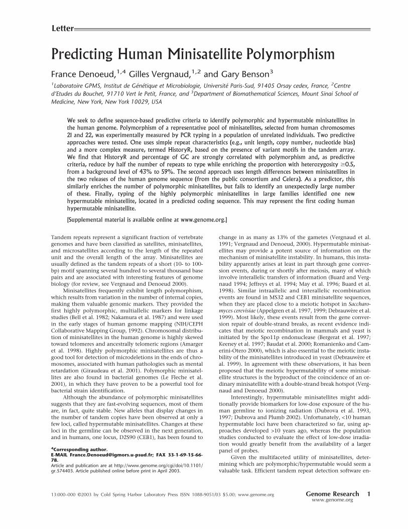
Predicting Human Minisatellite Polymorphism
France Denoeud,1,4 Gilles Vergnaud,1,2 and Gary Benson3
1Laboratoire GPMS, Institut de Genetique et Microbiologie, Universite Paris-Sud, 91405 Orsay cedex, France, 2Centre
d’Etudes du Bouchet, 91710 Vert le Petit, France, and 3Department of Biomathematical Sciences, Mount Sinai School of
Medicine, New York, New York 10029, USA
We seek to define sequence-based predictive criteria to identify polymorphic and hypermutable minisatellites in
the human genome. Polymorphism of a representative pool of minisatellites, selected from human chromosomes
21 and 22, was experimentally measured by PCR typing in a population of unrelated individuals. Two predictive
approaches were tested. One uses simple repeat characteristics (e.g., unit length, copy number, nucleotide bias)
and a more complex measure, termed HistoryR, based on the presence of variant motifs in the tandem array.
We find that HistoryR and percentage of GC are strongly correlated with polymorphism and, as predictive
criteria, reduce by half the number of repeats to type while enriching the proportion with heterozygosity �0.5,from a background level of 43% to 59%. The second approach uses length differences between minisatellites in
the two releases of the human genome sequence (from the public consortium and Celera). As a predictor, this
similarly enriches the number of polymorphic minisatellites, but fails to identify an unexpectedly large number
of these. Finally, typing of the highly polymorphic minisatellites in large families identified one new
hypermutable minisatellite, located in a predicted coding sequence. This may represent the first coding human
hypermutable minisatellite.
[Supplemental material is available online at www.genome.org.]
Tandem repeats represent a significant fraction of vertebrategenomes and have been classified as satellites, minisatellites,and microsatellites according to the length of the repeatedunit and the overall length of the array. Minisatellites areusually defined as the tandem repeats of a short (10- to 100-bp) motif spanning several hundred to several thousand basepairs and are associated with interesting features of genomebiology (for review, see Vergnaud and Denoeud 2000).
Minisatellites frequently exhibit length polymorphism,which results from variation in the number of internal copies,making them valuable genomic markers. They provided thefirst highly polymorphic, multiallelic markers for linkagestudies (Bell et al. 1982; Nakamura et al. 1987) and were usedin the early stages of human genome mapping (NIH/CEPHCollaborative Mapping Group, 1992). Chromosomal distribu-tion of minisatellites in the human genome is highly skewedtoward telomeres and ancestrally telomeric regions (Amargeret al. 1998). Highly polymorphic minisatellites are thus agood tool for detection of microdeletions in the ends of chro-mosomes, associated with human pathologies such as mentalretardation (Giraudeau et al. 2001). Polymorphic minisatel-lites are also found in bacterial genomes (Le Fleche et al.2001), in which they have proven to be a powerful tool forbacterial strain identification.
Although the abundance of polymorphic minisatellitessuggests that they are fast-evolving sequences, most of themare, in fact, quite stable. New alleles that display changes inthe number of tandem copies have been observed at only afew loci, called hypermutable minisatellites. Changes at theseloci in the germline can be observed in the next generation,and in humans, one locus, D2S90 (CEB1), has been found to
change in as many as 13% of the gametes (Vergnaud et al.1991; Vergnaud and Denoeud, 2000). Hypermutable minisat-ellites may provide a potent source of information on themechanism of minisatellite instability. In humans, this insta-bility apparently arises at least in part through gene conver-sion events, during or shortly after meiosis, many of whichinvolve interallelic transfers of information (Buard and Verg-naud 1994; Jeffreys et al. 1994; May et al. 1996; Buard et al.1998). Similar intraallelic and interallelic recombinationevents are found in MS32 and CEB1 minisatellite sequences,when they are placed close to a meiotic hotspot in Saccharo-
myces cerevisiae (Appelgren et al. 1997, 1999; Debrauwere et al.1999). Most likely, these events result from the gene conver-sion repair of double-strand breaks, as recent evidence indi-cates that meiotic recombination in mammals and yeast isinitiated by the Spo11p endonuclease (Bergerat et al. 1997;Keeney et al. 1997; Baudat et al. 2000; Romanienko and Cam-erini-Otero 2000), which is also essential to the meiotic insta-bility of the minisatellites introduced in yeast (Debrauwere et
al. 1999). In agreement with these observations, it has beenproposed that the meiotic hypermutability of some minisat-ellite structures is the byproduct of the coincidence of an or-dinary minisatellite with a double-strand break hotspot (Verg-naud and Denoeud 2000).
Interestingly, hypermutable minisatellites might addi-tionally provide biomarkers for low-dose exposure of the hu-man germline to ionizing radiation (Dubrova et al. 1993,1997; Dubrova and Plumb 2002). Unfortunately, <10 humanhypermutable loci have been characterized so far, using ap-proaches developed >10 years ago, whereas the populationstudies conducted to evaluate the effect of low-dose irradia-tion would greatly benefit from the availability of a largerpanel of probes.
Given the multifaceted utility of minisatellites, deter-
mining which are polymorphic/hypermutable would seem avaluable task. Efficient tandem repeat detection software en-
4Corresponding author.E-MAIL [email protected]; FAX 33-1-69-15-66-78.Article and publication are at http://www.genome.org/cgi/doi/10.1101/gr.574403. Article published online before print in April 2003.
Letter
13:000–000 ©2003 by Cold Spring Harbor Laboratory Press ISSN 1088-9051/03 $5.00; www.genome.org Genome Research 1www.genome.org

ables the identification of tandem repeats across entire ge-
nomes (Benson 1999; Vergnaud and Denoeud 2000), so that
testing for polymorphism is all that is required. But although
the polymorphism of the few dozen minisatellites usually
present in a small genome can be systematically assayed at a
reasonable cost (Le Fleche et al. 2001), this is not a realistic
option for the human genome. There, the number of mini-
satellite loci is estimated in the thousands (based on the se-
quence of chromosome 22; Vergnaud and Denoeud 2000),
the proportion of highly polymorphic minisatellites among
these is not known, and previous efforts to identify hyper-
mutable loci among minisatellites have produced only very
low yields (∼1% to 3% of those examined). Furthermore, se-
quence analysis of a few hypermutable loci has not yet re-
vealed specific features that might facilitate their identifica-
tion (Murray et al. 1999). Of need are predictive criteria that
can be applied before the expensive and labor-intensive step
of polymorphism typing.Earlier attempts at polymorphism prediction for tandem
repeats focused on microsatellites. Fondon III et al. (1998)
identified polymorphic loci by selecting microsatellites in
which the individual copies were at least 90% identical to a
core pattern, but that study did not include a control group to
test whether selection yielded higher polymorphism values
than the background rate. Wren et al. (2000) improved poly-
morphic microsatellite identification by requiring perfect ho-
mogeneity of the repetitive unit. Such results are in accor-
dance with the mutation process of microsatellites (replica-
tion slippage): They are stabilized by variant repeats (Weber
1990), the presence of which facilitates detection of slipped-
strand DNA by themismatch repair system (Strand et al. 1993;
Heale and Petes 1995). In the case of minisatellites, in which
internal conservation is not the rule at currently known hy-
permutable loci (Murray et al. 1999; Vergnaud and Denoeud
2000), such a high conservation requirement imposes too
great a restriction on the set of potentially useful repeats and,
as we report below, would preclude finding both highly poly-
morphic and hypermutable repeats.
The purpose of this report is to define inexpensive strat-egies to accelerate the search for highly polymorphic mini-
satellites. The goal has been the development of sequence-
based predictive criteria for polymorphism. Results are based
on the study of a representative pool of minisatellites selected
from human chromosomes 21 and 22. Polymorphism for
these loci was experimentally measured by typing in a popu-
lation of unrelated individuals. This was followed by typing
the most polymorphic loci across a number of large families
to test for hypermutability. Two predictive approaches were
tested. The most straightforward takes advantage of the avail-
ability of two different releases of the human genome se-
quence: one from the public genome sequencing project and
the other from the private Celera project. The second ap-
proach uses sequence-based characteristics of the repeats—
including such simple measures as unit length, copy number,
degree of conservation, percentage of GC (%GC)— and a
more complex measure based on the internal organization of
variant motifs in the tandem array. A repeat that contains
several distinct sets of nearly identical mutations exhibits
prima facie evidence of multiple rounds of expansion and
may be more likely to exist as multiple alleles than a repeat
that contains mostly unique mutations (Fig. 1). This later
measure is analyzed by using history reconstruction (Benson
and Dong 1999), a type of parsimony analysis that infers how
the present day sequence could have evolved from a single
ancestral copy while undergoing a minimum number of point
mutations interspersed with duplications.
RESULTS
Characterization of Chromosome 21 and
22 MinisatellitesHuman chromosomes 21 and 22 contain ∼15,000 tandem re-peats each (as detected by tandem repeats finder [TRF] in the
Figure 1 Multiple alignments of tandem repeats CEB252 andCEB233. In each alignment, the upper darker line is a consensuspattern for the basic unit, shown for reference, and the lighter linesare the individual copies, ordered from top to bottom as they occur inthe repeat. Only differences with the consensus are shown. Hetero-zygosity for CEB252 is 0.6. Note several redundant patterns of mu-tation resulting in a high HistoryR score. Heterozygosity for CEB233 iszero. No clear organization of mutations resulting in a low HistoryRscore.
Denoeud et al.
2 Genome Researchwww.genome.org

publicly available sequences that exclude heterochromatin;
Benson 1999). For this study, the empirical definition of mini-
satellites follows the suggestion made in Vergnaud and De-
noeud (2000), which is more stringent than the usual defini-
tion of minisatellites mentioned in the introduction: (1) unit
length �17 bp, (2) copy number �10, (3) total length �350bp, (4) percent matches �70%, and (5) GC bias (i.e., strand
asymmetry for G and C; see Methods section) �0.35. Thisdefinition includes repeats clearly classified as minisatellites,
not microsatellites, allows minisatellites shorter than the
�800 bp usually identified by Southern blotting (Vergnaud1989; Amarger et al. 1998) and removes repeats with highly
diverged copies. On chromosomes 21 and 22, 127 tandem
repeats fulfill these criteria. Table 1 indicates their position on
the chromosomes. As described before for minisatellites de-
rived by classical approaches (Amarger et al. 1998), they are
mainly located toward chromosome ends (both chromo-
somes are acrocentric). Analysis shows no statistically signifi-
cant differences between the minisatellites from chromosome
21 and 22 for any of the characteristics listed in Supplemen-
tary Table 1. The two chromosomes will subsequently be con-
sidered together.
PCR Typing ResultsPolymorphism results, that is, number of alleles observed and
heterozygosity, are given in Table 1, as well as dbSNP acces-
sion numbers for the polymorphic minisatellites that were
submitted to the SNP database (http://www.ncbi.nlm.nih.
gov/SNP/index.html). Supplementary data about polymor-
phism is also available at http://minisatellites.u-psud.fr. For
the minisatellites that were typed first (“training set”), the
study was made on a population of 76 unrelated individuals.
Results were comparable to those obtained with a subset of 28
unrelated individuals from the set of 76. Subsequent PCR typ-
ings (minisatellites from the “test set”) were performed only
on the 28 individuals, except for the most polymorphic loci
that were typed in all 76 individuals in order to evaluate their
polymorphism more accurately.
Among the 127 minisatellites, 118 were successfully am-plified (55 on chromosome 21 and 63 on chromosome 22) by
using the selected primer pair (Table 1). Not surprisingly, long
minisatellites (>2 kb) are the most difficult to amplify: Only
five among eight were successfully amplified under the con-
ditions used. Figure 2A shows the image of the gel obtained
for minisatellite CEB285 on 32 individuals (including 28 un-
related individuals): Six different alleles can be assigned.
About 75% of the minisatellites successfully amplified are
polymorphic (i.e., two alleles or more), and 42% have a het-
erozygosity value �0.5.
Polymorphism Prediction: Sequence Characteristics
and History Reconstruction
Training Set
Twenty-five out of 60 and 32 out of 67 minisatellites were
picked randomly, from chromosomes 21 and 22 respectively,
to be typed first: They form the training set. PCR amplifica-
tion was successful on 51 out of 57. A comparison of the
sequence and polymorphism characteristics between the
training set and the remaining minisatellites showed that the
two sets have comparable distributions except for percentage
of matches, purine/pyrimidine bias, and GC bias. To deter-
mine if some sequence characteristics are associated with high
polymorphism, correlations between sequence characteristics
and allele number or heterozygosity were calculated for the
training set. The greatest correlations were obtained for His-
toryR (a measure derived from the tandem repeats history
reconstruction algorithm [Benson and Dong 1999]; see Meth-
ods section) and %GC (Fig. 3). Weaker correlations were also
found for average entropy (strongly correlated with HistoryR),
and unit length (data not shown). Based on these observa-
tions, we chose to test three predictive criteria: criterion 1,
minisatellites with HistoryR � 0.54; criterion 2, minisatellites
with %GC � 48%; and criterion 3, minisatellites with Histo-ryR � 0.54 and %GC � 48%.
Test Set
Of the remaining 70 minisatellites, 67 were successfully am-
plified and used as a test set in order to confirm the predictive
criteria deduced from the training set. For each of the three
criteria, the test set was partitioned into two groups: a positive
group fitting the predictive criterion and a negative group.
Figure 4A illustrates the results: All three criteria are predic-
tive, that is, heterozygosity and allele number are signifi-
cantly higher in the positive group compared with the nega-
tive group. The best polymorphism prediction was obtained
with criterion 3 (HistoryR and %GC combined). It produces
an enrichment of repeats having heterozygosity �0.5 from43% (29 of 67) in the test set to 59% (19 of 32) in the positive
group and a diminishment of monomorphic repeats from
25% (17 of 67) in the test set to 6% (two of 32) in the positive
group. Criterion 3 thus reduces by half (67 to 32) the number
of minisatellites to type while eliminating most monomor-
phic minisatellites and keeping most polymorphic ones (Fig.
4A). One among five highly polymorphic minisatellites (het-
erozygosity �0.85%) would have been missed using crite-rion 3.
Polymorphism Prediction: Direct
Sequence ComparisonThe experimental polymorphism values measured here indi-
cate that greatly enhanced efficiency of polymorphic loci
identification is possible if the sequences of two independent
alleles for each locus are available. The reasoning is that two
random samples of a moderately or highly polymorphic locus
will, with high probability, yield different alleles, whereas for
a monomorphic or only slightly polymorphic locus, the al-
leles will likely be identical. Thus, selection based on observed
allele difference in the two samples should enhance the pro-
portion of loci obtained that are polymorphic. The applica-
bility of this approach was directly tested by comparing se-
quences from the Human International Genome Sequencing
Consortium (HGP) and Celera genomics. We establish selec-
tion criterion 4 to be different reported lengths in these two
sequences. For the 127 minisatellites previously identified in
the HGP sequence, repeat sizes in the sequence provided by
Celera (Venter et al. 2001) were obtained by BLAST with the
PCR primers. Three tandem repeats were not found in the
Celera sequence, including two that were typed (CEB230,
CEB256) and one long repeat (CEB215; length expected from
HGP = 2834 bp) that could not be typed. Of the remainder,
51% (29 of 57) have a different length in the two sequences
for chromosome 21 and 22% (15 of 67) for chromosome 22.
From the measured heterozygosity values, we would expect
37% (43 of 116) to have different lengths between the two
sequences, essentially the same as found. None of these
Predicting Human Minisatell ite Polymorphism
Genome Research 3www.genome.org
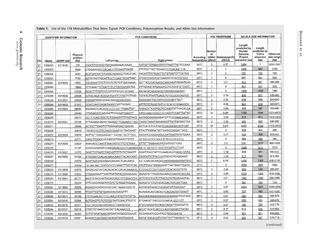
Table 1. List of the 118 Minisatellites That Were Typed: PCR Conditions, Polymorphism Results, and Allele Size Information
(continued)
Denoeudetal.
4G
enom
eR
esearch
www.genome.org

Table1.
(Continued
)
(continued)
Predicting Human Minisatell ite Polymorphism
Genome Research 5www.genome.org

Table1.
(Continued
)
Denoeud et al.
6 Genome Researchwww.genome.org

should be monomorphic, and ∼75% (32 of 43) should have a
heterozygosity value �0.5.Heterozygosity and allele number are significantly
higher in the positive group for criterion 4 (over the entire setof typed repeats) compared with the negative group (Fig. 4B).Criterion 4 produces an enrichment of repeats having hetero-zygosity �0.5 from 42% (49 of 118) in the whole set to 61%(25 of 41) in the positive group and a diminishment of mono-morphic repeats from 25% (30 of 118) in the whole set to 12%(5 of 41) in the positive group. Criterion 4 thus reduces tonearly one third (116 to 41) the number of minisatellites totype while eliminating most monomorphic minisatellites andretaining 50% of the most polymorphic ones. By comparison,criterion 3, if applied to the entire set of typed repeats, (Fig.4B) would reduce their number by roughly half (118 to 61),eliminating just two fewer monomorphs while retaining 69%
(34 of 49) of the most polymorphic repeats. Additionally, cri-terion 4 eliminates half (four of eight) of the highly polymor-phic (heterozygosity �0.85) minisatellites, whereas criterion
3 retains 75% (six of eight) of these.We note that for some highly polymorphic minisatel-
lites, (CEB202, CEB205, CEB310, CEB291), predicted lengthsare identical in the two sequences. In addition, the results forcriterion 4 are not uniform for the two chromosomes, owingto the much greater agreement on predicted loci length inchromosome 22. We presume that this reflects the fact thatthe Celera sequence was assembled by using both public andCelera sequence reads (Venter et al. 2001). More surprisingly,for five minisatellites, which we found to be monomorphic,predicted lengths differ (CEB214, CEB255, CEB264, CEB247,CEB289). These findings raise unresolved questions about theaccuracy of the HGP and Celera sequences with regard tominisatellites. Tandem arrays can present significant se-quence assembly problems, in particular when the internalarray contains regions of high homology and, potentiallymore seriously, when the repeat exhibits length polymor-phism and data are drawn from more than one individual, aswas done for the Celera sequence (Venter et al. 2001).
To examine this further, we compared the HGP and Cel-
era predictions to the alleles we detected (in Table 1, predictedlengths are underlined and not shaded when they correspondto an observed allele). In 65% (75 of 116) of the repeats, HGPand Celera predict an identical allele length, which corre-sponds to an observed allele length with five exceptions(Table 2) and is the most common allele in 81% of these cases.In 35% of the repeats (41 of 116), HGP and Celera predictdifferent length alleles (Table 2). The length predicted by theHGP sequence fits with an observed allele size in 36 cases(most common allele length in 20 of these), whereas the Cel-era prediction fits with an observed size in 10 cases (and wasonce the most common allele).
Among the tandem repeats that provide PCR products
unmatched by the HGP sequence, six sufficiently informativeones (CEB230, CEB253, CEB295, CEB298, CEB315, CEB269),with at least three different alleles among the four parentalchromosomes, were typed in large CEPH families to checktheir chromosomal origin. All map to the expected area ofchromosome 21 or 22, indicating that the discrepancy be-tween sequence data and PCR product size probably resultsfrom a sequencing error (or the sequencing of a very rareallele) and not from a PCR specificity problem.
�2 tests were used to examine whether the similarities in
prediction of the HGP and Celera findings could be explainedby chance (see Methods). Differences identified by the testshad, in all cases, less than one one-thousandth probability ofoccurring by chance. Specifically, cases in which predictionsdisagreed and both allele sizes were detected were underrep-resented (compared to expected frequency) in all tests, andcases in which only one or neither predicted size was detectedwere overrepresented in all but one test.
Identifying Hypermutable LociHypermutable minisatellites are expected to belong to theclass of highly polymorphic loci because they are, by defini-tion, subject to frequent rearrangements that generate newalleles. For practical reasons linked to the size of availablepedigrees, a minisatellite will usually be classified as hyper-mutable if its average mutation rate in the germline is >0.5%,that is, if an average of at least one or two mutant alleles isobserved among 100 children.
Figure 2 (A) Ethidium bromide–stained agarose gel showing PCRproducts for minisatellite CEB285. Six different alleles are scoredamong 32 individuals (in some cases, three bands are seen for oneindividual [the upper one is a PCR artifact as shown by segregationpatterns in families]; this artifact occurs only in heterozygotes [datanot shown], indicating a mechanism involving an interaction be-tween the two alleles). (B) Image of the gels obtained for minisatellitesCEB205 and CEB310 on CEPH families 884 and 1331, respectively.Two children inherit mutant alleles for CEB205, and one child inheritsa mutant allele for CEB310. For CEB205, larger alleles are missed inthe procedure used: The results were confirmed by Southern blot.
Predicting Human Minisatell ite Polymorphism
Genome Research 7www.genome.org

We typed the eight most polymorphic minisatellites (i.e.,
with heterozygosity �0.85) in the eight largest CEPH families(102 children) to search for mutant alleles. Comparing theresults obtained by PCR and Southern blotting shows thateven when some larger alleles are missing in the PCR prod-ucts, the estimated heterozygosity rate (see Methods) is closeto the heterozygosity rate obtained with Southern blots. Thishelps validate the simplified PCR-based polymorphism mea-surement. Among the eight minisatellites (CEB202, CEB205,CEB250, CEB310, CEB269, CEB291, CEB305, CEB324), twoshowed mutant alleles (CEB205 and CEB310; Fig. 2B). Bothyielded two mutant alleles among 204 meioses, that is,102 children (mutation rate, 0.12% to 3.5%; 95% confidenceinterval). For minisatellite CEB205, one mutation eventoccurred in the mother and the other in the father, whereasfor CEB310, both mutations occurred in the father. Theremaining six minisatellites yielded no mutant allele among102 children (mutation rate, 0 to 1.79%; 95% confidence in-terval). They were not investigated further but can not bestrictly excluded from being hypermutable. The two minisat-
ellites that appeared hypermutable among 102 children werethen typed in more families (32 other reference CEPH fami-lies). For CEB205, one new mutant allele was found among352 meioses (mutation rate, 0.54%; 95% confidence interval,0.11% to 1.57%), but no other mutant allele was detected forCEB310 among 476 additional meioses (mutation rate,0.29%; 95% confidence interval, 0.04% to 1.06%). Basedon these results, CEB205 appears to be hypermutable. It is aGC-rich minisatellite with a unit length of 33 bp repeated 10to 70 times, located at 1.5 Mb from the end of the chromo-some 22 sequence. It seems to be part of a predicted codingregion (gene LOC129238; see http://www.ncbi.nlm.nih.gov/LocusLink/LocRpt.cgi?l=129238, 31 July 2002 update).
DISCUSSIONThis study, performed on the scale
of entire human chromosomes,
provides a first global evaluation ofminisatellite polymorphism based
on genome sequence data. The re-
peats studied here, chosen by using
a detailed definition that is more
stringent than the broad definition
mentioned in the Introduction, are,
in majority (75%), polymorphic in
the population investigated, and
42% have a heterozygosity value
�0.5. Minisatellites from chromo-
somes 21 and 22 are similar in
physical distribution (higher fre-
quency toward chromosome ends),
sequence features, and polymor-
phism. Assuming that chromo-
somes 21 and 22 are representative
of all human chromosomes and
given that the two chromosomes
represent ∼2% of the genome, we
speculate that the entire human ge-
nome contains ∼6,000 minisatel-
lites that match our definition, in-
cluding 4,800 polymorphic and
2,500 very polymorphic ones. A few
10s of these might be expected to
qualify as hypermutable loci. Be-
cause our definition precluded many other potentially poly-morphic minisatellites, future research should seek to expandthe category of minisatellites that are tested against our poly-morphism prediction criteria.
Predicting PolymorphismWe showed that using the sequence properties %GC andHistoryR effectively improves polymorphic minisatellite se-lection. With them, we reduce the number of minisatellitesfor typing by about half while increasing the frequency ofrepeats with heterozygosity �0.5% from the background rateof 43% to 59%. Internal conservation, used as a polymor-phism predictor for microsatellites, is not applicable to mini-satellites, presumably owing to the greater complexity of theirmutation processes.
That %GC correlates with polymorphism is in agreementwith earlier observations. Some of the first minisatellites to becharacterized were detected via a shared 10- to 15-bp “core”sequence similar to the generalized recombination signal (�)
of Escherichia coli (GCTGTGG; Jeffreys et al. 1985). The ma-jority of classical minisatellites (mostly polymorphic and/orhypermutable ones) are GC-rich, with a strong purine/pyrimidine strand asymmetry (Vergnaud and Denoeud 2000).In other genomes, though, (for instance bacterial genomes),%GC does not seem to be associated with minisatellites poly-morphism (Le Fleche et al. 2001). Such a criterion may there-
fore not be universal, especially because GC content variessignificantly across genomes.
The HistoryR criterion is based on the hypothesis that
tandem repeats expand through multiple rounds of duplica-tion, with the new copies sharing the mutations that occurbefore duplication, whereas unique mutations accumulateonce the repeat is no longer evolving. For example (Fig. 1),
Figure 3 Criteria 1, 2, and 3 applied to the training set. For criteria 1 and 2, heterozygosity (28individuals) versus HistoryR (criterion 1) or percentage of GC (%GC; criterion 2) are plotted. Correla-tions are significant at the 0.01 level. For criterion 3, HistoryR versus %GC is plotted, with differentsymbols representing the polymorphism. Lines represent the selected thresholds, and shaded areascontain the minisatellites selected by the criteria (criterion 1, HistoryR �0.54; criterion 2, %GC �48%;criterion 3, criteria 1 and 2 combined). Plots show that criteria select most of the polymorphic mini-satellites and eliminate a majority of monomorphs or slightly polymorphic ones.
Denoeud et al.
8 Genome Researchwww.genome.org

minisatellite CEB252 shows several redundant patterns ofmutation, resulting in a high HistoryR score, whereas CEB233shows no clear organization of mutations, resulting in a lowHistoryR score.
This polymorphism criterion is likely to be applicable to
any genome, even though the history reconstruction algo-rithm makes simplifying assumptions about the possible bio-logical mechanisms involved in array expansion. Thesemechanisms, which include mutational events during mitoticreplication and meiotic recombination, comprising both in-traallelic and interallelic events, might occur independentlyor jointly. At present, there are no rules to predict whichmechanism will occur preferentially at which locus (Maleki etal. 2002). Moreover, the individual mechanisms themselvesare still poorly understood and, thus, impossible to model.Meiotic events, for instance, have been shown to result fromthe activity of nearby meiosis-specific double-strand breakhot-spots. The nature of these sites, better known in yeast, isstill unknown in the human genome (Debrauwere et al. 1999;Tamaki et al. 1999; Vergnaud and Denoeud 2000). In view ofthe current state of knowledge, it may be premature to hopefor a perfect polymorphism predictor based on apparent arrayexpansion.
Use of Two Human Sequences to Select for
Polymorphic Loci Is ProblematicThe availability of two versions of the human genome se-
quence provides an additional avenue to improve polymor-
phic minisatellite identification. However, in the repeats stud-
ied here, selection based on re-
ported length differences discardedhalf the highly polymorphic mini-satellites and, in particular, the hy-permutable one from chromosome22. In both chromosomes, thenumber of loci with different pre-dicted lengths in the HGP and Cel-era sequences that were nonethe-less both found was significantlyunderrepresented. This is appar-ently owing to the lack of indepen-dence resulting from sharing ofdata during assembly of the Celerasequence. In addition, in bothchromosomes, the number of lociin which only one or no predictedallele was found is overrepresented,apparently owing to assembly er-rors. Because the Celera sequence—which when not in agreement withthe HGP data—usually provides
copy numbers unobserved in any allele, it appears that theCelera sequence, at least with respect to minisatellites, is moreprone to assembly error. As a result of the lack of indepen-dence/assembly errors in the Celera/HGP data, polymorphismprediction based on sequence comparison did not perform aswell as anticipated.
One New Hypermutable Locus in a Coding RegionThis study revealed one hypermutable minisatellite, CEB205,showing three mutant alleles among 278 children (mutationrate, 0.54%; 95% confidence interval, 0.11% to 1.57%). Inter-estingly, CEB205, with a 33-bp pattern, may be part of a cod-ing region. The corresponding putative protein is 614 aminoacids long, half of which are derived from the tandem repeat(11 codon repetition) at the N terminus. Of the minisatellitesstudied here, 26 among 60 (43%) on chromosome 21, and 22among 67 (33%) on chromosome 22 belong to genes (i.e.,exons, introns, or UTRs), as determined by sequence similar-ity analyses in the human genome sequence (using BLASTand http://www.ncbi.nlm.nih.gov/genome/seq/, release ofNovember 2001). None except CEB205 appear to contributeto the coding sequence itself. Although the proportion of tan-dem repeats that contribute to coding regions is important inbacterial genomes, it is relatively low in the human genome,and CEB205 might represent the first known, coding hyper-mutable minisatellite.
CEB310—which exhibited meiotic mutation events, butwhich we do not here classify as hypermutable—is unusual inthat its sequence is 80% AT. It is reminiscent of the tandem
Table 2. Success of the Public Human Genome Project (HGP) and Celera Sequences in Predicting Alleles That Were ActuallyFound to Occur
116 tandem repeats (predicted by both sequences)
Predicted lengths match: n = 75 Predicted lengths differ: n = 41
Allele found with predicted length: Allele found with predicted length:
Yes No Yes, both predictions HGP prediction only Celera prediction only Neither prediction
70 5 10 26 0 5
Figure 4 (A) Application of criteria 1 (HistoryR �0.54), 2 (%GC �48%), and 3 (HistoryR �0.54 and%GC �48%) to the test set. For each criterion, the distributions of minisatellites (from monomorphsto highly polymorphic) between positive (retained by the criterion) and negative (excluded by thecriterion) sets are compared. All differences between sets + and � are statistically significant at the 0.01level. (B) On the whole set, comparison of the results obtained with criterion 4 and criterion 3.
Predicting Human Minisatell ite Polymorphism
Genome Research 9www.genome.org
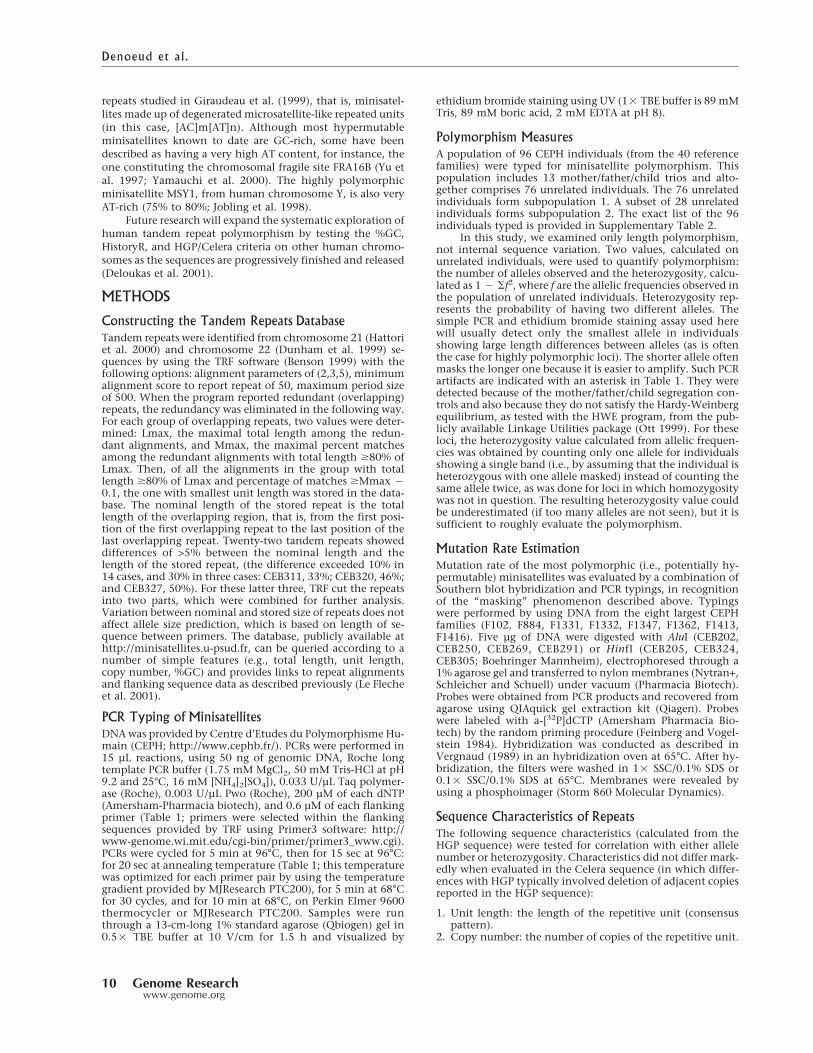
repeats studied in Giraudeau et al. (1999), that is, minisatel-
lites made up of degenerated microsatellite-like repeated units
(in this case, [AC]m[AT]n). Although most hypermutable
minisatellites known to date are GC-rich, some have been
described as having a very high AT content, for instance, the
one constituting the chromosomal fragile site FRA16B (Yu et
al. 1997; Yamauchi et al. 2000). The highly polymorphic
minisatellite MSY1, from human chromosome Y, is also very
AT-rich (75% to 80%; Jobling et al. 1998).
Future research will expand the systematic exploration ofhuman tandem repeat polymorphism by testing the %GC,
HistoryR, and HGP/Celera criteria on other human chromo-
somes as the sequences are progressively finished and released
(Deloukas et al. 2001).
METHODS
Constructing the Tandem Repeats Database
Tandem repeats were identified from chromosome 21 (Hattoriet al. 2000) and chromosome 22 (Dunham et al. 1999) se-quences by using the TRF software (Benson 1999) with thefollowing options: alignment parameters of (2,3,5), minimumalignment score to report repeat of 50, maximum period sizeof 500. When the program reported redundant (overlapping)repeats, the redundancy was eliminated in the following way.For each group of overlapping repeats, two values were deter-mined: Lmax, the maximal total length among the redun-dant alignments, and Mmax, the maximal percent matchesamong the redundant alignments with total length �80% ofLmax. Then, of all the alignments in the group with totallength �80% of Lmax and percentage of matches �Mmax �
0.1, the one with smallest unit length was stored in the data-base. The nominal length of the stored repeat is the totallength of the overlapping region, that is, from the first posi-tion of the first overlapping repeat to the last position of thelast overlapping repeat. Twenty-two tandem repeats showeddifferences of >5% between the nominal length and thelength of the stored repeat, (the difference exceeded 10% in14 cases, and 30% in three cases: CEB311, 33%; CEB320, 46%;and CEB327, 50%). For these latter three, TRF cut the repeatsinto two parts, which were combined for further analysis.Variation between nominal and stored size of repeats does notaffect allele size prediction, which is based on length of se-quence between primers. The database, publicly available athttp://minisatellites.u-psud.fr, can be queried according to anumber of simple features (e.g., total length, unit length,copy number, %GC) and provides links to repeat alignmentsand flanking sequence data as described previously (Le Flecheet al. 2001).
PCR Typing of Minisatellites
DNAwas provided by Centre d’Etudes du Polymorphisme Hu-main (CEPH; http://www.cephb.fr/). PCRs were performed in15 µL reactions, using 50 ng of genomic DNA, Roche longtemplate PCR buffer (1.75 mM MgCl2, 50 mM Tris-HCl at pH9.2 and 25°C, 16 mM [NH4]2[SO4]), 0.033 U/µL Taq polymer-ase (Roche), 0.003 U/µL Pwo (Roche), 200 µM of each dNTP(Amersham-Pharmacia biotech), and 0.6 µM of each flankingprimer (Table 1; primers were selected within the flankingsequences provided by TRF using Primer3 software: http://www-genome.wi.mit.edu/cgi-bin/primer/primer3_www.cgi).PCRs were cycled for 5 min at 96°C, then for 15 sec at 96°C:for 20 sec at annealing temperature (Table 1; this temperaturewas optimized for each primer pair by using the temperaturegradient provided by MJResearch PTC200), for 5 min at 68°Cfor 30 cycles, and for 10 min at 68°C, on Perkin Elmer 9600thermocycler or MJResearch PTC200. Samples were runthrough a 13-cm-long 1% standard agarose (Qbiogen) gel in0.5� TBE buffer at 10 V/cm for 1.5 h and visualized by
ethidium bromide staining using UV (1� TBE buffer is 89 mMTris, 89 mM boric acid, 2 mM EDTA at pH 8).
Polymorphism Measures
A population of 96 CEPH individuals (from the 40 referencefamilies) were typed for minisatellite polymorphism. Thispopulation includes 13 mother/father/child trios and alto-gether comprises 76 unrelated individuals. The 76 unrelatedindividuals form subpopulation 1. A subset of 28 unrelatedindividuals forms subpopulation 2. The exact list of the 96individuals typed is provided in Supplementary Table 2.
In this study, we examined only length polymorphism,not internal sequence variation. Two values, calculated onunrelated individuals, were used to quantify polymorphism:the number of alleles observed and the heterozygosity, calcu-lated as 1 � �f2, where f are the allelic frequencies observed inthe population of unrelated individuals. Heterozygosity rep-resents the probability of having two different alleles. Thesimple PCR and ethidium bromide staining assay used herewill usually detect only the smallest allele in individualsshowing large length differences between alleles (as is oftenthe case for highly polymorphic loci). The shorter allele oftenmasks the longer one because it is easier to amplify. Such PCRartifacts are indicated with an asterisk in Table 1. They weredetected because of the mother/father/child segregation con-trols and also because they do not satisfy the Hardy-Weinbergequilibrium, as tested with the HWE program, from the pub-licly available Linkage Utilities package (Ott 1999). For theseloci, the heterozygosity value calculated from allelic frequen-cies was obtained by counting only one allele for individualsshowing a single band (i.e., by assuming that the individual isheterozygous with one allele masked) instead of counting thesame allele twice, as was done for loci in which homozygositywas not in question. The resulting heterozygosity value couldbe underestimated (if too many alleles are not seen), but it issufficient to roughly evaluate the polymorphism.
Mutation Rate Estimation
Mutation rate of the most polymorphic (i.e., potentially hy-permutable) minisatellites was evaluated by a combination ofSouthern blot hybridization and PCR typings, in recognitionof the “masking” phenomenon described above. Typingswere performed by using DNA from the eight largest CEPHfamilies (F102, F884, F1331, F1332, F1347, F1362, F1413,F1416). Five µg of DNA were digested with AluI (CEB202,CEB250, CEB269, CEB291) or HinfI (CEB205, CEB324,CEB305; Boehringer Mannheim), electrophoresed through a1% agarose gel and transferred to nylonmembranes (Nytran+,Schleicher and Schuell) under vacuum (Pharmacia Biotech).Probes were obtained from PCR products and recovered fromagarose using QIAquick gel extraction kit (Qiagen). Probeswere labeled with a-[32P]dCTP (Amersham Pharmacia Bio-tech) by the random priming procedure (Feinberg and Vogel-stein 1984). Hybridization was conducted as described inVergnaud (1989) in an hybridization oven at 65°C. After hy-bridization, the filters were washed in 1� SSC/0.1% SDS or0.1� SSC/0.1% SDS at 65°C. Membranes were revealed byusing a phosphoimager (Storm 860 Molecular Dynamics).
Sequence Characteristics of Repeats
The following sequence characteristics (calculated from theHGP sequence) were tested for correlation with either allelenumber or heterozygosity. Characteristics did not differ mark-edly when evaluated in the Celera sequence (in which differ-ences with HGP typically involved deletion of adjacent copiesreported in the HGP sequence):
1. Unit length: the length of the repetitive unit (consensuspattern).
2. Copy number: the number of copies of the repetitive unit.
Denoeud et al.
10 Genome Researchwww.genome.org

3. Total length: the length of the entire tandem array.4. Percent matches: the frequency at which a nucleotide at aposition in one unit matches the corresponding nucleotidein the next unit (reading from left to right).
5. %GC: the percentage of nucleotides that are either G or C.6. GC bias: strand asymmetry for G and C, |%G � %C|/(%G + %C).
7. Purine/Pyrimidine bias: strand asymmetry for purines andpyrimidines, |%Pur � %Pyr|/(%Pur + %Pyr).
8. Average entropy: from the columns of a multiple align-ment of the repeat copies, the average, over all columns, ofthe entropy calculated from nucleotide frequencies.
9. HistoryR: described below.
HistoryR is derived from the tandem repeats history re-construction algorithm (Benson and Dong 1999), a greedyalgorithm that chooses a series of least-cost contractions toconvert a multicopy tandem array into a single putative an-cestral copy. Greedy algorithms are not guaranteed to find theoverall least-cost solution, but testing has shown this ap-proach to work very well on simulated sequences. Input is amultiple alignment,M, of the individual copies in the repeat,with n rows (number of copies) and k columns (length ofalignment). Mi,j represents the ith row and jth column of M,and each Mi,j contains one of the alphabet symbols(A,C,G,T,–). In a contraction, two or more consecutive, equal-length subsequences (the contraction copies) are replaced bya single subsequence (the merged copy) of the same length(all subsequences selected have length equal to a multiple ofk). Each contraction reduces the number of rows in M. If thecontraction copies are identical, then one becomes themerged copy. Otherwise at every position at which the con-traction copies differ, the merged copy contains the characterthat occurs most often, with ties being represented by an am-biguous character, that is, a set of all the most frequentlyoccurring characters at that position. An ambiguous charactercreated in one contraction may be converted to a single char-acter in a subsequent contraction. This method is analogousto that used by Sankoff (1975; Sankoff and Rousseau 1975).The cost of a contraction is a ratio. The numerator is the costof obtaining the contraction copies from the merged copy;that is, at each position of the merged copy, subtract the num-ber of times the most frequent character occurs in the con-traction copies from the total number of contraction copies,then sum all these differences. The denominator is the com-bined length of rows by which M is reduced, that is, thelength of all contraction copies minus the length of themerged copy.
History reconstruction yields four numerical values: (1)Max, the maximum possible history cost; (2) Min, the mini-mum possible history cost; (3) BinaryActual, the calculatedhistory cost when the number of contraction copies in everycontraction is restricted to exactly two; and (4) ManyActual,the calculated cost when the number of contraction copies isunrestricted. Max and Min are sums of column values fromthe original alignment M. In the case of Max, the value of asingle column is the number of characters that are not themost frequent character. Max is therefore the cost if the mostfrequent character is ancestral and if every character differentfrom the ancestral character was produced by its own muta-tion. For Min, the value is one less than the number of distinctcharacters in a column, that is, at most four. Min is the historycost if every distinct character different from the ancestralcharacter arose by a single mutation (with identical charactersproduced by duplication).
Combinations of the four numerical values were testedfor polymorphism prediction in the training set and HistoryR,which produced the highest correlation with heterozygosity,was used for the remainder of the study. It is defined as
History R = ��Max − BestActual���Max − Min� when Max � Min1 otherwise
where BestActual is the minimum of BinaryActual andManyActual. Usually, this was BinaryActual. The HistoryRvalue can be thought of as the proportion of mutations thatcould be accounted for by duplication that actually are. WhenMax � Min, HistoryR �1, with a higher ratio indicating moremutations accounted for by duplications (Fig. 1). WhenMax = Min, each mutation is unique, and we arbitrarily setthe ratio to one. This occurred in only one repeat with a totalof four mutations. The history reconstruction program isfreely available for interactive use at http://tandem.biomath.mssm.edu/cgi-bin/history/history.exe.
Statistical Analysis
All statistical analysis was done with the SPSS program exceptfor �2 tests which were done with StatXact 4. Correlationswere determined by three methods: Pearson correlation, andnonparametric Kendall’s ��b and Spearman’s �. Correlationsare considered significant at the 0.01 level (two-tailed) of thetest statistics. Group comparisons were determined by firstconducting two tests of normality, Kolmogorov-Smirnov andShapiro-Wilkinson, on the values within each group. Valueswere assumed to be normally distributed unless the test sta-tistic fell within the 0.05 level of significance. If the valueswere normally distributed in the two groups, then a t test wasused to compare the means, which were judged significantlydifferent at the 0.01 level of the statistic (two-tailed). If thevalues were not normally distributed in either of the twogroups, then a nonparametric Mann-Whitney test was used tocompare the distributions, which were judged significantlydifferent at the 0.01 level of the statistic (two-tailed).
�2 tests were used to analyze HGP/Celera prediction datafor chromosomes 21 and 22 separately. The data were dividedinto three categories: (1) identical predictions/allele size de-tected, (2) different predictions/both alleles sizes detected,and (3) one or neither predicted allele size detected. Two es-timates for frequency of unobserved alleles were used (in or-der to calculate the probability of alleles being detected): 10%which corresponds to the largest frequency in the populationfor which the chance of not appearing in our sample of 28individuals is�0.05, and an arbitrary low estimate of 1%. Theprobability of identical predictions in the HGP and Celerasequences was obtained by summing the estimated heterozy-gosity values calculated separately for each locus based onobserved frequencies in our sample (equivalent to using theaverage observed heterozygosity over all loci).
ACKNOWLEDGMENTSWe would like to thank Carol Bodian for extensive discus-sions and help with design of the �2 analysis. G.B. is sup-ported in part by NSF grants CCR-0073081 and DBI-0090789.F.D. and G.V. are supported by grants from Delegation Gen-erale de l’Armement.
The publication costs of this article were defrayed in partby payment of page charges. This article must therefore behereby marked “advertisement” in accordance with 18 USCsection 1734 solely to indicate this fact.
REFERENCESAmarger, V., Gauguier, D., Yerle, M., Apiou, F., Pinton, P.,
Giraudeau, F., Monfouilloux, S., Lathrop, M., Dutrillaux, B.,Buard, J., et al. 1998. Analysis of the human, pig, and ratgenomes supports a universal telomeric origin of minisatellitesequences. Genomics 52: 62–71.
Appelgren, H., Cederberg, H., and Rannug, U. 1997. Mutations atthe human minisatellite MS32 integrated in yeast occur withhigh frequency in meiosis and involve complex recombinationevents. Mol. Gen. Genet. 256: 7–17.
———. 1999. Meiotic interallelic conversion at the humanminisatellite MS32 in yeast triggers recombination in severalchromatids. Gene 239: 29–38.
Baudat, F., Manova, K., Yuen, J.P., Jasin M., and Keeney, S. 2000.Chromosome synapsis defects and sexually dimorphic meiotic
Predicting Human Minisatell ite Polymorphism
Genome Research 11www.genome.org

progression in mice lacking Spo11. Mol. Cell 6: 989–998.Bell, G.I., Serby M.J., and Rutter, W.J. 1982. The highly polymorphic
region near the human insulin gene is composed of simpletandemly repeating sequences. Nature 295: 31–35.
Benson, G. 1999. Tandem repeats finder: A program to analyze DNAsequences. Nucleic Acids Res. 27: 573–580.
Benson, G. and Dong, L. 1999. Reconstructing the duplicationhistory of a tandem repeat. Proc. Int. Conf. Intell. Syst. Mol. Biol.44–53.
Bergerat, A., de Massy, B., Gadelle, D., Varoutas, P.C., Nicolas, A.,and Forterre, P. 1997. An atypical topoisomerase II from archaeawith implication for meiotic recombination. Nature386: 414–417.
Buard, J. and Vergnaud, G. 1994. Complex recombination events atthe hypermutable minisatellite CEB1 (D2S90). EMBO J.13: 3203–3210.
Buard, J., Bourdet, A., Yardley, J., Dubrova Y., and Jeffreys, A.J. 1998.Influences of array size and homogeneity on minisatellitemutation. EMBO J. 17: 3495–3502.
Debrauwere, H., Buard, J., Tessier, J., Aubert, D., Vergnaud, G., andNicolas, A. 1999. Meiotic instability of human minisatellite CEB1in yeast requires DNA double-strand breaks. Nat. Genet.23: 367–371.
Deloukas, P., Matthews, L.H., Ashurst, J., Burton, J., Gilbert, J.G.,Jones, M., Stavrides, G., Almeida, J.P., Babbage, A.K., Bagguley,C.L., et al. 2001. The DNA sequence and comparative analysis ofhuman chromosome 20. Nature 414: 865–871.
Dubrova, Y.E. and Plumb, M.A. 2002. Ionising radiation andmutation induction at mouse minisatellite loci: The story of thetwo generations. Mutat. Res. 499: 143–150.
Dubrova, Y.E., Jeffreys, A.J., and Malashenko, A.M. 1993. Mouseminisatellite mutations induced by ionizing radiation. Nat.Genet. 5: 92–94.
Dubrova, Y.E., Nesterov, V.N., Krouchinsky, N.G., Ostapenko, V.A.,Vergnaud, G., Giraudeau, F., Buard J., and Jeffreys, A.J. 1997.Further evidence for elevated human minisatellite mutation ratein Belarus eight years after the Chernobyl accident. Mut. Res.381: 267–278.
Dunham, I., Shimizu, N., Roe, B.A., Chissoe, S., Hunt, A.R., Collins,J.E., Bruskiewich, R., Beare, D.M., Clamp, M., Smink, L.J., et al.1999. The DNA sequence of human chromosome 22. Nature402: 489–495.
Feinberg, A.P. and Vogelstein, B. 1984. Addendum: a technique forradiolabeling DNA restriction endonuclease fragments to highspecific activity. Anal. Biochem. 137: 266–267.
Fondon III, J.W., Mele, G.M., Brezinschek, R.I., Cummings, D.,Pande, A., Wren, J., O’Brien, K.M., Kupfer, K.C., Wei, M.H.,Lerman, M., et al. 1998. Computerized polymorphic markeridentification: Experimental validation and a predicted humanpolymorphism catalog. Proc. Natl. Acad. Sci. 95: 7514–7519.
Giraudeau, F., Petit, E., Avet-Loiseau, H., Hauck, Y., Vergnaud, G.,and Amarger, V. 1999. Finding new human minisatellitesequences in the vicinity of long CA-rich sequences. Genome Res.9: 647–653.
Giraudeau, F., Taine, L., Biancalana, V., Delobel, B., Journel, H.,Moncla, A., Bonneau, D., Lacombe, D., Moraine, C., Croquette,M.F., et al. 2001. Use of a set of highly polymorphic minisatelliteprobes for the identification of cryptic 1p36.3 deletions in a largecollection of patients with idiopathic mental retardation: Threenew cases. J. Med. Genet. 38: 121–125.
Hattori, M., Fujiyama, A., Taylor, T.D., Watanabe, H., Yada, T., Park,H.S., Toyoda, A., Ishii, K., Totoki, Y., Choi, D.K., et al. 2000. TheDNA sequence of human chromosome 21: The chromosome 21mapping and sequencing consortium. Nature 405: 311–319.
Heale, S.M. and Petes, T.D. 1995. The stabilization of repetitive tractsof DNA by variant repeats requires a functional mismatch repairsystem. Cell 83: 539–545.
Jeffreys, A.J., Wilson, V., and Thein, S.L. 1985. Hypervariable“minisatellite” regions in human DNA. Nature 314: 67–73.
Jeffreys, A.J., Tamaki, K., MacLeod, A., Monckton, D.G., Neil, D.L.,and Armour, J.A.L. 1994. Complex gene conversion events ingermline mutation at human minisatellites. Nat. Genet.6: 136–145.
Jobling, M.A., Bouzekri, N., and Taylor, P.G. 1998. Hypervariabledigital DNA codes for human paternal lineages: MVR-PCR at theY-specific minisatellite, MSY1 (DYF155S1). Hum. Mol. Genet.7: 643–653.
Keeney, S., Giroux, C.N., and Kleckner, N. 1997. Meiosis-specificDNA double-strand breaks are catalyzed by Spo11, a member of awidely conserved protein family. Cell 88: 375–384.
Le Fleche, P., Hauck, Y., Onteniente, L., Prieur, A., Denoeud, F.,Ramisse, V., Sylvestre, P., Benson, G., Ramisse, F., and Vergnaud,G. 2001. A tandem repeats database for bacterial genomes:application to the genotyping of Yersinia pestis and Bacillusanthracis. BMC Microbiol. 1: 2.
Maleki, S., Cederberg, H., and Rannug, U. 2002. The humanminisatellites MS1, MS32, MS205 and CEB1 integrated into theyeast genome exhibit different degrees of mitotic instability butare all stabilised by RAD27. Curr. Genet. 41: 333–341.
May, C.A., Jeffreys, A.J., and Armour, J.A.L. 1996. Mutation rateheterogeneity and the generation of allele diversity at the humanminisatellite MS205 (D16S309). Hum. Mol. Genet. 5: 1823–1833.
Murray, J., Buard, J., Neil, D.L., Yeramian, E., Tamaki, K., Hollies,C.R., and Jeffreys, A.J. 1999. Comparative sequence analysis ofhuman minisatellites showing meiotic repeat instability. GenomeRes. 9: 130–136.
Nakamura, Y., Leppert, M., O’Connell, P., Wolff, T., Holm, T.,Culver, M., Martin, C., Fujimoto, E., Hoff, M., Kumlin, E., et al.1987. Variable number of tandem repeat (VNTR) markers forhuman gene mapping. Science 235: 1616–1622.
NIH/CEPH collaborative mapping group. 1992. A comprehensivegenetic linkage map of the human genome. Science 258: 67–83.
Ott, J. 1999. Analysis of human genetic linkage, 3d ed. Johns HopkinsUniversity Press, Baltimore, MD.
Romanienko, P.J. and Camerini-Otero, R.D. 2000. The mouse Spo11gene is required for meiotic chromosome synapsis. Mol. Cell6: 975–987.
Sankoff, D. 1975. Minimal mutation trees of sequences. J. Appl.Math. 28: 35–42.
Sankoff, D. and Rousseau, P. 1975. Locating the vertices of a Steinertree in an arbitrary metric space. Math. Programming 9: 240–246.
Strand, M., Prolla, T.A., Liskay, R.M., and Petes, T.D. 1993.Destabilization of tracts of simple repetitive DNA in yeast bymutations affecting DNA mismatch repair. Nature 365: 274–276.
Tamaki, K., May, C.A., Dubrova, Y.E., and Jeffreys, A.J. 1999.Extremely complex repeat shuffling during germline mutation athuman minisatellite B6.7. Hum. Mol. Genet. 8: 879–888.
Venter, J.C., Adams, M.D., Myers, E.W., Li, P.W., Mural, R.J., Sutton,G.G., Smith, H.O., Yandell, M., Evans, C.A., Holt, R.A., et al.2001. The sequence of the human genome. Science291: 1304–1351.
Vergnaud, G. 1989. Polymers of random short oligonucleotidesdetect polymorphic loci in the human genome. Nucleic Acids Res.17: 7623–7630.
Vergnaud, G. and Denoeud, F. 2000. Minisatellites: Mutability andgenome architecture. Genome Res. 10: 899–907.
Vergnaud, G., Mariat, D., Apiou, F., Aurias, A., Lathrop, M., andLauthier, V. 1991. The use of synthetic tandem repeats to isolatenew VNTR loci: Cloning of a human hypermutable sequence.Genomics 11: 135–144.
Weber, J.L. 1990. Informativeness of human (dC-dA)n (dG-dT)npolymorphisms. Genomics 7: 524–530.
Wren, J.D., Forgacs, E., Fondon III, J.W., Pertsemlidis, A., Cheng,S.Y., Gallardo, T., Williams, R.S., Shohet, R.V., Minna, J.D., andGarner, H.R. 2000. Repeat polymorphisms within gene regions:phenotypic and evolutionary implications. Am. J. Hum. Genet.67: 345–356.
Yamauchi, M., Tsuji, S., Mita, K., Saito, T., and Morimyo, M. 2000. Anovel minisatellite repeat expansion identified at FRA16B in aJapanese carrier. Genes Genet. Syst. 75: 149–154.
Yu, S., Mangelsdorf, M., Hewett, D., Hobson, L., Baker, E., Eyre, H.J.,Lapsys, N., Le Paslier, D., Doggett, N.A., Sutherland, G.R., et al.1997. Human chromosomal fragile site FRA16B is an amplifiedAT-rich minisatellite repeat. Cell 88: 367–374.
WEB SITE REFERENCEShttp://minisatellites.u-psud.fr; the tandem repeats database.http://tandem.biomath.mssm.edu/cgi-bin/history/history.exe;
history reconstruction programhttp://www.ncbi.nlm.nih.gov/genome/seq/; Human Genome
Sequencing at NCBI.http://www-genome.wi.mit.edu/cgi-bin/primer/primer3_www.cgi;
Primer3 primer picking software.http://www.cephb.fr; Centre d’Etudes du Polymorphisme Humain.http://www.ncbi.nlm.nih.gov/LocusLink/LocRpt.cgi?l=129238;
locuslink at NCBI, predicted gene LOC129238.http://www.ncbi.nlm.nih.gov/SNP/index.html; dbSNP home page.
Received July 1, 2002; accepted in revised form January 28, 2003.
Denoeud et al.
12 Genome Researchwww.genome.org
87
2.3.3 Recherche de minisatellites potentiellement polymorphes dans les séquences codantes
2.3.3.1 Introduction
Comme nous l’avons vu précédemment (Vergnaud & Denoeud 2000), le génome humain est
riche en répétitions en tandem. Le chromosome 22, qui représente environ 1% du génome
humain, contient de l’ordre de 15000 répétitions en tandem (détectées par le Tandem Repeats
Finder (Benson 1999)) : on peut donc estimer le nombre de répétitions en tandem sur
l’ensemble du génome à 1500000, dont plus de 5000 appartiendraient à la classe des
minisatellites.
Cependant, seule une quinzaine de minisatellites polymorphes appartenant à des séquences
codantes, c’est-à-dire générant des répétitions en tandem au niveau de la séquence en acides
aminés de la protéine correspondante, ont été étudiés, la plupart appartenant à la famille des
mucines (Vinall 1998) : voir Tableau 9, paragraphe 1.2.3.3.3.2. D’autre part, l’étude menée
sur les chromosomes 21 et 22 (Denoeud 2003) a mis en évidence un minisatellite
hypermutable appartenant à une séquence codante prédite, mais l’existence de cette protéine
hypothétique nécessitera d’être vérifiée. Les répétitions survenant au niveau de séquences
codantes sont susceptibles de générer une variabilité au niveau des protéines, éventuellement
nécessaire à leur rôle physiologique (système immunitaire…) ou générant des pathologies. Il
semble donc intéressant d’étudier plus largement cette classe de répétitions en tandem encore
peu explorée.
Certains articles traitent plus particulièrement des microsatellites survenant dans les
séquences codantes : une étude menée sur des protéines de primates a par exemple montré que
les répétitions trinucléotidiques étaient sur-représentées dans les exons (Borstnik 2002). Wren
et collègues ont étudié les microsatellites polymorphes présents dans les ARNs messagers
humains : ils sont plus fréquents dans les séquences non-codantes (5’ ou 3’) que dans les
séquences codantes. Les auteurs prédisent toutefois que 3,7% des gènes humains
contiendraient des répétitions en tandem polymorphes, dont 92% correspondraient à une unité
répétée multiple de 3 (Wren 2000). Il apparaît en outre que certaines répétitions d’un acide
aminé (trinucléotides) sont plus fréquentes que d’autres : poly-alanine, proline, glycine,
glutamine, lysine, leucine et sérine (Borstnik 2002 ; Wren 2000).
Marcotte et al. ont mis en évidence que les protéines d’eucaryotes sont plus riches en
répétitions (principalement duplications) que les protéines de bactéries ou d’archées : ils ont
émis l’hypothèse que des mécanismes similaires seraient à l’origine des duplications dans les
trois règnes, mais que la machinerie de synthèse protéique eucaryote, prenant mieux en charge

88
les protéines multi-domaines, non-globulaires, permettrait le maintien des structures répétées
chez ces organismes préférentiellement (Marcotte 1999).
De Fonzo et al. ont mené une étude sur les répétitions en tandem (microsatellites comme
minisatellites) présentes dans des gènes associés à des pathologies (en particulier celles qui
sont associées à un phénomène d’anticipation) : certains candidats ont été trouvés à proximité
de gènes ou dans des gènes (leurs unités répétées étant alors systématiquement multiples de 3)
mais leur polymorphisme nécessite encore d’être testé (De Fonzo 1998).
2.3.3.2 Séquences traitées
Afin d’identifier les minisatellites appartenant à des séquences codantes, j’ai rapatrié toutes
les séquences d’ARNs messagers du génome humain présentes dans Refseq en juin 2003.
Refseq, « The Reference Sequence » collection (http://www.ncbi.nih.gov/RefSeq/) contient
l’ensemble des séquences non-redondantes (ADN génomique, transcrits (ARNs messagers) et
produits protéiques) pour différents organismes. Les ARNs messagers provenant de Refseq
appartiennent à deux catégories principales : les gènes, dont le produit protéique est connu, et
les modèles de gènes, prédits par des logiciels de détection de gènes et étayés par différents
types de preuves : alignement avec des ARNs messagers (ARNm) et/ou des EST (Expressed
Sequence Tags : séquences provenant de banques d’ADN complémentaire). Parmi les 37547
ARNm rapatriés, 18474 correspondent à des gènes et 19016 correspondent à des modèles. Les
autres ARNs messagers correspondent à des gènes d’ARNs ou des pseudogènes : ils n’ont pas
été considérés dans cette étude. Les ARNm considérés totalisent 76921544 paires de bases,
soit environ 2,5% de l’ensemble du génome humain.
2.3.3.3 Caractéristiques des répétitions en tandem des ARNs messagers
Tableau 10 : Caractéristiques des séquences des chromosomes
20, 21 et 22 et de l’ensemble des ARNs messagers du génome humain.
Chromosome 20 Chromosome 21 Chromosome 22 ARNs
messagers
Longueur de la séquence traitée (pb) 59422997 33824148 33563000 76921544
Densité en gènes (gènes/Mb) (Venter 2001)
16 13 23
% GC (Venter 2001)
44% 41% 48%
% de répétitions (dispersées…) (Venter 2001)
41% 38% 44%
Nombre de répétitions en tandem 24484 14996 14369 26153
Densité en répétitions en tandem (/Mb) 412 443 428 340
Nombre de minisatellites (définition au § 2.3.3.4)
51 / / 50
% de minisatellites parmi les répétitions en tandem
0,21 % / / 0,19 %
Densité en minisatellites (/Mb) 0,86 / / 0,65
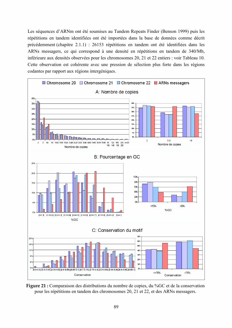
89
Les séquences d’ARNm ont été soumises au Tandem Repeats Finder (Benson 1999) puis les
répétitions en tandem identifiées ont été importées dans la base de données comme décrit
précédemment (chapitre 2.1.1) : 26153 répétitions en tandem ont été identifiées dans les
ARNs messagers, ce qui correspond à une densité en répétitions en tandem de 340/Mb,
inférieure aux densités observées pour les chromosomes 20, 21 et 22 entiers : voir Tableau 10.
Cette observation est cohérente avec une pression de sélection plus forte dans les régions
codantes par rapport aux régions intergéniques.
Figure 21 : Comparaison des distributions du nombre de copies, du %GC et de la conservation
pour les répétitions en tandem des chromosomes 20, 21 et 22, et des ARNs messagers.

90
Nous avons comparé les caractéristiques des répétitions en tandem des ARNs messagers à
celles des chromosomes 20, 21 et 22. Les histogrammes présentés sur la Figure 21 font
apparaître que les répétitions en tandem des ARNs messagers ont, par rapport à l’ensemble
des répétitions en tandem des trois chromosomes considérés :
- un plus faible nombre de copies (Figure 21A) : pour les ARNm, il y a un excès de
répétitions en tandem avec un nombre de copies entre 3 et 6 et un défaut de répétitions en
tandem avec un nombre de copies supérieur à 6 par rapport aux trois chromosomes
entiers. Cette observation reflète probablement le fait que les répétitions en tandem trop
longues peuvent nuire à la fonction des gènes et sont donc contre-sélectionnées.
- un plus fort pourcentage en GC (Figure 21B) : pour les ARNm, il y a un excès de
répétitions en tandem avec un pourcentage en GC supérieur à 50% par rapport aux trois
chromosomes entiers, même si leur situation est hétérogène : en effet, les chromosomes
20, 21, et 22 ont des %GC variables : 44%, 41%, et 48% respectivement (Venter 2001).
Le fait que les minisatellites d’ARNs messagers soient plus riches en GC reflète
probablement le fait que les régions riches en GC du génome humain sont plus riches en
gènes (Lander 2001 ; Venter 2001).
- un plus faible degré de conservation entre unités répétées (Figure 21C) : pour les ARNm,
il y a un excès de répétitions en tandem avec une conservation inférieure à 75% par
rapport aux chromosomes 20, 21 et 22.
Chez les bactéries, la plupart des répétitions en tandem a un motif répété d’une taille multiple
de 3 (Le Flèche 2001), ce qui reflète probablement le fait qu’une majorité des répétitions se
trouve dans des séquences codantes (en tout cas dans certaines espèces), les génomes
bactériens étant plus denses en gènes. Pour les ARNs messagers humains, nous avons donc
comparé la proportion de répétitions en tandem avec des unités répétées multiples de 3 entre
les ARNm et les chromosomes 20, 21, et 22 : voir Figure 22. Il y a un très net excès de
répétitions en tandem avec des unités répétées multiples de 3 dans les ARNs messagers par
rapport aux trois chromosomes considérés. On peut donc supposer qu’une proportion
conséquente de ces répétitions en tandem se situe dans les régions codantes et non pas dans
les régions non traduites en 5’ (5’UTR) et en 3’ (3’UTR).
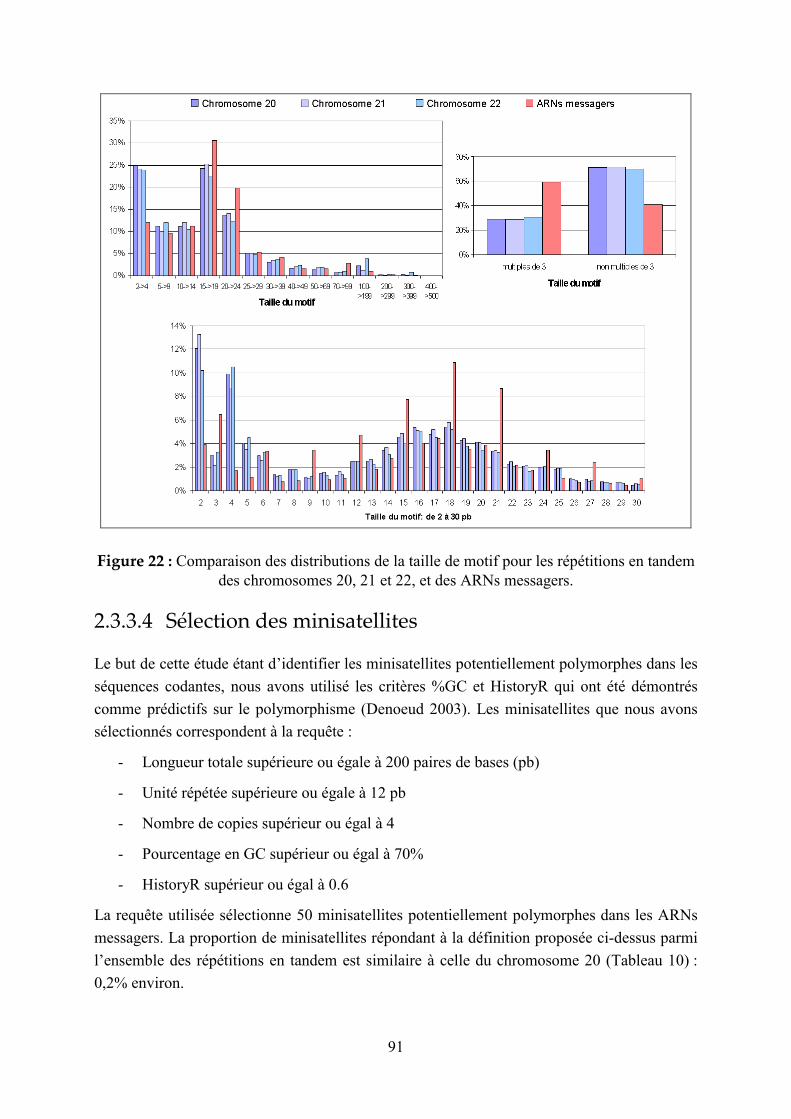
91
Figure 22 : Comparaison des distributions de la taille de motif pour les répétitions en tandem
des chromosomes 20, 21 et 22, et des ARNs messagers.
2.3.3.4 Sélection des minisatellites
Le but de cette étude étant d’identifier les minisatellites potentiellement polymorphes dans les
séquences codantes, nous avons utilisé les critères %GC et HistoryR qui ont été démontrés
comme prédictifs sur le polymorphisme (Denoeud 2003). Les minisatellites que nous avons
sélectionnés correspondent à la requête :
- Longueur totale supérieure ou égale à 200 paires de bases (pb)
- Unité répétée supérieure ou égale à 12 pb
- Nombre de copies supérieur ou égal à 4
- Pourcentage en GC supérieur ou égal à 70%
- HistoryR supérieur ou égal à 0.6
La requête utilisée sélectionne 50 minisatellites potentiellement polymorphes dans les ARNs
messagers. La proportion de minisatellites répondant à la définition proposée ci-dessus parmi
l’ensemble des répétitions en tandem est similaire à celle du chromosome 20 (Tableau 10) :
0,2% environ.

92
Les seuils sur la longueur totale et le nombre de répétitions sont peu stringents, car il est
probable, compte-tenu de la pression de sélection, que les minisatellites présents dans les
séquences codantes soient de taille relativement restreinte. Cette hypothèse est en accord avec
la définition élargie des minisatellites proposée dans l’introduction et dans la revue du
chapitre 2.3.1 : les répétitions en tandem de plus de 140 paires de bases peuvent être
considérées comme des minisatellites.
Lors de l’étude menée sur les minisatellites des chromosomes 21 et 22, présentée au chapitre
2.3.2 (Denoeud 2003) une requête sélectionnant les minisatellites avec %GC ≥ 48% et
HistoryR ≥ 0.54 produisait un enrichissement en minisatellites polymorphes de 43% à 59%
(spécificité : 59%) dans le lot sélectionné, et ne manquait que 30% des minisatellites
polymorphes (sensibilité : 70%). La requête utilisée ici est plus stringente : on peut donc
espérer qu’au moins 60% des minisatellites sélectionnés (soit 36 sur 50) soient effectivement
polymorphes, et que moins d’une vingtaine aient été manqués.
Figure 23 : Localisation chromosomique des minisatellites identifiés dans des séquences d’ARNm.
La Figure 23 représente la localisation des 50 minisatellites potentiellement polymorphes
détectés dans des ARNs messagers, sur le génome humain. Les minisatellites des ARNs
messagers montrent sensiblement la même répartition (télomérique) que celle indiquée par
(Amarger 1998) pour la population générale des minisatellites.

93
La Figure 24 montre la distribution des minisatellites correspondant à la même requête sur le
chromosome 20 (dans ou hors des ARNs messagers) : le biais de distribution télomérique est
similaire (31 minisatellites parmi 50 sont situés aux extrémités des chromosomes pour les
ARNs messagers, et 27/51 pour le chromosome 20).
Figure 24 : Localisation des minisatellites identifiés dans la séquence complète du chromosome 20
(à droite : illutration d’une requête de répartition dans la base de données des répétitions en tandem).
2.3.3.5 Caractéristiques des 50 minisatellites potentiellement polymorphes sélectionnés
L’Annexe 4 présente la description des 50 minisatellites et des ARNs messagers qui leur sont
associés. Pour les 50 minisatellites répondant à la requête, nous avons recherché si la
répétition en tandem était située dans la région codante (CDS) ou dans les régions non
traduites (UTRs), et si l’ARNm correspondait à un gène ou à un modèle de gène (prédiction).
Les résultats sont présentés dans le Tableau 11. Parmi les 50 minisatellites, 36 sont situés dans

94
des ARNs messagers correspondant à des prédictions de gènes (modèles), soit 72%. Dans
l’ensemble des ARNs messagers traités, seuls 51% correspondent à des modèles et 49% à des
gènes avec des produits protéiques de fonction connue, inférée, ou inconnue.
Tableau 11 : Distribution des minisatellites d’ARNs messagers, de motif multiple de 3 ou
non multiple de 3, dans les différentes classes de séquences (CDS, UTRs, gènes, modèles).
CDS UTRs Total : 50
minisatellites potentiellement polymorphes
gènes modèles gènes modèles
Motif multiple de 3 10 11 1 2
Motif non multiple de 3 1 14 2 9
Total 11 25 3 11
Le Tableau 11 présente également le nombre de minisatellites d’unité répétée multiple ou non
multiple de 3 situés dans des UTRs, dans des CDS, de gènes ou de modèles de gènes. On note
que les motifs « non multiples de 3 » sont dans leur très grande majorité rencontrés dans les
modèles (23 sur 27). Le seul gène mis en évidence possédant une répétition non multiple de 3
dans sa région codante correspond à une protéine hypothétique (certains ARNs messagers
sont en effet classés parmi les « gènes » et non les « modèles de gènes », alors que la protéine
correspondante est qualifiée d’hypothétique, de fonction inconnue). En principe, des
répétitions nucléiques non multiples de 3 peuvent générer des répétitions d’acides aminés si le
nombre de répétitions est suffisant : 3 unités répétées génèrent alors un « super-motif »
nucléotidique multiple de 3, qui codera pour une répétition d’acides aminés. La Figure 25
présente la traduction correspondant au minisatellite du gène du récepteur à la dopamine
(DRD4) , unité répétée : 48 pb, et du gène prédit « LOC349662 » (catégorie : « ab initio, with
EST support »). Cependant, le fait que de telles structures (motifs non multiple de 3 pb
codants) ne soient obtenues que pour des prédictions de gènes laisse présager que les modèles
de gènes contenant des répétitions nucléiques codantes mais non multiples de 3 seraient des
artéfacts ne correspondant pas à des gènes réels. D’une manière plus générale, on constate
que, en accord avec les observations menées sur la répartition des répétitions en tandem dans
les génomes (Li 2002), la majorité des minisatellites répondant à la requête d’enrichissement
en locus polymorphes et appartenant à des CDS ont des unités multiples de 3 (21/36 soit 58%,
ou si on ne considère pas les modèles, 10/11 soit 91%), et la majorité des minisatellites
appartenant à des UTRs ont des motifs non multiples de 3 (11/15 soit 73%). Pour les UTRs,
contrairement aux CDS, les proportions de minisatellites à motif multiple de 3 ne sont pas
significativement différentes entre gènes et modèles.

95
Figure 25 : Traduction des motifs répétés nucléiques en motifs répétés protéiques, pour deux
minisatellites (l’un d’unité répétée de 48 pb, multiple de 3, et l’autre d’unité répétée de 29 pb).

96
2.3.3.6 Minisatellites appartenant à des séquences codantes
Nous avons identifié 36 minisatellites appartenant à des séquences codantes, dont 25
correspondent à des modèles de gènes (prédits), 2 à des gènes ayant des produits protéiques
hypothétiques, et 9 à des gènes ayant des produits protéiques de fonction connue ou inférée :
ces 9 protéines sont décrites dans le Tableau 12. Parmi ces répétitions en tandem, 5 sont déjà
connues comme étant polymorphes : 3 d’entre elles (DRD4, CEL, OGFR) sont listées dans le
Tableau 9 (paragraphe 1.2.3.3.3.2) car leur polymorphisme a déjà été largement étudié. Les
deux autres correspondent à un minisatellite du chromosome 21 qui s’est révélé polymorphe
(2 allèles) lors de l’étude que j’ai menée sur les chromosomes 21 et 22 (Denoeud 2003) (gène
CLIC6 : ce minisatellite, CEB234, n’avait pas été localisé dans un gène lors d’analyses
effectuées en 2001, alors qu’il fait partie d’une séquence codante selon la mise à jour du
génome humain d’avril 2003), et à un ARN messager ayant plusieurs transcrits, provenant
d’un phénomène d’épissage alternatif, différant par leur nombre de répétitions en tandem
(gène MADCAM1) (Leung 1996). Quatre minisatellites potentiellement polymorphes, mais
encore peu étudiés, situés dans des gènes connus ont donc été identifiés grâce à cette étude :
ils appartiennent aux gènes GRINA, KRT10, GNAS1 et ESX1L, tous 4 impliqués dans des
pathologies. L’étude de leur polymorphisme et de son influence sur ces pathologies mériterait
donc d’être menée. De plus, les deux derniers gènes listés sont associés à des phénomènes
d’empreinte parentale, ce qui les rend particulièrement intéressants à étudier.
Parmi les 25 minisatellites appartenant à des séquences codantes de modèles de gènes, 11 ont
des motifs répétés de longueur multiple de 3 et sont donc plus susceptibles de correspondre à
des gènes réels, et surtout d’être polymorphes (en effet, il est très peu vraisemblable que des
répétitions polymorphes appartenant à des séquences codantes aient des motifs répétés non
multiples de 3 : ce n’est en tout cas observé pour aucune de celles listés dans le Tableau 9,
voir paragraphe 1.2.3.3.3.2, ni pour celles identifiées par De Fonzo et al. (De Fonzo 1998)). A
ces 11 minisatellites, on peut ajouter le minisatellite identifié dans un gène codant pour une
protéine hypothétique, de fonction inconnue, et qui a également un motif répété multiple de 3.
Le Tableau 13 présente ces 12 gènes qui, s’ils ont été correctement prédits, contiennent des
minisatellites potentiellement polymorphes dans leurs séquences codantes. Ces gènes, n’ayant
jamais été étudiés, mériteraient donc une attention particulière, surtout ceux pour lesquels la
répétition en tandem constitue la majeure partie de la protéine. Certaines de ces séquences
codantes sont en effet constituées en majorité -58%, 84%, 86%- par la répétition en tandem,
ce qui laisse supposer un rôle prépondérant de leurs répétitions d’acides aminés sur la
structure tri-dimensionnelle de la protéine, et donc sur sa fonction. De façon intéressante, pour
les 9 gènes présentés dans le Tableau 12, la proportion de la séquence protéique
correspondant à la répétition en tandem n’atteint pas plus de 32% (des proportions bien plus
importantes sont toutefois observées pour des protéines connues comme les mucines : voir
paragraphe 1.2.3.3.3.2 (Vinall 1998 ; Debailleul 1998)).
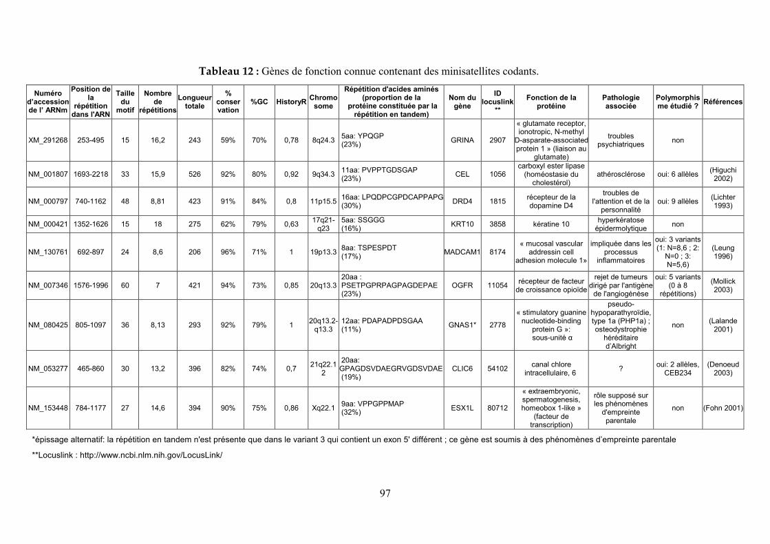
97
Tableau 12 : Gènes de fonction connue contenant des minisatellites codants.
*épissage alternatif: la répétition en tandem n'est présente que dans le variant 3 qui contient un exon 5' différent ; ce gène est soumis à des phénomènes d’empreinte parentale
**Locuslink : http://www.ncbi.nlm.nih.gov/LocusLink/
Numéro d’accession de l’ ARNm
Position de la
répétition dans l'ARN
Taille du
motif
Nombre de
répétitions
Longueur totale
% conser vation
%GC HistoryR Chromo
some
Répétition d'acides aminés (proportion de la
protéine constituée par la répétition en tandem)
Nom du gène
ID locuslink
**
Fonction de la protéine
Pathologie associée
Polymorphisme étudié ?
Références
XM_291268 253-495 15 16,2 243 59% 70% 0,78 8q24.3 5aa: YPQGP (23%)
GRINA 2907
« glutamate receptor, ionotropic, N-methyl
D-asparate-associated protein 1 » (liaison au
glutamate)
troubles psychiatriques
non
NM_001807 1693-2218 33 15,9 526 92% 80% 0,92 9q34.3 11aa: PVPPTGDSGAP (23%)
CEL 1056 carboxyl ester lipase
(homéostasie du cholestérol)
athérosclérose oui: 6 allèles (Higuchi
2002)
NM_000797 740-1162 48 8,81 423 91% 84% 0,8 11p15.5 16aa: LPQDPCGPDCAPPAPG (30%)
DRD4 1815 récepteur de la dopamine D4
troubles de l'attention et de la
personnalité oui: 9 allèles
(Lichter 1993)
NM_000421 1352-1626 15 18 275 62% 79% 0,63 17q21-
q23 5aa: SSGGG (16%)
KRT10 3858 kératine 10 hyperkératose
épidermolytique non
NM_130761 692-897 24 8,6 206 96% 71% 1 19p13.3 8aa: TSPESPDT (17%)
MADCAM1 8174 « mucosal vascular
addressin cell adhesion molecule 1»
impliquée dans les processus
inflammatoires
oui: 3 variants (1: N=8,6 ; 2:
N=0 ; 3: N=5,6)
(Leung 1996)
NM_007346 1576-1996 60 7 421 94% 73% 0,85 20q13.3 20aa : PSETPGPRPAGPAGDEPAE (23%)
OGFR 11054 récepteur de facteur
de croissance opioïde
rejet de tumeurs dirigé par l'antigène
de l'angiogénèse
oui: 5 variants (0 à 8
répétitions)
(Mollick 2003)
NM_080425 805-1097 36 8,13 293 92% 79% 1 20q13.2-
q13.3 12aa: PDAPADPDSGAA (11%)
GNAS1* 2778
« stimulatory guanine nucleotide-binding
protein G »: sous-unité α
pseudo-hypoparathyroïdie, type 1a (PHP1a) ; osteodystrophie
héréditaire d’Albright
non (Lalande
2001)
NM_053277 465-860 30 13,2 396 82% 74% 0,7 21q22.1
2
20aa: GPAGDSVDAEGRVGDSVDAE (19%)
CLIC6 54102 canal chlore
intracellulaire, 6 ?
oui: 2 allèles, CEB234
(Denoeud 2003)
NM_153448 784-1177 27 14,6 394 90% 75% 0,86 Xq22.1 9aa: VPPGPPMAP (32%)
ESX1L 80712
« extraembryonic, spermatogenesis, homeobox 1-like »
(facteur de transcription)
rôle supposé sur les phénomènes
d'empreinte parentale
non (Fohn 2001)
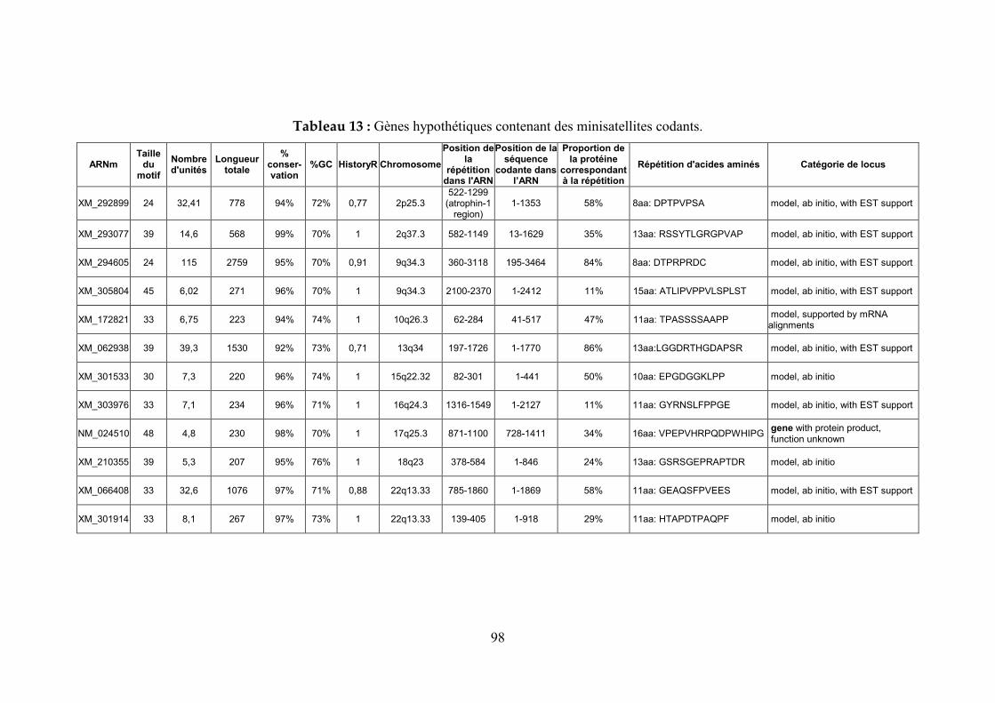
98
Tableau 13 : Gènes hypothétiques contenant des minisatellites codants.
ARNm Taille
du motif
Nombre d'unités
Longueur totale
% conser- vation
%GC HistoryR Chromosome
Position de la
répétition dans l'ARN
Position de la séquence
codante dans l’ARN
Proportion de la protéine
correspondant à la répétition
Répétition d'acides aminés Catégorie de locus
XM_292899 24 32,41 778 94% 72% 0,77 2p25.3 522-1299
(atrophin-1 region)
1-1353 58% 8aa: DPTPVPSA model, ab initio, with EST support
XM_293077 39 14,6 568 99% 70% 1 2q37.3 582-1149 13-1629 35% 13aa: RSSYTLGRGPVAP model, ab initio, with EST support
XM_294605 24 115 2759 95% 70% 0,91 9q34.3 360-3118 195-3464 84% 8aa: DTPRPRDC model, ab initio, with EST support
XM_305804 45 6,02 271 96% 70% 1 9q34.3 2100-2370 1-2412 11% 15aa: ATLIPVPPVLSPLST model, ab initio, with EST support
XM_172821 33 6,75 223 94% 74% 1 10q26.3 62-284 41-517 47% 11aa: TPASSSSAAPP model, supported by mRNA alignments
XM_062938 39 39,3 1530 92% 73% 0,71 13q34 197-1726 1-1770 86% 13aa:LGGDRTHGDAPSR model, ab initio, with EST support
XM_301533 30 7,3 220 96% 74% 1 15q22.32 82-301 1-441 50% 10aa: EPGDGGKLPP model, ab initio
XM_303976 33 7,1 234 96% 71% 1 16q24.3 1316-1549 1-2127 11% 11aa: GYRNSLFPPGE model, ab initio, with EST support
NM_024510 48 4,8 230 98% 70% 1 17q25.3 871-1100 728-1411 34% 16aa: VPEPVHRPQDPWHIPG gene with protein product, function unknown
XM_210355 39 5,3 207 95% 76% 1 18q23 378-584 1-846 24% 13aa: GSRSGEPRAPTDR model, ab initio
XM_066408 33 32,6 1076 97% 71% 0,88 22q13.33 785-1860 1-1869 58% 11aa: GEAQSFPVEES model, ab initio, with EST support
XM_301914 33 8,1 267 97% 73% 1 22q13.33 139-405 1-918 29% 11aa: HTAPDTPAQPF model, ab initio

99
2.3.3.7 Conclusions
Différentes observations nous laissent penser que certains modèles de gènes, provenant de
prédictions ab initio, ne doivent pas correspondre à de véritables gènes :
- On note un excédent de modèles de gènes parmi les ARNs messagers contenant des
minisatellites : une hypothèse pour expliquer cette observation serait que certains de ces
modèles sont faux.
- Seuls les modèles de gènes contiennent des répétitions nucléiques codantes d’unité non
multiple de 3. Si de telles structures étaient utilisées pour coder pour des répétitions
d’acides aminés (« super-motifs » de 3 motifs nucléiques codant pour un motif protéique :
voir Figure 25), on observerait vraisemblablement des co-mutations au niveau de la
séquence nucléique tous les 3 motifs répétés. Il est d’ailleurs probable que le Tandem
Repeats Finder (Benson 1999) aurait identifié de façon redondante des répétitions avec
des consensus 3 fois plus longs, et une meilleure conservation. L’analyse des séquences
nucléiques correspondantes ne montre rien de tel. Il est donc très probable que les
séquences codantes prédites, correspondant à des unités répétés non multiples de 3 paires
de bases sont en fait des erreurs de prédiction. On ne peut pas exclure toutefois que ces
répétitions en tandem soient monomorphes (et maintenues comme telles par la pression de
sélection) auquel cas la taille du motif aurait moins d’importance.
On peut donc supposer que les séquences codantes prédites (modèles de gènes) contenant des
minisatellites sont pour la plupart (en tout cas pour ceux ayant des motifs non multiples de 3)
des erreurs de prédiction, tout du moins en ce qui concerne les frontières entre introns et
exons. Il y a donc vraisemblablement moins de minisatellites appartenant à des séquences
codantes que nous n’en avions identifié en première intention, c’est pourquoi nous n’avons
décrit que les minisatellites à motif multiple de 3 (soit 21/36). Afin d’améliorer la prédiction
de gènes, des critères tels que « absence de répétitions en tandem à motif non multiple de 3 »
dans la séquence codante pourraient être appliqués. En effet, les minisatellites codants restent
très rares : si de telles structures sont identifiées (quelle que soit la longueur du motif), il
convient de vérifier la validité du gène prédit.
Les nouveaux minisatellites codants identifiés par cette étude sont potentiellement
polymorphes : ils ont été sélectionnés selon des critères corrélés au polymorphisme (Denoeud
2003). Ce polymorphisme nécessite d’être confirmé. Le fait que parmi les 9 minisatellites
codants isolés dans des gènes connus, 5 aient déjà été étudiés et caractérisés comme étant
polymorphes, laisse présumer que la requête utilisée est efficace pour sélectionner des
minisatellites polymorphes. Cependant, elle n’a permis d’identifier que 4 nouveaux
minisatellites dans des gènes connus et 12 dans des gènes hypothétiques. De plus, les
mucines, famille de protéines contenant des répétitions en tandem polymorphes (Vinall 1998),
n’ont pas été identifiées par cette requête. Certains minisatellites codants ont donc été
manqués : une requête moins stringente mériterait d’être testée. Par exemple, on pourrait

100
tâcher de trouver des caractéristiques communes aux minisatellites codants déjà étudiés
(Tableau 9) afin de générer une requête plus générale. En tout cas, cette approche pour
identifier des répétitions en tandem codantes, c’est-à-dire potentiellement fonctionnelles, est
prometteuse. En particulier, cette étude semble cibler de façon préférentielle des locus
impliqués dans des phénomènes d’empreinte parentale, comme le gène GNAS1, épissé de
façon différentielle selon l’origine maternelle ou paternelle des allèles (Lalande 2001), et dont
l’un des transcrits contient un minisatellite potentiellement polymorphe (Tableau 12).

101
3 Discussion et perspectives

102
3.1 La base de données des répétitions en tandem
Dans cette thèse, j’ai illustré l’utilité de la base de données des répétitions en tandem. Elle a
été mise à profit au laboratoire pour :
- L’étude de la répartition des minisatellites sur des chromosomes eucaryotes (Vergnaud
2000).
- L’identification de marqueurs polymorphes pour l’épidémiologie bactérienne (Le
Flèche 2001; Le Flèche 2002), en particulier grâce à la page de comparaison de souches
(Denoeud 2004).
- La mise en évidence de critères prédictifs sur le polymorphisme de minisatellites
humains (Denoeud 2003).
Cette base de données est accessible sur Internet, dans le but d’être utile à d’autres
utilisateurs, ce qui semble être le cas au vu des nombreuses requêtes qui y sont menées
quotidiennement, en particulier pour les génomes bactériens. Cependant, ce site reste
« artisanal » (j’en suis pour l’instant la seule administratrice) et on ne peut qu’espérer qu’un
des sites majeurs fournisseurs de services bioinformatiques trouve suffisamment d’intérêt à
cet outil pour en réaliser des développements plus performants et plus durables.
Certaines améliorations mériteraient encore d’être apportées à la base de données. Comme
nous l’avons décrit au paragraphe 2.1.1.1, l’élimination de la redondance dans les résultats du
logiciel Tandem Repeats Finder (Benson 1999) a fait l’objet de choix automatiques sur des
critères empiriques qui étaient dans certains cas peu judicieux. Par exemple, lorsqu’une
répétition en tandem « émerge » à l’intérieur d’une autre (comme nous l’avons vu dans le cas
des « VLTR » ou « variable length tandem repeat » (Hauth 2002) : Figure 11), le choix
effectué va masquer cette émergence en présentant la plus grande répétition en tandem. Des
ajustements pourraient permettre de compenser ce défaut : la base pourrait inclure par
exemple une mesure de la distorsion de la conservation interne, ce qui permettrait d’identifier
les répétitions en tandem qui en contiennent une autre, d’évolution plus récente, à motifs
souvent beaucoup mieux conservés. Une amélioration a déjà été apportée par rapport à la
première version de la base : les alignements retenus proposent un lien vers les autres
alignements (non retenus) de la région de redondance, afin de permettre à l’utilisateur de
choisir. Cette fonctionnalité n’est toutefois disponible que pour les génomes importés
récemment. C’est également le cas d’autres options telles que l’accès à la séquence brute des
répétitions en tandem (sans alignement des unités répétées avec le motif consensus).
Malgré ces quelques réserves, la base de données que j’ai développée reste à ce jour le seul
site spécialement dédié à l’identification de répétitions en tandem dans les génomes, utilisable
sans compétences ou goûts particuliers pour la bioinformatique. Au vu de l’intérêt croissant
porté ces dernières années au génotypage de souches par les répétitions en tandem, on peut

103
espérer que la page consacrée aux génomes bactériens (et notamment la page de comparaison
de souches) devienne un outil de référence pour les développeurs de tels moyens de typage.
A titre de comparaison, le site développé par Gary Benson (TRDB : Tandem Repeats
Database [http://tandem.bu.edu/cgi-bin/trdb/trdb.exe]) dédié à la détection et à l’analyse des
répétitions en tandem, n’a pas la même vocation que notre base de données. Il s’adresse plutôt
à des « spécialistes » des répétitions en tandem, en leur proposant des utilitaires de
visualisation et de classification des répétitions en tandem présentes dans les séquences qu’ils
soumettent. Ce site est toutefois très utile pour analyser les séquences de différents allèles de
répétitions en tandem polymorphes. En effet, pour le génotypage de bactéries, il est de plus en
plus fréquent de pousser l’analyse jusqu’au séquençage des allèles. Dans notre laboratoire,
différents locus polymorphes de bactéries pathogènes sont ainsi séquencés, et nous sommes
des utilisateurs réguliers de la base de Gary Benson, avec qui nous entretenons une
collaboration.
3.2 Répétitions en tandem et phylogénie
3.2.1 Intérêt phylogénique du typage de répétitions en tandem bactériennes
En épidémiologie bactérienne, les répétitions en tandem sont utilisées afin de distinguer
différents isolats ou souches. Les génotypes obtenus par typage des répétitions en tandem
permettent d’obtenir une classification des différentes souches, sous forme d’un arbre, ce qui
donne une idée de leur proximité. Cette proximité vis à vis des répétitions en tandem n’a
toutefois pas nécessairement une valeur phylogénique : l’arbre basé sur les répétitions en
tandem peut ne pas correspondre à l’arbre évolutif réel de l’espèce. En effet, certains allèles
peuvent être de taille identique sans avoir suivi le même chemin évolutif. Ce phénomène est
appelé homoplasie. Lorsque la répétition en tandem est imparfaite (variations entre motifs),
l’homoplasie peut se manifester sous la forme d’allèles de taille identique mais de séquence
différente. Par exemple, pour le minisatellite de l’insuline analysé par Jeffreys et collègues, 39
allèles peuvent être distingués d’après leur taille, qui correspondent à 189 codes MVR
distincts (Stead 2000). De plus, l’analyse des séquences ou des cartes MVR (Jeffreys 1991) de
certains minisatellites montre que des allèles de taille différente peuvent être bien plus
proches, du point de vue de l’agencement et des mutations internes des motifs, que des allèles
de taille identique. Il arrive donc que des allèles de même taille soient retrouvés dans des
branches distinctes de l’arbre provenant d’une analyse MLVA (multiple locus VNTR
analysis). En général, les classifications produites par typage de taille de répétitions en tandem
varient légèrement en fonction de la combinaison de marqueurs polymorphes utilisée.
Cependant, si on emploie un nombre suffisant de marqueurs, l’arbre obtenu est robuste, et en
104
concordance avec la phylogénie connue pour l’espèce considérée (inférée grâce à des critères
morphologiques, biochimiques, et/ou de spécificité d’hôte) : nous l’avons montré pour
Bacillus anthracis (Le Flèche 2001), Mycobacterium tuberculosis (Le Flèche 2002), et
d’autres espèces encore en cours d’analyse au laboratoire, comme Brucella. Ainsi, si le choix
des marqueurs polymorphes est judicieux (élimination des marqueurs soumis au phénomène
d’homoplasie et des marqueurs trop polymorphes donc peu informatifs d’un point de vue
évolutif), l’arbre produit par l’approche MLVA aura de bonnes chances de correspondre à la
phylogénie de l’espèce bactérienne.
3.2.2 Analyse des séquences de répétitions en tandem
Une façon simple de détecter des phénomènes d’homoplasie consiste à séquencer les
différents allèles de même taille, à condition que le locus ne soit pas une répétition en tandem
parfaite. Le séquençage des locus VNTR peut en outre s’avérer une source très puissante
d’information sur la phylogénie des espèces : il suffirait dans les cas favorables de séquencer
un ou quelques locus au lieu de typer une quinzaine de locus, pour aboutir au moins à un
premier niveau de classification. Cette approche se heurte toutefois à une limitation.
Actuellement, très peu de moyens existent pour analyser les séquences de répétitions en
tandem, et en particulier pour appréhender leur évolution. Gary Benson a proposé en 1999 un
logiciel de reconstruction de l’histoire des répétitions en tandem (Benson 1999). Ce
programme, décrit dans l’article présenté au chapitre 2.3.2 (Denoeud 2003), cherche à
contracter à moindre coût une répétition en tandem pour remonter à un motif ancestral. Cet
algorithme se base sur l’hypothèse que lorsque plusieurs motifs ou groupes de motifs arborent
les mêmes mutations internes, ils sont très probablement issus d’une duplication. Cette
approche, même si elle a un intérêt pour quantifier l’hétérogénéité interne d’allèles de
minisatellites considérés individuellement (nous le verrons au chapitre 3.3), ne peut pas
s’appliquer à la comparaison de plusieurs allèles. Elemento et collègues se sont intéressés plus
particulièrement à la reconstruction de l’histoire des duplications ayant conduit d’un gène
ancestral à différents gènes paralogues répétés en tandem, comme c’est le cas pour la région
«TRGV » (région variable de la chaîne gamma des récepteurs des lymphocytes T) constituée
de 9 gènes (Elemento 2002a ; Elemento 2002b) : ces programmes, d’une grande efficacité,
s’intéressent encore une fois aux événements de duplication survenus dans un seul allèle et ne
s’appliquent donc pas à la comparaison d’allèles.
Actuellement, plusieurs projets de séquençage de minisatellites sont menés au laboratoire, et
l’analyse des allèles est effectuée à la main, après un pré-traitement utilisant les outils de Gary
Benson [http://tandem.bu.edu/cgi-bin/trdb/trdb.exe]. Il s’agit, comme pour les cartes MVR, de
coder les minisatellites par la succession des différents types d’unités répétés qu’ils
contiennent : voir Figure 26.

105
Figure 26 : Codage d’allèles d’un minisatellite de M. tuberculosis : à chaque motif différent
est attribué un code, de A à J. Les deux allèles diffèrent par la présence/absence du motif D.
Ensuite, si le nombre de motifs possibles n’est pas trop grand et que le nombre d’allèles
comparés n’est pas excessif, il est possible de reconstruire manuellement les événements
évolutifs les plus probables ayant conduit d’un allèle à un autre (duplications, délétions,
mutations ponctuelles dans des motifs), ou tout au moins de se faire une idée, visuellement,
sur la proximité des allèles. De telles études devraient fournir des pistes sur les mécanismes
de mutation des minisatellites bactériens, encore très peu étudiés. On peut espérer que
l’analyse manuelle d’un certain nombre de locus minisatellites bactériens permettra de faire
des généralisations sur les mécanismes d’évolution de ces structures, afin de créer un
programme de comparaison automatique d’allèles. Il semble en effet prématuré de vouloir
traiter automatiquement la phylogénie des répétitions en tandem, alors que les mécanismes
sous-jacents restent énigmatiques et les données encore peu nombreuses.
Quelques tentatives ont toutefois été faites pour créer des algorithmes de phylogénie des
répétitions en tandem, en utilisant des simplifications du problème. Ainsi, Bérard & Rivals
proposent un algorithme d’alignement d’allèles de minisatellites (cartes MVR). Malgré le fait
que ce programme n’autorise que des événements de nature simple (il ne considère que les
délétions et amplifications d’une seule unité à la fois, et ne tient pas compte de la proximité
106
entre les différents types de motifs), les distances entre allèles qu’il a produites pour le
minisatellite humain MSY1 ont permis de reconstruire une phylogénie concordante avec celle
issue d’autres marqueurs (Berard 2003). Le minisatellite MSY1, situé sur le chromosome Y,
correspond à une situation haploïde : seuls des événements intra-alléliques peuvent y survenir,
ce qui est également le cas pour les minisatellites bactériens. Il serait intéressant d’appliquer
cet algorithme aux séquences obtenues dans notre laboratoire pour des minisatellites de
différentes espèces bactériennes (Brucella, Staphylococcus aureus…). Cependant, les études
déjà faites manuellement pour certains locus montrent qu’une généralisation est difficile, et il
est donc probable que ce modèle simple ne donne pas toujours des résultats satisfaisants. En
outre, il n’est clairement pas adapté pour ceux des minisatellites humains localisés sur les
autosomes qui peuvent subir des événements de recombinaison inter-allélique.
3.3 Prédiction du polymorphisme
3.3.1 Critères de séquence corrélés au polymorphisme
Pouvoir prédire le polymorphisme des répétitions en tandem à partir de la séquence d’un seul
allèle constituerait une avancée majeure, dans la mesure où elle permettrait d’éliminer les
étapes de typages préliminaires, parfois longues et coûteuses, visant à évaluer le
polymorphisme des locus. Nous avons montré que certaines caractéristiques de séquence sont
corrélées au polymorphisme des minisatellites humains : %GC et HistoryR (Denoeud 2003).
Ces deux critères, même s’ils améliorent la sélection de minisatellites polymorphes, ne
permettent pas de s’affranchir totalement de l’étape de typage préliminaire, puisque les
prédictions ne produisent que 60% environ de minisatellites effectivement polymorphes dans
le lot de minisatellites humains que nous avons testé. En outre, ils passent à côté d’un certain
nombre de locus d’intérêt (30%). Par exemple, même si la majorité des minisatellites
hypermutables actuellement connus est riche en GC (Vergnaud 2000), certains sont riches en
AT et seraient donc éliminés par une telle sélection. Il faut noter que les quelques
minisatellites riches en AT connus, comme FRA16B (Yu 1997), ne possèdent pas
suffisamment de variants internes pour qu’on puisse étudier les événements de mutation qui
s’y produisent. Il serait pourtant important de vérifier qu’ils sont bien soumis aux mêmes
mécanismes que les minisatellites riches en GC. De façon intéressante, le meilleur critère de
prédiction du polymorphisme des minisatellites humains, HistoryR, est aussi le plus
complexe : il reflète la facilité avec laquelle on peut remonter depuis la répétition en tandem
qu’on observe jusqu’à un motif « ancestral » unique, par des événements de contraction.
Même si ce paramètre est obtenu à partir d’un modèle d’évolution des répétitions en tandem
relativement simple, il reste plus informatif que des données de séquence brutes. On peut
donc espérer que si des programmes plus élaborés voient le jour, ils fourniront de meilleurs
prédicteurs du polymorphisme.

107
Chez les bactéries, nous avons également recherché des critères de séquence corrélés au
polymorphisme des répétitions en tandem (Le Flèche 2001 ; Denoeud 2004). Nous avons
effectué cette recherche sur des répétitions en tandem de longueur totale quelconque, donc
généralement bien inférieure à celle des minisatellites que nous avions étudiés chez l’Homme.
En effet, les minisatellites de grande taille (plusieurs centaines de paires de bases et plusieurs
dizaines de motifs) sont en nombre trop faible dans certains génomes bactériens et nous avons
donc étendu l’analyse aux répétitions ne comptant que quelques unités répétées. Pour des
nombres d’unités faibles, la valeur de HistoryR a de fortes chances d’être égale à 1 (le coût
maximal de reconstruction est égal au coût minimal) : ce critère est alors peu informatif, ce
qui explique qu’il ne soit pas corrélé au polymorphisme dans ces analyses. Deux
caractéristiques de séquence plus « classiques » sont en revanche corrélées au
polymorphisme : la conservation interne et le nombre de copies. Etant donné que nous avons
considéré des répétitions en tandem de taille assez restreinte, il n’est pas étonnant que des
critères influant sur l’instabilité des microsatellites soient corrélés au polymorphisme dans cet
échantillon. Cependant, la qualité prédictive de tels critères reste très variable selon les
espèces bactériennes considérées : ils ne sont donc pas satisfaisants pour étudier de nouvelles
espèces. Ainsi, la meilleure approche pour prédire le polymorphisme des répétitions en
tandem bactériennes reste la comparaison de souches. Cette approche devrait être de plus en
plus facile à mettre en œuvre car il est probable que nous disposions dans un avenir proche,
pour la quasi-totalité des espèces bactériennes d’intérêt médical et/ou économique, de la
séquence génomique de plusieurs souches.
3.3.2 Mécanismes de mutation
La prédiction du polymorphisme, comme la phylogénie basée sur les répétitions en tandem,
profiterait d’une meilleure compréhension des mécanismes de mutation sous-jacents. Comme
je l’ai décrit dans l’introduction, plusieurs types de mécanismes d’instabilité (méiotiques ou
mitotiques) sont invoqués pour les minisatellites humains. Selon les locus, ces différents
événements surviennent dans des proportions variables, encore impossibles à prédire. Il est
probable que pour certains mécanismes, la séquence des répétitions en tandem joue un rôle,
tandis que pour d’autres, seul l’environnement ait une importance. Ainsi, la compréhension
des mécanismes de mutation des minisatellites humains, qui ont pourtant été largement
étudiés, reste très partielle. Il semble que nous soyons encore dans une phase où les nouvelles
données obtenues compliquent notre vision des choses plus qu’elles ne la simplifient.
Chez les bactéries, organismes haploïdes où les événements de recombinaison inter-alléliques
ne peuvent pas se produire (sauf cas de transferts horizontaux), les mécanismes de mutation
des minisatellites n’ont quasiment pas été étudiés. Ces mécanismes pourraient impliquer,
comme chez l’Homme, des glissements lors de la réplication et la réparation de cassures
double-brin par l’invasion de la chromatide sœur. On peut espérer que des investigations
seront menées prochainement dans ce domaine.

108
Par ailleurs, lorsque les mécanismes d’évolution des répétitions en tandem seront mieux
élucidés, il faudra encore être en mesure, afin d’améliorer l’étude des séquences pour la
prédiction du polymorphisme et la phylogénie, de les modéliser in silico. Il s’agit là d’une
tâche bioinformatique ambitieuse.

109
Bibliographie

110
Aach, J., Bulyk, M. L., Church, G. M., Comander, J., Derti, A. et Shendure, J. 2001. Computational comparison of two draft sequences of the human genome. Nature 409: 856-9.
Achaz, G., Rocha, E. P., Netter, P. et Coissac, E. 2002. Origin and fate of repeats in bacteria. Nucleic Acids Res 30: 2987-94.
Achtman, M., Zurth, K., Morelli, G., Torrea, G., Guiyoule, A. et Carniel, E. 1999. Yersinia pestis, the cause of plague, is a recently emerged clone of Yersinia pseudotuberculosis. Proc Natl Acad Sci U S A 96: 14043-8.
Aharoni, A., Baran, N. et Manor, H. 1993. Characterization of a multisubunit human protein which selectively binds single stranded d(GA)n and d(GT)n sequence repeats in DNA. Nucleic Acids Res 21: 5221-8.
Alm, R. A. et al. 1999. Genomic-sequence comparison of two unrelated isolates of the human gastric pathogen Helicobacter pylori. Nature 397: 176-80.
Altschul, S. F., Gish, W., Miller, W., Myers, E. W. et Lipman, D. J. 1990. Basic local alignment search tool. J Mol Biol 215: 403-10.
Altschul, S. F., Boguski, M. S., Gish, W. et Wootton, J. C. 1994. Issues in searching molecular sequence databases. Nature Genet. 6: 119-129.
Altschul, S. F., Madden, T. L., Schaffer, A. A., Zhang, J., Zhang, Z., Miller, W. et Lipman, D. J. 1997. Gapped BLAST and PSI-BLAST: a new generation of protein database search programs. Nucleic Acids Res 25: 3389-402.
Amarger, V. et al. 1998. Analysis of the human, pig, and rat genomes supports a universal telomeric origin of minisatellite sequences. Genomics 52: 62-71.
Amos, W. & Rubinstzein, D. C. 1996. Microsatellites are subject to directional evolution. Nat Genet 12: 13-4.
Amos, W., Sawcer, S. J., Feakes, R. W. et Rubinsztein, D. C. 1996. Microsatellites show mutational bias and heterozygote instability. Nat Genet 13: 390-1.
Appelgren, H., Cederberg, H. et Rannug, U. 1997. Mutations at the human minisatellite MS32 integrated in yeast occur with high frequency in meiosis and involve complex recombination events. Mol. Gen. Genet. 256: 7-17.
Armour, J. A., Brinkworth, M. H. et Kamischke, A. 1999. Direct analysis by small-pool PCR of MS205 minisatellite mutation rates in sperm after mutagenic therapies. Mutat Res 445: 73-80.
Armour, J. A. L., Patel, I., Thein, S. L., Fey, M. F. et Jeffreys, A. J. 1989. Analysis of somatic mutations at human minisatellite loci in tumors and cell lines. Genomics 4: 328-334.
Ashley, T. 1994. Mammalian meiotic recombination: a reexamination. Hum Genet 94: 587-93.
Atkin, N. B. 2001. Microsatellite instability. Cytogenet Cell Genet 92: 177-81.
Aupetit, C., Drouet, M., Pinaud, E., Denizot, Y., Aldigier, J. C., Bridoux, F. et Cogne, M. 2000. Alleles of the alpha1 immunoglobulin gene 3' enhancer control evolution of IgA nephropathy toward renal failure. Kidney Int 58: 966-71.
Autexier, C. & Greider, C. W. 1996. Telomerase and cancer: revisiting the telomere hypothesis. Trends Biochem Sci 21: 387-91.
Awad, M., Pravica, V., Perrey, C., El Gamel, A., Yonan, N., Sinnott, P. J. et Hutchinson, I. V. 1999. CA repeat allele polymorphism in the first intron of the human interferon-gamma gene is associated with lung allograft fibrosis. Hum Immunol 60: 343-6.

111
Bachtrog, D., Weiss, S., Zangerl, B., Brem, G. et Schlotterer, C. 1999. Distribution of dinucleotide microsatellites in the Drosophila melanogaster genome. Mol Biol Evol 16: 602-10.
Bagli, M., Papassotiropoulos, A., Knapp, M., Jessen, F., Luise Rao, M., Maier, W. et Heun, R. 2000. Association between an interleukin-6 promoter and 3' flanking region haplotype and reduced Alzheimer's disease risk in a German population. Neurosci Lett 283: 109-12.
Bailly, S., di Giovine, F. S. et Duff, G. W. 1993. Polymorphic tandem repeat region in interleukin-1 alpha intron 6. Hum Genet 91: 85-6.
Bailly, S., Israel, N., Fay, M., Gougerot-Pocidalo, M. A. et Duff, G. W. 1996. An intronic polymorphic repeat sequence modulates interleukin-1 alpha gene regulation. Mol Immunol 33: 999-1006.
Baldi, P. & Baisnee, P. F. 2000. Sequence analysis by additive scales: DNA structure for sequences and repeats of all lengths. Bioinformatics 16: 865-89.
Bambara, R. A., Murante, R. S. et Henricksen, L. A. 1997. Enzymes and reactions at the eukaryotic DNA replication fork. J Biol Chem 272: 4647-50.
Basil, J. B., Goodfellow, P. J., Rader, J. S., Mutch, D. G. et Herzog, T. J. 2000. Clinical significance of microsatellite instability in endometrial carcinoma. Cancer 89: 1758-64.
Beckmann, J. S. & Weber, J. L. 1992. Survey of human and rat microsatellites. Genomics 12: 627-631.
Bell, G. I., Horita, S. et Karam, J. H. 1984. A polymorphic locus near the insulin gene is associated with insulin-dependent diabetes mellitus. Diabetes 33: 176-183.
Bennett, S. T. et al. 1995. Susceptibility to human type 1 diabetes at IDDM2 is determined by tandem repeat variation at the insulin gene minisatellite locus. Nat Genet. 9: 284-292.
Bennett, P. 2000. Demystified ... microsatellites. Mol Pathol 53: 177-83.
Benson, G. 1999. Tandem repeats finder: a program to analyze DNA sequences. Nucleic Acids Res 27: 573-580.
Benson, G. & Dong, L. 1999. Reconstructing the duplication history of a tandem repeat. Proc Int Conf Intell Syst Mol Biol: 44-53.
Berard, S. & Rivals, E. 2003. Comparison of minisatellites. J Comput Biol 10: 357-72.
Berg, E. S. & Olaisen, B. 1993. Characterization of the COL2A1 VNTR polymorphism. Genomics 16: 350-354.
Berg, I., Cederberg, H. et Rannug, U. 2000. Tetrad analysis shows that gene conversion is the major mechanism involved in mutation at the human minisatellite MS1 integrated in Saccharomyces cerevisiae. Genet Res 75: 1-12.
Berg, I., Neumann, R., Cederberg, H., Rannug, U. et Jeffreys, A. J. 2003. Two modes of germline instability at human minisatellite MS1 (locus D1S7): complex rearrangements and paradoxical hyperdeletion. Am J Hum Genet 72: 1436-47.
Bernal, A., Ear, U. et Kyrpides, N. 2001. Genomes OnLine Database (GOLD): a monitor of genome projects world-wide. Nucleic Acids Res 29: 126-7.
Biet, E., Sun, J. et Dutreix, M. 1999. Conserved sequence preference in DNA binding among recombination proteins: an effect of ssDNA secondary structure. Nucleic Acids Res 27: 596-600.
Bikker, H., Baas, F. et de Vijlder, J. J. 1992. Structure and characterization of a 50 bp repeat in intron 10 of the human thyroid peroxidase gene. Mol Cell Endocrinol 83: 21-8.

112
Bingen, E. H., Denamur, E. et Elion, J. 1994. Use of ribotyping in epidemiological surveillance of nosocomial outbreaks. Clin Microbiol Rev 7: 311-27.
Bishop, A. J., Louis, E. J. et Borts, R. H. 2000. Minisatellite Variants Generated in Yeast Meiosis Involve DNA Removal During Gene Conversion. Genetics 156: 7-20.
Boan, F., Rodriguez, J. M. et Gomez-Marquez, J. 1998. A non-hypervariable human minisatellite strongly stimulates in vitro intramolecular homologous recombination. J Mol Biol 278: 499-505.
Bobek, L. A., Tsai, H., Biesbrock, A. R. et Levine, M. J. 1993. Molecular cloning, sequence, and specificity of expression of the gene encoding the low molecular weight human salivary mucin (MUC7). J Biol Chem 268: 20563-9.
Bois, P. R. 2003. Hypermutable minisatellites, a human affair? Genomics 81: 349-55.
Bonthron, D. T., Smith, S. J. et Campbell, R. 1999. Complex patterns of intragenic polymorphism at the PDGFA locus. Hum Genet 105: 452-9.
Borensztajn, K., Sobrier, M. L., Fischer, A. M., Chafa, O., Amselem, S. et Tapon-Bretaudiere, J. 2003. Factor VII gene intronic mutation in a lethal factor VII deficiency: effects on splice-site selection. Blood 102: 561-3.
Borstnik, B. & Pumpernik, D. 2002. Tandem repeats in protein coding regions of primate genes. Genome Res 12: 909-15.
Bouzekri, N., Taylor, P. G., Hammer, M. F. et Jobling, M. A. 1998. Novel mutation processes in the evolution of a haploid minisatellite, MSY1: array homogenization without homogenization. Hum Mol Genet 7: 655-9.
Bowcock, A. M., Ray, A., Erlich, H. et Sehgal, P. B. 1989. Rapid detection and sequencing of alleles in the 3' flanking region of the interleukin-6 gene. Nucleic Acids Res 17: 6855-64.
Boyer, J. C., Umar, A., Risinger, J. I., Lipford, J. R., Kane, M., Yin, S., Barrett, J. C., Kolodner, R. D. et Kunkel, T. A. 1995. Microsatellite instability, mismatch repair deficiency, and genetic defects in human cancer cell lines. Cancer Res 55: 6063-70.
Brinkmann, B., Klintschar, M., Neuhuber, F., Huhne, J. et Rolf, B. 1998. Mutation rate in human microsatellites: influence of the structure and length of the tandem repeat. Am J Hum Genet 62: 1408-15.
Britten, R. J. & Kohne, D. E. 1968. Repeated sequences in DNA. Hundreds of thousands of copies of DNA sequences have been incorporated into the genomes of higher organisms. Science 161: 529-40.
Brohede, J. & Ellegren, H. 1999. Microsatellite evolution: polarity of substitutions within repeats and neutrality of flanking sequences. Proc R Soc Lond B Biol Sci 266: 825-33.
Brook, J. D. et al. 1992. Molecular basis of myotonic dystrophy: expansion of a trinucleotide (CTG) repeat at the 3' end of a transcript encoding a protein kinase family member. Cell 68: 799-808.
Brooks, B. P. & Fischbeck, K. H. 1995. Spinal and bulbar muscular atrophy: a trinucleotide-repeat expansion neurodegenerative disease. Trends Neurosci 18: 459-61.
Brown, S. M. 2003. Bioinformatics becomes respectable. Biotechniques 34: 1124-7.
Brusco, A., Saviozzi, S., Cinque, F., Bottaro, A. et DeMarchi, M. 1999. A recurrent breakpoint in the most common deletion of the Ig heavy chain locus (del A1-GP-G2-G4-E). J Immunol 163: 4392-8.

113
Buard, J. & Vergnaud, G. 1994. Complex recombination events at the hypermutable minisatellite CEB1 (D2S90). EMBO J. 13: 3203-3210.
Buard, J., Bourdet, A., Yardley, J., Dubrova, Y. et Jeffreys, A. J. 1998. Influences of array size and homogeneity on minisatellite mutation. Embo J 17: 3495-502.
Buard, J., Collick, A., Brown, J. et Jeffreys, A. J. 2000. Somatic versus germline mutation processes at minisatellite CEB1 (D2S90) in humans and transgenic mice. Genomics 65: 95-103.
Buchs, N., Silvestri, T., di Giovine, F. S., Chabaud, M., Vannier, E., Duff, G. W. et Miossec, P. 2000. IL-4 VNTR gene polymorphism in chronic polyarthritis. The rare allele is associated with protection against destruction. Rheumatology (Oxford) 39: 1126-31.
Bugert, P., Kenck, C. et Kovacs, G. 1998. A 33 bp minisatellite repeat upstream of the 'mutated in colon cancer' gene at chromosome 5q21. Electrophoresis 19: 1362-5.
Burn, T. C. et al. 1995. Analysis of the genomic sequence for the autosomal dominant polycystic kidney disease (PKD1) gene predicts the presence of a leucine-rich repeat. The American PKD1 Consortium (APKD1 Consortium). Hum Mol Genet 4: 575-82.
Caligo, M. A., Ghimenti, C., Sensi, E., Piras, A. et Rainaldi, G. 1999. Microsatellite instability is co-selectable with gene amplification in a mammalian mutator phenotype. Anticancer Res 19: 1271-5.
Campbell, T. A., Palmer, M. S., Will, R. G., Gibb, W. R., Luthert, P. J. et Collinge, J. 1996. A prion disease with a novel 96-base pair insertional mutation in the prion protein gene. Neurology 46: 761-6.
Campuzano, V. et al. 1996. Friedreich's ataxia: autosomal recessive disease caused by an intronic GAA triplet repeat expansion. Science 271: 1423-7.
Cancel, G. et al. 1995. Marked phenotypic heterogeneity associated with expansion of a CAG repeat sequence at the spinocerebellar ataxia 3/Machado-Joseph disease locus. Am J Hum Genet 57: 809-16.
Castelo, A. T., Martins, W. et Gao, G. R. 2002. TROLL--tandem repeat occurrence locator. Bioinformatics 18: 634-6.
Chaillet, J. R., Bader, D. S. et Leder, P. 1995. Regulation of genomic imprinting by gametic and embryonic processes. Genes Dev 9: 1177-87.
Chang, D. K., Metzgar, D., Wills, C. et Boland, C. R. 2001. Microsatellites in the eukaryotic DNA mismatch repair genes as modulators of evolutionary mutation rate. Genome Res 11: 1145-6.
Charlesworth, B., Sniegowski, P. et Stephan, W. 1994. The evolutionary dynamics of repetitive DNA in eukaryotes. Nature 371: 215-220.
Chinen, K., Takahashi, E. et Nakamura, Y. 1996. Isolation and mapping of a human gene (SEC14L), partially homologous to yeast SEC14, that contains a variable number of tandem repeats (VNTR) site in its 3' untranslated region. Cytogenet Cell Genet 73: 218-23.
Chou, P. Y. & Fasman, G. D. 1978. Prediction of the secondary structure of proteins from their amino acid sequence. Adv Enzymol Relat Areas Mol Biol 47: 45-148.
Citti, C. & Rosengarten, R. 1997. Mycoplasma genetic variation and its implication for pathogenesis. Wien Klin Wochenschr 109: 562-8.
Cohen, H., Sears, D. D., Zenvirth, D., Hieter, P. et Simchen, G. 1999. Increased instability of human CTG repeat tracts on yeast artificial chromosomes during gametogenesis. Mol Cell Biol 19: 4153-8.

114
Cole, S. T. et al. 1998. Deciphering the biology of Mycobacterium tuberculosis from the complete genome sequence. Nature 393: 537-544.
Cole, S. T., Supply, P. et Honore, N. 2001. Repetitive sequences in Mycobacterium leprae and their impact on genome plasticity. Lepr Rev 72: 449-61.
Csink, A. K. & Henikoff, S. 1998. Something from nothing: the evolution and utility of satellite repeats. Trends Genet 14: 200-4.
Davies, P. A., Pistis, M., Hanna, M. C., Peters, J. A., Lambert, J. J., Hales, T. G. et Kirkness, E. F. 1999. The 5-HT3B subunit is a major determinant of serotonin-receptor function. Nature 397: 359-63.
De Bolle, X., Bayliss, C. D., Field, D., van de Ven, T., Saunders, N. J., Hood, D. W. et Moxon, E. R. 2000. The length of a tetranucleotide repeat tract in Haemophilus influenzae determines the phase variation rate of a gene with homology to type III DNA methyltransferases. Mol Microbiol 35: 211-22.
De Fonzo, V., Bersani, E., Aluffi-Pentini, F., Castrignano, T. et Parisi, V. 1998. Are only repeated triplets guilty? J Theor Biol 194: 125-42.
Debailleul, V., Laine, A., Huet, G., Mathon, P., d'Hooghe, M. C., Aubert, J. P. et Porchet, N. 1998. Human mucin genes MUC2, MUC3, MUC4, MUC5AC, MUC5B, and MUC6 express stable and extremely large mRNAs and exhibit a variable length polymorphism. An improved method to analyze large mRNAs. J Biol Chem 273: 881-90.
Debrauwère, H., Buard, J., Tessier, J., Aubert, D., Vergnaud, G. et Nicolas, A. 1999. Meiotic instability of human minisatellite CEB1 in yeast requires DNA double-strand breaks. Nat Genet 23: 367-71.
Deloukas, P. et al. 2001. The DNA sequence and comparative analysis of human chromosome 20. Nature 414: 865-71.
Denizot, Y., Pinaud, E., Aupetit, C., Le Morvan, C., Magnoux, E., Aldigier, J. C. et Cogne, M. 2001. Polymorphism of the human alpha1 immunoglobulin gene 3' enhancer hs1,2 and its relation to gene expression. Immunology 103: 35-40.
Denney, R. M., Koch, H. et Craig, I. W. 1999. Association between monoamine oxidase A activity in human male skin fibroblasts and genotype of the MAOA promoter-associated variable number tandem repeat. Hum Genet 105: 542-51.
Denoeud, F., Vergnaud, G. et Benson, G. 2003. Predicting Human Minisatellite Polymorphism. Genome Res 13: 856-867.
Denoeud, F. & Vergnaud, G. 2004. Identification of polymorphic tandem repeats by direct comparison of genome sequence from different bacterial strains : a Web-based ressource. BMC Bioinformatics 5: 4.
Desseyn, J. L., Guyonnet-Duperat, V., Porchet, N., Aubert, J. P. et Laine, A. 1997. Human mucin gene MUC5B, the 10.7-kb large central exon encodes various alternate subdomains resulting in a super-repeat. Structural evidence for a 11p15.5 gene family. J Biol Chem 272: 3168-78.
Desseyn, J. L., Buisine, M. P., Porchet, N., Aubert, J. P., Degand, P. et Laine, A. 1998. Evolutionary history of the 11p15 human mucin gene family. J Mol Evol 46: 102-6.
Desseyn, J. L., Rousseau, K. et Laine, A. 1999. Fifty-nine bp repeat polymorphism in the uncommon intron 36 of the human mucin gene MUC5B. Electrophoresis 20: 493-6.

115
Destro-Bisol, G., Belledi, M., Capelli, C., Maviglia, R. et Spedini, G. 2000. Genetic variation at the ApoB 3' HVR minisatellite locus in the Mbenzele Pygmies from the Central African Republic. Am J Human Biol 12: 588-592.
Dib, C. et al. 1996. A comprehensive genetic map of the human genome based on 5,264 microsatellites. Nature 380: 152-154.
Ding, S., Larson, G. P., Foldenauer, K., Zhang, G. et Krontiris, T. G. 1999. Distinct mutation patterns of breast cancer-associated alleles of the HRAS1 minisatellite locus. Hum Mol Genet 8: 515-21.
Doege, K. J., Coulter, S. N., Meek, L. M., Maslen, K. et Wood, J. G. 1997. A human-specific polymorphism in the coding region of the aggrecan gene. Variable number of tandem repeats produce a range of core protein sizes in the general population. J Biol Chem 272: 13974-9.
Doucette-Stamm, L. A., Blakely, D. J., Tian, J., Mockus, S. et Mao, J. I. 1995. Population genetic study of the human dopamine transporter gene (DAT1). Genet Epidemiol 12: 303-8.
Dubrova, Y. E., Jeffreys, A. J. J. et Malashenko, A. M. 1993. Mouse minisatellite mutations induced by ionizing radiation. Nat Genet. 5: 92-94.
Dubrova, Y. E., Nesterov, V. N., Krouchinsky, N. G., Ostapenko, V. A., Neumann, R., Neil, D. L. et Jeffreys, A. J. 1996. Human minisatellite mutation rate after the Chernobyl accident. Nature 380: 683-686.
Dubrova, Y. E., Nesterov, V. N., Krouchinsky, N. G., Ostapenko, V. A., Vergnaud, G., Giraudeau, F., Buard, J. et Jeffreys, A. J. 1997. Further evidence for elevated human minisatellite mutation rate in Belarus eight years after the Chernobyl accident. Mut. Res. 381: 267-278.
Dubrova, Y. E., Plumb, M., Brown, J., Fennelly, J., Bois, P., Goodhead, D. et Jeffreys, A. J. 1998. Stage specificity, dose response, and doubling dose for mouse minisatellite germ-line mutation induced by acute radiation. Proc Natl Acad Sci U S A 95: 6251-5.
Dubrova, Y. E., Grant, G., Chumak, A. A., Stezhka, V. A. et Karakasian, A. N. 2002. Elevated minisatellite mutation rate in the post-chernobyl families from ukraine. Am J Hum Genet 71: 801-9.
Dubrova, Y. E., Bersimbaev, R. I., Djansugurova, L. B., Tankimanova, M. K., Mamyrbaeva, Z., Mustonen, R., Lindholm, C., Hulten, M. et Salomaa, S. 2002. Nuclear weapons tests and human germline mutation rate. Science 295: 1037.
Dunham, I. et al. 1999. The DNA sequence of human chromosome 22. Nature 402: 489-95.
Dutreix, M. 1997. (GT)n repetitive tracts affect several stages of RecA-promoted recombination. J Mol Biol 273: 105-13.
Eichler, E. E., Holden, J. J. A., Popovich, B. W., Reiss, A. L., Snow, K., Thibodeau, S. N., Richards, C. S., Ward, P. A. et Nelson, D. L. 1994. Length of uninterrupted CGG repeats determines instability in the FMR1 gene. Nature Genet. 8: 88-94.
Elemento, O. & Gascuel, O. 2002a. An efficient and accurate distance based algorithm to reconstruct tandem duplication trees. Bioinformatics 18 Suppl 2: S92-S99.
Elemento, O., Gascuel, O. et Lefranc, M. P. 2002b. Reconstructing the duplication history of tandemly repeated genes. Mol Biol Evol 19: 278-88.
Ellegren, H., Lindgren, G., Primmer, C. R. et Moller, A. P. 1997. Fitness loss and germline mutations in barn swallows breeding in Chernobyl. Nature 389: 593-6.
Ellegren, H. 2000a. Microsatellite mutations in the germline: implications for evolutionary inference. Trends Genet 16: 551-8.
116
Ellegren, H. 2000b. Heterogeneous mutation processes in human microsatellite DNA sequences. Nat Genet 24: 400-2.
Engelmann, K., Baldus, S. E. et Hanisch, F. G. 2001. Identification and topology of variant sequences within individual repeat domains of the human epithelial tumor mucin MUC1. J Biol Chem 276: 27764-9.
Epplen, C., Melmer, G., Siedlaczck, I., Schwaiger, F. W., Maueler, W. et Epplen, J. T. 1993. On the essence of "meaningless" simple repetitive DNA in eukaryote genomes. Exs 67: 29-45.
Escande, F., Aubert, J. P., Porchet, N. et Buisine, M. P. 2001. Human mucin gene MUC5AC: organization of its 5'-region and central repetitive region. Biochem J 358: 763-72.
Feng, D. F. & Doolittle, R. F. 1987. Progressive sequence alignment as a prerequisite to correct phylogenetic trees. J Mol Evol 25: 351-60.
Fleischmann, R. D. et al. 1995. Whole-genome random sequencing and assembly of Haemophilus influenzae Rd. Science 269: 496-512.
Fohn, L. E. & Behringer, R. R. 2001. ESX1L, a novel X chromosome-linked human homeobox gene expressed in the placenta and testis. Genomics 74: 105-8.
Foster, P. L. & Trimarchi, J. M. 1994. Adaptive reversion of a frameshift mutation in Escherichia coli by simple base deletions in homopolymeric runs. Science 265: 407-409.
Fraser, C. M. et al. 1995. The minimal gene complement of Mycoplasma genitalium. Science 270: 397-403.
Fraser, C. M. et al. 1997. Genomic sequence of a Lyme disease spirochaete, Borrelia burgdorferi. Nature 390: 580-6.
Fraser, C. M. et al. 1998. Complete genome sequence of Treponema pallidum, the syphilis spirochete. Science 281: 375-88.
Fraser, C. M., Eisen, J. A. et Salzberg, S. L. 2000. Microbial genome sequencing. Nature 406: 799-803.
Fraser, C. M., Eisen, J. A., Nelson, K. E., Paulsen, I. T. et Salzberg, S. L. 2002. The value of complete microbial genome sequencing (you get what you pay for). J Bacteriol 184: 6403-5; discusion 6405.
Freudenreich, C. H., Kantrow, S. M. et Zakian, V. A. 1998. Expansion and length-dependent fragility of CTG repeats in yeast. Science 279: 853-6.
Frothingham, R. & Meeker-O'Connell, W. A. 1998. Genetic diversity in the Mycobacterium tuberculosis complex based on variable numbers of tandem DNA repeats. Microbiology 144: 1189-1196.
Fu, Y.-H. et al. 1991. Variation of the CGG repeat at the Fragile X site results in genetic instability: resolution of the Sherman paradox. Cell 67: 1047-1058.
Fujisawa, T., Ikegami, H., Kawaguchi, Y., Yamato, E., Nakagawa, Y., Shen, G. Q., Fukuda, M. et Ogihara, T. 1999. Length rather than a specific allele of dinucleotide repeat in the 5' upstream region of the aldose reductase gene is associated with diabetic retinopathy. Diabet Med 16: 1044-7.
Gallegos-Arreola, M., Rivas-Solis, F., Flores-Martinez, S., Zuniga-Gonzalez, G., Sandoval-Ramirez, L., Cantu-Garza, J. M., Ranaji, C., Figuera, L., Moran-Moguel, M. C. et Sanchez Corona, J. 1999. Linkage disequilibrium between IDUA kpnI-VNTR haplotype in Mexican patients with MPS-I. Arch Med Res 30: 375-9.

117
Garcia, E., Carvalho, F., Amorim, A. et David, L. 1997. MUC6 gene polymorphism in healthy individuals and in gastric cancer patients from northern Portugal. Cancer Epidemiol Biomarkers Prev 6: 1071-4.
Gardner, M. J. et al. 1998. Chromosome 2 sequence of the human malaria parasite Plasmodium falciparum. Science 282: 1126-32.
Garza, J. C., Slatkin, M. et Freimer, N. B. 1995. Microsatellite allele frequencies in humans and chimpanzees, with implications for constraints on allele size. Mol Biol Evol 12: 594-603.
Gebhardt, F., Zanker, K. S. et Brandt, B. 1999. Modulation of epidermal growth factor receptor gene transcription by a polymorphic dinucleotide repeat in intron 1. J Biol Chem 274: 13176-80.
Gebhardt, F., Burger, H. et Brandt, B. 2000. Modulation of EGFR gene transcription by secondary structures, a polymorphic repetitive sequence and mutations--a link between genetics and epigenetics. Histol Histopathol 15: 929-36.
Gendler, S., Taylor-Papadimitriou, J., Duhig, T., Rothbard, J. et Burchell, J. 1988. A highly immunogenic region of a human polymorphic epithelial mucin expressed by carcinomas is made up of tandem repeats. J Biol Chem 263: 12820-3.
Gendrel, C. G., Boulet, A. et Dutreix, M. 2000. (CA/GT)(n) microsatellites affect homologous recombination during yeast meiosis. Genes Dev 14: 1261-8.
Genereux, D. P. & Logsdon, J. M., Jr. 2003. Much ado about bacteria-to-vertebrate lateral gene transfer. Trends Genet 19: 191-5.
Giraud, T., Fortini, D., Levis, C. et Brygoo, Y. 1998. The minisatellite MSB1, in the fungus Botrytis cinerea, probably mutates by slippage. Mol Biol Evol 15: 1524-31.
Giraudeau, F., Aubert, D., Young, I., Horsley, S., Knight, S., Kearney, L., Vergnaud, G. et Flint, J. 1997. Molecular-cytogenetic detection of a deletion of 1p36.3 leads to a revised estimate of the frequency of subtelomeric rearrangements in idiopathic mental retardation. J. Med. Genet. 34: 314-317.
Giraudeau, F. et al. 2001. Use of a set of highly polymorphic minisatellite probes for the identification of cryptic 1p36.3 deletions in a large collection of patients with idiopathic mental retardation : three new cases. J Med Genet 38: 121-125.
Glew, M. D., Baseggio, N., Markham, P. F., Browning, G. F. et Walker, I. D. 1998. Expression of the pMGA genes of Mycoplasma gallisepticum is controlled by variation in the GAA trinucleotide repeat lengths within the 5' noncoding regions. Infect Immun 66: 5833-41.
Goltsov, A. A., Eisensmith, R. C., Konecki, D. S., Lichter-Konecki, U. et Woo, S. L. 1992. Associations between mutations and a VNTR in the human phenylalanine hydroxylase gene. Am J Hum Genet 51: 627-36.
Gonzalez-Conejero, R., Lozano, M. L., Rivera, J., Corral, J., Iniesta, J. A., Moraleda, J. M. et Vicente, V. 1998. Polymorphisms of platelet membrane glycoprotein Ib associated with arterial thrombotic disease. Blood 92: 2771-6.
Goodier, J. L. & Davidson, W. S. 1998. Characterization of novel minisatellite repeat loci in Atlantic salmon (Salmo salar) and their phylogenetic distribution. J Mol Evol 46: 245-55.
Gordenin, D. A., Kunkel, T. A. et Resnick, M. A. 1997. Repeat expansion--all in a flap? Nat Genet 16: 116-8.
Gravekamp, C., Rosner, B. et Madoff, L. C. 1998. Deletion of repeats in the alpha C protein enhances the pathogenicity of group B streptococci in immune mice. Infect Immun 66: 4347-54.
118
Grillot, I. 2003. Small-angle neutron scattering study of a world-wide known emulsion:
Le Pastis. Colloids and Surfaces A: Physicochem. Eng. Aspects 225: 153-60.
Guerin, M., Robichon, N., Geiselmann, J. et Rahmouni, A. R. 1998. A simple polypyrimidine repeat acts as an artificial Rho-dependent terminator in vivo and in vitro. Nucleic Acids Res 26: 4895-900.
Gum, J. R., Byrd, J. C., Hicks, J. W., Toribara, N. W., Lamport, D. T. et Kim, Y. S. 1989. Molecular cloning of human intestinal mucin cDNAs. Sequence analysis and evidence for genetic polymorphism. J Biol Chem 264: 6480-7.
Gum, J. R., Hicks, J. W., Swallow, D. M., Lagace, R. L., Byrd, J. C., Lamport, D. T., Siddiki, B. et Kim, Y. S. 1990. Molecular cloning of cDNAs derived from a novel human intestinal mucin gene. Biochem Biophys Res Commun 171: 407-15.
Gyapay, G., Morisette, J., Vignal, A., Dib, C., Fizames, C., Milasseau, P., Marc, S., Bernardi, G., Lathrop, M. et Weissenbach, J. 1994. The 1993-94 Généthon human genetic linkage map. Nature Genet. 7: 246-339.
Gyllensten, U. B., Jakobsson, S., Temrin, H. et Wilson, A. C. 1989. Nucleotide sequence and genomic organization of bird minisatellites. Nucleic Acids Res 17: 2203-14.
Haber, J. E. & Louis, E. J. 1998. Minisatellite origins in yeast and humans. Genomics 48: 132-5.
Hancock, J. M. & Santibanez-Koref, M. F. 1998. Trinucleotide expansion diseases in the context of micro- and minisatellite evolution, Hammersmith Hospital, April 1-3, 1998. Embo J 17: 5521-4.
Handt, O., Sutherland, G. R. et Richards, R. I. 2000. Fragile sites and minisatellite repeat instability. Mol Genet Metab 70: 99-105.
Harr, B. & Schlotterer, C. 2000. Long microsatellite alleles in Drosophila melanogaster have a downward mutation bias and short persistence times, which cause their genome-wide underrepresentation. Genetics 155: 1213-20.
Hartmann, C., Johnk, L., Sasaki, H., Jenkins, R. B. et Louis, D. N. 2002. Novel PLA2G4C polymorphism as a molecular diagnostic assay for 19q loss in human gliomas. Brain Pathol 12: 178-82.
Hattori, M. et al. 2000. The DNA sequence of human chromosome 21. The chromosome 21 mapping and sequencing consortium. Nature 405: 311-9.
Hauth, A. M. & Joseph, D. A. 2002. Beyond tandem repeats: complex pattern structures and distant regions of similarity. Bioinformatics 18 Suppl 1: S31-7.
Hayette, S., Gadoux, M., Martel, S., Bertrand, S., Tigaud, I., Magaud, J. P. et Rimokh, R. 1998. FLRG (follistatin-related gene), a new target of chromosomal rearrangement in malignant blood disorders. Oncogene 16: 2949-54.
He, Q., Cederberg, H., Armour, J. A., May, C. A. et Rannug, U. 1999. Cis-regulation of inter-allelic exchanges in mutation at human minisatellite MS205 in yeast. Gene 232: 143-53.
He, Q., Cederberg, H. et Rannug, U. 2002. The influence of sequence divergence between alleles of the human MS205 minisatellite incorporated into the yeast genome on length-mutation rates and lethal recombination events during meiosis. J Mol Biol 319: 315-27.
Hedenskog, M., Sjogren, M., Cederberg, H. et Rannug, U. 1997. Induction of germline-length mutations at the minisatellites PC-1 and PC-2 in male mice exposed to polychlorinated biphenyls and diesel exhaust emissions. Environ Mol Mutagen 30: 254-9.

119
Heilig, R. et al. 2003. The DNA sequence and analysis of human chromosome 14. Nature 421: 601-7.
Heise, C. E. et al. 2000. Characterization of the human cysteinyl leukotriene 2 receptor. J Biol Chem 275: 30531-6.
Henderson, E., Hardin, C. C., Walk, S. K., Tinoco, I., Jr. et Blackburn, E. H. 1987. Telomeric DNA oligonucleotides form novel intramolecular structures containing guanine-guanine base pairs. Cell 51: 899-908.
Henderson, S. T. & Petes, T. D. 1992. Instability of simple sequence DNA in Saccharomyces cerevisiae. Molecular and Cellular Biology 12: 2749-2757.
Henderson, I. R., Owen, P. et Nataro, J. P. 1999. Molecular switches--the ON and OFF of bacterial phase variation. Mol Microbiol 33: 919-32.
Henikoff, S. 2001. Chromosomes on the move. Trends Genet 17: 689-90.
Heringa, J. 1998. Detection of internal repeats: how common are they? Curr Opin Struct Biol 8: 338-45.
Hewett, D. R., Handt, O., Hobson, L., Mangelsdorf, M., Eyre, H. J., Baker, E., Sutherland, G. R., Schuffenhauer, S., Mao, J. I. et Richards, R. I. 1998. FRA10B structure reveals common elements in repeat expansion and chromosomal fragile site genesis. Mol Cell 1: 773-81.
Heyer, E., Puymirat, J., Dieltjes, P., Bakker, E. et de Knijff, P. 1997. Estimating Y chromosome specific microsatellite mutation frequencies using deep rooting pedigrees. Hum Mol Genet 6: 799-803.
Higuchi, S., Nakamura, Y. et Saito, S. 2002. Characterization of a VNTR polymorphism in the coding region of the CEL gene. J Hum Genet 47: 213-5.
Hillier, L. W. et al. 2003. The DNA sequence of human chromosome 7. Nature 424: 157-64.
Himmelreich, R., Hilbert, H., Plagens, H., Pirkl, E., Li, B. C. et Herrmann, R. 1996. Complete sequence analysis of the genome of the bacterium Mycoplasma pneumoniae. Nucleic Acids Res 24: 4420-49.
Hofferbert, S., Schanen, N. C., Chehab, F. et Francke, U. 1997. Trinucleotide repeats in the human genome : size distributions for all possible triplets and detection of expanded disease alleles in a group of Huntington disease individuals by the Repeat Expansion Detection method. Human Molec. Genet. 6: 77-83.
Hogenesch, J. B., Ching, K. A., Batalov, S., Su, A. I., Walker, J. R., Zhou, Y., Kay, S. A., Schultz, P. G. et Cooke, M. P. 2001. A comparison of the Celera and Ensembl predicted gene sets reveals little overlap in novel genes. Cell 106: 413-5.
Hollingshead, S. K., Fischetti, V. A. et Scott, J. R. 1987. Size variation in group A streptococcal M protein is generated by homologous recombination between intragenic repeats. Mol Gen Genet 207: 196-203.
Holmlund, G. & Lindblom, B. 1998. Different ancestor alleles: a reason for the bimodal fragment size distribution in the minisatellite D2S44 (YNH24). Eur J Hum Genet 6: 597-602.
Hood, D. W., Deadman, M. E., Jennings, M. P., Bisercic, M., Fleischmann, R. D., Venter, J. C. et Moxon, E. R. 1996. DNA repeats identify novel virulence genes in Haemophilus influenzae. Proc Natl Acad Sci U S A 93: 11121-5.
Horowitz, H. & Haber, J. E. 1984. Subtelomeric regions of yeast chromosomes contain a 36 base-pair tandemly repeated sequence. Nucleic Acids Res 12: 7105-21.
120
Huang, L.-S. & Breslow, J. L. 1987. A unique AT-rich hypervariable minisatellite 3' to the ApoB gene defines a high information restriction fragment length polymorphism. J. Biol. Chem. 262: 8952-8955.
Imbert, G. et al. 1996. Cloning of the gene for spinocerebellar ataxia 2 reveals a locus with high sensitivity to expanded CAG/glutamine repeats. Nat Genet 14: 285-91.
Irshaid, N. M., Chester, M. A. et Olsson, M. L. 1999. Allele-related variation in minisatellite repeats involved in the transcription of the blood group ABO gene. Transfus Med 9: 219-26.
Ito, K. et al. 2002. A variable number of tandem repeats in the serotonin transporter gene does not affect the antidepressant response to fluvoxamine. Psychiatry Res 111: 235-9.
Jackson, P. J. et al. 1997. Characterization of the variable-number tandem repeats in vrrA from different Bacillus anthracis isolates. Appl Environ Microbiol 63: 1400-5.
Jacob, S. & Praz, F. 2002. DNA mismatch repair defects: role in colorectal carcinogenesis. Biochimie 84: 27-47.
Jakupciak, J. P. & Wells, R. D. 2000. Gene conversion (recombination) mediates expansions of CTG.CAG repeats. J Biol Chem 275: 40003-13.
Jankowski, C., Nasar, F. et Nag, D. K. 2000. Meiotic instability of CAG repeat tracts occurs by double-strand break repair in yeast. Proc Natl Acad Sci U S A 97: 2134-2139.
Jansen, R., Embden, J. D., Gaastra, W. et Schouls, L. M. 2002. Identification of genes that are associated with DNA repeats in prokaryotes. Mol Microbiol 43: 1565-75.
Jeffreys, A. J., Wilson, V. et Thein, S. L. 1985a. Hypervariable 'minisatellite' regions in human DNA. Nature 314: 67-73.
Jeffreys, A. J., Wilson, V. et Thein, S. L. 1985b. Individual-specific 'fingerprints' of human DNA. Nature 316: 76-79.
Jeffreys, A. J., Royle, N. J., Wilson, V. et Wong, Z. 1988. Spontaneous mutation rates to new length alleles at tandem-repetitive hypervariable loci in human DNA. Nature 332: 278-281.
Jeffreys, A. J., MacLeod, A., Tamaki, K., Neil, D. L. et Monckton, D. G. 1991. Minisatellite repeat coding as a digital approach to DNA typing. Nature 354: 204-209.
Jeffreys, A. J., Tamaki, K., MacLeod, A., Monckton, D. G., Neil, D. L. et Armour, J. A. L. 1994. Complex gene conversion events in germline mutation at human minisatellites. Nat. Genet. 6: 136-145.
Jeffreys, A. J. & Neumann, R. 1997. Somatic mutation processes at a human minisatellite. Hum. Mol. Genet. 6: 129-136.
Jeffreys, A. J. et al. 1999. Human minisatellites, repeat DNA instability and meiotic recombination. Electrophoresis 20: 1665-75.
Jeffreys, A. J. & Neumann, R. 2002. Reciprocal crossover asymmetry and meiotic drive in a human recombination hot spot. Nat Genet 31: 267-71.
Jernigan, D. B. et al. 2002. Investigation of bioterrorism-related anthrax, United States, 2001: epidemiologic findings. Emerg Infect Dis 8: 1019-28.
Jilma-Stohlawetz, P., Homoncik, M., Jilma, B., Knechtelsdorfer, M., Unger, P., Mannhalter, C., Santoso, S. et Panzer, S. 2003. Glycoprotein Ib polymorphisms influence platelet plug formation under high shear rates. Br J Haematol 120: 652-5.
Jin, L., Macaubas, C., Hallmayer, J., Kimura, A. et Mignot, E. 1996. Mutation rate varies among alleles at a microsatellite locus: phylogenetic evidence. Proc Natl Acad Sci U S A 93: 15285-8.

121
Jobling, M. A., Bouzekri, N. et Taylor, P. G. 1998. Hypervariable digital DNA codes for human paternal lineages: MVR-PCR at the Y-specific minisatellite, MSY1 (DYF155S1). Hum Mol Genet 7: 643-53.
Johannsdottir, J. T., Jonasson, J. G., Bergthorsson, J. T., Amundadottir, L. T., Magnusson, J., Egilsson, V. et Ingvarsson, S. 2000. The effect of mismatch repair deficiency on tumourigenesis; microsatellite instability affecting genes containing short repeated sequences. Int J Oncol 16: 133-9.
Jurado, L. A., Coloma, A. et Cruces, J. 1999. Identification of a human homolog of the Drosophila rotated abdomen gene (POMT1) encoding a putative protein O-mannosyl-transferase, and assignment to human chromosome 9q34.1. Genomics 58: 171-80.
Kalikin, L. M., Bugeaud, E. M., Palmbos, P. L., Lyons, R. H., Jr. et Petty, E. M. 2001. Genomic characterization of human SEC14L1 splice variants within a 17q25 candidate tumor suppressor gene region and identification of an unrelated embedded expressed sequence tag. Mamm Genome 12: 925-9.
Kamerbeek, J. et al. 1997. Simultaneous detection and strain differentiation of Mycobacterium tuberculosis for diagnosis and epidemiology. J Clin Microbiol 35: 907-14.
Karlin, S. 1998. Global dinucleotide signatures and analysis of genomic heterogeneity. Curr Opin Microbiol 1: 598-610.
Katsuyama, Y., Inoko, H., Imanishi, T., Mizuki, N., Gojobori, T. et Ota, M. 1998. Genetic relationships among Japanese, northern Han, Hui, Uygur, Kazakh, Greek, Saudi Arabian, and Italian populations based on allelic frequencies at four VNTR (D1S80, D4S43, COL2A1, D17S5) and one STR (ACTBP2) loci. Hum Hered 48: 126-37.
Kayser, M. et al. 2000. Characteristics and frequency of germline mutations at microsatellite loci from the human Y chromosome, as revealed by direct observation in father/son pairs. Am J Hum Genet 66: 1580-8.
Keim, P., Price, L. B., Klevytska, A. M., Smith, K. L., Schupp, J. M., Okinaka, R., Jackson, P. J. et Hugh-Jones, M. E. 2000. Multiple-Locus Variable-Number Tandem Repeat Analysis Reveals Genetic Relationships within Bacillus anthracis. J Bacteriol 182: 2928-2936.
Kinarsky, L., Suryanarayanan, G., Prakash, O., Paulsen, H., Clausen, H., Hanisch, F. G., Hollingsworth, M. A. et Sherman, S. 2003. Conformational studies on the MUC1 tandem repeat glycopeptides: implication for the enzymatic O-glycosylation of the mucin protein core. Glycobiology.
Kirkbride, H. J., Bolscher, J. G., Nazmi, K., Vinall, L. E., Nash, M. W., Moss, F. M., Mitchell, D. M. et Swallow, D. M. 2001. Genetic polymorphism of MUC7: allele frequencies and association with asthma. Eur J Hum Genet 9: 347-54.
Kodaira, M., Satoh, C., Hiyama, K. et Toyama, K. 1995. Lack of effects of atomic bomb radiation on genetic instability of tandem-repetitive elements in human germ cells. Am. J. Hum. Genet. 57: 1275-1283.
Kohl, S. et al. 2000. Mutations in the CNGB3 gene encoding the beta-subunit of the cone photoreceptor cGMP-gated channel are responsible for achromatopsia (ACHM3) linked to chromosome 8q21. Hum Mol Genet 9: 2107-16.
Kokoska, R. J., Stefanovic, L., Tran, H. T., Resnick, M. A., Gordenin, D. A. et Petes, T. D. 1998. Destabilization of yeast micro- and minisatellite DNA sequences by mutations affecting a nuclease involved in Okazaki fragment processing (rad27) and DNA polymerase delta (pol3-t). Mol Cell Biol 18: 2779-88.

122
Kokoska, R. J., Stefanovic, L., Buermeyer, A. B., Liskay, R. M. et Petes, T. D. 1999. A mutation of the yeast gene encoding PCNA destabilizes both microsatellite and minisatellite DNA sequences. Genetics 151: 511-9.
Kolpakov, R., Bana, G. et Kucherov, G. 2003. mreps: Efficient and flexible detection of tandem repeats in DNA. Nucleic Acids Res 31: 3672-8.
Kominato, Y., Tsuchiya, T., Hata, N., Takizawa, H. et Yamamoto, F. 1997. Transcription of human ABO histo-blood group genes is dependent upon binding of transcription factor CBF/NF-Y to minisatellite sequence. J Biol Chem 272: 25890-8.
Korotkov, E. V., Korotkova, M. A. et Tulko, J. S. 1997. Latent sequence periodicity of some oncogenes and DNA-binding protein genes. Comput Appl Biosci 13: 37-44.
Kovalchuk, O., Dubrova, Y. E., Arkhipov, A., Hohn, B. et Kovalchuk, I. 2000. Wheat mutation rate after Chernobyl. Nature 407: 583-4.
Krasilnikova, M. M., Samadashwily, G. M., Krasilnikov, A. S. et Mirkin, S. M. 1998. Transcription through a simple DNA repeat blocks replication elongation. Embo J 17: 5095-102.
Krontiris, T. G., Devlin, B., Karp, D. D., Robert, N. J. et Risch, N. 1993. An association between the risk of cancer and mutations in the HRAS1 minisatellite locus. N. Engl. J. Med. 329: 517-523.
Kruglyak, S., Durrett, R. T., Schug, M. D. et Aquadro, C. F. 1998. Equilibrium distributions of microsatellite repeat length resulting from a balance between slippage events and point mutations. Proc Natl Acad Sci U S A 95: 10774-8.
Kurtz, S. & Schleiermacher, C. 1999. REPuter: fast computation of maximal repeats in complete genomes. Bioinformatics 15: 426-7.
Kurtz, S., Ohlebusch, E., Schleiermacher, C., Stoye, J. et Giegerich, R. 2000. Computation and visualization of degenerate repeats in complete genomes. Proc Int Conf Intell Syst Mol Biol 8: 228-38.
Kyte, J. & Doolittle, R. F. 1982. A simple method for displaying the hydropathic character of a protein. J Mol Biol 157: 105-32.
La Spada, A. R. and J. P. Taylor. 2003. Polyglutamines placed into context. Neuron 38: 681-4.
Laken, S. J. et al. 1997. Familial colorectal cancer in Ashkenazim due to a hypermutable tract in APC. Nat Genet 17: 79-83.
Lalande, M. 2001. Imprints of disease at GNAS1. J Clin Invest 107: 793-4.
Lalioti, M. D., Scott, H. S., Buresi, C., Rossier, C., Bottani, A., Morris, M. A., Malafosse, A. et Antonarakis, S. E. 1997. Dodecamer repeat expansion in cystatin B gene in progressive myoclonus epilepsy. Nature 386: 847-51.
Landau, G. M., Schmidt, J. P. et Sokol, D. 2001. An algorithm for approximate tandem repeats. J Comput Biol 8: 1-18.
Lander, E. S. et al. 2001. Initial sequencing and analysis of the human genome. Nature 409: 860-921.
Langdon, J. A. & Armour, J. A. 2003. Evolution and population genetics of the H-ras minisatellite and cancer predisposition. Hum Mol Genet 12: 891-900.
Lanz, R. B., Wieland, S., Hug, M. et Rusconi, S. 1995. A transcriptional repressor obtained by alternative translation of a trinucleotide repeat. Nucleic Acids Res. 23: 138-145.
123
Laplanche, J. L., Delasnerie-Laupretre, N., Brandel, J. P., Dussaucy, M., Chatelain, J. et Launay, J. M. 1995. Two novel insertions in the prion protein gene in patients with late-onset dementia. Hum Mol Genet 4: 1109-11.
Larson, G. P., Ding, S., Lafreniere, R. G., Rouleau, G. A. et Krontiris, T. G. 1999. Instability of the EPM1 minisatellite. Hum Mol Genet 8: 1985-8.
Le Flèche, P., Hauck, Y., Onteniente, L., Prieur, A., Denoeud, F., Ramisse, V., Sylvestre, P., Benson, G., Ramisse, F. et Vergnaud, G. 2001. A tandem repeats database for bacterial genomes: application to the genotyping of Yersinia pestis and Bacillus anthracis. BMC Microbiol 1: 2.
Le Flèche, P., Fabre, M., Denoeud, F., Koeck, J. L. et Vergnaud, G. 2002. High resolution, on-line identification of strains from the Mycobacterium tuberculosis complex based on tandem repeat typing. BMC Microbiol 2: 37.
Lee, S. & Park, M. S. 2002. Human FEN-1 can process the 5'-flap DNA of CTG/CAG triplet repeat derived from human genetic diseases by length and sequence dependent manner. Exp Mol Med 34: 313-7.
Leem, S. H. et al. 2002. The human telomerase gene: complete genomic sequence and analysis of tandem repeat polymorphisms in intronic regions. Oncogene 21: 769-77.
Leung, E., Greene, J., Ni, J., Raymond, L. G., Lehnert, K., Langley, R. et Krissansen, G. W. 1996. Cloning of the mucosal addressin MAdCAM-1 from human brain: identification of novel alternatively spliced transcripts. Immunol Cell Biol 74: 490-6.
Leung, W. K., Kim, J. J., Kim, J. G., Graham, D. Y. et Sepulveda, A. R. 2000. Microsatellite instability in gastric intestinal metaplasia in patients with and without gastric cancer. Am J Pathol 156: 537-43.
Levinson, G. & Gutman, G. A. 1987. High frequency of short frameshifts in poly-CA/TG tandem repeats borne by bacteriophage M13 in Escherichia coli K-12. Nucleic Acids Res. 15: 5323-5338.
Li, X., Li, J., Harrington, J., Lieber, M. R. et Burgers, P. M. 1995. Lagging strand DNA synthesis at the eukaryotic replication fork involves binding and stimulation of FEN-1 by proliferating cell nuclear antigen. J Biol Chem 270: 22109-12.
Li, T. et al. 1997. Association analysis of the dopamine D4 gene exon III VNTR and heroin abuse in Chinese subjects. Mol Psychiatry 2: 413-6.
Li, Y., Fahima, T., Korol, A. B., Peng, J., Roder, M. S., Kirzhner, V., Beiles, A. et Nevo, E. 2000. Microsatellite diversity correlated with ecological-edaphic and genetic factors in three microsites of wild emmer wheat in North Israel. Mol Biol Evol 17: 851-62.
Li, Y. C., Korol, A. B., Fahima, T., Beiles, A. et Nevo, E. 2002. Microsatellites: genomic distribution, putative functions and mutational mechanisms: a review. Mol Ecol 11: 2453-65.
Lichter, J. B., Barr, C. L., Kennedy, J. L., Van Tol, H. H., Kidd, K. K. et Livak, K. J. 1993. A hypervariable segment in the human dopamine receptor D4 (DRD4) gene. Hum Mol Genet 2: 767-73.
Lievers, K. J., Kluijtmans, L. A., Heil, S. G., Boers, G. H., Verhoef, P., van Oppenraay-Emmerzaal, D., den Heijer, M., Trijbels, F. J. et Blom, H. J. 2001. A 31 bp VNTR in the cystathionine beta-synthase (CBS) gene is associated with reduced CBS activity and elevated post-load homocysteine levels. Eur J Hum Genet 9: 583-9.
Lin, X. et al. 1999. Sequence and analysis of chromosome 2 of the plant Arabidopsis thaliana. Nature 402: 761-8.
124
Lin, J. J., Yueh, K. C., Chang, D. C., Chang, C. Y., Yeh, Y. H. et Lin, S. Z. 2003. The homozygote 10-copy genotype of variable number tandem repeat dopamine transporter gene may confer protection against Parkinson's disease for male, but not to female patients. J Neurol Sci 209: 87-92.
Liu, B. et al. 1995. Mismatch repair gene defects in sporadic colorectal cancers with microsatellite instability. Nature Genet. 9: 48-55.
Livshits, L. A. et al. 2001. Children of Chernobyl Cleanup Workers do not Show Elevated Rates of Mutations in Minisatellite Alleles. Radiat Res 155: 74-80.
Lopes, J., Debrauwere, H., Buard, J. et Nicolas, A. 2002. Instability of the human minisatellite CEB1 in rad27Delta and dna2-1 replication-deficient yeast cells. Embo J 21: 3201-11.
Lopez, J. A., Ludwig, E. H. et McCarthy, B. J. 1992. Polymorphism of human glycoprotein Ib alpha results from a variable number of tandem repeats of a 13-amino acid sequence in the mucin-like macroglycopeptide region. Structure/function implications. J Biol Chem 267: 10055-61.
Ma, P., Chen, D., Pan, J. et Du, B. 2002. Genomic polymorphism within interleukin-1 family cytokines influences the outcome of septic patients. Crit Care Med 30: 1046-50.
MacNeill, S. A. 2001. DNA replication: partners in the Okazaki two-step. Curr Biol 11: R842-4.
Madoff, L. C., Michel, J. L., Gong, E. W., Kling, D. E. et Kasper, D. L. 1996. Group B streptococci escape host immunity by deletion of tandem repeat elements of the alpha C protein. Proc Natl Acad Sci U S A 93: 4131-6.
Maeng, J. H. & Yoon, J. B. 1998. The human PTFgamma/SNAP43 gene: structure, chromosomal location, and identification of a VNTR in 5'-UTR. J Biochem (Tokyo) 124: 23-7.
Mahtani, M. M. & Willard, H. F. 1993. A polymorphic X-linked tetranucleotide repeat locus displaying a high rate of new mutation: implications for mechanisms of mutation at short tandem repeat loci. Human Molecular Genetics 2: 431-437.
Maiden, M. C. et al. 1998. Multilocus sequence typing: a portable approach to the identification of clones within populations of pathogenic microorganisms. Proc Natl Acad Sci U S A 95: 3140-5.
Majewski, J. & Ott, J. 2000. GT repeats are associated with recombination on human chromosome 22. Genome Res 10: 1108-14.
Maleki, S., Cederberg, H. et Rannug, U. 1997. Mutations occurring at the human minisatellite MS1 integrated in haploid yeast are similar to MS1 mutations in humans. Mol Gen Genet 254: 37-42.
Malik, H. S. & Henikoff, S. 2001. Adaptive evolution of Cid, a centromere-specific histone in Drosophila. Genetics 157: 1293-8.
Manuck, S. B., Flory, J. D., Ferrell, R. E., Mann, J. J. et Muldoon, M. F. 2000. A regulatory polymorphism of the monoamine oxidase-A gene may be associated with variability in aggression, impulsivity, and central nervous system serotonergic responsivity. Psychiatry Res 95: 9-23.
Marcotte, E. M., Pellegrini, M., Yeates, T. O. et Eisenberg, D. 1999. A census of protein repeats. J Mol Biol 293: 151-60.
Markowitz, S. et al. 1995. Inactivation of the type II TGF-beta receptor in colon cancer cells with microsatellite instability. Science 268: 1336-8.
125
Martin-Farmer, J. & Janssen, G. R. 1999. A downstream CA repeat sequence increases translation from leadered and unleadered mRNA in Escherichia coli. Mol Microbiol 31: 1025-38.
Masepohl, B., Gorlitz, K. et Bohme, H. 1996. Long tandemly repeated repetitive (LTRR) sequences in the filamentous cyanobacterium Anabaena sp. PCC 7120. Biochim Biophys Acta 1307: 26-30.
Matsunami, H., Montmayeur, J. P. et Buck, L. B. 2000. A family of candidate taste receptors in human and mouse. Nature 404: 601-4.
May, C. A., Jeffreys, A. J. et Armour, J. A. L. 1996. Mutation rate heterogeneity and the generation of allele diversity at the human minisatellite MS205 (D16S309). Human Molecular Genetics 5: 1823-1833.
May, C. A., Tamaki, K., Neumann, R., Wilson, G., Zagars, G., Pollack, A., Dubrova, Y. E., Jeffreys, A. J. et Meistrich, M. L. 2000. Minisatellite mutation frequency in human sperm following radiotherapy. Mutat Res 453: 67-75.
Mays, P. K., Tromp, G., Kuivaniemi, H., Ryynanen, M. et Prockop, D. J. 1992. A 15 base-pair AT-rich variable number tandem repeat in the type III procollagen gene (COL3A1) as an informative marker for 2q31-2q32.3. Matrix 12: 44-9.
McMurray, C. T. 1999. DNA secondary structure: a common and causative factor for expansion in human disease. Proc Natl Acad Sci U S A 96: 1823-5.
Meloni, R., Albanese, V., Ravassard, P., Treilhou, F. et Mallet, J. 1998. A tetranucleotide polymorphic microsatellite, located in the first intron of the tyrosine hydroxylase gene, acts as a transcription regulatory element in vitro. Hum Mol Genet 7: 423-8.
Meneveri, R., Agresti, A. et Ginelli, E. 1984. Distribution of repeated DNA families in the human genome. Biochem Biophys Res Commun 124: 400-6.
Messier, W., Li, S. H. et Stewart, C. B. 1996. The birth of microsatellites. Nature 381: 483.
Metzgar, D., Bytof, J. et Wills, C. 2000. Selection against frameshift mutations limits microsatellite expansion in coding DNA. Genome Res 10: 72-80.
Meulenbelt, I., Bijkerk, C., De Wildt, S. C., Miedema, H. S., Breedveld, F. C., Pols, H. A., Hofman, A., Van Duijn, C. M. et Slagboom, P. E. 1999. Haplotype analysis of three polymorphisms of the COL2A1 gene and associations with generalised radiological osteoarthritis. Ann Hum Genet 63 ( Pt 5): 393-400.
Mill, J., Asherson, P., Browes, C., D'Souza, U. et Craig, I. 2002. Expression of the dopamine transporter gene is regulated by the 3' UTR VNTR: Evidence from brain and lymphocytes using quantitative RT-PCR. Am J Med Genet 114: 975-9.
Mitas, M. 1997. Trinucleotide repeats associated with human disease. Nucleic Acids Res 25: 2245-54.
Miyahara, K. et al. 1994. Cloning and structural characterization of the human endothelial nitric-oxide-synthase gene. Eur J Biochem 223: 719-26.
Mojica, F. J., Diez-Villasenor, C., Soria, E. et Juez, G. 2000. Biological significance of a family of regularly spaced repeats in the genomes of Archaea, Bacteria and mitochondria. Mol Microbiol 36: 244-6.
Mollick, J. A., Hodi, F. S., Soiffer, R. J., Nadler, L. M. et Dranoff, G. 2003. MUC1-like tandem repeat proteins are broadly immunogenic in cancer patients. Cancer Immun 3: 3.

126
Monckton, D. G., Neumann, R., Guram, T., Fretwell, N., Tamaki, K., MacLeod, A. et Jeffreys, A. J. 1994. Minisatellite mutation rate variation associated with a flanking DNA sequence polymorphism. Nat genetics 8: 162-170.
Monckton, D. G. & Caskey, C. T. 1995. Unstable triplet repeat diseases. Circulation 91: 513-20.
Moore, H., Greenwell, P. W., Liu, C. P., Arnheim, N. et Petes, T. D. 1999. Triplet repeats form secondary structures that escape DNA repair in yeast. Proc Natl Acad Sci U S A 96: 1504-9.
Morgante, M., Hanafey, M. et Powell, W. 2002. Microsatellites are preferentially associated with nonrepetitive DNA in plant genomes. Nat Genet 30: 194-200.
Morral, N., Nunes, V., Casals, T. et Estivill, X. 1991. CA/GT microsatellite alleles within the cystic fibrosis transmembrane conductance regulator (CFTR) gene are not generated by unequal crossingover. Genomics 10: 692-8.
Mout, R., Willemze, R. et Landegent, J. E. 1991. Repeat polymorphisms in the interleukin-4 gene (IL4). Nucleic Acids Res 19: 3763.
Moxon, E. R., Rainey, P. B., Nowak, M. A. et Lenski, R. E. 1994. Adaptive evolution of highly mutable loci in pathogenic bacteria. Curr Biol 4: 24-33.
Muckian, C., Fitzgerald, A., O'Neill, A., O'Byrne, A., Fitzgerald, D. J. et Shields, D. C. 2002. Genetic variability in the extracellular matrix as a determinant of cardiovascular risk: association of type III collagen COL3A1 polymorphisms with coronary artery disease. Blood 100: 1220-3.
Murray, R. E., McGuigan, F., Grant, S. F., Reid, D. M. et Ralston, S. H. 1997. Polymorphisms of the interleukin-6 gene are associated with bone mineral density. Bone 21: 89-92.
Murray, J., Buard, J., Neil, D. L., Yeramian, E., Tamaki, K., Hollies, C. R. et Jeffreys, A. J. 1999. Comparative sequence analysis of human minisatellites showing meiotic repeat instability. Genome Res. 9: 130-136.
Myers, E. W., Sutton, G. G., Smith, H. O., Adams, M. D. et Venter, J. C. 2002. On the sequencing and assembly of the human genome. Proc Natl Acad Sci U S A 99: 4145-6.
Nagafuchi, S. et al. 1994. Dentatorubral and pallidoluysian atrophy expansion of an unstable CAG trinucleotide on chromosome 12p. Nature Genet. 6: 14-18.
Nakamura, Y., Lathrop, M., O'Connell, P., Leppert, M., Lalouel, J.-M. et White, R. 1988. A primary map of ten DNA markers and two serological markers for human chromosome 19. Genomics 3: 67-71.
Nakamura, Y., Koyama, K. et Matsushima, M. 1998. VNTR (variable number of tandem repeat) sequences as transcriptional, translational, or functional regulators. J Hum Genet 43: 149-52.
Needleman, S. B. & Wunsch, C. D. 1970. A general method applicable to the search for similarities in the amino acid sequence of two proteins. J Mol Biol 48: 443-53.
Nelson, K. E., Paulsen, I. T., Heidelberg, J. F. et Fraser, C. M. 2000. Status of genome projects for nonpathogenic bacteria and archaea. Nat Biotechnol 18: 1049-54.
Neumann, B., Kubicka, P. et Barlow, D. P. 1995. Characteristics of imprinted genes. Nat Genet 9: 12-13.
Nollet, S., Moniaux, N., Maury, J., Petitprez, D., Degand, P., Laine, A., Porchet, N. et Aubert, J. P. 1998. Human mucin gene MUC4: organization of its 5'-region and polymorphism of its central tandem repeat array. Biochem J 332 ( Pt 3): 739-48.

127
Ogilvie, A. D., Battersby, S., Bubb, V. J., Fink, G., Harmar, A. J., Goodwim, G. M. et Smith, C. A. 1996. Polymorphism in serotonin transporter gene associated with susceptibility to major depression. Lancet 347: 731-3.
O'Hara, P. J. & Grant, F. J. 1988. The human factor VII gene is polymorphic due to variation in repeat copy number in a minisatellite. Gene 66: 147-158.
Okladnova, O., Syagailo, Y. V., Tranitz, M., Stober, G., Riederer, P., Mossner, R. et Lesch, K. P. 1998. A promoter-associated polymorphic repeat modulates PAX-6 expression in human brain. Biochem Biophys Res Commun 248: 402-5.
Olive, D. M. & Bean, P. 1999. Principles and applications of methods for DNA-based typing of microbial organisms. J Clin Microbiol 37: 1661-9.
Olivieri, N. F. & Weatherall, D. J. 1998. The therapeutic reactivation of fetal haemoglobin. Hum Mol Genet 7: 1655-8.
Onteniente, L., S. Brisse, P. T. Tassios and G. Vergnaud. 2003. Evaluation of the polymorphisms associated with tandem repeats for Pseudomonas aeruginosa strain typing. J Clin Microbiol 41: 4991-7.
Orr, H. T., Chung, M., Banfi, S., Kwiatkowski, T. J., Servadio, A., Beaudet, A. L., McCall, A. E., Duvick, L. A., Ranum, L. P. W. et Zoghbi, H. Y. 1993. Expansion of an unstable trinucleotide CAG repeat in spinocerebellar ataxia type 1. Nature Genet. 4: 221-226.
Paques, F., Richard, G. F. et Haber, J. E. 2001. Expansions and contractions in 36-bp minisatellites by gene conversion in yeast. Genetics 158: 155-66.
Parniewski, P. & Staczek, P. 2002. Molecular mechanisms of TRS instability. Adv Exp Med Biol 516: 1-25.
Pausova, Z., Morgan, K., Fujiwara, M., Bourdon, J., Goltzman, D. et Hendy, G. N. 1993. Molecular characterization of an intragenic minisatellite (VNTR) polymorphism in the human parathyroid hormone-related peptide gene in chromosome region 12p12.1-p11.2. Genomics 17: 243-244.
Pearson, W. R. & Lipman, D. J. 1988. Improved tools for biological sequence comparison. Proc Natl Acad Sci U S A 85: 2444-8.
Pellegrini, M., Marcotte, E. M. et Yeates, T. O. 1999. A fast algorithm for genome-wide analysis of proteins with repeated sequences. Proteins 35: 440-6.
Perez-Vilar, J. & Hill, R. L. 1999. The structure and assembly of secreted mucins. J Biol Chem 274: 31751-4.
Petes, T. D., Greenwell, P. W. et Dominska, M. 1997. Stabilization of microsatellite sequences by variant repeats in the yeast Saccharomyces cerevisiae. Genetics 146: 491-8.
Pigny, P. et al. 1996. Human mucin genes assigned to 11p15.5: identification and organization of a cluster of genes. Genomics 38: 340-52.
Pinaud, E., Aupetit, C., Chauveau, C. et Cogne, M. 1997. Identification of a homolog of the C alpha 3'/hs3 enhancer and of an allelic variant of the 3'IgH/hs1,2 enhancer downstream of the human immunoglobulin alpha 1 gene. Eur J Immunol 27: 2981-5.
Pizza, M. et al. 2000. Identification of vaccine candidates against serogroup B meningococcus by whole-genome sequencing [see comments]. Science 287: 1816-20.
Pourcel, C., Vidgop, Y., Ramisse, F., Vergnaud, G. et Tram, C. 2003. Characterization of a Tandem Repeat Polymorphism in Legionella pneumophila and Its Use for Genotyping. J Clin Microbiol 41: 1819-1826.

128
Read, T. D. et al. 2002. Comparative genome sequencing for discovery of novel polymorphisms in Bacillus anthracis. Science 296: 2028-33.
Rice, P., Longden, I. et Bleasby, A. 2000. EMBOSS: the European Molecular Biology Open Software Suite. Trends Genet 16: 276-7.
Richard, G. F. & Paques, F. 2000. Mini- and microsatellite expansions: the recombination connection. EMBO Rep 1: 122-6.
Robitaille, Y., I. Lopes-Cendes, M. Becher, G. Rouleau and A. W. Clark. 1997. The neuropathology of CAG repeat diseases: review and update of genetic and molecular features. Brain Pathol 7: 901-26.
Roversi, G., Beghini, A., Zambruno, G., Paradisi, M. et Larizza, L. 2003. Identification of two novel RECQL4exonic SNPs and genomic characterization of the IVS12 minisatellite. J Hum Genet 48: 107-9.
Royle, N. J., Clarkson, R. E., Wong, Z. et Jeffreys, A. J. 1988. Clustering of hypervariable minisatellites in the proterminal regions of human autosomes. Genomics 3: 352-360.
Sabol, S. Z., Hu, S. et Hamer, D. 1998. A functional polymorphism in the monoamine oxidase A gene promoter. Hum Genet 103: 273-9.
Sagot, M. F. & Myers, E. W. 1998. Identifying satellites and periodic repetitions in biological sequences. J Comput Biol 5: 539-53.
Saitou, N. & Nei, M. 1987. The neighbor-joining method: a new method for reconstructing phylogenetic trees. Mol Biol Evol 4: 406-25.
Sandberg, G. & Schalling, M. 1997. Effect of in vitro promoter methylation and CGG repeat expansion on FMR-1 expression. Nucleic Acids Res 25: 2883-7.
Saunders, N. J., Peden, J. F., Hood, D. W. et Moxon, E. R. 1998. Simple sequence repeats in the Helicobacter pylori genome. Mol Microbiol 27: 1091-8.
Scharf, S. J., Bowcock, A. M., McClure, G., Klitz, W., Yandell, D. W. et Erlich, H. A. 1992. Amplification and characterization of the retinoblastoma gene VNTR by PCR. Am J Hum Genet 50: 371-81.
Scherer, S. W. et al. 2003. Human chromosome 7: DNA sequence and biology. Science 300: 767-72.
Schlotterer, C. & Tautz, D. 1992. Slippage synthesis of simple sequence DNA. Nucleic Acids Res 20: 211-5.
Schlotterer, C., Ritter, R., Harr, B. et Brem, G. 1998. High mutation rate of a long microsatellite allele in Drosophila melanogaster provides evidence for allele-specific mutation rates. Mol Biol Evol 15: 1269-74.
Schueler, M. G., Higgins, A. W., Rudd, M. K., Gustashaw, K. et Willard, H. F. 2001. Genomic and genetic definition of a functional human centromere. Science 294: 109-15.
Schweitzer, J. K. & Livingston, D. M. 1998. Expansions of CAG repeat tracts are frequent in a yeast mutant defective in Okazaki fragment maturation. Hum Mol Genet 7: 69-74.
Scott, H. S., Nelson, P. V., MacDonald, M. E., Gusella, J. F., Hopwood, J. J. et Morris, C. P. 1992. An 86-bp VNTR within IDUA is the basis of the D4S111 polymorphic locus. Genomics 14: 1118-20.
Sehouli, J. & Mustea, A. 2002. Interleukin-1 receptor antagonist gene polymorphism and cancer. Clin Infect Dis 34: 1535-6.

129
Semple, C. A., Morris, S. W., Porteous, D. J. et Evans, K. L. 2002. Computational comparison of human genomic sequence assemblies for a region of chromosome 4. Genome Res 12: 424-9.
Seznec, H., Lia-Baldini, A. S., Duros, C., Fouquet, C., Lacroix, C., Hofmann-Radvanyi, H., Junien, C. et Gourdon, G. 2000. Transgenic mice carrying large human genomic sequences with expanded CTG repeat mimic closely the DM CTG repeat intergenerational and somatic instability. Hum Mol Genet 9: 1185-94.
Shaikh, T. H. et al. 2000. Chromosome 22-specific low copy repeats and the 22q11.2 deletion syndrome: genomic organization and deletion endpoint analysis. Hum Mol Genet 9: 489-501.
Shopsin, B., Gomez, M., Waddington, M., Riehman, M. et Kreiswirth, B. N. 2000. Use of coagulase gene (coa) repeat region nucleotide sequences for typing of methicillin-resistant Staphylococcus aureus strains. J Clin Microbiol 38: 3453-6.
Sia, E. A., Kokoska, R. J., Dominska, M., Greenwell, P. et Petes, T. D. 1997. Microsatellite instability in yeast: dependence on repeat unit size and DNA mismatch repair genes. Mol Cell Biol 17: 2851-8.
Silva, F. et al. 2001. MUC1 gene polymorphism in the gastric carcinogenesis pathway. Eur J Hum Genet 9: 548-52.
Simon, M., Phillips, M. et Green, H. 1991. Polymorphism due to variable number of repeats in the human involucrin gene. Genomics 9: 576-580.
Smith, T. F. & Waterman, M. S. 1981. Identification of common molecular subsequences. J Mol Biol 147: 195-7.
Song, J., Yoon, Y., Park, K. U., Park, J., Hong, Y. J., Hong, S. H. et Kim, J. Q. 2003. Genotype-specific influence on nitric oxide synthase gene expression, protein concentrations, and enzyme activity in cultured human endothelial cells. Clin Chem 49: 847-52.
Spire-Vayron de la Moureyre, C., Debuysere, H., Fazio, F., Sergent, E., Bernard, C., Sabbagh, N., Marez, D., Lo Guidice, J. M., D'Halluin J, C. et Broly, F. 1999. Characterization of a variable number tandem repeat region in the thiopurine S-methyltransferase gene promoter. Pharmacogenetics 9: 189-98.
Staden, R. 1979. A strategy of DNA sequencing employing computer programs. Nucleic Acids Res 6: 2601-10.
Staden, R. 1980. A new computer method for the storage and manipulation of DNA gel reading data. Nucleic Acids Res 8: 3673-94.
Stead, J. D. & Jeffreys, A. J. 2000. Allele diversity and germline mutation at the insulin minisatellite. Hum Mol Genet 9: 713-23.
Stothard, D. R., Van Der Pol, B., Smith, N. J. et Jones, R. B. 1998. Effect of serial passage in tissue culture on sequence of omp1 from Chlamydia trachomatis clinical isolates. J Clin Microbiol 36: 3686-8.
Strand, M., Prolla, T. A., Liskay, R. M. et Petes, T. D. 1993. Destabilization of tracts of simple repetitive DNA in yeast by mutations affecting DNA mismatch repair. Nature 365: 274-276.
Sun, X., Wahlstrom, J. et Karpen, G. 1997. Molecular structure of a functional Drosophila centromere. Cell 91: 1007-19.
Supply, P., Magdalena, J., Himpens, S. et Locht, C. 1997. Identification of novel intergenic repetitive units in a mycobacterial two-component system operon. Mol Microbiol 26: 991-1003.

130
Supply, P., Mazars, E., Lesjean, S., Vincent, V., Gicquel, B. et Locht, C. 2000. Variable human minisatellite-like regions in the Mycobacterium tuberculosis genome. Mol Microbiol 36: 762-771.
Sutherland, G. R., Baker, E. et Richards, R. I. 1998. Fragile sites still breaking. Trends Genet 14: 501-6.
Swanson, J. et al. 2000. Attention deficit/hyperactivity disorder children with a 7-repeat allele of the dopamine receptor D4 gene have extreme behavior but normal performance on critical neuropsychological tests of attention. Proc Natl Acad Sci U S A 97: 4754-9.
Sybenga, J. 1999. What makes homologous chromosomes find each other in meiosis? A review and an hypothesis. Chromosoma 108: 209-19.
Sylvestre, P., Couture-Tosi, E. et Mock, M. 2003. Polymorphism in the collagen-like region of the Bacillus anthracis BclA protein leads to variation in exosporium filament length. J Bacteriol 185: 1555-63.
Tamaki, K., May, C. A., Dubrova, Y. E. et Jeffreys, A. J. 1999. Extremely complex repeat shuffling during germline mutation at human minisatellite B6.7. Hum Mol Genet 8: 879-88.
Tammi, M. T., Arner, E. et Andersson, B. 2003. TRAP: Tandem Repeat Assembly Program produces improved shotgun assemblies of repetitive sequences. Comput Methods Programs Biomed 70: 47-59.
Taylor, J. S., Sanny, J. S. et Breden, F. 1999. Microsatellite allele size homoplasy in the guppy (Poecilia reticulata). J Mol Evol 48: 245-7.
Taylor, J. S. & Breden, F. 2000. Slipped-strand mispairing at noncontiguous repeats in Poecilia reticulata: a model for minisatellite birth. Genetics 155: 1313-20.
Tenover, F. C., Arbeit, R. D., Goering, R. V., Mickelsen, P. A., Murray, B. E., Persing, D. H. et Swaminathan, B. 1995. Interpreting chromosomal DNA restriction patterns produced by pulsed-field gel electrophoresis: criteria for bacterial strain typing. J Clin Microbiol 33: 2233-9.
Timchenko, L. T. & Caskey, C. T. 1999. Triplet repeat disorders: discussion of molecular mechanisms. Cell Mol Life Sci 55: 1432-47.
Tishkoff, D. X., Filosi, N., Gaida, G. M. et Kolodner, R. D. 1997. A novel mutation avoidance mechanism dependent on S. cerevisiae RAD27 is distinct from DNA mismatch repair. Cell 88: 253-263.
Tomb, J. F. et al. 1997. The complete genome sequence of the gastric pathogen Helicobacter pylori. Nature 388: 539-47.
Tonjum, T., Caugant, D. A., Dunham, S. A. et Koomey, M. 1998. Structure and function of repetitive sequence elements associated with a highly polymorphic domain of the Neisseria meningitidis PilQ protein. Mol Microbiol 29: 111-24.
Toribara, N. W., Gum, J. R., Jr., Culhane, P. J., Lagace, R. E., Hicks, J. W., Petersen, G. M. et Kim, Y. S. 1991. MUC-2 human small intestinal mucin gene structure. Repeated arrays and polymorphism. J Clin Invest 88: 1005-13.
Toribara, N. W., Roberton, A. M., Ho, S. B., Kuo, W. L., Gum, E., Hicks, J. W., Gum, J. R., Jr., Byrd, J. C., Siddiki, B. et Kim, Y. S. 1993. Human gastric mucin. Identification of a unique species by expression cloning. J Biol Chem 268: 5879-85.
Toth, G., Gaspari, Z. et Jurka, J. 2000. Microsatellites in different eukaryotic genomes: survey and analysis. Genome Res 10: 967-81.

131
Tran, H. T., Gordenin, D. A. et Resnick, M. A. 1996. The prevention of repeat-associated deletions in Saccharomyces cerevisiae by mismatch repair depends on size and origin of deletions. Genetics 143: 1579-87.
Treco, D. & Arnheim, N. 1986. The evolutionarily conserved repetitive sequence d(TG.AC)n promotes reciprocal exchange and generates unusual recombinant tetrads during yeast meiosis. Mol Cell Biol 6: 3934-47.
Turri, M. G., Cuin, K. A. et Porter, A. C. 1995. Characterisation of a novel minisatellite that provides multiple splice donor sites in an interferon-induced transcript. Nucleic Acids Res 23: 1854-1861.
Ugarkovic, D. & Plohl, M. 2002. Variation in satellite DNA profiles--causes and effects. Embo J 21: 5955-9.
Urquhart, A. & Gill, P. 1993. Tandem-repeat internal mapping (TRIM) of the involucrin gene: repeat number and repeat-pattern polymorphism within a coding region in human populations. Am. J. Hum. Genet. 53: 279-286.
Vamvakopoulos, J. E., Taylor, C. J., Morris-Stiff, G. J., Green, C. et Metcalfe, S. 2002. The interleukin-1 receptor antagonist gene: a single-copy variant of the intron 2 variable number tandem repeat (VNTR) polymorphism. Eur J Immunogenet 29: 337-40.
van Belkum, A., Scherer, S., van Alphen, L. et Verbrugh, H. 1998. Short-sequence DNA repeats in prokaryotic genomes. Microbiol Mol Biol Rev 62: 275-93.
van Belkum, A. 1999a. The role of short sequence repeats in epidemiologic typing. Curr Opin Microbiol 2: 306-11.
van Belkum, A. 1999b. Short sequence repeats in microbial pathogenesis and evolution. Cell Mol Life Sci 56: 729-34.
van Belkum, A., Scherer, S., van Leeuwen, W., Willemse, D., van Alphen, L. et Verbrugh, H. 1997. Variable number of tandem repeats in clinical strains of Haemophilus influenzae. Infect Immun 65: 5017-27.
van Belkum, A., Struelens, M., de Visser, A., Verbrugh, H. et Tibayrenc, M. 2001. Role of genomic typing in taxonomy, evolutionary genetics, and microbial epidemiology. Clin Microbiol Rev 14: 547-60.
van der Ende, A., Hopman, C. T., Zaat, S., Essink, B. B., Berkhout, B. et Dankert, J. 1995. Variable expression of class 1 outer membrane protein in Neisseria meningitidis is caused by variation in the spacing between the -10 and -35 regions of the promoter. J Bacteriol 177: 2475-80.
van Embden, J. D., van Gorkom, T., Kremer, K., Jansen, R., van Der Zeijst, B. A. et Schouls, L. M. 2000. Genetic variation and evolutionary origin of the direct repeat locus of Mycobacterium tuberculosis complex bacteria. J Bacteriol 182: 2393-401.
van Ham, S. M., van Alphen, L., Mooi, F. R. et van Putten, J. P. 1993. Phase variation of H. influenzae fimbriae: transcriptional control of two divergent genes through a variable combined promoter region. Cell 73: 1187-96.
Van Klinken, B. J., Van Dijken, T. C., Oussoren, E., Buller, H. A., Dekker, J. et Einerhand, A. W. 1997. Molecular cloning of human MUC3 cDNA reveals a novel 59 amino acid tandem repeat region. Biochem Biophys Res Commun 238: 143-8.
Vandenbergh, D. J., Persico, A. M., Hawkins, A. L., Griffin, C. A., Li, X., Jabs, E. W. et Uhl, G. R. 1992. Human dopamine transporter gene (DAT1) maps to chromosome 5p15.3 and displays a VNTR. Genomics 14: 1104-6.

132
Vandenbroeck, K., Fiten, P., Ronsse, I., Goris, A., Porru, I., Melis, C., Rolesu, M., Billiau, A., Marrosu, M. G. et Opdenakker, G. 2000. High-resolution analysis of IL-6 minisatellite polymorphism in Sardinian multiple sclerosis: effect on course and onset of disease. Genes Immun 1: 460-3.
Venter, J. C. et al. 2001. The sequence of the human genome. Science 291: 1304-51.
Vergnaud, G. 1989. Polymers of random short oligonucleotides detect polymorphic loci in the human genome. Nucleic Acids Res. 17: 7623-7630.
Vergnaud, G., Mariat, D., Apiou, F., Aurias, A., Lathrop, M. et Lauthier, V. 1991. The use of synthetic tandem repeats to isolate new VNTR loci: cloning of a human hypermutable sequence. Genomics 11: 135-144.
Vergnaud, G. & Denoeud, F. 2000. Minisatellites: Mutability and Genome Architecture. Genome Res 10: 899-907.
Verkerk, A. J. M. H. et al. 1991. Identification of a gene (FMR-1) containing a CGG repeat coincident with a breakpoint cluster region exhibiting length variation in fragile X syndrome. Cell 65: 905-914.
Vinall, L. E., Hill, A. S., Pigny, P., Pratt, W. S., Toribara, N., Gum, J. R., Kim, Y. S., Porchet, N., Aubert, J. P. et Swallow, D. M. 1998. Variable number tandem repeat polymorphism of the mucin genes located in the complex on 11p15.5. Hum Genet 102: 357-66.
Vinall, L. E. et al. 2000. Polymorphism of human mucin genes in chest disease: possible significance of MUC2. Am J Respir Cell Mol Biol 23: 678-86.
Vogt, P. H. et al. 1997. Report of the Third International Workshop on Y Chromosome Mapping 1997. Heidelberg, Germany, April 13-16, 1997. Cytogenet Cell Genet 79: 1-20.
Volfovsky, N., Haas, B. J. et Salzberg, S. L. 2001. A clustering method for repeat analysis in DNA sequences. Genome Biol 2: RESEARCH0027.
Vos, P. et al. 1995. AFLP: a new technique for DNA fingerprinting. Nucleic Acids Res 23: 4407-14.
Wahls, W. P., Wallace, L. J. et Moore, P. D. 1990. The Z-DNA motif d(TG)30 promotes reception of information during gene conversion events while stimulating homologous recombination in human cells in culture. Mol. Cell. Biol. 10: 785.
Wahls, W. P. & Moore, P. D. 1998. Recombination hotspot activity of hypervariable minisatellite DNA requires minisatellite DNA binding proteins. Somat Cell Mol Genet 24: 41-51.
Warpeha, K. M., Xu, W., Liu, L., Charles, I. G., Patterson, C. C., Ah-Fat, F., Harding, S., Hart, P. M., Chakravarthy, U. et Hughes, A. E. 1999. Genotyping and functional analysis of a polymorphic (CCTTT)(n) repeat of NOS2A in diabetic retinopathy. Faseb J 13: 1825-32.
Waterston, R. H., Lander, E. S. et Sulston, J. E. 2002. On the sequencing of the human genome. Proc Natl Acad Sci U S A 99: 3712-6.
Weber, J. L. 1990. Informativeness of human (dC-dA)n (dG-dT)n polymorphisms. Genomics 7: 524-530.
Weiser, J. N., Love, J. M. et Moxon, E. R. 1989. The molecular mechanism of phase variation of H. influenzae lipopolysaccharide. Cell 59: 657-65.
Weiser, J. N., Williams, A. et Moxon, E. R. 1990. Phase-variable lipopolysaccharide structures enhance the invasive capacity of Haemophilus influenzae. Infect Immun 58: 3455-7.
133
Weitzel, J. N., Ding, S., Larson, G. P., Nelson, R. A., Goodman, A., Grendys, E. C., Ball, H. G. et Krontiris, T. G. 2000. The HRAS1 minisatellite locus and risk of ovarian cancer. Cancer Res 60: 259-61.
Wells, R. D. 1996. Molecular basis of genetic instability of triplet repeats. J Biol Chem 271: 2875-8.
Wierdl, M., Dominska, M. et Petes, T. D. 1997. Microsatellite instability in yeast: dependence on the length of the microsatellite. Genetics 146: 769-79.
Williams, J. G. K., Kubelik, A. R., Livak, K. J., Rafalski, J. A. et Tingey, S. V. 1990. DNA polymorphisms amplified by arbitrary primers are useful as genetic markers. Nucleic Acids Res. 18: 6531-6535.
Winter, E. & Varshavsky, A. 1989. A DNA binding protein that recognizes oligo(dA).oligo(dT) tracts. Embo J 8: 1867-77.
Woerner, S. M., Benner, A., Sutter, C., Schiller, M., Yuan, Y. P., Keller, G., Bork, P., Doeberitz, M. K. et Gebert, J. F. 2003. Pathogenesis of DNA repair-deficient cancers: a statistical meta-analysis of putative Real Common Target genes. Oncogene 22: 2226-35.
Wong, Z., Wilson, V., Patel, I., Povey, S. et Jeffreys, A. J. 1987. Characterization of a panel of highly variable minisatellites cloned from human DNA. Annu. Hum. Genet. 51: 269-288.
Wooster, R., Bignell, G., Lancaster, J., Swift, S., Seal, S., Mangion, J., Collins, N., Gregory, S., Gumbs, C. et Micklem, G. 1995. Identification of the breast cancer susceptibility gene BRCA2. Nature 378: 789-92.
Wren, J. D., Forgacs, E., Fondon, J. W., 3rd, Pertsemlidis, A., Cheng, S. Y., Gallardo, T., Williams, R. S., Shohet, R. V., Minna, J. D. et Garner, H. R. 2000. Repeat polymorphisms within gene regions: phenotypic and evolutionary implications. Am J Hum Genet 67: 345-56.
Wyman, A. R. & White, R. 1980. A highly polymorphic locus in human DNA. Proc. Natl. Acad. Sci. USA 77: 6754-6758.
Xu, G. & Goodridge, A. G. 1998. A CT repeat in the promoter of the chicken malic enzyme gene is essential for function at an alternative transcription start site. Arch Biochem Biophys 358: 83-91.
Xu, X., Peng, M. et Fang, Z. 2000. The direction of microsatellite mutations is dependent upon allele length. Nat Genet 24: 396-9.
Yamada, N. A., Smith, G. A., Castro, A., Roques, C. N., Boyer, J. C. et Farber, R. A. 2002. Relative rates of insertion and deletion mutations in dinucleotide repeats of various lengths in mismatch repair proficient mouse and mismatch repair deficient human cells. Mutat Res 499: 213-25.
Yamauchi, M., Tsuji, S., Mita, K., Saito, T. et Morimyo, M. 2000. A novel minisatellite repeat expansion identified at FRA16B in a Japanese carrier. Genes Genet Syst 75: 149-54.
Yan, L., Zhang, S., Eiff, B., Szumlanski, C. L., Powers, M., O'Brien, J. F. et Weinshilboum, R. M. 2000. Thiopurine methyltransferase polymorphic tandem repeat: genotype-phenotype correlation analysis. Clin Pharmacol Ther 68: 210-9.
Yang, F., Hanson, N. Q., Schwichtenberg, K. et Tsai, M. Y. 2000. Variable number tandem repeat in exon/intron border of the cystathionine beta-synthase gene: a single nucleotide substitution in the second repeat prevents multiple alternate splicing. Am J Med Genet 95: 385-90.

134
Yauk, C. L. & Quinn, J. S. 1996. Multilocus DNA fingerprinting reveals high rate of heritable genetic mutation in herring gulls nesting in an industrialized urban site. Proc. Natl. Acad. Sci. USA 93: 12137-12141.
Yeramian, E. & Buc, H. 1999. Tandem repeats in complete bacterial genome sequences: sequence and structural analyses for comparative studies. Res Microbiol 150: 745-54.
Yoshida, T., Obata, N. et Oosawa, K. 2000. Color-coding reveals tandem repeats in the Escherichia coli genome. J Mol Biol 298: 343-9.
Young, E. T., Sloan, J. S. et Van Riper, K. 2000. Trinucleotide repeats are clustered in regulatory genes in Saccharomyces cerevisiae. Genetics 154: 1053-68.
Yousef, G. M., Bharaj, B. S., Yu, H., Poulopoulos, J. et Diamandis, E. P. 2001. Sequence analysis of the human kallikrein gene locus identifies a unique polymorphic minisatellite element. Biochem Biophys Res Commun 285: 1321-9.
Yu, S. et al. 1997. Human chromosomal fragile site FRA16B is an amplified AT-rich minisatellite repeat. Cell 88: 367-374.
Zagon, I. S., Verderame, M. F., Allen, S. S. et McLaughlin, P. J. 2000. Cloning, sequencing, chromosomal location, and function of cDNAs encoding an opioid growth factor receptor (OGFr) in humans. Brain Res 856: 75-83.
Zagursky, R. J., Olmsted, S. B., Russell, D. P. et Wooters, J. L. 2003. Bioinformatics: how it is being used to identify bacterial vaccine candidates. Expert Rev Vaccines 2: 417-36.
Zhuchenko, O., Bailey, J., Bonnen, P., Ashizawa, T., Stockton, D. W., Amos, C., Dobyns, W. B., Subramony, S. H., Zoghbi, H. Y. et Lee, C. C. 1997. Autosomal dominant cerebellar ataxia (SCA6) associated with small polyglutamine expansions in the alpha 1A-voltage-dependent calcium channel. Nat Genet 15: 62-9.
Zivanovic, Y., Lopez, P., Philippe, H. et Forterre, P. 2002. Pyrococcus genome comparison evidences chromosome shuffling-driven evolution. Nucleic Acids Res 30: 1902-10.
Zorkol'tseva, I. V., Liubinskii, O. A., Sharipov, R. N., Zaidman, A. M., Aksenovich, T. I. et Dymshits, G. M. 2002. Analysis of polymorphism of the number of tandem repeats in the aggrecan gene exon G3 in the families with idiopathic scoliosis. Genetika 38: 259-63.

135
Annexes

136
Annexe 1 : Extrait du programme Perl utilisé pour générer les tables
à importer dans la base de données des répétitions en tandem
sub detection_redondance{# Détecte la redondance : le tableau groupe contiendra 0 aux indicescorrespondant à des sorties non-redondantes, et il contiendra le numéro dugroupe de redondance aux indices correspondant à des sorties redondantes (aumoins 2 sorties auront le même numéro de groupe)
#taille_tabl = dimension de nos tableaux#pos_left, pos_right = tableaux contenant les indices de début et de fin#groupe = tableau contenant les numéros de groupes#deb_plage et fin_plage = positions de début et de fin de la plage union desplages chevauchantes
my ($i, $j);
# initialisation du tableau "groupe":for ($i=0; $i<= $taille_tabl; $i++){
$groupe[$i] = 0;}
$d1 = $pos_left[0];$f1 = $pos_right[0];
$j = 1; #numéro de groupe$dans_groupe = 0; # vaudra 1 si on est dans un groupe
for ($i = 1; $i <= $taille_tabl; $i++){
$d2 = $pos_left[$i];$f2 = $pos_right[$i];
if ($d2 > $f1){ #plages 1 et 2 non chevauchantes
$d1 = $d2;$f1 = $f2;if ($dans_groupe == 1){ # Au tour précédent, nous étions dans un groupe : on vient de
quitter un groupe$groupe[$i-1] = $j; # On remplit groupe(indice du dernier du
groupe)$j++;$dans_groupe = 0;
}}else{ #plages 1 et 2 chevauchantes
$d1 = min($d1, $d2);$f1 = max($f1, $f2);#on prend l'union des 2 plages$deb_plage[$j] = $d1;$fin_plage[$j] = $f1;$groupe[$i - 1] = $j;if ($i == $taille_tabl)

137
{ #on ne rentrera plus dans la boucle : il faut donc remplirgroupe(indice du dernier du groupe) soit groupe(taille_tabl)
$groupe[$i] = $j;$j++; # pr calculer Nb_groupes
}$dans_groupe = 1; #on signale qu'on était dans un groupe à ce tour
}}}##########################################################################
sub traitement_redondance{# Traite la redondance en sélectionnant le plus petit motif répété parmi ceuxqui ont une longueur totale maximale à 20% près et ayant les meilleurs tauxd'alignement. On conservera l'union des étendues chevauchantes.# Recalcule aussi la longueur totale et le nombre de répétitions et modifie lescases correspondantes.
# On est à l'intérieur d'un groupe de redondance# ideb = indice du début du groupe# ifin = indice de la fin du groupe# debut_pl et fin_pl = début et fin de plage
my($i, $ind);
# 1. On recherche l'indice correspondant à la longueur totale maximale :ind_maxlong et l'indice correspondant au %matches maximal pour une longueurtotale maximale, à 20% près
$ind_maxlong=max_tabl($ideb, $ifin);
$ind_maxmatch = $ind_maxlong; #il faut initialiser à un indice répondant aucritère de longueur totale
for ($i = $ideb; $i <= $ifin; $i++){
if ($long_tot[$i] >= $long_tot[$ind_maxlong] * 0.8){
if ($matches[$i] > $matches[$ind_maxmatch]){
$ind_maxmatch = $i;}
}}
# 2. On recherche le motif minimal, pour des séquences répondant aux deuxcritères suivants: longueur totale maximale à 20% près, et %matches maximal à10 près
$ind_motifmin = $ind_maxmatch;#initialise dans la plage considérée (conditions sur Ltot et %M vérifiées)
for ($i = $ideb; $i <= $ifin; $i++){
if ($long_tot[$i] >= $long_tot[$ind_maxlong] * 0.8 && $matches[$i] >=$matches[$ind_maxmatch]- 10)
{if ($U[$i] < $U[$ind_motifmin]){
$ind_motifmin = $i;
138
}if ($U[$i] == $U[$ind_motifmin]) #on privilegie la plus grande plage{
if ($long_tot[$i] > $long_tot[$ind_motifmin]){$ind_motifmin=$i;}
}}
}$ind=$ind_motifmin;#pour raccourcir la ligne à écrire
#3. Modification de la sequence: sequence_cor
$ileft=$ideb;#ileft est l'indice de la séquence la plus à gauche: on commencera par elle# (correspond à ideb car pos_left classées dans l'ordre croissant)$fin_seq=$pos_right[$ileft];$seq=$sequence[$ileft];$i=$ideb;while ($fin_seq < $fin_pl)
{if ($pos_right[$i] > $fin_seq && $pos_left[$i] <= $fin_seq){
$ecart=$pos_right[$i]-$fin_seq;$deb = length($sequence[$i]) - $ecart;$seq=$seq.substr($sequence[$i],$deb);$fin_seq = $pos_right[$i];
}$i++;
}
$L= $fin_pl - $debut_pl + 1;if (length($seq) == $L){
$sequence_cor[$ind] = $seq;}else{
$sequence_cor[$ind] = "probleme!";print "pb seq_cor!_n";
}
#4. On écrit la ligne correspondante
$plage = $debut_pl."--".$fin_pl;$N = $L / $U[$ind];$N=int($N*100)/100; #arrondit à deux chiffres après la virguleif ($hist == 1){$ligne = $plage."\t".$U[$ind]."\t".$N."\t".$cons_size[$ind]."\t".$matches[$ind]."\t".$indel[$ind]."\t".$score[$ind]."\t".$pA[$ind]."\t".$pC[$ind]."\t".$pG[$ind]."\t".$pT[$ind]."\t".$ent[$ind]."\t".$L."\t".$nom_seq."\t".$chemin_align[$ind]."\ta\ta\ta\t".$B_GC[$ind]."\t".$B_AT[$ind]."\t".$B_pp[$ind]."\t".$consensus[$ind]."\t".$pos_left[$ind]."\t".$pos_right[$ind]."\t".$sequence[$ind]."\ty\t".$sequence_cor[$ind]."\t".$avg_ent[$ind]."\t".$historyR[$ind]."\n";}else{
139
$ligne = $plage."\t".$U[$ind]."\t".$N."\t".$cons_size[$ind]."\t".$matches[$ind]."\t".$indel[$ind]."\t".$score[$ind]."\t".$pA[$ind]."\t".$pC[$ind]."\t".$pG[$ind]."\t".$pT[$ind]."\t".$ent[$ind]."\t".$L."\t".$nom_seq."\t".$chemin_align[$ind]."\ta\ta\ta\t".$B_GC[$ind]."\t".$B_AT[$ind]."\t".$B_pp[$ind]."\t".$consensus[$ind]."\t".$pos_left[$ind]."\t".$pos_right[$ind]."\t".$sequence[$ind]."\ty\t".$sequence_cor[$ind]."\t-1\t-1\n";}print OUT_FILE $ligne;
#5. Modification du fichier d'alignement
@al=split(/\/+/, $chemin_align[$ind]);$align=join("\/","./alignements",$al[4],$al[5]);
open (ALIGN_FILE, ">>".$align) || die "Unable to open alignment file $align";print ALIGN_FILE "\nThe Tandem Repeat Finder software suggested alternativeways to present\nthe alignment for this (or part of this) tandem repeat (see <ahref=\"http://iech5.igmors.u-psud.fr/ALIGNEMENTS/base_ms/overlapping.html\">explanation</a>)\nOther alignments:\n";
for ($i = $ideb; $i <= $ifin; $i++){
if ($i != $ind){
print ALIGN_FILE "<a href = $chemin_align[$i]>positions$pos_left[$i] to $pos_right[$i]: $N[$i]x$U[$i] bp<\/a>\n";
}}print ALIGN_FILE "\nEntire sequence: from positions $debut_pl to$fin_pl\n$sequence_cor[$ind]\n";close (ALIGN_FILE);}

140
Annexe 2 : Extrait du script Perl utilisé pour le Blast
de couples d’amorces PCR
#!/usr/bin/perl
# Réception argument d'entrée
$dossier_blast = $ARGV[0];
# Ce nom est celui du dossier avec les seq blast, du fichier .nt contenudans ce dossier, ET de la table correspondante dans base_ms.mdb# les fichiers blasts .nt devront contenir les seq nommées:
>nom_base.posdebTR--posfinTR.descriptif# descriptif=left flanking sequence ou right flanking sequence ou TR
if (substr($dossier_blast, 0, 7) ne "GENOMES"){
$seq=$dossier_blast;$base_blast=”D:/denoeud/projet_blast/".$dossier_blast."/".$seq.".nt";
}else{
$seq=substr($dossier_blast, 8);$base_blast="D:/denoeud/projet_blast/".$dossier_blast."/".$seq.".nt";
}$fichier_input_left="C:/Inetpub/wwwroot/ASPSamp/base_ms/Blast/tmp/query_lef
t.txt";$fichier_input_right="C:/Inetpub/wwwroot/ASPSamp/base_ms/Blast/tmp/query_ri
ght.txt";
#lancement des blasts:
$fichier_temp_left="C:/Inetpub/wwwroot/ASPSamp/base_ms/Blast/tmp/temp_blast_left.txt";
$fichier_temp_right="C:/Inetpub/wwwroot/ASPSamp/base_ms/Blast/tmp/temp_blast_right.txt";
`blastall -p blastn -d $base_blast -i $fichier_input_left -F F -o$fichier_temp_left -e 0.3`;
`blastall -p blastn -d $base_blast -i $fichier_input_right -F F -o$fichier_temp_right -e 0.3`;
########### Ecriture entete fichier HTMl sortie ############
print "<HTML><TITLE>BLAST Search Results</TITLE><BODY BGCOLOR=\"#ffffc0\"LINK=\"#0000FF\" VLINK=\"#660099\" ALINK=\"#660099\"><FONT face=\'couriernew\' size=2>";
if (substr($dossier_blast, 0, 7) ne "GENOMES"){print "<b><font face =arial size +2 color=#FF6600> BLAST OF PCR PRIMERS INTHE <a href=\"http://minisatellites.u-psud.fr\">TANDEM REPEATS DATABASE</a></font><BR>";}else{print "<b><font face =arial size +2 color=#FF6600> BLAST of PCR primers in:
$seq </font><BR>";}
141
print "<br> BLASTN 2.2.1 [Apr-13-2001]</b><b><ahref=\"http://www.ncbi.nlm.nih.gov/htbin-post/Entrez/query?uid=9254694&form=6&db=m&Dopt=r\"><BR>";print "Reference</a>:</b><BR>Altschul, Stephen F., Thomas L. Madden,
Alejandro A. Schäffer, Jinghui Zhang, Zheng Zhang, Webb Miller, andDavid J. Lipman (1997), \"Gapped BLAST and PSI-BLAST: a new generation ofprotein database search programs\", Nucleic Acids Res. 25:3389-3402.</b><BR><BR>";print "<BR><font face=arial size +1><b> List of primer pairs
matches:</B></font><BR><BR>";
########## Analyse des résultats du blast ##########
blast($fichier_temp_left);# retourne la pos 1 du primer même si le match ne va pas jusqu'à 1 et qq
soit le sens +/+ ou +/-# les positions de match sont rangées dans les tableaux matchP (+/+) et
matchM (+/-)
positionne(); ## Calcule pos réelle match sur la séquence (corrige @matchPet @matchM)
@matchLP = @matchP;@matchLM = @matchM;@exactLP=@match_exactP;@exactLM=@match_exactM;@nomLP=@nom_seqP;@nomLM=@nom_seqM;@redondLP=@groupe_redondP;@redondLM=@groupe_redondM;$nb_groupesLP=$num_groupeP;$nb_groupesLM=$num_groupeM;
#print "matchs left +: @matchLP\t exact: @exactLP\tseq: @nomLP <BR> matchsleft -: @matchLM\texact:@exactLM\tseq: @nomLM <BR>";
blast($fichier_temp_right);positionne(); ## Calcule pos réelle match sur la séquence
@matchRP = @matchP;@matchRM = @matchM;@exactRP=@match_exactP;@exactRM=@match_exactM;@nomRP=@nom_seqP;@nomRM=@nom_seqM;@redondRP=@groupe_redondP;@redondRM=@groupe_redondM;$nb_groupesRP=$num_groupeP;$nb_groupesRM=$num_groupeM;
############### Analyse des matchs ##################match();
########### Ecriture fin fichier HTML ###############print "</BODY></HTML>";
#####################################################END
{unlink ($fichier_temp_left, $fichier_input_left, $fichier_temp_right,$fichier_input_right) or die "Pb suppr fichiers tmp!\n"

142
Annexe 3 : Tableaux supplémentaires associés à l’article (Denoeud, 2003)
Tableau supplémentaire 1 :
IDENTIFIER INFORMATION SEQUENCE CHARACTERISTICS POLYMORPHISM MEASURES ALLELE SIZE INFORMATION PREDICTIVE CRITERIA
chr SET Name
Physical
position
(kb)
Unit
length
(bp)
Copy
number
Total
length
(bp)
percent
matches%GC
GC
bias
PurPyr
bias
avg
entropyHistoryR
number
of
alleles
(28ind)
heterozygosity
(28ind)
number
of
alleles
(76ind)
heterozygosity
(76ind)
Length
predicted
by Human
Genome
Project
sequence
(bp)
Length
predicted
by Celera
sequence
(bp)
observed
size range
(bp)
crit1 crit2 crit3 crit4
21 training CEB256 220 26 49 1277 85% 53% 0.4 0.1 38.35 0.705 3 0.57 / / 1454 ? 900-1450 + + + ?
21 training CEB255 3948 38 25 962 83% 8% 0.5 0.03 44.25 0.469 1 0 1 0 1045 682 1050 - - - +
21 test CEB258 4445 22 29 633 91% 27% 0.41 0.29 26.94 0.706 1 0 / / 720 720 720 + - - -
21 test CEB259 6028 26 97 2523 73% 10% 0.6 0.08 45.32 0.556 failed failed / / 2638 2461 failed + - - +
21 test CEB260 7748 73 10 748 93% 35% 0.43 0.2 13.84 0.286 1 0 / / 891 891 890 - - - -
21 test CEB261 10501 25 15 370 82% 1% 1 0.04 41.84 0.563 4 0.72 / / 543 491 490-550 + - - +
21 test CEB262 19540 30 16 481 85% 5% 1 0.22 37.36 0.455 failed failed / / 669 779 failed - - - +
21 test CEB263 19963 29 28 811 71% 34% 0.47 0.28 63.1 0.272 1 0 / / 917 917 920 - - - -
21 test CEB264 21556 30 32 959 79% 52% 0.69 0.13 46.92 0.387 1 0 / / 1086 1026 1090 - + - +
21 training CEB215 22291 28 97 2705 87% 25% 0.44 0.22 39.74 0.788 failed failed failed failed 2834 ? failed + - - ?
21 test CEB265 23006 54 13 680 74% 55% 0.38 0.24 54.57 0.429 2 0.14 / / 830 831 830-880 - + - -
21 training CEB234 23639 30 13 396 82% 74% 0.38 0.41 34.99 0.7 2 0.36 2 0.28 636 636 610-640 + + + -
21 test CEB266 24912 45 13 565 72% 41% 0.9 0.27 46.95 0.438 2 0.36 / / 821 819 820-870 - - - -
21 test CEB267 25371 24 18 419 90% 3% 1 0.16 28.15 0.4 failed failed / / 1109 1112 failed - - - -
21 test CEB268 26682 22 17 383 72% 60% 1 0.98 47.01 0.516 6 0.64 / / 498 357 500-690 - + - +
21 test CEB269 28940 74 14 1021 97% 48% 0.37 0.46 4.75 0.6 14* 0.88* / / 1192 1044 1600-4500 + + + +
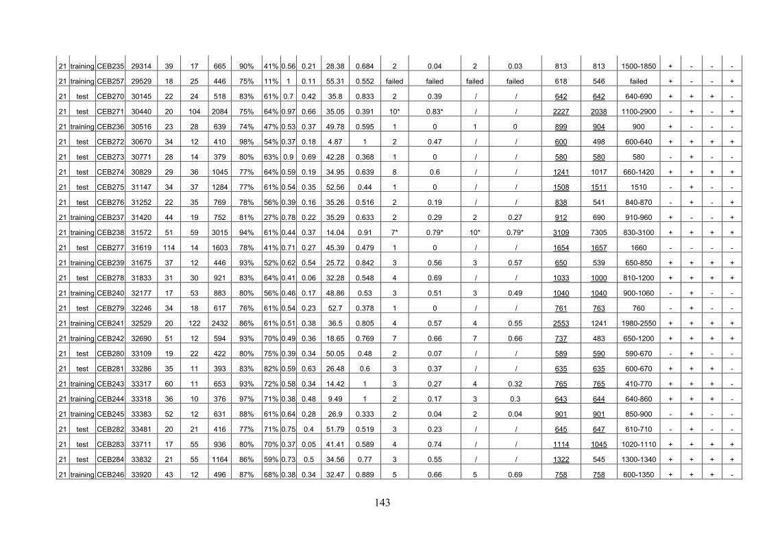
143
21 training CEB235 29314 39 17 665 90% 41% 0.56 0.21 28.38 0.684 2 0.04 2 0.03 813 813 1500-1850 + - - -
21 training CEB257 29529 18 25 446 75% 11% 1 0.11 55.31 0.552 failed failed failed failed 618 546 failed + - - +
21 test CEB270 30145 22 24 518 83% 61% 0.7 0.42 35.8 0.833 2 0.39 / / 642 642 640-690 + + + -
21 test CEB271 30440 20 104 2084 75% 64% 0.97 0.66 35.05 0.391 10* 0.83* / / 2227 2038 1100-2900 - + - +
21 training CEB236 30516 23 28 639 74% 47% 0.53 0.37 49.78 0.595 1 0 1 0 899 904 900 + - - -
21 test CEB272 30670 34 12 410 98% 54% 0.37 0.18 4.87 1 2 0.47 / / 600 498 600-640 + + + +
21 test CEB273 30771 28 14 379 80% 63% 0.9 0.69 42.28 0.368 1 0 / / 580 580 580 - + - -
21 test CEB274 30829 29 36 1045 77% 64% 0.59 0.19 34.95 0.639 8 0.6 / / 1241 1017 660-1420 + + + +
21 test CEB275 31147 34 37 1284 77% 61% 0.54 0.35 52.56 0.44 1 0 / / 1508 1511 1510 - + - -
21 test CEB276 31252 22 35 769 78% 56% 0.39 0.16 35.26 0.516 2 0.19 / / 838 541 840-870 - + - +
21 training CEB237 31420 44 19 752 81% 27% 0.78 0.22 35.29 0.633 2 0.29 2 0.27 912 690 910-960 + - - +
21 training CEB238 31572 51 59 3015 94% 61% 0.44 0.37 14.04 0.91 7* 0.79* 10* 0.79* 3109 7305 830-3100 + + + +
21 test CEB277 31619 114 14 1603 78% 41% 0.71 0.27 45.39 0.479 1 0 / / 1654 1657 1660 - - - -
21 training CEB239 31675 37 12 446 93% 52% 0.62 0.54 25.72 0.842 3 0.56 3 0.57 650 539 650-850 + + + +
21 test CEB278 31833 31 30 921 83% 64% 0.41 0.06 32.28 0.548 4 0.69 / / 1033 1000 810-1200 + + + +
21 training CEB240 32177 17 53 883 80% 56% 0.46 0.17 48.86 0.53 3 0.51 3 0.49 1040 1040 900-1060 - + - -
21 test CEB279 32246 34 18 617 76% 61% 0.54 0.23 52.7 0.378 1 0 / / 761 763 760 - + - -
21 training CEB241 32529 20 122 2432 86% 61% 0.51 0.38 36.5 0.805 4 0.57 4 0.55 2553 1241 1980-2550 + + + +
21 training CEB242 32690 51 12 594 93% 70% 0.49 0.36 18.65 0.769 7 0.66 7 0.66 737 483 650-1200 + + + +
21 test CEB280 33109 19 22 422 80% 75% 0.39 0.34 50.05 0.48 2 0.07 / / 589 590 590-670 - + - -
21 test CEB281 33286 35 11 393 83% 82% 0.59 0.63 26.48 0.6 3 0.37 / / 635 635 600-670 + + + -
21 training CEB243 33317 60 11 653 93% 72% 0.58 0.34 14.42 1 3 0.27 4 0.32 765 765 410-770 + + + -
21 training CEB244 33318 36 10 376 97% 71% 0.38 0.48 9.49 1 2 0.17 3 0.3 643 644 640-860 + + + -
21 training CEB245 33383 52 12 631 88% 61% 0.64 0.28 26.9 0.333 2 0.04 2 0.04 901 901 850-900 - + - -
21 test CEB282 33481 20 21 416 77% 71% 0.75 0.4 51.79 0.519 3 0.23 / / 645 647 610-710 - + - -
21 test CEB283 33711 17 55 936 80% 70% 0.37 0.05 41.41 0.589 4 0.74 / / 1114 1045 1020-1110 + + + +
21 test CEB284 33832 21 55 1164 86% 59% 0.73 0.5 34.56 0.77 3 0.55 / / 1322 545 1300-1340 + + + +
21 training CEB246 33920 43 12 496 87% 68% 0.38 0.34 32.47 0.889 5 0.66 5 0.69 758 758 600-1350 + + + -

144
21 training CEB247 34282 19 27 522 84% 73% 0.45 0.44 37.72 0.571 1 0 1 0 649 539 650 + + + +
21 test CEB285 34327 36 25 895 86% 64% 0.47 0.27 37.25 0.444 6 0.3 / / 1092 1092 880-1090 - + - -
21 training CEB248 34384 36 37 1339 70% 53% 0.66 0.15 32.33 0.5 5 0.64 6 0.63 1510 1514 830-1800 - + - -
21 test CEB286 34390 18 34 609 82% 72% 0.61 0.22 31.44 0.776 5 0.66 / / 713 713 560-850 + + + -
21 test CEB287 34454 44 10 448 77% 73% 0.45 0.31 44.91 0.227 3 0.51 / / 570 570 430-570 - + - -
21 test CEB288 34474 66 11 694 86% 70% 0.4 0.31 31.47 0.667 3 0.57 / / 822 394 690-820 + + + +
21 training CEB249 34683 51 22 1132 88% 63% 0.49 0.46 27.01 0.486 2 0.5 2 0.48 1305 430 1300-1350 - + - +
21 test CEB289 34741 45 45 2030 75% 44% 0.55 0.33 52.83 0.678 1 0 / / 2144 3451 5000 + - - +
21 test CEB290 34830 52 12 627 91% 63% 0.43 0.28 18.19 0.682 4 0.64 / / 833 374 670-920 + + + +
21 test CEB291 34854 28 22 602 85% 66% 0.52 0.22 37.67 0.706 10 0.87 12 0.88 772 775 770-1300 + + + -
21 training CEB250 34932 23 32 744 94% 72% 0.53 0.46 12.11 0.8 19 0.93 25 0.94 1045 487 540-1850 + + + +
21 test CEB292 34950 33 17 552 91% 79% 0.44 0.4 19.43 0.727 5 0.55 / / 647 648 580-1100 + + + -
21 training CEB251 34992 39 40 1575 88% 43% 0.72 0.12 29.91 0.435 3 0.39 3 0.38 1737 ? 1740-1960 - - - ?
21 training CEB252 35084 50 17 843 88% 56% 0.36 0.14 25.59 0.757 4 0.61 5 0.62 983 1033 980-1330 + + + +
21 training CEB253 35145 45 14 629 97% 57% 0.37 0.29 7.46 0.833 6 0.49 6 0.62 798 663 870-1100 + + + +
21 training CEB254 35165 24 20 483 80% 64% 0.44 0.34 43.33 0.784 3 0.2 3 0.18 656 539 630-680 + + + +
22 training CEB224 1528 41 21 855 91% 63% 0.71 0.46 26.31 0.609 6 0.75 7 0.77 1066 1019 940-1500 + + + +
22 training CEB213 1571 62 25 1548 94% 62% 0.39 0.24 13.36 0.538 5 0.53 / / 1401 1092 900-1700 - + - +
22 test CEB293 3522 36 20 719 86% 40% 0.4 0.34 33.64 0.64 3 0.35 / / 858 858 820-910 + - - -
22 training CEB208 3635 20 149 2987 77% 69% 0.48 0.31 32.46 0.538 failed failed failed failed 3126 3125 failed - + - -
22 training CEB225 4095 19 24 459 77% 61% 0.57 0.25 42.31 0.538 2 0.23 2 0.22 555 555 550-600 - + - -
22 training CEB216 10395 23 59 1362 79% 57% 0.65 0.41 46.71 0.6 2 0.07 3 0.09 1563 1290 1300-1600 + + + +
22 test CEB294 11741 43 12 506 73% 49% 0.39 0.08 40.85 0.3 3 0.5 / / 624 628 630-800 - + - -
22 training CEB226 12766 21 26 521 79% 55% 0.53 0.09 43.89 0.647 1 0 1 0 650 652 650 + + + -
22 test CEB295 13055 29 23 681 87% 52% 0.88 0.74 32.07 0.829 9* 0.8* 10* 0.73* 805 809 1020-4700 + + + -
22 training CEB222 15802 23 26 587 84% 57% 0.96 0.5 41.32 0.423 1 0 1 0 822 822 1200 - + - -
22 test CEB296 16228 21 83 1735 72% 57% 0.65 0.04 33.99 0.375 5 0.52 / / 1866 1868 1860-2200 - + - -
22 training CEB227 16333 37 14 515 82% 17% 0.41 0.22 35.32 0.304 1 0 1 0 670 671 670 - - - -
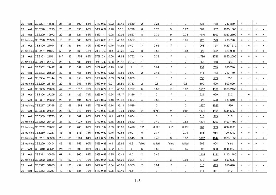
145
22 test CEB297 16658 21 28 602 85% 71% 0.63 0.22 33.42 0.649 2 0.24 / / 738 738 740-880 + + + -
22 test CEB298 18295 20 20 395 86% 58% 0.97 0.96 37.6 0.778 8 0.78 9 0.77 569 567 1080-1300 + + + -
22 test CEB299 19972 22 28 621 86% 53% 1 0.98 39.06 0.567 8 0.79 9 0.78 1016 1993 1020-2500 + + + +
22 training CEB228 20986 18 33 588 80% 45% 0.38 0.01 43.63 0.567 1 0 2 0.01 723 723 700-720 + - - -
22 test CEB300 21044 18 47 851 80% 63% 0.84 0.45 41.92 0.491 3 0.56 / / 968 758 1420-1670 - + - +
22 training CEB201 21327 59 11 668 78% 70% 0.4 0.3 40.26 0.75 3 0.58 7 0.63 820 2301 320-900 + + + +
22 test CEB301 21541 147 12 1752 88% 57% 0.4 0.06 37.64 0.762 3 0.33 / / 1790 1796 1650-2090 + + + -
22 training CEB214 22157 25 19 480 91% 4% 0.5 0.08 23.42 0.727 1 0 / / 668 418 660 + - - +
22 test CEB302 22447 57 10 552 97% 51% 0.45 0.29 6.91 1 2 0.04 / / 737 739 680-740 + + + -
22 test CEB303 23529 30 15 455 81% 57% 0.82 0.52 47.86 0.577 2 0.13 / / 713 713 710-770 + + + -
22 test CEB304 25144 29 12 356 87% 55% 0.64 0.53 27.94 0.889 1 0 / / 533 533 530 + + + -
22 training CEB229 26130 22 16 353 88% 35% 0.54 0.01 27.89 0.733 2 0.5 2 0.5 500 500 500-520 + - - -
22 test CEB305 27086 47 28 1313 76% 61% 0.74 0.61 45.56 0.727 14 0.89 16 0.92 1557 1109 1060-2150 + + + +
22 test CEB306 27209 20 21 426 74% 62% 0.71 0.64 47.17 0.389 1 0 / / 629 629 630 - + - -
22 test CEB307 27382 26 15 401 85% 74% 0.57 0.48 28.33 0.667 4 0.58 / / 528 528 430-640 + + + -
22 training CEB217 27398 20 68 1364 82% 47% 0.36 0.14 36.11 0.529 1 0 1 0 1527 1527 1530 - - - -
22 test CEB308 27654 28 33 918 91% 77% 0.43 0.39 18.94 0.872 7* 0.78* 7* 0.8* 1191 1191 600-1200 + + + -
22 test CEB309 27773 35 11 387 80% 28% 0.5 0.1 42.69 0.654 1 0 / / 513 513 513 + - - -
22 training CEB212 28490 36 29 1037 88% 37% 0.89 0.56 28.54 0.652 4 0.49 4 0.52 1201 1203 1100-1400 + - - -
22 training CEB202 29067 41 18 703 82% 59% 0.8 0.33 35.83 0.476 19* 0.92* 21* 0.93* 907 909 630-1900 - + - -
22 training CEB230 30257 35 15 513 71% 65% 0.69 0.46 52.56 0.591 5 0.77 7 0.79 683 684 720-1200 + + + -
22 training CEB231 30336 20 88 1761 84% 43% 0.77 0.15 33.19 0.545 3 0.45 3 0.57 1862 1844 1620-1850 + - - +
22 training CEB209 30404 46 16 755 90% 74% 0.38 0.4 23.66 0.6 failed failed failed failed 906 904 failed + + + -
22 test CEB310 30541 24 25 595 98% 20% 0.6 0.02 8.79 1 12 0.85 12 0.85 998 998 900-1550 + - - -
22 test CEB311 30860 67 14 960 82% 59% 0.49 0.25 36.41 0.5 2 0.46 1115 1111 1110-1180 - + - -
22 training CEB232 31534 17 22 373 70% 39% 0.64 0.05 65.06 0.324 1 0 3 0.04 572 572 500-605 - - - -
22 test CEB312 31865 19 23 439 81% 64% 0.78 0.54 45.61 0.565 2 0.04 / / 615 615 610-640 + + + -
22 test CEB313 32217 40 17 685 79% 51% 0.45 0.25 50.49 0.6 1 0 / / 811 811 810 + + + -
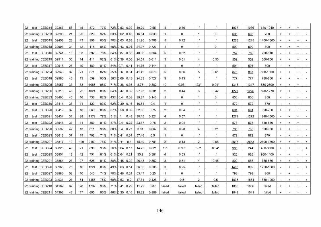
146
22 test CEB314 32267 58 15 872 77% 72% 0.53 0.39 49.29 0.55 4 0.56 / / 1037 1036 630-1040 + + + -
22 training CEB203 32298 21 25 529 92% 63% 0.62 0.46 16.84 0.833 1 0 1 0 695 695 700 + + + -
22 test CEB315 32458 23 43 996 80% 70% 0.63 0.63 31.95 0.788 5 0.72 / / 1228 1245 1400-1600 + + + +
22 training CEB218 32693 34 12 418 88% 56% 0.43 0.04 24.97 0.727 1 0 1 0 590 590 600 + + + -
22 test CEB316 32741 18 33 592 78% 64% 0.87 0.63 40.96 0.364 5 0.62 / / 757 759 700-810 - + - -
22 training CEB219 32911 30 14 411 92% 61% 0.38 0.06 24.51 0.611 3 0.51 4 0.53 559 559 500-700 + + + -
22 test CEB317 32915 26 19 489 81% 54% 0.7 0.41 44.76 0.444 1 0 / / 594 594 600 - + - -
22 training CEB204 32948 32 21 671 82% 55% 0.6 0.31 41.49 0.679 5 0.66 5 0.61 875 867 850-1500 + + + -
22 test CEB318 32980 43 13 559 90% 56% 0.68 0.43 24.33 0.727 3 0.43 / / 777 777 730-860 + + + -
22 training CEB205 33057 33 33 1086 96% 71% 0.38 0.36 8.75 0.882 19* 0.93* 23* 0.94* 1318 1317 550-2500 + + + -
22 training CEB206 33318 45 22 1024 88% 64% 0.47 0.32 27.93 0.581 2 0.44 3 0.47 1227 1226 820-1270 + + + -
22 training CEB233 33400 45 16 736 82% 43% 0.4 0.08 39.87 0.143 1 0 1 0 856 856 850 - - - -
22 test CEB319 33414 38 11 420 93% 62% 0.39 0.16 16.51 0.4 1 0 / / 572 572 570 - + - -
22 test CEB320 33419 32 18 563 86% 67% 0.58 0.39 32.65 0.75 2 0.04 / / 691 691 690-760 + + + -
22 test CEB321 33434 31 38 1172 77% 51% 1 0.48 38.15 0.321 4 0.57 / / 1272 1272 1240-1500 - + - -
22 test CEB322 33545 33 11 359 91% 57% 0.4 0.22 23.67 0.75 2 0.04 / / 578 578 540-580 + + + -
22 training CEB220 33592 47 13 611 98% 60% 0.4 0.27 3.81 0.667 3 0.28 4 0.21 785 785 600-930 + + + -
22 test CEB323 33618 37 19 702 71% 71% 0.41 0.34 57.46 0.5 1 0 / / 872 872 870 - + - -
22 training CEB207 33817 19 129 2459 76% 51% 0.41 0.3 48.19 0.701 2 0.13 2 0.08 2617 2663 2600-3500 + + + +
22 test CEB324 33825 43 21 890 93% 56% 0.64 0.17 14.25 0.621 19* 0.93* 27* 0.94* 985 244 400-3500 + + + +
22 test CEB325 33854 18 42 751 81% 61% 0.64 0.21 35.2 0.381 4 0.53 / / 926 928 930-1400 - + - -
22 training CEB221 33864 23 27 625 91% 58% 0.45 0.22 26.43 0.852 3 0.51 4 0.46 802 686 750-830 + + + +
22 test CEB326 33965 75 16 1224 83% 49% 0.63 0.14 36.35 0.508 3 0.25 / / 1408 802 1250-1680 - + - +
22 test CEB327 33983 52 10 543 74% 70% 0.46 0.24 53.47 0.25 1 0 / / 793 793 800 - + - -
22 training CEB223 34031 27 54 1456 75% 60% 0.53 0.2 47.81 0.428 2 0.5 2 0.5 1836 1864 1850-1950 - + - +
22 training CEB210 34182 62 28 1732 93% 71% 0.41 0.29 11.72 0.87 failed failed failed failed 1880 1886 failed + + + -
22 training CEB211 34393 43 17 695 95% 46% 0.35 0.16 18.22 0.889 failed failed failed failed 1048 1041 failed + - - -
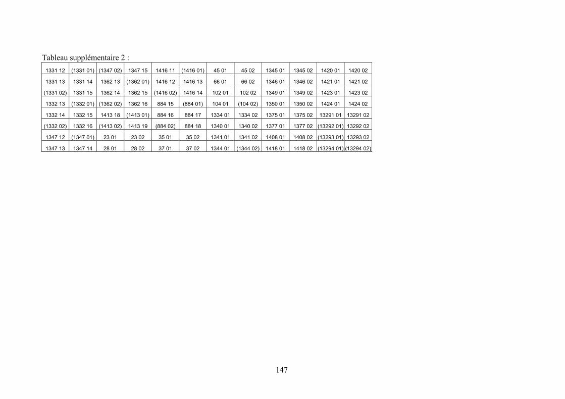
147
Tableau supplémentaire 2 :
1331 12 (1331 01) (1347 02) 1347 15 1416 11 (1416 01) 45 01 45 02 1345 01 1345 02 1420 01 1420 02
1331 13 1331 14 1362 13 (1362 01) 1416 12 1416 13 66 01 66 02 1346 01 1346 02 1421 01 1421 02
(1331 02) 1331 15 1362 14 1362 15 (1416 02) 1416 14 102 01 102 02 1349 01 1349 02 1423 01 1423 02
1332 13 (1332 01) (1362 02) 1362 16 884 15 (884 01) 104 01 (104 02) 1350 01 1350 02 1424 01 1424 02
1332 14 1332 15 1413 18 (1413 01) 884 16 884 17 1334 01 1334 02 1375 01 1375 02 13291 01 13291 02
(1332 02) 1332 16 (1413 02) 1413 19 (884 02) 884 18 1340 01 1340 02 1377 01 1377 02 (13292 01) 13292 02
1347 12 (1347 01) 23 01 23 02 35 01 35 02 1341 01 1341 02 1408 01 1408 02 (13293 01) 13293 02
1347 13 1347 14 28 01 28 02 37 01 37 02 1344 01 (1344 02) 1418 01 1418 02 (13294 01) (13294 02)

148
Annexe 4 : description des 50 minisatellites isolés dans des ARNs messagers
ARNm Taille
du motif
Nombre d'unités
Longueur totale
% conservation
%GC HistoryR Chromosome
Position de la
répétition dans l'ARN
5', 3', Codant ?
Répétition d'acides aminés ? Catégorie de locus
XM_114067 16 14,75 236 63% 81% 0,7 1p36.22 15-236 5'-codant non model, supported by EST alignments
XM_303126 49 8,22 403 97% 72% 1 1p36.32 900-1302 codant 49aa: VRQGWCWQLCGGAGLLGTAGVVLAALWRCRAAGYGRGGAGSSVAVPGCR
model, ab initio, with EST support
NM_152384 175 5,38 942 87% 77% 1 2q31.1 1851-2792 3' / gene with protein product, function unknown
XM_209485 127 4,87 619 92% 78% 1 2p23.3 442-1060 codant-3' non model, supported by mRNA alignments
XM_210394 20 56,5 1130 89% 78% 0,78 2p25.3 38-1167 codant 20aa: ACGAPGVPAEPPACLRSPLR model, ab initio, with EST support
XM_292899 24 32,41 778 94% 72% 0,77 2p25.3 522-1299
(atrophin-1 region)
codant 8aa: DPTPVPSA model, ab initio, with EST support
XM_293077 39 14,6 568 99% 70% 1 2q37.3 582-1149 codant 13aa: RSSYTLGRGPVAP model, ab initio, with EST support
XM_293082 38 28,2 1071 97% 78% 0,85 2q37.3 21-1091 codant 38aa: IAAPASLSAAGSPSPPRRLSPLRGAHRRPGVSLRCGEP
model, ab initio, with EST support
NM_138385 42 5 212 95% 78% 1 4p16.3 225-436 5' / gene with protein product, function unknown
XM_209692 41 13,02 534 61% 78% 0,78 5p13.1 439-972 codant-3' non model, supported by mRNA alignments
XM_302163 43 41,3 1789 95% 73% 0,74 5p15.33 3778-5566 codant 43aa: SGDGDDTGPRRGVQLRGRRRHRPPQRGSAQGTATTQAPAEGFS
model, ab initio, with EST support
XM_209786 37 10,48 388 52% 78% 0,81 6p21.31 362-749 codant-3' non model, supported by mRNA alignments
XM_294174 38 30,9 1184 90% 70% 0,73 7q36.3 1033-2216 codant 38aa (et blocs de 13): DATPGAARKSPPQTLRQAPTGRAHHRRYVRRRPEEPTT)
model, ab initio, with EST support
XM_300953 49 5,2 255 91% 82% 1 7q36.2 434-688 codant-3' non model, supported by mRNA alignments
XM_302324 38 19,9 758 96% 71% 0,83 7q36.3 868-1625 codant 38aa: ALPVSSGRGRPPARCLLPQEEDGLPRAAGFLRKRTASR
model, ab initio, with EST support
XM_302334 56 7,5 420 92% 70% 0,8 7p22.3 191-610 codant 56aa: SGPRTASELPHATASSRGTQDRGRPPSSPTPRHPAEGLRTEDGLRALPRRGIRQRG
model, ab initio
XM_291268 15 16,2 243 59% 70% 0,78 8q24.3 253-495 codant 5aa: YPQGP gene with protein product, function known or inferred: GRINA

149
ARNm Taille
du motif
Nombre d'unités
Longueur totale
% conservation
%GC HistoryR Chromosome
Position de la
répétition dans l'ARN
5', 3', Codant ?
Répétition d'acides aminés ? Catégorie de locus
NM_001807 33 15,9 526 92% 80% 0,92 9q34.3 1693-2218 codant 11aa:GAPPVPPTGDS gene with protein product, function known or inferred: CEL
NM_024718 44 16,9 744 95% 71% 0,78 9q34.3 1140-1883 3' / gene with protein product, function unknown
XM_209730 51 4,58 234 97% 83% 1 9q32 422-655 codant-3' non model, supported by mRNA alignments
XM_294605 24 115 2759 95% 70% 0,91 9q34.3 360-3118 codant 8aa: DTPRPRDC model, ab initio, with EST support
XM_305804 45 6,02 271 96% 70% 1 9q34.3 2100-2370 codant 15aa: ATLIPVPPVLSPLST model, ab initio, with EST support
XM_172821 33 6,75 223 94% 74% 1 10q26.3 62-284 codant 11aa: TPASSSSAAPP model, supported by mRNA alignments
NM_000797 48 8,81 423 91% 84% 0,8 11p15.5 740-1162 codant 16aa: PPAPGLPRGPCGPDCA gene with protein product, function known or inferred:DRD4
XM_296010 32 10,34 331 92% 70% 0,84 11q13.2 2-332 codant 33aa: DLPQPTLPEEEWTYPSPSSQRGGPTPAQAPRGV
model, ab initio, with EST support
XM_303443 29 8,1 235 97% 74% 1 11q25 277-511 codant 29aa: PALGGRRVGDLPSVDGVWATCPLWTASGR
model, ab initio, with EST support
XM_208611 49 4,4 216 93% 84% 1 12q21.2 677-892 3' / model, supported by mRNA alignments
XM_062938 39 39,3 1530 92% 73% 0,71 13q34 197-1726 codant 13aa:LGGDRTHGDAPSR model, ab initio, with EST support
XM_292156 79 4,4 352 89% 72% 0,85 13q14.11 434-785 codant 26aa: APIAGGEAPPTRQGLRASPSSPLALK(répétés 2,2 fois seulement)
model, ab initio, with EST support
XM_208767 40 10,35 414 74% 74% 0,72 14q32.33 371-784 codant-3' non model, supported by mRNA alignments
XM_301533 30 7,3 220 96% 74% 1 15q22.32 82-301 codant 10aa: EPGDGGKLPP model, ab initio
XM_303976 33 7,1 234 96% 71% 1 16q24.3 1316-1549 codant 11aa: GYRNSLFPPGE model, ab initio, with EST support
NM_000421 15 18 275 62% 79% 0,63 17q21-q23 1352-1626 codant 5aa: SSGGG gene with protein product, function known or inferred: KRT10
NM_024510 48 4,8 230 98% 70% 1 17q25.3 871-1100 codant 16aa: VPEPVHRPQDPWHIPG gene with protein product, function unknown
XM_302704 59 4,1 242 94% 72% 0,66 17 844-1085 codant
59aa: AGTSRASSRPRLHLLWDWPPQAPPGHPPAPGCSFCGTEPRRHLPGILPPQAAPSVGLSP
model, supported by mRNA and EST alignments
XM_046434 59 4,1 242 94% 72% 0,66 17q25.3 848-1089 codant
59aa: AGTSRASSRPRLHLLWDWPPQAPPGHPPAPGCSFCGTEPRRHLPGILPPQAAPSVGLSP
gene with protein product, function unknown
XM_210355 39 5,3 207 95% 76% 1 18q23 378-584 codant 13aa: GSRSGEPRAPTDR model, ab initio
XM_301656 31 24,87 771 97% 74% 1 18p11.32 342-1112 codant 31aa: PPRTSARPPDPSSDLGQTSGPLLGPRPDLRT (et 10:ALLGPRPDLRT)
model, ab initio
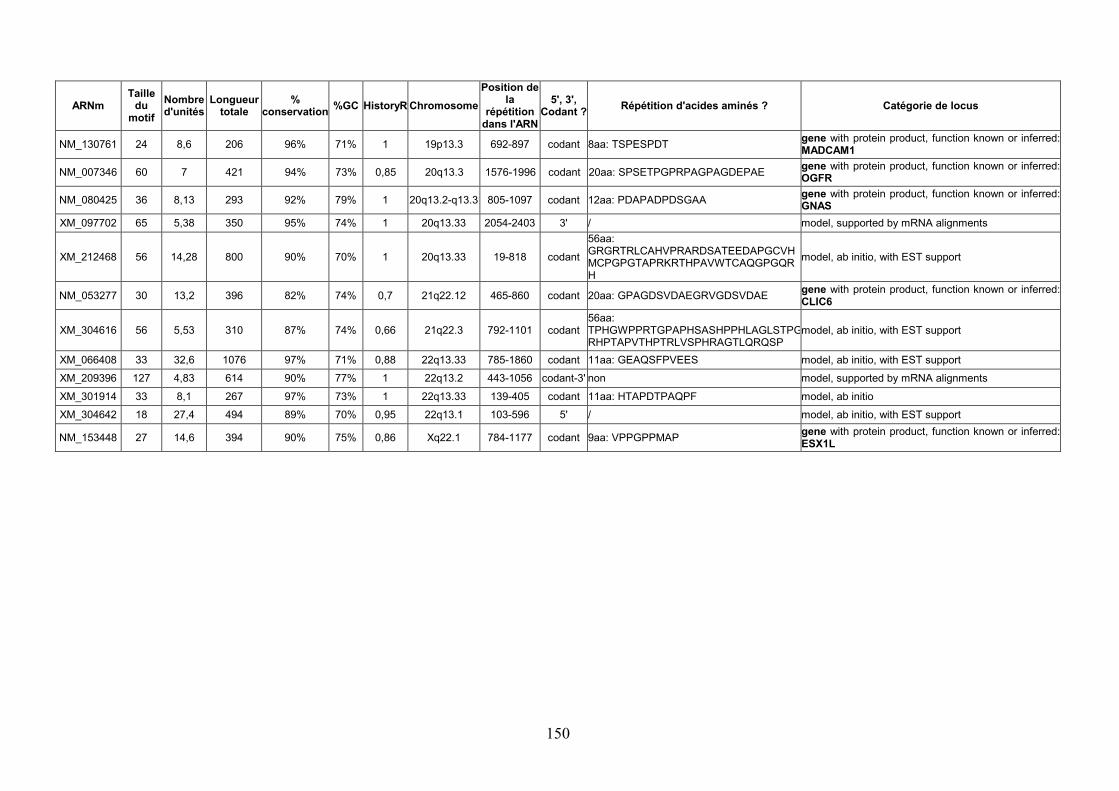
150
ARNm Taille
du motif
Nombre d'unités
Longueur totale
% conservation
%GC HistoryR Chromosome
Position de la
répétition dans l'ARN
5', 3', Codant ?
Répétition d'acides aminés ? Catégorie de locus
NM_130761 24 8,6 206 96% 71% 1 19p13.3 692-897 codant 8aa: TSPESPDT gene with protein product, function known or inferred: MADCAM1
NM_007346 60 7 421 94% 73% 0,85 20q13.3 1576-1996 codant 20aa: SPSETPGPRPAGPAGDEPAE gene with protein product, function known or inferred: OGFR
NM_080425 36 8,13 293 92% 79% 1 20q13.2-q13.3 805-1097 codant 12aa: PDAPADPDSGAA gene with protein product, function known or inferred: GNAS
XM_097702 65 5,38 350 95% 74% 1 20q13.33 2054-2403 3' / model, supported by mRNA alignments
XM_212468 56 14,28 800 90% 70% 1 20q13.33 19-818 codant
56aa: GRGRTRLCAHVPRARDSATEEDAPGCVHMCPGPGTAPRKRTHPAVWTCAQGPGQRH
model, ab initio, with EST support
NM_053277 30 13,2 396 82% 74% 0,7 21q22.12 465-860 codant 20aa: GPAGDSVDAEGRVGDSVDAE gene with protein product, function known or inferred: CLIC6
XM_304616 56 5,53 310 87% 74% 0,66 21q22.3 792-1101 codant 56aa: TPHGWPPRTGPAPHSASHPPHLAGLSTPGRHPTAPVTHPTRLVSPHRAGTLQRQSP
model, ab initio, with EST support
XM_066408 33 32,6 1076 97% 71% 0,88 22q13.33 785-1860 codant 11aa: GEAQSFPVEES model, ab initio, with EST support
XM_209396 127 4,83 614 90% 77% 1 22q13.2 443-1056 codant-3' non model, supported by mRNA alignments
XM_301914 33 8,1 267 97% 73% 1 22q13.33 139-405 codant 11aa: HTAPDTPAQPF model, ab initio
XM_304642 18 27,4 494 89% 70% 0,95 22q13.1 103-596 5' / model, ab initio, with EST support
NM_153448 27 14,6 394 90% 75% 0,86 Xq22.1 784-1177 codant 9aa: VPPGPPMAP gene with protein product, function known or inferred: ESX1L
Résumé Les répétitions en tandem sont constituées de successions de motifs d’ADN. Ces structures
sont présentes dans tous les organismes, procaryotes comme eucaryotes et, même si leur rôle
biologique est encore peu compris, elles ont des applications dans de nombreux domaines.
Tout d’abord, chez les bactéries, les répétitions en tandem polymorphes, dont le nombre
d’unités varie, se révèlent un outil puissant pour l’identification de souches à des fins
épidémiologiques. Par ailleurs, certaines répétitions en tandem humaines ont la propriété de
muter à des fréquences élevées : les minisatellites hypermutables sont les éléments les plus
instables du génome humain. Ils peuvent être utilisés comme biomarqueurs d’exposition à des
agents potentiellement mutagènes tels que les radiations ionisantes. D’un point de vue plus
fondamental, ils sont également un modèle d’étude des mécanismes d’instabilité des
génomes. Dans cette thèse, nous mettons à profit les données issues du séquençage afin
d’identifier des répétitions en tandem polymorphes. Nous avons tout d’abord élaboré une base
de données des répétitions en tandem accessible sur le web (http://minisatellites.u-psud.fr),
qui fournit un accès aux répétitions en tandem de génomes entiers. Ensuite, dans le but de
sélectionner les répétitions en tandem polymorphes, plusieurs stratégies ont été mises en
œuvre. D’une part, chez les bactéries pour lesquelles les séquences de plusieurs souches
étaient disponibles, nous avons créé un utilitaire de comparaison de souches, afin d’identifier
des marqueurs polymorphes utilisables en épidémiologie. D’autre part, une étude menée sur
les minisatellites humains a permis de définir des critères prédictifs du polymorphisme à
partir de la séquence d’un seul allèle de minisatellite, et a en outre mis en évidence un
nouveau minisatellite hypermutable situé dans une séquence codante putative. Les critères
prédictifs ont également été appliqués à l’identification de minisatellites codants
potentiellement polymorphes dans le génome humain.
Abstract
Tandem repeats are consecutive occurrences of a DNA unit. Such structures are found in all
organisms, prokaryotes as well as eukaryotes. Although their biological function is not fully
understood, they have diverse practical applications. In bacteria, polymorphic tandem repeats
(with varying copy numbers), are powerful tools for strain identification in bacterial
epidemiology. In humans, some tandem repeats mutate at a very high rate: hypermutable
minisatellites are the most unstable elements in the human genome. They can be used as
biomarkers for the monitoring of genotoxic agents such as ionizing radiations. More
fundamentally, they are a model to study genome instability. In this thesis, we take advantage
of the release of genome sequence data in order to identify polymorphic tandem repeats. First,
we developed a tandem repeats database freely accessible on the web (http://minisatellites.u-
psud.fr), which provides tandem repeats from complete genomes. Then, we used several
approaches to identify polymorphic tandem repeats. The first one consisted in developing a
strain comparison page for bacteria with several sequenced strains, which directly selects
polymorphic markers for use in epidemiology. The second approach, applied to human
minisatellites, sought to define polymorphism predictive criteria from the sequence of one
single allele. This study also identified a hypermutable minisatellite located in a putative
coding sequence. The predictive criteria were subsequently used to search for potentially
polymorphic coding minisatellites in the human genome.