Embed Size (px)
Citation preview

8/4/2019 Analisis Coprologico Www.librosdelqfb.blogspot
http://slidepdf.com/reader/full/analisis-coprologico-wwwlibrosdelqfbblogspot 1/17

8/4/2019 Analisis Coprologico Www.librosdelqfb.blogspot
http://slidepdf.com/reader/full/analisis-coprologico-wwwlibrosdelqfbblogspot 2/17

8/4/2019 Analisis Coprologico Www.librosdelqfb.blogspot
http://slidepdf.com/reader/full/analisis-coprologico-wwwlibrosdelqfbblogspot 3/17
8/4/2019 Analisis Coprologico Www.librosdelqfb.blogspot
http://slidepdf.com/reader/full/analisis-coprologico-wwwlibrosdelqfbblogspot 4/17

8/4/2019 Analisis Coprologico Www.librosdelqfb.blogspot
http://slidepdf.com/reader/full/analisis-coprologico-wwwlibrosdelqfbblogspot 5/17

8/4/2019 Analisis Coprologico Www.librosdelqfb.blogspot
http://slidepdf.com/reader/full/analisis-coprologico-wwwlibrosdelqfbblogspot 6/17
Introduction and Summary
Cysts and trophozoites must be examined carefully in different fields of view and measurement is
often essential. Objects such as epithelial cells and macrophages are around the same size as
amoebic trophozoites: the latter may also move and contain red blood cells. White blood cells, plant
and vegetable cells, fat globules, muscle fibres, pollen grains, yeasts cells and air bubbles may be
confused with cysts or eggs. Air bubbles trapped under adhesive tape often resemble Enterobius
eggs. Plant hairs and fibres are easily confused with larvae; algae such as Psorospermium haeckelii may be found in the faeces of patients who have eaten crayfish. Earthworms may resemble
roundworms. A variety of non-pathogenic ova, cysts and parasites resemble pathogens in terms of
size and morphology and careful examination is essential. Eggs of Heterodera, a parasitic nematode
of root vegetables, may resemble hookworm eggs. Eggs originating from harmless mites in cereals
or flour could be confused with hookworm ova but are usually larger. We recently encountered 160
micron "Schistosome ova" in the urine of a patient complaining of haematuria: we suspected
Schistosoma haematobium but, on closer analysis, the eggs contained unidentified insects. This
volume provides examples of artefacts that may be confused for parasitic life stages. Artefacts should
be considered on the basis of size, shape, lack of organelles and defining feature, and variable
reactivity with common stains
Red and White Blood Cells
Red blood cells and a variety of white blood cells can be easily mistaken for parasitic cells or cysts
when observed with microscopy.
Figure 1: Image illustrating red and white blood cells in a slide preparation
White Blood Cells
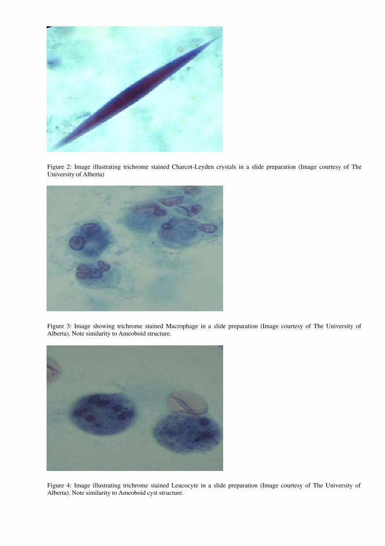
Charcot-Leyden crystals are a product of eosinophil breakdown and are, therefore, occasionally
found in faeces of patients suffering from parasitic disease. They appear red when stained with in a
trichrome faecal preparation.
8/4/2019 Analisis Coprologico Www.librosdelqfb.blogspot
http://slidepdf.com/reader/full/analisis-coprologico-wwwlibrosdelqfbblogspot 7/17
Figure 2: Image illustrating trichrome stained Charcot-Leyden crystals in a slide preparation (Image courtesy of The
University of Alberta)
Figure 3: Image showing trichrome stained Macrophage in a slide preparation (Image courtesy of The University of
Alberta). Note similarity to Ameoboid structure.
Figure 4: Image illustrating trichrome stained Leucocyte in a slide preparation (Image courtesy of The University of
Alberta). Note similarity to Ameoboid cyst structure.

8/4/2019 Analisis Coprologico Www.librosdelqfb.blogspot
http://slidepdf.com/reader/full/analisis-coprologico-wwwlibrosdelqfbblogspot 8/17
Figure 5: Image illustrating Red Blood Cells in slide preparation. RBC’s may appear to
have a central body and a rim of cytoplasm or granules which could be mistaken forBlastocystis hominis . (Image Courtesy of the University of Alberta)
Fat Globules
Fat globules present in a faecal slide
preparation may appear similar to parasitic cysts or cell bodies.
Figure 6: Image illustrating Fat Globules in slide preparation
Emulsifying agents are a useful tool to eliminate potential confusion involving fat globules. The
removal of such particles from slide preparations will undoubtedly reduce cases of misdiagnosis.
Yeast Cells

8/4/2019 Analisis Coprologico Www.librosdelqfb.blogspot
http://slidepdf.com/reader/full/analisis-coprologico-wwwlibrosdelqfbblogspot 9/17
Yeast may resemble protozoan cysts because they are uniform in colour, have fewinclusions and no nucleus. Yeast could also be confused with small protozoans like E. nana or with Cryptosporidium or Cyclospora oocysts in wet preparation. In acid-fast stains, theoocysts of Cryptosporidium and Cyclospora species stain pink to red. Yeasts are not acidfast and stain green.
Figure 7: Image illustrating Yeast Cells in slide preparation
Figure 8: Image illustrating Yeast Cells in slide preparation. Note similarity to parasiticoocysts

8/4/2019 Analisis Coprologico Www.librosdelqfb.blogspot
http://slidepdf.com/reader/full/analisis-coprologico-wwwlibrosdelqfbblogspot 10/17
Vegetable Cells
Plant cells and associated elements seen in faeces may resemble eggs,cysts or cell bodies.Plant cells are often identified by a more irregular outer membrane.
Figure 9: Image illustrating Vegetable cell in slide preparation. Note similarity toParagonimus eggs.
Figure 10: Image illustrating Vegetable cell in slide preparation. Note similarity to Dipylidium caninum egg packets

8/4/2019 Analisis Coprologico Www.librosdelqfb.blogspot
http://slidepdf.com/reader/full/analisis-coprologico-wwwlibrosdelqfbblogspot 11/17
Figure 11: Image illustrating Vegetable cell in slide preparation
Figure 12: Image illustrating a Vegetable Spiral in slide preparation. Such spirals may appear similar to proglottids
(Image courtesy of the University of Alberta)

8/4/2019 Analisis Coprologico Www.librosdelqfb.blogspot
http://slidepdf.com/reader/full/analisis-coprologico-wwwlibrosdelqfbblogspot 12/17
Figure 13: Image illustrating Vegetable Spiral in slide preparation
Pollen
Pollen grains are often misinterpreted as
parasite eggs, but can often be discerned through size and the presence or absence of important
structural elements.
Figure 14: Image illustrating pollen in slide preparation using a colour filter

8/4/2019 Analisis Coprologico Www.librosdelqfb.blogspot
http://slidepdf.com/reader/full/analisis-coprologico-wwwlibrosdelqfbblogspot 13/17
Figure 15: Image illustrating pollen in slide preparation that could be mistaken for a Taenia egg. The shell is thinner, of non-uniform thickness, and no hooks are visible. Imagecourtesy of CDC Division of Parasitic Diseases.
Figure 16: Image illustrating pollen resembling a Hymenolepis nana egg. Hooks and polarfilaments are not visible. Courtesy of CDC Division of Parasitic Diseases.

8/4/2019 Analisis Coprologico Www.librosdelqfb.blogspot
http://slidepdf.com/reader/full/analisis-coprologico-wwwlibrosdelqfbblogspot 14/17
Figure 17: Image illustrating geranium pollen cells in slide preparation
Figure 18: Image illustrating pollen cells in slide preparation. Similar to Taenia eggs, but distinguished by uneven
thickness of the wall and lack of internal contents do not suggest an egg.
Hair
Animal and plant hairs are most often and easily mistaken for parasitic nematode worms. Their size
and shape may be comparable in many cases, but a lack of internal definition will identify the artefact
when compared to the worm. Although nematodes are non-segmented and externally simple
organisms, they will often show unique structural characteristics under close examination.

8/4/2019 Analisis Coprologico Www.librosdelqfb.blogspot
http://slidepdf.com/reader/full/analisis-coprologico-wwwlibrosdelqfbblogspot 15/17
Figure 19: Image illustrating peach hair in slide preparation. Note the similarity toStrongyloides stercoralis.
Figure 20: Image illustrating vegetable hairs in slide preparation

8/4/2019 Analisis Coprologico Www.librosdelqfb.blogspot
http://slidepdf.com/reader/full/analisis-coprologico-wwwlibrosdelqfbblogspot 16/17
Insect Eggs
Figure 21: Image illustrating Insect eggs in slide preparation
Plant Parasites

8/4/2019 Analisis Coprologico Www.librosdelqfb.blogspot
http://slidepdf.com/reader/full/analisis-coprologico-wwwlibrosdelqfbblogspot 17/17
Figure 22: Image illustrating Heterodera spp. in slide preparation. Such arasitic nematodes attack root vegetables such as
beetroot, turnips and radishes. Their eggs are 80-120m by 25-40 m and can conceivably be confused with hookworm
eggs.
Earthworms
Figure 23: Image illustrating an Annelid earthworm in detritus. Theybelong to the Annelida (Lumbricus and Allolobophora) and are
elongated, segmented and circular in section and are occasionallyconfused with Ascaris . They have a purple-brown dorsal surface and a
paler ventral surface, swell out at around segment 12 and possess amarked thickening (the clitellum) a third of the way from the anterior
end