Embed Size (px)
Citation preview

An Update on EBUS Cytopathology
Walid E Khalbuss, MD PhD FIAC
Professor of Pathology; University of Pittsburgh
Medical Director of GE Clarinet; NGHA-Riyadh

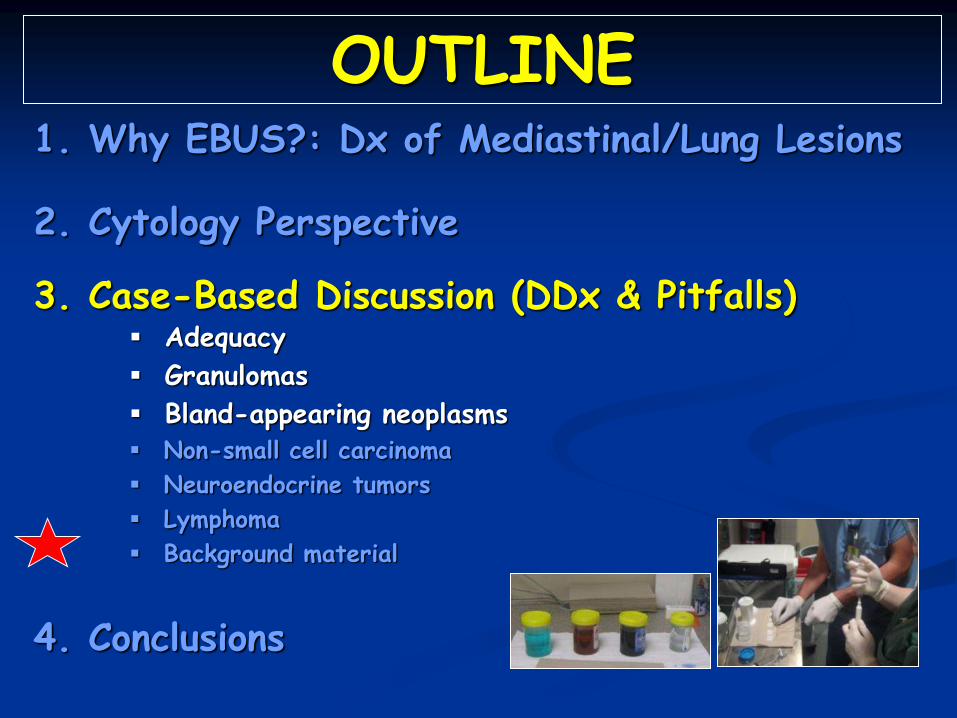
OUTLINE 1. Why EBUS?: Dx of Mediastinal/Lung Lesions
Approach to Diagnosis of Mediastinal Lesions EBUS/EUS FNA Clinical Perspective
2. Cytology Perspective Rapid On-Site Evaluation (ROSE) Adequacy Assessment
3. Case-Based Discussion (DDx & Pitfalls)
Cases
4. Conclusions

Mediastinal Mass: DDx
Non-neoplastic Neoplastic
RLH
Sarcoidosis
Granulomatous Other Metastases Lymphoma
Lung Ca Other
Primary
NHL HL Infectious
5T’s

Imaging & Diagnosis of Mediastinal/Lung Lesions
Imaging Modalities Chest Xray CT Scan PET CT Scan
Diagnostic Modalities Sputum/BAL/BB/BW/Pl Fl CT-Guided FNA Transbronchial FNA (Wang Bx) Mediastinoscopy/Thoracoscopy EBUS & EUS guided FNA
CLINICAL Sx
IMAGE
DIAGNOSE
STAGE
TREAT

EBUS/EUS FNA
1st available in 2004-2005
Minimally invasive
Real-time image guidance
Indications: Staging
Dx of lung or mediastinal mass
Dx of +CT/PET findings
Advantages & Disadvantages
Among patients with clinical stage IIIA, 40% of patients were down-staged with
EBUS-FNA Gilbert S et al. JTCVS 2009

EBUS/EUS FNA Advantages:
Minimally invasive Image guidance Tissue confirmation of +PET/CT
findings & evaluation of LNs <1 cm Broad sampling capability On-site evaluation triage
Disadvantages: Inability to access all LNs Not universally available Time requirement Experience Non-diagnostic specimens
Varela-Lema L et al., Eur Repir J, 2009
Remember Special Situations!
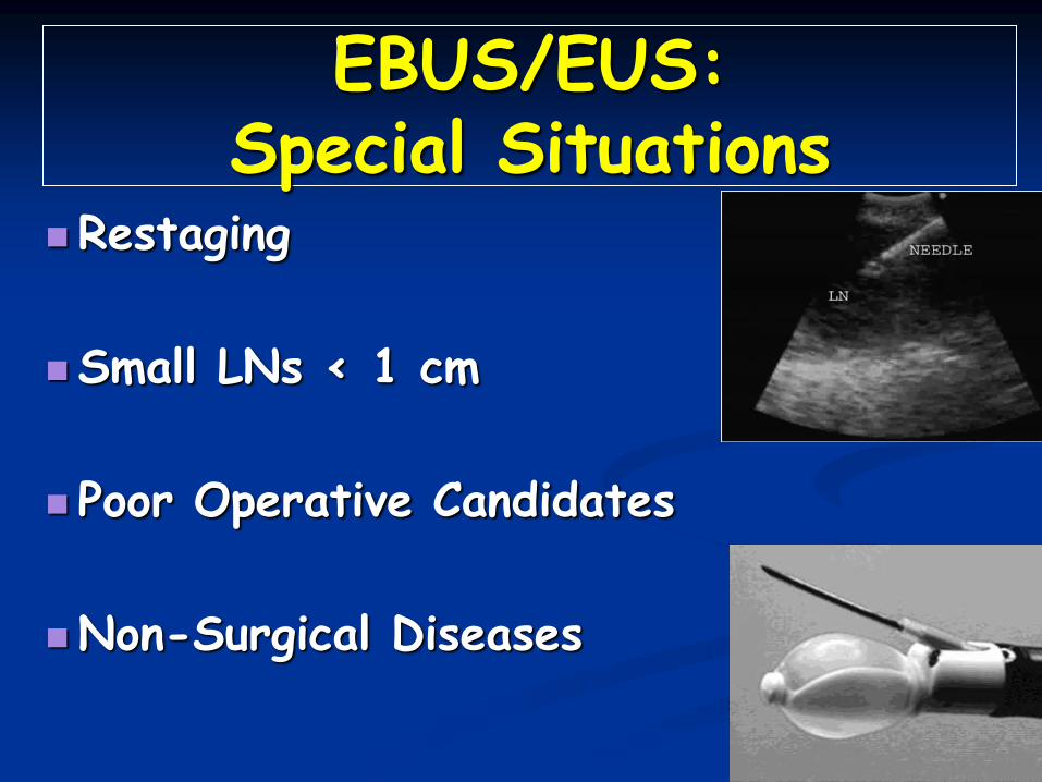
EBUS/EUS: Special Situations
Restaging
Small LNs < 1 cm
Poor Operative Candidates
Non-Surgical Diseases

Cost of EBUS/EUS FNA
EBUS/EUS-FNA Cost= $2,000
Mediastinoscopy Cost= $8,000
Thoracotomy Cost= $26,000
Cut cost
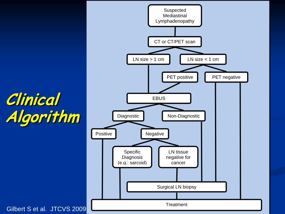
Clinical Algorithm
Suspected Mediastinal
Lymphadenopathy
CT or CT/PET scan
LN size > 1 cm LN size < 1 cm
EBUS
Negative
Non-Diagnostic Diagnostic
Positive
Surgical LN biopsy
Treatment
Specific Diagnosis
(e.g.: sarcoid)
PET negative
LN tissue negative for
cancer
PET positive
Gilbert S et al. JTCVS 2009
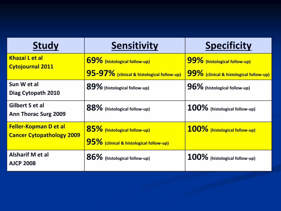
Study Sensitivity Specificity Khazai L et al
Cytojournal 2011 69% (histological follow-up)
95-97% (clinical & histological follow-up)
99% (histological follow-up)
99% (clinical & histological follow-up)
Sun W et al
Diag Cytopath 2010 89% (histological follow-up) 96% (histological follow-up)
Gilbert S et al
Ann Thorac Surg 2009 88% (histological follow-up) 100% (histological follow-up)
Feller-Kopman D et al
Cancer Cytopathology 2009 85% (histological follow-up)
95% (clinical & histological follow-up)
100% (histological follow-up)
Alsharif M et al
AJCP 2008 86% (histological follow-up) 100% (histological follow-up)

EBUS FNA USG thru bronchus
Anterior mediastinum
Limitation: Inability to access
posterior & inferior
Sen generally >80%
Spec generally >98%
EUS FNA USG thru esophagus
Posterior mediastinum
Limitation: Inability to access
anterior & superior
Sen generally >80%
Spec generally >98%
EBUS FNA & EUS FNA for Mediastinal Masses
School Bus

Yasufuku, K. et al. Chest 2006;130:710-718
Sampling techniques and their diagnostic reach of mediastinal and hilar lymph node stations (1, highest mediastinal; 2, upper paratracheal; 4, lower
paratracheal; 5, subaortic; 7, subcarinal; 8, paraesophageal; 9, pulmonary ligament; 10, hilar; 11, interlobar; and 12, lobar)
Yasufuku K et al. Chest 2006; 130: 710-18
Yasufuku, K. et al. Chest 2006;130:710-718
Sampling techniques and their diagnostic reach of mediastinal and hilar lymph node stations (1, highest mediastinal; 2, upper paratracheal; 4, lower
paratracheal; 5, subaortic; 7, subcarinal; 8, paraesophageal; 9, pulmonary ligament; 10, hilar; 11, interlobar; and 12, lobar)
EBUS/EUS FNA
LN Stations

EBUS FNA: Technical Aspects
Location: Bronchoscopy lab (conscious sedation)
Operating room (general anesthesia)
Equipment: Olympus bronchoscope + US probe + 22G FNA needle
Target: Identify with PET-CT
Confirm with real-time US guidance

Subcarinal (Level 7)
Herth FJF et al. J Bronchol 2006; 13(2): 84-91
EBUS Procedure
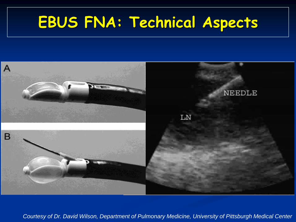
EBUS FNA: Technical Aspects
Courtesy of Dr. David Wilson, Department of Pulmonary Medicine, University of Pittsburgh Medical Center

● Cancer vs. No Cancer
● Adequate/sufficient for ancillary studies?
● ? More tissue needed: mediastinoscopy
● Adequacy of specimen: Need more passes?
● Communication: surgeon and cytology team
EBUS/EUS: Clinical Perspective

OUTLINE 1. Why EBUS?: Dx of Mediastinal/Lung Lesions
Approach to Diagnosis of Mediastinal Lesions EBUS/EUS FNA Clinical Perspective
2. Cytology Perspective Rapid On-Site Evaluation (ROSE) Adequacy Assessment
3. Case-Based Discussion (DDx & Pitfalls)
Cases 1-12 Additional Cases (static images) Additional Cases (virtual cases)
4. Conclusions
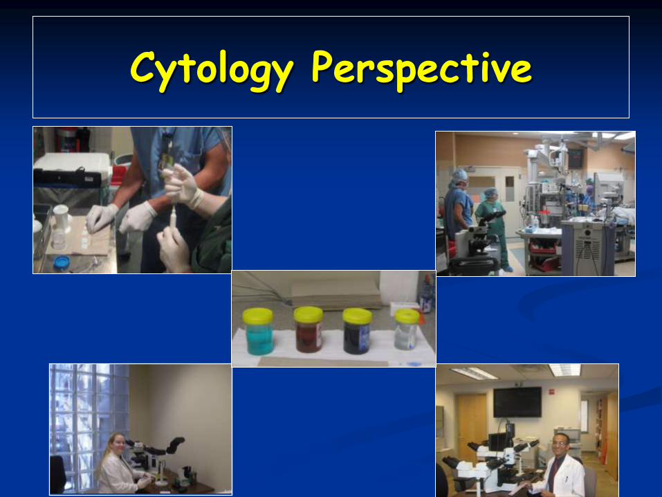
Cytology Perspective

EBUS/EUS FNA
Evaluation by Cytopathology
? Mediastinoscopy

Why Rapid On-Site Evaluation (ROSE)?
1.Immediate feedback
2.Assessment of adequacy
3.High-quality smears/CB
4.Triage: Flow Cytometry, Cultures, etc
5.Rapid Diagnosis: Management
ROSE
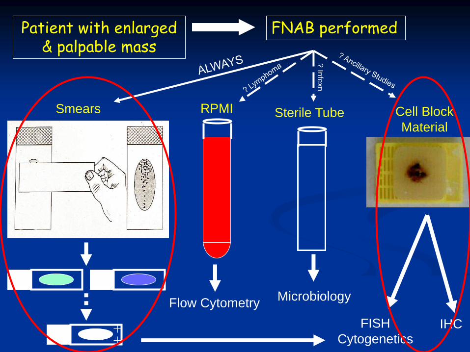
Patient with enlarged & palpable mass
FNAB performed
Smears RPMI Cell Block
Material Sterile Tube
IHC FISH
Cytogenetics
Flow Cytometry Microbiology
? In
fexn

EBUS FNA: Inadequate/NonDx EBUS FNA: Adequate/Positive
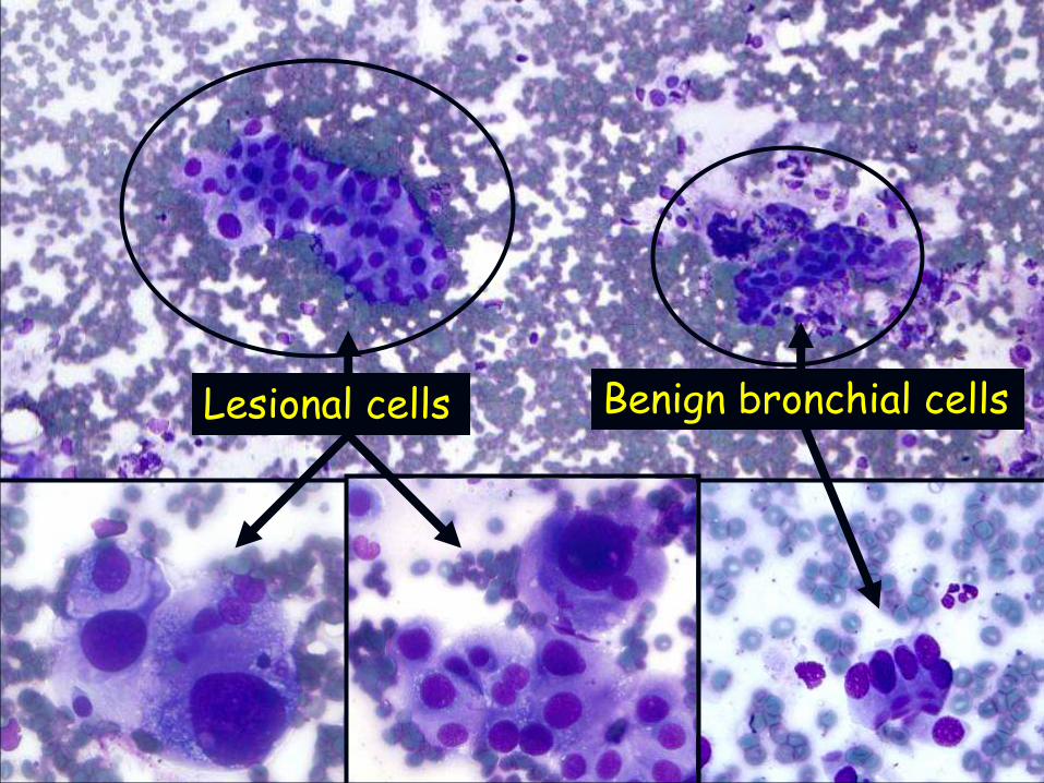
Benign bronchial cells Lesional cells

Squamous cells
Cartilage Bronchial Cells Lymphocytes
Anthracotic Pigment Laden Macrophages Mucus
Benign Components in EBUS-FNA

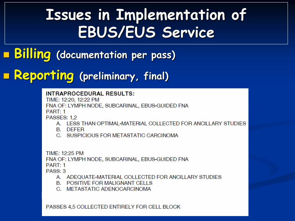
Issues in Implementation of EBUS/EUS Service
Time requirement (longer at start; # days coverage)
Where will you go? (OR vs. Bronch Lab)
What will you use? (cart,FS room,own space; light; masks)
Who will go? (cytotech, fellow, pathologist)
How to collect specimen? (formalin, Thin Prep, other media)
ROSE? Telecytology?

Issues in Implementation of EBUS/EUS Service
Time requirement (longer at start; # days coverage)
Where will you go? (OR vs. Bronch Lab)
What will you use? (cart,FS room,own space; light; masks)
Who will go? (cytotech, fellow, pathologist)
How to collect specimen? (formalin, other media)
ROSE? Telecytology?
STAGGERING
STATIONS 1st pass LN#1
1st pass LN#2
1st pass LN#3
2nd pass LN#1
2nd pass LN#2
2nd pass LN#3
Issues in Implementation of EBUS/EUS Service
Billing (documentation per pass)
Reporting (preliminary, final)
OUTLINE 1. Why EBUS?: Dx of Mediastinal/Lung Lesions
2. Cytology Perspective
3. Case-Based Discussion (DDx & Pitfalls) Adequacy
Granulomas
Bland-appearing neoplasms Non-small cell carcinoma
Neuroendocrine tumors
Lymphoma
Background material
4. Conclusions

Case 1: EBUS FNA
69/F with NSCLC and mediastinal LAD.
EBUS FNA of Rt hilar LN.

Case 1
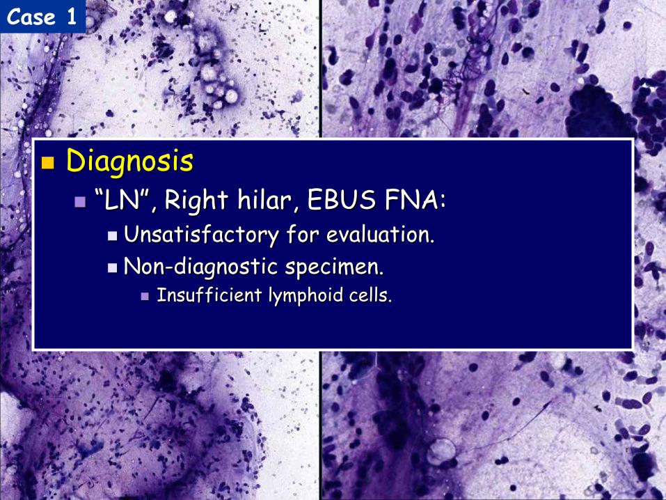
Case 1
Diagnosis “LN”, Right hilar, EBUS FNA:
Unsatisfactory for evaluation.
Non-diagnostic specimen. Insufficient lymphoid cells.

Case 2
Diagnosis LN, Subcarinal, EBUS FNA:
Adequate.
Negative for malignant cells.

Unsatisfactory Satisfactory- Negative Satisfactory- Positive
Spectrum of Diagnoses in EBUS-FNA

Usually 3-5 passes
Adequate if: Malignant lesion is identified
Lesional material (e.g. granulomas)
Sufficient nodal tissue is obtained Numerous lymphocytes
Anthracotic pigment-laden macrophages
Germinal center fragments
EBUS FNA Adequacy

Anthracotic Pigment Laden Macrophages E
BU
S
SLID
E
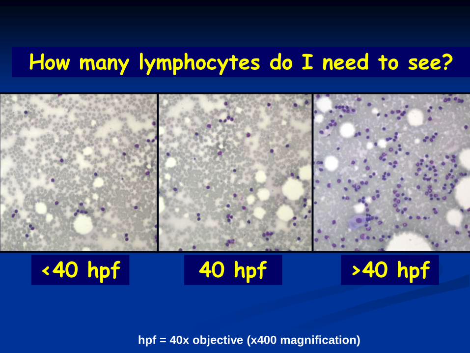
How many lymphocytes do I need to see?
<40 hpf 40 hpf >40 hpf
hpf = 40x objective (x400 magnification)
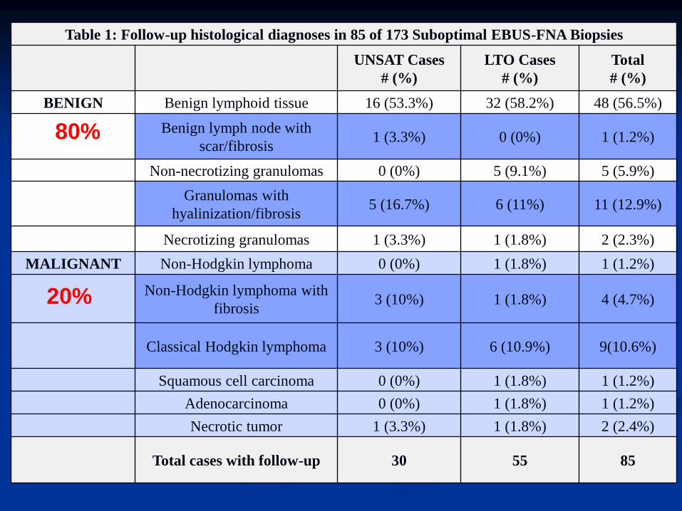
Table 1: Follow-up histological diagnoses in 85 of 173 Suboptimal EBUS-FNA Biopsies
UNSAT Cases
# (%)
LTO Cases
# (%)
Total
# (%)
BENIGN Benign lymphoid tissue 16 (53.3%) 32 (58.2%) 48 (56.5%)
Benign lymph node with
scar/fibrosis 1 (3.3%) 0 (0%) 1 (1.2%)
Non-necrotizing granulomas 0 (0%) 5 (9.1%) 5 (5.9%)
Granulomas with
hyalinization/fibrosis 5 (16.7%) 6 (11%) 11 (12.9%)
Necrotizing granulomas 1 (3.3%) 1 (1.8%) 2 (2.3%)
MALIGNANT Non-Hodgkin lymphoma 0 (0%) 1 (1.8%) 1 (1.2%)
Non-Hodgkin lymphoma with
fibrosis 3 (10%) 1 (1.8%) 4 (4.7%)
Classical Hodgkin lymphoma 3 (10%) 6 (10.9%) 9(10.6%)
Squamous cell carcinoma 0 (0%) 1 (1.8%) 1 (1.2%)
Adenocarcinoma 0 (0%) 1 (1.8%) 1 (1.2%)
Necrotic tumor 1 (3.3%) 1 (1.8%) 2 (2.4%)
Total cases with follow-up 30 55 85
80%
20%
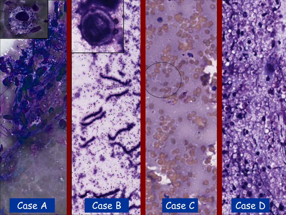
Case A Case D Case B Case C

74/M
• Cohesion;
• Low N/C ratio
• No nuclear enlargement, x2
• No significant pleomorphism
• Only reactive/degenerative atypia
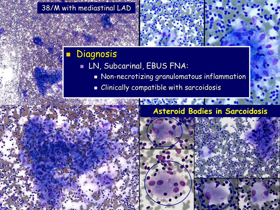
38/M with mediastinal LAD
Asteroid Bodies in Sarcoidosis
Diagnosis LN, Subcarinal, EBUS FNA:
Non-necrotizing granulomatous inflammation
Clinically compatible with sarcoidosis

68/M with mediastinal LAD. EUS FNA performed of LN.
Diagnosis Lymph node, Mediastinal.
EUS FNA: Positive for malignant cells.
Malignant NHL, high-grade.
Pitfall: Tumor with granulomas
Dx: Metastatic Seminoma.
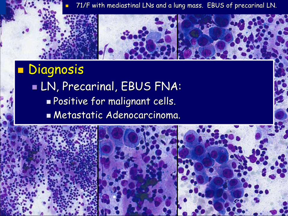
Diagnosis LN, Precarinal, EBUS FNA:
Positive for malignant cells.
Metastatic Adenocarcinoma.
71/F with mediastinal LNs and a lung mass. EBUS of precarinal LN.

60/M with multiple FDG-avid lung nodules. EUS FNA: PTC
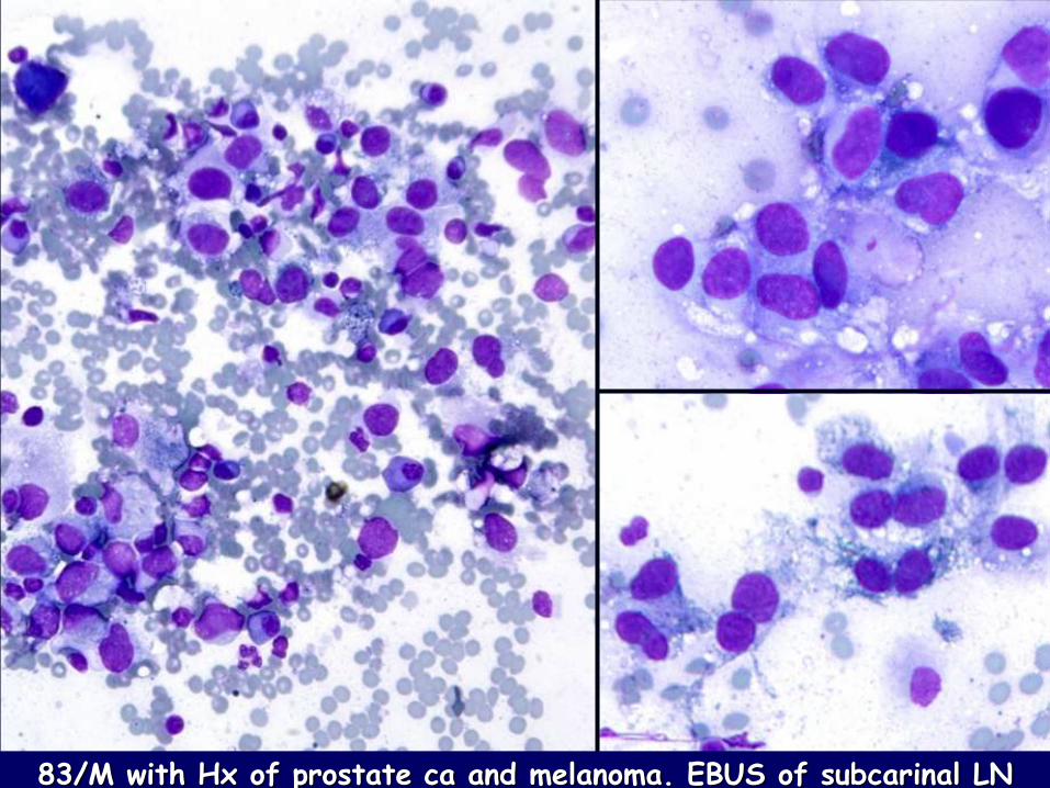
83/M with Hx of prostate ca and melanoma. EBUS of subcarinal LN
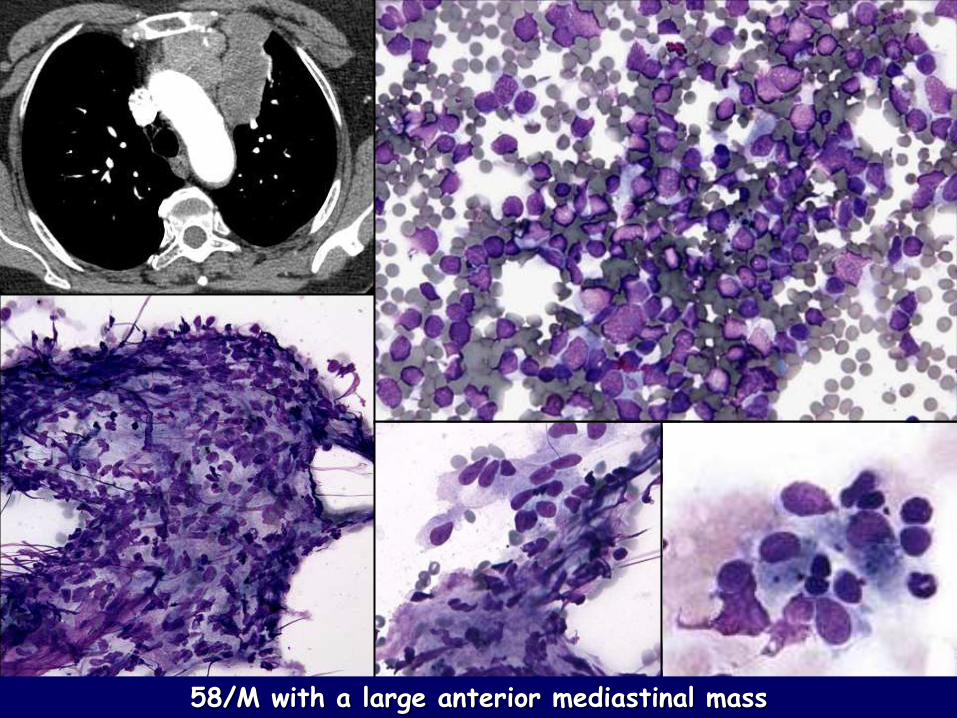
58/M with a large anterior mediastinal mass
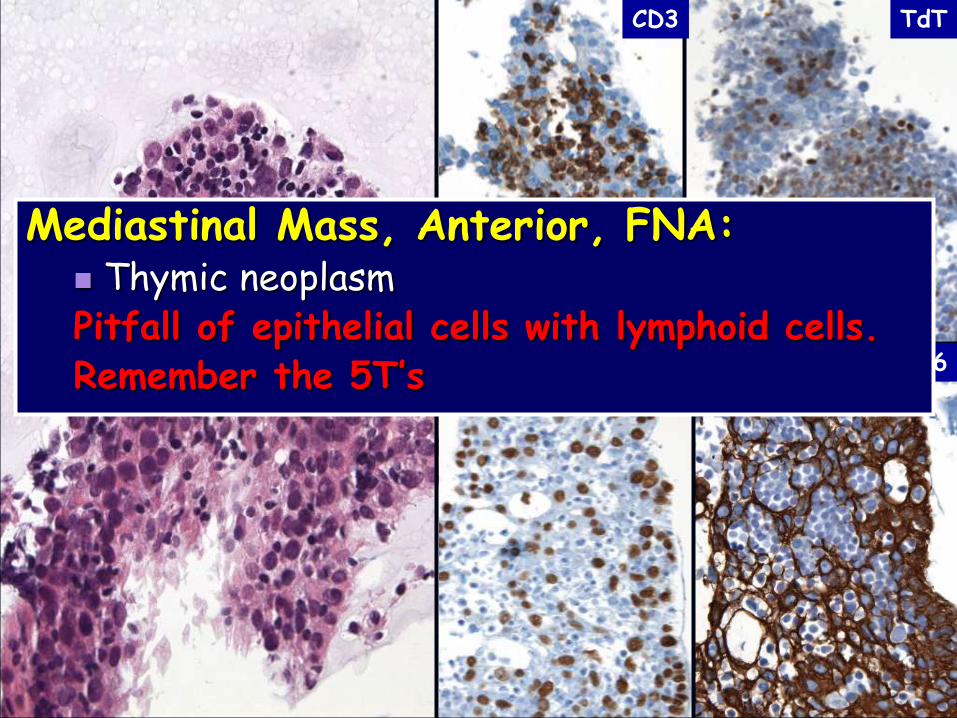
CD3 TdT
p63 CK5/6
Mediastinal Mass, Anterior, FNA: Thymic neoplasm Pitfall of epithelial cells with lymphoid cells. Remember the 5T’s

Conclusions EBUS/EUS FNA have changed
the way that thoracic & mediastinal lesions are approached
The EBUS/EUS FNA diagnoses
can be difficult & challenging Be aware of pitfalls Consider the wide spectrum of
conditions in the mediatinum Cytomorphology & ancillary studies
can help to make a definitive dx in most cases